Article

Terpenoids from Myrrh and Their Cytotoxic Activity against HeLa Cells

1
Lehrstuhl Pharmazeutische Biologie, Universitätsstraße 31, D-93053 Regensburg, Germany
2
Repha GmbH Biologische Arzneimittel, Alt-Godshorn 87, D-30855 Langenhagen, Germany
3
Institut für Pharmazeutische Biologie und Phytochemie, Corrensstr. 48, D-48149 Münster, Germany
*
Author to whom correspondence should be addressed.
These authors contributed equally to this work.
Molecules 2023, 28(4), 1637; https://doi.org/10.3390/molecules28041637
Submission received: 20 December 2022 / Revised: 31 January 2023 / Accepted: 1 February 2023 / Published: 8 February 2023
(This article belongs to the Special Issue Biological Activities of Traditional Medicinal Plants)

Abstract

:
The oleo–gum resin of Commiphora myrrha (Nees) Engl. has a long history of medicinal use, although many of its constituents are still unknown. In the present investigation, 34 secondary metabolites were isolated from myrrh resin using different chromatographic techniques (silica flash chromatography, CPC, and preparative HPLC) and their structures were elucidated with NMR spectroscopy, HRESIMS, CD spectroscopy, and ECD calculations. Among the isolated substances are seven sesquiterpenes (17), one disesquiterpene (8), and two triterpenes (23, 24), which were hitherto unknown, and numerous substances are described here for the first time for C. myrrha or the genus Commiphora. Furthermore, the effects of selected terpenes on cervix cancer cells (HeLa) were studied in an MTT-based in vitro assay. Three triterpenes were observed to be the most toxic with moderate IC50 values of 60.3 (29), 74.5 (33), and 78.9 µM (26). Due to the different activity of the structurally similar triterpenoids, the impact of different structural elements on the cytotoxic effect could be discussed and linked to the presence of a 1,2,3-trihydroxy substructure in the A ring. The influence on TNF-α dependent expression of the intercellular adhesion molecule 1 (ICAM-1) in human microvascular endothelial cells (HMEC-1) was also tested for 46, 911, 17, 18, 20, and 27 in vitro, but revealed less than 20% ICAM-1 reduction and, therefore, no significant anti-inflammatory activity.

Graphical Abstract

1. Introduction

Myrrh is the aromatic oleo–gum resin obtained from trees or shrubs of the genus Commiphora (Burseraceae), mainly Commiphora myrrha, and consists of water-soluble gum, alcohol-soluble resin, and volatile oil [1]. More than 300 molecules have already been described for the genus Commiphora whereby sesqui- and triterpenes are the predominating classes [2]. Several monoterpenes are also present in the volatile oil of some Commiphora species, such as C. opobalsamum, C. mukul, C. tenuis, and C. cyclophylla [3,4,5], but these were not detected by modern gas chromatography–mass spectrometry (GC-MS) analyses in C. myrrha [6,7,8,9,10,11]. Diterpenes of the abietane type occur only in some species, including C. myrrha [2,12].
Regarding sesquiterpenes, the most common types in the essential oil of C. myrrha are eudesmanes, elemanes, germacranes, and cadinanes, with furanoeudesma-1,3-diene, curzerene, and lindestrene representing the main components [6,7,9,10]. Myrrh resin also contains nonvolatile sesquiterpenoids that share structural similarities with the components of the essential oil, when excluding possible lactone structures [13,14,15,16,17]. Additionally, rather rare seco-eudesmanes and seco-cadinanes, which exhibit C-C cleavages, were isolated [18,19]. Moreover, 23 sesquiterpene dimers with varying monomers, connecting functions and, thus, new and varied skeletons, were discovered [20,21,22,23,24,25,26]. The identification of dimerized sesquiterpenes has become more feasible due to advances in technology in recent years, showing that they occur in the Asteraceae, Chloranthaceae, and Annonaceae (especially in the genus Xylopia) [27,28]. The dimerized sesquiterpenes are constituents of the resin, along with the nonvolatile monomeric sesquiterpenes and (mostly tetracyclic) triterpenes. Among the triterpenes from myrrh, multiple oxygenated dammaranes and cycloartanes are the most abundant in C. myrrha [12,15,16].
Myrrh shows both in vitro and in vivo anti-inflammatory, antiproliferative, analgesic, and antibacterial effects; however, the active compounds remain mostly unidentified [2,29]. In the clinic, Myrrhinil-intest®, a traditional herbal medicinal product containing a combination of myrrh, chamomile flower extract and coffee charcoal, is available in Germany for the treatment of gastrointestinal disorders with diarrhoea. This product also demonstrated efficacy in the maintenance therapy of remission in ulcerative colitis in a double-blind, double-dummy clinical trial, which provided evidence for its noninferior application against the gold-standard mesalazine [30].
Myrrh also shows antiproliferative effects against several cancer cell lines in vitro [31,32,33] and was able to show antitumour potential comparable to cyclophosphamide in a mouse study [34]. Although the antiproliferative activity of various triterpenes has been investigated many times, as well as its anti-inflammatory [35], antioxidative [36], antiviral [37], antibacterial [38], and antifungal [39] effects, data regarding triterpenes from myrrh are rare. For most triterpenes, their cytotoxic properties are limited in cancer cells, and just a low impact with normal cells is observed [40].
The aim of this study was to identify new compounds from myrrh and to complete the complex secondary metabolite profile of the plant resin. This will contribute to a better understanding of its composition and various pharmacological effects. Previous research has already shown that sesquiterpenes from myrrh can induce a reduction in ICAM-1 expression [18]. Therefore, selected and mainly sesquiterpenoid isolates were analysed in vitro in a tumour necrosis factor α (TNF-α)-dependent ICAM-1 expression assay in HMEC-1 cells to test anti-inflammatory activity. Moreover, mainly newly discovered triterpenoids were applied in an MTT assay to monitor their effects on the viability of a human tumour cell line (HeLa) and, therefore, broaden the knowledge on the relationship between the triterpenoid structure and cytotoxic activity.

2. Results

An ethanolic extract was gained by alternating maceration and percolation of myrrh resin and split into two parts by a liquid–liquid partition between methanol and n-heptane. Both fractions were processed using various chromatographic techniques including flash chromatography on silica gel, centrifugal partition chromatography (CPC), and preparative HPLC. Hereby, 22 sesquiterpenes and 12 triterpenes were isolated and their structures were elucidated with NMR, high resolution electrospray ionisation mass spectrometry (HRESIMS), circular dichroism (CD) spectra, and electronic circular dichroism (ECD) calculations. Among the isolated substances were ten compounds (18, 23, and 24) that were previously unknown, seven substances (9, 1721, and 34) that are described here for the first time for the genus Commiphora, and five (28 and 3033) that have not been detected in C. myrrha until now. For some known compounds, characterization was completed or revised (10, 11, 13, 30, and 33). The isolation of compounds 15 and 27 from a myrrh extract was recently published [41].

2.1. Sesquiterpenoids

All isolated sesquiterpenoids are shown in Figure 1.
Compound 1 (1.0 mg) was obtained as a colourless oil and its molecular formula C15H18O2 was determined with positive-mode HRESIMS analysis (m/z 231.1383 [M+H]+, calc. 231.1380). The 1H and 13C NMR spectra showed signals assignable to four methyls (δH 1.29 (d, H3-9), 1.47 (brs, H3-13), 2x 2.24 (s, H3-14/15); δC 18.7, 8.3, 2x 20.3, respectively), one sp3 methylene (δH 3.64/3.72 (d/d, H2-6); δC 28.3), one oxygenated sp3 methine (δH 4.72 (q, H-8); δC 79.1), three aromatic methines (δH 2x 7.02 (d, H-1/3), 7.09 (dd, H-2); δC 2x 128.5, 127.3, respectively), five quaternary carbons (δC 123.4 (C-11), 132.7 (C-5), 2x 136.8 (C-4/10), 160.9 (C-7)) and one carbonyl carbon (δC 174.4 (C-12)) (Table 1). The doubled integrals of the conjunct proton signals of pos. 1/3 and 14/15 together with the HMBC correlations of H-1/3 with C1/3 indicate a symmetric 2,6-dimethylphenyl moiety. Based on HMBC correlations, the moiety is linked via pos. 6 to a second five-membered ring, which was elucidated as an α,β-unsaturated γ-lactone with two methyl groups by COSY and HMBC correlations (Figure 2a). Thus, the molecule was identified as a novel seco-eudesmane-type sesquiterpene. This structural class is characterized by a cleaved C-C bond between pos. 9 and 10 and the scaffold has only been described once for two compounds of C. myrrha [18]. The proposed name for compound 1 is 9,10-seco-isolindestrenolide.
Compound 2 (4.1 mg) was isolated as a yellow oil with the molecular formula C15H16O2, deduced from positive-mode HRESIMS (m/z 229.1226 [M+H]+, calc. 229.1223). Its NMR data showed similarities to 1 and led to the elucidation of an identical 2,6-dimethylphenyl moiety. In 2, this moiety is linked to a methyl-furancarboxaldehyde validated by an aromatic methine (δH 7.37 (s, H-12); δC 144.8), three quaternary carbons (δC 135.9 (C-7), 149.2 (C-8), 123.5 (C-11)), a methyl (δH 1.83 (s, H3-13); δC 8.1) and an aldehyde function (δH 8.84 (s, H-9); δC 178.5) (Table 1, Figure 2b). The elucidated achiral structure, 9-oxo-9,10-seco-isolindestrene, is the second newly found representative of a seco-eudesmane-type sesquiterpene, after 1.
Compound 3 (1.2 mg) was extracted as a colourless oil and its molecular formula C17H26O3 was determined with positive-mode HRESIMS (m/z 279.1950 [M+H]+, calc. 279.1955). According to NMR data, the structure contains an acetyl substituent (δH 2.00 (s, H3-2′); δC 21.2 (C-2′) and 169.7 (C-1′)), three methyls (δH 1.79 (s, H3-13), 1.06 (s, H3-14), 1.76 (s, H3-15); δC 18.5, 18.6, 24.9, respectively), two sp3 methylenes (δH 1.54/2.35 (ddd/dd, H2-6), 1.56/2.06 (dd/dd, H2-9); δC 33.3, 38.3, respectively), three sp2 methylenes (δH 4.90/4.93 (brd/dd, H2-2), 4.69/4.90 (brs/brs, H2-3), 4.95/5.05 (brs/brs, H2-12); δC 110.7, 112.8, 112.4, respectively), two sp3 methines (δH 2.57 (dd, H-5), 4.97 (m, H-8); δC 46.2, 72.3, respectively), one sp2 methine (δH 5.83 (dd, H-1); δC 149.6) and four quaternary carbons (δC 146.8 (C-4), 73.9 (C-7), 39.0 (C-10), 148.5 (C-11)) (Table 1). Based on 2D NMR data, an elemene-type sesquiterpene was elucidated showing two typical isopropenyl substituents. Positions 7 and 8 are oxygenated, as deduced from characteristic 13C-NMR shifts and the acetyl group is located to pos. 8 due to HMBC signals (Figure 2c). Analysis of NOESY spectra of elemane-type sesquiterpenes is challenging because of the spatial flexibility of the substituents, resulting in signals to neighbours on both sides of the cyclohexane. Nonetheless, a signal between distant H3-14 and H3-2′ indicates these groups are located on the same side of the ring. The missing signal between H3-14 and H-5 together with the large coupling constant between H-5 and H-6b (δH 2.35; 3JH-5, H-6b = 13.5 Hz) suggests opposite axial positions of H3-14 and H-5. Missing signals between axial H-5 to the isopropenyl substituent H2-12/H3-13 indicate that its location is on the other side of the cyclohexane (Figure 2d). Thus, the molecule is rel-8S-acetyloxy-7R-hydroxy-5R,10R-β-elemene. Related structures, such as β-elemene itself, are known as constituents of myrrh [42], whereas the substitution pattern of 3 is described here for the first time.
Compound 4 (0.8 mg), a colourless oil, was found to have a molecular formula of C16H20O3 using positive-mode HRESIMS (m/z 261.1490 [M+H]+, calc. 261.1485). NMR data revealed four methyls including one methoxy group (δH 1.55 (d, H3-13), 2.37 (s, H3-14), 1.13 (d, H3-15), 3.44 (H3-1′); δC 15.4, 19.3, 22.1, 56.2, respectively), two sp3 methylenes (δH 1.28/2.28 (m/m, H2-3), 2.24/2.72 (m/ddd, H2-5); δC 34.0, 36.2, respectively), three sp3 methines (δH 4.33 (dd, H-1), 2.08 (m, H-4), 3.64 (m, H-11); δC 73.9, 22.7, 38.2, respectively), one aromatic methine (δH 6.84 (s, H-9); δC 110.7), five aromatic quaternary carbons (δC 130.1 (C-1), 134.8 (C-6), 123.3 (C-7), 152.6 (C-8), 140.1 (C-10)) and a carbonyl carbon (δC 178.6 (C-12)) (Table 2). On the basis of 2D NMR spectra, the structure was identified as a cadinane-type sesquiterpene with an aromatic B-ring, a methoxy function in pos. 2 and a γ-lactone moiety, previously described as commiterpene D [18] (Figure 3a). In accordance with nomenclature established by Xu et al. [13], compound 4 was named commiterpene E.
NOESY experiments were carried out to determine the relative configuration of the three stereocenters in compound 4. The substituents in pos. 2 and 4 must be on opposite sides of the ring due to the strong NOESY correlations between H3-1′ to H-3b (δH 2.28) and H-3b to H-4. The coupling constants of H-2/H-3a,b and H-5b (δH 2.72)/H-4 being less than 5 Hz indicate their equatorial position, resulting in an opposing orientation that necessitates their location under and above the ring, respectively. This corroborates the position of H3-13 on the same side as H3-15, due to the strong NOESY signal to H-5a (δH 2.24), whereas H-11 correlates with H-5b (Figure 3c). Thus, the configuration of compound 4 was determined to be rel-2R,4S,11R.
Compounds 5 and 6 (2.9 and 6.5 mg) were isolated as white crystals. The positive-mode HRESIMS suggested that they were isomers with the same molecular formula of C16H22O3 (m/z (5) 263.1646 [M+H]+, m/z (6) 263.1646 [M+H]+, calc. 263.1642). Further investigations of NMR data showed that 5 and 6 had similar resonances. Compound 5 has four methyls (δH 1.88 (brs, H3-13), 1.44 (s, H3-14), 1.61 (s, H3-15), 3.35 (s, H3-1‘); δC 9.3, 19.6, 18.0, 57.4, respectively), three sp3 methylenes (δH 1.96/2.55 (dd/dd, H2-3), 3.14/3.34 (dd/dd, H2-6), 2.23/3.06 (dd/dd, H2-9); δC 45.9, 27.0, 42.9, respectively), two sp3 methines (δH 4.13 (ddd, H-2), 5.12 (dd, H-8); δC 78.2, 83.3, respectively), six sp2 hybridized positions (δH 4.99 (d, H-1), 4.96 (dd, H-5); δC 132.1 (C-1), 134.8 (C-4), 124.9 (C-5), 162.0 (C-7), 135.4 (C-10), 125.9 (C-11)), and one carbonyl carbon (δC 173.5 (C-12)) (Table 2). Compound 6 also has four methyls (δH 1.87 (brs, H3-13), 1.57 (s, H3-14), 1.67 (brs, H3-15), 3.34 (s, H3-1‘); δC 8.9, 17.4, 18.0, 57.4, respectively), three sp3 methylenes (δH 1.94/2.54 (dd/dd, H2-3), 2.88/3.42 (dd/brd, H2-6), 2.12/3.09 (dd/dd, H-9); δC 45.3, 27.7, 46.8, respectively), two sp3 methines (δH 4.16 (ddd, H-2), 4.93 (dd, H-8); δC 77.2, 82.6, respectively), six sp2 hybridized positions (δH 4.89 (d, H-1), 4.56 (dd, H-5); δC 133.3 (C-1), 133.3 (C-4), 126.1 (C-5), 134.5 (C-10), 126.3 (C-11), 162.2 (C-7)) and one carbonyl carbon (δC 173.6 (C-12)) (Table 2), indicating that 5 and 6 are diastereomers.
1D and 2D NMR data of compounds 5 and 6 revealed strong similarities to the already-known germacranolide glechomanolide (9, Table S1). Both compounds possess an additional methoxy group, indicated by methyl signals H 3.35 ppm (5), 3.34 ppm (6); δC 57.4) with a typical chemical shift and an oxygenation in pos. 2 (δH 4.13 ppm (5), 4.16 ppm (6); δC 78.2 (5), 77.2 (6)) and HMBC correlations between H3-1‘ and C-2 (Figure 3b). Nevertheless, 1H and 13C NMR data of 5 and 6 had different shift values and coupling constants of positions 5, 6, 8, 9 and 14, as do the two known acetyloxy-glechomanolide derivatives 10 and 11, whose relative configurations have already been determined by Greve et al. [15]. This indicates that the two pairs of diastereomers correspond in their configurations. Furthermore, the strong similarity of 5 to 10 and 6 to 11 can be confirmed by their CD spectra (Figure 4). For a safe stereochemical determination of both stereocenters per molecule, the CD spectra of both possible diastereomeres would be necessary. Although, as the spectra are both so closely corresponding to one diastereomer just in the substituent-differing compounds, the authors are daring to propose the same relative configurations: a (rel-2R,8S) configuration can be postulated for 5, whereas for compound 6, a (rel-2R,8R) configuration can be assumed. In analogy to 10 and 11, the semitrivial names 2β-methoxyglechomanolide and 8-epi-2β-methoxyglechomanolide are suggested for the previously unknown compounds 5 and 6.
Compound 7 (1.8 mg) with a molecular formula of C15H16O2, as determined with positive-mode HRESIMS (m/z 229.1228 [M+H]+, calc. 229.1223), was isolated as a colourless oil. NMR spectra showed three methyls (δH 1.93 (brs, H3-13), 1.02 (s, H3-14), 1.90 (brs, H3-15); δC 8.6, 17.9, 20.0, respectively), one sp3 hybridized methylene (δH 2.54/2.96 (ddd/dd, H2-6); δC 21.3), one sp3 methine (δH 2.87 (brd, H-5); δC 44.3), four sp2 methines (δH 5.72 (d, H-1), 5.86 (dd, H-2), 5.79 (m, H-3), 5.77 (s, H-9); δC 135.0, 123.7, 120.9, 117.7, respectively), five quaternary carbons including four olefinic carbons (δC 135.8 (C-4), 148.5 (C-7), 147.9 (C-8), 37.8 (C-10), 121.1 (C-11)) and a carbonyl carbon (δC 171.2 (C-12)) (Table 3). The investigation of 2D NMR data of 7 led to a structure similar to isohydroxylindestrenolide [15]. The previously unknown dehydroisolindestrenolide was determined to have an analogue opposite position of C-14 and H-5, based on NOESY data (Figure 5a,b).
Compound 8 (0.9 mg) was obtained as a white solid, and its molecular formula was determined with positive-mode HRESIMS to be C32H40O6 (m/z 521.2904 [M+H]+, calc. 521.2898). Based on NMR spectra, a dimerized sesquiterpenoid was identified and the following signals were assigned: two methoxy groups (δH 3.33 (s, H3-16), 3.29 (s, H3-16′); δC 56.3, 56.6, respectively), five methyls (δH 2.21 (s, H3-13), 1.18 (s, H3-14), 1.12 (d, H3-15), 2.01 (s, H3-13′), 1.15 (d, H3-15′); δC 10.2, 25.6, 19.4, 9.4, 18.5, respectively), four sp3 methylenes (δH 1.14/2.01 (m/m, H2-3), 2.39/2.46 (d/dd, H2-5′), 2.59/2.96 (d/d, H2-9′), 1.84/2.39 (d/m, H2-14′); δC 39.8, 48.5, 35.1, 42.9, respectively), seven sp3 methines, of which two were oxygenated (δH 2.71 (d, H-1), 3.48 (m, H-2), 2.70 (m, H-4), 2.29 (dd, H-5), 3.50 (s, H-9), 3.03 (dd, H-3′), 2.57 (m, H-4′); δC 57.5, 83.5, 33.1, 59.2, 54.7, 88.9, 37.4, respectively), four sp2 methines, of which two were aromatic (δH 7.16 (s, H-12), 5.49 (d, H-1′), 5.58 (d, H-2′), 6.92 (s, H-12′); δC 139.2, 143.4, 133.6, 138.3, respectively) and ten quaternary carbons, of which two were aliphatic including the spiro carbon (δC 36.1 (C-10), 42.0 (C-10′)), six were aromatic (δC 122.5 (C-7), 159.0 (C-8), 121.1 (C-11), 117.6 (C-7′), 153.5 (C-8′), 128.6 (C-11′)) and two were carbonyl carbons (δC 196.5 (C-6), 202.4 (C-6′)) (Table 3).
Analysis of 2D NMR data revealed a dimer composed of a guaiane and a germacrene-type sesquiterpene, connected by a four-membered spirocycle (Figure 6a). More precisely, the spectra revealed a structure resembling commiphorine A [21]. The guaiane moiety (part I) is the C-1 epimer of the known monomer guaiane myrrhterpenoid O (22) [15] and is, thus, the same as described for the dimers commiphorine A and commiphoratone B [22]. Part (II) varies from commiphorine A. First, the double bond between C-1′ and C-2′ is E-configurated, deduced from NOESY signals as well as the large coupling constant of 3JH-1’, H-2’ = 16.0 Hz. Second, an additional methoxy substituent is located at C-3′. Third, according to the NOESY signals, the stereochemistry at the spiro-C-10′ is reversed, resulting in contrary axial chirality. The germacrene moiety (part II) was also isolated as a monomer (14), which is already known for C. myrrha as 3S-methoxy-4R-furanogermacra-1E,10(14)-dien-6-one [43,44]. Key HMBC signals between H-9 to C-1′/C-10′ and H-9b’ to C-9 indicate a connection between the two sesquiterpene monomers (Figure 6a).
The relative configuration of compound 8 was partially elucidated by the NOESY experiments. Its spatial arrangement at C-10’ is deviating from that of commiphorine A indicated by signals between H-9 and H-1′/H-2′ on the one hand and the correlation between H-1 and H-9a’ on the other hand (Figure 6b). Due to the distance, the configuration of the stereocenters C-3′ and C-4′ was not determinable in relation to the rest of the molecule, resulting in four possible absolute configurations. Therefore, the CD spectrum was compared to those of related molecules, and the determination of the absolute configuration was then attempted via EDC calculations.
As described above, compound 8 was found to be closely related to commiphorine A, reported by Dong et al. in 2019 [21] as a constituent of myrrh resin (Resina Commiphora). The authors also published the absolute configuration of their compound, which was based on the comparison of the measured electronic CD (ECD) spectrum with simulated quantum-mechanical computations (see Figure 7).
The ECD spectrum simulated in the present study for the postulated structure of commiphorine A [21] (Figure 7d, blue curve) is opposite to the experimental spectrum and simulation of the previous authors (Figure 7c, blue curve); i.e., the Cotton effect (CE) at the longest wavelength (about 290 nm) is positive, not negative. Due to this discrepancy, the structural models underlying the published ECD, shown in the Supplementary Materials of [21], were carefully inspected. There, the models used a compound with a 1,5-cis-guaianolide moiety, as opposed to the 1,5-trans structure postulated for commiphorine A, as determined from NMR spectroscopy (see Figure S1, Supplementary Materials). For comparison, we also simulated the ECD spectrum using the 1,5-cis-configured structures (see Figure S1, Supplementary Materials). These molecular models (from the original coordinates published in the Supplementary Materials of [21]) yield an ECD spectrum with a negative CE at their lowest energy transition, but they do not reflect the postulated 1,5-trans-configured chemical structure.
In conclusion, the experimental ECD spectrum of commiphorine A actually corresponds to the enantiomeric structure postulated in [21]. The ECD spectrum of the enantiomer (red line in Figure 7d) fits the compound’s experimental spectrum (red line in Figure 7c). On these grounds, we revised the structure of commiphorine A to be the enantiomer of the one previously published (red structure in Figure 7a).
The ECD spectrum of compound 8 also closely resembles commiphorine A. The spectrum, along with simulated spectra for two of the four possible absolute structures, is shown in Figure 8. A very prominent negative CE at about 290 nm is indicative of a guaiane moiety with the same absolute configuration as the revised structure of commiphorine A (compare Figure 8 with Figure 7). As the two calculated spectra are similar and neither fits the experimental one significantly better, the absolute configuration of commiphorine C (8) can be assumed using the proposed biosynthesis as presented in the discussion chapter (Figure 14). As the germacrane moiety was also isolated separately as a monomer (14), the absolute configuration of part II in the dimerized molecule is proposed to have the same: 3′S,4′R (determined previously with CD spectra). The structure of the new compound 8 is, thus, suggested to be (2R,2aR,6aR,6’R,7S,7’S,9S,9aR,9bS,E)- 7’,9-dimethoxy-3’,5,6’,7,9b-pentamethyl-6a,6’,7,7’,8,9,9a,9b-octahydro-1H,5’H-spiro[cyclo-buta[7,8]azuleno[6,5-b]furan-2,10’-cyclodeca[b]furan]-4’,6(2aH,11’H)-dione, as depicted in blue in Figure 8b. We propose the name commiphorine C for this new dimeric sesquiterpenoid.
Compounds 10 and 11 (10.9 and 11.3 mg) were identified as 2β-acetyloxyglechomanolide and 8-epi-2β-acetyloxyglechomanolide by matching their NMR data with the ones published by Greve et al. who isolated them in a 2:1 mixture from C. myrrha [15]. In the present work, these compounds were separated for the first time with preparative HPLC on a biphenyl column and could be characterized by UV, CD spectra and polarimetry.
Compounds 13 and 22 were isolated and elucidated as a mixture (1.6 mg in total). The 1H and 13C NMR data of compound 13 (Table S2) are consistent with a structure called 1(10)Z,4Z-furanodienone, which has been isolated from C. myrrha before [45]. NOESY signals between H-5 and H-3a (δH 1.80), H-1 and H3-14, and H2-2 and H-9b (δH 3.59) (Figure 6c) indicate another, not-yet-published configurational isomer: 1(10)Z,4E-isofuranodienone. This is also supported by the chemical shifts in the methyl carbons C-14 (δC 22.7) and C-15 (δC 19.2) above and below 20 ppm, which indicate a cis-configurated 1(10) double bond as well as a 4E configuration [46]. Therefore, the structure of 1(10)Z,4Z-furanodienone can be corrected to 1(10)Z,4E-isofuranodienone, in analogy to the Curcuma zedoaria molecule 1(10)E,4Z-isofuranodienone [47].
The other compounds were also elucidated from their NMR spectra and identified as described in the literature: glechomanolide (9) [48], 2α-methoxy-6-oxogermacra-1(10),7(11)-dien-8,12-olide (12) [49], and 3S-methoxy-4R-furanogermacra-1E,10(14)-dien-6-one (14) [43], which could be established in its absolute configuration by comparison with Santoro et al.’s calculated CD spectra [44], 2-methoxy-5-acetoxyfuranogermacr-1(10)-en-6-one (15) [50], rel-2R-methyl-5S-acetoxy-4R-furanogermacr-1(10)Z-en-6-one (16) [51], (1S,4S,5S,10S)-germacron-1(10),4-diepoxide (17) [52,53], dehydro-lindestrenolide (18) [54], tubipolide B (19) [55], atractylenolide II (20) [56], 8-epi-isogermafurenolide (21) [57] and myrrhterpenoid O (22). For myrrhterpenoid O, NOESY data were too weak for a confirmation of the configuration. This molecule is probably the correct structure of 2-methoxyfuranoguaia-9-ene-8-one, first published in 1983, as their NMR signals are consistent (Table S5) [15,45].

2.2. Triterpenoids

The structures of all isolated triterpenoids are presented in Figure 9.
Compound 23 (1.7 mg, white crystals) was assigned to a molecular formula of C34H56O4 based on negative-mode HRESIMS (m/z 573.4169 [M+HCOO], calc. for 573.4161). NMR data showed nine methyls (δH 0.72 (s, H3-18), 1.00 (s, H3-19), 0.93 (m, H3-21), 1.61 (s, H3-26), 1.69 (s, H3-27), 0.93 (s, H3-28), 0.91 (d, H3-29), 0.99 (d, H3-4′/H3-5′); δC 15.7, 18.2, 18.7, 17.6, 25.6, 24.9, 15.0, 22.4, 22.5, respectively), nine sp3 methylenes (δH 1.32/1.80 (m/m, H2-6), 2.04/2.10 (m/m, H2-7), 2.12/2.22 (m/m, H2-11), 1.75/1.82 (m/m, H2-12), 1.21/1.60 (m/m, H2-15), 1.33/1.93 (m/m, H2-16), 1.05/1.45 (m/m, H2-22), 1.86/2.04 (m/m, H2-23), 2.27 (m, H2-2′); δC 20.1, 25.8, 21.5, 30.8, 30.8, 28.1, 36.3, 25.0, 43.7, respectively), eight sp3 methines (δH 3.94 (brs, H-1), 3.75 (dd, H-2), 4.77 (dd, H-3), 1.63 (m, H-4), 1.62 (m, H-5), 1.52 (m, H-17), 1.40 (m, H-20), 2.16 (m, H-3′); δC 75.1, 72.3, 80.0, 34.8, 39.3, 50.3, 36.3, 25.7, respectively), three quaternary carbon (δC 41.8 (C-10), 44.6 (C-13), 50.1 (C-14)), four sp2 hybridized positions (δH 5.10 (dd, H-24); δC 138.6 (C-8), 130.0 (C-9), 125.0 (C-24), 130.9 (C-25)) and one carbonyl carbon (δC 175.2 (C-1′)) (Table 4).
The NMR data of compound 23 show many similarities to another isolated triterpene (33) with one major deviation; H-3 is shifted downfield to 4.77 ppm versus 3.32 ppm in 33. This effect was also observed in the other O-substituted triterpenes 3032, which also show downfield protons at the substituted hydroxyl groups. The molecular formulas determined by HRESIMS and the additional NMR signals of 3032 confirm a substitution with an isovalerate moiety. Additionally, the position of the substituent is verified by a HMBC signal between H-3 and C-1′ (see also Tables S7 and S8).
The relative configuration of compound 23 in the A ring was determined based on coupling constants and NOESY correlations. A relatively high coupling constant of 9.9 Hz between H-3, -2 and -4 indicates an axial position of these three protons, while the lower coupling constant between H-1 and -2 (3JH-1, H-2 = 2.9 Hz) infers an axial-equatorial coupling and, thus, an equatorial position of H-1. This can be confirmed by NOESY signals between H-1 and -2 as well as -19 and between H-3 and -29 (Figure 10).
Compound 23 can, thus, be assigned a rel-(1α,2α,3β) configuration and, reflecting trivial names of known triterpenes, the name 3β-isovaleroyloxy-29-nor-lanost-8,24-diene-1α,2α-diol is suggested for the hitherto-unknown substance.
Compound 24 (2.0 mg) was isolated as white crystals. The positive-mode HRESIMS revealed a molecular formula of C29H46O2 (m/z 427.3581 [M+H]+, calc. 427.3571) and the NMR spectra also showed analogies to other isolated triterpenes: seven methyls (δH 0.74 (s, H3-18), 1.06 (s, H3-19), 0.93 (d, H3-21), 1.61 (s, H3-26), 1.69 (s, H3-27), 0.91 (s, H3-28), 0.97 (d, H3-29); δC 15.7, 17.9, 18.7, 17.6, 25.7, 24.3, 16.3, respectively), eight sp3 methylenes (δH 1.33/1.74 (m/m, H2-6), 2.03 (m, H2-7), 2.23/2.29 (m/m, H2-11), 1.76/1.82 (m/m, H2-12), 1.18/1.57 (m/m, H2-15), 1.33/1.93 (m/m, H2-16), 1.05/1.45 (m/m, H2-22), 1.87/2.04 (m/m, H2-23); δC 19.2, 24.5, 22.3, 30.7, 30.6, 28.1, 36.3, 24.9, respectively), seven sp3 methines (δH 3.22 (d, H-1), 3.12 (d, H-2), 3.47 (m, H-3), 1.29 (m, H-4), 1.21 (m, H-5), 1.52 (m, H-17), 1.40 (m, H-20); δC 59.6, 27.5, 72.8, 36.1, 35.6, 50.2, 36.3, respectively), three quaternary carbons (δC 37.6 (C-10), 44.5 (C-13), 49.9 (C-14)) and four sp2 hybridized positions (δH 5.10 (dd, H-24), δC 136.5 (C-8), 130.3 (C-9), 125.2 (C-24), 131.0 (C-25)) (Table 4)
The NMR data of compound 24 show many similarities to 33, such as the doublet for H-29 (0.97 ppm) and double bonds between C-8, -9, -24 and -25, indicating a 29-nor-lanost-8,24-diene backbone. In comparison with 33, two protons and carbons in pos. 1 and 2 are shifted in high field (δH 3.22 and 3.12 ppm (24) compared to 3.94 and 3.62 ppm (33); and δC 59.6 and 57.5 ppm (24) versus 74.9 and 73.8 ppm (33)). These chemical shifts are typical for an epoxide and indicate an epoxidation of compound 24 at C-1/-2, which is confirmed by the HRESIMS formula.
Stereochemistry in the A ring was deduced from NOESY signals showing correlations between H3-18 and H-19 to confirm a β-configuration of the methyl group at pos. 19. Analogously, a correlation between H-5 and H3-28 indicates an α-configuration of H-5. Thus, the orientation at pos. 3 and 4 can be clarified by signals above (H3-19, H-4) and below (H-3, -5) the ring (Figure 11).
Furthermore, the orientation of the epoxy moiety is of interest, because a cis or trans configuration is possible. The cis arrangement is characterized by an equatorial position of the protons H-1 and -2, while in the trans configuration, they are axially apart. H-1 and H-2 show a strong NOESY signal, which is atypical for axially located protons. Furthermore, correlations of both protons to groups above (H3-19) or below (H-3) the ring can be observed (Figure 12) also suggesting an equatorial orientation. Consequently, a cis configuration of the epoxy moiety is postulated. Our methods cannot determine whether the epoxide is in the α or β position, as this does not affect the orientation of the molecule in space, and thus the NMR data (Figure 12). Compound 24 has not been described in the literature, and, therefore, the name 29-nor-1,2-cis-epoxy-lanost-8,24-diene-3β-triol is suggested.
Compound 30 (2.0 mg) was obtained as white crystals and assigned a molecular formula of C32H52O3 by HRLIFDIMS (m/z 484.3900 M+, calc. for 484.3911). The NMR data elucidated that the structure and relative configuration of 30 are in accordance with a compound isolated in 1988 by Provan et al. [58]. The complete data set of α-acetoxy-9,19-cyclolanost-24-ene-3β-ol (30) is presented here for the first time (Table S7, Figure S3).
Compound 33 (32.7 mg, white crystals) was assigned a molecular formula of C29H48O3 using positive-mode HRESIMS (m/z 445.3675 [M+H]+, calc. for 445.3676) and identified as previously isolated 29-nor-lanost-8,24-diene-1α,2α,3β-triol [58]. The NMR data of this substance, published in 1988, are fragmentary and could now be completed. Additionally, the assignment of some carbons was modified, affecting pos. 1–3, 7–9, 11, 12, 16, 19 and 26–28 (Table S8).
Other triterpenoids were elucidated from their NMR spectra such as rel-20S-hydroxy-dammar-24-en-3,16-dione (25) [15], mansumbinol (26) [59], 3,4-seco-mansumbinoic acid (27) [59,60], cycloartan-24-ene-1α,3β-diol (28) [61], cycloartan-24-ene-1α,2α,3β-triol (29) [62,63], α-acetoxycycloartan-24-ene-2α,3β-diol (31) [61], 3β-isovaleroyloxycycloartan-24-ene-1α,2α-diol (32) [61] and hydroxydammarenone II (34) [64,65].

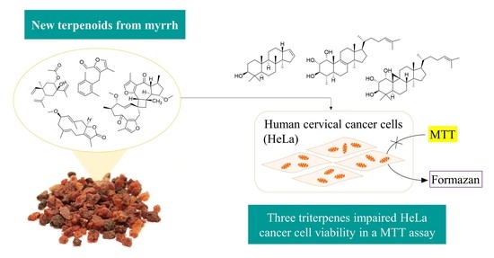
2.3. The Cytotoxicity of Selected Compounds against HeLa Cells

For investigation of their biological activity, selected and mainly sesquiterpenoid compounds, which were obtained in sufficient amount and HPLC-DAD purity (>90%) (46, 911, 17, 18, 20 and 27), were tested in an ICAM-1 in vitro assay, as described before [66]. In this assay, only a small reduction (less than 20%) of TNF-α dependent ICAM-1 expression in HMEC-1 cells was observed, and, hence, no relevant anti-inflammatory activity could be detected. The results are provided in the Supplementary Material in Figure S14.
Additionally, cytotoxic effects of selected isolates (2, 2629, 31, 33, and 34) on human cervical cancer cells (HeLa) were studied with special attention to the triterpenoids. For this, an MTT viability assay was used and carried out with some modifications according to Mosman [67] monitoring the viability of the cells via their (mitochondrial) reductase activity. The results are presented in relation to the untreated control (u.c.). To exclude the possibility of solvent effects, cells were also treated with the highest used DMSO concentration (negative control).
A significant reduction in the viability of HeLa cells could be observed for five of the tested substances: 26, 29, 31, 33 and 34. Two substances (31, 34) showed weak but significant concentration-dependent effects. The three isolates with the most cytotoxic activity were compounds 26 (mansumbinol), 29 (cycloartan-24-ene-1α,2α,3β-triol) and 33 (29-nor-lanost-8,24-diene- 1α,2α,3β-triol) with IC50 values of 78.9, 60.3 and 74.5 µM, respectively (Figure 13).

3. Discussion and Conclusions

The presented 34 terpenoids from myrrh are only a small excerpt of its secondary metabolites and illustrate their broad heterogeneity. Sesquiterpenes of six different structural types (germacranes, eudesmanes, seco-eudesmanes, elemanes, cadinane, and guaiane) were found, in addition to one sesquiterpene dimer and triterpenoids of four different scaffolds (mansumbinanes, lanostanes, dammaranes, and cycloartanes). The analysis of the structural richness is crucial for the further development of the standardized quality analytics of the drug, which are still based on a TLC experiment detecting the volatile sesquiterpenes in the European Pharmacopoeia [68]. The examination of the composition is also the basis for finding the molecular targets of the traditionally used drug, which are still unknown.
Concerning the sesquiterpene dimer (8), the accompanying isolation of its monomers with a double bond at the linking position strongly supports Dong et al.’s (2019) suggestion of a 2,2-cycloaddition as biosynthetic pathway (Figure 14) [21].
To test the anti-inflammatory activity of the isolated molecules, the ICAM-1 expression on human endothelial cells was chosen as a pathway. Interestingly, one of the active compounds found in earlier work [18] only differs in a missing double bond from as inactive analysed compounds 18 (position C-8,9) or an additional one from 20 (position C-1,2). The negative results of all the tested substances indicate that the proven anti-inflammatory activity of myrrh is either caused by other compounds [18] or triggered via another pathway. Investigation of the molecular mechanisms of action, therefore, remains an interesting field for further research.
The observed cytotoxic effect of compounds 26, 29 and 33 on HeLa cells provide a first hint of cytotoxic activity of these myrrh triterpenoids against cancer cells. For a more comprehensive and robust investigation of cytotoxicity a broader set of triterpenoids as well as a second quantitative analytical method for purity control, must be included. Interestingly, an A ring cleavage of mansumbinol (26) resulting in a carboxylic acid and an isopropenyl partial structure led to a complete loss of activity for compound 27. Compound 29, with three hydroxyl groups at C-1-3, showed significant cytotoxicity, whereas similar substances with a different substitution pattern in the A ring, as in 28 or 31, were less potent. This was also observed for 33, which is a lanostane-type triterpene but corresponds to 29 with the A ring substitution. Thus, it can be hypothesized that the 1,2,3-trihydroxy substructure in the A ring contributes to a cytotoxic effect, whereas the type of triterpene skeleton has less influence on the activity.
To our knowledge, this is the first time that myrrh triterpenes are reported to have cytotoxic effects against HeLa cells. Previous investigations occasionally describe the cytotoxic activity of myrrh oil or single myrrh sesquiterpenes on both normal and cancer cells, but activity is often selective for the former [33,69]. Further cell viability experiments with other tumour and noncancer cell lines will be necessary to characterize their activity better, especially regarding possibly selective anticancer activity or general cytotoxicity.

4. Materials and Methods

4.1. Chemicals

Extraction was performed with ethanol of technical quality from CSC Jäcklechemie (Nuremberg, Germany) after purification by evaporation. Methanol, n-heptane, ethyl acetate, dichloromethane, diethyl ether and toluene (all per analysis quality; p.a.) were purchased from Fisher Scientific (Hampton, New Hampshire, USA). Anisaldehyde (4-methoxybenzaldehyde for synthesis), sulfuric acid (95–97%, p.a.) and acetonitrile (HPLC-grade) were obtained from Merck Chemicals (Darmstadt, Germany), and n-hexane (p.a.), chloroform-d (99.8%), Dulbecco’s Phosphate-Buffered Saline and Minimum Essential Medium Eagle were from Sigma-Aldrich (St. Louis, Missouri, USA). Formic acid (p.a.) and DMSO (p.a. ≥ 99.5% for molecular biology) were provided by Carl Roth (Karlsruhe, Germany). Foetal bovine serum (FBS) Superior, L-glutamine, nonessential amino acids and trypsin/EDTA for cell culturing were purchased from Biochrom (Berlin, Germany), and 3-(4,5-dimethylthiazol-2-yl)-2,5-diphenyltetrazoliumbromide (MTT) as well as sodiumdodecylsulfate (SDS) from Sigma-Aldrich (Taufkirchen, Germany).

4.2. Plant Material and Extraction

Powdered myrrh resin of C. myrrha (Myrrha, Ph. Eur. 2016) was obtained from Lomapharm® (lot NM0160, Rudolf Lohmann GmbH KG, Emmerthal, Germany). 3 kg of powdered resin was extracted by maceration and percolation with ethanol 96% (v/v) as described before [18].

4.3. Isolation

The isolation process was carried out mostly as published [18] and is, thus, roughly summarized and complemented with the new procedures.

4.3.1. Liquid-Liquid Partition

Portions of ethanolic extract were solved in methanol and partitioned with n-heptane in a separatory funnel to gain a methanol (MeOH, 328.13 g) and an n-heptane-soluble fraction (HEP, 69.99 g).

4.3.2. Solid Phase Extraction: MeOH Fraction

To further divide the methanol fraction, solid-phase extraction by silica gel (Geduran Si 60 (0.063–0.200 mm), Merck Chemicals, Darmstadt, Germany) was performed. Therefore, 150 g of MeOH fraction was mortared with silica gel and submitted dryly in portions on wet-packed columns. The first elution step with ethyl acetate resulted in fraction M1 (107.0 g) and the second step with methanol in M2 (42.3 g).

4.3.3. Silica Flash Chromatography 1

HEP Fraction

Using silica flash chromatography (Spot flash system (Armen Instrument, Paris, France), SVP D40 silica column (13 × 4 cm, SI60 15–40 μm, 90 g, Götec Labortechnik GmbH, Bickenbach, Germany) with an n-hexane (A)/ethyl acetate (B) gradient (30 mL/min; 0–30 min 5% B, 30-90 min 5–20% B, 90–110 min 20–100% B, 110–140 min 100% B), the HEP fraction was partitioned in ten fractions F1-10. F5 (1301-1875 mL, 2.5 g), F6 (1876–2450 mL, 1.1 g), F7 (2451–2950 mL, 1.6 g), and F9 (3201–3550 mL, 1.9 g) were further investigated (retention volume and weight).

MeOH Fraction M1

Using the same flash system, 51 g of M1 was separated into ten subfractions M1.1-1.10 using a silica column (100 × 3.6 cm, SI60 0.063–0.2 mm, approx. 520 g, Merck KGaA, Darmstadt, Germany) and an n-hexane (A)/ethyl acetate (B)/methanol (C) gradient (10 mL/min; 0–5 min 80% A/20% B, 5-245 min 20-100% B, 245-395 min 100% B, 395–491 min 0–100% C, 491–585 min 100% C). Fraction M1.2 (1150-1575 mL retention volume, 0.6 g weight) was further investigated.

4.3.4. Centrifugal Partition Chromatography (CPC)

A Spot centrifugal partition chromatography (CPC) device with a 250 mL rotor (Armen Instrument, Paris, France), a 510 HPLC pump (Waters GmbH, Eschborn, Germany) and a 2111 Multirac Fraction Collector (LKB-Produkter AB, Bromma, Sweden) was used for the separation of fractions F5-7, F9 and M1.2. The separation was performed at a rotation speed of 1000 rpm and a flow rate of 5 mL/min, with a solvent system consisting of n-hexane, acetonitrile and methanol (40/25/10 v/v/v) [70], and was executed in two stages. First, the lower phase (LP) was used as a stationary phase in ascending mode (ASC) for 800 mL for F5-7/F9 or 925 mL for M1.2, respectively. Second, phases were switched, and the process was conducted in descending mode (DSC) for another 200 mL or 275 mL, respectively. Subsequently, subfractions (F5C1-6, F6C1-8, F7C1-8, F9C1-4, and M1.2C1-7) were formed, of which the following were processed further: fraction (mode, retention volume, and weight): F5C2 (ASC 181–280 mL, 70.6 mg), F5C5 (DSC 21–135 mL, 641.3 mg), F6C3 (ASC 191–275 mL, 20.6 mg), F6C4 (ASC 276–385 mL, 24.5 mg), F6C5 (ASC 386–520 mL, 46.6 mg), F6C7 (DSC 16–190 mL, 386.2 mg), F7C2 (ASC 196–480 mL, 43.4 mg), F7C3 (ASC 481–720 mL, 41.3 mg), F7C7 (DSC 11–50 mL, 508.4 mg), F9C2 (ASC 364–450 mL, 20.5 mg), F9C3 (ASC 451–585 mL, 118.1 mg), F9C4 (ASC 586–800 mL, 77.6 mg), M1.2C3 (ASC 311–620 mL, 107.0 mg), M1.2C6 (DSC 986–1060 mL, 129.7 mg) and M1.2C7 (DSC, 1061–1200 mL, 73.5 mg).

4.3.5. Silica Flash Chromatography 2

The fractions F6C7 and F7C7 were subjected to a second flash separation. A SVF D26 silica column (9 × 2.8 cm, SI60 15–40 μm, 30 g, Götec Labortechnik GmbH, Bickenbach, Germany) was used with a flow rate of 15 mL/min and a dichloromethane (A)/ethyl acetate (B) gradient (0–60 min 1–6% B, 60–90 min 6–15% B, 90–92 min 15–100% B, 92–110 min 100% B). Subfractions (F6C7F1-5 and F7C7F1-5) were combined according to thin-layer chromatography (TLC) control, and some were used for further isolation: fraction (retention volume and weight): F6C7F3 (256–510 mL, 64.5 mg), F7C7F1 (1–390 mL, 90.2 mg), F7C7F2 (391–465 mL, 40.4 mg) and F7C7F3 (466–660 mL, 52.6 mg).

4.3.6. Thin-Layer Chromatography (TLC)

For flash chromatography and CPC, fractions were checked and combined by TLC control on silica gel 60 F254 (Merck, Darmstadt, Germany) with a mobile phase consisting of toluene and ethyl acetate 95/5, 80/20 or 50/50 (v/v) for HEP fractions and M1.2C1-7 or hexane, ethyl acetate and methanol 15/80/5 (v/v/v) for M1.1-1.10. Derivatization was conducted with anisaldehyde reagent R (Ph. Eur.) and documented by a Camag TLC visualizer (Camag AG, Muttenz, Switzerland).

4.3.7. Preparative HPLC

For the isolation of pure compounds, a preparative high-performance liquid chromatography (HPLC) device equipped with a 1260 Infinity binary pump, a 1260 Infinity manual injector, a 1260 Infinity fraction collector, a 1260 Infinity diode array detector (all Agilent Technologies, Santa Clara, USA) and a Kinetex® column (Biphenyl, 100 Å, 5 μm, 21.2 × 250 mm, Phenomenex, Aschaffenburg, Germany) at a flow rate of 21 mL/min or—for fractions M1.2C6 and M1.2C7—a NucleodurTM C18 Isis column (RP18, 5 µm, 10 × 250 mm, Macherey-Nagel, Düren, Germany) at a flow rate of 5 mL/min was used. Separation was achieved by gradients consisting of acetonitrile (A)/water (B) and peaks were detected at 200 nm. After the elimination of acetonitrile via evaporation, the water fractions were partitioned four times with diethyl ether or ethyl acetate (M1.2C6 and M1.2C7) and organic phases were dried under a nitrogen stream. The gradients used for the separation can be found in Table 5.

4.4. NMR

NMR spectra (1D-1H, 1D-13C as well as 2D-1H,13C HSQC, 1H,13C HMBC, 1H,1H COSY, and 1H,1H NOESY) were recorded in CDCl3 in 507-HP-8 NMR tubes (Norell Inc, Morganton, USA) on an AVANCE III 600 NMR equipped with a 5 mm TBI CryoProbe (1H NMR 600.25 MHz, 13C NMR 150.95 MHz, 298 K) or an AVANCE III HD NMR (1H NMR 400.13 MHz, 13C NMR 100.63 MHz, 298 K) (Bruker Corporation, Billerica, MA, USA). For structure elucidation, the data were subsequently processed with TopSpin 3.1 or 3.5.b.91 pl 7 (Bruker Corporation).

4.5. UHPLC-MS

Isolates were analysed using ultra-high-performance liquid chromatography coupled to mass spectrometry (UHPLC-MS) (1290 Infinity UHPLC and Q-TOF 6540 UHD, Agilent Technologies, Santa Clara, USA) on a ZORBAX Eclipse column (XDB-C18 RRHD, 2.1 × 100 mm, 1.8 µm, Agilent Technologies) with the following gradient: solvent A (water with 0.1% formic acid); solvent B (acetonitrile with 0.1% formic acid); 0–10 min, 20-98% B; 10–12 min, 98% B; 12–12.1 min, 98-20% B; 12.1–13.5 min, 20% B; flow: 0.5 mL/min; column temperature: 50 °C; and injection volume: 1 µL. MS analysis was subsequently performed by electrospray ionization (ESI) in positive and negative mode or liquid injection field desorption ionisation (LIFDI) as an even softer ionisation method for compound 30 to avoid the cleavage of the ester.

4.6. Optical Methods

For further characterization of the isolates, optical data were gathered by using solutions in methanol. Specific optical rotation was measured by an UniPol L 1000 polarimeter (Schmidt + Haensch GmbH & Co., Berlin, Germany) using a microtube (50 mm, 550 μL) at 589 nm. UV-spectra were recorded by a Cary 50 Scan UV-spectrophotometer (Varian Deutschland GmbH, Darmstadt, Germany) in a quartz cuvette (QS, 1.0 cm, Hellma GmbH & Co. KG, Müllheim, Germany) in a range of 200–800 nm. Additionally, CD spectra were measured on a J-715 spectropolarimeter (JASCO Deutschland GmbH, Gross-Umstadt, Germany) with a 0.1 cm quartz cuvette (Type: 100-QSQ, Hellma GmbH & Co. KG). Thereby, ten scans were recorded at 22 °C from 190 to 300 or 400 nm with a scanning rate of 50, 100 or 200 nm/min in 0.5 nm steps and the Savitzky–Golay algorithm was used for spectra smoothing (convolution width: 15).

4.7. Simulation of ECD Spectra

Molecular models of commiphorine A and compound 8 (commiphorine C) were generated with the Molecular Operations Environment (MOE, v. 2020.09, Chemical Computing Group, Montreal, Canada) using the MMFF94x force field. After a conformational search with the low mode dynamics (LMD) method, the lowest energy conformers were energy-minimized with the semiempirical method PM3. In each case, the lowest energy conformer was used for the spectra simulation. The structure was exported to Gaussian (v. Gaussian03W, Gaussian Inc., Pittsburgh, Pa., U.S.A.) and fully minimized by density functional theory (DFT) with the B3LYP density functional and the 6−31G(d,p) basis set. The minimized structures were then subjected to a time-dependent DFT (TDDFT) calculation using the same functional and basis set, which yielded the electronic transition vectors determining the ECD spectra. Typically, the first (i.e., lowest in frequency) 12–18 electronic transitions were computed. The Gaussian output for electronic transition energies (E in eV) and rotator strengths (R, dipole length in cgs) was used to simulate ECD spectra by applying a Gaussian shape function with a band width at 1/e height σ = 0.15 eV. The resulting spectra shown in Figure 8 and Figure 9 were scaled by factor 0.5 and blue-shifted by 0.15 eV.
The structures of three conformers of 1,5-cis-configured “commiphorine A” were built directly from the atomic coordinates published by the authors of the previous study [21] in their supplementary file. The ECD spectra of these models were simulated as described above. There was no significant difference between the three spectra, and so only the spectrum of structure 1a1 [21] is shown in Figure S1.

4.8. Purity

The purity of isolates was determined through HPLC-DAD analysis (190-400 nm for the HEP fraction or 200–400 nm for the MeOH fraction) on an Elite LaChrom system consisting of an autosampler L-2200, a pump L-2130, a column oven L-2350, a diode array detector L-2455 (all Hitachi, Tokyo, Japan) and a Kinetex® biphenyl column (100 Å, 5 µm, 4.6 × 250 mm, Phenomenex, Aschaffenburg, Germany) or a NucleodurTM C18 Isis column (RP18, 5 µm, 4.6 × 250 mm, Macherey-Nagel, Düren, Germany). For analyses, gradients described in 0 were conducted at a flow rate of 1 mL/min and an injection volume of 5 or 10 µL (acetonitrile, 1 mg/mL). Thus, the purity was calculated as the proportion of the integral of the main peak in the chromatogram using the maxplot adjusted by a blank (EZChrom Elite 3.1.7, Hitachi).

4.9. Isolated Compounds

As software for drawing the chemical structures, ChemDraw Professional 20.0 (PerkinElmer Informatics Inc., Waltham, MA, USA) was used. Three-dimensional models were generated with Chem3D Ultra 20.0 from the same company.

4.9.1. 9,10-Seco-isolindestrenolide (1)

1.0 mg, colourless oil; [ α ] D 22 +238 (c 1.11, MeOH); UV (MeOH) λmax (log ε): 203 nm (3.97); CD: Figure S2; 1H and 13C NMR data (CDCl3, 600 and 150 MHz, respectively) in Table 1 and Figure S4; HRESIMS m/z 231.1383 [M+H]+ (calc. for C15H19O2, 231.1380); purity (according 4.8.) 89.9%.

4.9.2. 9-Oxo-9,10-seco-isolindestrene (2)

4.1 mg, yellow oil; UV (MeOH) λmax (log ε): 286 nm (3.43); 1H and 13C NMR data (CDCl3, 600 and 150 MHz, respectively) in Table 1 and Figure S5; HRESIMS m/z 229.1226 [M+H]+ (calc. for C15H17O2, 229.1223); purity (according 4.8.) 97.6%.

4.9.3. rel-8S-Acetyloxy-7R-hydroxy-5R,10R-β-elemene (3)

1.2 mg, colourless oil; [ α ] D 25 +36 (c 1.28, MeOH); UV (MeOH) λmax (log ε): 202 nm (3.80), 276 nm (3.57); CD: Figure S2; 1H and 13C NMR data (CDCl3, 600 and 150 MHz, respectively) in Table 2 and Figure S6; HRESIMS m/z 279.1950 [M+H]+ (calc. for C17H27O3, 279.1955); purity (according 4.8.) 88.3%.

4.9.4. Commiterpene E (4)

0.8 mg, colourless oil; [ α ] D 25 +18 (c 0.90, MeOH); UV (MeOH) λmax (log ε): 203 nm (4.26); CD: Figure S2; 1H and 13C NMR data (CDCl3, 600 and 150 MHz, respectively) in Table 2 and Figure S7; HRESIMS m/z 261.1490 [M+H]+ (calc. for C16H21O3, 261.1485); purity (according 4.8.) 93.5%.

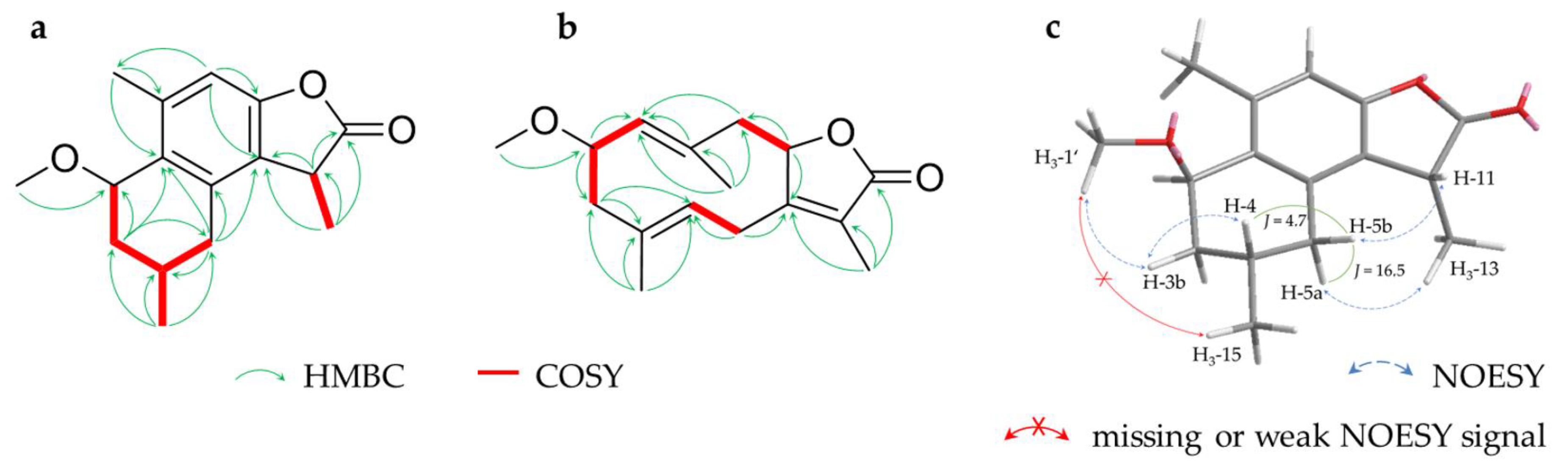
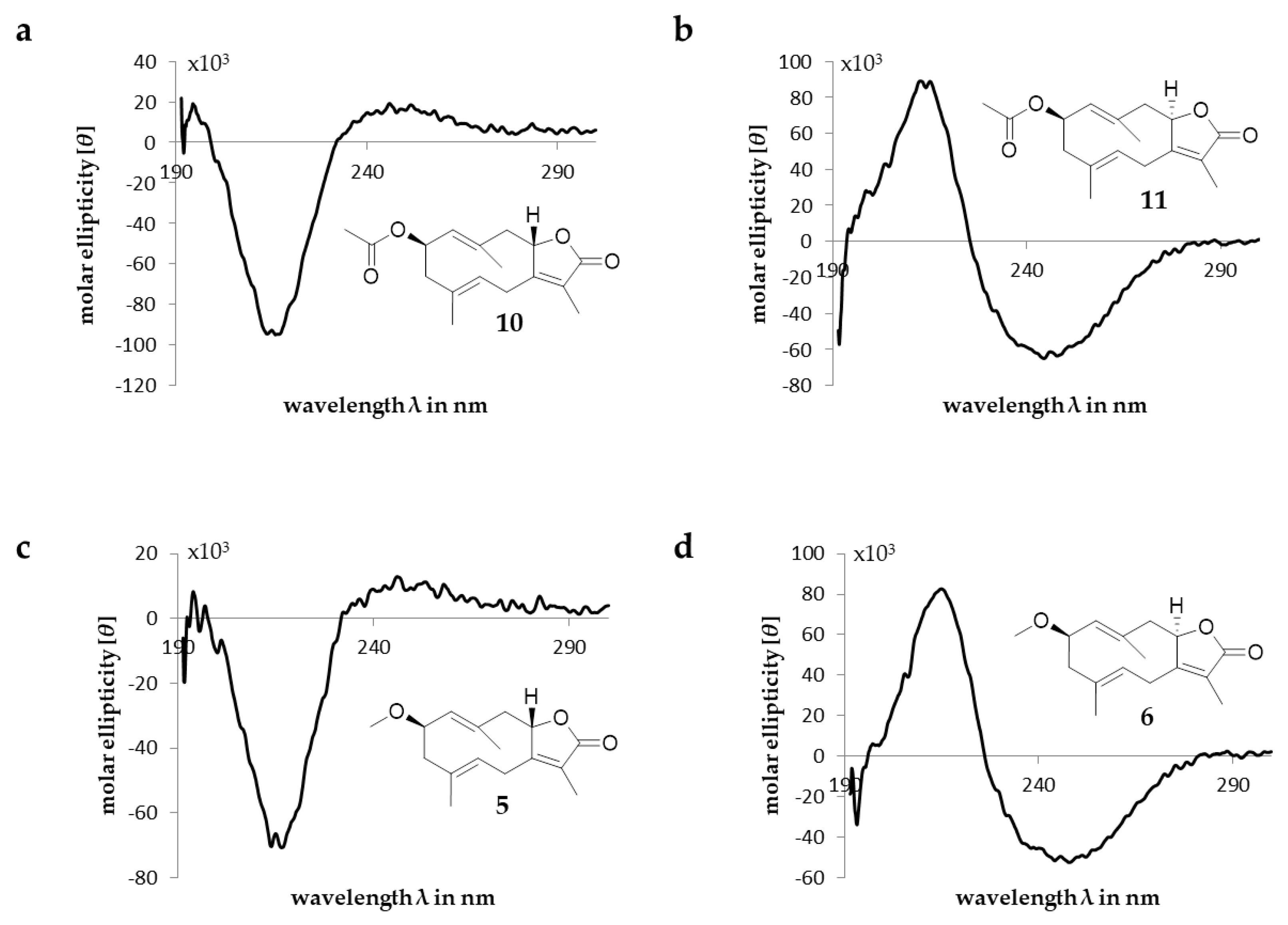
4.9.5. 2β-Methoxyglechomanolide (5)

2.9 mg, white crystals; [ α ] D 25 +53; UV (c 1.60, MeOH); λmax (log ε): 218 nm (3.98); CD: Figure S2; 1H and 13C NMR data (CDCl3, 600 and 150 MHz, respectively) in Table 2 and Figure S8; HRESIMS m/z 263.1646 [M+H]+ (calc. for C16H23O3, 263.1642); purity (according 4.8.) 90.9%.

4.9.6. 8-epi-2β-Methoxyglechomanolide (6)

6.5 mg, white crystals; [ α ] D 25 −69; UV (c 2.23, MeOH); λmax (log ε): 215 nm (4.17); CD: Figure S2; 1H and 13C NMR data (CDCl3, 400 and 100 MHz, respectively) in Table 2 and Figure S9; HRESIMS m/z 263.1646 [M+H]+ (calc. for C16H23O3, 263.1642); purity (according 4.8.) 96.9%.

4.9.7. Dehydroisolindestrenolide (7)

1.8 mg, colourless oil; [ α ] D 24 +34 (c 1.94, MeOH); UV (MeOH) λmax (log ε): 202 nm (3.94), 275 nm (3.79); CD: Figure S2; 1H and 13C NMR data (CDCl3, 600 and 150 MHz, respectively) in Table 3 and Figure S10; HRESIMS m/z 229.1228 [M+H]+ (calc. for C15H17O2, 229.1223); purity (according 4.8.) 89.5%.

4.9.8. Commiphorine C (8)

0.9 mg, white solid; [ α ] D 27 -33 (c 0.97, MeOH); UV (MeOH) λmax (log ε): 202 nm (4.06), 277 nm (3.48); CD: Figure S2; 1H and 13C NMR data (CDCl3, 600 and 150 MHz, respectively) in Table 3 and Figure S11; HRESIMS m/z 521.2904 [M+H]+ (calc. for C32H41O6, 521.2898); purity (according 4.8.) 85.3%.

4.9.9. Glechomanolide (9)

5.7 mg, white crystals; [ α ] D 25 +6 (c 1.84, MeOH); UV (MeOH) λmax (log ε): 214 nm (4.10); CD: Figure S2; 1H and 13C NMR data (CDCl3, 400 and 100 MHz, respectively) in Table S1; HRESIMS m/z 233.1540 [M+H]+ (calc. for C15H21O2, 233.1536); purity (according 4.8.) 97.2%.

4.9.10. 2β-Acetyloxyglechomanolide (10)

10.9 mg, white crystals; [ α ] D 25 +26 (c 2.31, MeOH); UV (MeOH) λmax (log ε): 216 nm (4.15), 285 nm (2.91); CD: Figure S2; 1H and 13C NMR data (CDCl3, 400 and 100 MHz, respectively) in Table S1; HRESIMS m/z 291.1595 [M+H]+ (calc. for C17H23O4, 291.1591); purity (according 4.8.) 95.4%.

4.9.11. 8-epi-2β-Acetyloxyglechomanolide (11)

11.3 mg, white crystals; [ α ] D 25 +26 (c 2.27, MeOH); UV (MeOH) λmax (log ε): 214 nm (4.20); CD: Figure S2; 1H and 13C NMR data (CDCl3, 400 and 100 MHz, respectively) in Table S1; HRESIMS m/z 291.1598 [M+H]+ (calc. for C17H23O4, 291.1591); purity (according 4.8.) 92.0%.

4.9.12. 2α-Methoxy-6-oxogermacra-1(10),7(11)-dien-8,12-olide (12)

1.1 mg, white crystals; [ α ] D 25 +107 (c 1.12, MeOH); UV (MeOH) λmax (log ε): 230 nm (3.76); CD: Figure S2; 1H and 13C NMR data (CDCl3, 600 and 150 MHz, respectively) in Table S2; HRESIMS m/z 279.1598 [M+H]+ (calc. for C16H23O4, 279.1591); purity (according 4.8.) 89.3%.

4.9.13. 1(10)Z,4E-Isofuranodienone (13)

1.6 mg (including 22), colourless oil; 1H and 13C NMR data (CDCl3, 600 and 150 MHz, respectively) in Table S2; HRESIMS m/z 231.1381 [M+H]+ (calc. for C15H19O2, 231.1380); purity (according 4.8.) 77.3% (including 22).

4.9.14. 3S-Methoxy-4R-furano-1E,10(14)-germacradien-6-one (14)

6.6 mg, colourless oil; [ α ] D 23 +43 (c 2.20, MeOH); UV (MeOH) λmax (log ε): 216 nm (3.97); CD: Figure S2; 1H and 13C NMR data (CDCl3, 400 and 100 MHz, respectively) in Table S2; HRESIMS m/z 261.1486 [M+H]+ (calc. for C16H21O3, 261.1485); purity (according 4.8.) 92.7%.

4.9.15. 2-Methoxy-5-acetoxyfuranogermacr-1(10)-en-6-one (15)

9.2 mg, white crystals; [ α ] D 25 −21 (c 2.22, MeOH); UV (MeOH) λmax (log ε): 207 nm (4.19), 284 nm (3.27); CD: Figure S2; 1H and 13C NMR data (CDCl3, 400 and 100 MHz, respectively) in Table S3; HRESIMS m/z 321.1699 [M+H]+ (calc. for C18H25O5, 321.1697); purity (according 4.8.) 98.5%.

4.9.16. rel-2R-Methoxy-5S-acetoxy-4R-furanogermacr-1(10)Z-en-6-one (16)

1.8 mg, colourless oil; [ α ] D 25 +56 (c 1.80, MeOH); UV (MeOH) λmax (log ε): 275 nm (3.36); CD: Figure S2; 1H and 13C NMR data (CDCl3, 600 and 150 MHz, respectively) in Table S3; HRESIMS m/z 321.1702 [M+H]+ (calc. for C18H25O5, 321.1697); purity (according 4.8.) 92.2%.

4.9.17. (1S,4S,5S,10S)-Germacron-1(10),4-diepoxide (17)

3.6 mg, white crystals; [ α ] D 25 +3 (c 1.80, MeOH); UV (MeOH) λmax (log ε): 248 nm (3.50); CD: Figure S2; 1H and 13C NMR data (CDCl3, 600 and 150 MHz, respectively) in Table S3; HRESIMS m/z 251.1647 [M+H]+ (calc. for C15H23O3, 251.1642); purity (according 4.8.) 88.2%.

4.9.18. Dehydrolindestrenolide (18)

1.9 mg, colourless oil; [ α ] D 23 +60 (c 1.86, MeOH); UV (MeOH) λmax (log ε): 202 nm (3.96), 204 nm (3.91), 276 nm (3.74); CD: Figure S3; 1H and 13C NMR data (CDCl3, 600 and 150 MHz, respectively) in Table S4; HRESIMS m/z 229.1225 [M+H]+ (calc. for C15H17O2, 229.1223); purity (according 4.8.) 89.5%.

4.9.19. Tubipolide B (19)

2.2 mg (including 21), colourless oil; 1H and 13C NMR data (CDCl3, 600 and 150 MHz, respectively) in Table S4; HRESIMS m/z 231.1380 [M+H]+ (calc. for C15H19O2, 231.1380); purity (according 4.8.) 93.1% (including 21).

4.9.20. Atractylenolide II (20)

1.2 mg, colourless oil; [ α ] D 25 +16 (c 1.31, MeOH); UV (MeOH) λmax (log ε): 202 nm (3.52), 205 nm (3.51), 221 nm (3.94); CD: Figure S3; 1H and 13C NMR data (CDCl3, 600 and 150 MHz, respectively) in Table S4; HRESIMS m/z 233.1545 [M+H]+ (calc. for C15H21O2, 233.1536); purity (according 4.8.) 96.5%.

4.9.21. 8-epi-Isogermafurenolide (21)

2.2 mg (including 19), colourless oil; 1H and 13C NMR data (CDCl3, 600 and 150 MHz, respectively) in Table S5; HRESIMS m/z 233.1536 [M+H]+ (calc. for C15H21O2, 233.1536); purity (according 4.8.) 93.1% (including 19).

4.9.22. Myrrhterpenoid O (22)

1.6 mg (including 13), colourless oil; [ α ] D 23 +152 (c about 0.66, MeOH); CD: Figure S3; 1H and 13C NMR data (CDCl3, 600 and 150 MHz, respectively) in Table S5; HRESIMS m/z 261.1490 [M+H]+ (calc. for C16H21O3, 261.1485); purity (according 4.8.) 77.3% (including 13).

4.9.23. 3β-Isovaleroyloxy-29-nor-lanost-8,24-diene-1α,2α-diol (23)

1.7 mg, white crystals; [ α ] D 25 +28 (c 1.70, MeOH); CD: Figure S3; 1H and 13C NMR data (CDCl3, 600 and 150 MHz, respectively) in Table 4 and Figure S12; HRESIMS m/z 573.4169 [M+HCOO] (calc. for C35H57O6, 573.4161); purity (according 4.8.) 60.9%.

4.9.24. 29-Nor-1,2-cis-epoxylanost-8,24-diene-3β-triol (24)

2.0 mg, white crystals; [ α ] D 25 +32 (c 2.00, MeOH); CD: Figure S3; 1H and 13C NMR data (CDCl3, 600 and 150 MHz, respectively) in Table 4 and Figure S13; HRESIMS m/z 427.3581 [M+H]+ (calc. for C29H47O2, 427.3571); purity (according 4.8.) 83.2%.

4.9.25. rel-20S-Hydroxydammar-24-en-3,16-dione (25)

6.2 mg, white crystals; [ α ] D 25 +1 (c 2.05, MeOH); CD: Figure S3; 1H and 13C NMR data (CDCl3, 600 and 150 MHz, respectively) in Table S6; HRESIMS m/z 501.3592 [M+HCOO] (calc. for C31H49O5, 501.3585); purity (according 4.8.) 71.6%.

4.9.26. Mansumbinol (26)

16.1 mg, white needles; [ α ] D 25 −19 (c 2.64, MeOH); CD: Figure S3; 1H and 13C NMR data (CDCl3, 400 and 100 MHz, respectively) in Table S6; HRESIMS m/z 334.3105 [M+NH4]+ (calc. for C22H40ON, 334.3104); purity (according 4.8.) 83.1%.

4.9.27. 3,4-Seco-mansumbinoic acid (27)

9.4 mg, white crystals; [ α ] D 25 +1 (c 1.85, MeOH); CD: Figure S3; 1H and 13C NMR data (CDCl3, 400 and 100 MHz, respectively) in Table S6; HRESIMS m/z 329.2491 [M-H] (calc. for C22H33O2, 329.2486); purity (according 4.8) 93.4%.

4.9.28. Cycloartan-24-ene-1α,3β-diol (28)

6.4 mg, white crystals; [ α ] D 25 +64 (c 2.02, MeOH); CD: Figure S3; 1H and 13C NMR data (CDCl3, 400 and 100 MHz, respectively) in Table S7; HRESIMS m/z 487.3797 [M+HCOO] (calc. for C31H51O4, 487.3793); purity (according 4.8.) 90.5%.

4.9.29. Cycloartan-24-ene-1α,2α,3β-triol (29)

48.4 mg, white crystals; [ α ] D 25 +38 (c 1.74, MeOH); CD: Figure S3; 1H and 13C NMR data (CDCl3, 400 and 100 MHz, respectively) in Table S7; HRESIMS m/z 476.4100 [M+NH4]+ (calc. for C30H54O3N, 476.4098); purity (according 4.8.) 90.6%.

4.9.30. 1α-Acetoxy-9,19-cyclolanost-24-ene-3β-ol (30)

2.0 mg, white crystals; [ α ] D 25 +46 (c 2.07, MeOH); CD: Figure S3; 1H and 13C NMR data (CDCl3, 600 and 150 MHz, respectively) in Table S7; HRLIFDIMS m/z 484.3900 M●+ (calc. for C32H52O3, 484.3911); purity (according 4.8.) 68.8%.

4.9.31. 1α-Acetoxycycloartan-24-ene-2α,3β-diol (31)

6.0 mg, white crystals; [ α ] D 25 +30 (c 1.92, MeOH); CD: Figure S3; 1H and 13C NMR data (CDCl3, 400 and 100 MHz, respectively) in Table S8; HRESIMS m/z 523.3751 [M+Na]+ (calc. for C32H52O4Na, 523.3758); purity (according 4.8.) 89.2%.

4.9.32. 3β-Isovaleroyloxycycloartan-24-ene-1α,2α-diol (32)

2.4 mg, white crystals; [ α ] D 25 +25 (c 2.58, MeOH); CD: Figure S3; 1H and 13C NMR data (CDCl3, 600 and 150 MHz, respectively) in Table S8; HRESIMS m/z 565.4233 [M+Na]+ (calc. for C33H58O4Na, 565.4227); purity (according 4.8.) 54.9%.

4.9.33. 29-Nor-lanost-8,24-diene-lα,2α,3β-triol (33)

32.7 mg, white crystals; [ α ] D 25 +83 (c 1.08, MeOH); CD: Figure S3; 1H and 13C NMR data (CDCl3, 400 and 100 MHz, respectively) in Table S8; HRESIMS m/z 445.3675 [M+H]+ (calc. for C29H49O3, 445.3676); purity (according 4.8.) 93.9%.

4.9.34. Hydroxydammarenone II (34)

4.1 mg, white crystals; [ α ] D 25 +42 (c 2.00, MeOH); CD: Figure S3; 1H and 13C NMR data (CDCl3, 600 and 150 MHz, respectively) in Table S9; HRESIMS m/z 443.3897 [M+H]+ (calc. for C30H51O2, 443.3884); purity (according 4.8.) 89.8%.

4.10. Cell Culture

4.10.1. Cultivation

The human cervical cancer cell line HeLa (CCL-2TM) was obtained from the American Type Culture Collection (ATTC®, Manassas, VA, USA). The cells were cultured in Minimum Essential Medium Eagle supplemented with 10% FBS, 1% L-glutamine and 1% nonessential amino acids.
The human microvascular endothelial cell line (HMEC-1) was provided by Dr. E. Ades und F.-J. Candel (CDC, Atlanta, GA, USA), as well as Dr. T. Lawley (Emory University, Atlanta, GA, USA). These cells were cultured in EASY Endothelial Cell Growth Medium supplemented with 10% FBS, 50 ng/mL amphotericin B and 50 ng/mL gentamicin.
For both the used cell lines, mycoplasma contamination was excluded by PCR and culture from GATC Biotech AG (Konstanz, Germany) and they were cultured in an atmosphere of 5% CO2 and 90% relative humidity at 37 °C.

4.10.2. MTT Assay

The MTT assay was conducted with both cell lines and differed (besides the different respective medium) only in the seeded cell density: HeLa experiments, 8 × 10⁴ cells/well, HMEC-1 experiments 9 × 10⁴ cells/well. They were seeded (in 100 μL/well) in 96-well plates and incubated at 37 °C in an atmosphere of 5% CO2 and 90% relative humidity. After 24 h, the medium was replaced by a sample solution in medium (25–100 µM, max. 0.2% DMSO, v/v) and kept for another 24 h at the same conditions. Subsequently, the supernatant was removed and 100 μL of an MTT solution in medium (0.4 mg/mL) was added and incubated for three hours. Thereafter, cells were treated with 10% sodium dodecyl sulfate (SDS) in water and stored at room temperature in the dark until the formazan crystals were dissolved and the absorbance at 560 nm could be determined using a Tecan microplate reader (Tecan Trading AG, Maennedorf, Switzerland). The cell viability was calculated as proportion compared to the average absorbance of the negative control group (only medium). To preclude the possibility of solvent effects, some cells were also treated with the highest used DMSO concentration. All tests were performed three times independently in hexaplicates.

4.10.3. ICAM-1 Assay

This assay was performed as previously described [66]. Confluently grown HMEC-1 cells from a culture flask (150 cm2) were split (1:3), suspended in 13 mL of medium, and seeded in a 24-well plate (500 µL/well). After cultivation for 48 h at 37 °C in an atmosphere of 5% CO2 and 90% relative humidity and the formation of a monolayer, the supernatant was replaced by substance dilutions in medium (6–70 µM) containing a maximum of 0.15% DMSO (v/v) and incubated for 30 min before stimulation with TNFα (10 ng/mL). In each performance, an unstimulated negative control (0.15% DMSO, v/v), an untreated control (medium), and a positive control (parthenolide, 5 µM) were included. 24 h later, the cells were washed with phosphate-buffered saline (PBS), detached by trypsin/EDTA, fixed by formalin 10% for 15 min, and treated with a murine fluorescein-isothiocyanate (FITC)-marked IgG1 antibody against ICAM-1 (Bio-Rad, Kidlington, UK) for 30 min. The cell suspension in PBS was analysed by a FACSCanto II (BD, Lakes, NJ, USA) (Flow 60 µL/min, FSC: 1 V, SSC: 320 V; FITC: 320 V). The ICAM-1 expression was calculated as a proportion of the mean obtained for the untreated control. The test was performed three times independently in duplicate.

4.11. Statistics

Significance levels were calculated in a one-way ANOVA followed by Tukey-HSD in SPSS 26 (IBM, Armonk, NY, USA), whereas nonlinear regression curves were determined by GraphPad Prism 5.0.0 (GraphPad Software, San Diego, CA, USA).

Supplementary Materials

The following supporting information can be downloaded at: https://www.mdpi.com/article/10.3390/molecules28041637/s1, Figure S1. (a) Molecular structures of three conformers of commiphorine A underlying the ECD simulations published by previous authors [21]. All structures represent a compound with 1,5-cis ring fusion in the guaiane part (H-1 and H-5 are cis-oriented; the latter is marked by a dashed circle). This arrangement does not correspond to the structure of commiphorine A with a 1,5-trans-guaiane moiety postulated in the same work [21]. (b) Experimental ECD spectrum of commiphorine A (red curve) and simulated ECD spectrum (blue curve) obtained with the structures shown in (a) according to [21]. (c) ECD spectrum simulated for commiphorine A with the erroneous 1,5-cis structure (blue curve) and with the correct 1,5-trans structure (red curve). Table S1. 1H and 13C NMR data (400 and 100 MHz, respectively) for compounds 911 (CDCl3, δ in ppm, J in Hz, s: singlet, d: doublet, br: broad, and m: multiplet). Table S2. 1H and 13C NMR data (600 and 150 MHz, respectively) for compounds 12, 13 and (400 and 100 MHz, respectively) for 14 (CDCl3, δ in ppm, J in Hz, s: singlet, d: doublet, br: broad, and m: multiplet). Table S3. 1H and 13C NMR data (400 and 100 MHz, respectively) for compound 15 and (600 and 150 MHz, respectively) for 16 and 17 (CDCl3, δ in ppm, J in Hz, s: singlet, d: doublet, br: broad, and m: multiplet). Table S4. 1H and 13C NMR data (600 and 150 MHz, respectively) for compounds 1820 (CDCl3, δ in ppm, J in Hz, s: singlet, d: doublet, br: broad, and m: multiplet). Table S5. 1H and 13C NMR data (600 and 150 MHz, respectively) for compounds 21 and 22 (CDCl3, δ in ppm, J in Hz, s: singlet, d: doublet, br: broad, and m: multiplet). Table S6. 1H and 13C NMR data (600 and 150 MHz, respectively) for compound 25 and (400 and 100 MHz, respectively) for 26 and 27 (CDCl3, δ in ppm, J in Hz, s: singlet, d: doublet, br: broad, and m: multiplet). Table S7. 1H and 13C NMR data (600 and 150 MHz, respectively) for compounds 28, 29 and (400 and 100 MHz, respectively) for 30 (CDCl3, δ in ppm, J in Hz, s: singlet, d: doublet, br: broad, and m: multiplet). Table S8. 1H and 13C NMR data (400 and 100 MHz, respectively) for compounds 31 and 32 and (600 and 150 MHz, respectively) for 33 (CDCl3, δ in ppm, J in Hz, s: singlet, d: doublet, br: broad, and m: multiplet). Table S9. 1H and 13C NMR data (600 and 150 MHz, respectively) for compound 34 (CDCl3, δ in ppm, J in Hz, s: singlet, d: doublet, br: broad, and m: multiplet). Figure S2. CD spectra of compounds 1, 312 and 1417 in methanol. [θ] in [(°cm2) · dmol−1]. Figure S3. CD spectra of compounds 18, 20 and 2234 in methanol. [θ] in [(°cm2) · dmol−1]. Figure S4. 1H (600 MHz, A) and 13C NMR spectra (150 MHz, B) of 1 (in CDCl3). Figure S5. 1H (600 MHz, A) and 13C NMR spectra (150 MHz, B) of 2 (in CDCl3). Figure S6. 1H (600 MHz, A) and 13C NMR spectra (150 MHz, B) of 3 (in CDCl3). Figure S7. 1H (600 MHz, A) and 13C NMR spectra (150 MHz, B) of 4 (in CDCl3). Figure S8. 1H (600 MHz, A) and 13C NMR spectra (150 MHz, B) of 5 (in CDCl3). Figure S9. 1H (400 MHz, A) and 13C NMR spectra (100 MHz, B) of 6 (in CDCl3). Figure S10. 1H (600 MHz, A) and 13C NMR spectra (150 MHz, B) of 7 (in CDCl3). Figure S11. 1H (600 MHz, A) and 13C NMR spectra (150 MHz, B) of 8 (in CDCl3). Figure S12. 1H (600 MHz, A) and 13C NMR spectra (150 MHz, B) of 23 (in CDCl3). Figure S13. 1H (600 MHz, A) and 13C NMR spectra (150 MHz, B) of 24 (in CDCl3). Figure S14. Influence of compounds 46, 911, 17, 18, 20 and 27 on the viability of HMEC-1 cells in the MTT assay (a) and on their TNFα-induced ICAM-1 expression (b). The test was performed with medium containing DMSO (0.15%, v/v) without stimulation (u.c.), with TNFα (10 ng/mL, n.c.), and with both TNFα and parthenolide (5 µM, Parth.) as positive control. Substances were applied in concentrations between 13 and 70 µM. Data are presented as mean ± SEM (n = 3).

Author Contributions

Conceptualization, J.H. and G.J.; methodology, K.K., A.U., and T.J.S. (ECD calculations); formal analysis, K.K. and A.U.; investigation, K.K. and A.U.; resources, J.H.; writing—original draft preparation, K.K. and A.U.; writing—review and editing, S.S., J.H., G.J., and B.L.; visualization, K.K. and A.U.; supervision, S.S., J.H., G.J., and B.L.; project administration, J.H.; funding acquisition, J.H. All authors have read and agreed to the published version of the manuscript.

Funding

This research was funded by Repha GmbH Biologische Arzneimittel. The APC was funded by the publication fund of University of Regensburg, Germany.

Institutional Review Board Statement

Not applicable.

Informed Consent Statement

Not applicable.

Data Availability Statement

The data presented in this study are available on request from J.H., K.K., and A.U.

Acknowledgments

Thanks are due to Repha GmbH Biologische Arzneimittel for scientific discussions and financial support. Fritz Kastner is kindly acknowledged for performing the 1D and 2D NMR experiments, as are Josef Kiermaier and Wolfgang Söllner for acquiring the MS data (all: Zentrale Analytik, Faculty of Chemistry and Pharmacy, University of Regensburg). The authors also would like to thank the American Type Culture Collection (ATTC®, Manassas, USA) for providing the HeLa cell line. Furthermore, thanks are due to Laura Humphrys for the fine-tuning of the language and to Thomas Gruber for the photo of myrrh depicted in the graphical abstract.

Conflicts of Interest

Bartosz Lipowicz is employed by Repha GmbH Biologische Arzneimittel. All other authors (K.K., A.U., S.S., G.J., T.S. and J.H.) have declared no conflicts of interest.

Sample Availability

Samples of the compounds 134 are available from the authors.

References

  1. Tucker, A.O. Frankincense and myrrh. Econ. Bot. 1986, 40, 425–433. [Google Scholar] [CrossRef]
  2. Shen, T.; Li, G.-H.; Wang, X.-N.; Lou, H.-X. The genus Commiphora: A review of its traditional uses, phytochemistry and pharmacology. J. Ethnopharmacol. 2012, 142, 319–330. [Google Scholar] [CrossRef]
  3. Abegaz, B.; Dagne, E.; Bates, C.; Waterman, P.G. Monoterpene-rich resins from two ethiopian species of Commiphora. Flavour Fragr. J. 1989, 4, 99–101. [Google Scholar] [CrossRef]
  4. Asres, K.; Tei, A.; Moges, G.; Sporer, F.; Wink, M. Terpenoid composition of the wound-induced bark exudate of Commiphora tenuis from Ethiopia. Planta Med. 1998, 64, 473–475. [Google Scholar] [CrossRef]
  5. Hanuš, L.O.; Řezanka, T.; Dembitsky, V.M.; Moussaieff, A. Myrrh-Commiphora chemistry. Biomed. Papers 2005, 149, 3–28. [Google Scholar] [CrossRef]
  6. Dekebo, A.; Dagne, E.; Sterner, O. Furanosesquiterpenes from Commiphora sphaerocarpa and related adulterants of true myrrh. Fitoterapia 2002, 73, 48–55. [Google Scholar] [CrossRef]
  7. Morteza-Semnani, K.; Saeedi, M. Constituents of the essential oil of Commiphora myrrha (Nees) Engl. var. molmol. J. Essent. Oil Res. 2003, 15, 50–51. [Google Scholar] [CrossRef]
  8. Madia, V.N.; de Angelis, M.; de Vita, D.; Messore, A.; de Leo, A.; Ialongo, D.; Tudino, V.; Saccoliti, F.; de Chiara, G.; Garzoli, S.; et al. Investigation of Commiphora myrrha (Nees) Engl. oil and its main components for antiviral activity. Pharmaceuticals 2021, 14, 243. [Google Scholar] [CrossRef]
  9. Marongiu, B.; Piras, A.; Porcedda, S.; Scorciapino, A. Chemical composition of the essential oil and supercritical CO2 extract of Commiphora myrrha (Nees) Engl. and of Acorus calamus L. J. Agric. Food Chem. 2005, 53, 7939–7943. [Google Scholar] [CrossRef]
  10. Nikolic, M.; Smiljkovic, M.; Markovic, T.; Ciric, A.; Glamoclija, J.; Markovic, D.; Sokovic, M. Sensitivity of clinical isolates of Candida to essential oils from Burseraceae family. EXCLI J. 2016, 15, 280–289. [Google Scholar] [CrossRef]
  11. Hanuš, L.O.; Rosenthal, D.; Řezanka, T.; Dembitsky, V.M.; Moussaief, A. Fast and easy GC/MS identification of myrrh resins. Pharm. Chem. J. 2008, 42, 719–720. [Google Scholar] [CrossRef]
  12. Su, S.L.; Duan, J.A.; Tang, Y.P.; Zhang, X.; Yu, L.; Jiang, F.R.; Zhou, W.; Luo, D.; Ding, A.W. Isolation and biological activities of neomyrrhaol and other terpenes from the resin of Commiphora myrrha. Planta Med. 2009, 75, 351–355. [Google Scholar] [CrossRef]
  13. Xu, J.; Guo, Y.; Zhao, P.; Xie, C.; Jin, D.; Hou, W.; Zhang, T. Neuroprotective cadinane sesquiterpenes from the resinous exudates of Commiphora myrrha. Fitoterapia 2011, 82, 1198–1201. [Google Scholar] [CrossRef]
  14. Xu, J.; Guo, Y.; Li, Y.; Zhao, P.; Liu, C.; Ma, Y.; Gao, J.; Hou, W.; Zhang, T. Sesquiterpenoids from the resinous exudates of Commiphora myrrha and their neuroprotective effects. Planta Med. 2011, 77, 2023–2028. [Google Scholar] [CrossRef]
  15. Greve, H.L.; Kaiser, M.; Schmidt, T.J. Investigation of antiplasmodial effects of terpenoid compounds isolated from myrrh. Planta Med. 2020, 86, 643–654. [Google Scholar] [CrossRef]
  16. Shen, T.; Wan, W.-Z.; Wang, X.-N.; Yuan, H.-Q.; Ji, M.; Lou, H.-X. A triterpenoid and sesquiterpenoids from the resinous exudates of Commiphora myrrha. Helv. Chim. Acta 2009, 92, 645–652. [Google Scholar] [CrossRef]
  17. Ayyad, S.-E.N.; Hoye, T.R.; Alarif, W.M.; Al Ahmadi, S.M.; Basaif, S.A.; Ghandourah, M.A.; Badria, F.A. Differential cytotoxic activity of the petroleum ether extract and its furanosesquiterpenoid constituents from Commiphora molmol resin. Z. Naturforsch. 2015, 70, 87–92. [Google Scholar] [CrossRef]
  18. Kuck, K.; Jürgenliemk, G.; Lipowicz, B.; Heilmann, J. Sesquiterpenes from myrrh and their ICAM-1 inhibitory activity in vitro. Molecules 2020, 26, 42. [Google Scholar] [CrossRef]
  19. Ahmed, F.; Ali, M.; Singh, O. New compounds from Commiphora myrrha (Nees) Engl. Pharmazie 2006, 61, 728–731. [Google Scholar] [CrossRef]
  20. Wang, C.-C.; Liang, N.-Y.; Xia, H.; Wang, R.-Y.; Zhang, Y.-F.; Huo, H.-X.; Zhao, Y.-F.; Song, Y.-L.; Zheng, J.; Tu, P.-F. Cytotoxic sesquiterpenoid dimers from the resin of Commiphora myrrha Engl. Phytochemistry 2022, 204, 113443. [Google Scholar] [CrossRef]
  21. Dong, L.; Qin, D.-P.; Di, Q.-Q.; Liu, Y.; Chen, W.-L.; Wang, S.-M.; Cheng, Y.-X. Commiphorines A and B, unprecedented sesquiterpenoid dimers from Resina Commiphora with striking activities on anti-inflammation and lipogenesis inhibition. Org. Chem. Front. 2019, 6, 3825–3833. [Google Scholar] [CrossRef]
  22. Liu, J.-W.; Liu, Y.; Yan, Y.-M.; Yang, J.; Lu, X.-F.; Cheng, Y.-X. Commiphoratones A and B, two sesquiterpene dimers from Resina Commiphora. Org. Lett. 2018, 20, 2220–2223. [Google Scholar] [CrossRef]
  23. Hu, B.-Y.; Qin, D.-P.; Wang, S.-X.; Qi, J.-J.; Cheng, Y.-X. Novel terpenoids with potent cytotoxic activities from Resina Commiphora. Molecules 2018, 23, 3239. [Google Scholar] [CrossRef]
  24. Zhu, C.-Z.; Hu, B.-Y.; Liu, J.-W.; Cai, Y.; Chen, X.-C.; Qin, D.-P.; Cheng, Y.-X.; Zhang, Z.-D. Anti-Mycobacterium tuberculosis terpenoids from Resina Commiphora. Molecules 2019, 24, 1475. [Google Scholar] [CrossRef]
  25. Liu, J.-W.; Zhang, M.-Y.; Yan, Y.-M.; Wei, X.-Y.; Dong, L.; Zhu, Y.-X.; Cheng, Y.-X. Characterization of sesquiterpene dimers from Resina Commiphora that promote adipose-derived stem cell proliferation and differentiation. J. Org. Chem. 2018, 83, 2725–2733. [Google Scholar] [CrossRef]
  26. Dong, L.; Jiang, J.-B.; Yan, Y.-M.; Wang, S.-M.; Cheng, Y.-X. Commiphoroids G1—G3, H and I, Five Terpenoid Dimers as Extracellular Matrix Inhibitors from Resina Commiphora. Chin. J. Chem. 2021, 39, 2172–2180. [Google Scholar] [CrossRef]
  27. Ma, Y.-H.; Dou, X.-X.; Tian, X.-H. Natural disesquiterpenoids: An overview of their chemical structures, pharmacological activities, and biosynthetic pathways. Phytochem. Rev. 2020, 19, 983–1043. [Google Scholar] [CrossRef]
  28. Zhan, Z.-J.; Ying, Y.-M.; Ma, L.-F.; Shan, W.-G. Natural disesquiterpenoids. Nat. Prod. Rep. 2011, 28, 594–629. [Google Scholar] [CrossRef]
  29. Cao, B.; Wei, X.-C.; Xu, X.-R.; Zhang, H.-Z.; Luo, C.-H.; Feng, B.; Xu, R.-C.; Zhao, S.-Y.; Du, X.-J.; Han, L.; et al. Seeing the unseen of the combination of two natural resins, frankincense and myrrh: Changes in chemical constituents and pharmacological activities. Molecules 2019, 24, 3076. [Google Scholar] [CrossRef]
  30. Langhorst, J.; Varnhagen, I.; Schneider, S.B.; Albrecht, U.; Rueffer, A.; Stange, R.; Michalsen, A.; Dobos, G.J. Randomised clinical trial: A herbal preparation of myrrh, chamomile and coffee charcoal compared with mesalazine in maintaining remission in ulcerative colitis—A double-blind, double-dummy study. Aliment. Pharmacol. Ther. 2013, 38, 490–500. [Google Scholar] [CrossRef]
  31. Boffa, L.; Binello, A.; Boscaro, V.; Gallicchio, M.; Amisano, G.; Fornasero, S.; Cravotto, G. Commiphora myrrha (Nees) Engl. extracts: Evaluation of antioxidant and antiproliferative activity and their ability to reduce microbial growth on fresh-cut salad. Int. J. Food Sci. Technol. 2016, 51, 625–632. [Google Scholar] [CrossRef]
  32. Shoemaker, M.; Hamilton, B.; Dairkee, S.H.; Cohen, I.; Campbell, M.J. In vitro anticancer activity of twelve chinese medicinal herbs. Phytother. Res. 2005, 19, 649–651. [Google Scholar] [CrossRef]
  33. Tipton, D.A.; Lyle, B.; Babich, H.; Dabbous, M.K. In vitro cytotoxic and anti-inflammatory effects of myrrh oil on human gingival fibroblasts and epithelial cells. Toxicol. In Vitro 2003, 17, 301–310. [Google Scholar] [CrossRef]
  34. Al-Harbi, M.M.; Qureshi, S.; Raza, M.; Ahmed, M.M.; Giangreco, A.B.; Shah, A.H. Anticarcinogenic effect of Commiphora molmol on solid tumors induced by Ehrlich carcinoma cells in mice. Chemotherapy 1994, 40, 337–347. [Google Scholar] [CrossRef]
  35. Liaw, C.-C.; Chen, Y.-C.; Huang, G.-J.; Tsai, Y.-C.; Chien, S.-C.; Wu, J.-H.; Wang, S.-Y.; Chao, L.K.; Sung, P.-J.; Huang, H.-C.; et al. Anti-inflammatory lanostanoids and lactone derivatives from Antrodia camphorata. J. Nat. Prod. 2013, 76, 489–494. [Google Scholar] [CrossRef]
  36. Silva, M.d.L.e.; David, J.P.; Silva, L.C.R.C.; Santos, R.A.F.; David, J.M.; Lima, L.S.; Reis, P.S.; Fontana, R. Bioactive oleanane, lupane and ursane triterpene acid derivatives. Molecules 2012, 17, 12197–12205. [Google Scholar] [CrossRef]
  37. Baltina, L.A.; Flekhter, O.B.; Nigmatullina, L.R.; Boreko, E.I.; Pavlova, N.I.; Nikolaeva, S.N.; Savinova, O.V.; Tolstikov, G.A. Lupane triterpenes and derivatives with antiviral activity. Bioorg. Med. Chem. Lett. 2003, 13, 3549–3552. [Google Scholar] [CrossRef]
  38. Ahmed, Y.; Sohrab, M.H.; Al-Reza, S.M.; Tareq, F.S.; Hasan, C.M.; Sattar, M.A. Antimicrobial and cytotoxic constituents from leaves of Sapium baccatum. Food Chem. Toxicol. 2010, 48, 549–552. [Google Scholar] [CrossRef]
  39. Mokoka, T.A.; McGaw, L.J.; Mdee, L.K.; Bagla, V.P.; Iwalewa, E.O.; Eloff, J.N. Antimicrobial activity and cytotoxicity of triterpenes isolated from leaves of Maytenus undata (Celastraceae). BMC Complement. Altern. Med. 2013, 13, 111. [Google Scholar] [CrossRef]
  40. Chudzik, M.; Korzonek-Szlacheta, I.; Król, W. Triterpenes as potentially cytotoxic compounds. Molecules 2015, 20, 1610–1625. [Google Scholar] [CrossRef] [Green Version]
  41. Weber, L.; Kuck, K.; Jürgenliemk, G.; Heilmann, J.; Lipowicz, B.; Vissiennon, C. Anti-inflammatory and barrier-stabilising effects of myrrh, coffee charcoal and chamomile flower extract in a co-culture cell model of the intestinal mucosa. Biomolecules 2020, 10, 1033. [Google Scholar] [CrossRef]
  42. Provan, G.J.; Gray, A.I.; Waterman, P.G. Sesquiterpenes from the myrrh-type resins of some kenyan Commiphora species. Flavour Fragr. J. 1987, 2, 109–113. [Google Scholar] [CrossRef]
  43. Brieskorn, C.H.; Noble, P. Drei neue Furanogermacrene aus Myrrhe. Tetrahedron Lett. 1980, 21, 1511–1514. [Google Scholar] [CrossRef]
  44. Santoro, E.; Messina, F.; Marcotullio, M.C.; Superchi, S. Absolute configuration of bioactive furanogermacrenones from Commiphora erythraea (Ehrenb) Engl. by computational analysis of their chiroptical properties. Tetrahedron 2014, 70, 8033–8039. [Google Scholar] [CrossRef]
  45. Brieskorn, C.H.; Noble, P. Furanosesquiterpenes from the essential oil of myrrh. Phytochemistry 1983, 22, 1207–1211. [Google Scholar] [CrossRef]
  46. Lange, G.L.; Lee, M. 13C NMR determination of the configuration of methyl-substituted double bonds in medium- and large-ring terpenoids. Magn. Reson. Chem. 1986, 24, 656–658. [Google Scholar] [CrossRef]
  47. Hikino, H.; Konno, C.; Agatsuma, K.; Takemoto, T.; Horibe, I.; Tori, K.; Ueyama, M.; Takeda, K. Sesquiterpenoids. Part XLVII. Structure, configuration, conformation, and thermal rearrangement of furanodienone, isofuranodienone, curzerenone, epicurzerenone, and pyrocurzerenone, sesquiterpenoids of Curcuma zedoaria. J. Chem. Soc. Perkin Trans. 1975, 1, 478–484. [Google Scholar] [CrossRef]
  48. Stahl, E.; Datta, S.N. Neue sesquiterpenoide Inhaltsstoffe der Gundelrebe (Glechoma hederacea L.). Justus Liebigs Ann. Chem. 1972, 757, 23–32. [Google Scholar] [CrossRef]
  49. Shen, T.; Wan, W.-Z.; Wang, X.-N.; Sun, L.-M.; Yuan, H.-Q.; Wang, X.-L.; Ji, M.; Lou, H.-X. Sesquiterpenoids from the resinous exudates of Commiphora opobalsamum (Burseraceae). Helv. Chim. Acta 2008, 91, 881–887. [Google Scholar] [CrossRef]
  50. Zhao, N.; Yang, G.-C.; Li, D.-H.; Li, X.-Y.; Li, Z.-L.; Bai, J.; Liu, X.-Q.; Hua, H.-M. Two new sesquiterpenes from myrrh. Helv. Chim. Acta 2015, 98, 1332–1336. [Google Scholar] [CrossRef]
  51. Zhu, N.; Kikuzaki, H.; Sheng, S.; Sang, S.; Rafi, M.M.; Wang, M.; Nakatani, N.; DiPaola, R.S.; Rosen, R.T.; Ho, C.-T. Furanosesquiterpenoids of Commiphora myrrha. J. Nat. Prod. 2001, 64, 1460–1462. [Google Scholar] [CrossRef]
  52. Ulubelen, A.; Gören, N.; Jakupovic, J. Germacrane derivatives from the fruits of Smyrnium creticum. Phytochemistry 1986, 26, 312–313. [Google Scholar] [CrossRef]
  53. Gao, J.-F.; Xie, J.-H.; Iitaka, Y.; Inayama, S. The stereostructure of wenjine and related (1S,10S),(4S,5S)-germacrone-1(10),4-diepoxide isolated from Curcuma wenyujin. Chem. Pharm. Bull. 1989, 37, 233–236. [Google Scholar] [CrossRef]
  54. Tada, H.; Minato, H.; Takeda, K. Components of the root of Lindera strychnifolia Vill. Part XVIII. Neosericenyl acetate and dehydrolindestrenolide. J. Chem. Soc. C 1971, 1070–1073. [Google Scholar] [CrossRef]
  55. Duh, C.-Y.; Chen, K.-J.; El-Gamal, A.A.H.; Dai, C.-F. Sesquiterpenes from the formosan stolonifer Tubipora musica. J. Nat. Prod. 2001, 64, 1430–1433. [Google Scholar] [CrossRef]
  56. Endo, K.; Taguchi, T.; Taguchi, F.; Hikino, H.; Yamahara, J.; Fujimura, H. Antiinflammatory principles of Atractylodes rhizomes. Chem. Pharm. Bull. 1979, 27, 2954–2958. [Google Scholar] [CrossRef]
  57. Friedrich, D.; Bohlmann, F. Total synthesis of various elemanolides. Tetrahedron 1988, 44, 1369–1392. [Google Scholar] [CrossRef]
  58. Provan, G.J.; Waterman, P.G. Major triterpenes from the resins of Commiphora incisa and C. kua and their potential chemotaxonomic significance. Phytochemistry 1988, 27, 3841–3843. [Google Scholar] [CrossRef]
  59. Provan, G.J.; Waterman, P.G. The mansumbinanes: Octanordammaranes from the resin of Commiphora incisa. Phytochemistry 1986, 25, 917–922. [Google Scholar] [CrossRef]
  60. Rahman, M.M.; Garvey, M.; Piddock, L.J.V.; Gibbons, S. Antibacterial terpenes from the oleo-resin of Commiphora molmol (Engl.). Phytother. Res. 2008, 22, 1356–1360. [Google Scholar] [CrossRef]
  61. Shen, T.; Yuan, H.-Q.; Wan, W.-Z.; Wang, X.-L.; Wang, X.-N.; Ji, M.; Lou, H.-X. Cycloartane-type triterpenoids from the resinous exudates of Commiphora opobalsamum. J. Nat. Prod. 2008, 71, 81–86. [Google Scholar] [CrossRef]
  62. Shen, T.; Wan, W.; Yuan, H.; Kong, F.; Guo, H.; Fan, P.; Lou, H. Secondary metabolites from Commiphora opobalsamum and their antiproliferative effect on human prostate cancer cells. Phytochemistry 2007, 68, 1331–1337. [Google Scholar] [CrossRef]
  63. Gao, W.; Su, X.; Dong, X.; Chen, Y.; Zhou, C.; Xin, P.; Yu, C.; Wei, T. Cycloartan-24-ene-1α,2α,3β-triol, a cycloartane-type triterpenoid from the resinous exudates of Commiphora myrrha, induces apoptosis in human prostatic cancer PC-3 cells. Oncol. Rep. 2015, 33, 1107–1114. [Google Scholar] [CrossRef]
  64. Mills, J.S.; Werner, A.E.A. The chemistry of dammar resin. J. Chem. Soc. 1955, 3132–3140. [Google Scholar] [CrossRef]
  65. Asakawa, J.; Kasai, R.; Yamasaki, K.; Tanaka, O. 13C NMR study of ginseng sapogenins and their related dammarane type triterpenes. Tetrahedron 1977, 33, 1935–1939. [Google Scholar] [CrossRef]
  66. Freischmidt, A.; Jürgenliemk, G.; Kraus, B.; Okpanyi, S.N.; Müller, J.; Kelber, O.; Weiser, D.; Heilmann, J. Contribution of flavonoids and catechol to the reduction of ICAM-1 expression in endothelial cells by a standardised willow bark extract. Phytomedicine 2012, 19, 245–252. [Google Scholar] [CrossRef]
  67. Mosmann, T. Rapid colorimetric assay for cellular growth and survival: Application to proliferation and cytotoxicity assays. J. Immunol. Methods 1983, 65, 55–63. [Google Scholar] [CrossRef]
  68. Monographie Myrrhe, Myrrha. Europäisches Arzneibuch 10. Ausgabe, Grundwerk 2020: Amtliche Deutsche Ausgabe (Ph. Eur. 10.0); Deutscher Apotheker Verlag: Stuttgart, Germany, 2020; ISBN 9783769275155. [Google Scholar]
  69. Alqahtani, A.S.; Nasr, F.A.; Noman, O.M.; Farooq, M.; Alhawassi, T.; Qamar, W.; El-Gamal, A. Cytotoxic evaluation and anti-angiogenic effects of two furano-sesquiterpenoids from Commiphora myrrh resin. Molecules 2020, 25, 1318. [Google Scholar] [CrossRef]
  70. Marston, A.; Borel, C.; Hostettmann, K. Separation of natural products by centrifugal partition chromatography. J. Chromatogr. A 1988, 450, 91–99. [Google Scholar] [CrossRef]
Figure 1. Chemical structures of isolated sesquiterpenes (17 and 922) and the sesquiterpene dimer (8) in their relative configuration. For compound 14, absolute stereochemistry is depicted.
Molecules 28 01637 g001
Figure 2. Key HMBC and COSY correlations for compounds 1 (a), 2 (b) and 3 (c) and relevant (or missing) NOESY signals for compound 3 (d); J in Hz.
Molecules 28 01637 g002
Figure 3. Key HMBC and COSY correlations for compounds 4 (a), the diastereomers 5 and 6 (b) as well as relevant (or missing) NOESY signals for compound 4 (c); J in Hz.
Molecules 28 01637 g003
Figure 4. CD spectra and structures of compounds 10, 11, 5 and 6 (ad). [θ] in °∙cm2∙dmol−1.
Molecules 28 01637 g004
Figure 5. Key HMBC, COSY (a) and (missing) NOESY (b) correlations for compound 7.
Molecules 28 01637 g005
Figure 6. Key HMBC, COSY (a) and NOESY correlations for compound 8 (b) and key (missing) NOESY signals for compound 13 (c).
Molecules 28 01637 g006
Figure 7. (a) Blue: Structure of commiphorine A according to [21], red: Enantiomeric structure; (b) minimum energy conformer of the blue structure depicted in (a) after a conformational search and full energy minimization (MMFF94x force field/low mode dynamics/energy minimization by MOPAC/PM3 followed by DFT B3LYP/6−31G(d,p)). (c) Experimental ECD spectrum of commiphorine A (red curve) and simulated ECD spectrum (blue curve) according to [21]. (d) Blue: ECD spectrum simulated by TDDFT (B3LYP/6−31G(d,p)) in the present study with the postulated [21] structure of commiphorine A (blue structure in a). Red: Simulated ECD spectrum of the enantiomer of the postulated [21] structure (red structure in a). Note the very close fit of the red curve with the experimental spectrum shown in (c).
Molecules 28 01637 g007
Figure 8. Structures and corresponding simulated ECD spectra of two possible absolute configurations of compound 8 are represented in (a,b), each shown in the same colour. The experimental ECD spectrum of commiphorine C (8) is depicted as a black line in both diagrams. The simulation was carried out with TDDFT (B3LYP/6−31G(d,p)).
Molecules 28 01637 g008
Figure 9. Chemical structures of isolated triterpenoids (2334).
Molecules 28 01637 g009
Figure 10. Key NOESY signals of 23 for determination of the relative configuration in the A ring.
Molecules 28 01637 g010
Figure 11. Key NOESY signals of 24 for determination of the relative configuration in the A ring.
Molecules 28 01637 g011
Figure 12. 3D model of 29-nor-1β,2β-cis-epoxy-lanost-8,24-diene-3β-triol (blue) and 29-nor-1α2α-cis-epoxy-lanost-8,24-diene-3β-triol (violet).
Molecules 28 01637 g012
Figure 13. (a) Influence of compounds 2, 2629, 31, 33 and 34 on the viability of HeLa cells in the MTT assay. The test was performed with pure medium (u.c.), the highest used concentration of DMSO (0.2%, v/v) and substance concentrations between 25 and 100 μM. Data are presented as mean +/− SEM; * p < 0.05, ** p < 0.01, *** p < 0.001 vs. u.c. (Tukey-HSD, n = 3). Additionally, nonlinear regression curves of compounds 26 (b), 29 (c) and 33 (d) and their corresponding IC50 values are presented.
Molecules 28 01637 g013
Figure 14. Plausible biosynthetic pathway for commiphorine C (8) by 2,2-cycloaddition of two monomer sesquiterpenes according to Dong et al. [21].
Molecules 28 01637 g014
Table 1. 1H and 13C NMR data for compounds 13 (600 and 150 MHz, respectively) in CDCl3 (δ in ppm, J in Hz, s: singlet, d: doublet, br: broad, and m: multiplet).
No.123
δHδCδHδCδHδC
17.02 (1H, d, 7.1)128.57.04 (1H, d, 7.7)128.65.83 (1H, dd, 11.0, 17.6)149.6
27.09 (1H, dd, 6.9, 8.0)127.37.10 (1H, dd, 7.1, 8.2)127.24.90 (1H, brd, 18.7)
4.93 (1H, dd, 1.4, 12.4)
110.7
37.02 (1H, d, 7.1)128.57.04 (1H, d, 7.7)128.64.69 (1H, brs)
4.90 (1H, brs)
112.8
4 136.8 136.8 146.8
5 132.7 134.82.57 (1H, dd, 3.0, 13.5)46.2
63.64 (1H, d, 17.6)
3.72 (1H, d, 18.2)
28.34.09 (2H, s)25.61.54 (1H, ddd, 1.8, 2.9, 14.2)
2.35 (1H, dd, 13.8, 13.8)
33.3
7 160.9 135.9 73.9
84.72 (1H, q, 7.0)79.1 149.24.97 1 (1H, m)72.3
91.29 (3H, d, 6.6)18.78.84 (1H, s)178.51.56 (1H, dd, 3.0, 15.1)
2.06 (1H, dd, 3.3, 14.9)
38.3
10 136.8 136.8 39.0
11 123.4 123.5 148.5
12 174.47.37 (1H, s)144.84.95 (1H, brs)
5.05 (1H, brs)
112.4
131.47 (3H, brs)8.31.83 (3H, s)8.11.79 (3H, s)18.5
142.24 (3H, s)20.32.26 (3H, s)20.21.06 (3H, s)18.6
152.24 (3H, s)20.32.26 (3H, s)20.21.76 (3H, s)24.9
1′ 169.7
2′ 2.00 (3H, s)21.2
1 overlapped signal.
Table 2. 1H and 13C NMR data for compounds 4, 5 (600 and 150 MHz, respectively) and 6 (400 and 100 MHz, respectively) in CDCl3 (δ in ppm, J in Hz, s: singlet, d: doublet, br: broad, and m: multiplet).
No.456
δHδCδHδCδHδC
1 130.44.99 (1H, d, 10.8)132.14.89 (1H, d, 10.0)133.3
24.33 (1H, dd, 2.8, 2.8)73.94.13 (1H, ddd, 5.0, 10.1, 10.5)78.24.16 (1H, ddd, 5.0, 10.1, 10.1)77.2
31.28 1 (1H, m)
2.28 1 (1H, m)
34.01.96 (1H, dd, 11.3, 11.3)
2.55 (1H, dd, 5.0, 11.3)
45.91.94 (1H, dd, 11.0, 11.0)
2.54 (1H, dd, 5.0, 10.1)
45.3
42.08 (1H, m)22.7 134.8 133.3
52.24 1 (1H, m)
2.72 (1H, ddd, 1.7, 4.7, 16.5)
36.24.96 (1H, dd, 4.4, 10.5)124.94.56 (1H, brd, 11.2)126.1
6 134.83.14 (1H, dd, 3.3, 18.2)
3.34 (1H, dd, 10.5, 17.9)
27.02.88 (1H, dd, 11.2, 14.9)
3.42 (1H, brd, 14.7)
27.7
7 123.3 162.0 162.2
8 152.65.12 (1H, dd, 6.6, 6.6)83.34.93 (1H, dd, 4.3, 11.7)82.6
96.84 (1H, s)110.72.23 (1H, dd, 6.6, 14.5)
3.06 (1H, dd, 6.6, 14.5)
42.92.12 (1H, dd, 11.7, 12.4)
3.09 (1H, dd, 4.3, 12.8)
46.8
10 140.1 135.4 134.5
113.64 1 (1H, m)38.2 125.9 126.3
12 178.6 173.5 173.6
131.55 (3H, d, 7.7)15.41.88 (3H, brs)9.31.87 (3H, brs)8.9
142.37 (3H, s)19.31.44 (3H, s)19.61.57 (3H, s)17.4
151.13 (3H, d, 6.6)22.11.61 (3H, s)18.01.67 (3H, brs)18.0
1′3.44 (3H, s)56.23.35 (3H, s)57.43.34 (3H, s)57.4
1 overlapped signal.
Table 3. 1H and 13C NMR data for compounds 7 and 8 (600 and 150 MHz, respectively) in CDCl3 (δ in ppm, J in Hz, s: singlet, d: doublet, br: broad, m: multiplet).
No.78 (Part I)8 (Part II)
δHδCδHδCNo.δHδC
15.72 (1H, d, 8.8)135.02.71 1 (1H, d, 8.8)57.5 1’5.49 (1H, d, 16.0)143.4
25.86 (1H, dd, 5.0, 9.4)123.73.48 1 (1H, m)83.52’5.58 (1H, dd, 9.1, 15.7)133.6
35.79 (1H, m)120.91.14 1 (1H, m)
2.01 1 (1H, m)
39.83’3.03 (1H, dd, 9.1, 9.1)88.9
4 135.82.70 1 (1H, m)33.14’2.57 1 (1H, m)37.4
52.87 (1H, brd, 14.3)44.32.29 (1H, dd, 9.6, 9.6)59.25’2.39 1 (1H, m)
2.46 (1H, dd, 11.8, 14.6)
48.5
62.54 (1H, ddd, 2.0, 2.0, 16.2)
2.96 (1H, dd, 4.4, 17.1)
21.3 196.56’ 202.4
7 148.5 122.57’ 117.6
8 147.9 159.08’ 153.5
95.77 (1H, s)117.73.50 (1H, s)54.79’2.59 (1H, d, 14.3)
2.96 (1H, d, 14.3)
35.1
10 37.8 36.110’-42.0
11 121.1 121.111’-128.6
12 171.27.16 (1H, s)139.212’6.92 (1H, s)138.3
131.93 (3H, brs)8.62.21 (3H, s)10.213’2.01 (3H, s)9.4
141.02 (3H, s)17.91.18 (3H, s)25.614’1.84 (1H, d, 11.6)
2.39 1 (1H, m)
42.9
151.90 (3H, brs)20.01.12 (3H, d, 6.6)19.415’1.15 1 (3H, d, 6.1)18.5
163.44 (3H, s)56.23.33 (3H, s)56.316’3.29 (3H, s)56.6
1 overlapped signal.
Table 4. 1H and 13C NMR data for compounds 23 and 24 (600 and 150 MHz, respectively) in CDCl3 (δ in ppm, J in Hz, s: singlet, d: doublet, br: broad, and m: multiplet).
No.2324
δHδCδHδC
13.94 (1H, brs)75.13.22 (1H, d, 3.9)59.6
23.75 (1H, dd, 2.9, 9.8)72.33.12 (1H, d, 3.9)57.5
34.77 (1H, dd, 9.9, 9.9)80.03.47 1 (1H, m)72.8
41.63 1 (1H, m)34.81.29 1 (1H, m)36.1
51.62 1 (1H, m)39.31.21 1 (1H, m)35.6
61.32 1 (1H, m)
1.80 1 (1H, m)
20.11.33 1 (1H, m)
1.74 1 (1H, m)
19.2
72.04 1 (1H, m)
2.10 1 (1H, m)
25.82.03 1 (2H, m)24.5
8 138.6 136.5
9 130.0 130.3
10 41.8 37.6
112.12 1 (1H, m)
2.22 1 (1H, m)
21.52.23 (1H, m)
2.29 (1H, m)
22.3
121.75 1 (1H, m)
1.82 1 (1H, m)
30.81.76 1 (1H, m)
1.82 1(1H, m)
30.7
13 44.6 44.5
14 50.1 49.9
151.21 1 (1H, m)
1.60 1 (1H, m)
30.81.18 1 (1H, m)
1.57 1 (1H, m)
30.6
161.33 1 (1H, m)
1.93 1 (1H, m)
28.11.33 1 (1H, m)
1.93 (1H, m)
28.1
171.52 1 (1H, m)50.31.52 1 (1H, m)50.2
180.72 (3H, s)15.70.74 1 (3H, s)15.7
191.00 1 (3H, s)18.21.06 (3H, s)17.9
201.40 1 (1H, m)36.31.40 1 (1H, m)36.3
210.93 1 (3H, m)18.70.93 (3H, d, 6.6)18.7
221.05 1 (1H, m)
1.45 1 (1H, m)
36.31.05 1 (1H, m)
1.45 1 (1H, m)
36.3
231.86 1 (1H, m)
2.04 1 (1H, m)
25.01.87 1 (1H, m)
2.04 1 (1H, m)
24.9
245.10 (1H, dd, 7.2, 7.2)125.05.10 (1H, dd, 7.2, 7.2)125.2
25 130.9 131.0
261.61 (3H, s)17.61.61 (3H, s)17.6
271.69 (3H, s)25.61.69 (3H, s)25.7
280.93 (3H, s)24.90.91 (3H, s)24.3
290.91 (3H, d, 6.1)15.00.97 (3H, d, 6.6)16.3
30
1′ 175.2
2′2.27 1 (2H, m)43.7
3′2.16 1 (1H, m)25.7
4′0.99 (3H, d, 6.8)22.4
5′0.99 (3H, d, 6.8)22.5
1 overlapped signal.
Table 5. The gradients consisting of water and ACN used to separate the respective fractions and the retention times of the isolates. A biphenyl column as specified in the text was used for the separation of all fractions except M1.2C6 and M1.2C7, for which the Nucleodur C18 was employed.
FractionGradientRetention Time [min], Isolate FractionGradient Retention Time [min], Isolate
Time [min]ACN [%] Time [min]ACN [%]
F5C20
6
10
11
16
65
79
88
100
100
8.8, 26F7C7F20
19
20
25
30
50
100
100
16.1, 6
15.4, 12
F5C50
16
17
22
45
53
100
100
16.0, 9
11.9, 15
F7C7F30
19
20
25
25
45
100
100
18.1, 5
F6C30
15
20
70
100
100
11.6, 23F9C20
15
16
21
60
80
100
100
14.7, 31
F6C40
20
21
26
60
80
100
100
17.3, 24F9C30
15
16
21
60
80
100
100
13.6, 28
11.8, 29
F6C50
13
14
19
60
68
100
100
11.1, 34F9C40
12
13
16
60
76
100
100
9.7, 33
F6C7F30
19
23
30
35
30
50
80
100
100
21.6, 16
11.6, 17
26.8, 25
M1.2C30
15
25
26
31
40
50
60
95
95
20.7, 13 and 22
F7C20
26
36
41
60
70
100
100
30.2, 30
33.8, 32
M1.2C60
20
21
26
50
60
90
90
14.0, 1
17.4, 19 and 21
18.8, 20
20.6, 4
21.2, 3
27.2, impure 8
F7C30
10
11
17
60
80
100
100
9.9, 2727.2, from
M1.2C6
0
16
17
26
57
68
90
90
15.2, 8
F7C7F10
13
14
19
40
54
100
100
11.3, 10
12.4, 11
M1.2C70
20
21
31
55
65
100
100
11.8, 14
13.1, 2
16.2, 7
16.9, 18
Disclaimer/Publisher’s Note: The statements, opinions and data contained in all publications are solely those of the individual author(s) and contributor(s) and not of MDPI and/or the editor(s). MDPI and/or the editor(s) disclaim responsibility for any injury to people or property resulting from any ideas, methods, instructions or products referred to in the content.

