

GC/MS Analysis, Cytotoxicity, and Antiviral Activities of Annona glabra Hexane Extract Supported by In Silico Study
 ,
,  , ,
, ,  and
and
Abstract
:1. Introduction
2. Results
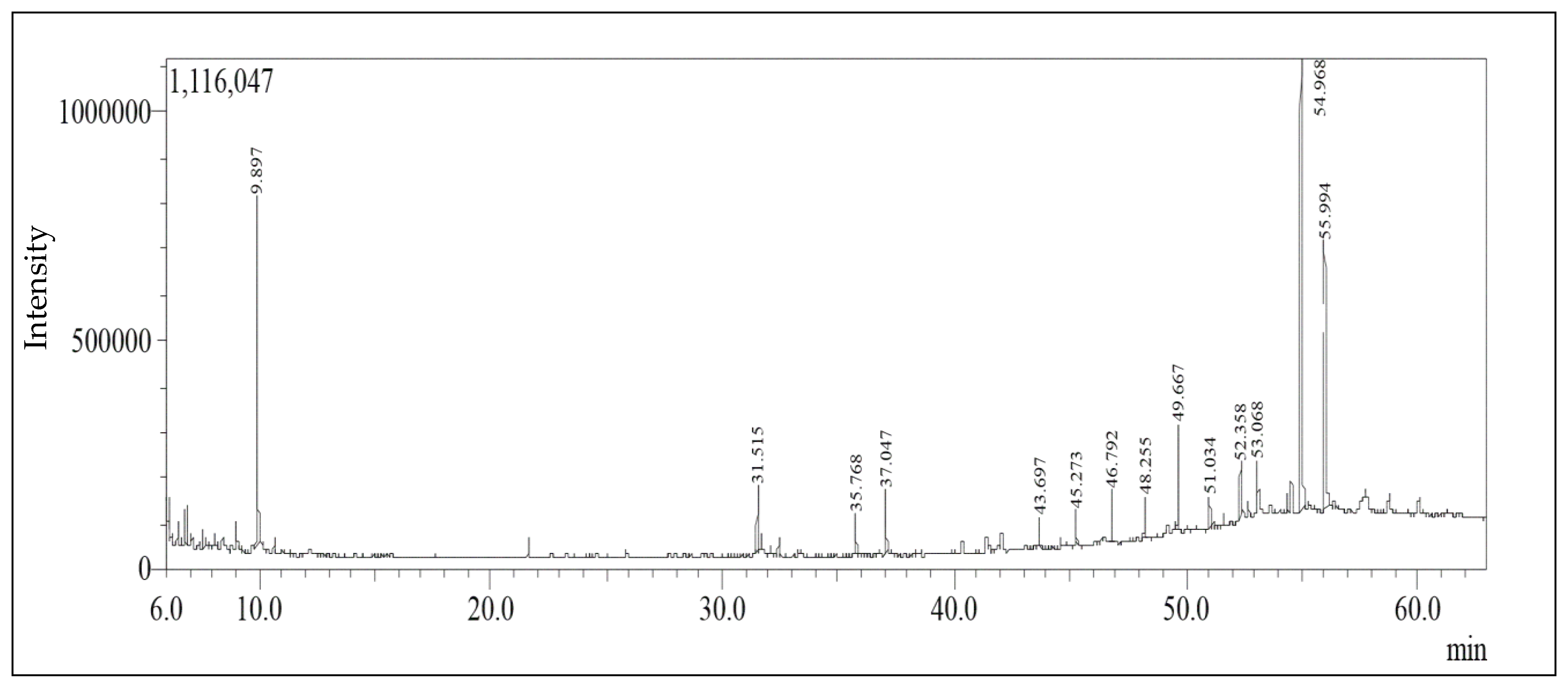
2.1. GC/MS Analysis Identification and Quantification of the Chemical Constituents
2.2. In Vitro Cytotoxicity Assay
Cytotoxic Effect of Annona Extract
2.3. Antiviral Assay
2.3.1. Annona glabra Sample MNTC Determination
2.3.2. Antiviral Efficacy of the Annona glabra Extract versus HAV and HSV1
Comparison of the Effect of the Extract versus HSV1 vs. Acyclovir with Different Protocols
Comparison of the Effect of the Annona Extract versus HAV vs. Acyclovir with Different Protocols
2.4. Molecular Docking
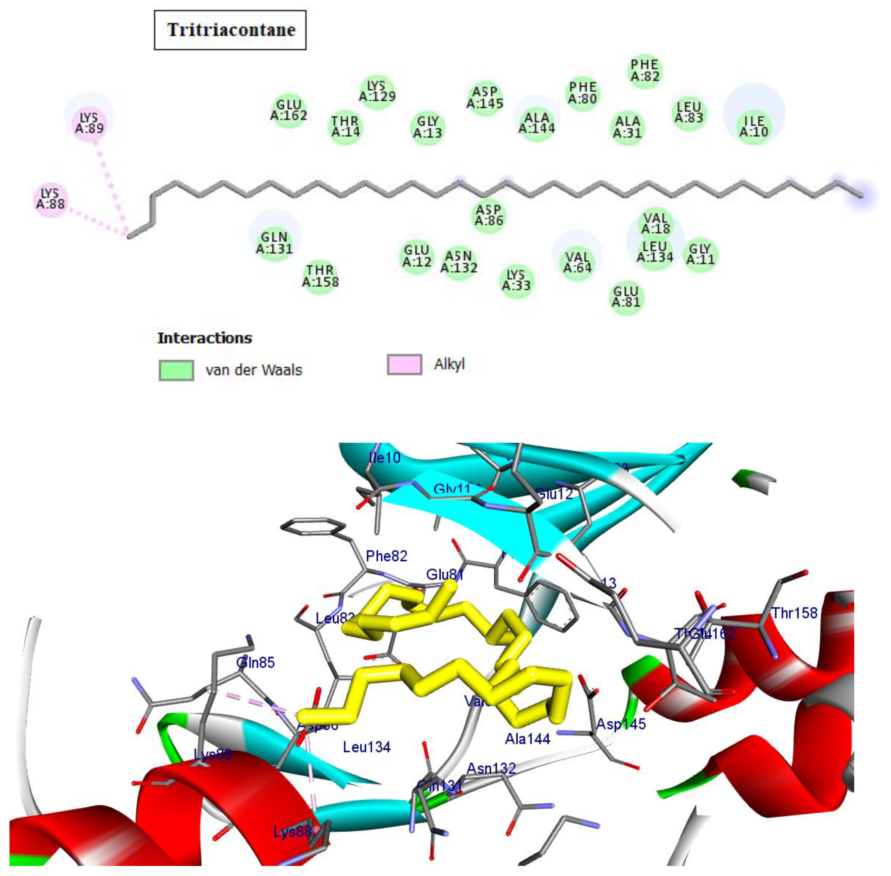
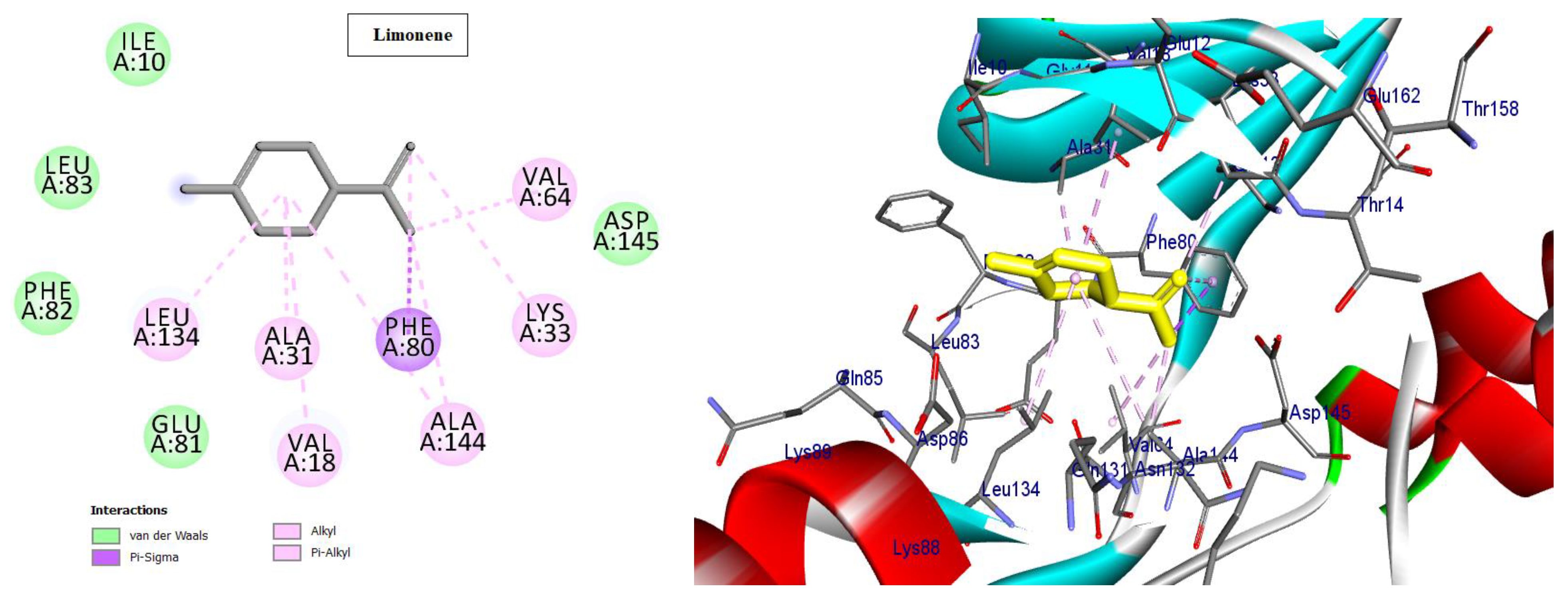
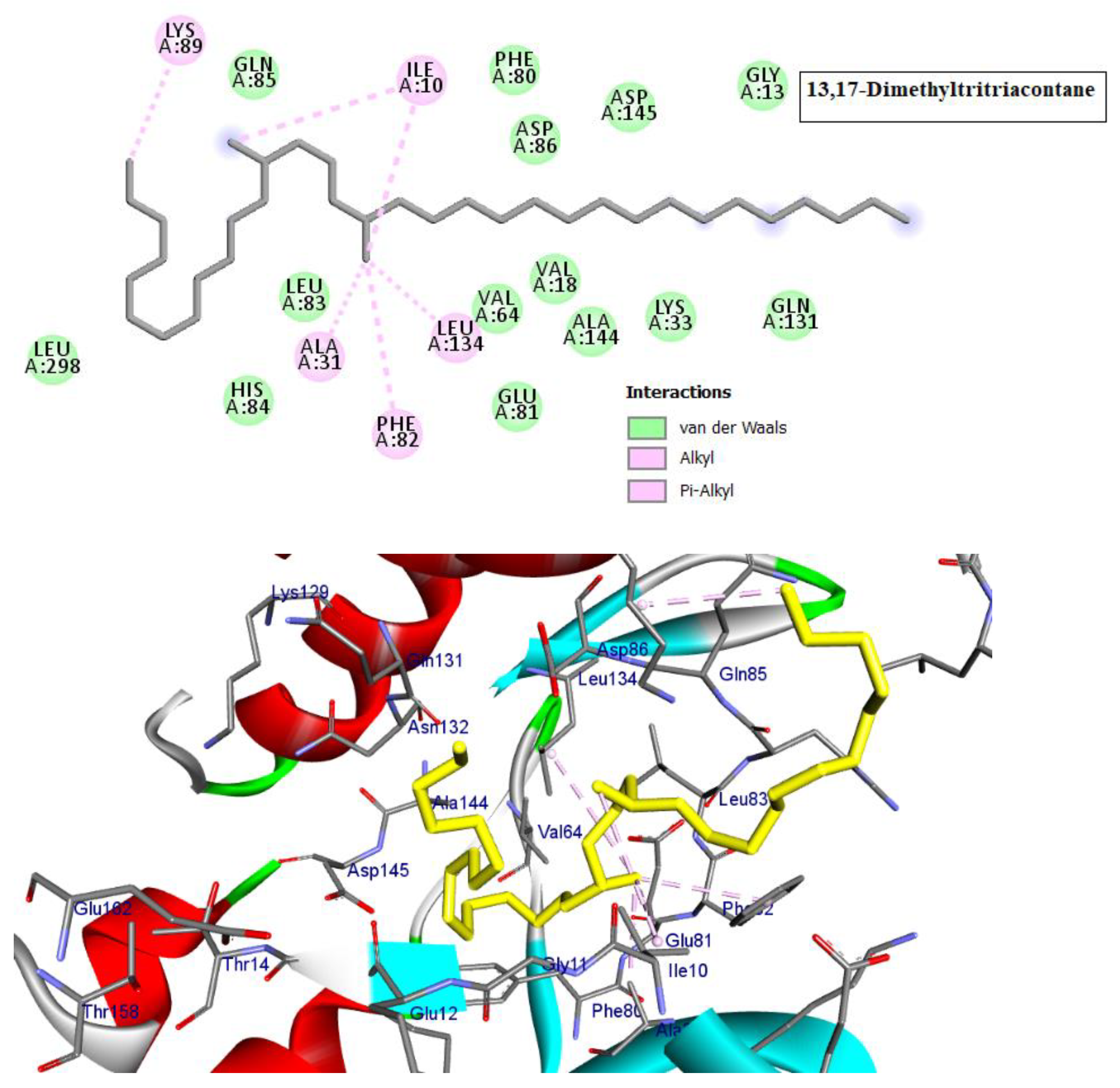
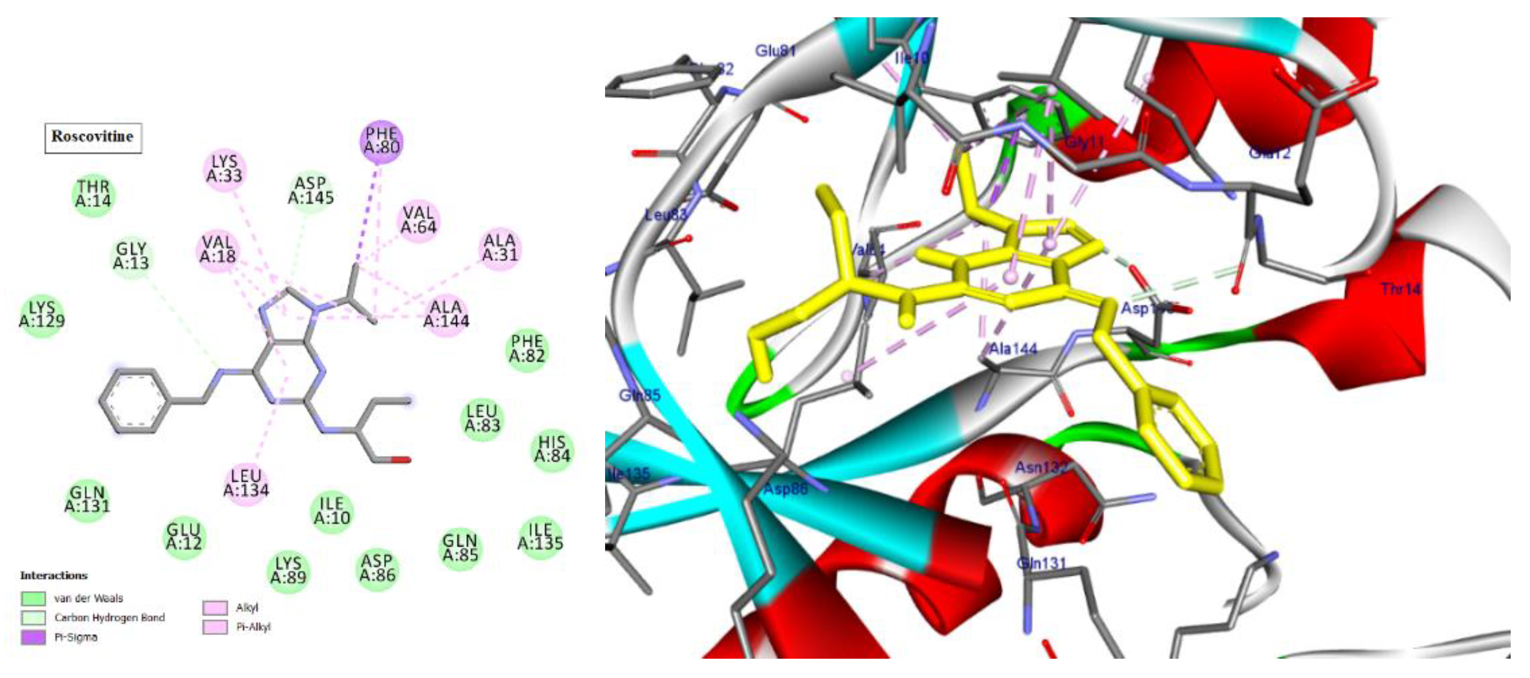
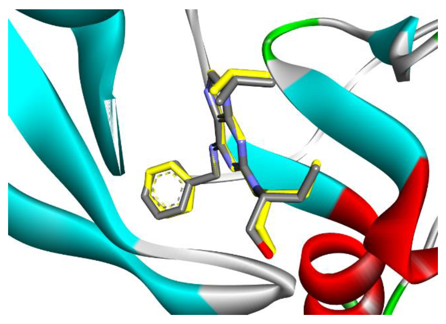
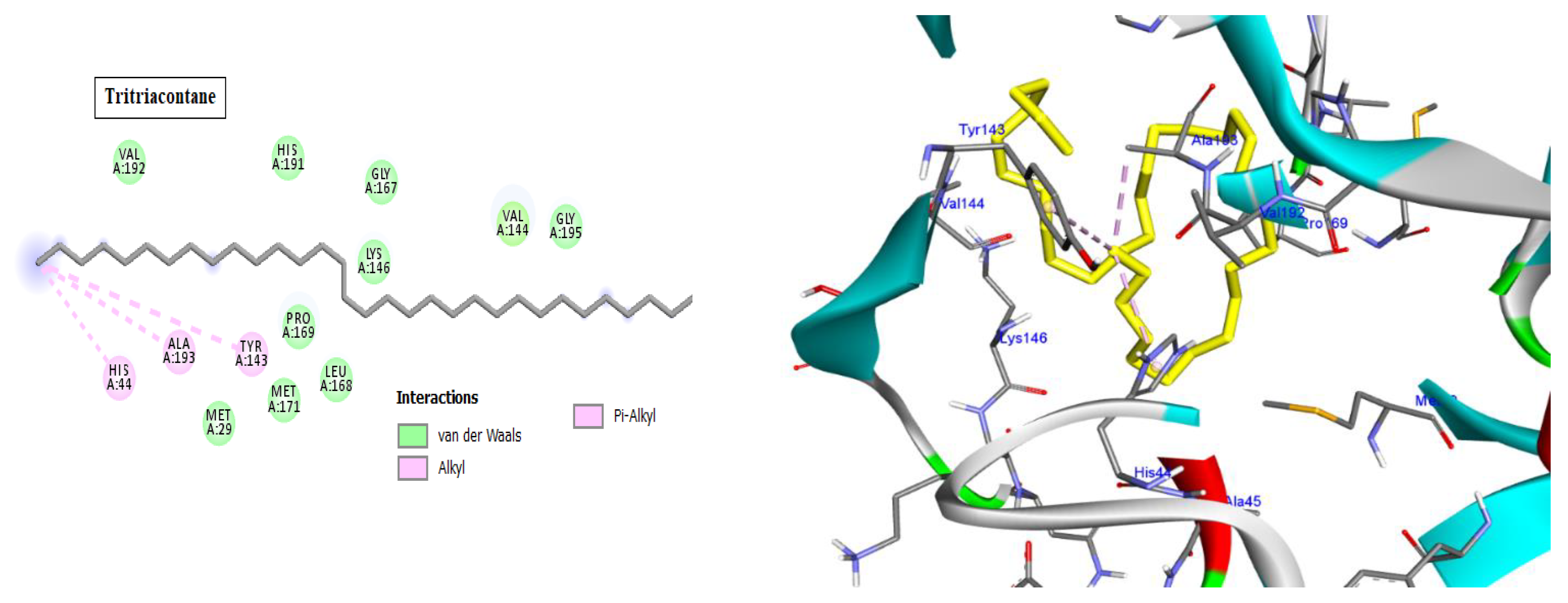
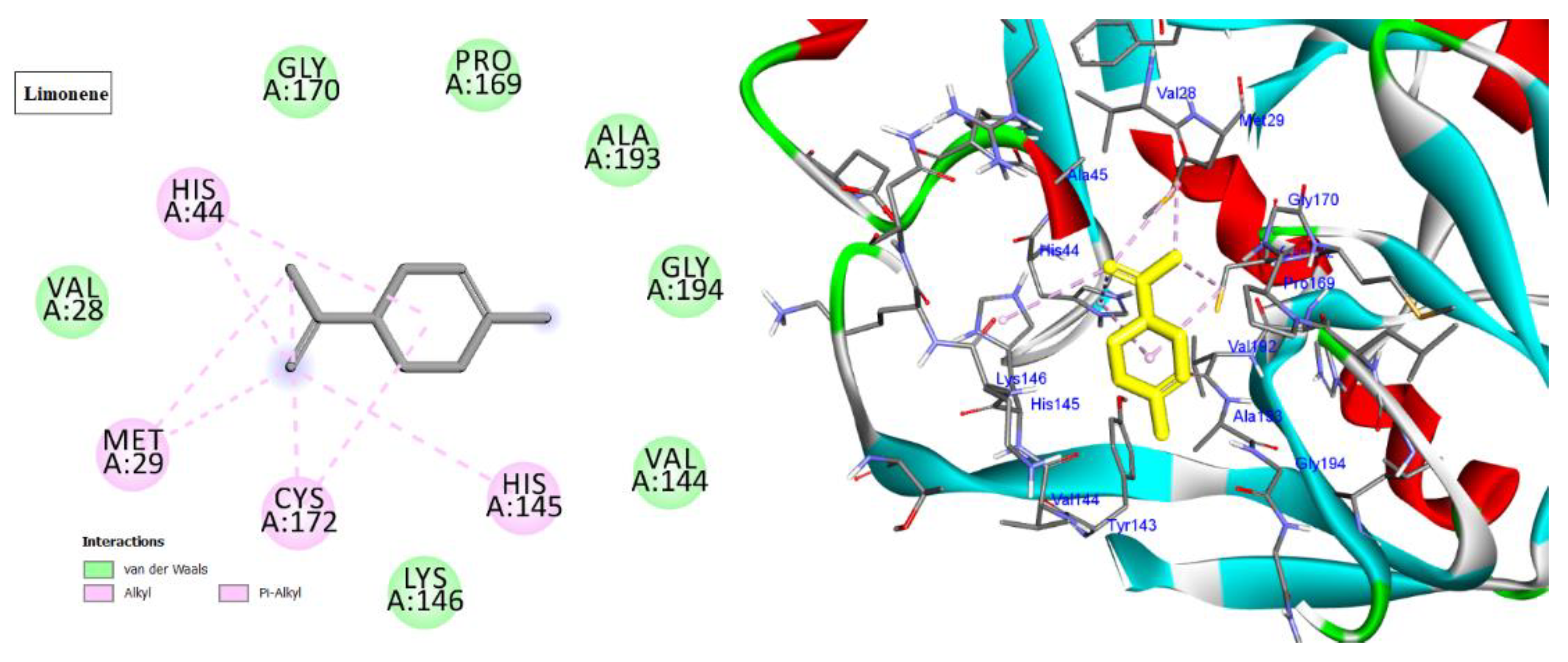
2.4.1. Docking into Cyclin-Dependent Kinase 2 Active Site
2.4.2. Docking into Hepatitis A Virus 3C (HAV 3C) Protease Enzyme Active Site
3. Discussion
4. Materials and Methods
4.1. Plant Material
4.2. Preparation of the Crude Extract
4.3. GC-FID and GC-MS Analysis of A. glabra Hexane Extract
4.4. Preparation of Stock Solution of the Plant Hexane Extract
4.5. In Vitro Cytotoxicity Assay (Viability Assay)
4.5.1. Cancer Cell Lines and Culture
4.5.2. Determination of Cytotoxicity by MTT Assay
4.6. Antiviral Assays
4.6.1. Cell Culture and Viruses
4.6.2. Virus Stock Preparation
4.6.3. Cytotoxicity Assay
4.6.4. Antiviral Protocols
Protocol A: Virus Pretreatment (Anti-Infective Activity) [51]
Protocol B: Postinfection Treatment (Anti-Replicative Activity) [48,50]
Protocol C: Cell Pretreatment (Protective Activity) [49,50]
4.7. Molecular Docking
4.8. Statistical Analysis
5. Conclusions
Author Contributions
Funding
Institutional Review Board Statement
Informed Consent Statement
Data Availability Statement
Acknowledgments
Conflicts of Interest
References
- Elkousy, R.H.; Said, Z.N.A.; Abd El-Baseer, M.A.; Abu El Wafa, S.A. Antiviral activity of castor oil plant (Ricinus communis) leaf extracts. J. Ethnopharmacol. 2021, 271, 113878. [Google Scholar] [CrossRef]
- Cochrane, C.B.; Raveendran Nair, P.K.; Melnick, S.J.; Resek, A.P.; Ramachandran, C. Anticancer effects of Annona glabra plant extracts in human leukemia cell lines. Anticancer Res. 2008, 28, 965–972. [Google Scholar] [PubMed]
- Mostafa, N.M.; Abd El-Ghffar, E.A.; Hegazy, H.G.; Eldahshan, O.A. New Methoxyflavone from Casimiroa sapota and the Biological Activities of Its Leaves Extract against Lead Acetate Induced Hepatotoxicity in Rats. Chem. Biodivers. 2018, 15, e1700528. [Google Scholar] [CrossRef]
- Ghoneim, A.I.; Eldahshan, O.A. Anti-apoptotic effects of tamarind leaves against ethanol-induced rat liver injury. J. Pharm. Pharmacol. 2012, 64, 430–438. [Google Scholar] [CrossRef] [PubMed]
- El-Nashar, H.A.S.; Mostafa, N.M.; Eldahshan, O.A.; Singab, A.N.B. A new antidiabetic and anti-inflammatory biflavonoid from Schinus polygama (Cav.) Cabrera leaves. Nat. Prod. Res. 2022, 36, 1182–1190. [Google Scholar] [CrossRef]
- Abdelghffar, E.A.; El-Nashar, H.A.S.; Al-Mohammadi, A.G.A.; Eldahshan, O.A. Orange fruit (Citrus sinensis) peel extract attenuates chemotherapy-induced toxicity in male rats. Food Funct. 2021, 12, 9443–9455. [Google Scholar] [CrossRef]
- Lúcio, A.S.; Almeida, J.R.; Da-Cunha, E.V.; Tavares, J.F.; Barbosa Filho, J.M. Alkaloids of the Annonaceae: Occurrence and a compilation of their biological activities. Alkaloids Chem. Biol. 2015, 74, 233–409. [Google Scholar] [CrossRef] [PubMed]
- Badrie, N.; Schauss, A.G. Soursop (Annona muricata L.). In Bioactive Foods in Promoting Health; Watson, R.R., Preedy, V.R., Eds.; Academic Press: Oxford, UK, 2010; pp. 2–6. ISBN 978-0-12-374628-3. [Google Scholar]
- Kalidindi, N.; Thimmaiah, N.V.; Jagadeesh, N.V.; Nandeep, R.; Swetha, S.; Kalidindi, B. Antifungal and antioxidant activities of organic and aqueous extracts of Annona squamosa Linn. Leaves. J. Food Drug Anal. 2015, 23, 795–802. [Google Scholar] [CrossRef]
- Bhattacharya, A.; Chakraverty, R. The pharmacological properties of Annona squamosa Linn: A Review. Int. J. Pharm. Eng. (IJPE) 2016, 4, 692–699. [Google Scholar]
- Doe, P.; Iddrisu, A.; Lartey, P.; Elijah, K.; Issaka, S.; Enock, D.A. Evaluation of the anti-diarrheal activity of the ethanolic seed extract of Annona muricata. J. Phytopharmaco. 2019, l8, 199–202. [Google Scholar] [CrossRef]
- Bhardwaj, R.; Pareek, S.; Sagar, N.A.; Vyas, N. Bioactive Compounds of Annona. In Bioactive Compounds in Underutilized Fruits and Nuts; Murthy, H.N., Bapat, V.A., Eds.; Springer, Cham: Cham, Switzerland; Manhattan, NY, USA, 2019; pp. 1–26. ISBN 978-3-030-06120-3_5-1. [Google Scholar]
- Eva González-Trujano, M.; Tapia, E.; López-Meraz, L.; Navarrete, A.; Reyes-Ramírez, A.; Martínez, A. Anticonvulsant effect of Annona diversifolia Saff. and palmitone on penicillin-induced convulsive activity. A behavioral and EEG study in rats. Epilepsia 2006, 47, 1810–1817. [Google Scholar] [CrossRef] [PubMed]
- Zorofchian, S.; Moghadamtousi; Rouhollahi, E.; Karimian, H.; Fadaeinasab, M.; Abdulla, M.A.; Kadir, H.A. Gastroprotective activity of Annona muricata leaves against ethanol-induced gastric injury in rats via Hsp70/Bax involvement. Drug Des. Dev. Ther. 2014, 8, 2099–2111. [Google Scholar] [CrossRef]
- Verma, A.; Ajay Kumar, P.; Kavitha, D.; Anurag, K.B. Anti denaturation and antioxidant activities of Annona cherimola in-vitro. Int. J. Pharm. Boil. Sci. 2011, 2, 1–6. [Google Scholar]
- Adeyemi, D.O.; Komolafe, O.A.; Adewole, O.S.; Obuotor, E.M.; Adenowo, T.K. Antihyperglycemic activities of Annona muricata (Linn). Afr. J. Tradit. Complement. Altern. Med. 2008, 6, 62–69. [Google Scholar] [CrossRef]
- Bhalke, R.D.; Chavan, M.J. Analgesic and CNS depressant activities of extracts of Annona reticulate Linn. Bark. J. Phytopharm. 2011, 1, 160–165. [Google Scholar]
- Betancur-Galvis, L.; Saez, J.; Granados, H.; Salazar, A.; Ossa, J. Antitumor and antiviral activity of Colombian medicinal plant extracts. Mem. Inst. Oswaldo Cruz 1999, 94, 531–535. [Google Scholar] [CrossRef]
- Grzybowski, A.; Tiboni, M.; Silva, M.A.; Chitolina, R.F.; Passos, M.; Fontana, J.D. Synergistic larvicidal effect and morphological alterations induced by ethanolic extracts of Annona muricata and Piper nigrum against the dengue fever vector Aedes aegypti. Pest Manag. Sci. 2013, 69, 589–601. [Google Scholar] [CrossRef]
- Zhang, Y.H.; Peng, H.Y.; Xia, G.H.; Wang, M.Y.; Han, Y. Anticancer effect of two diterpenoid compounds isolated from Annona glabra Linn. Acta Pharmacol. Sin. 2004, 25, 937–942. [Google Scholar] [PubMed]
- Singh, T.P.; Singh, R.K.; Malik, P. Analgesic and Anti-inflammtory Activities of Annona squamosa Linn bark. J. Sci. Innov. Res. 2014, 3, 60–64. [Google Scholar] [CrossRef]
- Eldahshan, O.A. Rhoifolin: A potent antiproliferative effect on cancer cell lines. Br. J. Pharm. Res. 2013, 3, 46–53. [Google Scholar] [CrossRef]
- Hien, N.T.; Nhiem, N.X.; Yen, D.T.; Hang, D.T.; Tai, B.H.; Quang, T.H.; Tuan Anh, H.L.; Kiem, P.V.; Minh, C.V.; Kim, E.J.; et al. Chemical constituents of the Annona glabra fruit and their cytotoxic activity. Pharm. Biol. 2015, 53, 1602–1607. [Google Scholar] [CrossRef] [PubMed]
- Liu, X.X.; Alali, F.Q.; Pilarinou, E.; McLaughlin, J.L. Glacins A and B: Two novel bioactive mono-tetrahydrofuran macetogenins from Annona glabra. J. Nat. Prod. 1998, 61, 620–624. [Google Scholar] [CrossRef]
- Chang, F.R.; Yang, P.Y.; Lin, J.Y.; Lee, K.H.; Wu, Y.C. Bioactive Kaurane Diterpenoids from Annona glabra. J. Nat. Prod. 1998, 61, 437–439. [Google Scholar] [CrossRef] [PubMed]
- Tatar, O.; Sriamornsak, P.; Dass, C.R. Doxorubicin: An update on anticancer molecular action, toxicity, and novel drug delivery systems. Pharm. Pharmacol. 2013, 65, 70–157. [Google Scholar] [CrossRef]
- Gamal El-Din, M.I.; Youssef, F.S.; Ashour, M.L.; Eldahshan, O.A.; Singab, A.N.B. Comparative Analysis of Volatile Constituents of Pachira Aquatica Aubl. and Pachira glabra Pasq., their Anti-Mycobacterial and Anti-Helicobacter pylori Activities and their Metabolic Discrimination using Chemometrics. J. Essent. Oil Bear. Plants 2018, 21, 1550–1567. [Google Scholar] [CrossRef]
- Younis, I.Y.; El-Hawary, S.S.; Eldahshan, O.A.; Abdel-Aziz, M.M.; Ali, Z.Y. Green synthesis of magnesium nanoparticles mediated from Rosa floribunda charisma extract and its antioxidant, antiaging, and antibiofilm activities. Sci. Rep. 2021, 11, 16868. [Google Scholar] [CrossRef]
- Schonbrunn, E.; Betzi, S.; Alam, R.; Martin, M.P.; Becker, A.; Han, H.; Francis, R.; Chakrasali, R.; Jakkaraj, S.; Kazi, A.; et al. Development of Highly Potent and Selective Diaminothiazole Inhibitors of Cyclin-Dependent Kinases. J. Med. Chem. 2013, 56, 3768–3782. [Google Scholar] [CrossRef]
- Peyressatre, M.; Prevel, C.; Pellerano, M.; Morris, M.C. Targeting Cyclin-Dependent Kinases in Human Cancers: From Small Molecules to Peptide Inhibitors. Cancers 2015, 7, 179–237. [Google Scholar] [CrossRef]
- Chohan, T.A.; Qian, H.Y.; Pan, Y.L.; Chen, J.Z. Molecular Simulation Studies on the Binding Selectivity of 2-Anilino-4-(Thiazol-5-yl)-Pyrimidines in Complexes with CDK2 and CDK7. Mol. Biosyst. 2016, 12, 145–161. [Google Scholar] [CrossRef]
- Zhou, J.; Wang, D.; Xi, Y.; Zhu, X.; Yang, Y.; Lv, M.; Luo, C.; Chen, J.; Ye, X.; Fang, L.; et al. Assessing activity of Hepatitis A virus 3C protease using a cyclized luciferase-based biosensor. Biochem. Biophys. Res. Commun. 2017, 488, 621–627. [Google Scholar] [CrossRef]
- Ben-Shabat, S.; Yarmolinsky, L.; Porat, D.; Dahan, A. Antiviral effect of phytochemicals from medicinal plants: Applications and drug delivery strategies. Drug Deliv. Transl. Res. 2020, 10, 354–367. [Google Scholar] [CrossRef] [PubMed]
- Chen, C.H.; Hsieh, T.J.; Liu, T.Z.; Chern, C.L.; Hsieh, P.Y.; Chen, C.Y. Annoglabayin, a novel dimeric kaurane diterpenoid, and apoptosis in hep G2 cells of annomontacin from the fruits of Annona glabra. J. Nat. Prod. 2004, 67, 1942–1946. [Google Scholar] [CrossRef] [PubMed]
- Liu, X.X.; Alali, F.Q.; Pilarinou, E.; McLaughlin, J.L. Two bioactive mono-tetrahydrofuran acetogenins, annoglacins A and B, from Annona glabra. Phytochemistry 1999, 5, 815–821. [Google Scholar] [CrossRef]
- Anh, H.T.; Hien, N.T.; Hang, D.T.; Ha, T.M.; Nhiem, N.X.; Hien, T.T.; Thu, V.K.; Thao, D.T.; Van Minh, C.; Kiem, P.V. Ent-Kaurane diterpenes from Annona glabra and their cytotoxic activities. Nat. Prod. Commun. 2014, 9, 1681–1682. [Google Scholar] [CrossRef]
- O’Brien, J.J.; Campoli-Richards, D.M. Acyclovir. An updated review of its antiviral activity, pharmacokinetic properties, and therapeutic efficacy. Drugs 1989, 37, 233–309. [Google Scholar] [CrossRef]
- Jingjing, Y.; Lei, Z.; Ren, L.; Yunzheng, Y.; Jiye, Y.; Qingsong, D.; Xiaojia, G.; Wei, L.; Yuexiang, L.; Miaomiao, L.; et al. In vitro and in vivo antiviral activity of Maqian (Zanthoxylum myriacanthum var. pubescent) essential oil and its major constituents against strains of influenza virus. Ind. Crops Prod. 2022, 177, 114524. [Google Scholar] [CrossRef]
- Fadilah, N.Q.; Jittmittraphap, A.; Leaungwutiwong, P.; Pripdeevech, P.; Dhanushka, D.; Mahidol, C.; Ruchirawat, S.; Kittakoop, P. Virucidal Activity of Essential Oils From Citrus x aurantium L. Against Influenza A Virus H1N1: Limonene as a Potential Household Disinfectant Against Virus. Nat. Prod. Commun. 2022, 17, 1–7. [Google Scholar] [CrossRef]
- Astani, A.; Schnitzler, P. Antiviral activity of monoterpenes beta-pinene and limonene against herpes simplex virus in vitro. Iran. J. Microbiol. 2014, 6, 149–155. [Google Scholar]
- Konappa, N.; Udayashankar, A.C.; Krishnamurthy, S.; Pradeep, C.K.; Chowdappa, S.; Jogaiah, S. GC–MS analysis of phytoconstituents from Amomum nilgiricum and molecular docking interactions of bioactive serverogenin acetate with target proteins. Sci. Rep. 2020, 10, 16438. [Google Scholar] [CrossRef]
- Viegas, D.J.; Edwards, T.G.; Bloom, D.C.; Abreu, P.A. Virtual screening identified compounds that bind to cyclin dependent kinase 2 and prevent herpes simplex virus type 1 replication and reactivation in neurons. Antivir. Res. 2019, 172, 104621. [Google Scholar] [CrossRef]
- Schang, L.M.; Bantly, A.; Schaffer, P.A. Explant-induced reactivation of herpes simplex virus occurs in neurons expressing nuclear cdk2 and cdk4. J. Virol. 2002, 76, 7724–7735. [Google Scholar] [CrossRef] [PubMed]
- Schang, L.M.; St Vincent, M.R.; Lacasse, J.J. Five years of progress on cyclin-dependent kinases and other cellular proteins as potential targets for antiviral drugs. Antivir. Chem. Chemother. 2006, 17, 293–320. [Google Scholar] [PubMed]
- Takeuchi, H.; Baba, M.; Shigeta, S. An application of tetrazolium (MTT) colorimetric assay for the screening of anti-herpes simplex virus compounds. J. Virol. Methods 1991, 33, 61–71. [Google Scholar] [CrossRef] [PubMed]
- Reed, L.; Muench, H. A simple method of estimating 50% endpoints. Am. J. Hyg. 1938, 27, 493–497. [Google Scholar]
- Dulbecco, R.; Vogt, M. Plaque formation and isolation of pure lines with poliomyelitis viruses. J. Exp. Med. 1954, 99, 167–182. [Google Scholar] [CrossRef] [Green Version]
- Kaul, T.; Middleton, E.; Ogra, P. Antiviral effect of flavonoids on human viruses. J. Med. Virol. 1985, 15, 71–79. [Google Scholar] [CrossRef]
- Kaye, S. Antiviral methods and protocols. J. Antimicrob. Chemother. 2000, 46, 521. [Google Scholar] [CrossRef]
- Gescher, K.; Kühn, J.; Hafezi, W.; Louis, A.; Derksen, A.; Deters, A.; Lorentzen, E.; Hensel, A. Inhibition of viral adsorption and penetration by an aqueous extract from Rhododendron ferrugineum L. as antiviral principle against herpes simplex virus type-1. Fitoterapia 2011, 82, 408–413. [Google Scholar] [CrossRef]
- Flechas, M.; Ocazionez, R.; Stashenko, E. Evaluation of in vitro antiviral activity of essential oil compounds against dengue virus. Pharmacogn. Rev. J. 2017, 10, 55–59. [Google Scholar] [CrossRef]
- De Azevedo, W.F.; Leclerc, S.; Meijer, L.; Havlicek, L.; Strnad, M.; Kim, S.H. Inhibition of cyclin-dependent kinases by purine analogues: Crystal structure of human cdk2 complexed with roscovitine. Eur. J. Biochem. 1997, 243, 518–526. [Google Scholar] [CrossRef]
- Yin, J.; Bergmann, E.M.; Cherney, M.M.; Lall, M.S.; Jain, R.P.; Vederas, J.C.; James, M.N. Dual modes of modification of hepatitis A virus 3C protease by a serine-derived beta-lactone: Selective crystallization and formation of a functional catalytic triad in the active site. J. Mol. Biol. 2005, 354, 854–871. [Google Scholar] [CrossRef] [PubMed]
- Morris, G.M.; Huey, R.; Lindstrom, W.; Sanner, M.F.; Belew, R.K.; Goodsell, D.S.; Olson, A.J. AutoDock4 and AutoDockTools4: Automated docking with selective receptor flexibility. J. Comput. Chem. 2009, 30, 2785–2791. [Google Scholar] [CrossRef] [PubMed]
- Trott, O.; Olson, A.J. AutoDock Vina: Improving the speed and accuracy of docking with a new scoring function, efficient optimization, and multithreading. J. Comput. Chem. 2010, 31, 455–461. [Google Scholar] [CrossRef] [PubMed] [Green Version]


| Peak No. | Compounds | tR | Molecular Formula | KI (Cal.) | KI (Rep) | Area % | Method of Identification |
|---|---|---|---|---|---|---|---|
| 1. | Limonene | 9.897 | C10H16 | 1028 | 1029 | 18.97 | KI, MS |
| 2. | E-Nerolidyl isobutyrate | 31.515 | C19H32O2 | 1833 | 1826 | 3.61 | KI, MS |
| 3. | Unidentified | 35.768 | - | 2048 | - | 1.97 | - |
| 4. | Phytol | 37.047 | C20H40O | 2115 | 2116 | 3.75 | KI, MS |
| 5. | n-Pentacosane | 43.697 | C25H52 | 2498 | 2500 | 1.54 | KI, MS |
| 6. | Hexacosane | 45.273 | C26H54 | 2598 | 2600 | 1.69 | KI, MS |
| 7. | Heptacosane | 46.792 | C27H56 | 2698 | 2700 | 2.46 | KI, MS |
| 8. | Octacosane | 48.255 | C28H58 | 2798 | 2800 | 2.14 | KI, MS |
| 9. | Nonacosane | 49.667 | C29H60 | 2897 | 2900 | 4.94 | KI, MS |
| 10. | Triacontane | 51.034 | C30H62 | 2998 | 3000 | 1.59 | KI, MS |
| 11. | Untriacontane | 52.358 | C31H64 | 3098 | 3100 | 2.63 | KI, MS |
| 12. | Vitamin E | 53.068 | C29H50O2 | 3152 | 3149 | 2.04 | KI, MS |
| 13. | Tritriacontane | 54.968 | C33H68 | 3289 | 3300 | 30.23 | KI, MS |
| 14. | 13,17-dimethyl-tritriacontane | 55.994 | C35H72 | 3352 | 3358 | 22.44 | KI |
| Vero | CACO-2 | HepG-2 | PANC-1 | MCF-7 | PC-3 | A-549 | |||||||
|---|---|---|---|---|---|---|---|---|---|---|---|---|---|
| IC50 | IC50 | SI | IC50 | SI | IC50 | SI | IC50 | SI | IC50 | SI | IC50 | SI | |
| AE | 179.88 ± 0.28 A | 47 ± 0.74 A | 3.82 | 57.01 ± 0.85 A | 3.10 | 57.34 ± 0.77 A | 3.10 | 80.31 ± 4.13 A | 2.20 | 81.86 ± 3.26 A | 2.20 | 56.82 ± 0.92 A | 3.10 |
| Doxo. | 34.26 ± 0.55 B | 31.91 ± 0.81 B | 1.07 | 5.4 ± 0.22 B | 6.00 | 19.07 ± 0.2 B | 1.60 | 15.48 ± 0.84 B | 2.00 | 32.9 ± 1.74 B | 1.04 | 23.39 ± 0.43 B | 1.40 |
| ug/mL | O.D. | Mean O.D. | SD. E. | Viability % | Toxicity % | IC50 | |||
|---|---|---|---|---|---|---|---|---|---|
| Vero | --- | 0.482 | 0.458 | 0.473 | 0.471 | 0.007 | 100 | 0 | |
| Extract | 1000 | 0.015 | 0.016 | 0.015 | 0.015333 | 0.000333 | 3.255484784 | 96.74451522 | 22.94 |
| 500 | 0.018 | 0.016 | 0.017 | 0.017 | 0.000577 | 3.609341826 | 96.39065817 | ||
| 250 | 0.016 | 0.018 | 0.018 | 0.017333 | 0.000667 | 3.680113234 | 96.31988677 | ||
| 125 | 0.019 | 0.02 | 0.018 | 0.019 | 0.000577 | 4.033970276 | 95.96602972 | ||
| 62.5 | 0.034 | 0.021 | 0.039 | 0.031333 | 0.005364 | 6.652512385 | 93.34748762 | ||
| 31.25 | 0.106 | 0.112 | 0.135 | 0.117667 | 0.008838 | 24.98230715 | 75.01769285 | ||
| 15.62 | 0.345 | 0.322 | 0.316 | 0.327667 | 0.008838 | 69.56829441 | 30.43170559 | ||
| 7.81 | 0.48 | 0.461 | 0.472 | 0.471 | 0.005508 | 100 | 0 | ||
| Acyclovir | 1000 | 0.038 | 0.048 | 0.04 | 0.042 | 0.003055 | 10.9947644 | 89.0052356 | 360.92 |
| 500 | 0.092 | 0.105 | 0.129 | 0.108667 | 0.010837 | 28.44677138 | 71.55322862 | ||
| 250 | 0.274 | 0.271 | 0.246 | 0.263667 | 0.008876 | 69.02268761 | 30.97731239 | ||
| 125 | 0.314 | 0.293 | 0.326 | 0.311 | 0.00644 | 81.41361257 | 18.58638743 | ||
| 62.5 | 0.365 | 0.388 | 0.391 | 0.381333 | 0.008212 | 99.82547993 | 0.17452007 | ||
| 31.25 | 0.371 | 0.381 | 0.388 | 0.38 | 0.004933 | 99.47643979 | 0.523560209 | ||
| ID | Antiviral Activity % | |
|---|---|---|
| HAV | HSV1 | |
| Acyclovir | 46.17 ± 1.67 A | 83.76 ± 5.67 A |
| AE | 24.26 ± 9.77 B | 75.78 ± 7.51 A |
| ID | Antiviral Activity % | ||
|---|---|---|---|
| Protocol A | Protocol B | Protocol C | |
| Acyclovir | 83.76 ± 5.67 AB | 92.69 ± 1.32 A | 68.44 ± 7.62 B |
| AE | 70.91 ± 7.02 AB | 61.91 ± 3.51 B | 79.55 ± 1.67 AB |
| ID | Antiviral Activity % | ||
|---|---|---|---|
| Protocol A | Protocol B | Protocol C | |
| Acyclovir | 46.17 ± 1.67 A | 54.8 ± 11.7 A | 36.89 ± 6.61 AB |
| AE | 36.81 ± 2.67 AB | 20.13 ± 3.1 B | 48.08 ± 3.46 A |
| Cyclin-Dependent Kinase 2 (Pdb; 2a4l) | ||
|---|---|---|
| Cpd. | Amino Acids, Bond Type, Distance in (Å) | Docking Energy Scores in kcal/mol |
| Roscovitine | Gly13, Carbon Hydrogen Bond, 3.71 Asp145, Carbon Hydrogen Bond, 3.16 Phe80, Pi-Sigma, 3.64 Ala31, Alkyl, 3.50 Ala144, Alkyl, 3.75 Val64, Alkyl, 4.30 Val18, Alkyl, 4.84 Phe80, Pi-Alkyl, 4.37 Val18, Pi-Alkyl, 4.61 Leu134, Pi-Alkyl, 5.41 Val18, Pi-Alkyl, 4.57 Lys33, Pi-Alkyl, 4.86 Ala144, Pi-Alkyl, 4.91 | −7.5 |
| Tritriacontane | Lys88, Alkyl, 3.84 Lys89, Alkyl, 89 | −6.6 |
| Limonene | Ala144, Alkyl, 5.48 Val18, Alkyl, 5.12 Leu134, Alkyl, 4.82 Phe80, Pi-Alkyl, 4.17 Lys33, Alkyl, 4.08 Val64, Alkyl, 4.08 Ala31, Alkyl, 3.89 Ala144, Alkyl, 3.55 Phe80, Pi-Sigma, 3.49 | −7.5 |
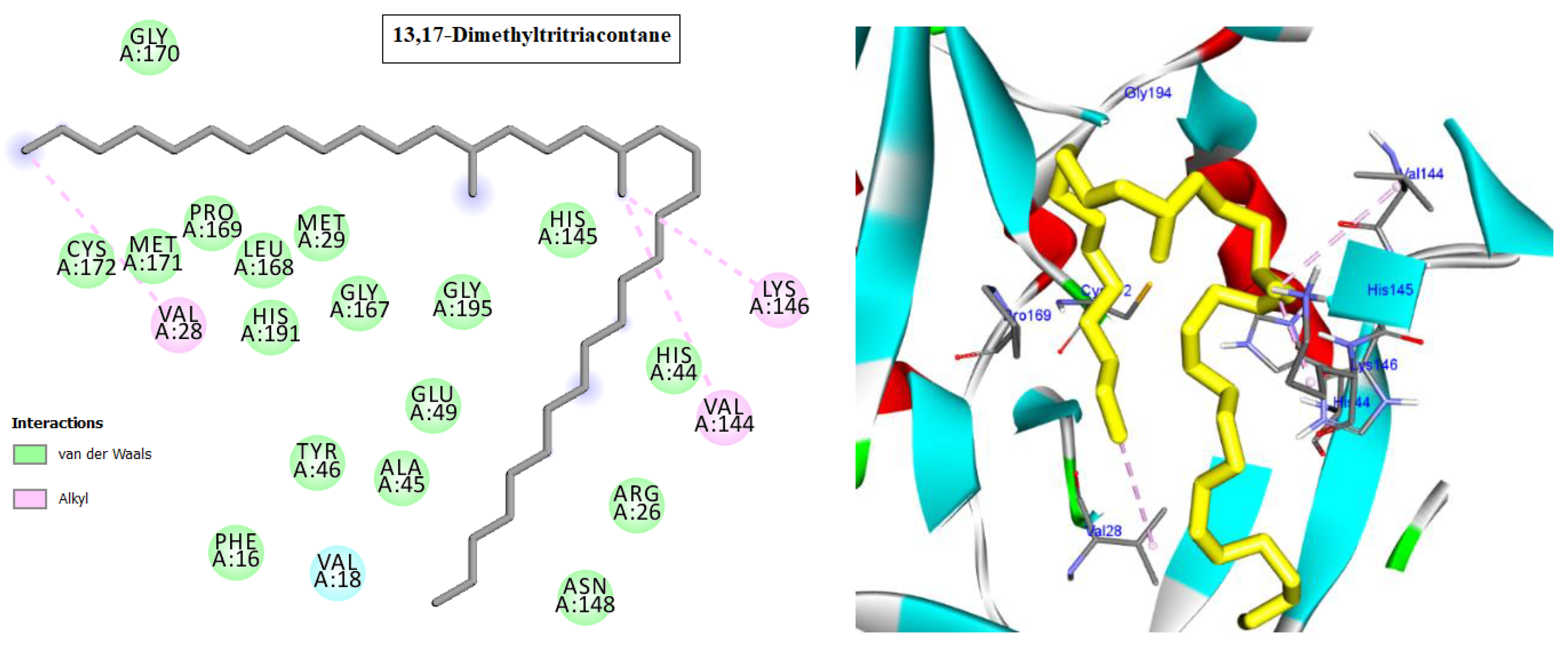
| 13,17-dimethyltritriacontane | Ala31, Alkyl, 3.72 Ile10, Alkyl, 4.67 Leu134, Alkyl, 5.17 Ile10, Alkyl, 5.04 Lys89, Alkyl, 4.02 Phe82, Pi-Alkyl, 4.36 | −7.3 |
| Hepatitis A Virus 3C Protease (Pdb; 2cxv) | ||
| Cpd. | Amino acids, Bond type, Distance in (Å) | Docking energy scores in kcal/mol |
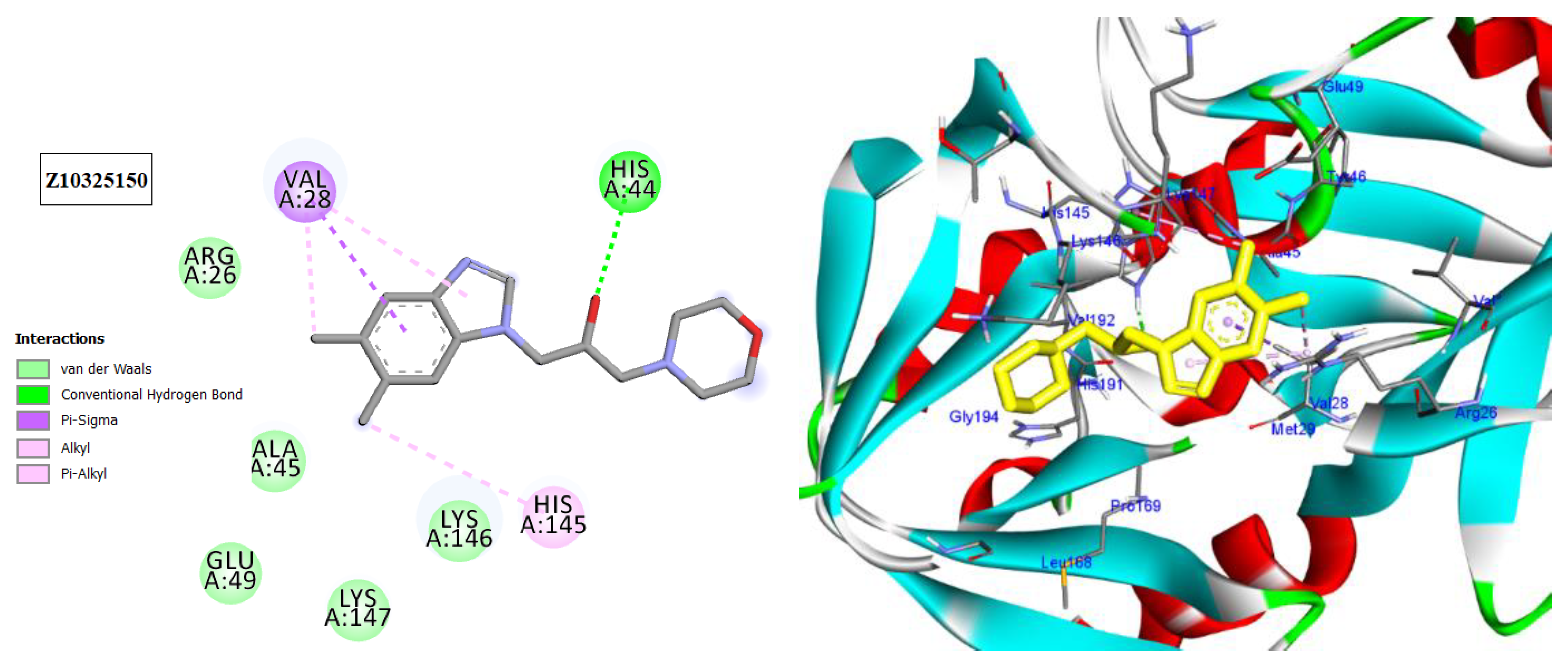
| Z10325150 | His44, Hydrogen Bond, 2.16 Val28, Pi-Sigma, 3.52 Val28, Alkyl, 3.66 His145, Pi-Alkyl, 4.57 Val28, Pi-Alkyl, 5.07 | −6.9 |
| Tritriacontane | Ala193, Alkyl,3.88 His44, Pi-Alkyl, 4.34 Try143, Pi-Alkyl, 5.27 | −6.3 |
| Limonene | Cys172, Alkyl, 4.84 Met29, Alkyl, 5.15 Met29, Alkyl, 4.40 Cys172, Alkyl, 3.67 His44, Pi-Alkyl, 5.24 His44, Pi-Alkyl, 4.73 His145, Pi-Alkyl, 4.90 | −6.9 |
| 13,17-dimethyltritriacontane | Val144, Alkyl, 4.90 Lys146, Alkyl, 3.64 Val28, Alkyl, 4.84 | −6.7 |
Disclaimer/Publisher’s Note: The statements, opinions and data contained in all publications are solely those of the individual author(s) and contributor(s) and not of MDPI and/or the editor(s). MDPI and/or the editor(s) disclaim responsibility for any injury to people or property resulting from any ideas, methods, instructions or products referred to in the content. |
© 2023 by the authors. Licensee MDPI, Basel, Switzerland. This article is an open access article distributed under the terms and conditions of the Creative Commons Attribution (CC BY) license (https://creativecommons.org/licenses/by/4.0/).
Share and Cite
Soleman, D.M.; Eldahshan, O.A.; Ibrahim, M.H.; Ogaly, H.A.; Galal, H.M.; Batiha, G.E.-S.; Elkousy, R.H. GC/MS Analysis, Cytotoxicity, and Antiviral Activities of Annona glabra Hexane Extract Supported by In Silico Study. Molecules 2023, 28, 1628. https://doi.org/10.3390/molecules28041628
Soleman DM, Eldahshan OA, Ibrahim MH, Ogaly HA, Galal HM, Batiha GE-S, Elkousy RH. GC/MS Analysis, Cytotoxicity, and Antiviral Activities of Annona glabra Hexane Extract Supported by In Silico Study. Molecules. 2023; 28(4):1628. https://doi.org/10.3390/molecules28041628
Chicago/Turabian StyleSoleman, Dalia M., Omayma A. Eldahshan, Mona H. Ibrahim, Hanan A. Ogaly, Heba M. Galal, Gaber El-Saber Batiha, and Rawah H. Elkousy. 2023. "GC/MS Analysis, Cytotoxicity, and Antiviral Activities of Annona glabra Hexane Extract Supported by In Silico Study" Molecules 28, no. 4: 1628. https://doi.org/10.3390/molecules28041628