
Inducible Nitric Oxide Synthase Embedded in Alginate/Polyethyleneimine Hydrogel as a New Platform to Explore NO-Driven Modulation of Biological Function

 , and
, and 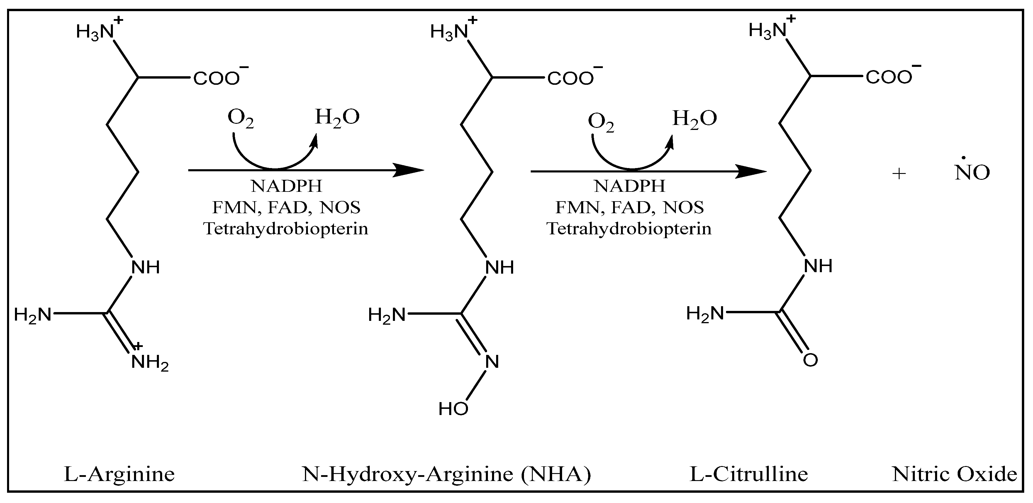
Abstract
:1. Introduction
2. Results and Discussion
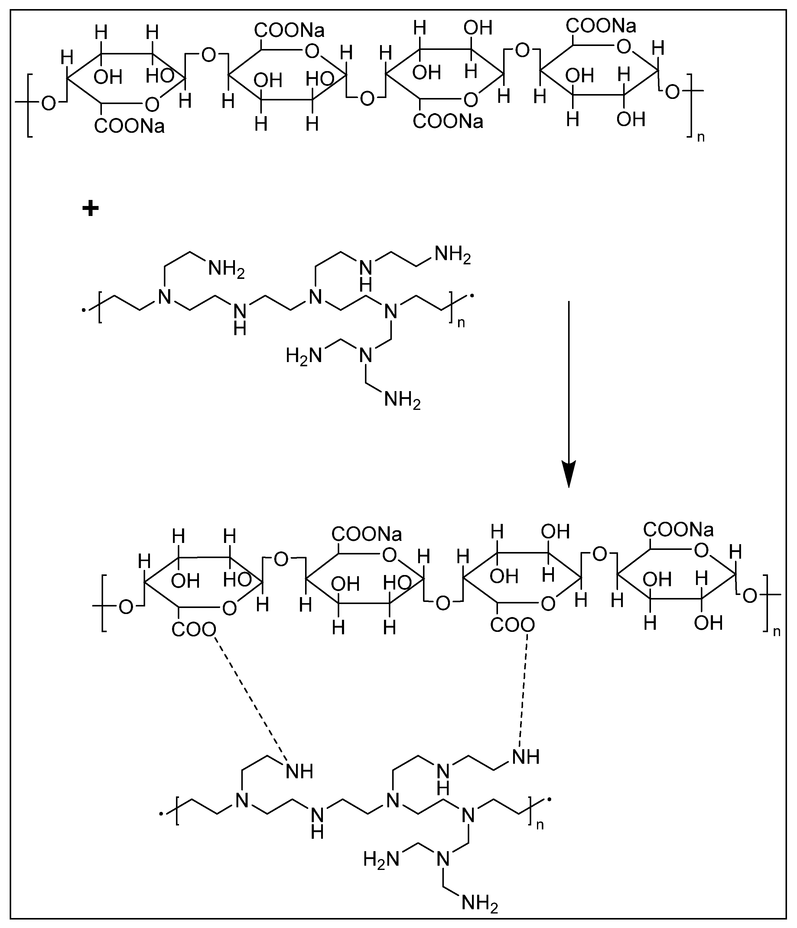
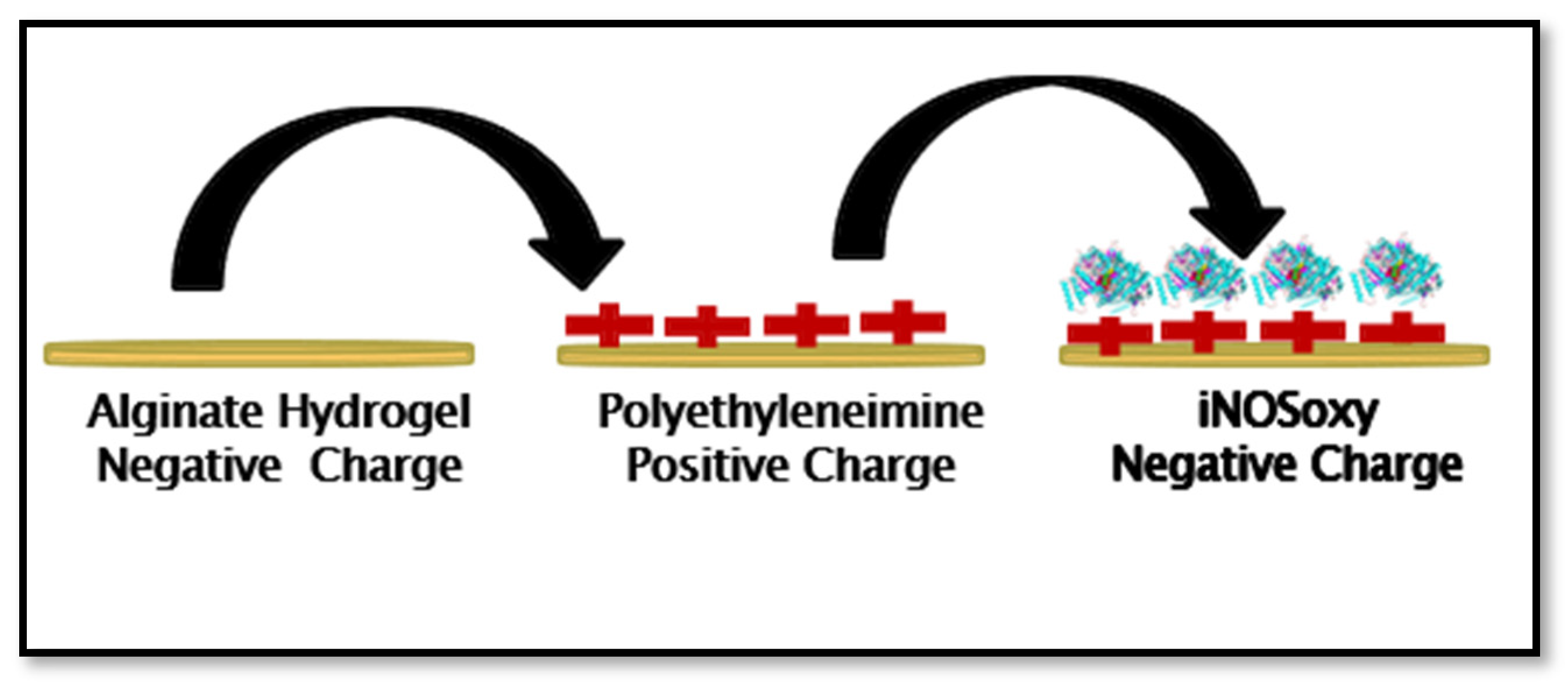
2.1. SA/PEI/iNOSoxy Hydrogel Preparation
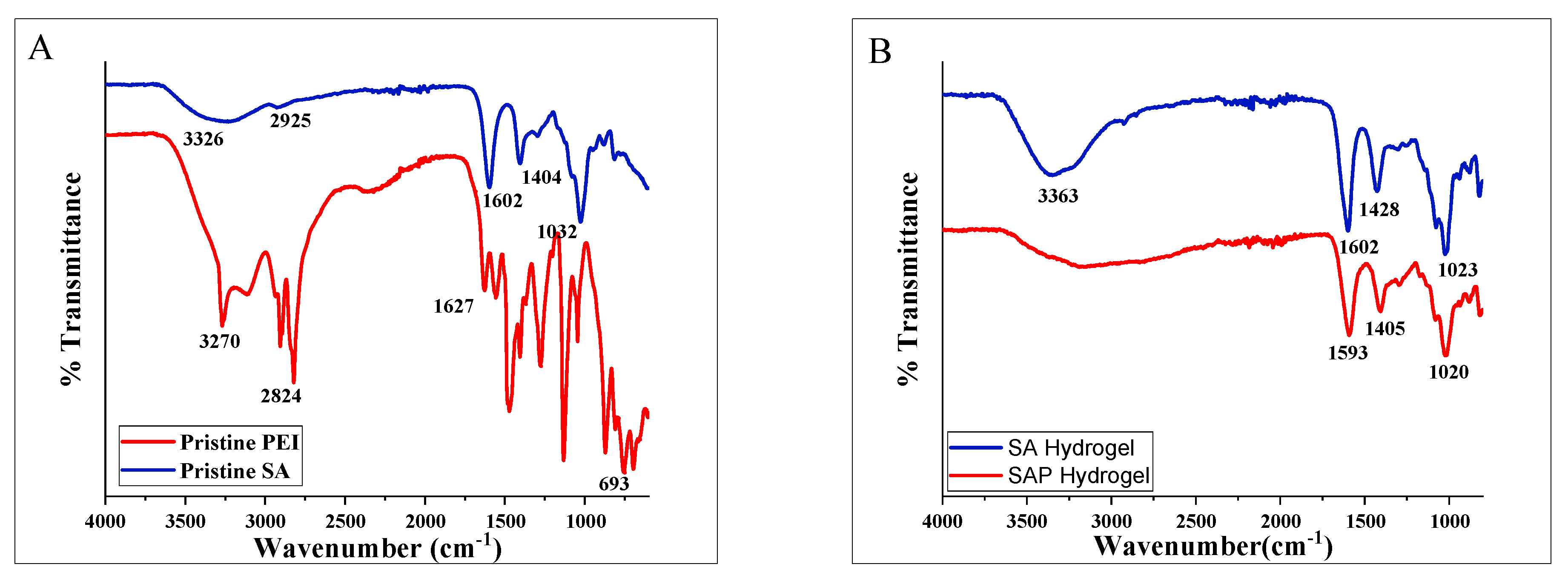
2.2. FT-IR Measurements
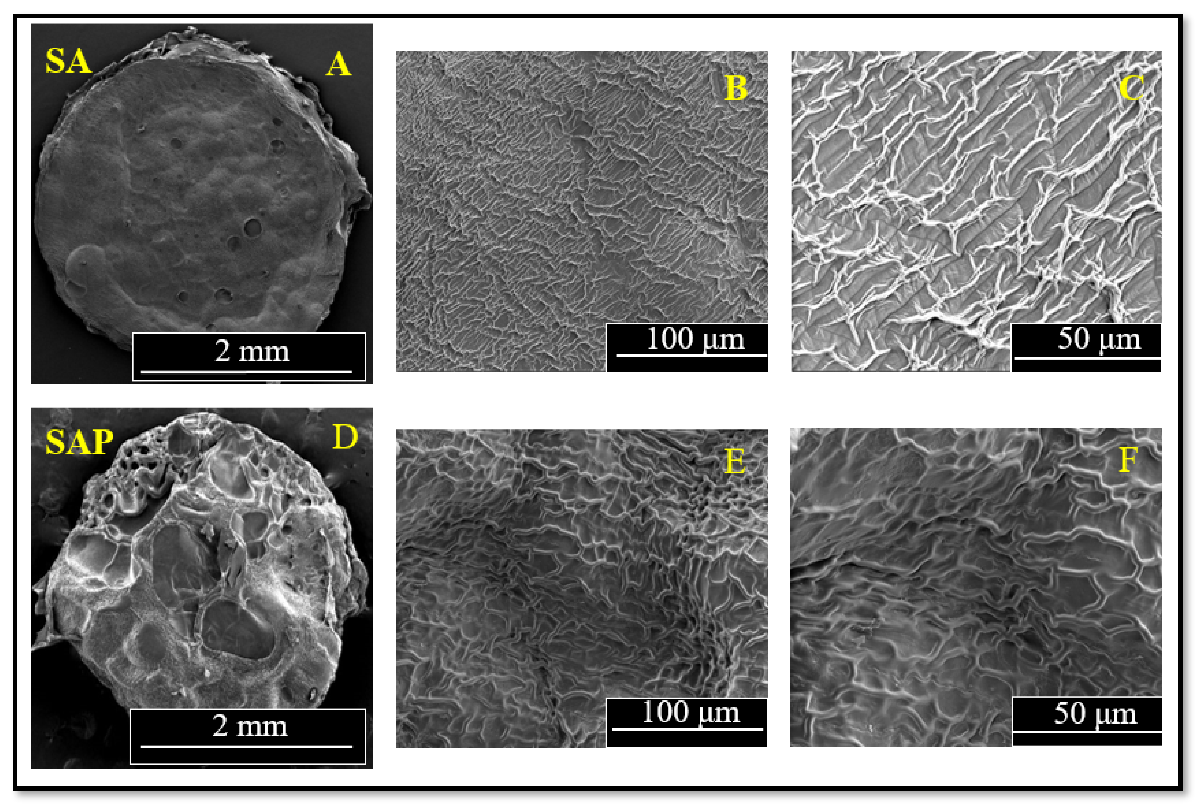
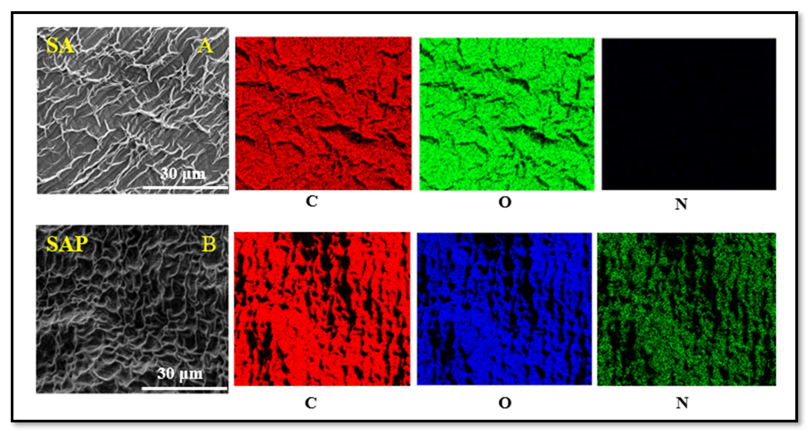
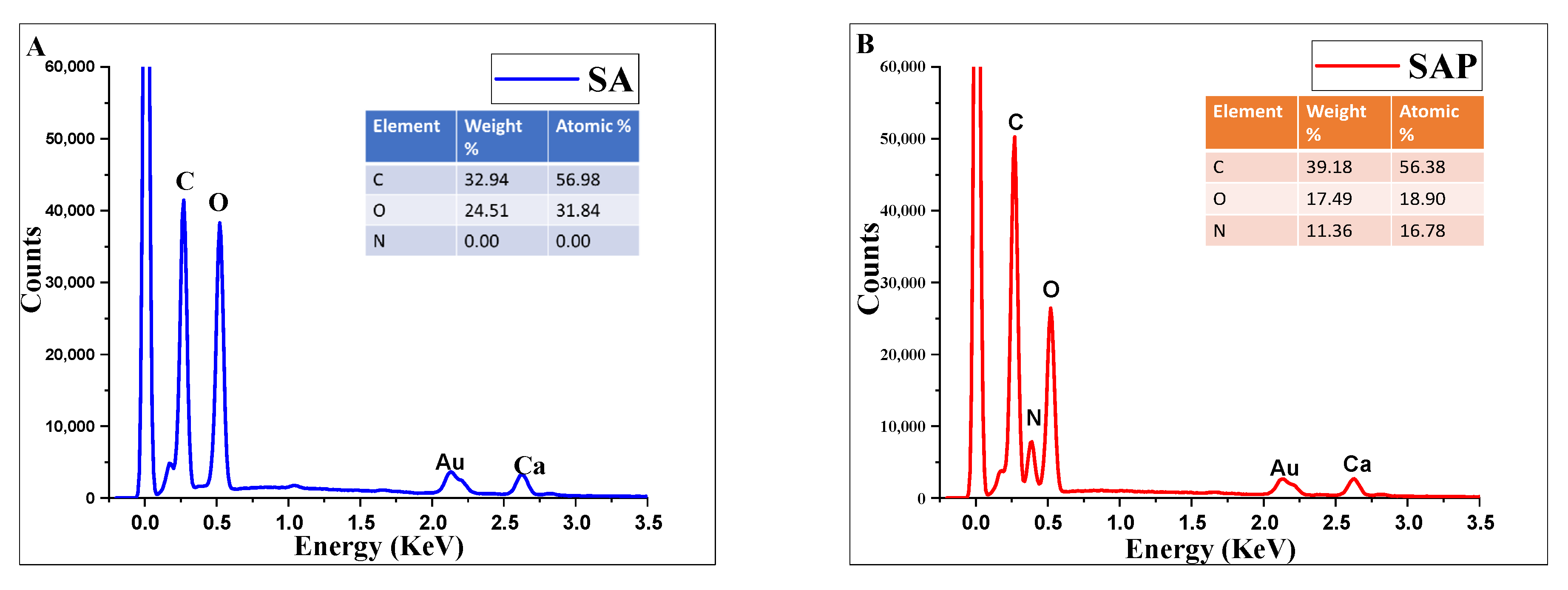
2.3. Scanning Electron Microscopy/Energy-Dispersive X-ray Spectroscopic Characterization
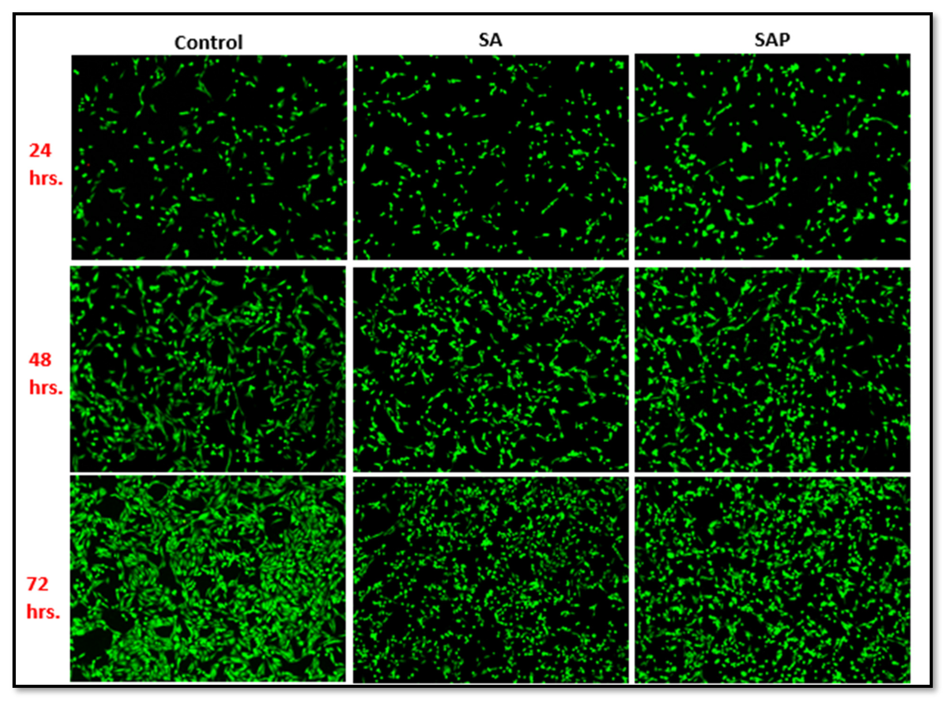
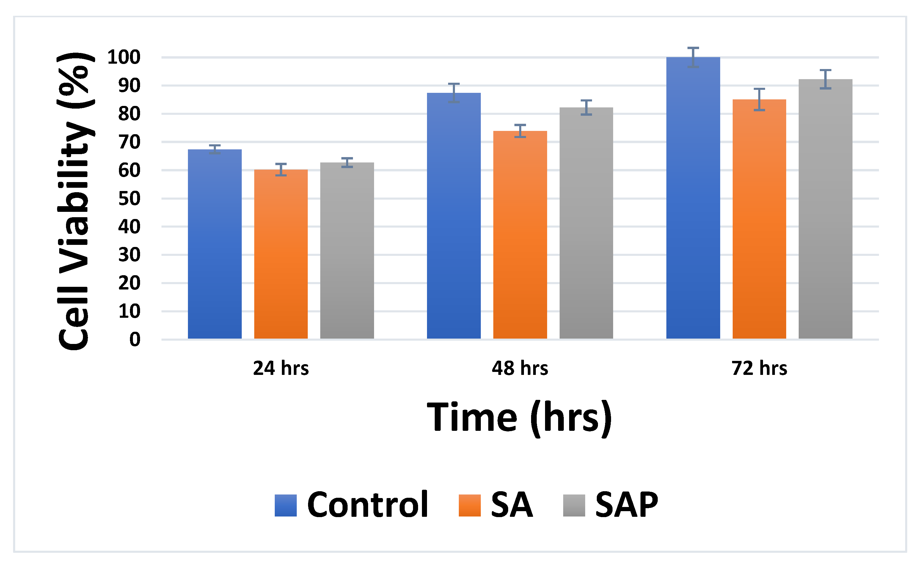
2.4. Biocompatibility Testing of Alginate/PEI Hydrogel Using MDA-MB-231
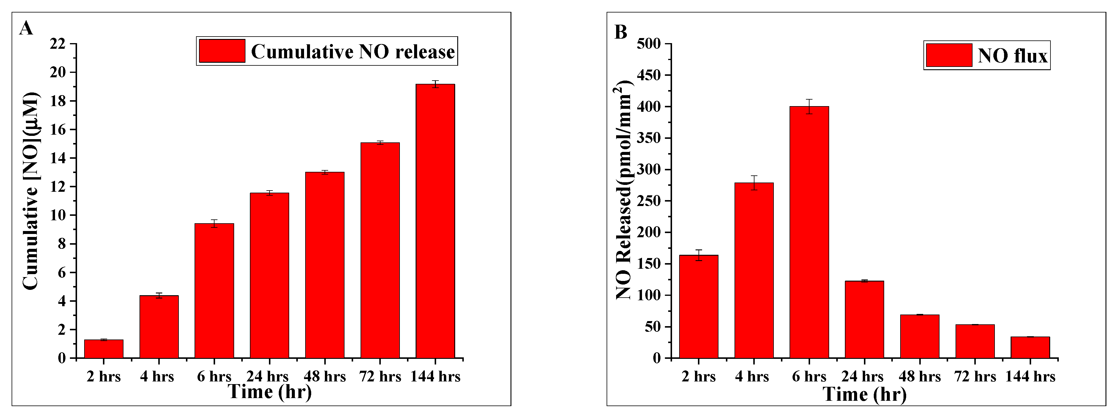
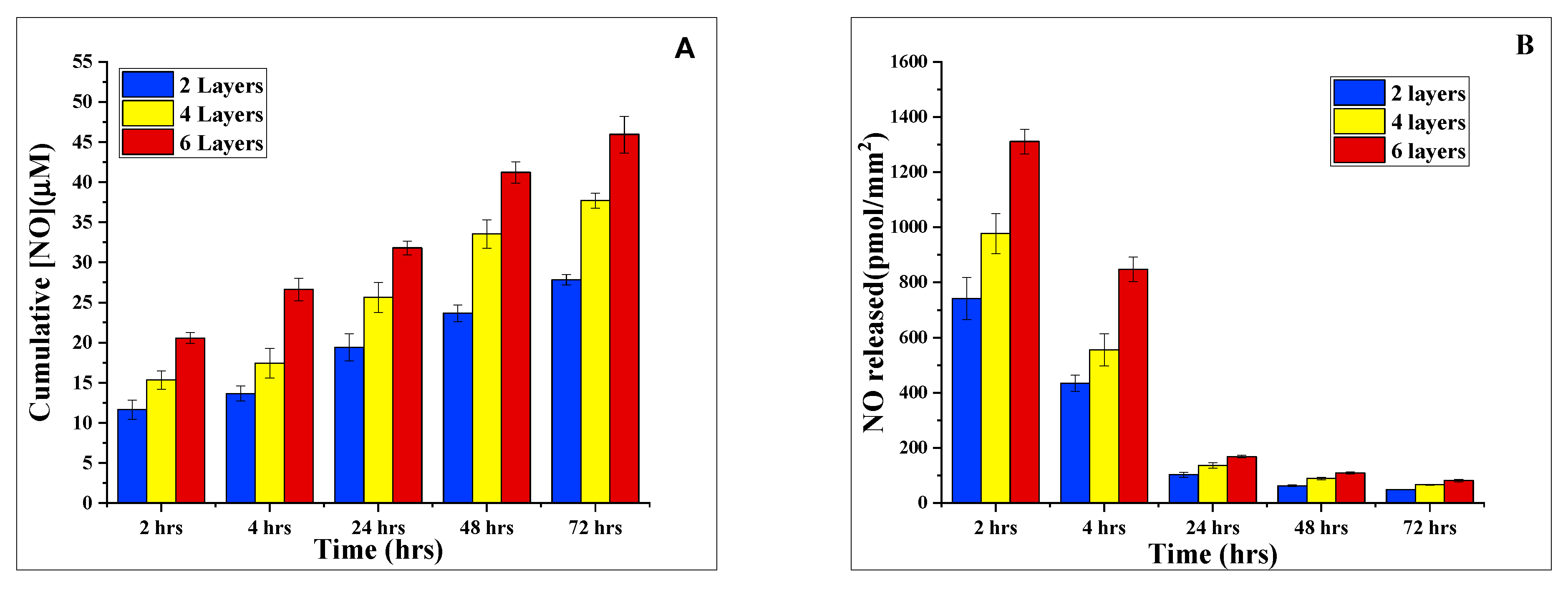
2.5. Nitric Oxide Production Activity of SAP/iNOSoxy Hydrogel
3. Materials and Methods
3.1. Materials
3.2. Preparation of Sodium Alginate/Polyethyleneimine/iNOS Hydrogel (Symbolized in This Work as SA/PEI/iNOSoxy)
3.2.1. iNOS Oxygenase Domain Expression and Purification
3.2.2. Preparation of SA/PEI/iNOSoxy Hydrogel
3.2.3. Water Content/Swelling Ratio of the SA/PEI Hydrogel as Prepared
3.2.4. FT-IR Characterization
3.2.5. Scanning Electron Microscopy/Energy-Dispersive X-ray Spectroscopy
3.3. Biocompatibility Testing of Alginate/PEI Hydrogel Using MDA-MB-231
3.4. Nitric Oxide Production Activity of SA/PEI/iNOSoxy Hydrogel
4. Conclusions
Author Contributions
Funding
Institutional Review Board Statement
Informed Consent Statement
Data Availability Statement
Conflicts of Interest
Sample Availability
References
- Furchgott, R.F.; Zawadzki, J.V. The obligatory role of endothelial cells in the relaxation of arterial smooth muscle by acetylcholine. Nature 1980, 288, 373–376. [Google Scholar] [CrossRef] [PubMed]
- Dattilo, J.B.; Makhoul, R.G. The role of nitric oxide in vascular biology and pathobiology. Ann. Vasc. Surg. 1997, 11, 307–314. [Google Scholar] [CrossRef] [PubMed]
- LloydJones, D.M.; Bloch, K.D. The vascular biology of nitric oxide and its role in atherogenesis. Annu. Rev. Med. 1996, 47, 365–375. [Google Scholar] [CrossRef] [PubMed]
- Walford, G.; Loscalzo, J. Nitric oxide in vascular biology. J. Thromb. Haemost. 2003, 1, 2112–2118. [Google Scholar] [CrossRef]
- Stuehr, D.J.; Kwon, N.S.; Nathan, C.F.; Griffith, O.W.; Feldman, P.; Wiseman, J. N omega-hydroxy-L-arginine is an intermediate in the biosynthesis of nitric oxide from L-arginine. J. Biol. Chem. 1991, 266, 6259–6263. [Google Scholar] [CrossRef]
- Wink, D.A.; Ridnour, L.A.; Hussain, S.P.; Harris, C.C. The reemergence of nitric oxide and cancer. Nitric Oxide Biol. Chem. Off. J. Nitric Oxide Soc. 2008, 19, 65. [Google Scholar] [CrossRef]
- Wink, D.; Vodovotz, Y.; Cook, J.; Krishna, M.; Kim, S.; Coffin, D.; DeGraff, W.; Deluca, A.; Liebmann, J.; Mitchell, J. The role of nitric oxide chemistry in cancer treatment. Biochem. C/C Biokhimiia 1998, 63, 802–809. [Google Scholar]
- Wink, D.A.; Vodovotz, Y.; Laval, J.; Laval, F.; Dewhirst, M.W.; Mitchell, J.B. The multifaceted roles of nitric oxide in cancer. Carcinogenesis 1998, 19, 711–721. [Google Scholar] [CrossRef]
- Lala, P.K.; Orucevic, A. Role of nitric oxide in tumor progression: Lessons from experimental tumors. Cancer Metastasis Rev. 1998, 17, 91–106. [Google Scholar] [CrossRef] [PubMed]
- Ducrocq, C.; Blanchard, B.; Pignatelli, B.; Ohshima, H. Peroxynitrite: An endogenous oxidizing and nitrating agent. Cell. Mol. Life Sci. CMLS 1999, 55, 1068–1077. [Google Scholar] [CrossRef]
- Szabó, C.; Ischiropoulos, H.; Radi, R. Peroxynitrite: Biochemistry, pathophysiology and development of therapeutics. Nat. Rev. Drug Discov. 2007, 6, 662–680. [Google Scholar] [CrossRef]
- Glynn, S.A.; Boersma, B.J.; Dorsey, T.H.; Yi, M.; Yfantis, H.G.; Ridnour, L.A.; Martin, D.N.; Switzer, C.H.; Hudson, R.S.; Wink, D.A. Increased NOS2 predicts poor survival in estrogen receptor–negative breast cancer patients. J. Clin. Investig. 2010, 120, 3843–3854. [Google Scholar] [CrossRef]
- Lee, J.; Cuddihy, M.J.; Kotov, N.A. Three-dimensional cell culture matrices: State of the art. Tissue Eng. Part B Rev. 2008, 14, 61–86. [Google Scholar] [CrossRef]
- Gunasekera, B.; Abou Diwan, C.; Altawallbeh, G.; Kalil, H.; Maher, S.; Xu, S.; Bayachou, M. Functional layer-by-layer thin films of inducible nitric oxide (NO) synthase oxygenase and polyethylenimine: Modulation of enzyme loading and NO-release activity. ACS Appl. Mater. Interfaces 2018, 10, 7745–7755. [Google Scholar] [CrossRef]
- Pereira, R.; Tojeira, A.; Vaz, D.C.; Mendes, A.; Bártolo, P. Preparation and characterization of films based on alginate and aloe vera. Int. J. Polym. Anal. Charact. 2011, 16, 449–464. [Google Scholar] [CrossRef]
- Rajaram, A.; Schreyer, D.J.; Chen, D.X. Use of the polycation polyethyleneimine to improve the physical properties of alginate–hyaluronic acid hydrogel during fabrication of tissue repair scaffolds. J. Biomater. Sci. Polym. Ed. 2015, 26, 433–445. [Google Scholar] [CrossRef]
- Zhao, J.; Li, Q.; Zhang, X.; Xiao, M.; Zhang, W.; Lu, C. Grafting of polyethylenimine onto cellulose nanofibers for interfacial enhancement in their epoxy nanocomposites. Carbohydr. Polym. 2017, 157, 1419–1425. [Google Scholar] [CrossRef] [PubMed]
- Yun, H.-J.; Hong, H.; Lee, J.; Choi, C.-J. Chemical and structural properties of polyethyleneimine film coated on a SiO2 substrate in different concentrations. Mater. Trans. 2014, 55, 801–805. [Google Scholar] [CrossRef]
- Akbarzadeh, M.; Oskuee, R.K.; Gholami, L.; Mahmoudi, A.; Malaekeh-Nikouei, B. BR2 cell penetrating peptide improved the transfection efficiency of modified polyethyleneimine. J. Drug Deliv. Sci. Technol. 2019, 53, 101154. [Google Scholar] [CrossRef]
- Calixto, G.M.F.; Victorelli, F.D.; Dovigo, L.N.; Chorilli, M. Polyethyleneimine and chitosan polymer-based mucoadhesive liquid crystalline systems intended for buccal drug delivery. AAPS PharmSciTech 2018, 19, 820–836. [Google Scholar] [CrossRef] [PubMed]
- Hlaing, S.P.; Kim, J.; Lee, J.; Hasan, N.; Cao, J.; Naeem, M.; Lee, E.H.; Shin, J.H.; Jung, Y.; Lee, B.-L. S-Nitrosoglutathione loaded poly (lactic-co-glycolic acid) microparticles for prolonged nitric oxide release and enhanced healing of methicillin-resistant Staphylococcus aureus-infected wounds. Eur. J. Pharm. Biopharm. 2018, 132, 94–102. [Google Scholar] [CrossRef] [PubMed]
- Wang, Z.; Zhu, S.; Wang, L.; Chang, L.; Wang, J.; Li, J.; Guan, S. Preparing a novel magnesium-doped hyaluronan/polyethyleneimine nanoparticle to improve endothelial functionalisation. IET Nanobiotechnol. 2019, 14, 142–147. [Google Scholar] [CrossRef] [PubMed]
- Martinsen, A.; Skjåk-Bræk, G.; Smidsrød, O. Alginate as immobilization material: I. Correlation between chemical and physical properties of alginate gel beads. Biotechnol. Bioeng. 1989, 33, 79–89. [Google Scholar] [CrossRef] [PubMed]
- Martinsen, A.; Storrø, I.; Skjårk-Bræk, G. Alginate as immobilization material: III. Diffusional properties. Biotechnol. Bioeng. 1992, 39, 186–194. [Google Scholar] [CrossRef]
- Amsden, B.; Turner, N. Diffusion characteristics of calcium alginate gels. Biotechnol. Bioeng. 1999, 65, 605–610. [Google Scholar] [CrossRef]
- Carew, E.O.; Cooke, F.W.; Lemons, J.E.; Ratner, B.D.; Vesely, I.; Vogler, E. Properties of materials. In Biomaterials Science: An Introduction to Materials in Medicine; Elsevier Academic Press: Cambridge, MA, USA, 2004; pp. 23–32. [Google Scholar]
- Hollinger, J.O.; Guelcher, S.A. An Introduction to Biomaterials; CRC Press/Taylor & Francis Boca Raton: Boca Raton, FL, USA, 2012. [Google Scholar]
- Zhang, J.; Zhu, Y.; Song, J.; Yang, J.; Pan, C.; Xu, T.; Zhang, L. Novel balanced charged alginate/pei polyelectrolyte hydrogel that resists foreign-body reaction. ACS Appl. Mater. Interfaces 2018, 10, 6879–6886. [Google Scholar] [CrossRef]
- Saha, A.K.; Ray, S.D. Effect of cross-linked biodegradable polymers on sustained release of sodium diclofenac-loaded microspheres. Braz. J. Pharm. Sci. 2013, 49, 873–888. [Google Scholar] [CrossRef]
- Yeh, C.-J.G.; Hsi, B.-L.; Faulk, W.P. Propidium iodide as a nuclear marker in immunofluorescence. II. Use with cellular identification and viability studies. J. Immunol. Methods 1981, 43, 269–275. [Google Scholar] [CrossRef]
- Tanke, H.; Van der Linden, P.; Langerak, J. Alternative fluorochromes to ethidium bromide for automated read out of cytotoxicity tests. J. Immunol. Methods 1982, 52, 91–96. [Google Scholar] [CrossRef]
- Jacobs, D.B.; Pipho, C. Use of propidium iodide staining and flow cytometry to measure antibody-mediated cytotoxicity: Resolution of complement-sensitive and resistant target cells. J. Immunol. Methods 1983, 62, 101–108. [Google Scholar] [CrossRef]
- Khoshnood, N.; Zamanian, A.; Abbasi, M. The potential impact of polyethylenimine on biological behavior of 3D-printed alginate scaffolds. Int. J. Biol. Macromol. 2021, 178, 19–28. [Google Scholar] [CrossRef] [PubMed]
- Godiya, C.B.; Xiao, Y.; Lu, X. Amine functionalized sodium alginate hydrogel for efficient and rapid removal of methyl blue in water. Int. J. Biol. Macromol. 2020, 144, 671–681. [Google Scholar] [CrossRef] [PubMed]
- Rogero, S.O.; Malmonge, S.M.; Lugão, A.B.; Ikeda, T.I.; Miyamaru, L.; Cruz, Á.S. Biocompatibility study of polymeric biomaterials. Artif. Organs. 2003, 27, 424–427. [Google Scholar] [CrossRef]
- Bank, H. Assessment of islet cell viability using fluorescent dyes. Diabetologia 1987, 30, 812–816. [Google Scholar] [CrossRef]
- West, S.S. Fluorescence microspectrophotometry of supravitally stained cells. Phys. Tech. Biol. Res. 1969, 3, 253–321. [Google Scholar]
- Pantazis, C.G.; Kniker, W.T. Assessment of blood leukocyte microbial killing by using a new fluorochrome microassay. J. Reticuloendothel. Soc. 1979, 26, 155–170. [Google Scholar]
- Kapuscinski, J.; Darzynkiewicz, Z.; Melamed, M.R. Interactions of acridine orange with nucleic acids: Properties of complexes of acridine orange with single stranded ribonucleic acid. Biochem. Pharmacol. 1983, 32, 3679–3694. [Google Scholar] [CrossRef] [PubMed]
- Waring, M. Ethidium and propidium. In Mechanism of Action of Antimicrobial and Antitumor Agents; Springer: Berlin/Heidelberg, Germany, 1975; pp. 141–165. [Google Scholar]
- Frost, M.C.; Reynolds, M.M.; Meyerhoff, M.E. Polymers incorporating nitric oxide releasing/generating substances for improved biocompatibility of blood-contacting medical devices. Biomaterials 2005, 26, 1685–1693. [Google Scholar] [CrossRef] [PubMed]
- Abou Diwan, C. Nitric Oxide Synthase-Based Biopolymers; Towards Novel Thromboresistant No-Release Materials; Cleveland State University: Cleveland, OH, USA, 2009. [Google Scholar]
- Stuehr, D.J.; Cho, H.J.; Kwon, N.S.; Weise, M.F.; Nathan, C.F. Purification and characterization of the cytokine-induced macrophage nitric oxide synthase: An FAD-and FMN-containing flavoprotein. Proc. Natl. Acad. Sci. USA 1991, 88, 7773–7777. [Google Scholar] [CrossRef]
- Hetrick, E.M.; Schoenfisch, M.H. Analytical chemistry of nitric oxide. Annu. Rev. Anal. Chem. 2009, 2, 409–433. [Google Scholar] [CrossRef]
- Griess, P. Comments on the essay of HH Weselsky and Benedict ‘About some azo compounds’. Chem. Ber. 1879, 12, 426–428. [Google Scholar] [CrossRef] [Green Version]


Disclaimer/Publisher’s Note: The statements, opinions and data contained in all publications are solely those of the individual author(s) and contributor(s) and not of MDPI and/or the editor(s). MDPI and/or the editor(s) disclaim responsibility for any injury to people or property resulting from any ideas, methods, instructions or products referred to in the content. |
© 2023 by the authors. Licensee MDPI, Basel, Switzerland. This article is an open access article distributed under the terms and conditions of the Creative Commons Attribution (CC BY) license (https://creativecommons.org/licenses/by/4.0/).
Share and Cite
Maher, S.; Smith, L.A.; El-Khoury, C.A.; Kalil, H.; Sossey-Alaoui, K.; Bayachou, M. Inducible Nitric Oxide Synthase Embedded in Alginate/Polyethyleneimine Hydrogel as a New Platform to Explore NO-Driven Modulation of Biological Function. Molecules 2023, 28, 1612. https://doi.org/10.3390/molecules28041612
Maher S, Smith LA, El-Khoury CA, Kalil H, Sossey-Alaoui K, Bayachou M. Inducible Nitric Oxide Synthase Embedded in Alginate/Polyethyleneimine Hydrogel as a New Platform to Explore NO-Driven Modulation of Biological Function. Molecules. 2023; 28(4):1612. https://doi.org/10.3390/molecules28041612
Chicago/Turabian StyleMaher, Shaimaa, Lauren A. Smith, Celine A. El-Khoury, Haitham Kalil, Khalid Sossey-Alaoui, and Mekki Bayachou. 2023. "Inducible Nitric Oxide Synthase Embedded in Alginate/Polyethyleneimine Hydrogel as a New Platform to Explore NO-Driven Modulation of Biological Function" Molecules 28, no. 4: 1612. https://doi.org/10.3390/molecules28041612