Article

In Vitro and In Silico Studies of Neolignans from Magnolia grandiflora L. Seeds against Human Cannabinoids and Opioid Receptors

by
Pankaj Pandey
1,*,
Mallika Kumarihamy
1,†,
Krishna Chaturvedi
2,
Mohamed A. M. Ibrahim
1,
Janet A. Lambert
3,‡,
Murrell Godfrey
2,
Robert J. Doerksen
3 and
Ilias Muhammad
1,*
1
National Center for Natural Products Research, Research Institute of Pharmaceutical Sciences, School of Pharmacy, The University of Mississippi, University, MS 38677, USA
2
Department of Chemistry and Biochemistry, The University of Mississippi, University, MS 38677, USA
3
Department of BioMolecular Sciences, Division of Medicinal Chemistry and Research Institute of Pharmaceutical Sciences, School of Pharmacy, The University of Mississippi, University, MS 38677, USA
*
Authors to whom correspondence should be addressed.
Current address: United States Department of Agriculture, Agriculture Research Services, New Orleans, LA 70124, USA.
Current address: Department of Pharmacology, School of Medicine, University of Nevada, Reno, NV 89557, USA.
Molecules 2023, 28(3), 1253; https://doi.org/10.3390/molecules28031253
Submission received: 6 December 2022 / Revised: 17 January 2023 / Accepted: 21 January 2023 / Published: 27 January 2023
(This article belongs to the Special Issue New Insights into Biomolecular Structures and Interactions)

Abstract

:
Magnolia grandiflora L. (Magnoliaceae) is a plant of considerable medicinal significance; its flowers and seeds have been used in various traditional remedies. Radioligand binding assays of n-hexane seeds extract showed displacement of radioligand for cannabinoid (CB1 and CB2) and opioid δ (delta), κ (kappa), and µ (mu) receptors. Bioactivity-guided fractionation afforded 4-O-methylhonokiol (1), magnolol (2), and honokiol (3), which showed higher binding to cannabinoid rather than opioid receptors in radioligand binding assays. Compounds 13, together with the dihydro analog of 2 (4), displayed selective affinity towards CB2R (Ki values of 0.29, 1.4, 1.94, and 0.99 μM, respectively), compared to CB1R (Ki 3.85, 17.82, 14.55, and 19.08 μM, respectively). An equal mixture of 2 and 3 (1:1 ratio) showed additive displacement activity towards the tested receptors compared to either 2 or 3 alone, which in turn provides an explanation for the strong displacement activity of the n-hexane extract. Due to the unavailability of an NMR or X-ray crystal structure of bound neolignans with the CB1 and CB2 receptors, a docking study was performed to predict ligand–protein interactions at a molecular level and to delineate structure-activity relationships (SAR) of the neolignan analogs with the CB1 and CB2 receptors. The putative binding modes of neolignans 1–3 and previously reported related analogs (4, 4a, 5, 5a, 6, 6a, and 6b) into the active site of the CB1 and CB2 receptors were assessed for the first time via molecular docking and binding free-energy (∆G) calculations. The docking and ∆G results revealed the importance of a hydroxyl moiety in the molecules that forms strong H-bonding with Ser383 and Ser285 within CB1R and CB2R, respectively. The impact of a shift from a hydroxyl to the methoxy group on experimental binding affinity to CB1R versus CB2R was explained through ∆G data and the orientation of the alkyl chain within the CB1R. This comprehensive SAR, influenced by the computational study and the observed in vitro displacement binding affinities, has indicated the potential of magnolia neolignans for developing new CB agonists for potential use as analgesics, anti-inflammatory agents, or anxiolytics.

Graphical Abstract

1. Introduction

The cannabinoid (CB) and opioid receptors are seven-transmembrane domain G-protein coupled receptors (GPCRs) known to modulate various cellular, neuronal, and cardiovascular functions [1]. Cannabinoid receptor type 1 (CB1R) antagonists/inverse agonists have the potential for treating obesity [2], obesity-related cardiometabolic disorders [3], and drug/substance abuse [4]; however, there is no such drug currently available on the market. CB1R is abundant in the central nervous system (CNS) and in peripheral tissues [5,6]. The cannabinoid receptor type 2 (CB2R) is a target for the discovery of several categories of therapeutics, such as for neuro-inflammation, cardiometabolic disorder, renal ischemia-reperfusion injury, and other diseases/disorders, including multiple sclerosis and arthritis [7,8,9,10]. Opioid receptors include three subtypes, δ- (delta), κ- (kappa), and µ- (mu), and agonism of these receptors regulates pain inhibitory pathways of the CNS [11]. Research studies have shown that both CB and opioid receptors share several pharmacological properties and act synergistically in analgesic effects at lower doses with fewer side effects [12]. Medicinal plants and their constituents have been used extensively as supplements to treat various neurological disorders, including pain, anxiety, convulsions, epilepsy, hysteria, and inflammation [13].
In the United States, Magnolia grandiflora L. (Magnoliaceae), commonly known as Southern magnolia, is a plant of medicinal significance that is often grown as an ornamental tree [14]. Its flowers and seeds are used in various traditional herbal remedies, including fever, rheumatism, and inflammation [15,16]. From the same genus, the bark of M. officinalis plays an important role in traditional Chinese and Japanese herbal medicine for the treatment of anxiety, sleep-related disorders, and allergic diseases [16]. In addition, magnolia-based products have been used for smoking cessation therapy and as aphrodisiacs, anti-depressants, and sedatives due to their hypothesized cannabimimetic and GABA-ergic-like effects [17]. Extensive research has been carried out on magnolia extracts and their major constituents to study their anti-cancer [18], anti-anxiety [19], anti-depressant [16,20], cardiovascular [21], and anti-inflammatory [22,23] activities. These pharmacological effects were proposed [16] to be primarily mediated by the presence of the neolignans 4-O-methylhonokiol (1), magnolol (2), honokiol (3), and 2′s hydrogenated derivative, tetrahydromagnolol (4) (Figure 1). The presence of these neolignans in the other species of the genus Magnolia, including M. officinalis, M. obovata, and M. virginiana, has led to the use of supplements prepared from these extracts as anti-depressants, analgesics, aphrodisiacs, and appetite suppressants or stimulants [17]. Earlier studies [24] have indicated that 1 and 2 could potentiate γ-aminobutyric acid A (GABAA) receptors and could act as positive allosteric modulators (PAMs); however, these neolignans have shown no direct interaction with the norepinephrine transporter [25]. In 2004, Kong et al., evaluated the MAO inhibitory potential of 2 and 3 using rat brain mitochondria; however, no significant rat brain MAO inhibitory activity was noted [26]. Remarkably, M. officinalis extract, its constituents, and their synthetic analogs have been reported as CB2 agonists and inverse agonists [27,28,29,30] and similarly as CB-related orphan receptor GPR55 inhibitors [27]; however, the safety and toxicological properties of 2, 3, and bark extracts of M. officinalis and M. obovate have been reported as safe for consumption [31].
In a continuation to our research efforts to establish potential leads towards human cannabinoids and opioid receptors, M. grandiflora seed extract showed significant binding affinities to CB and opioid receptors, and therefore a detailed investigation of its active constituents and their structure-activity relationships (SAR) has been conducted using in silico studies that utilize the CB2 X-ray crystal structure. A bioassay-guided isolation of the hexane extract, which showed significant displacement of radioligand in cannabinoid and opioid receptors (64% CB1, 74% CB2, δ 93%, κ 61%, and µ 85%), yielded 4-O-methylhonokiol (1), magnolol (2), and honokiol (3) with selective CB2 activity (Ki 0.29, 1.40, and 1.94 μM, respectively). The structures of these neolignans are closely related to the structures of the potent CB1 agonist Δ9-tetrahydrocannabinol (Δ9-THC) and the CB1/CB2 non-selective agonist CP55,940 (Figure 1). In addition to CB and opioid activity, we report the first systematic in silico molecular docking studies of neolignans (14) and their structurally-related previously reported analogs (4a, 5, 5a, 6, 6a, 6b) [29], using the active-state X-ray crystal structures of CB1R and CB2R to gain an understanding of the various protein–ligand interaction patterns, their putative binding modes, and the observed SAR of these ligands. A detailed understanding of the SAR of the isolated metabolites, as well as the additive effect between compounds 2 and 3, has been reported for the first time, as supported via the in vitro data and the in silico docking studies that take advantage of the first CB2 X-ray crystal structure, which was released in 2020 [32].

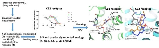
2. Results and Discussion

The n-hexane extract of M. grandiflora showed significant radioligand displacement of CB1, CB2, δ, κ, and µ opioid receptors (Table 1). The extract was subjected to centrifugal preparative thin-layer chromatography (CPTLC; for details, see Experimental Section) to afford 4-O-methylhonokiol (1), magnolol (2), and honokiol (3), together with marked fatty acids. The structures of the isolated compounds were identified based on 1D- and 2D- nuclear magnetic resonance (NMR) and electrospray ionization mass spectrometry (ESI-MS). Compounds 13, as well as 4 (tetrahydromagnolol, purchased from Sigma-Aldrich), were evaluated using in vitro binding assays with CB and opioid receptors (Table 1 and Table 2 and Figure 1, Figure 2 and Figure 3). The preliminary results determined at a concentration of 10 µM revealed strong displacement by 1 towards both CB1R and CB2R (i.e., 91.3% and 82.2%, respectively). The mixture of 2 + 3 (1:1) showed similar displacement of radioligand (99.8% and 91.0%, respectively), compared to either compound 2 or 3 alone (Table 1). Based on the secondary assay results, compounds 13 displayed strong selective affinity towards CB2R compared to CB1R, with Ki values of 1 towards CB2R as being 0.29 ± 0.022 μM, which is significantly better than those of 2 and 3 (Ki 1.44 ± 0.138 and 1.94 ± 0.162 µM, respectively), while tetrahydromagnolol (4) showed similar displacement to that of 2 (Figure 2 and Figure 3).
The hexane extract displayed significant radioligand displacement at all three opioid receptors: δ (93%), κ (60.8%), and µ (84.5%); however, all the isolated neolignans (13) were devoid of binding affinity at δ and κ. Nevertheless, the isolated neolignans showed high micromolar binding affinity at the µ receptor. Indeed, a 1:1 mixture of 2 and 3 showed better additive percentage displacement (%) of radioligand at δ (93.3%), κ (59.2%), and µ (74.0%) receptors, compared to 13 alone, but was similar to those of the hexane extract (Table 1), which provides an explanation for the strong displacement activity of the n-hexane extract. Additionally, 2 and 3 showed a low affinity for µ opioid receptors during IC50 and Ki determinations (Figure 4).
CB binding affinities of compounds 14 have been previously reported [27,28,29], and are in accordance with the results obtained for CB2R in our laboratory. Furthermore, compounds 14 were reported to possess binding affinities (Ki) between 29 μM for CB1R [27,28,29]. However, in our lab, this observation was only true for 4-O-methylhonokiol (1); the remaining three compounds possessed binding affinities (Ki) between 1420 μM (Table 2). Rempel et al. (2013) have reported magnolol (2), and tetrahydromagnolol (4) as partial CB2R agonists (Ki 1.44 and 0.41 μM, respectively), and honokiol (3) was considered as a CB2R antagonist or inverse agonist (Ki 5.61 μM) [27]. Schuehly et al. (2011) reported intriguing nonspecific heteroactive behavior of 4-O-methylhonokiol (1) at the CB2R as an inverse agonist at Gi/o and as a full agonist regarding intracellular Ca2+ [Ca2+]i. In contrast, Fuchs et al. (2013) reported 1 as an agonist at both CB receptor subtypes in forskolin-induced cAMP (3,5-cyclic adenosine monophosphate) accumulation assays [29]. Our CB binding affinity data of 14 are in close agreement with those reported previously [27,28,29].

Protein-Ligand Interaction Study

Due to the unavailability of an NMR or X-ray crystal structure of bound neolignans with the CB1 and CB2 receptors, a docking study was performed to predict ligand–protein interactions at a molecular level. The in silico docking studies were performed by taking advantage of the first CB2 X-ray crystal structure (PDB ID: 6KPC) [32] which was released in 2020 and the CB1 receptor (PDB ID: 5XRA) [33]. The isolated neolignans (14) and structurally-related previously published compounds (4a, 5, 5a, 6, 6a, and 6b) [29] were studied to understand the SAR using docking and binding free-energy calculations. The docking protocol was validated by a self-docking approach in which native ligands, 8D3 and E3R, were docked into their corresponding protein structures, CB1 and CB2, respectively. Further, we calculated the Root Mean Square Deviation (RMSD) between docked poses and experimental poses of native ligands with the CB1 and CB2 receptors. The overlay of experimental poses of native ligands with the docked poses showed an identical conformation, with very small RMSD differences of 0.35 Å and 0.64 Å, respectively. The binding affinity data from Table 3 indicates that the replacement of one hydroxyl by a methoxy group (4a) in tetrahydromagnolol (4) led to a significant increase in CB1R affinity. Simultaneously, the selectivity towards CB2R was lost. Our computational results also justified this SAR through analysis of the binding free energy data. The replacement of one hydroxyl by a methoxy group in 4a showed better-predicted binding free-energy (ΔG = −76.78 kcal/mol) compared to tetrahydromagnolol (4) (ΔG = −71.06 kcal/mol) at the CB1R.
The replacement of the hydroxyl group with the methoxy group resulted in a steric clash of the methoxy moiety with Phe170. Thus, the methoxy-containing phenyl ring of 4a is inverted towards Phe200, and the alkyl chain (n = 3) is oriented towards the small hydrophobic pocket in a similar fashion to the cocrystallized ligand AM1152 in the 5XRA crystal structure wherein the dimethyl heptyl moiety is positioned in that pocket (Figure 5). The strong hydrophobic interaction of the methoxy group of 4a with Phe200 and the other lesser hydrophobic interactions of the propyl chain with Leu193, Val196, Phe200, Leu276, Trp279, Met363, Cys386, and Leu387 support a better CB1R affinity compared to 4.
Like methoxytetrahydromagnolol (4a), the methylated compound 5a, which differs at the R2 position (pentyl chain), displayed a 21-fold higher binding affinity towards CB1R than the parent biphenyl 5, which is clearly explained by binding free-energy data. Similarly, compound 5a exhibited better binding free-energy (ΔG = −83.77 kcal/mol) compared to 5 (ΔG = −78.78 kcal/mol).
The docking orientation of 5 and 5a is very similar in the active site of the CB1R (Figure 6), except for the pentyl and propyl chains, which are interchanged. Like compound 4a, the strong hydrophobic interactions between Phe200 and Cys386 of CB1R and the methoxy moiety of 5a were observed, which might explain the better affinity of 5a to CB1R compared to 5.
The impact of the methoxy group on CB1R affinity was found to be more pronounced compared to the CB2R. When the methoxy group was introduced in the para-position of the short propyl residue of 6, a remarkable enhancement in CB1R affinity (a 15-fold increase) was observed (6a).
The docking poses of 6 and 6a in the active site of CB1R are markedly similar (Figure 7), and the alkyl chain is also oriented in the same manner, which was different in the docking pose of 5 and 5a. The additional strong hydrophobic interactions between the methoxy moiety of 6a and Phe200, Phe170, and Cys386 (also H-bonding) of CB1R might explain the increased affinity (Ki = 0.00957 µM) of 6a to CB1R compared to 6. Compared to the unmethylated 5 and 6, the methylated 5a and 6a exhibited strong predicted CB1R affinity (Figure 6 and Figure 7C), while predicted CB2R binding affinity was almost unchanged (Figure 6D and Figure 7D); however, the introduction of a methoxy group in the para-position to the hexyl residue (6b) drastically reduced the affinity for the CB2R (from Ki = 0.0294 µM to 0.234 µM, Figure 7C). Interestingly, 6a, with a functional group rearrangement (methoxy group in the para-position with respect to the propyl residue), displayed a 12-fold increase in CB2R affinity (Ki= 0.0238 µM). Analysis of the docking pose of 6a and 6b in the active site of CB2R clearly explained the activity difference between 6a and 6b. In 6a, the presence of the methoxy group oriented towards Thr114 (distance Thr (O–H) and –O–CH3 = 4.27 Å), the unmethylated hydroxyl group forming H-bonding with Ser285, and the biphenyl contributing to π–π stacking with Phe87 and Phe183 allowed the alkyl chain (n = 6) to be directed towards the small hydrophobic pocket similar to the cocrystallized ligand AM1152 in the 5XRA crystal structure that positions the dimethyl heptyl moiety in that pocket. This also afforded lower binding free-energy (ΔG = −80.56 kcal/mol) compared to 6b (ΔG = −69.06 kcal/mol); however, in 6b, the methoxy moiety moved slightly upwards towards Ser90, and the alkyl chain (n = 3) was redirected towards the small hydrophobic pocket similar to the cocrystallized ligand AM1152 in the 5XRA crystal structure [33]. The identical orientation of the hexyl alkyl chain of 6a towards the hydrophobic pocket where the alkyl chain of the cocrystallized ligand AM1152 in the 5XRA CB1 crystal structure is situated led to the strong activity of 6a compared to 6b.
Both honokiol (3) and 4-O-methylhonokiol (1) docked in similar orientations within the active site of CB1R and showed strong π–π interactions with Phe170/Phe174 and Phe268, as well as H-bonding with one of the hydroxyls of 3 and 1 with Ser383 (Figure 8). Furthermore, the methoxy group of 1 exhibited additional strong hydrophobic interactions with Ser173 and Phe177, which led to better binding free-energy (ΔG = −78.20 kcal/mol) compared to 3 (ΔG = −71.19 kcal/mol). These observed data do not match with Fuchs et al. (2013) [29] experimental in vitro data, whereas 3 (Ki = 6.46 µM) is more active at CB1R than 1 (Ki = 8.34 µM). Interestingly, our in-house testing of the binding affinity of 1 (Ki = 3.85 µM) and 3 (Ki = 14.55 µM) against CB1R matches closely with the computational binding free-energy data. In the case of CB2R, 4-O-methylhonokiol (1) and honokiol (3) docked in a similar fashion; however, 4-O-methylhonokiol (1) shifted ~1.6 Å intracellularly from the position of the honokiol (3) atoms, which allows this molecule (1) to form strong hydrogen bonding (through the phenolic hydroxyl moiety) with Ser285 and strong π–π interactions with Phe87 and Phe183. These interactions were absent in the honokiol (3) docking pose with CB2R. The only interactions observed between honokiol (3) and CB2R were hydrophobic. Remarkably, the binding free-energy (ΔG) data matches well with the experimental activity data for these two molecules at the CB2R.

3. Materials and Methods

3.1. Extraction and Bioassay-Guided Isolation of Compounds

Mature seeds of M. grandiflora were collected at the University of Mississippi (MS 38,677) in November 2013. The voucher specimen (NCNPR #15,895) is deposited at the University of Mississippi. The air-dried seeds (107 g) were powdered and extracted with n-hexane (200 mL × 2 for 24 h) followed by 95% EtOH. The combined hexane extracts were evaporated under reduced pressure. Two grams of hexane extract were chromatographed over a centrifugal preparative thin layer chromatographer (CPTLC, Chromatotron®, Analtech Inc., Newark, DE, USA) using a 6 mm silica gel rotor. The sample was dissolved in dichloromethane (DCM), applied to the rotor, and then eluted with n-hexane, followed by DCM and MeOH (200 mL each) to yield eighteen fractions. These fractions later yielded three major lignans, 4-O-methylhonokiol (1, 36 mg), honokiol (2, 20 mg), and magnolol (3, 15 mg), together with marked fatty acids. All fractions were monitored and collected via TLC analysis (silica gel; solvents: n-hexane-EtOAc; 75:25).
4-O-methylhonokiol (1); UPHPLC/APCI-MS m/z 281.3 ([M + H])+ C19H20O2 + H; the 1H and 13C NMR were indistinguishable to those reported [34].
Honokiol (2); UPHPLC/APCI-MS m/z 267.3 ([M + H])+ C18H18O2 + H; the 1H and 13C NMR were indistinguishable to those reported [35].
Magnolol (3); UPHPLC/APCI-MS m/z 267.3 ([M + H])+ C18H18O2 + H; the 1H and 13C NMR were indistinguishable to those reported [35].

3.2. Cannabinoid and Opioid Receptor Binding Assay

3.2.1. Reagents

CP55,940 was purchased from Tocris Bioscience (Minneapolis, MN, USA). BSA, TrizmaTM hydrochloride (Tris-HCl), penicillin, streptomycin, and nonenzymatic cell dissociation solution were purchased from Sigma-Aldrich (St. Louis, MO, USA). Radioligands, GF/C, GF/B 96-well plates, and MicroScintTM-20, were purchased from PerkinElmer (Waltham, MA, USA). Membrane preparation was made using a 50 mM Tris-HCl buffer with pH 7.4. Tetrahydromagnolol (4) was purchased from Sigma-Aldrich (St. Louis, MO, USA) (% purity ≥ 95).

3.2.2. Cell Culture and Membrane Preparation

Human embryonic kidney 293 (HEK293) cells (ATCC) were stably transfected with cannabinoid receptor subtypes 1 and 2 and maintained and harvested as described [36,37]. HEK293 cells stably transfected with δ, κ, and μ opioid subtypes were a generous gift from Roth Laboratories (University of North Carolina at Chapel Hill, NC, USA). Opioid cells were maintained as previously described [37,38]. Membranes were made by washing the cells with cold PBS. The cells were then scraped in cold 50 mM Tris-HCl, pH 7.4 buffer. The solution was centrifuged at 5200× g for 10 min at 4 °C. Next, the supernatant was discarded, and the pellet was washed with more Tris-HCl buffer, homogenized via Sonic Dismembrator (Fisher Scientific, Pittsburgh, PA, USA) and then centrifuged at 24,000× g for 40 min at 4 °C. Finally, the pellet was re-suspended in cold 50 mM Tris-HCl buffer, aliquoted into 2 mL vials, and stored at −80 °C. The total membrane protein concentration was measured using a Pierce BCA Protein Assay Kit (Thermo Scientific, Rockford, IL, USA) as per the manufacturer’s protocol.

3.2.3. Competitive Radioligand Binding Assays

Cannabinoid and opioid competitive radioligand binding assays were performed as previously described [5,36,37,38]. Saturation experiments were performed for all the receptors to determine receptor concentration and radioligand dissociation constant (Kd) for the membrane. Percent displacements were evaluated for all the cannabinoid and opioid subtypes with a triplicate of a fixed concentration (10 µg/mL for extracts and fractions, 10 μM for purified compounds). The samples competed with a tritium-labeled ligand with a known affinity of the receptor of interest-{[3H]-CP55,940 for CB1R and CB2R, [3H]-U-69,593 for κ, [3H]-DAMGO for μ, or [3H]-enkephalin (DPDPE) for δ}, with the radioligand concentration equal to its Kd. Control/test compounds were dissolved in DMSO at 10 μg/mL for extracts and fractions and 10 μM for purified compounds. Dilutions of the membrane, radioligand, and control/test compounds were made in a Tris-EDTA buffer (50 mM Tris-HCl (pH 7.4), 20 mM EDTA, 154 mM NaCl, and 0.2% fatty-acid free BSA), with pH = 7.4 for cannabinoids and 50 mM Tris-HCl (pH 7.4) for opioids. The competitive binding assays were performed using 12 serial dilutions of each compound ranging from 0.002–300 μM (control compounds were serially diluted from 10 μM to 0.06 nM). The cannabinoid assays were incubated for 90 min at 37 °C with gentle agitation. The opioid assays were incubated for 60 min at room temperature. Bound radioligand was collected on GF/C (cannabinoid) or GF/B plates (opioid), washed 10 times with ice-cold 50 mM Tris-HCl (pH 7.4)/0.1% BSA (cannabinoid) or ice-cold 50 mM Tris-HCl (pH 7.4) (opioid). Radiodetection was measured with 50 µL (cannabinoid) or 25 µL (opioid) MicroScintTM-20 on a TopCount NXT HTS Microplate Scintillation Counter (PerkinElmer, Waltham, MA, USA). The IC50 and Ki values were calculated by a non-linear curve fit model using GraphPad Prizm 5.0 software (GraphPad Software Inc., San Diego, CA, USA). Each compound was tested in triplicate unless stated otherwise.
Percent displacement [5] was calculated to represent the ability of the samples to displace the radioligand binding for a given cannabinoid or opioid receptor subtype.
% displacement was calculated as follows:
100   binding   of   compound nonspecific   binding specific   binding × 100

3.3. Computational Method

The X-ray crystal structures of the active-state of cannabinoid receptors 1 (PDB ID: 5XRA) [33] and 2 (PDB ID: 6KPC) [32] were downloaded from the Protein Data Bank website. CB1 and CB2 protein structures were prepared by adding hydrogen atoms, bond orders, and missing side chain residues and by proper ionization at a physiological pH of 7.4 using the Protein Preparation Wizard (PPW) [39] module implemented in the Schroödinger software. We used CP55,940 as a reference compound for the current study. The ligands magnolol (2), CP55,940, honokiol (3), tetrahydromagnolol (4), 4-O-methylhonokiol (1), 4a, 5, 5a, 6, 6a, and 6b were sketched in Maestro [Schrödinger Release 2020-4: Maestro, Schrödinger, LLC, New York, NY, USA, 2020] and energy-minimized with the LigPrep [Schrödinger Release 2020-4: LigPrep, Schrödinger, LLC, New York, NY, USA, 2020] module of the Schrödinger suite using the OPLS3e (optimized potential for liquid simulations 3e) force field [40]. The grids for CB1R and CB2R were prepared using the centroid of the co-crystalized ligands in the respective X-ray structures of CB1R and CB2R. The van der Waals radius-scaling factor and partial charge cutoff were maintained at 1 and 0.25, respectively. No additional constraints were used when preparing the grid or for docking. The docking of the ligands into the active states of CB1R and CB2R was performed using the Extra Precision (XP) [41] method of Glide [42] using the OPLS3e force field [40]. The docking protocol was validated by redocking of native co-crystallized ligands of CB1 and CB2 receptors in their corresponding protein structures. During docking, ligand sampling was kept flexible while the receptor (protein) was kept rigid. After docking, the binding free energies (Prime MM-GBSA free energies) [43] of the docked structures were calculated using the Prime [43] module of the Schrödinger software.

4. Conclusions

Bioassay-guided isolation revealed that neolignans 13 from M. grandiflora seeds and an analog of 2 (4) displayed various degrees of displacement affinities against cannabinoid and opioid receptors. These neolignans were previously reported as CB2 agonists [29]. The observed in vitro displacement binding affinities and previously published functional data provided further evidence that neolignan 1 acts as a promising CB2 agonist. An in-depth understanding of the SAR of the isolated compounds as well as the additive effect between compounds 2 and 3 has been reported for the first time, supported via the in vitro data and the in silico docking studies that take advantage of the first CB2 X-ray crystal structure, which was released in 2020. Methylation of 2 at the p-position and the replacement of the hydroxyl group with a methoxy substituent increase the displacement activity towards CB1 as shown in Table 1. Similarly, an equal (1:1) mixture of 2 and 3 showed additive displacement activity towards the tested receptors as compared to either 2 or 3 alone, which in turn provides an explanation for the strong displacement activity of the n-hexane extract. The in silico docking studies of these neolignans (14) and their related previously reported analogs (4a, 5, 5a, 6, 6a, and 6b) [29] with the active states X-ray crystal structures of CB1R and CB2R revealed the putative binding mechanism, selectivity, and interaction profile of the protein–ligand complex. The docking and binding free-energy results indicate that one of the hydroxyl moieties of the molecules in the present study formed strong H-bonding through Ser383 and Ser285 with CB1R and CB2R, respectively. The impact of the methoxy group on the affinity towards CB1R and CB2R was explained in terms of binding free-energy data and the orientation of the alkyl chain within the CB1R. The additional strong hydrophobic interactions between the methoxy moiety of 5a and 6a, and Phe200 and Cys386 of CB1R, as well as the orientation of the pentyl and propyl chains, are interchanged across 5 and 5a, which could explain the increased affinity towards CB1R compared to the CB2R. We believe that insights gained from this study could provide a platform for medicinal chemists working in this important area of cannabinoid research to utilize a new scaffold for the design of new analogs from neolignans. This, in turn, can lead to the identification of new synthetic compounds with improved affinity, functional activity, and/or selectivity for the CB receptors.
Our findings suggest the potential utility of Magnolia neolignans and their derivatives for the development of new CB agonists for use as analgesic or anti-inflammatory lead compounds. Furthermore, the combined effect of potent cannabinoid (CB) and weak μ opioid binding affinity of a mixture of magnolol and honokiol analogs offers a new window for the treatment of opioid dependence and opioid withdrawal symptoms; however, further in vivo studies should be implemented to help deepen the understanding of the mechanisms of action of these new lead natural compounds.

Author Contributions

I.M., P.P. and R.J.D. conceptualized the study and planned the experiments and analysis for chemistry and molecular docking work; I.M., M.K. and M.A.M.I. organized and executed the isolation, LCMS, NMR and HRMS work; J.A.L. planned and executed the radioligand binding assays of CB and opioid receptors; P.P., K.C., R.J.D. and M.G. planned and executed the molecular docking work and analyzed the data; R.J.D. and M.G. planned and supervised the molecular docking study; I.M., P.P. and M.K. prepared the original draft of the manuscript. All of the authors contributed to the writing and editing of the manuscript. All authors have read and agreed to the published version of the manuscript.

Funding

This project is supported by the National Institute of General Medical Sciences (NIGMS), NIH Grant Number P20GM104932, COBRE, CORE-NPN (Project-1), Research Core A, C. Its contents are solely the responsibility of the authors and do not necessarily represent the official view of NIGMS or NIH, and USDA ARS co-operative agreement # 58-6060-6-015. This publication was also made possible in part by NIST Grant # 70NANB19H104.

Institutional Review Board Statement

Not applicable.

Informed Consent Statement

Not applicable.

Data Availability Statement

Not applicable.

Acknowledgments

The authors sincerely thank the National Center for Natural Products Research for providing facilities to execute all of the chemical and biological work of this project.

Conflicts of Interest

The authors declare no conflict of interest.

Sample Availability

Samples of compounds 14 are available from the authors.

References

  1. Hauser, A.S.; Attwood, M.M.; Rask-Andersen, M.; Schiöth, H.B.; Gloriam, D.E. Trends in GPCR drug discovery: New agents, targets and indications. Nat. Rev. Drug Discov. 2009, 16, 829. [Google Scholar] [CrossRef]
  2. Quarta, C.; Cota, D. Anti-obesity therapy with peripheral CB1 blockers: From promise to safe(?) practice. Int. J. Obes. 2020, 44, 2179–2193. [Google Scholar] [CrossRef] [PubMed]
  3. Kunos, G.; Tam, J. The case for peripheral CB(1) receptor blockade in the treatment of visceral obesity and its cardiometabolic complications. Br. J. Pharmacol. 2011, 163, 1423–1431. [Google Scholar] [CrossRef] [PubMed] [Green Version]
  4. Soler-Cedeno, O.; Xi, Z.-X. Neutral CB1 Receptor Antagonists as Pharmacotherapies for Substance Use Disorders: Rationale, Evidence, and Challenge. Cells 2022, 11, 3262. [Google Scholar] [CrossRef] [PubMed]
  5. Pandey, P.; Roy, K.K.; Liu, H.; Ma, G.; Pettaway, S.; Alsharif, W.F.; Gadepalli, R.S.; Rimoldi, J.M.; McCurdy, C.R.; Cutler, S.J.; et al. Structure-based identification of potent natural product chemotypes as cannabinoid receptor 1 inverse agonists. Molecules 2018, 23, 2630. [Google Scholar] [CrossRef] [Green Version]
  6. GGaliegue, S.; Mary, S.; Marchand, J.; Dussossoy, D.; Carriere, D.; Carayon, P.; Bouaboula, M.; Shire, D.; Le Fur, G.; Casellas, P. Expression of central and peripheral cannabinoid receptors in human immune tissues and leukocyte subpopulations. Eur. J. Biochem. 1995, 232, 54–61. [Google Scholar] [CrossRef]
  7. Ashton, J.C.; Glass, M. The cannabinoid CB2 receptor as a target for inflammation-dependent neurodegeneration. Curr. Neuropharmacol. 2007, 5, 73–80. [Google Scholar] [CrossRef] [Green Version]
  8. Defer, N.; Wan, J.; Souktani, R.; Escoubet, B.; Perier, M.; Caramelle, P.; Manin, S.; Deveaux, V.; Bourin, M.C.; Zimmer, A.; et al. The cannabinoid receptor type 2 promotes cardiac myocyte and fibroblast survival and protects against ischemia/reperfusion-induced cardiomyopathy. FASEB J. 2009, 23, 2120–2130. [Google Scholar] [CrossRef]
  9. Han, S.; Thatte, J.; Buzard, D.J.; Jones, R.M. Therapeutic utility of cannabinoid receptor type 2 (CB(2)) selective agonists. J. Med. Chem. 2013, 56, 8224–8256. [Google Scholar] [CrossRef]
  10. Pressly, J.D.; Mustafa, S.M.; Adibi, A.H.; Alghamdi, S.; Pandey, P.; Roy, K.K.; Doerksen, R.J.; Moore, B.M., Jr.; Park, F. Selective cannabinoid 2 receptor stimulation reduces tubular epithelial cell damage after renal ischemia-reperfusion Injury. J. Pharmacol. Exp. Ther. 2018, 364, 287–299. [Google Scholar] [CrossRef] [Green Version]
  11. Trang, T.; Al-Hasani, R.; Salvemini, D.; Salter, M.W.; Gutstein, H.; Cahill, C.M. Pain and poppies: The good, the bad, and the ugly of opioid analgesics. J. Neurosci. 2015, 35, 13879–13888. [Google Scholar] [CrossRef] [PubMed] [Green Version]
  12. Abrams, D.I.; Couey, P.; Shade, S.B.; Kelly, M.E.; Benowitz, N.L. Cannabinoid-opioid interaction in chronic pain. Clin. Pharmacol. Ther. 2011, 90, 844–851. [Google Scholar] [CrossRef] [PubMed]
  13. Sharma, R.; Singla, R.K.; Banerjee, S.; Sinha, B.; Shen, B.; Sharma, R. Role of Shankhpushpi (Convolvulus pluricaulis) in neurological disorders: An umbrella review covering evidence from ethnopharmacology to clinical studies. Neurosci. Biobehav. Rev. 2022, 140, 104795. [Google Scholar] [CrossRef]
  14. von Linnaeus, C. Magnolia grandiflora L. Syst. Nat. Ed. 1759, 10, 1082. [Google Scholar]
  15. Schühly, W.; Khan, S.I.; Fischer, N.H. Neolignans from North American Magnolia species with cyclooxygenase 2 inhibitory activity. Inflammopharmacology 2009, 17, 106–110. [Google Scholar] [CrossRef]
  16. Lee, Y.J.; Lee, Y.M.; Lee, C.K.; Jung, J.K.; Han, S.B.; Hong, J.T. Therapeutic applications of compounds in the Magnolia family. Pharmacol. Ther. 2011, 130, 157–176. [Google Scholar] [CrossRef] [PubMed]
  17. Schifano, F.; Guarino, V.; Papanti, D.G.; Baccarin, J.; Orsolini, L.; Corkery, J.M. Is there a potential of misuse for Magnolia officinalis compounds/metabolites? Hum. Psychopharmacol. 2017, 32, 2595. [Google Scholar] [CrossRef] [Green Version]
  18. Singh, T.; Katiyar, S.K. Honokiol, a phytochemical from Magnolia spp., inhibits breast cancer cell migration by targeting nitric oxide and cyclooxygenase-2. Int. J. Oncol. 2011, 38, 769–776. [Google Scholar] [CrossRef] [PubMed] [Green Version]
  19. Han, H.; Jung, J.K.; Han, S.B.; Nam, S.Y.; Oh, K.W.; Hong, J.T. Anxiolytic-like effects of 4-O-methylhonokiol isolated from Magnolia officinalis through enhancement of GABAergic transmission and chloride influx. J. Med. Food. 2011, 14, 724–731. [Google Scholar] [CrossRef]
  20. Zhang, B.; Wang, P.P.; Hu, K.L.; Li, L.N.; Yu, X.; Lu, Y.; Chang, H.S. Antidepressant-Like effect and mechanism of action of honokiol on the mouse lipopolysaccharide (LPS) depression model. Molecules 2019, 24, 2035. [Google Scholar] [CrossRef] [Green Version]
  21. Singha, S.K.; Muhammad, I.; Ibrahim, M.A.; Wang, M.; Ashpole, N.M.; Shariat-Madar, Z. 4-O-Methylhonokiol influences normal cardiovascular development in medaka embryo. Molecules 2019, 24, 475. [Google Scholar] [CrossRef] [PubMed] [Green Version]
  22. Kuo, W.L.; Chung, C.Y.; Hwang, T.L.; Chen, J.J. Biphenyl-type neolignans from Magnolia officinalis and their anti-inflammatory activities. Phytochemistry 2013, 85, 153–160. [Google Scholar] [CrossRef] [PubMed]
  23. Khalid, S.; Ullah, M.Z.; Khan, A.U.; Afridi, R.; Rasheed, H.; Khan, A.; Ali, H.; Kim, Y.S.; Khan, S. Antihyperalgesic properties of honokiol in inflammatory pain models by targeting of NF-κB and Nrf2 signaling. Front. Pharmacol. 2018, 9, 140. [Google Scholar] [CrossRef] [PubMed] [Green Version]
  24. Fuchs, A.; Baur, R.; Schoeder, C.; Sigel, E.; Müller, C.E. Structural analogues of the natural products magnolol and honokiol as potent allosteric potentiators of GABA(A) receptors. Bioorg. Med. Chem. 2014, 22, 6908–6917. [Google Scholar] [CrossRef]
  25. Stout, K.; Bernaskova, M.; Miller, G.W.; Hufner, A.; Schuehly, W. Bioinspired honokiol analogs and their evaluation for activity on the norepinephrine transporter. Molecules 2018, 23, 2536. [Google Scholar] [CrossRef] [Green Version]
  26. Kong, L.D.; Cheng, C.H.; Tan, R.X. Inhibition of MAO A and B by some plant-derived alkaloids, phenols and anthraquinones. J. Ethnopharmacol. 2004, 91, 351–355. [Google Scholar] [CrossRef]
  27. Rempel, V.; Fuchs, A.; Hinz, S.; Karcz, T.; Lehr, M.; Koetter, U.; Müller, C.E. Magnolia extract, magnolol, and metabolites: Activation of cannabinoid CB2 receptors and blockade of the related GPR55. ACS Med. Chem. Lett. 2013, 4, 41–45. [Google Scholar] [CrossRef] [Green Version]
  28. Anavi-Goffer, S.; Gertsch, J. CB2 Receptor Ligands for the Treatment of Psychiatric Disorders. Google Patents 1498379858369718886-09486419, 8 November 2016. Available online: storage.googleapis.com (accessed on 4 January 2022).
  29. Fuchs, A.; Rempel, V.; Müller, C.E. The natural product magnolol as a lead structure for the development of potent cannabinoid receptor agonists. PLoS ONE 2013, 8, e77739. [Google Scholar] [CrossRef] [Green Version]
  30. Schuehly, W.; Paredes, J.M.; Kleyer, J.; Huefner, A.; Anavi-Goffer, S.; Raduner, S.; Altmann, K.H.; Gertsch, J. Mechanisms of osteoclastogenesis inhibition by a novel class of biphenyl-type cannabinoid CB(2) receptor inverse agonists. Chem. Biol. 2011, 18, 1053–1064. [Google Scholar] [CrossRef] [Green Version]
  31. Sarrica, A.; Kirika, N.; Romeo, M.; Salmona, M.; Diomede, L. Safety and toxicology of magnolol and honokiol. Planta Med. 2018, 84, 1151–1164. [Google Scholar] [CrossRef] [Green Version]
  32. Li, X.; Hua, T.; Vemuri, K.; Ho, J.H.; Wu, Y.; Wu, L.; Popov, P.; Benchama, O.; Zvonok, N.; Locke, K.; et al. Crystal structure of the human cannabinoid receptor CB2. Cell 2019, 176, 459–467.e13. [Google Scholar] [CrossRef] [Green Version]
  33. Hua, T.; Vemuri, K.; Nikas, S.P.; Laprairie, R.B.; Wu, Y.; Qu, L.; Pu, M.; Korde, A.; Jiang, S.; Ho, J.H.; et al. Crystal structures of agonist-bound human cannabinoid receptor CB1. Nature 2017, 547, 468–471. [Google Scholar] [CrossRef] [PubMed] [Green Version]
  34. Liu, X.; Tang, Y.; Wei, S.; Yu, H.; Lv, H.; Ge, H. Isolation and purification of phenolic compounds from magnoliae officinalis by preparative high-performance liquid chromatography. J. Liq. Chromatogr. Relat. Technol. 2010, 33, 431–440. [Google Scholar] [CrossRef]
  35. Chen, Y.H.; Lu, M.H.; Guo, D.S.; Zhai, Y.Y.; Miao, D.; Yue, J.Y.; Yuan, C.H.; Zhao, M.M.; An, D.R. An antifungal effect of magnolol and honokiol from Magnolia officinalis on Alternaria alternata causing Tobacco Brown Spot. Molecules 2019, 24, 2140. [Google Scholar] [CrossRef] [PubMed] [Green Version]
  36. Husni, A.S.; McCurdy, C.R.; Radwan, M.M.; Ahmed, S.A.; Slade, D.; Ross, S.A.; ElSohly, M.A.; Cutler, S.J. Evaluation of phytocannabinoids from high potency Cannabis sativa using in vitro bioassays to determine structure-activity relationships for cannabinoid receptor 1 and cannabinoid receptor 2. Med. Chem. Res. 2014, 23, 4295–4300. [Google Scholar] [CrossRef] [PubMed] [Green Version]
  37. Tarawneh, A.H.; Pandey, P.; Lo’ay, A.; Gadetskaya, A.V.; Abu-Orabi, S.T.; Doerksen, R.J.; Cutler, S.J. 1, 2, 3-Triazole derivatives as highly selective cannabinoid receptor type 2 (CB2) agonists. Arab J. Chem. 2022, 15, 103545. [Google Scholar] [CrossRef]
  38. Giacometti, R.D.; Duchek, J.; Werner, L.; Husni, A.S.; McCurdy, C.R.; Cutler, S.J.; Cox, D.P.; Hudlicky, T. Heteroatom analogues of hydrocodone: Synthesis and biological activity. J. Org. Chem. 2013, 78, 2914–2925. [Google Scholar] [CrossRef] [Green Version]
  39. Sastry, G.M.; Adzhigirey, M.; Day, T.; Annabhimoju, R.; Sherman, W. Protein and ligand preparation: Parameters, protocols, and influence on virtual screening enrichments. J. Comput. Aid. Mol. Des. 2013, 27, 221–234. [Google Scholar] [CrossRef]
  40. Harder, E.; Damm, W.; Maple, J.; Wu, C.; Reboul, M.; Xiang, J.Y.; Wang, L.; Lupyan, D.; Dahlgren, M.K.; Knight, J.L.; et al. OPLS3: A Force field providing broad coverage of drug-like small molecules and proteins. J. Chem. Theory Comput. 2016, 12, 281–296. [Google Scholar] [CrossRef]
  41. Friesner, R.A.; Murphy, R.B.; Repasky, M.P.; Frye, L.L.; Greenwood, J.R.; Halgren, T.A.; Sanschagrin, P.C.; Mainz, D.T. Extra precision glide: Docking and scoring incorporating a model of hydrophobic enclosure for protein-ligand complexes. J. Med. Chem. 2006, 49, 6177–6196. [Google Scholar] [CrossRef] [Green Version]
  42. Friesner, R.A.; Banks, J.L.; Murphy, R.B.; Halgren, T.A.; Klicic, J.J.; Mainz, D.T.; Repasky, M.P.; Knoll, E.H.; Shelley, M.; Perry, J.K.; et al. Glide: A new approach for rapid, accurate docking and scoring. 1. Method and assessment of docking accuracy. J. Med. Chem. 2004, 47, 1739–1749. [Google Scholar] [CrossRef] [PubMed]
  43. Jacobson, M.P.; Friesner, R.A.; Xiang, Z.; Honig, B. On the role of the crystal environment in determining protein side-chain conformations. J. Mol. Biol. 2002, 320, 597–608. [Google Scholar] [CrossRef] [PubMed]
Figure 1. Compounds isolated/identified (14) from M. grandiflora seed and related (4a6b) compounds [29] were used for molecular docking studies.
Molecules 28 01253 g001
Figure 2. The radioligand displacement curves were obtained for compounds (14) for the CB1R. CP55,940 was used as a positive control.
Molecules 28 01253 g002
Figure 3. The radioligand displacement curves were obtained for compounds (14) for the CB2R. CP55,940 was used as a positive control.
Molecules 28 01253 g003
Figure 4. The radioligand displacement curves were obtained for compounds (2 and 3) for the µ opioid receptor. Naloxone hydrochloride was used as a positive control.
Molecules 28 01253 g004
Figure 5. 2D interaction diagrams of (A) 4 and (B) 4a along with the 3D overlaid representations of (C) 4 (carbon in orange) with 4a (carbon in plum) against the CB1R and (D) 4 (carbon in orange) with 4a (carbon in plum) against the CB2R. The key residues are shown in the ball and stick model (carbon in grey).
Molecules 28 01253 g005
Figure 6. 2D interaction diagrams of (A) 5 and (B) 5a along with the 3D overlaid representations of (C) 5 (carbon in yellow) with 5a (carbon in magenta) against the CB1R and (D) 5 (carbon in yellow) with 5a (carbon in magenta) against the CB2R. The key residues are shown in the ball and stick model (carbon in grey).
Molecules 28 01253 g006
Figure 7. 2D interaction diagrams of (A) 6 and (B) 6a, along with the 3D overlaid representations of (C) 6 (carbon in orange) with 6a (carbon in plum) against the CB1R and (D) 6 (carbon in orange) with 6a (carbon in plum) and 6b (carbon in yellow) against the CB2R. The key residues are shown in the ball and stick model (carbon in grey).
Molecules 28 01253 g007
Figure 8. 2D interaction diagrams of (A) 1 and (B) 3 along with the 3D overlaid representations of (C) 1 (carbon in blue) with 3 (carbon in orange) against the CB1R and (D) 1 (carbon in blue) with 3 (carbon in orange) against the CB2R. The key residues are shown in the ball and stick model (carbon in grey).
Molecules 28 01253 g008
Table 1. The binding affinities of extracts/compounds towards human cannabinoids and opioid receptors.
10 μM Compounds or
10 μg/mL Extracts *
Cannabinoid Receptors (% Displacement) Opioid Receptors
(% Displacement)
CB1CB2Δκµ
n-Hexanes extract63.873.793.060.884.5
Ethanol extract55.494.776.223.172.6
4-O-Methylhonokiol (1)91.382.231.418.346.2
Magnolol (2)52.774.77.310.752.9
Honokiol (3)50.865.7NANA54.2
2 + 3 (1:1)99.891.093.359.274.0
Tetrahydromagnolol (4)30.878.2NANANA
CP55,94082.5101.3NTNTNT
NaloxoneNTNT97.0100.299.8
* All purified compounds and 2 + 3 were tested at a concentration of 10 μM. For the cannabinoid binding assay, the CB agonist CP55,940 was used as the positive control. For the opioid receptor binding affinity assay, the opioid receptor antagonist naloxone was used as the positive control. NA = Not active (no displacement), NT = Not tested.
Table 2. The binding affinities (Ki and IC50) of selected compounds against CB1R, CB2R, and µ opioid receptors.
CompoundCB1R (μM)CB2R (μM)µ Opioid Receptor (μM)
IC50Ki ± SEMIC50Ki ± SEMIC50Ki ± SEM
17.693.85 ± 0.890.590.29 ± 0.02n/an/a
235.6417.82 ± 3.432.891.40 ± 0.14106.20 *53.12 *
329.1114.55 ± 2.473.881.94 ± 0.1691.06 *182.10 *
438.1719.08 ± 0.791.990.99 ± 0.14NANA
CP55,940 #0.0068590.0033 ± 0.001280.002870.001439 ± 0.00027NTNT
Naloxone ^NTNTNTNT0.004090.002049 ± 0.000179
* Run as a singlet (no S.E.M. calculated). # For the cannabinoid binding assay, the CB agonist CP55,940 was used as the positive control. ^ For the opioid receptor binding affinity assay, the opioid receptor antagonist naloxone was used as the positive control. IC50; the concentration required for 50% displacement of 3H-labeled ligand. n/a; not applicable. NA = Not active, NT = Not tested.
Table 3. Summary of docking scores and binding free energies (ΔG) of Magnolia compounds to the CB1 and CB2 receptors.
CompoundCB1R Agonist
Ki (nM) *
CB2R Agonist
Ki (nM) *
CB1RCB2R
GlideScore (kcal/mol)ΔG (kcal/mol)GlideScore (kcal/mol)ΔG (kcal/mol)
CP55,9401.281.42−12.045−85.71−12.156−81.21
2 (Magnolol)31501440−10.243−74.55−9.765−64.48
4 (Tetrahydromagnolol)2260416−10.346−71.06−11.194−73.41
3 (Honokiol)64605610−10.854−71.17−8.989−59.73
1 (4-O-methylhonokiol)8340 ± 320043.3 ± 17.1−11.106−78.20−10.564−73.06
4a267 ± 58221 ± 57−11.275−76.78−10.251−71.46
5362 ± 11337.5 ± 7.8−12.307−78.78−11.195−77.88
5a17.3 ± 1.431.0 ± 9.9−11.652−83.77−10.401−76.00
6145 ± 4829.4 ± 9.0−12.228−80.02−11.577−81.06
6a9.57 ± 5.4323.8 ± 7.1−12.311−88.39−11.224−80.56
6b313281 ± 101NDND−10.944−69.06
* Data used here are obtained from the original publication [29]. ND = not determined.
Disclaimer/Publisher’s Note: The statements, opinions and data contained in all publications are solely those of the individual author(s) and contributor(s) and not of MDPI and/or the editor(s). MDPI and/or the editor(s) disclaim responsibility for any injury to people or property resulting from any ideas, methods, instructions or products referred to in the content.

