Article

Identifications of Surfactin-Type Biosurfactants Produced by Bacillus Species Isolated from Rhizosphere of Vegetables

1
Department of Microbiology, Faculty of Science and Informatics, University of Szeged, Közép Fasor 52, H-6726 Szeged, Hungary
2
Department of Biotechnology, Faculty of Chemical Engineering, Ho Chi Minh University of Technology (HCMUT), 268 Ly Thuong Kiet Street, District 10, Ho Chi Minh City 72607, Vietnam
3
Vietnam National University Ho Chi Minh City, Linh Trung Ward, Thu Duc District, Ho Chi Minh City 71351, Vietnam
*
Author to whom correspondence should be addressed.
These authors contributed equally to this work.
Molecules 2023, 28(3), 1172; https://doi.org/10.3390/molecules28031172
Submission received: 2 January 2023 / Revised: 11 January 2023 / Accepted: 17 January 2023 / Published: 25 January 2023

Abstract

:
Surfactins are cyclic lipopeptides consisting of a β-hydroxy fatty acid of variable chain length and a peptide ring of seven amino acids linked together by a lactone bridge, forming the cyclic structure of the peptide chain. These compounds are produced mainly by Bacillus species and are well regarded for their antibacterial, antifungal, and antiviral activities. For their surfactin production profiling, several Bacillus strains isolated from vegetable rhizospheres were identified by their fatty acid methyl ester profiles and were tested against phytopathogen bacteria and fungi. The isolates showed significant inhibition against of E. amylovora, X. campestris, B. cinerea, and F. culmorum and caused moderate effects on P. syringae, E. carotovora, A. tumefaciens, F. graminearum, F. solani, and C. gloeosporioides. Then, an HPLC-HESI-MS/MS method was applied to simultaneously carry out the quantitative and in-depth qualitative characterisations on the extracted ferment broths. More than half of the examined Bacillus strains produced surfactin, and the MS/MS spectra analyses of their sodiated precursor ions revealed a total of 29 surfactin variants and homologues, some of them with an extremely large number of peaks with different retention times, suggesting a large number of variations in the branching of their fatty acid chains.

1. Introduction

Surfactins are cyclic lipopeptide-type biosurfactants first described in 1968 by Arima et al. [1] and are mainly produced by gram-positive Bacillus species, such as B. subtilis, B. pumilus, B. mojavensis, B. licheniformis, and B. amyloliquefaciens [2,3,4,5,6]. These molecules were isolated in the form of white, needle-shaped crystals and were named due to their potent surface activity properties. Surfactin consists of a hydrophobic β-hydroxy fatty acid chain of variable length (C12–C18) linked to a ring of seven amino acids. The cyclic structure is formed by a lactone bridge connecting the β-hydroxyl functional group of the fatty acid to the C-terminal of the heptapeptide [7]. These compounds show numerous different biological activities, such as anti-mycoplasmic [8], anti-tumour [9], anti-inflammatory [10], and antiviral activities [11]. Owing to their surface effect, the research of surfactins in therapeutical, environmental and agricultural applications is also a subject of increasing interest [12,13,14,15].
Surfactins possess a high variability in their fatty acid chain lengths and amino acid sequences; therefore, they bear numerous variants and isoforms. On the basis of their heptapeptide sequences, 10 naturally produced variants were outlined in a summary written by Bonmatin et al. [7]. More recent studies revealed additional groups of surfactin molecules containing Val in the second amino acid position [16], or esterified forms of Glu (glutamic acid 5-methyl ester—GME) [17] and Asp (aspartic acid 4-methyl ester—AME) [17,18,19,20].
Although a large proportion of this lipopeptide group resulting from its potential variability has been well described, and several characteristic structural variations have been reported, the identified surfactin molecule profiles of the different lipopeptide-producing microorganisms are lacking proper comparison. This could be the first step in building a library of the different bacteria with their respective production profiles, which could be used for targeted cultivation with the intention of selectively promoting the production of one surfactin molecule or group in order to compare their biological effects or to characterise their exact structures by the respective techniques. This article aims to be the start of such aspirations.
Different Bacillus species and strains produce distinct variants of surfactins in variable ratios, thus affecting the biological and environmental characteristics of the varying ferment broths. These dissimilarities have been reported, for example, in the case of B. subtilis, B. pumilus, and B. licheniformis [21,22]. The alteration of cultivation parameters, including the application of different carbon sources and metal ions [23] or the pH control of the ferment broth [24], is also proven to influence the total surfactin production of the microorganisms and the proportion of the different variants in their ferment broths. The isolation location of the biosurfactant-producing bacteria may also have an important role on the composition of lipopeptide molecules, which may be a key factor from an agricultural standpoint.
In the present study, a fast and easily evaluable HPLC-HESI-MS method was applied in SIM/SRM mode for simultaneously acquiring both quantitative and qualitative information on the surfactin produced by 25 different Bacillus strains isolated from vegetable rhizospheres. The surfactin profilings were performed in depth as much as possible, whereby differentiating 158 congeners from the ferment broth. The isolates were identified at the species level, and their in vitro bactericidal and fungicidal properties were determined against phytopathogen microorganisms.

2. Results

2.1. Identification of the Isolated Strains

Altogether, 25 strains were isolated from the rhizospheres of five types of vegetables, including tomato, pepper, paprika, carrot, and sweet potato on Hungarian and Serbian agricultural areas. The taxonomic identification was carried out by the Sherlock CAS method based on cellular FAME profiling analysis. This system was already successfully used for the discrimination of closely related Bacillus species [25] by applying a Similarity Index (SI) together with the constructed libraries in order to identify the isolates. The SI is an interrelation between analysing FA profiles and the mean FA composition of the library’s database as its match. As a consequence, the isolates were identified as B. atrophaeus, B. cereus, B. megaterium, B. pumilus, B. subtilis, and B. velezensis (Table 1) with a high SI (SI > 0.5) and proper SI separations (>0.1), confirming that these strains are typical isolates with high confidence.
In addition, the primary fatty acid methyl ester compositions in Bacillus species are shown in Table 2. Interestingly, FA compositions in B. cereus, B. megaterium, and B. pumilus have been divided into two distinguishable groups, named GC subgroup A and B. These species possess a higher content of branched-odd FAs, including 13:0 iso, 15:0 iso, 15:0 anteiso, 17:0 iso, and 17:0 anteiso, as common features of Bacillus’ taxonomy [26]. In our cases, the B. cereus and B. megaterium isolates belonged to the GC subgroup A, while the B. pumilus strains were members of the GC subgroup B within the species.

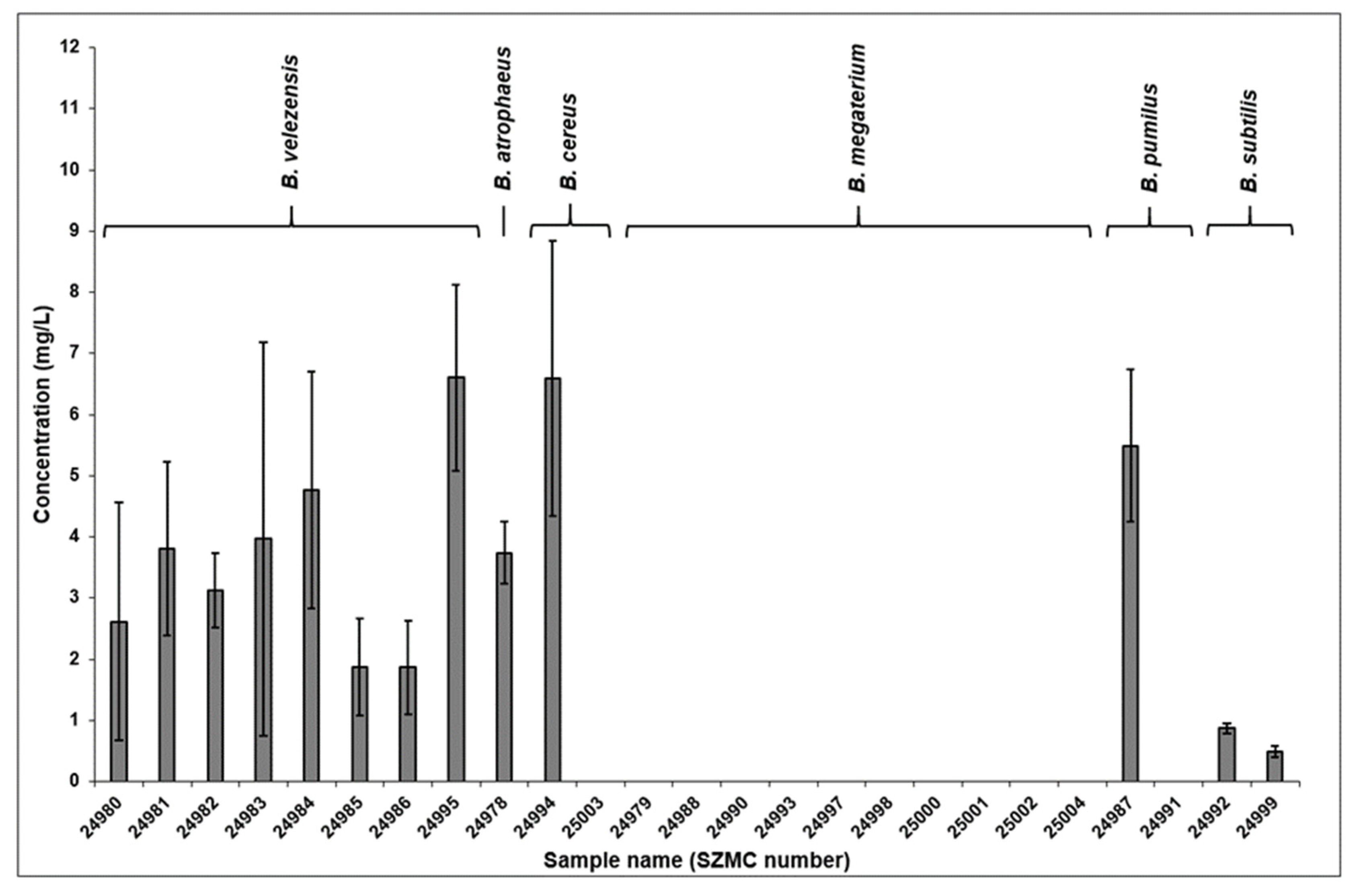
2.2. Quantitative Results of the Total Surfactin Production

The lipopeptide concentration in the ferment broth of each sample was carried out after calibration with surfactin standard by calculating the integrated peak areas of the total ion chromatograms (TIC) measured in SIM mode set to the m/z values of the sodiated precursor ions (Figure 1).
The comparison of the results of the quantitative measurements is shown in Figure 2. Observing the diagram, it can be seen that none of the B. megaterium strains examined in this study produced surfactin at all. The extracted ferment broths of the five B. velezensis samples contained surfactin in the 2–5 mg/L concentration range, except for strain SZMC 24995 which bore the highest surfactin content among the examined samples by almost reaching 7 mg/L. The B. atrophaeus strain SZMC 24978 possessed biosurfactant production properties similar to most of the aforementioned strains. As a peculiar result in the case of B. cereus, the SZMC 24994 strain produced surfactins in one of the highest quantities among the examined samples, although, the ferment broth of the SZMC 25003 strain did not contain the observed lipopeptides. Results were similar in the case of the B. pumilus samples, in which strain SZMC 24991 did not produce the examined molecules, while strain SZMC 24987 possessed surfactins in the third highest concentration in its ferment broth. The quantity of biosurfactants in both B. subtilis strains were detected below 1 mg/L.

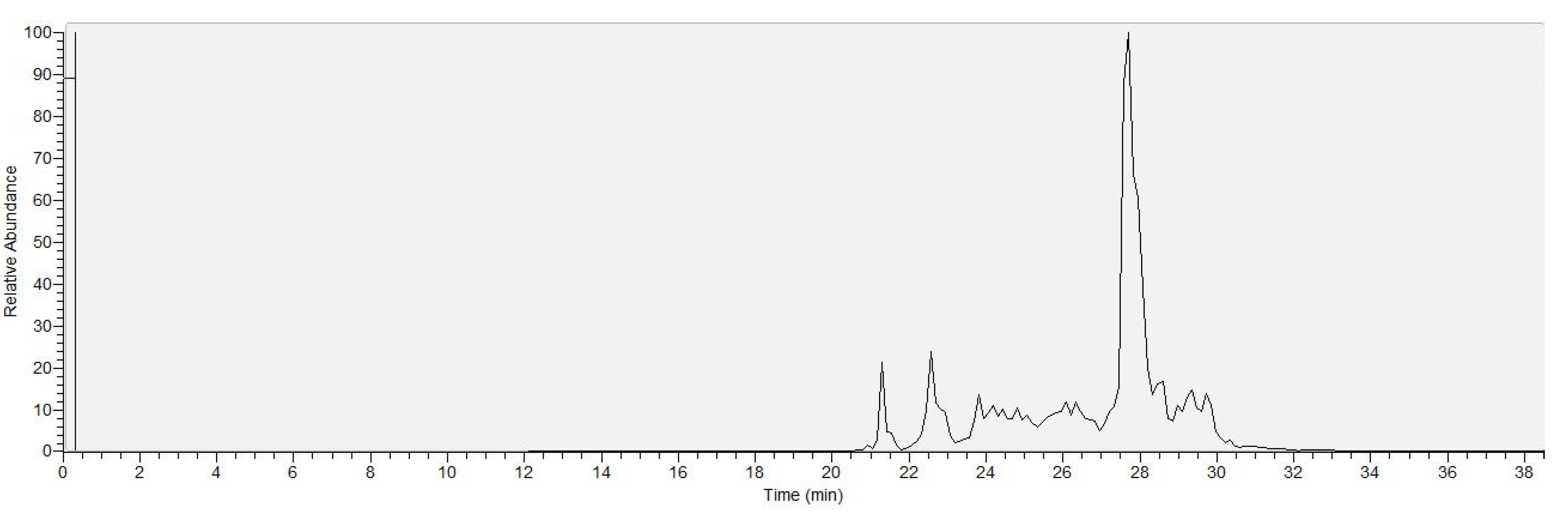
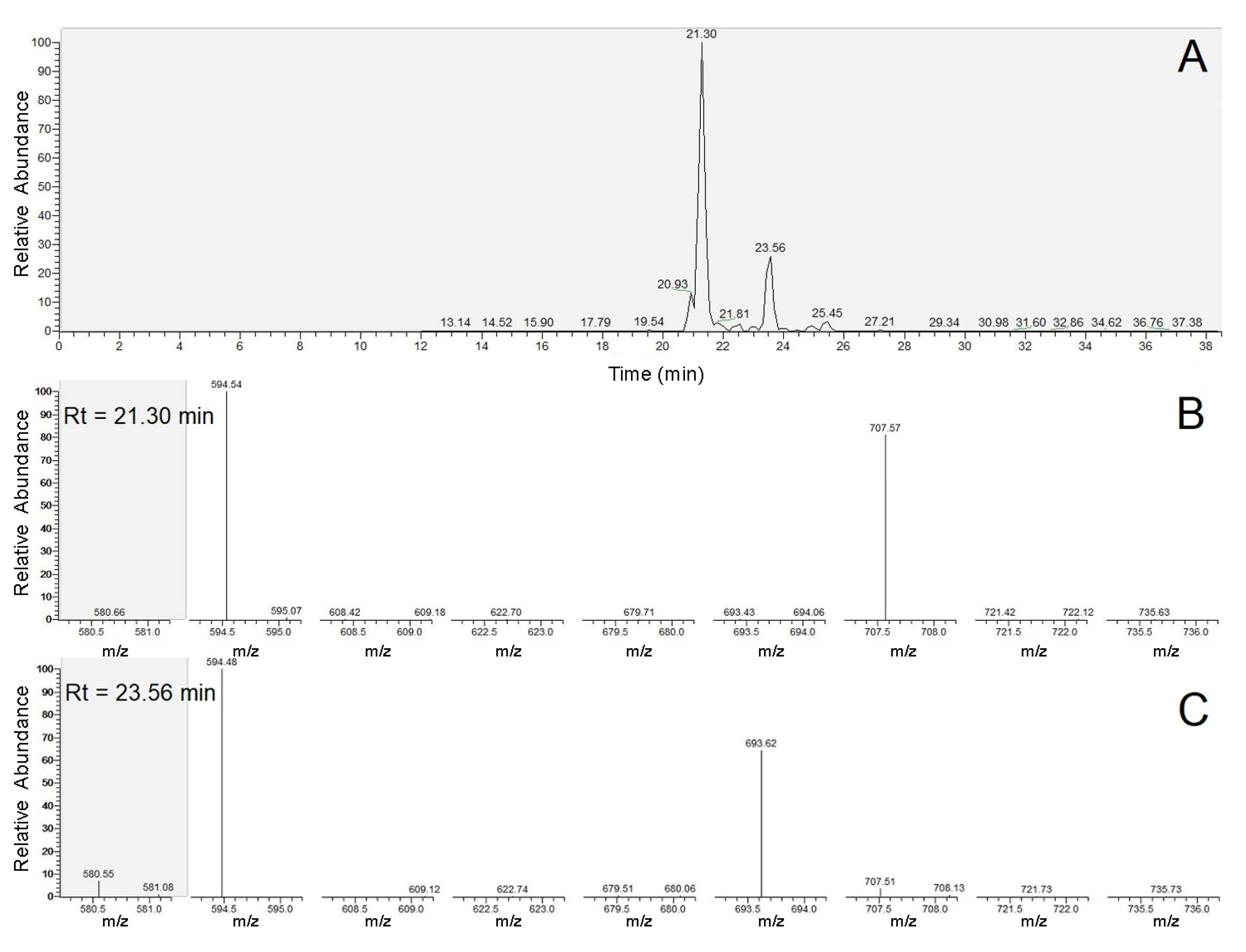
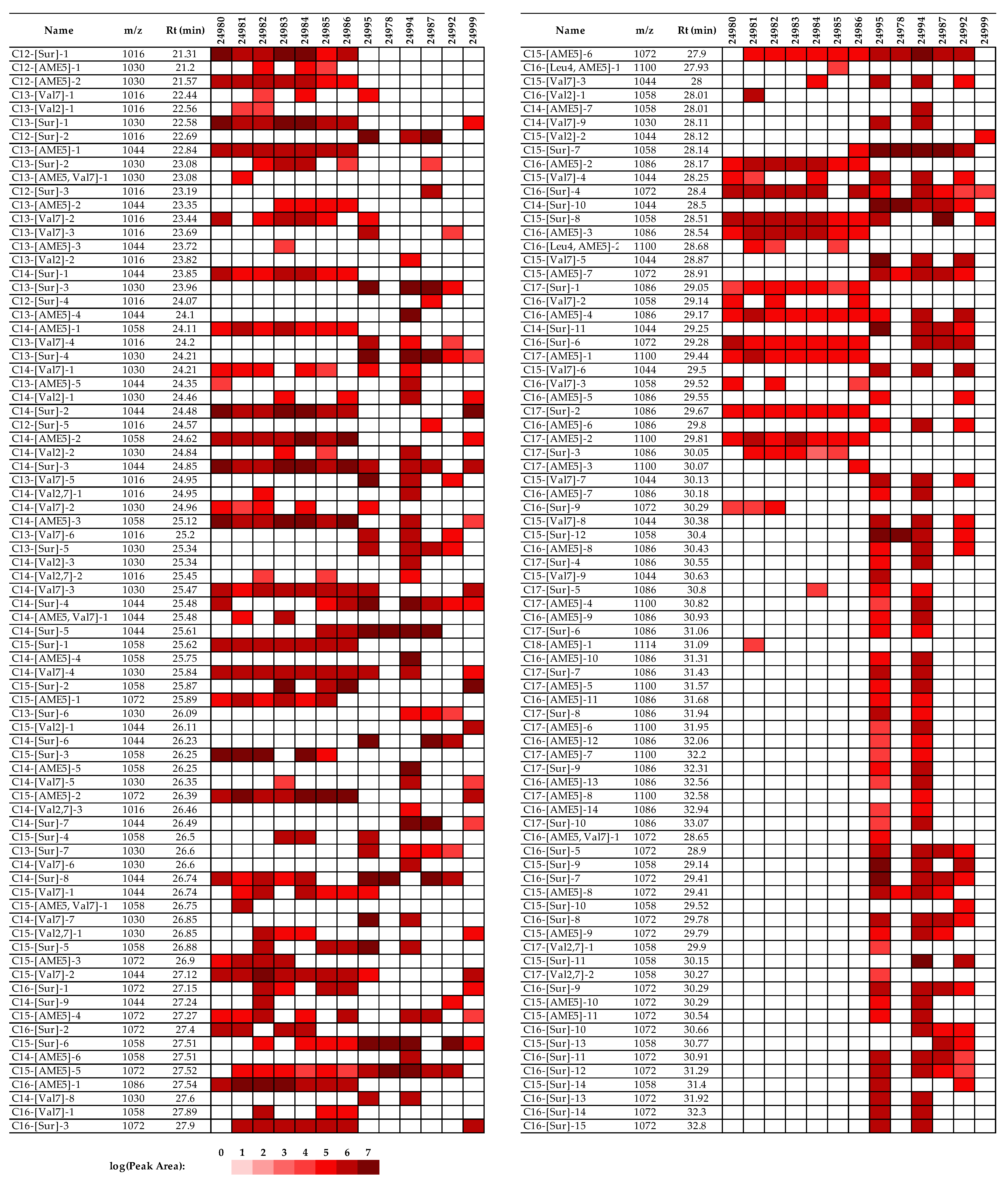
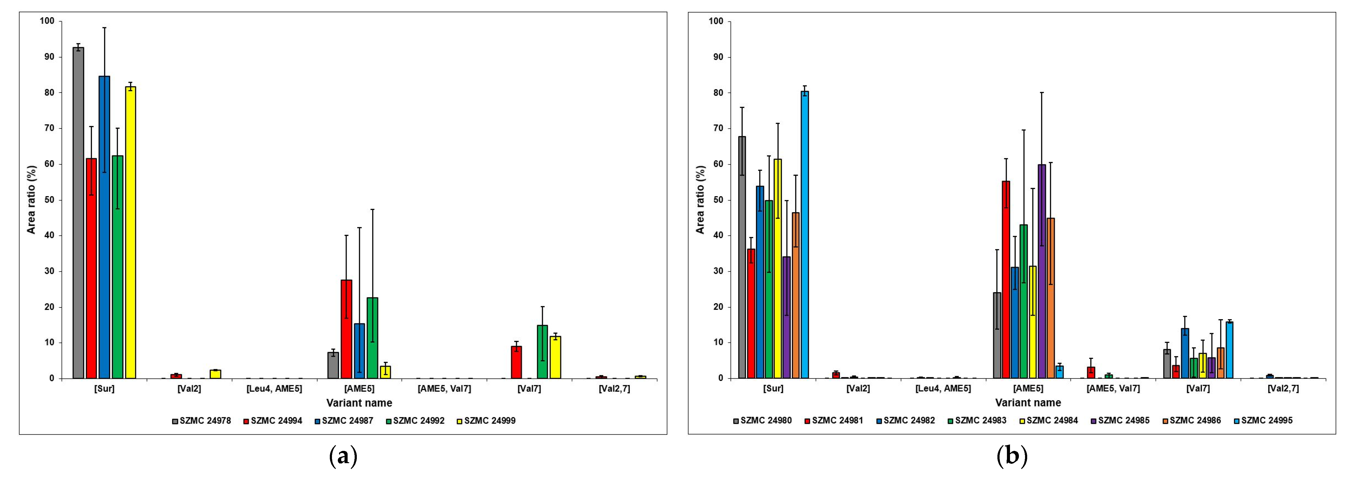
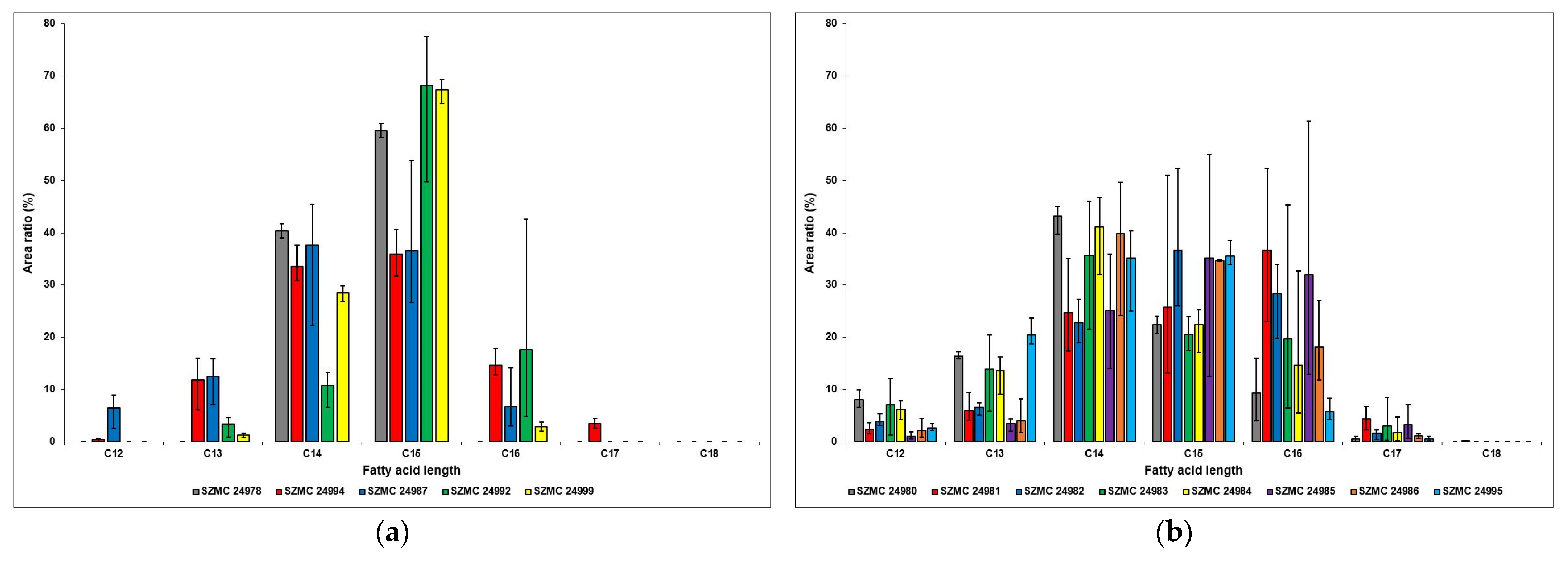
2.3. Identification of the Detected Surfactins

Identification of the different surfactin molecules was carried out based on the mass differences of the precursor ions and the y6 + H2O internal fragment ions as well as the m/z values of the overlapping peaks of y6 + H2O and y6b6 + H2O fragment ions. An example for the evaluation process is shown in Figure 3. Observing the MS/MS spectra of the two peaks, the first peak at the retention time of 21.30 min definitely belongs to a [Sur] variant, while the second one at Rt = 23.56 min marks the presence of a [Val7] surfactin molecule due to the corresponding m/z values of y6 + H2O and y6b6 + H2O fragment ions. Subtracting the m/z values of the y6 + H2O fragment ions from those of the sodiated precursor ions, their mass differences indicate the two homologues to be C12 and C13, respectively; thus, the two molecules can be identified as C12-[Sur] and C13-[Val7] simply from these acquired data.
By examining all of the peaks on the EICs in the case of every lipopeptide-producing sample, the detected surfactin variants and homologues were identified and listed in Figure 4 and in the Supplementary Material (Tables S1 and S2). There were 29 surfactin molecules with different structures, with most of them having multiple peaks with distinct retention times, suggesting changes in the branching of the fatty acid chains. As a result of the appearance of these specific peaks in vast numbers, 158 instances of them with particular retention times were detected altogether in all of the examined samples combined. Most of them occur in the B. cereus strain SZMC 24994 with 90 peaks of different surfactin molecules, while in evaluating the MS/MS spectra of the B. atrophaeus strain SZMC 24978, only 10 peaks were detected. While this number varies in a wide range in the other samples (10, 90, 36, 45, and 24 instances in SZMC 24978, 24994, 24987, 24992, and 24999, respectively), the strains of B. velezensis are consistent in that regard, resulting in a range from 41–55 instances with relatively high similarities with the only, rather peculiar, exception being the SZMC 24995 strain (Figure 4).

2.4. Comparison of the Surfactin Production Profiles

After the successful identification of the different surfactin variants and homologues, their relative quantitative relations were examined by combining their respective integrated peak areas and comparing their area ratio percentages in diagrams (Figure 5 and Figure 6). Regarding the different variants, one main similarity can be observed between the strains of B. atrophaeus, B. cereus, B. pumilus, and B. subtilis, namely that the relative amount of [Sur] molecules is the most dominant, reaching over 60% in all cases and extending over 90% in strain SZMC 24978 (Figure 5). The variant with the second most dominant area ratio is [AME5], except for strain SZMC 24999, as it possesses [Val7] surfactins in higher amounts. A rather peculiar result is that the ferment broth of the B. atrophaeus strain SZMC 24978 contained only three molecules with different structures—C14-[Sur], C15-[Sur], and C15-[AME5]—although their total concentration has proven to be above average compared with the other examined Bacillus strains (Figure 2). The area ratios of the variants [Val2] and [Val2,7] are close to negligible, and the [Leu4, AME5] and [AME5, Val7] surfactins were not even detected in the samples of these four Bacillus species.
The surfactin variants with the highest relative amounts are also the [Sur], [AME5], and [Val7] isoforms in the case of the B. velezensis samples; however, the ratio of the first two are much closer together, except for strain SZMC 24995; the [AME5] variant even surpasses the [Sur] molecules in that regard in strains SZMC 24981 and SZMC 24985 (Figure 5). After those, the [Val7] isoforms are produced in the highest relative amounts, although only exceeding 10% in the SZMC 24982 and SZMC 24995 strains. The area ratios of the other surfactin variants are below 4% in all samples, not even reaching the 1% mark in most cases.
In observing the relative amounts of the different surfactin homologues in the strains of B. atrophaeus, B. cereus, B. pumilus, and B. subtilis, the diagram shows a common characteristic among these samples, which is the dominance of C14 and C15 molecules. The only exception is the SZMC 24992 strain, in which the C16 homologues have the second largest area ratio after the C15 surfactins (Figure 6). As a result of its unique production properties described above, no other homologues were detected in the ferment broth of the B. atrophaeus strain SZMC 24978. In the case of the B. cereus strain, all of the homologues were detected, except for the C18 surfactins, while the samples of B. subtilis are lacking the presence of C12, C17, and C18 molecules. However, both the SZMC 24992 and the SZMC 24999 strains produced C15 homologues in relative amounts of approximately 70%, which is the highest compared with all other samples.
Results of the B. velezensis strains show that the area ratios of C16 molecules are more proportionate to their C14 and C15 counterparts; in the SZMC 24981 strain, these were observed in the highest relative amounts (Figure 6). This is also the only sample in which C18 homologues were detected, although only in a mere 0.05% ratio, while all the other fatty acid chain lengths between 12 and 17 carbon atoms were present in all strains.

2.5. The Biocontrol Properties of the Examined Bacillus Isolates

Due to bacterial growth inhibitions, the clearance zones showed potential biocontrol activities of Bacillus isolates. Altogether, 16 of the 25 isolates exhibited such properties on pathogenic bacteria, with inhibition zones ranging from 1.0 to 16.67 mm. The B. velezensis strains were considered to have substantial potential for biocontrol, antagonizing against all test pathogens (Table 3). In a peculiar way, E. amylovora and X. campestris were significantly antagonized by a wide range of isolates. Accordingly, 36.0% of isolates exhibited notable activities against E. amylovora, with inhibition zone diameters of ≥5mm; moreover, 28.0% of those were ≥10mm. Subsequently, 28.0% of isolates exhibited activities against X. campestris, with inhibition zone diameters of ≥5mm and 4.0% of those ≥10mm. In addition, isolates slightly antagonized A. tumefaciens, P. syringae, and E. carotovora with lower activities. In all cases, the inhibitions were statistically significant based on ANOVA.
For the examination of biocontrol properties of the Bacillus isolates on pathogenic fungi, 14 of the 25 testing isolates were potent in the control of various phytopathogenic fungi, with inhibition rates ranging from 34.4% to 83.8%. The isolates of B. subtilis and B. velezensis showed significant activities in fungal growth inhibition. To sum up, 40%, 52%, 40%, 40%, and 36% of isolates inhibited more than 50% growth of F. graminearum, B. cinerea, F. solani, F. culmorum, and C. gloeosporioides, respectively. Furthermore, 44%, 12% and 4% of isolates effectively inhibited more than 70% growth of B. cinerea, F. culmorum, and C. gloeosporioides, respectively. In all cases, the inhibitions were statistically significant based on ANOVA (Table 4).

3. Discussion

The taxonomy identification relied on the Sherlock CAS method which revealed Bacillus isolates as B. atrophaeus, B. cereus, B. megaterium, B. pumilus, B. subtilis, and B. velezensis. Accordingly, FA composition has been diverse, drawing a distinction between Bacillus species as taxonomic biomarkers. As a rather peculiar result, FA compositions in B. cereus, B. megaterium, and B. pumilus have been divided into two distinguishable groups, named GC subgroup A and B. These species possess a higher content of branched-odd FAs, including 13:0 iso, 15:0 iso, 15:0 anteiso, 17:0 iso, and 17:0 anteiso, as common features of Bacillus’ taxonomy [26].
A combined SIM/SRM mass spectrometric method has also been developed, capable of performing quantitative measurements of the total surfactin concentration on the extracted ferment broths of the different strains and simultaneously identifying the different surfactin molecules based on the m/z values of their sodiated precursor ions and the first two internal fragment ions, whereby monitoring 80 mass transitions altogether. Out of the aforementioned 25 strains, 13 produced surfactins in average concentrations of 0.5–6.6 g/L while 12 samples, including the ferment broths of all B. megaterium strains, contained no surfactins whatsoever. As a result of our qualitative measurements, 29 surfactin molecules in total were identified, with 158 detected instances with different retention times, suggesting numerous variations of branching within their fatty acid chains apart from alterations in their chain lengths and amino acid sequences. In comparing the relative amounts of the different surfactin variants and homologues, the resulting data showed that the [Sur], [AME5], and [Val7] isoforms were the most dominant in all cases, while regarding the occurrence of surfactins with different fatty acid chain lengths, the C14–C16 molecules had the largest area ratios. Results supported the conclusions of our earlier studies stating that the appearance of a previously rarely encountered group of surfactins with methyl esterified aspartic acid in their fifth amino acid position could be encountered in considerable numbers, and the fatty acid chain lengths could vary between 12 and 18 carbon atoms.
The inhibitory results demonstrated that the Bacillus isolates have a broad range in the biocontrol potential against various phytopathogens. The use of bactericidal and fungicidal microbes as natural mechanisms may prevent the production losses in agriculture caused by phytopathogens and limit the effects of chemical pesticides and antibiotics on the environment and ecosystem [27]. Moreover, many rhizosphere-associated Bacillus exhibiting significant inhibitory activity towards phytopathogens have been reported in agreement that Bacillus species are ideal biocontrol candidates [27,28,29,30].
The present results exhibited the significantly effective biocontrol activity of Bacillus species against E. amylovora, X. campestris, B. cinerea, and F. culmorum. In addition, the strains displayed moderate effects on P. syringae, E. carotovora, A. tumefaciens, F. graminearum, F. solani, and C. gloeosporioides. Accordingly, it was determined that the strains belonging to the B. subtilis species complex, namely B. atrophaeus, B. subtilis, and B. velezensis, could inhibit test phytopathogens as effective biocontrol agents. Together with gene clusters encoding non-ribosomal synthesis of lipopeptides and polyketides, the Bacillus group has been reflected as a producer of diverse bioactive secondary metabolites [31]. The Bacillus species were described in 2005 [32] regarding plant–pathogen-inhibiting and plant–growth-promoting potentials [33,34]. The present investigation determined great potential, especially in the B. velezensis species which contains many gene clusters toward non-ribosomal synthesis of versatile metabolites [35].
Generally, it can be stated that the best surfactin producers are the members of the B. velezensis species because all studied members of this species produced these biosurfactants, which released in remarkably high variabilities (Figure 4). The number of the detected congeners varied in the range of 41–84 and 24–45 for B. velezensis and B. subtilis, respectively, while this number was 10, 90, and 30 for B. atrophaeus, B. cereus, and B. pumilus, respectively (Figure 4).
In the fungicidal assays, the B. velezensis and B. subtilis isolates showed the highest inhibition rates against fungi, which typically ranged from 30–50%. Currently, there is no possibility to provide direct relationships between the surfactin productions and the biological activities based on the gathered result; however, in examining the surfactin profiles of these isolates to find common features, it can be concluded that strains of both species produced C14-[Sur] and C15-[Sur] in high amounts. Furthermore, the B. velezensis isolates showed the most effective antibacterial activities against the phytopathogen bacteria, producing C14-[Sur], C14-[Val7], C15-[Val7], C15-[Sur], and C16-[AME5] as shared features of their surfactin production.
Based on our knowledge, our results have provided the most detailed surfactin characterisation of the Bacillus isolates, which can be extended in the future with the inclusion of novel strains belonging to other species or isolated from different locations/sources in order to achieve deeper insight into the surfactin production features of Bacillus strains.

4. Materials and Methods

4.1. Strains Maintenance

The examined Bacillus strains were isolated from different vegetable rhizospheres (Table 1). The strains were derived from the Szeged Microbiology Collection (SZMC; www.szmc.hu (accessed on 16 January 2023)), maintained on nutrient agar (5 g/L peptone, 3 g/L yeast extract, 5 g/L NaCl, 15 g/L agar) slants, and stored at 4 °C.

4.2. Nomenclature of Surfactin Variants

Surfactin variants were designated according to Grangemard et al. [36] and Bóka et al. [16]. Briefly, the first discovered surfactin sequence (Glu-Leu-Leu-Val-Asp-Leu-Leu) was denoted as [Sur], and any changes in the peptide sequence were indicated with the abbreviation and position of the altered amino acid, for example, [Val2], [Val7], and [Val2,7]. The esterified form of aspartic acid and glutamic acid at the side chain carboxyl group were abbreviated as AME and GME, respectively. As the applied mass spectrometric technique could not distinguish between the Leu and Ile isobaric residues, this sequence element was marked as Lxx in this paper. The amino acid residues present in the sequences of the surfactins are designated in general by AAn, the superscript ‘n’ indicating the position number of each amino acid from the N-terminal end of the peptide chain. Furthermore, the fragment ions on the MS2 spectra were designated according to the terminology published by Roepstorff and Fohlman [37], as well as Biemann [38], while the internal fragments of sodiated fragment ions were designated by the ynbm nomenclature [16,39].

4.3. Culture Conditions and Sample Preparation for surfactin Analysis

For the surfactin production, a liquid ferment broth was applied according to Besson et al. [40] containing 10 g/L glucose, 5 g/L glutamic acid, 1 g/L KH2PO4, 1 g/L K2HPO4, 1 g/L KCl, 500 mg/L MgSO4 × 7 H2O, 5 mg/L FeSO4 × 7 H2O, and 160 µg/L CuSO4 × 5 H2O. Bacteria (5 × 10⁷ cells) were inoculated into a 20 mL medium in 100 mL Erlenmeyer flasks followed by incubation on a rotary shaker at 120 rpm for five days at 25 °C.
The bacterial cells were separated from the ferment broths via centrifugation at 8000 rpm for 15 min at 4 °C. The pH of the supernatant was decreased to 2 with HCl, and the lipopeptides were precipitated overnight at 4 °C. The pellets were collected by centrifugation (8000 rpm, 15 min, 4 °C) and resolved in 1 mL methanol [23].
All chemicals and reagents mentioned above were AR purity and were purchased from Molar Chemicals Ltd. (Budapest, Hungary).

4.4. Identification of Bacillus Isolates by Fatty Acid Methyl Ester (FAME) Analysis

The MIDI Sherlock® Microbial Identification System (MIS, Microbial ID Inc., Newark, NJ, USA) was applied for the identification. The composition of whole-cell fatty acids was determined by the Sherlock CAS Software operating on a gas chromatography platform, Shimadzu’s GC-2010/2030, equipped with an HP-Ultra 2, 25 m × 0.2 mm × 0.33 µm thickness fused silica capillary column (Agilent, Santa Clara, CA, USA) as a stationary phase [25,41]. Briefly, the sample processing was prepared following The SherlockTM Operating CAS Manual. The bacteria were cultured on Trypticase Soy Broth Agar (Becton, Dickinson and Company, Sparks, NV, USA) at 28 °C for 24 ± 2 h. Then, cells were harvested, saponificated, methylated, and extracted producing total FAMEs. The whole-cell FAME profiles with database were analysed by the method RTSBA6 and the library RTSBA6 and RTSBA7. In the method, injector and detector temperatures were 250 °C and 300 °C, respectively. Carrier gas was hydrogen at a flow rate of 1.48 mL/min, while the detector gases were nitrogen (make up), oxygen, and hydrogen with the flows of 30, 30, and 350 mL/min, respectively. Samples were introduced in an injection volume of 2 µL in split mode with a 40:1 split ratio. The oven program started at 168.1 °C, which ramped up to 291 °C at 28 °C per min and then up to 300 °C at 60 °C per min, holding at this temperature for 1.50 min. The total column oven program time was 6.04 min. The 1300-C rapid calibration standard mix (Microbial ID Inc., Newark, DE, USA) was used for RT calibration and system suitability purposes as well as for the fine tuning of the pressure and temperature parameters at the system setup.
The B. subtilis ATCC 6633 and pure hexane were considered as the positive and negative control, respectively.

4.5. Analytical Parameters

The HPLC-HESI-MS/MS examinations of the different surfactin molecules were carried out based on the work of Büchner et al. [42]. The applied instrument was a Nexera XR HPLC system containing a DGU-20A5R degasser, an LC-20ADXR pump, a SIL-20AXR autosampler and a CTO-10ASVP column oven (Shimadzu Corporation, Kyoto, Japan), coupled with a TSQ Quantum Access triple quadrupole mass spectrometer (Thermo Scientific, Waltham, MA, USA).
The gradient solvent delivery system consisted of two solvents: A was H2O, and B was a mixture of acetonitrile/methanol (1:1, v/v %). Both solvents were supplemented with 0.1% acetic acid. The applied reverse phase gradient elution time program was the following: 5% eluent B for 2 min, increased to 80% in the following 2 min, and then gradually raised to 95% for 24 min. This rate was held for 9 min and then dropped to 5% in 0.5 min, followed by a 5 min long equilibration stage, ending the run of 38.5 min in total. The flow rates were 0.2 mL/min and the column heater temperature was 30 °C. The applied column was a Gemini-NX (3µ, C18, 150 × 2 mm). The injection volume was 10 µL.
Both the quantitative and qualitative measurements were carried out using a combined method in selected ion monitoring (SIM) and single reaction monitoring (SRM) modes running in parallel, with a heated electrospray ionization (HESI) ion source and in positive polarity. The spray voltage was +4000 V; the vaporiser temperature was 285 °C; the capillary temperature was 350 °C; the sheath gas pressure was 10 psi; and the auxiliary gas pressure was 15 psi. The SRM mode analyses for the identification of the different surfactin variants were performed with a collision energy of 60 V and a collision gas pressure of 1 mTorr. The m/z values of sodiated surfactin molecules were set as parent ion masses (m/z 1016.7, 1030.7, 1044.7, 1058.7, 1072.7, 1086.7, 1100.7, 1114.7) and for every parent ion, the first two internal fragment ions of every natural surfactin variant were set as a daughter ion (m/z 580.7, 594.7, 608.7, 622.7, 679.7, 693.7, 707.7, 721.7, 735.7). The SIM mode measurements were also used to examine the parent ion m/z values listed above. With these two modes, 80 different mass transitions ran in parallel for a total scan time of 0.11 sec/scan. The instrument control and the data processing were performed using the TraceFinder General Quan 4.1 software (Thermo Fisher Scientific, Waltham, MA, USA) while the Xcalibur software v. 4.0 (Thermo Fisher Scientific, Waltham, MA, USA) was applied for the spectral examinations.
The standard used for the quantification was surfactin (S3523) from Sigma-Aldrich (Budapest, Hungary). All chemicals used as eluents and for sample preparation are HPLC-MS purity and were purchased from VWR International Ltd. (Debrecen, Hungary).

4.6. Inhibition Assays on Phytopathogen Microorganisms

The tests were determined by an agar diffusion technique [30] using the yeast extract–glucose medium (YEG) (glucose 0.2%, yeast extract 0.2%, bacto-agar 2%; purchased from VWR International Ltd. (Debrecen, Hungary)).
The following bacteria were prepared in YEG broth overnight: Pseudomonas syringae SZMC 16160, Erwinia amylovora SZMC 21402, Erwinia carotovora SZMC 6190, Xanthomonas campestris SZMC 6182, Agrobacteria tumefaciens SZMC 14554 and Bacillus isolates. A quantity of 200 µL of phytopathogenic suspension (~5 × 10⁷ CFU/mL) was spread on YEG agar, and then 6 mm diameter paper discs with 5 µL of Bacillus suspension (~2.5 × 10⁷ CFU/mL) were put in place. The antibacterial effects were evaluated by measuring inhibition zones after 1–2 days. Three replicates were conducted for each experiment.
The following fungi were prepared on PDA plates: Fusarium graminearum SZMC 11030, Botrytis cinerea SZMC 21047, Fusarium solani SZMC 16084, Fusarium culmorum SZMC 11039, and Colletotrichum gloeosporioides SZMC 16087. In addition, Bacillus isolates were prepared in YEG broth overnight. Subsequently, 6 mm in diameter paper discs with 5 µL of Bacillus suspension (~2.5 × 10⁷ CFU/mL) and 6 mm in diameter mycelia from cultured fungi were inoculated on YEG agar by using a direct dual culture method with a 3 cm spacing distance. The control test was performed without Bacillus. Three replicates were conducted for each experiment. The fungicidal activity was determined by the values of the inhibition rate (%) after the 7-day incubation.
The rate of inhibition was measured using the formula: The inhibition rate (%) = ((diameter of control − diameter of treatment)/diameter of control) × 100%.

4.7. Statistical Analysis

Data were displayed as Mean ± SD. Analysis of variance (ANOVA) was run using the R package. Values with p < 0.05 were considered statistically significant.

5. Conclusions

In this study, 25 Bacillus strains in total isolated from vegetable rhizospheres were identified by their FAME profiles and were tested against phytopathogen bacteria and fungi. Their surfactin production profiles were also examined in detail by a fast and easily evaluable HPLC-HESI-MS/MS method in SIM/SRM mode, differentiating 158 surfactin variants from the ferment broths.
Based on our knowledge, our results report the most comprehensive surfactin profiling, which could promote the further targeted selection of strains and cultivation conditions to produce certain surfactin molecules responsible mainly for biological effects.

Supplementary Materials

The following supporting information can be downloaded at: https://www.mdpi.com/article/10.3390/molecules28031172/s1, Table S1: Relative amounts of the detected surfactin molecules of B. atrophaeus (SZMC 24978), B. cereus (SZMC 24994), B. pumilus (SZMC 24987) and B. subtilis (SZMC 24992, SZMC 24999); Table S2: Relative amounts of the detected surfactin molecules of B. velezensis strains.

Author Contributions

Conceptualization, A.S. and C.V.; methodology, A.B. and T.H.; software, A.B. and T.H.; validation, C.V.; formal analysis, A.B., A.S. and T.H.; investigation, A.B., A.K., H.A., M.V. (Mónika Vörös), O.K. and T.H.; resources, A.S., C.V. and L.K.; data curation, M.V. (Mónika Varga); writing—original draft preparation, A.B. and T.H.; writing—review and editing, A.S., C.V. and L.K.; visualization, A.B. and A.S.; supervision, A.S.; project administration, A.S. and M.V. (Mónika Varga); funding acquisition, A.S. All authors have read and agreed to the published version of the manuscript.

Funding

This work was supported by the grant OTKA K-128659 from the Hungarian Scientific Research Fund.

Institutional Review Board Statement

Not applicable.

Informed Consent Statement

Not applicable.

Data Availability Statement

Not applicable.

Acknowledgments

We thank Craig Kunitsky, Gary Jackoway, Mike Alexander and the Supporting MIDI Team (MIDI Inc. (Newark, Delaware)) and the whole company for technical help and continuous support to adopt the Sherlock MIDI system at our site.

Conflicts of Interest

The authors declare no conflict of interest.

Sample Availability

Not applicable.

References

  1. Arima, K.; Kakinuma, A.; Tamura, G. Surfactin, a crystalline peptidelipid surfactant produced by Bacillus subtilis: Isolation, characterization and its inhibition of fibrin clot formation. Biochem. Biophys. Res. Commun. 1968, 31, 488–494. [Google Scholar] [CrossRef] [PubMed]
  2. Chen, W.-C.; Juang, R.-S.; Wei, Y.-H. Applications of a lipopeptide biosurfactant, surfactin, produced by microorganisms. Biochem. Eng. J. 2015, 103, 158–169. [Google Scholar] [CrossRef]
  3. Nieminen, T.; Rintaluoma, N.; Andersson, M.; Taimisto, A.-M.; Ali-Vehmas, T.; Seppälä, A.; Priha, O.; Salkinoja-Salonen, M. Toxinogenic Bacillus pumilus and Bacillus licheniformis from mastitic milk. Vet. Microbiol. 2007, 124, 329–339. [Google Scholar] [CrossRef] [PubMed]
  4. From, C.; Hormazabal, V.; Hardy, S.P.; Granum, P.E. Cytotoxicity in Bacillus mojavensis is abolished following loss of surfactin synthesis: Implications for assessment of toxicity and food poisoning potential. Int. J. Food Microbiol. 2007, 117, 43–49. [Google Scholar] [CrossRef] [PubMed]
  5. Pecci, Y.; Rivardo, F.; Martinotti, M.G.; Allegrone, G. LC/ESI-MS/MS characterisation of lipopeptide biosurfactants produced by the Bacillus licheniformis v9t14 strain. J. Mass Spectrom. 2010, 4, 772–778. [Google Scholar] [CrossRef] [PubMed]
  6. Alvarez, F.; Castro, M.; Príncipe, A.; Borioli, G.; Fischer, S.; Mori, G.; Jofré, E. The plant-associated Bacillus amyloliquefaciens strains MEP218 and ARP23 capable of producing the cyclic lipopeptides iturin or surfactin and fengycin are effective in biocontrol of sclerotinia stem rot disease. J. Appl. Microbiol. 2011, 112, 159–174. [Google Scholar] [CrossRef]
  7. Bonmatin, J.-M.; Laprevote, O.; Peypoux, F. Diversity among microbial cyclic lipopeptides: Iturins and surfactins. Activity-structure relationships to design new bioactive agents. Comb. Chem. High Throughput Screen. 2003, 6, 541–556. [Google Scholar] [CrossRef] [PubMed]
  8. Vollenbroich, D.; Pauli, G.; Ozel, M.; Vater, J. Antimycoplasma properties and application in cell culture of surfactin, a lipopeptide antibiotic from Bacillus subtilis. Appl. Environ. Microbiol. 1997, 63, 44–49. [Google Scholar] [CrossRef] [Green Version]
  9. Duarte, C.; Gudiña, E.J.; Lima, C.F.; Rodrigues, L.R. Effects of biosurfactants on the viability and proliferation of human breast cancer cells. AMB Express 2014, 4, 40. [Google Scholar] [CrossRef] [Green Version]
  10. Zhang, Y.; Liu, C.; Dong, B.; Ma, X.; Hou, L.; Cao, X.; Wang, C. Anti-inflammatory activity and mechanism of surfactin in lipopolysaccharide-activated macrophages. Inflammation 2015, 38, 756–764. [Google Scholar] [CrossRef]
  11. Wang, X.; Hu, W.; Zhu, L.; Yang, Q. Bacillus subtilis and surfactin inhibit the transmissible gastroenteritis virus from entering the intestinal epithelial cells. Biosci. Rep. 2017, 37, BSR20170082. [Google Scholar] [CrossRef] [PubMed] [Green Version]
  12. Mulligan, C.N. Environmental applications for biosurfactants. Environ. Pollut. 2005, 133, 183–198. [Google Scholar] [CrossRef] [PubMed]
  13. Albino, J.D.; Nambi, I.M. Effect of biosurfactants on the aqueous solubility of PCE and TCE. J. Environ. Sci. Health Part A 2009, 44, 1565–1573. [Google Scholar] [CrossRef] [PubMed]
  14. Zou, A.; Liu, J.; Garamus, V.M.; Zheng, K.; Willumeit, R.; Mu, B. Interaction between the natural lipopeptide [Glu1, Asp5] surfactin-C15 and hemoglobin in aqueous solution. Biomacromolecules 2010, 11, 593–599. [Google Scholar] [CrossRef]
  15. Hafeez, F.Y.; Naureen, Z.; Sarwar, A. Surfactin: An emerging biocontrol tool for agriculture sustainability. In Plant Growth Promoting Rhizobacteria for Agricultural Sustainability, 1st ed.; Kumar, A., Meena, V., Eds.; Springer: Singapore, 2019; pp. 203–213. [Google Scholar]
  16. Bóka, B.; Manczinger, L.; Kecskeméti, A.; Chandrasekaran, M.; Kadaikunnan, S.; Alharbi, N.S.; Vágvölgyi, C.; Szekeres, A. Ion trap mass spectrometry of surfactins produced by Bacillus subtilis SZMC 6179J reveals novel fagmentation features of cyclic lipopeptides. Rapid Commun. Mass Spectrom. 2016, 30, 1581–1590. [Google Scholar] [CrossRef] [PubMed]
  17. Tang, J.-S.; Gao, H.; Hong, K.; Yu, J.; Jiang, M.-M.; Lin, H.-P.; Ye, W.-C.; Yao, X.-S. Complete assignments of 1H and 13C NMR spectral data of nine surfactin isomers. Magn. Reson. Chem. 2007, 45, 792–796. [Google Scholar] [CrossRef]
  18. Tang, J.-S.; Zhao, F.; Gao, H.; Dai, Y.; Yao, Z.-H.; Hong, K.; Li, J.; Ye, W.-C.; Yao, X.-S. Characterization and online detection of surfactin isomers based on HPLC-MSn analyses and their inhibitory effects on the overproduction of nitric oxide and the release of TNF-α and IL-6 in LPS-induced macrophages. Mar. Drugs 2010, 8, 2605–2618. [Google Scholar] [CrossRef] [Green Version]
  19. Moro, G.V.; Almeida, R.T.R.; Napp, A.P.; Porto, C.; Pilau, E.J.; Lüdtke, D.S.; Moro, A.V.; Vainstein, M.H. Identification and ultra-high-performance liquid chromatography coupled with high-resolution mass spectrometry characterization of biosurfactants, including a new surfactin, isolated from oil-contaminated environments. Microb. Biotechnol. 2018, 11, 759–769. [Google Scholar] [CrossRef]
  20. Kecskeméti, A.; Bartal, A.; Bóka, B.; Kredics, L.; Manczinger, L.; Shine, K.; Alharby, N.S.; Khaled, J.M.; Varga, M.; Vágvölgyi, C.; et al. High-frequency occurrence of surfactin monomethyl isoforms in the ferment broth of a Bacillus subtilis strain revealed by ion trap mass spectrometry. Molecules 2018, 23, 2224. [Google Scholar] [CrossRef] [Green Version]
  21. Slivinski, C.T.; Mallmann, E.; de Araújo, J.M.; Mitchell, D.A.; Krieger, N. Production of surfactin by Bacillus pumilus UFPEDA 448 in solid-state fermentation using a medium based on okara with sugarcane bagasse as a bulking agent. Process Biochem. 2012, 47, 1848–1855. [Google Scholar] [CrossRef]
  22. Joshi, S.J.; Al-Wahaibi, Y.M.; Al-Bahry, S.N.; Elshafie, A.E.; Al-Bemani, A.S.; Al-Mandhari, M.S. Production, characterization, and application of Bacillus licheniformis W16 biosurfactant in enhancing oil recovery. Front. Microbiol. 2016, 7, 1853. [Google Scholar] [CrossRef] [PubMed]
  23. Bartal, A.; Vigneshwari, A.; Bóka, B.; Vörös, M.; Takács, I.; Kredics, L.; Manczinger, L.; Varga, M.; Vágvölgyi, C.; Szekeres, A. Effects of different cultivation parameters on the production of surfactin variants by a Bacillus subtilis strain. Molecules 2018, 23, 2675. [Google Scholar] [CrossRef] [PubMed] [Green Version]
  24. Czinkóczky, R.; Németh, Á. The effect of pH on biosurfactant production by Bacillus subtilis DSM10. Hung. J. Ind. Chem. 2020, 48, 37–43. [Google Scholar] [CrossRef]
  25. Huynh, T.; Vörös, M.; Kedves, O.; Turbat, A.; Sipos, G.; Leitgeb, B.; Kredics, L.; Vágvölgyi, C.; Szekeres, A. Discrimination between the two closely related species of the operational group B. amyloliquefaciens based on whole-cell fatty acid profiling. Microorganisms 2022, 10, 418. [Google Scholar] [CrossRef]
  26. Diomandé, S.E.; Nguyen-The, C.; Guinebretière, M.H.; Broussolle, V.; Brillard, J. Role of fatty acids in Bacillus environmental adaptation. Front. Microbiol. 2015, 6, 813. [Google Scholar] [CrossRef] [PubMed] [Green Version]
  27. Foysal, M.J.; Lisa, A.K. Isolation and characterization of Bacillus sp. strain BC01 from soil displaying potent antagonistic activity against plant and fish pathogenic fungi and bacteria. J. Genet. Eng. Biotechnol. 2018, 16, 387–392. [Google Scholar] [CrossRef] [PubMed]
  28. Zalila-Kolsi, I.; Ben Mahmoud, A.; Ali, H.; Sellami, S.; Nasfi, Z.; Tounsi, S.; Jamoussi, K. Antagonist effects of Bacillus spp. strains against Fusarium graminearum for protection of durum wheat (Triticum turgidum L. subsp. durum). Microbiol. Res. 2016, 192, 148–158. [Google Scholar] [CrossRef] [PubMed]
  29. Kumar, P.; Dubey, R.C.; Maheshwari, D.K. Bacillus strains isolated from rhizosphere showed plant growth promoting and antagonistic activity against phytopathogens. Microbiol. Res. 2012, 167, 493–499. [Google Scholar] [CrossRef]
  30. Vágvölgyi, C.; Sajben-Nagy, E.; Bóka, B.; Vörös, M.; Berki, A.; Palágyi, A.; Krisch, J.; Skrbić, B.; Durišić-Mladenović, N.; Manczinger, L. Isolation and characterization of antagonistic Bacillus strains capable to degrade ethylenethiourea. Curr. Microbiol. 2013, 66, 243–250. [Google Scholar] [CrossRef]
  31. Fan, B.; Blom, J.; Klenk, H.P.; Borriss, R. Bacillus amyloliquefaciens, Bacillus velezensis, and Bacillus siamensis form an “Operational group B. amyloliquefaciens” within the B. subtilis species complex. Front. Microbiol. 2017, 8, 22. [Google Scholar] [CrossRef]
  32. Ruiz-García, C.; Béjar, V.; Martínez-Checa, F.; Llamas, I.; Quesada, E. Bacillus velezensis sp. nov., a surfactant-producing bacterium isolated from the river Vélez in Málaga, southern Spain. Int. J. Syst. Evol. Microbiol. 2005, 55, 191–195. [Google Scholar] [CrossRef] [PubMed] [Green Version]
  33. Grady, E.N.; MacDonald, J.; Ho, M.T.; Weselowski, B.; McDowell, T.; Solomon, O.; Renaud, J.; Yuan, Z.C. Characterization and complete genome analysis of the surfactin-producing, plant-protecting bacterium Bacillus velezensis 9D-6. BMC Microbiol. 2019, 19, 5. [Google Scholar] [CrossRef] [PubMed] [Green Version]
  34. Wang, C.; Zhao, D.; Qi, G.; Mao, Z.; Hu, X.; Du, B.; Liu, K.; Ding, Y. Effects of Bacillus velezensis FKM10 for promoting the growth of Malus hupehensis Rehd. and inhibiting Fusarium verticillioides. Front. Microbiol. 2020, 10, 2889. [Google Scholar] [CrossRef]
  35. Liu, G.; Kong, Y.; Fan, Y.; Geng, C.; Peng, D.; Sun, M. Data on genome analysis of Bacillus velezensis LS69. Data Brief 2017, 13, 1–5. [Google Scholar] [CrossRef] [PubMed]
  36. Grangemard, I.; Peypoux, F.; Wallach, J.; Das, B.C.; Labbé, H.; Caille, A.; Genest, M.; Maget-Dana, R.; Ptak, M.; Bonmatin, J.-M. Lipopeptides with improved properties: Structure by NMR, purification by HPLC and structure–activity relationships of new isoleucyl-rich surfactins. J. Pept. Sci. 1997, 3, 145–154. [Google Scholar] [CrossRef]
  37. Roepstorff, P.; Fohlman, J. Proposal for a common nomenclature for sequence ions in mass spectra of peptides. Biol. Mass Spectrom. 1984, 11, 601–605. [Google Scholar] [CrossRef] [PubMed]
  38. Biemann, K. Sequencing of peptides by tandem mass spectrometry and high-energy collision-induced dissociation. Methods Enzymol. 1990, 193, 455–479. [Google Scholar]
  39. Hue, N.; Serani, L.; Laprévote, O. Structural investigation of cyclic peptidolipids from Bacillus subtilis by high-energy tandem mass spectrometry. Rapid Commun. Mass Spectrom. 2001, 15, 203–209. [Google Scholar] [CrossRef]
  40. Besson, F.; Chevanet, C.; Michel, G. Influence of the culture medium on the production of iturin A by Bacillus subtilis. J. Gen. Microbiol. 1987, 133, 767–772. [Google Scholar] [CrossRef] [Green Version]
  41. MIDI Inc. The Sherlock Chromatographic Analysis System Operating Manual, 6th ed.; MIDI, Inc.: Newark, DE, USA, 2018. [Google Scholar]
  42. Büchner, R.; Vörös, M.; Allaga, H.; Varga, A.; Bartal, A.; Szekeres, A.; Varga, S.; Bajzát, J.; Bakos-Barczi, N.; Misz, A.; et al. Selection and characterization of a Bacillus strain for potential application in industrial production of white button mushroom (Agaricus bisporus). Agronomy 2022, 12, 467. [Google Scholar] [CrossRef]
Figure 1. The TIC of the SIM mode measurement of B. velezensis strain SZMC 24980.
Molecules 28 01172 g001
Figure 2. Concentrations of surfactin molecules detected in the examined Bacillus strains.
Molecules 28 01172 g002
Figure 3. The extracted ion chromatogram (EIC) of m/z = 1016.7 of B. velezensis strain SZMC 24983 (A), and the MS2 spectra of the peaks at Rt = 21.30 min (B) and Rt = 23.56 min (C).
Molecules 28 01172 g003
Figure 4. Relative amounts of the detected surfactin molecules of B. atrophaeus (SZMC 24978), B. cereus (SZMC 24994), B. pumilus (SZMC 24987), B. subtilis (SZMC 24992, SZMC 24999), and B. velezensis (SZMC 24980, SZMC 24981, SZMC 24982, SZMC 24983, SZMC 24984, SZMC 24985, SZMC 24986, SZMC 24995) strains.
Molecules 28 01172 g004
Figure 5. The ratios of all surfactin isoforms produced (a) by B. atrophaeus (SZMC 24978), B. cereus (SZMC 24994), B. pumilus (SZMC 24987), and B. subtilis (SZMC 24992, SZMC 24999) as well as (b) by B. velezensis (SZMC 24980, SZMC 24981, SZMC 24982, SZMC 24983, SZMC 24984, SZMC 24985, SZMC 24986, SZMC 24995) strains.
Molecules 28 01172 g005
Figure 6. The ratios of all surfactin homologues produced (a) by B. atrophaeus (SZMC 24978), B. cereus (SZMC 24994), B. pumilus (SZMC 24987), and B. subtilis (SZMC 24992, SZMC 24999) as well as (b) by B. velezensis (SZMC 24980, SZMC 24981, SZMC 24982, SZMC 24983, SZMC 24984, SZMC 24985, SZMC 24986, SZMC 24995) strains.
Molecules 28 01172 g006
Table 1. List of the identified Bacillus isolates.
Culture Collection Number 1SourceIdentification
SZMC 24978Totovo selo, Serbia, soil sample, tomatoB. atrophaeus
SZMC 24979Szeged, Hungary, soil sample, tomatoB. megaterium
SZMC 24980Szeged, Hungary soil sample, pepperB. velezensis
SZMC 24981Szeged, Hungary soil sample, pepperB. velezensis
SZMC 24982Szeged, Hungary soil sample, pepperB. velezensis
SZMC 24983Szeged, Hungary soil sample, pepperB. velezensis
SZMC 24984Szeged, Hungary soil sample, pepperB. velezensis
SZMC 24985Szeged, Hungary soil sample, pepperB. velezensis
SZMC 24986Szeged, Hungary soil sample, tomatoB. velezensis
SZMC 24987Cantavir, Serbia, tomatoB. pumilus
SZMC 24988Szeged, Hungary soil sample, tomatoB. megaterium
SZMC 24990Szeged, Hungary soil sample, tomatoB. megaterium
SZMC 24991Szeged, Hungary soil sample, tomatoB. pumilus
SZMC 24992Cantavir, Serbia, pepperB. subtilis
SZMC 24993Szeged, Hungary soil sample, pepperB. megaterium
SZMC 24994Cantavir, Serbia, tomatoB. cereus
SZMC 24995Cantavir, Serbia, tomatoB. velezensis
SZMC 24997Szeged, Hungary soil sample, carrotB. megaterium
SZMC 24998Szeged, Hungary soil sample, carrotB. megaterium
SZMC 24999Szeged, Hungary soil sample, carrotB. subtilis
SZMC 25000Szeged, Hungary soil sample, paprikaB. megaterium
SZMC 25001Madaras, Hungary, pepperB. megaterium
SZMC 25002Szeged, Hungary soil sample, paprikaB. megaterium
SZMC 25003Szeged, Hungary soil sample, paprikaB. cereus
SZMC 25004Szeged, Hungary soil sample, sweet potatoB. megaterium
1 SZMC—Szeged Microbiology Collection.
Table 2. Cellular fatty acid compositions (%) of the identified species.
FeaturesB. atrophaeusB. cereus
(GC-A) 1
B. megaterium (GC-A) 1B. pumilus (GC-B) 1B. subtilisB. velezensis
13:0 iso-9.69-0.50-0.93
14:0 iso1.373.614.310.790.851.05
14:0-2.911.721.23-3.04
15:0 iso13.5334.7838.0250.8324.2430.25
15:0 anteiso46.283.9341.5623.4839.1832.54
16:0 iso4.115.560.621.442.371.56
16:1 w11c1.82-3.851.881.741.71
16:02.974.972.704.153.3913.00
17:1 iso w10c1.814.690.572.262.440.82
17:0 iso6.299.001.868.1210.948.18
17:1 iso w5c-4.50----
17:0 anteiso17.920.963.254.5612.345.43
1 GC-A: GC subgroup A and GC-B: GC subgroup B.
Table 3. Inhibition of bacterial pathogens by isolates.
SpeciesStrain Number P. syringaeE. amylovoraE. carotovoraX. campestrisA. tumefaciens
Inhibition Zones *
B. atrophaeusSZMC 24978-+++++
B. cereusSZMC 25003-+---
B. megateriumSZMC 24979-+++++++
SZMC 24988----+
SZMC 25000-+---
B. pumilusSZMC 24991----+
B. subtilisSZMC 24992-++++
SZMC 24999-+---
B. velezensisSZMC 24980+++++++
SZMC 24981++++++++
SZMC 24982++++++++
SZMC 24983++++++++
SZMC 24984+++++++++
SZMC 24985-+++++++
SZMC 24986++++-++
SZMC 24995+++-+
* Inhibition zone: no inhibition (-), 1–5 mm (+), 5.1–10 mm (++), and >10 mm (+++).
Table 4. Inhibition of bacterial pathogens by isolates.
SpeciesStrain Number F. graminearumB. cinereaF. solaniF. culmorumC. gloeosporioides
Inhibition Rates *
B. atrophaeusSZMC 24978-+++---
B. cereusSZMC 25003-++--+
B. megateriumSZMC 24979++-++--
B. pumilusSZMC 24987-++---
B. subtilisSZMC 24992+++++++++-
SZMC 24999++++++++++++
B. velezensisSZMC 24980++++++++++
SZMC 24981++++++++++++
SZMC 24982+++++++++++
SZMC 24983+++++++++++
SZMC 24984+++++++++++
SZMC 24985+++++++++++
SZMC 24986+++++++++++
SZMC 24995++++++++++++
* Inhibition rate: no inhibition (-), 30–50% (+), 51–70% (++), and >70% (+++).
Disclaimer/Publisher’s Note: The statements, opinions and data contained in all publications are solely those of the individual author(s) and contributor(s) and not of MDPI and/or the editor(s). MDPI and/or the editor(s) disclaim responsibility for any injury to people or property resulting from any ideas, methods, instructions or products referred to in the content.

