Communication

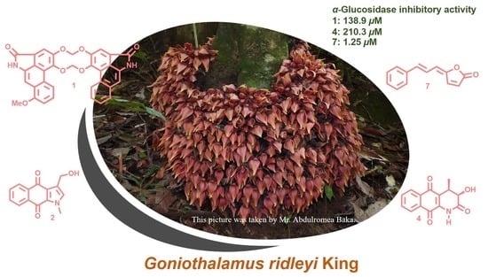
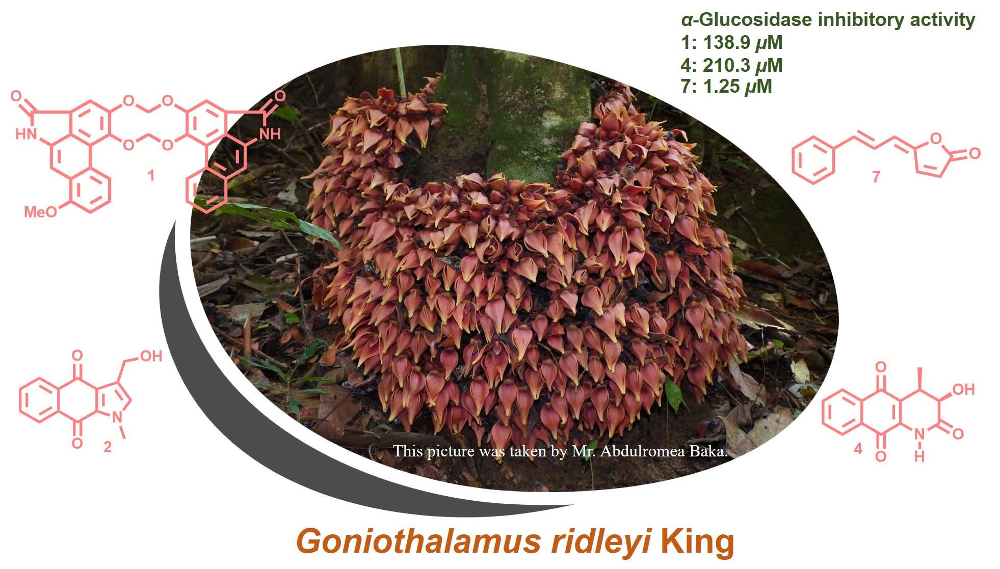
Alkaloids and Styryl lactones from Goniothalamus ridleyi King and Their α-Glucosidase Inhibitory Activity

by
Isaraporn Polbuppha
1,
Passakorn Teerapongpisan
1,
Piyaporn Phukhatmuen
1,
Virayu Suthiphasilp
2,
Tharakorn Maneerat
1,3,
Rawiwan Charoensup
3,4,
Raymond J. Andersen
5,* and
Surat Laphookhieo
1,3,*
1
Center of Chemical Innovation for Sustainability (CIS) and School of Science, Mae Fah Luang University, Chiang Rai 57100, Thailand
2
Department of Industrial Technology and Innovation Management, Faculty of Science and Technology, Pathumwan Institute of Technology, Bangkok 10330, Thailand
3
Medicinal Plant Innovation Center, Mae Fah Luang University, Chiang Rai 57100, Thailand
4
School of Integrative Medicine, Mae Fah Luang University, Chiang Rai 57100, Thailand
5
Department of Chemistry and Department of Earth, Ocean & Atmospheric Sciences, University of British Columbia, 2036, Main Mall, Vancouver, BC V6T 1Z1, Canada
*
Authors to whom correspondence should be addressed.
Molecules 2023, 28(3), 1158; https://doi.org/10.3390/molecules28031158
Submission received: 23 December 2022 / Revised: 11 January 2023 / Accepted: 20 January 2023 / Published: 24 January 2023

Abstract

:
Gonioridleylactam (1), a new compound, is a unique dimeric aristolactam isolated from the EtOAc extract of the twigs of Goniothalamus ridleyi King. The structure of gonioridleylactam (1) consists of two different aristolactams linked together with two methylenedioxy bridges at C–3/C–3′ and C–4/C–4′, generating a ten-membered ring of [1,3,6,8]tetraoxecine. A new natural product, gonioridleyindole (3-hydroxymethyl-1-methyl-1H-benz[f]indole-4,9-dione, 2), together with eight known compounds (3–10) were also isolated from this plant. Their structures were extensively characterized by spectroscopic methods and comparisons were made with the literature. Compounds 1–4, 7, and 9 were evaluated for their α-glucosidase inhibitory activity. Of these, 3,5-demethoxypiperolide (7) displayed the highest α-glucosidase inhibitory activity, with an IC50 value of 1.25 µM.

Graphical Abstract

1. Introduction

Goniothalamus ridleyi King belongs to the Annonaceae family, widely distributed throughout Thailand (southern part), Peninsular Malaysia, and Sumatra. Plants in the Goniothalamus genus produce diverse bioactive compounds, including styryl lactones, acetogenins, flavonoids, and alkaloids [1,2,3,4]. Many of these compounds have been reported to exhibit cytotoxic, antibacterial, larvicidal, and antitubercular activities [3,4,5,6,7]. For example, two styryl lactones, (−)-5-acetoxygoniothalamin and (Z)-6-styryl-5,6-dihydro-2-pyranone, displayed cytotoxicity against a colon cancer cell line (HCT116), with IC50 values of 8.6 and 22.2 μM, respectively [4]. (+)-3-Acetylaltholactone and (−)-nordicentrine showed antiplasmodial activity against the parasite Plasmodium falciparum, with IC50 values of 2.6 and 0.3 μg/mL, respectively [8].
From a previous phytochemical investigation of G. ridleyi, five simple styryl lactones [9,10] and 5-hydroxy-6-[(E)-2-phenylethenyl]-5,6-dihydro-2H-pyran-2-one were isolated and identified. A preliminary bioassay screen conducted by our group revealed that the EtOAc extract of G. ridleyi twigs inhibited α-glucosidase activity (97% at 250 μg/mL), prompting us to isolate the α-glucosidase inhibitory compounds from this plant. This paper describes the isolation, structure elucidation, and α-glucosidase inhibitory activity of compounds from G. ridleyi.

2. Results and Discussion

The EtOAc extract of the twig of G. ridleyi was separated and purified by various chromatographic techniques to afford a new dimeric aristrolactam (1), a new natural indole alkaloid (2), and eight known compounds (3–10) (Figure 1). The known compounds were identified as goniochelienic acid B (3) [3], griffithazanone A (4) [11], ethenol (5) [12], goniobutenolide B (6) [13], (5-(3-phenyl-2-propenylidene)-2(5H)-furanone) (7) [14], (−)-goniothalamin (8) [15], (−)-5-hydroxygoniothalamin (9) [16], and (−)-5-acetyl goniothalamin (10) [17] by extensive NMR spectroscopic data and comparisons made with spectroscopic data reported in the literature.

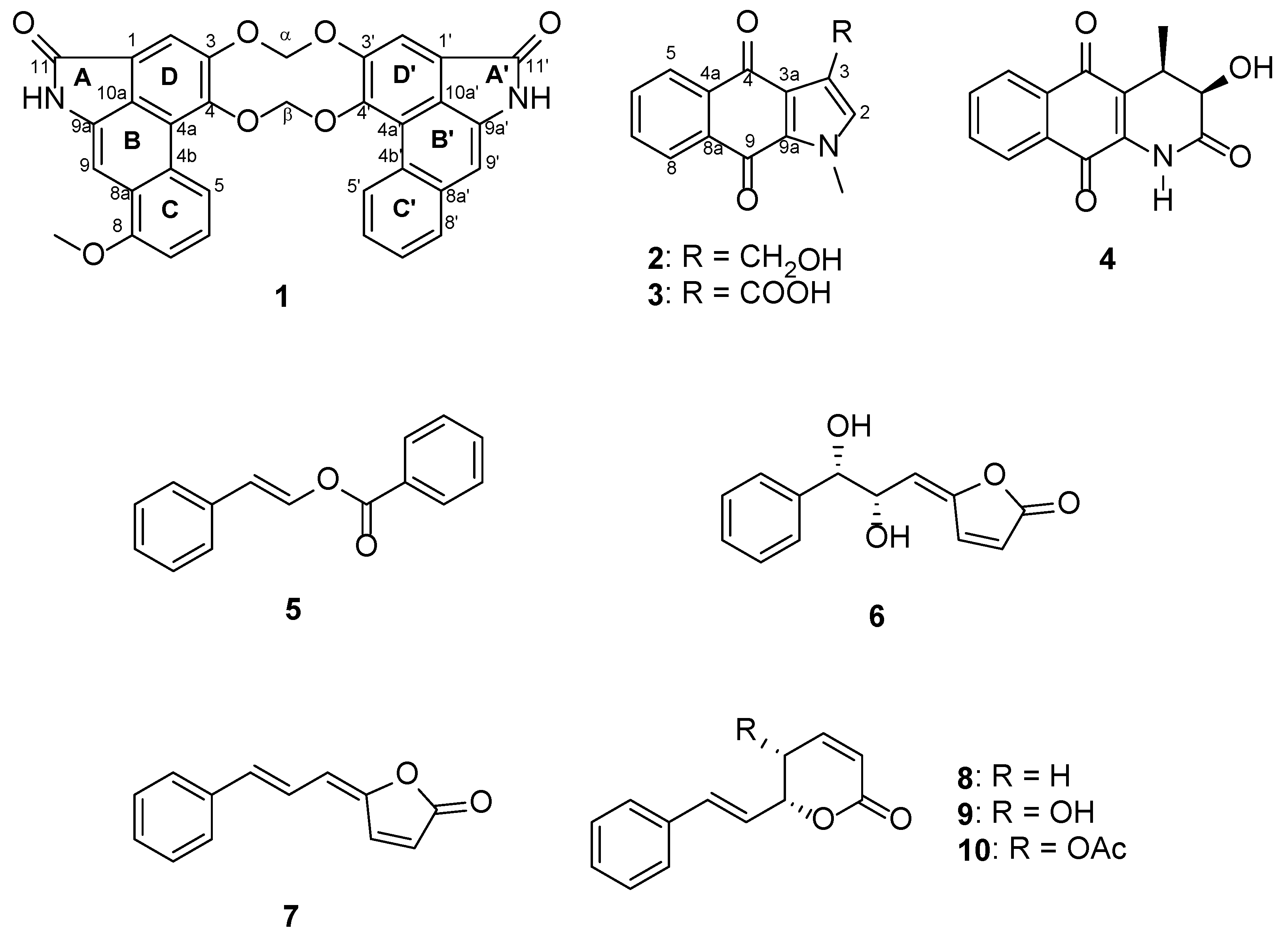
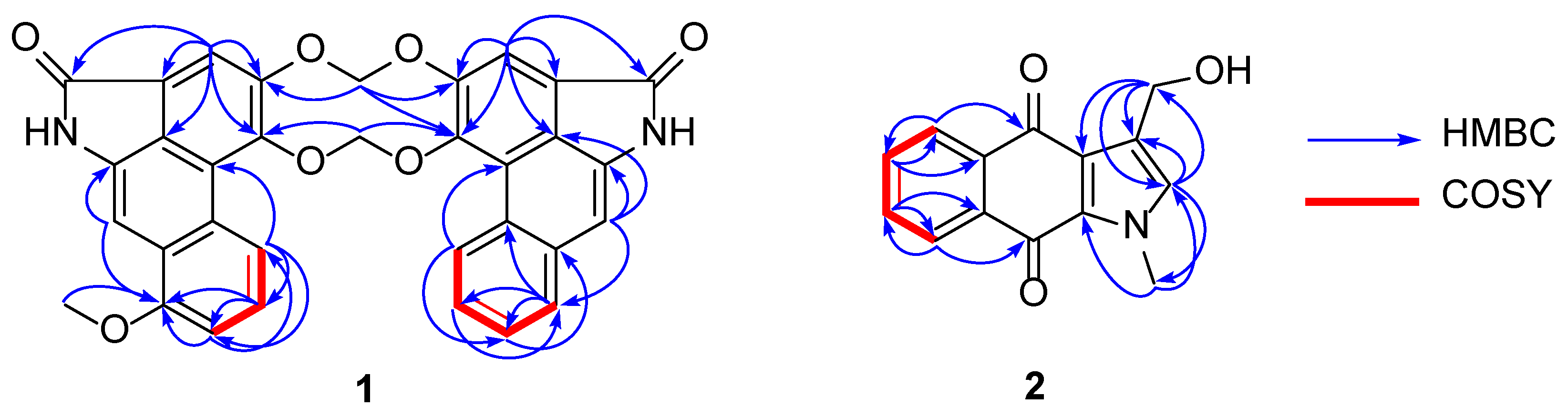
2.1. Structural Elucidation

Gonioridleylactam (1) was isolated as a yellowish solid. The HRESITOFMS spectrum of 1 displayed a sodium adduct ion at m/z 556.1271, corresponding to the molecular formula of C33H20N2O7. The UV spectrum showed maxima absorption bands at λmax 250, 277, and 322 nm, while the IR spectrum displayed the presence of NH (3394 cm−1) and amide carbonyl (1688 cm−1) functionalities. Following an intensive analysis of NMR spectroscopic data, the structure of 1 was determined as a dimeric aristolactam linked together with two different aristolactams (aristolactam units A and B). The 13C NMR data, in combination with DEPT and HMQC, displayed 33 carbon resonances, including 1 methyl (δC 55.3), 2 methylenes (δC 103.3 (×2)), 11 methines (δC 128.7, 127.6, 126.8, 125.6, 125.2, 119.2, 108.0, 105.3, 105.2, 104.2, and 98.1), and 17 non-protonated carbons (δC 167.9 (×2), 155.9, 149.3, 149.2, 147.5, 147.3, 135.5, 134.6, 134.7, 125.5 (×2), 124.8, 124.7, 119.8 (×2), 111.8 (×2), and 104.2). Aristolactam unit A displayed the 1H and 13C NMR resonances as follows: a singlet NH proton [δH 9.74 (br s, NH′)], a set of ABC aromatic protons [δH 8.25 (d, J = 8.2 Hz, H-5)/δC 119.2, 7.52 (t, J = 8.2 Hz, H-6)/δC 125.6, and 7.20 (d, J = 8.2 Hz, H-7)/δC 108.0], two singlet aromatic protons [δH 7.58 (s, H-2)/δC 105.0, and 7.54 (1H, s, H-9)/δC 98.1], and one methoxy group [δH 4.05 (s, 8-OMe)/δC 55.3]. The methoxy group was placed at C-8 due to the HMBC cross peaks between H-6, H-7, MeO-8, and H-9 with C-8 (δC 155.9) (Figure 2 and Table 1). In the case of aristolactam unit B, the 1H and 13C NMR data were similar to those of aristolactam unit A. The main difference between aristolactam units A and B is that the OMe-8 resonance of aristolactam unit B was not observed. Aristolactam unit B displayed four aromatic protons of 1,2-disubstituted benzene at δH 8.63 (1H, dd, J = 7.8, 1.5 Hz, H-5′)/δC 126.8, 7.58 (1H, td, J = 7.8, 1.5 Hz, H-6′)/δC 125.2, 7.61 (1H, td, J = 7.8, 1.5 Hz, H-7′)/δC 127.6, and 7.92 (1H, dd, J = 7.8, 1.5 Hz, H-8′)/δC 128.7. These were supported by HMBC correlations from H-5′ (δH 8.63) to C-4a′ (δC 111.8), C-7′ (δC 127.6), and C-8a′ (δC 134.7), from H-6′ (δH 7.58) to C-4b′ (δC 124.8), C-7′ (δC 127.6), from H-7′ (δH 7.61) to C-8a′ (δC 134.7), C-5′ (δC 126.8), and from H-8′ (δH 7.92) to C-4b′ (δC 124.8), C-6′ (δC 125.2), and C-9′ (δC 104.2) (Figure 2). Aristolactam units A and B linked together with two methylenedioxy bridges at C–3/C–3′ [6.49 (2H, s, H-α/103.3)] and C–4/C–4′ [6.50 (2H, s, H-β/103.3)], generating a ten-membered ring of [1,3,6,8]tetraoxecine. The observed HMBC cross peaks of H-α (δH 6.49) to C-3 (δC 147.3) and C-3′ (δC 147.5) and H-β (δH 6.50) to C-4 (δC 149.2) and C-4′ (δC 149.3) (Figure 2) supported these assignments. In addition, the HRESITOFMS ions of aristolactam units A and B at m/z 293.0692 and 263.0585 (Figure 3), respectively (Figures S10 and S11, Supplementary Materials), also supported the linkage of aristolactam units A and B. The full assignment of NMR data and HMBC correlations is shown in Figure 2 and Table 1, respectively. The structure of 1 was the first example of a ten-membered ring of [1,3,6,8]tetraoxecine dimeric aristolactam found in Annonaceae.
The HRESITOFMS spectrum of compound 2, a new natural product, displayed an [M + Na]+ ion at m/z 264.0635 (Figure S18, Supplementary Materials), corresponding to the molecular formula of C14H11NO3 (Tgt. for C14H11NO3, 241.0742). The UV spectrum showed maxima absorption bands at λmax 250, 277, and 322 nm suggesting the benzo[f]indole-4,9-dione framework [3], while the IR spectrum revealed absorption bands for carbonyl (1709 cm−1) and hydroxy (3404 cm−1) functionalities. The 1H and 13C NMR spectroscopic data of 2 (Table 2) showed resonances for four aromatic protons of 1,2-disubstituted benzene [δH 7.70 (m, H-5 and H-8)/δC 126.8 (C-5), 126.7 (C-8) and 8.16 (m, H-6 and H-7)/δC 133.6 (C-6) and 133.3 (C-7)], an olefinic proton [δH 6.85 (s, H-2)/δC 129.3], a hydroxymethylene proton [δH 4.72 (s, H2-1′/δC 57.1)], and an N-methyl group [δH 4.07 (s, N-CH3)/δC 36.8]. The structure of 2 was further supported by the following key HMBC correlations (Figure 2): δH 6.85 (H-2) with C-3 (δC 126.2), C-1′ (δC 36.8), and N-CH3 (δC 57.1); δH 4.72 (H2-1′) with C-2 (δC 129.3), C-3 (δC 126.2), and C-3a (δC 162.2); δH 4.15 (N-CH3) with C-2 (δC 129.3) and C-9a (δC 131.8). Thus, compound 2 was named as gonioridleyindole (3-hydroxymethyl-1-methyl-1H-benzo[f]indole-4,9-dione). Many 3-hydroxymethylindolequinone derivatives, including compound 2, had previously been synthesized and functionalized to other indolequinones [18]. However, compound 2 was first isolated from nature.

2.2. α-Glucosidase Inhibitory Activity

α-Glucosidase is a carbohydrate hydrolyzing enzyme that maintains postprandial blood glucose and insulin levels [19]. The inhibition of this enzyme can delay intestinal carbohydrate digestion to control hyperglycemia in diabetes mellitus [20]. Exploration for new α-glucosidase inhibitors and other antidiabetic drugs from natural sources has increased in recent years [21]. There are reports of the presence of α-glucosidase inhibitors such as flavonoids (quercetin) [22], terpenoids (wallitaxanes) [23], and alkaloids (5-hydroxynoracronycin) [19]. In this study, compounds isolated with a sufficient amount, 1–4, 7, and 9, were evaluated for their α-glucosidase inhibitory activity. Of these, 3,5-demethoxypiperolid (7) showed the highest α-glucosidase inhibitory activity with an IC50 value of 1.25 µM, which is better than that of the standard control (acarbose). Other tested compounds were weak or inactive (Table 3). The observed α-glucosidase inhibitory activity of 3,5-demethoxypiperolid (7) indicates that this compound may have the potential as a lead compound for the further development of anti-diabetes agents.

3. Conclusions

Phytochemical investigation of the EtOAc extract of the twigs of Goniothalamus ridleyi King resulted in the discovery of a unique dimeric aristolactam and nine other compounds. The dimeric aristolactam contained two different aristolactam units linked together with two methylenedioxy bridges forming a [1,3,6,8]tetraoxecine ten-membered ring. The discovery of styryl lactones and alkaloids in this study as the major compounds was in good agreement with previous reports. In addition, the result of preliminary α-glucosidase inhibitory assay suggested that (5-(3-phenyl-2-propenylidene)-2(5H)-furanone) may have potential as the lead compound for the development of α-glucosidase inhibitory agent.

4. Materials and Methods

4.1. General Experimental Procedures

Melting points were measured with a Büchi B-540 melting point apparatus (Flawil, Sankt Gallen, Switzerland). UV–vis spectra were recorded with a Varian Cary 5000 UV–vis–NIR spectrophotometer (Agilent Technologies, Santa Clara, CA, USA). The IR spectra were recorded using a Perkin-Elmer FTS FT-IR spectrophotometer (Waltham, MA, USA). The NMR spectra were measured using 400, 500, or 600 MHz Bruker spectrometers (Billerica, MA, USA). HRESITOFMS and LRESIMS spectra were carried out on a Bruker-Hewlett-Packard 1100 Esquire-LC system mass spectrometer (Billerica, MA, USA) and Waters 2695 HPLC (Milford, MA, USA), Waters ZQ equipped with ESCI ion source mass spectrometer, respectively. All quick column chromatography (QCC) and column chromatography (CC) were carried out on silica gel 60 (5–40 μm, SiliCycle Inc., Québec, QC G1P 4S6, Canada) and silica gel 100 (63–200 μm, SiliCycle Inc.), respectively. Sephadex LH-20, when indicated, was also used for CC. Precoated thin-layer chromatography (TLC) plates of silica gel 60 F254 were used for analytical purposes.

4.2. Plant Material

The twigs of G. ridleyi [24] were collected in April 2021 from Narathiwat Province, Thailand. This plant was identified by Mr. Abdulromae Baka (Independent Research Group on Plant Diversity in Thailand, Sichon, Nakhon Si Thammarat, 80120, Thailand). A voucher specimen (MFU-NPR0206) was deposited at the Natural Products Research Laboratory, School of Science, Mae Fah Luang University. Plant materials were dried and stored at room temperature.

4.3. Extraction and Isolation

Air-dried twigs of G. ridleyi (1.5 kg) were extracted with EtOAc (3 × 20 L) at room temperature and concentrated under reduced pressure to give an EtOAc extract (58.5 g). The twig extract was subjected to quick column chromatography (QCC) over silica gel (100% hexanes to 100% acetone) to give seven fractions (1A–1G). Fraction 1C (5.3 g) was subjected to CC over Sephadex LH-20 CC (100% MeOH) to give four subfractions (2A–2D). Subfraction 2B (3.6 g) was further purified by silica gel CC (1:9 v/v, acetone–hexanes) to give compound 5 (5.8 mg) and four subfractions (3A–3D). Compound 7 (3.1 mg) was obtained from subfraction 3B (97.2 mg) by silica gel CC (100% CH2Cl2), while compound 6 (1.0 mg) was isolated from subfraction 3C (68.2 mg) by silica gel CC (1:19 v/v, acetone-hexanes). Subfraction 1D (2.6 g) was further separated by CC over Sephadex LH-20 (100% MeOH) to afford five subfractions (4A–4E). Purification of subfraction 4D (53.5 mg) by silica gel CC (100% CH2Cl2) yielded compound 8 (1.5 mg). Fraction 1E (23.5 g) was subjected to QCC over silica gel (100% hexanes to 100% EtOAc) to give four subfractions (5A–5D). Upon standing at room temperature, the white solid was precipitate from subfraction 5B (778.9 mg), which was washed by MeOH to give compound 10 (5.7 mg). Subfraction 5C (13.0 g) was separated by CC over Sephadex LH-20 (100% MeOH) to give four subfractions (6A–6D). Subfraction 6B (1.0 g) was further separated by CC over Sephadex LH-20 CC (100% MeOH) to afford compound 3 (1.9 mg) and two subfractions (7A–7B). Subfraction 7A (887.4 mg) was purified by silica gel CC (1:24 v/v, EtOAc–CH2Cl2) to give compound 9 (1.2 mg). Compounds 2 (3.5 mg) and 4 (2.2 mg) were obtained from subfraction 7B (18.6 mg) by silica gel CC (1:24 v/v, EtOAc–CH2Cl2). Purification of subfraction 6D (4.3 mg) by silica gel CC (1:24 v/v, EtOAc-CH2Cl2) gave compound 1 (2.9 mg) (%yield = 0.019%).

4.3.1. Gonioridleylactam (1)

Yellow solid; mp 296–298 °C; UV (MeOH) λmax (log ε) 259 (4.7), 276 (4.5), 287 (4.5), 328 (4.1), and 390 (4.0) nm; IR (neat) vmax 3394, 2923, 1688, 1376, 1261, 1093, 1261, 1041, and 801 cm−1; 1H NMR (600 MHz, acetone-d6) and 13C NMR (150 MHz, acetone-d6), see Table 1; HRESITOFMS m/z 579.1162 [M + Na]+ (C33H20N2O7, 556.1271, Tgt. Mass, 556.1271), see Figure S8, Supplementary Materials.

4.3.2. Gonioridleyindole (3-Hydroxymethyl-1-methyl-1H-benz[f]indole-4,9-dione, 2)

Yellow solid; mp 218–220 °C; UV (MeOH) λmax (log ε) 250 (4.5), 277 (4.2), 332 (3.8), and 399 (2.4) nm; IR (neat) vmax 3404, 2984, 1709, 1377, 1300, 1191, 1104, 1035, 969, and 693 cm−1; 1H NMR (400 MHz, CDCl3) and 13C NMR (100 MHz, CDCl3), see Table 2; HRESITOFMS m/z 264.0635 [M + Na]+ (C14H11NO3, 241.0742, Tgt. Mass, C14H11NO3, 241.0742), see Figure S18, Supplementary Materials.

4.4. α-Glucosidase Inhibitory Activity

The previously reported approach was used to perform a colorimetric α-glucosidase assay [25]. Briefly, the tested samples (50 µL) were combined with 50 µL of the α-glucosidase enzyme solution (0.05 U/mL) and preincubated at 37 °C for 5 min. The substrate (50 µL, c 1 mM), p-nitrophenyl α-D-glucoside, was added and incubated at 37 °C for 30 min. Then, 50 µL of Na2CO3 (0.3 M) was added. The absorption of the mixture was measured at 405 nm. Acarbose was used as a positive control (185.7 µM).

Supplementary Materials

The following supporting information can be downloaded at: https://www.mdpi.com/article/10.3390/molecules28031158/s1, Figure S1: 1H NMR (600 MHz, acetone-d6) of gonioridleyilactam (1); Figure S2: 13C NMR (150 MHz, acetone-d6) of gonioridleyilactam (1); Figure S3: HSQC of gonioridleyilactam (1); Figure S4: HMBC of gonioridleyilactam (1); Figure S5: COSY of gonioridleyilactam (1); Figure S6: NOESY of gonioridleyilactam (1); Figure S7: LRESIMS (low-resolution electrospray ionization mass spectrometry) spectrum of gonioridleyilactam (1); Figure S8: HRESIMS spectrum of gonioridleyilactam (1); Figure S9: HRESIMS spectrum of Unit A (aristolactam II) in 1; Figure S10: HRESIMS spectrum of Unit B (aristolactam I) in 1; Figure S11: IR spectrum of gonioridleyilactam (1); Figure S12: 1H NMR (400 MHz, CDCl3) of 3-hydroxymethyl-1-methyl-1H-benz[f]indole-4,9-dione (2); Figure S13: 13C NMR (100 MHz, CDCl3) of 3-hydroxymethyl-1-methyl-1H-benz[f]indole-4,9-dione (2); Figure S14: HSQC of 3-hydroxymethyl-1-methyl-1H-benz[f]indole-4,9-dione (2); Figure S15: HMBC of 3-hydroxymethyl-1-methyl-1H-benz[f]indole-4,9-dione (2); Figure S16: COSY of 3-hydroxymethyl-1-methyl-1H-benz[f]indole-4,9-dione (2); Figure S17: NOESY of 3-hydroxymethyl-1-methyl-1H-benz[f]indole-4,9-dione (2); Figure S18: HRESIMS spectrum of 3-hydroxymethyl-1-methyl-1H-benz[f]indole-4,9-dione (2); Figure S19: IR spectrum of 3-hydroxymethyl-1-methyl-1H-benz[f]indole-4,9-dione (2); Figure S20: The isolation and purification of isolated compounds 1–10 from the twig extract of G. ridleyi.

Author Contributions

Conceptualization, S.L.; methodology, I.P., P.T. and P.P.; formal analysis, I.P. and S.L.; investigation, I.P.; resources, S.L. and R.C.; data curation, I.P.; writing—original draft preparation, I.P., V.S. and S.L.; writing—review and editing, I.P., V.S., T.M., R.C., R.J.A. and S.L.; funding acquisition, S.L. All authors have read and agreed to the published version of the manuscript.

Funding

This project is funded by the National Research Council of Thailand (NRCT) and Mae Fah Luang University (N42A650373).

Institutional Review Board Statement

Not applicable.

Informed Consent Statement

Not applicable.

Data Availability Statement

Not applicable.

Acknowledgments

We would like to thank the National Research Council of Thailand (NRCT) and Mae Fah Luang University (N42A650373) for financial support. The Postdoctoral Fellowship from Mae Fah Luang University to Isaraporn Polbuppha is also acknowledged. The University of British Columbia and Mae Fah Luang University are recognized for their laboratory facilities. We also thank Abdulromea Baka for plant identification.

Conflicts of Interest

The authors declare no conflict of interest.

Sample Availability

Samples of the compounds are not available from the authors.

References

  1. Blázquez, M.A.; Bermejo, A.; Zafra-Polo, M.C.; Cortes, D. Styryl-lactones from Goniothalamus species–A review. Phytochem. Anal. 1999, 10, 161–170. [Google Scholar] [CrossRef]
  2. Aslam, M.S.; Ahmad, M.S.; Mamat, A.S.; Ahmad, M.Z.; Salam, F. Goniothalamus: Phytochemical and ethnobotanical review. Recent Adv. Biol. Med. 2016, 2, 34–47. [Google Scholar] [CrossRef]
  3. Jaidee, W.; Andersen, R.J.; Patrick, B.O.; Pyne, S.G.; Muanprasat, C.; Borwornpinyo, S.; Laphookhieo, S. Alkaloids and styryllactones from Goniothalamus cheliensis. Phytochemistry 2019, 157, 8–20. [Google Scholar] [CrossRef] [PubMed]
  4. Meesakul, P.; Jaidee, W.; Richardson, C.; Andersen, R.J.; Patrick, B.O.; Willis, A.C.; Muanprasat, C.; Wang, J.; Lei, X.G.; Hadsadee, S.; et al. Styryllactones from Goniothalamus tamirensis. Phytochemistry 2020, 171, 112248. [Google Scholar] [CrossRef] [PubMed]
  5. Lian Ee, G.C.; Lee, H.L.; Goh, S.H. Larvicidal activity of Malaysian Goniothalamus species. Nat. Prod. Lett. 1999, 13, 137–142. [Google Scholar] [CrossRef]
  6. Noor-Zarina, A.W.; Saleha, S.; Halimah, A.S.; Azimahtol, H.L.P.; Nazlina, I. Antioxidant, antibacterial and antiviral properties of Goniothalamus umbrosus leaves methanolic extract. Afr. J. Microbiol. Res. 2011, 5, 3138–3143. [Google Scholar] [CrossRef]
  7. Macabeo, A.P.G.; Lopez, A.D.A.; Schmidt, S.; Heilmann, J.; Dahse, H.M.; Alejandro, G.J.D.; Franzblau, S.G. Antitubercular and cytotoxic constituents from Goniothalamus gitingensis. Rec. Nat. Prod. 2014, 8, 41–45. [Google Scholar]
  8. Lekphrom, R.; Kanokmedhakul, S.; Kanokmedhakul, K. Bioactive styryllactones and alkaloid from flowers of Goniothalamus laoticus. J. Ethnopharmacol. 2009, 125, 47–50. [Google Scholar] [CrossRef]
  9. Jusoh, S.; Din, L.B.; Zakaria, Z.; Khaledi, H. 5-Hydroxy-6-[(E)-2-phenylethenyl]-5, 6-dihydro-2H-pyran-2-one isolated from Goniothalamus ridleyi. Acta Cryst. 2012, 68, 2274. [Google Scholar]
  10. Jusoh, S.; Zakaria, Z.; Ahmad, F.B.; Din, L.B. Isolation and characterization of styryl lactone of Goniothalamus ridleyi. Sains Malays. 2015, 44, 365–370. [Google Scholar] [CrossRef]
  11. Zhang, Y.J.; Kong, M.; Chen, R.Y.; Yu, D.Q. Alkaloids from the roots of Goniothalamus griffithii. J. Nat. Prod. 1999, 62, 1050–1052. [Google Scholar] [CrossRef] [PubMed]
  12. Ye, S.; Leong, W.K. Regio- and stereoselective addition of carboxylic acids to phenylacetylene catalyzed by cyclopentadienyl ruthenium complexes. J. Organomet. Chem. 2006, 691, 1117–1120. [Google Scholar] [CrossRef]
  13. Fang, X.P.; Anderson, J.E.; Chang, C.J.; McLaughlin, J.L. Three new bioactive styryllactones from Goniothalamus giganteus (Annonaceae). Tetrahedron 1991, 47, 9751–9758. [Google Scholar] [CrossRef]
  14. Tran, D.T.; Thi Mai, H.D.; Pham, V.C.; Nguyen, V.H.; Litaudon, M.; Guéritte, F.; Nguyen, Q.V.; Tran, T.A.; Chau, V.M. Alkaloids and styryllactones from the leaves of Goniothalamus tamirensis. Phytochem. Lett. 2013, 6, 79–83. [Google Scholar] [CrossRef]
  15. Fátima, Â.; Kohn, L.K.; Carvalho, J.E.; Pillia, R.A. Cytotoxic activity of (S)–goniothalamin and analogues against human cancer cells. Bioorgan. Med. Chem. 2006, 14, 622–631. [Google Scholar] [CrossRef]
  16. Goh, S.H.; Ee, G.C.L.; Chuah, C.H.; Mak, T.C.W. 5β-Hydroxygoniothalamin, a styrylpyrone derivative from Goniothalamus dolichocarpus (Annonaceae). Nat. Prod. Lett. 2006, 5, 255–259. [Google Scholar] [CrossRef]
  17. Ahmad, F.B.; Tukol, W.A.; Omar, S.; Sharif, A.M. 5-Acetyl goniothalamin, a styryl dihydropyrone from Goniothalamus uvaroides. Phytochemistry 1991, 30, 2430–2431. [Google Scholar] [CrossRef]
  18. Inman, M.; Visconti, A.; Yan, C.; Siegel, D.; Ross, D.; Moody, C.J. Antitumour indolequinones: Synthesis and activity against human pancreatic cancer cells. Org. Biomol. Chem. 2014, 12, 4848–4861. [Google Scholar] [CrossRef] [Green Version]
  19. Trinh, D.H.; Tran, P.T.; Trinh, B.T.; Nguyen, H.T.; Nguyen, H.D.; Ha, L.D.; Nguyen, L.H.D. Coumarins and acridone alkaloids with α-glucosidase inhibitory and antioxidant activity from the roots of Paramignya trimera. Phytochem. Lett. 2006, 35, 94–98. [Google Scholar] [CrossRef]
  20. Le, T.K.D.; Danova, A.; Aree, T.; Duong, T.H.; Koketsu, M.; Ninomiya, M.; Chavasiri, W. α-Glucosidase inhibitors from the stems of Knema globularia. J. Nat. Prod. 2022, 85, 776–786. [Google Scholar] [CrossRef]
  21. Rangel-Grimaldo, M.; Rivero-Cruz, I.; Madariaga-Mazón, A.; Figueroa, M.; Mata, R. α-Glucosidase inhibitors from Preussia minimoides. J. Nat. Prod. 2017, 80, 582–587. [Google Scholar] [CrossRef]
  22. Ghani, U.; Nur-e-Alam, M.; Yousaf, M.; Ul-Haq, Z.; Noman, O.M.; Al-Rehaily, A.J. Natural flavonoid α-glucosidase inhibitors from Retama raetam: Enzyme inhibition and molecular docking reveal important interactions with the enzyme active site. Bioorg. Chem. 2019, 87, 736–742. [Google Scholar] [CrossRef]
  23. Dang, P.H.; Nguyen, H.X.; Duong, T.T.T.; Tran, T.K.T.; Nguyen, P.T.; Vu, T.K.T.; Awale, S. α-Glucosidase inhibitory and cytotoxic taxane diterpenoids from the stem bark of Taxus wallichiana. J. Nat. Prod. 2017, 80, 1087–1095. [Google Scholar] [CrossRef] [PubMed]
  24. Saunders, R.M.K.; Chalermglin, P. A synopsis of Goniothalamus species (Annonaceae) in Thailand, with descriptions of three new species. Bot. J. Linn. 2008, 156, 355–384. [Google Scholar] [CrossRef]
  25. Kittiwisut, S.; Amnuoypol, S.; Pathompak, P.; Setharaksa, S. α-Glucosidase and α-amylase inhibitory effects with anti-oxidative activity of Tetracera loureiri (Finet & Gagnep.) Pierre ex Craib leaf extracts. Pharm. Sci. Asia 2021, 48, 175–184. [Google Scholar]
Figure 1. Compounds isolated from the twig extract of G. ridleyi.
Molecules 28 01158 g001
Figure 2. COSY (1H—1H) and selected HMBC (1H→13C) correlations of 1 and 2.
Molecules 28 01158 g002
Figure 3. Mass spectral fragmentation of 1.
Molecules 28 01158 g003
Table 1. 1H (600 MHz) and 13C (150 MHz) NMR spectroscopic data of 1 in acetone-d6.
PositionδCδH [mult, J in Hz]HMBC (1H→13C)
1119.8
2105.07.58 (s)1, 3, 4, 4a, 11
3147.3
4149.2
4a111.8
4b124.7
5119.28.25 (d, 8.2)4b, 6, 7, 8
6125.67.52 (t, 8.2)5, 7, 8
7108.07.20 (d, 8.2)5, 6, 8
8155.9
8a104.2
998.17.54 (s)8, 9a,10a
9a134.6
10a125.5
11167.9
1′119.8
2′105.37.57 (s)1′, 3′, 4′, 10a′, 11′
3′147.5
4′149.3
4a′111.8
4b′124.8
5′126.88.63 (dd, 7.8, 1.5)4a′, 7′, 8a′
6′125.27.58 (td, 7.8, 1.5)8′
7′127.67.61 (td, 7.8, 1.5)5′, 8a′
8′128.77.92 (dd, 7.8, 1.5)4b′, 6′, 9′
8a′134.7
9′104.27.16 (s)4a′, 8′, 9a′, 10a′
9a′135.5
10a′125.5
11′167.9
α103.36.49 (s)3, 3′
β103.36.50 (s)4, 4′
N-H 9.76 (s)
N-H′ 9.74 (s)
8-OMe55.34.05 (s)8′
Table 2. 1H (400 MHz) and 13C (MHz) NMR spectroscopic data of 2 in CDCl3.
PositionδCδH [mult, J in Hz]HMBC (1H→13C)
2129.36.85 (s)3, 3a, 9a, 1′, N-CH3
3126.2
3a162.2
4183.0
4a133.9
5126.87.70 (m)4, 6
6133.68.16 (m)4a, 5
7133.38.16 (m)8, 8a
8126.77.70 (m)7, 9
8a133.8
9176.4
9a131.8
1′36.84.72 (s)2, 3, 3a
N-CH357.14.07 (s)2, 9a
Table 3. α-Glucosidase inhibitory activity of some isolated compounds from G. ridleyi.
Compounds%Inhibition at 250 µg/mLIC50, µM
199.6138.9 ± 0.9
299.3inactive
399.5inactive
498.0inactive
798.61.25 ± 0.4
999.8inactive
Acarbose88.1185.7 ± 0.3
Disclaimer/Publisher’s Note: The statements, opinions and data contained in all publications are solely those of the individual author(s) and contributor(s) and not of MDPI and/or the editor(s). MDPI and/or the editor(s) disclaim responsibility for any injury to people or property resulting from any ideas, methods, instructions or products referred to in the content.

