Article

A Natural Compound Containing a Disaccharide Structure of Glucose and Rhamnose Identified as Potential N-Glycanase 1 (NGLY1) Inhibitors

1
School of Medical Technology and Information Engineering, Zhejiang Chinese Medical University, Hangzhou 310053, China
2
School of Basic Medical Sciences, Zhejiang Chinese Medical University, Hangzhou 310053, China
3
Department of Clinical Laboratory, Jiaxing Maternity and Child Health Care Hospital, Jiaxing 314001, China
4
Central Laboratory, Ningbo Hospital, Renji Hospital, Shanghai Jiao Tong University School of Medicine, Ningbo 315336, China
5
Key Laboratory of Medical Molecular Virology (MOE/NHC/CAMS), School of Basic Medical Sciences, Fudan University, Shanghai 200032, China
*
Authors to whom correspondence should be addressed.
These authors contributed equally to this work.
Molecules 2023, 28(23), 7758; https://doi.org/10.3390/molecules28237758
Submission received: 23 October 2023 / Revised: 13 November 2023 / Accepted: 23 November 2023 / Published: 24 November 2023

Abstract

:
N-glycanase 1 (NGLY1) is an essential enzyme involved in the deglycosylation of misfolded glycoproteins through the endoplasmic reticulum (ER)-associated degradation (ERAD) pathway, which could hydrolyze N-glycan from N-glycoprotein or N-glycopeptide in the cytosol. Recent studies indicated that NGLY1 inhibition is a potential novel drug target for antiviral therapy. In this study, structure-based virtual analysis was applied to screen candidate NGLY1 inhibitors from 2960 natural compounds. Three natural compounds, Poliumoside, Soyasaponin Bb, and Saikosaponin B2 showed significantly inhibitory activity of NGLY1, isolated from traditional heat-clearing and detoxifying Chinese herbs. Furthermore, the core structural motif of the three NGLY1 inhibitors was a disaccharide structure with glucose and rhamnose, which might exert its action by binding to important active sites of NGLY1, such as Lys238 and Trp244. In traditional Chinese medicine, many compounds containing this disaccharide structure probably targeted NGLY1. This study unveiled the leading compound of NGLY1 inhibitors with its core structure, which could guide future drug development.

Graphical Abstract

1. Introduction

N-Glycanase 1 (NGLY1) is a de-N-glycosylating enzyme that catalyzes the hydrolysis of the amide bond between the proximal N-acetylglucosamine (GlcNAc) residue and the Asn side chain to which it is attached, removing N-glycans from glycosylated proteins in the cytosol [1,2,3]. Discovered in 1993, NGLY1 is known to participate in clearing misfolded glycoproteins during the process of glycoprotein synthesis through the endoplasmic reticulum (ER)-associated degradation (ERAD) pathway [4]. NGLY1 deficiency leads to disrupted ERAD function. Histological analysis of Ngly1−/− rats demonstrated cytoplasmic ubiquitinated protein accumulation in neurons of the thalamus and spinal cord [5]. In 2014, it was reported that NGLY1 deficiency could lead to NGLY1-congenital disorder of deglycosylation (CDDG), a disease characterized by developmental delay, intellectual disability, absence of or reduced tears and sweating, abnormal liver function, and motor dysfunction [6,7,8]. It was noteworthy that NGLY1-CDDG patients were less susceptible to viral infections [9]. Additionally, it has been reported that generated Ngly1 knockout (Ngly1−/−) murine embryonic fibroblasts (MEFs) inhibited vesicular stomatitis virus (VSV) replication [10]. siRNA knockdown of NGLY1 or Z-VAD-FMK (benzyloxycarbonyl-Val-Ala-Asp-fluoromethyl ketone) inhibits NGLY1, restricting the infection of enterovirus 71 (EV71) and coxsackievirus A16 (CA16) in RD cells [11]. Yang et al. [10] also found NGLY1-deficient human and mouse cells, resulting in severely fragmented mitochondria and the activation of cGAS–STING pathways, leading to elevated IFN-stimulated genes (ISGs). A schematic diagram is shown in Figure 1. NGLY1 represents a potential drug target for antiviral therapy. NGLY1 inhibitors screening is an important pathway for discovering potential antiviral lead compounds. Currently, Z-VAD-FMK is reported to inhibit NGLY1 irreversibly by covalently binding to the active site, but it also inhibits pan-caspase [12,13]. Hence, the pursuit of milder NGLY1 inhibitors holds critical significance.
NGLY1 is a unique protein that plays a role in the ERDA pathway as well as in the activation of key signal proteins such as Nrf1 [3], and its regulation may provide new insights into health and disease. Although efforts have been made on compounds for the regulation of glycosylation from molecule compounds [13] and natural compounds [14], as well as those obtained through synthetic methods [15,16], reports of natural compounds targeting NGLY1 are rare. Commonly used natural compounds from plants, animals, and microorganisms have various beneficial effects on our health, and natural compounds have fewer side effects and are, thus, advantageous for therapeutic purposes [17,18,19]. One example is artemisinin isolated from artemisia annua L., which has been used for the treatment of malaria [20]. Similarly, paclitaxel, isolated from taxus brevifolia, has been used to treat some malignancies, such as lung, breast, and pancreas cancers [21]. The natural compound has been found to exhibit various effects. Yang et al. [22] found that the natural compound green tea polyphenols exhibited inhibitory effects on proteasomes, suggesting their potential applications in the prevention and treatment of cancers. It also has been reported that two substituted derivatives of the natural compound salacinol showed marginal activity against O-GlcNAcase [23]. Furthermore, many traditional Chinese medicines are known for their heat-clearing and detoxifying properties [24], and natural compounds isolated from these medicines exhibit potential antiviral effects. It has been reported that the natural compound flavonoids acted at different stages of viral infection, such as viral entrance and replication [25]. They had the potential to impede the attachment and entry of viruses into cells, disrupt various stages of viral DNA replication, protein translation, and poly-protein processing [26].
Molecular docking, based on a protein structure, is a method employed to predict the binding modes and affinity of ligands within complexes [27,28,29]. This approach is crucial in screening ligands within chemical libraries, providing valuable insights into their interactions with biological targets. Consequently, molecular docking studies have emerged as highly accurate and powerful tools for analyzing the interactions between active compounds and potential targets [30,31].
In this study, we employed computer-aided drug screening methods to identify three compounds capable of inhibiting NGLY1 from a library of 2960 natural compounds. The inhibitory effects were further validated through an electrophoretic mobility shift assay. They were isolated from monomers of traditional Chinese medicine for clearing heat and detoxification. We also found that the core structure of NGLY1 inhibitors consists of a disaccharide structure composed of glucose and rhamnose. These compounds might act as lead compounds of NGLY1 inhibitors and may have antiviral effects.

2. Results

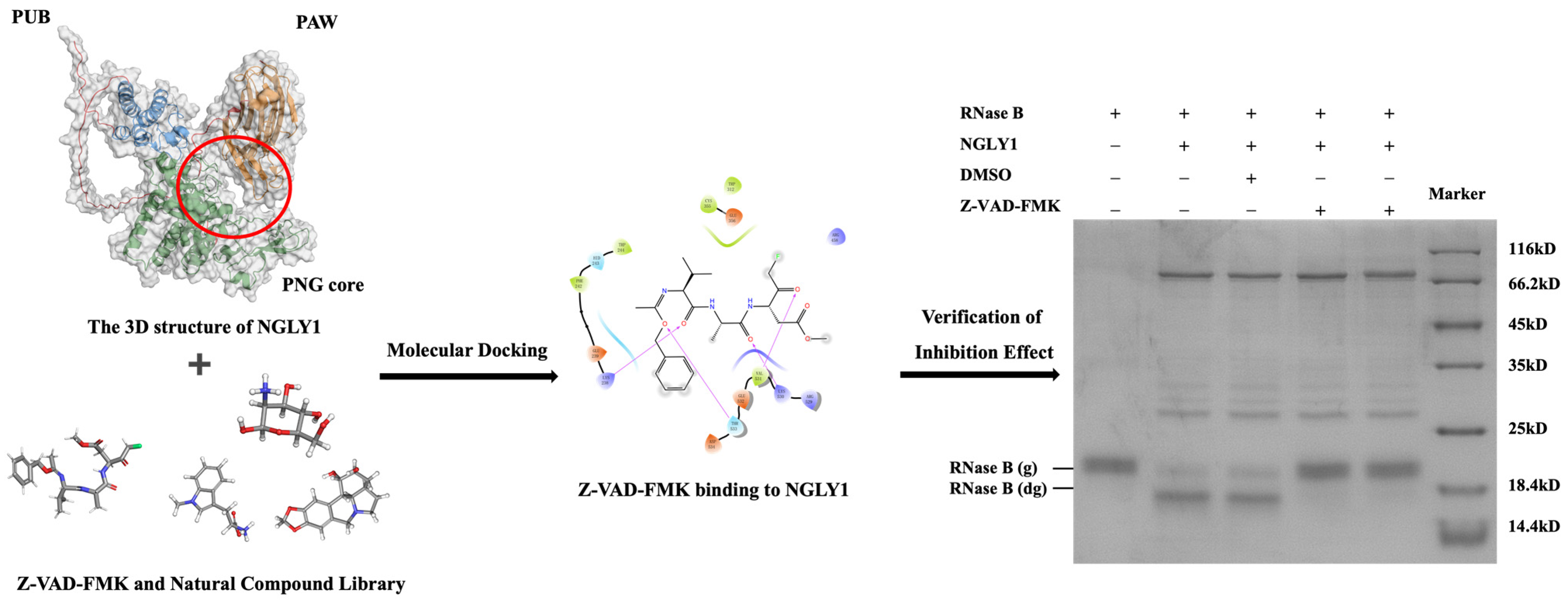
2.1. Establishment of a Screening Method for Targeting NGLY1 Lead Compounds

Molecular docking plays a pivotal role in the drug screening process. In this study, we developed a natural lead compound screening method targeting NGLY1. By predicting the three-dimensional structures of NGLY1, we performed molecular docking with 2960 natural compounds derived from plants, animals, or microorganisms. To identify potential inhibitors, we compared the docking scores of these compounds with that of the known NGLY1 inhibitor Z-VAD-FMK. Compounds exhibiting docking scores lower than −4.56 were then selected as primary screening compounds. The enzyme NGLY1 exhibits the same cleavage effect as PNGase F (Figure S1), and the inhibitory effects of the primary screening compounds were validated through an electrophoretic mobility shift assay. We validated the feasibility of this screening method using Z-VAD-FMK, as shown in Figure 2.
The lower binding affinity energy indicates stronger binding affinity. Among the 2960 compounds screened, a total of 215 compounds exhibited docking scores lower than −4.56. Considering the known biological activities of these compounds, we further narrowed down the selection to 17 compounds associated with glycosylation or possessing anti-inflammatory, antioxidant, and anti-tumor properties (Table S1). Among these 17 compounds, 13 of them contained glycan structures. Therefore, we speculated that compounds with glycan structures might have a higher affinity with NGLY1.

2.2. Verification of Inhibitory Effect of Primary Screening Compounds

The inhibitory effects of primary screening compounds were validated through the electrophoretic mobility shift assay. Specifically, Poliumoside, Soyasaponin Bb, and Saikosaponin B2 demonstrated effective inhibition of NGLY1 activity. The minimum inhibitory concentrations of Z-VAD-FMK, Poliumoside, Soyasaponin Bb, and Saikosaponin B2 were found to be 1 mM, 25 mM, 25 mM, and 50 mM, respectively (Figure 3). It is noteworthy that all three compounds were isolated from traditional heat-clearing and detoxifying Chinese herbs. The strongest dockings of NGLY1 inhibitors and their binding sites, interactions, and distances, are shown in Figure 3 and Table 1. NGLY1 inhibitors mainly bind to amino acids, such as Lys238, Glu239, Trp244, and Glu356, through hydrogen bonding.

2.3. The Analysis of Inhibitory Mechanism of NGLY1 Inhibitor

We compared the chemical structures of Poliumoside, Soyasaponin Bb, and Saikosaponin B2 (Figure 4A) and found that Poliumoside contains d-glucose and l-rhamnose, Soyasaponin Bb contains d-galactose (an enantiomer of d-glucose) and l-rhamnose, and Saikosaponin B2 contains d-glucose and d-fucose (an enantiomer of l-rhamnose). The results illustrated that the natural compounds with a core disaccharide structure of (d-glucose/d-galactose) and (l-rhamnose/d-fucose) may have inhibitory effects on NGLY1. We identified a compound, Rutinose (Figure 4A), which is a disaccharide structure containing d-glucose and l-rhamnose. The electrophoretic mobility shift assay showed that Rutinose inhibited NGLY1 in a dose-dependent manner at high concentrations (Figure 4B). As Poliumoside exhibited the highest potency, and the activity of Rutinose was confirmed, we utilized the disaccharide structure containing glucose and rhamnose as the core structural element for the NGLY1 inhibitor in this study.
The molecular docking results between Rutinose and NGLY1 showed five binding sites, Lys238, Glu239, Trp244, Thr533, and Asp534 (Figure 4C). Mutations were added to each of the five binding sites, and the results showed that E239K and T533A had no effect on NGLY1 activity, while K238D and D534K mutations led to the partial inactivation of NGLY1. The W244A mutation resulted in complete inactivation of NGLY1 (Figure 4D). Lys238, Trp244, and Asp534 were important catalytic sites of NGLY1. Poliumoside, Soyasaponin Bb, and Saikosaponin B2 might inhibit NGLY1 by binding to Lys238 or Trp244.

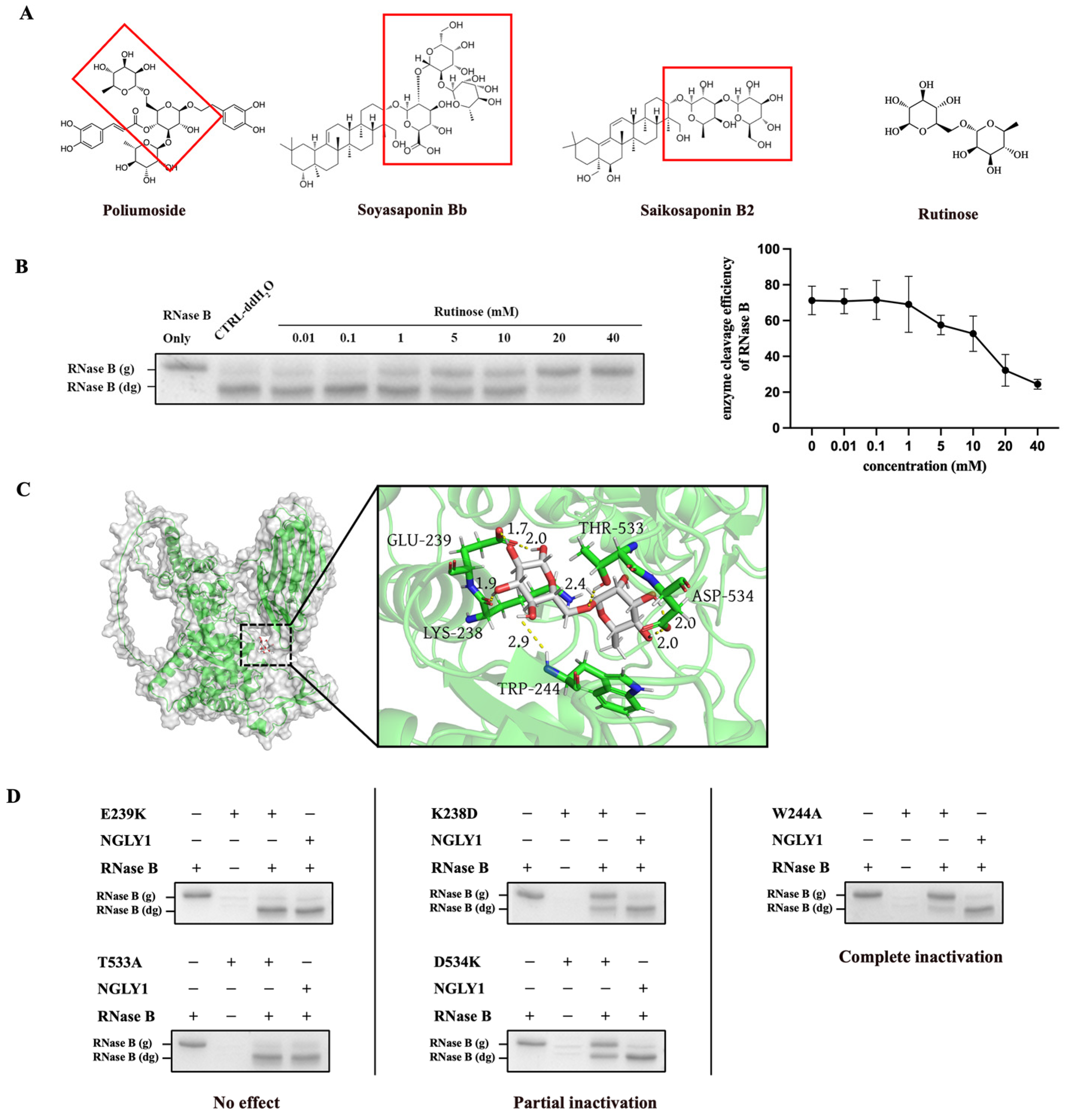
2.4. The Inhibitory Effects of Rutinose as the Core Structural Element for NGLY1 Inhibitor

To validate Rutinose as a core structure for NGLY1 inhibition, we identified two other compounds, Doismin and Rutin, which also contain D-glucose and L-rhamnose in their glycoside portion (Figure 5A,C). Through in vitro enzymatic assays, they were found to inhibit NGLY1 activity as well (Figure 5B,D), but their minimum inhibitory concentration was different. Therefore, we hypothesized that the glycoside composed of glucose and arabinose played a major inhibitory role but exhibited varying inhibitory effects due to different saponin ligands.

2.5. Toxicity Analysis

To ensure the safety of candidate drugs, toxicity testing of compounds was an important step. Calculation-based electronic toxicity measurement is widely utilized due to its accuracy and accessibility, providing information on any natural compound [32]. In order to determine the toxicity and adverse effects of the four NGLY1 inhibitors, we used the freely available testing tool ADMETlab 2.0 [33,34]. The website evaluates several toxicological parameters, such as human hepatotoxicity (H-HT), drug-induced liver injury (DILI), skin sensitization, carcinogenicity, eye corrosion, and eye irritation (Table 2). According to ADMETlab 2, Z-VAD-FMK and Poliumoside were highly probable to cause a drug-induced liver injury, with Poliumoside also posing a potential risk of skin allergy. Conversely, Soyasaponin Bb and Saikosaponin B2 exhibited a lower probability of toxicities.
Subsequently, we also performed CCK-8 assays of HEK293T cells treated with four NGLY1 inhibitors. After 24 h of compound exposure, Soyasaponin Bb did not significantly impact cell viability and the impact of Poliumoside, Soyasaponin Bb, and Saikosaponin B2 on cell viability was less than that of Z-VAD-FMK (Figure 6). In summary, the compound Soyasaponin Bb demonstrated a higher safety profile compared to Z-VAD-FMK, whereas Poliumoside was predicted to have potential compound toxicity.
The results were predicted by the ADMETlab 2.0 website (https://admetmesh.scbdd.com (accessed on 10 October 2023)), but the actual toxicity of the drugs should be determined through real experiments.

3. Discussion

In this study, we screened three natural compounds that can inhibit NGLY1 activity. Leveraging the predictive structure of NGLY1, we conducted computational virtual screening using a natural compound library. In the initial round of screening, seventeen natural compounds were evaluated, leading to the identification of three promising NGLY1 inhibitors: Poliumoside, Soyasaponin Bb, and Saikosaponin B2. An intriguing revelation emerged upon comparing the chemical structures of these inhibitors. Remarkably, we observed a disaccharide structure composed of glucose and rhamnose, which had been proven to be effective in inhibiting NGLY1 and could serve as a core structural element for NGLY1 inhibitors. This core disaccharide structure could also serve as a lead structure for the design and development of NGLY1 inhibitors. Based on the molecular interaction analysis, we proposed that Lys238, Trp244, and Asp534 are important catalytic sites of NGLY1. Our findings suggest that NGLY1 inhibitors likely impede its function by binding to Lys238 or Trp244. Consequently, during the screening process, it was crucial to focus on compounds that interact with Lys238 and Trp244, as they may serve as a basis for subsequent drug screening for NGLY1 inhibitors.
Comparatively, Poliumoside, Soyasaponin Bb, and Saikosaponin B2 demonstrated less potent inhibitory effects on NGLY1 when compared to Z-VAD-FMK. However, it is worth noting that these compounds are natural products derived from plants with fewer drug-related side effects. We conducted preliminary toxicity predictions using ADMETlab 2.0 website (https://admetmesh.scbdd.com (accessed on 10 October 2023)) and assessed their effects on cellular viability for the three specific compounds, and found that the compounds Soyasaponin Bb demonstrated higher safety profiles compared to Z-VAD-FMK, whereas Poliumoside was predicted to have potential compound toxicity. Further research is necessary to investigate the impact of these compounds on intracellular NGLY1 activity within cells and to conduct metabolomics-based toxicological research, focusing on aspects such as nephrotoxicity, hepatotoxicity, cardiotoxicity, and central nervous system toxicity [35,36,37].
It has been reported that NGLY1 deficiency could upregulate the expression of ISGs, enhancing the body’s innate antiviral capabilities [10,38]. Consequently, NGLY1 inhibitors have the potential to complement traditional antiviral drugs by contributing to an enhanced antiviral response. NGLY1 inhibitors have demonstrated not only antiviral properties but also significant implications in cancer treatment. In the treatment of blood cancers like multiple myeloma (MM) and mantle cell lymphoma (MCL), proteasome inhibitors play a crucial role [39,40]. However, resistance to protease inhibitors primarily arises from the upregulation of proteasome subunit (PSM) levels, with the expression of the PSM gene being regulated by the transcription factor Nuclear Factor, Erythroid 2 Like 1 (NFE2L1 or Nrf1) [39,41]. Importantly, Nrf1 is an N-glycosylated transmembrane protein, and its activation process involves the participation of NGLY1 [42,43]. Tomlin et al. [3] found that inhibiting NGLY1 in cultured cells disrupts Nrf1 function, enhancing the cytotoxicity of protease inhibitors. Notably, NGLY1 is highly expressed in certain human cancer cells, such as melanoma and ovarian cancer, while being low in their corresponding normal tissues like skin and ovary [44]. It has also been reported that the downregulation of NGLY1 results in melanoma cell death and a slowdown in tumor growth [45]. Compounds, like Poliumoside, Soyasaponin Bb, and Saikosaponin, exhibit promising therapeutic potential in various tumor diseases, including melanoma, multiple myeloma, and acute lymphocytic leukemia.
Our future work will encompass a comprehensive assessment of the broad-spectrum antiviral properties associated with NGLY1 inhibitors, employing various types of viruses for thorough evaluation. Additionally, we will also investigate the potential therapeutic utility of NGLY1 inhibitors in the treatment of melanoma, multiple myeloma, and acute lymphocytic leukemia. Current efforts are concentrated on scaffold optimization to refine the molecular structure of the inhibitors, aiming to enhance their inhibitory potential. Dubbu et al. [15] utilized the Prins cyclization to synthesize 2-Deoxy-β-C-aryl/alkyl glycosides, while Chennaiah et al. [16] synthesized vicinal 1,2-azidoacetates catalyzed by TMSOTf, offering a method for structural optimization of NGLY1 inhibitors.

4. Materials and Methods

4.1. Molecular Docking

A three-dimensional structure of NGLY1 was predicted online using AlphaFold2 based on the NGLY1 amino acid sequence (https://colab.research.google.com/github/sokrypton/ColabFold/blob/main/AlphaFold2.ipynb?pli=1#scrollTo=kOblAo-xetgx (accessed on 10 January 2022)). The SDF format structures of the natural compound library consist of 2960 compound structures’ information obtained from TargetMol (Boston, MA, USA).
NGLY1 consists of three domains: PUB, PNG Core, and PAW. The PNG Core plays the primary catalytic role. Preliminary experiments have shown that mutations of amino acids Trp236, Trp244, Cys283, Leu318, Cys355, Glu356, and Asp386 in the PNG Core domain result in protein inactivation. The receptor pocket was generated with the aforementioned amino acids as the center (x = 5.47, y = 4.28, z = 6.22). The receptor pocket was set as a cubic grid with a side length of 20 Å. Semi-flexible docking was employed. Structural analysis of the docking results was performed using PyMOL (2.5.2) software.

4.2. Construction of NGLY1 Mutant

Plasmid pET28a-NGLY1 (WT) was preserved by our laboratory. All NGLY1 mutants were generated using the reverse complement primers for PCR (Takara, Beijing, China) with the template of pET28a-NGLY1 (WT) plasmid. The constructed pET28a-NGLY1 (WT) plasmid and pET28a-NGLY1 (mutant) plasmid were transformed into E. coli BL21 (DE3) (Tiangen, Beijing, China) separately for protein expression. The primers can be found in Table 3.

4.3. Protein Expression and Purification of NGLY1 and NGLY1 Mutant

Escherichia coli BL21(DE3) cells harboring the NGLY1 and NGLY1 mutant expression plasmid were grown at 37 °C in a Luria-Bertani (LB) medium containing 50 μg/mL kanamycin. When the culture reached OD600 of 0.6–0.8, 1 mM isopropyl β-d-thiogalactoside (IPTG) (Sangon Biotech, Shanghai, China) was added to the medium and incubated further for 12 h at 28 °C.
The frozen cells were resuspended in a lysis buffer (20 mM Tris, 300 mM NaCl, and 10 mM imidazole; pH 7.4) and then disrupted by sonication, and insoluble materials were removed by centrifugation at 12,000 rpm for 30 min at 4 °C. The supernatant was loaded onto a Ni-NTA resin column (1 mL bed volume, Thermo Scientific, Walthamm, MA, USA) and incubated for 30 min at 4 °C. The column was washed with lysis buffers and wash buffers (20 mM Tris, 300 mM NaCl, and 25 mM imidazole; pH 7.4), and the protein was eluted with an elution buffer (20 mM Tris, 300 mM NaCl, and 250 mM imidazole; pH 7.4). The purified protein was concentrated using an amicon ultrafiltration concentrator (30kDa, Millipore, MA, USA). The protein concentration was determined by NanoDrop One spectrophotometer (Thermo Scientific, Walthamm, MA, USA).

4.4. The Electrophoretic Mobility Shift Assay

The natural compounds were purchased from Targetmol (Boston, MA, USA). Recombinant NGLY1 (2 μg) was incubated with a primary screening compound for 60 min at 37 °C, at which time denatured RNase B (New England Biolabs, Ipswich, MA, USA) (2 μg) was added. The mixture was incubated for 12–16 h at 37 °C before separation by SDS-PAGE (15%) and Coomassie staining. Further quantification was performed using Image J software. RNase B had a molecular weight of ~17 kDa. After NGLY1 mediated deglycosylation, the molecular weight of RNase B decreased to ~13.7 kDa. RNase B remained at ~17 kDa when NGLY1 activity was inhibited.

4.5. Toxicity Test

The safety profile of natural compounds was analyzed through calculation-based methods. ADMETlab 2.0 (https://admetmesh.scbdd.com (accessed on 10 October 2023)) was used to analyze the toxic effects of the NGLY1 inhibitors [33,34]. The ADMETlab 2.0 predicts the human hepatotoxicity (H-HT), drug induced liver injury (DILI), skin sensitization, carcinogenicity, eye corrosion, and eye irritation of the query compounds.

4.6. CCK-8 Analysis of Cell Viability Assay

HEK293T cells were cultured in Dulbecco’s modified Eagle’s medium (DMEM; Gibico, Shanghai, China) supplemented with 10% fetal bovine serum (FBS; Gibico, NSW, Australia) and 1% penicillin-streptomycin (Beyotime, Beijing, China) at 37 ℃ with 5% CO2. 5000 cells were seeded in each well of a 96-well plate. Cell viability was measured using the Cell Counting Kit-8 (Beyotime, Beijing, China) according to the manufacturer’s instructions.

5. Conclusions

In conclusion, NGLY1, an essential enzyme involved in the deglycosylation of misfolded glycoproteins through the ERAD pathway, has emerged as a pivotal enzyme with diverse therapeutic potential. Through a comprehensive structure-based virtual analysis of a natural compound library, and by validating the inhibitory effects of compounds using electrophoretic mobility shift assays, we identified three promising NGLY1 inhibitors: Poliumoside, Soyasaponin Bb, and Saikosaponin B2. These compounds were derived from traditional Chinese herbs with heat-clearing and detoxifying properties, displaying inhibitory activity against NGLY1. Preliminary toxicity predictions conducted through computer programs and analysis of cellular toxicity using the CCK-8 assays indicated that Soyasaponin Bb exhibited a higher safety profile in comparison to Z-VAD-FMK, while Poliumoside raised concerns regarding potential compound toxicity.
Moreover, it was revealed that the core structural motif shared by these inhibitors is a disaccharide structure featuring glucose and rhamnose. This structural insight suggested that their mode of action might involve binding to critical active sites of NGLY1, including Lys238 and Trp244, as they might serve as a basis for subsequent drug screening for NGLY1 inhibitors. Furthermore, natural compounds containing a disaccharide structure composed of glucose and rhamnose might also exert inhibitory effects on NGLY1.
Furthermore, NGLY1 inhibitors offer a multifaceted approach to address critical challenges in healthcare. They present a compelling solution to protease inhibitor resistance in blood cancer treatment and hold potential application in the management of melanoma. This study not only highlights the promise of Poliumoside, Soyasaponin Bb, and Saikosaponin B2 as lead compounds for NGLY1 inhibition but also provides a valuable foundation for future drug development efforts in the pursuit of effective antiviral and anticancer therapies.

Supplementary Materials

The following supporting information can be downloaded at: https://www.mdpi.com/article/10.3390/molecules28237758/s1. Refs [46,47,48,49,50,51,52,53,54,55,56,57,58,59,60,61,62] are cited in the Supplementary Material.

Author Contributions

Conceptualization, L.C.; methodology, R.L. and Y.Y.; software, S.Z. and Y.Z.; validation, J.G.; formal analysis, Q.L.; investigation, X.L.; data curation, Y.T. and S.L.; writing—original draft preparation, R.L.; writing—review and editing, G.S.; visualization, S.Y. and Y.C.; supervision, L.C.; project administration, G.S.; funding acquisition, G.S. All authors have read and agreed to the published version of the manuscript.

Funding

This research was funded by the Natural Science Foundation of Zhejiang Province (LY20C050002) and through a Research Project of Zhejiang Chinese Medical University (2023GJYY19).

Institutional Review Board Statement

Not applicable.

Informed Consent Statement

Not applicable.

Data Availability Statement

The datasets used and/or analyzed during the current study are available from the corresponding author on reasonable request.

Conflicts of Interest

The authors declare no conflict of interest.

References

  1. Srinivasan, B.; Zhou, H.; Mitra, S.; Skolnick, J. Novel small molecule binders of human N-glycanase 1, a key player in the endoplasmic reticulum associated degradation pathway. Bioorg. Med. Chem. 2016, 24, 4750–4758. [Google Scholar] [CrossRef] [PubMed]
  2. Suzuki, T.; Huang, C.; Fujihira, H. The cytoplasmic peptide: N-glycanase (NGLY1)—Structure, expression and cellular functions. Gene 2016, 577, 1–7. [Google Scholar] [CrossRef]
  3. Tomlin, F.M.; Gerling-Driessen, U.I.M.; Liu, Y.C.; Flynn, R.A.; Vangala, J.R.; Lentz, C.S.; Clauder-Muenster, S.; Jakob, P.; Mueller, W.F.; Ordoñez-Rueda, D.; et al. Inhibition of NGLY1 Inactivates the Transcription Factor Nrf1 and Potentiates Proteasome Inhibitor Cytotoxicity. ACS Cent. Sci. 2017, 3, 1143–1155. [Google Scholar] [CrossRef] [PubMed]
  4. Li, X.; Raihan, M.A.; Reynoso, F.J.; He, M. Glycosylation Analysis for Congenital Disorders of Glycosylation. Curr. Protoc. Hum. Genet. 2015, 86, 17.18.1–17.18.22. [Google Scholar] [CrossRef] [PubMed]
  5. Asahina, M.; Fujinawa, R.; Nakamura, S.; Yokoyama, K.; Tozawa, R.; Suzuki, T. Ngly1−/− rats develop neurodegenerative phenotypes and pathological abnormalities in their peripheral and central nervous systems. Hum. Mol. Genet. 2020, 29, 1635–1647. [Google Scholar] [CrossRef] [PubMed]
  6. Might, M.; Wilsey, M. The shifting model in clinical diagnostics: How next-generation sequencing and families are altering the way rare diseases are discovered, studied, and treated. Genet. Med. 2014, 16, 736–737. [Google Scholar] [CrossRef] [PubMed]
  7. Abuduxikuer, K.; Zou, L.; Wang, L.; Chen, L.; Wang, J.S. Novel NGLY1 gene variants in Chinese children with global developmental delay, microcephaly, hypotonia, hypertransaminasemia, alacrimia, and feeding difficulty. J. Hum. Genet. 2020, 65, 387–396. [Google Scholar] [CrossRef]
  8. Kariminejad, A.; Shakiba, M.; Shams, M.; Namiranian, P.; Eghbali, M.; Talebi, S.; Makvand, M.; Jaeken, J.; Najmabadi, H.; Hennekam, R.C. NGLY1 deficiency: Novel variants and literature review. Eur. J. Med. Genet. 2021, 64, 104146. [Google Scholar] [CrossRef]
  9. Lam, C.; Wolfe, L.; Need, A.; Shashi, V.; Enns, G. NGLY1-Related Congenital Disorder of Deglycosylation. In GeneReviews®; Adam, M.P., Mirzaa, G.M., Pagon, R.A., Wallace, S.E., Bean, L.J.H., Gripp, K.W., Amemiya, A., Eds.; University of Washington: Seattle, WA, USA, 2018. [Google Scholar]
  10. Yang, K.; Huang, R.; Fujihira, H.; Suzuki, T.; Yan, N. N-glycanase NGLY1 regulates mitochondrial homeostasis and inflammation through NRF1. J. Exp. Med. 2018, 215, 2600–2616. [Google Scholar] [CrossRef]
  11. Wu, K.X.; Phuektes, P.; Kumar, P.; Goh, G.Y.; Moreau, D.; Chow, V.T.; Bard, F.; Chu, J.J. Human genome-wide RNAi screen reveals host factors required for enterovirus 71 replication. Nat. Commun. 2016, 7, 13150. [Google Scholar] [CrossRef]
  12. Misaghi, S.; Pacold, M.E.; Blom, D.; Ploegh, H.L.; Korbel, G.A. Using a small molecule inhibitor of peptide: N-glycanase to probe its role in glycoprotein turnover. Chem. Biol. 2004, 11, 1677–1687. [Google Scholar] [CrossRef] [PubMed]
  13. Needs, S.H.; Bootman, M.D.; Grotzke, J.E.; Kramer, H.B.; Allman, S.A. Off-target inhibition of NGLY1 by the polycaspase inhibitor Z-VAD-fmk induces cellular autophagy. FEBS J. 2022, 289, 3115–3131. [Google Scholar] [CrossRef] [PubMed]
  14. Bhambhani, S.; Kondhare, K.R.; Giri, A.P. Diversity in Chemical Structures and Biological Properties of Plant Alkaloids. Molecules 2021, 26, 3374. [Google Scholar] [CrossRef] [PubMed]
  15. Dubbu, S.; Chennaiah, A.; Verma, A.K.; Vankar, Y.D. Stereoselective synthesis of 2-deoxy-β-C-aryl/alkyl glycosides using Prins cyclization: Application in the synthesis of C-disaccharides and differently protected C-aryl glycosides. Carbohydr. Res. 2018, 468, 64–68. [Google Scholar] [CrossRef] [PubMed]
  16. Chennaiah, A.; Bhowmick, S.; Vankar, Y.D. Conversion of glycals into vicinal-1,2-diazides and 1,2-(or 2,1)-azidoacetates using hypervalent iodine reagents and Me 3 SiN 3. Application in the synthesis of N-glycopeptides, pseudo-trisaccharides and an iminosugar. RSC Adv. 2017, 7, 41755–41762. [Google Scholar] [CrossRef]
  17. Yamasaki, S.; Asakura, M.; Neogi, S.B.; Hinenoya, A.; Iwaoka, E.; Aoki, S. Inhibition of virulence potential of Vibrio cholerae by natural compounds. Indian J. Med. Res. 2011, 133, 232–239. [Google Scholar]
  18. Zhang, L.; Song, J.; Kong, L.; Yuan, T.; Li, W.; Zhang, W.; Hou, B.; Lu, Y.; Du, G. The strategies and techniques of drug discovery from natural products. Pharmacol. Ther. 2020, 216, 107686. [Google Scholar] [CrossRef]
  19. Guo, X.; Yin, X.; Liu, Z.; Wang, J. Non-Alcoholic Fatty Liver Disease (NAFLD) Pathogenesis and Natural Products for Prevention and Treatment. Int. J. Mol. Sci. 2022, 23, 15489. [Google Scholar] [CrossRef]
  20. Ma, N.; Zhang, Z.; Liao, F.; Jiang, T.; Tu, Y. The birth of artemisinin. Pharmacol. Ther. 2020, 216, 107658. [Google Scholar] [CrossRef]
  21. Yu, D.L.; Lou, Z.P.; Ma, F.Y.; Najafi, M. The interactions of paclitaxel with tumour microenvironment. Int. Immunopharmacol. 2022, 105, 108555. [Google Scholar] [CrossRef]
  22. Yang, H.; Landis-Piwowar, K.R.; Chen, D.; Milacic, V.; Dou, Q.P. Natural compounds with proteasome inhibitory activity for cancer prevention and treatment. Curr. Protein Pept. Sci. 2008, 9, 227–239. [Google Scholar] [CrossRef] [PubMed]
  23. Choubdar, N.; Bhat, R.G.; Stubbs, K.A.; Yuzwa, S.; Pinto, B.M. Synthesis of 2-amido, 2-amino, and 2-azido derivatives of the nitrogen analogue of the naturally occurring glycosidase inhibitor salacinol and their inhibitory activities against O-GlcNAcase and NagZ enzymes. Carbohydr. Res. 2008, 343, 1766–1777. [Google Scholar] [CrossRef] [PubMed]
  24. Zhen, D.; Xuan, T.Q.; Hu, B.; Bai, X.; Fu, D.N.; Wang, Y.; Wu, Y.; Yang, J.; Ma, Q. Pteryxin attenuates LPS-induced inflammatory responses and inhibits NLRP3 inflammasome activation in RAW264.7 cells. J. Ethnopharmacol. 2022, 284, 114753. [Google Scholar] [CrossRef] [PubMed]
  25. Badshah, S.L.; Faisal, S.; Muhammad, A.; Poulson, B.G.; Emwas, A.H.; Jaremko, M. Antiviral activities of flavonoids. Biomed. Pharmacother. 2021, 140, 111596. [Google Scholar] [CrossRef] [PubMed]
  26. Ahmad, A.; Kaleem, M.; Ahmed, Z.; Shafiq, H. Therapeutic potential of flavonoids and their mechanism of action against microbial and viral infections—A review. Food Res. Int. 2015, 77, 221–235. [Google Scholar] [CrossRef]
  27. Śledź, P.; Caflisch, A. Protein structure-based drug design: From docking to molecular dynamics. Curr. Opin. Struct. Biol. 2018, 48, 93–102. [Google Scholar] [CrossRef]
  28. Ballante, F.; Kooistra, A.J.; Kampen, S.; de Graaf, C.; Carlsson, J. Structure-Based Virtual Screening for Ligands of G Protein-Coupled Receptors: What Can Molecular Docking Do for You? Pharmacol. Rev. 2021, 73, 527–565. [Google Scholar] [CrossRef]
  29. Kitchen, D.B.; Decornez, H.; Furr, J.R.; Bajorath, J. Docking and scoring in virtual screening for drug discovery: Methods and applications. Nat. Rev. Drug Discov. 2004, 3, 935–949. [Google Scholar] [CrossRef]
  30. Zhang, L.; Xu, J.; Guo, J.; Wang, Y.; Wang, Q. Elucidation of Pharmacological Mechanism Underlying the Anti-Alzheimer’s Disease Effects of Evodia rutaecarpa and Discovery of Novel Lead Molecules: An In Silico Study. Molecules 2023, 28, 5846. [Google Scholar] [CrossRef]
  31. Lu, F.; Luo, G.; Qiao, L.; Jiang, L.; Li, G.; Zhang, Y. Virtual Screening for Potential Allosteric Inhibitors of Cyclin-Dependent Kinase 2 from Traditional Chinese Medicine. Molecules 2016, 21, 1259. [Google Scholar] [CrossRef]
  32. Luo, L.; Zhong, A.; Wang, Q.; Zheng, T. Structure-Based Pharmacophore Modeling, Virtual Screening, Molecular Docking, ADMET, and Molecular Dynamics (MD) Simulation of Potential Inhibitors of PD-L1 from the Library of Marine Natural Products. Mar. Drugs 2021, 20, 29. [Google Scholar] [CrossRef] [PubMed]
  33. Xiong, G.; Wu, Z.; Yi, J.; Fu, L.; Yang, Z.; Hsieh, C.; Yin, M.; Zeng, X.; Wu, C.; Lu, A.; et al. ADMETlab 2.0: An integrated online platform for accurate and comprehensive predictions of ADMET properties. Nucleic Acids Res. 2021, 49, W5–W14. [Google Scholar] [CrossRef]
  34. Alkahtani, H.M.; Almehizia, A.A.; Al-Omar, M.A.; Obaidullah, A.J.; Zen, A.A.; Hassan, A.S.; Aboulthana, W.M. In Vitro Evaluation and Bioinformatics Analysis of Schiff Bases Bearing Pyrazole Scaffold as Bioactive Agents: Antioxidant, Anti-Diabetic, Anti-Alzheimer, and Anti-Arthritic. Molecules 2023, 28, 7125. [Google Scholar] [CrossRef] [PubMed]
  35. Chen, D.Q.; Chen, H.; Chen, L.; Tang, D.D.; Miao, H.; Zhao, Y.Y. Metabolomic application in toxicity evaluation and toxicological biomarker identification of natural product. Chem. Biol. Interact. 2016, 252, 114–130. [Google Scholar] [CrossRef] [PubMed]
  36. Rendedula, D.; Doddipalla, R.; Shaik, H.; Gupta, H.P.; Godugu, D.; Kristipati, R.R.; Mudiam, M.K.R. Multiplatform Metabolomics to Understand the Imidacloprid-Induced Toxicity in Drosophila. Chem. Res. Toxicol. 2023, 36, 1574–1583. [Google Scholar] [CrossRef] [PubMed]
  37. Zhou, J.; Chen, B.; Cai, Z. Metabolomics-based approach for assessing the toxicity mechanisms of dibutyl phthalate to abalone (Haliotis diversicolor supertexta). Environ. Sci. Pollut. Res. Int. 2015, 22, 5092–5099. [Google Scholar] [CrossRef]
  38. Decout, A.; Katz, J.D.; Venkatraman, S.; Ablasser, A. The cGAS-STING pathway as a therapeutic target in inflammatory diseases. Nat. Rev. Immunol. 2021, 21, 548–569. [Google Scholar] [CrossRef] [PubMed]
  39. Manasanch, E.E.; Orlowski, R.Z. Proteasome inhibitors in cancer therapy. Nat. Rev. Clin. Oncol. 2017, 14, 417–433. [Google Scholar] [CrossRef]
  40. Dou, Q.P.; Zonder, J.A. Overview of proteasome inhibitor-based anti-cancer therapies: Perspective on bortezomib and second generation proteasome inhibitors versus future generation inhibitors of ubiquitin-proteasome system. Curr. Cancer Drug Targets 2014, 14, 517–536. [Google Scholar] [CrossRef]
  41. Radhakrishnan, S.K.; Lee, C.S.; Young, P.; Beskow, A.; Chan, J.Y.; Deshaies, R.J. Transcription factor Nrf1 mediates the proteasome recovery pathway after proteasome inhibition in mammalian cells. Mol. Cell 2010, 38, 17–28. [Google Scholar] [CrossRef]
  42. Radhakrishnan, S.K.; den Besten, W.; Deshaies, R.J. p97-dependent retrotranslocation and proteolytic processing govern formation of active Nrf1 upon proteasome inhibition. eLife 2014, 3, e01856. [Google Scholar] [CrossRef]
  43. Suzuki, T.; Yoshida, Y. Ever-expanding NGLY1 biology. J. Biochem. 2022, 171, 141–143. [Google Scholar] [CrossRef] [PubMed]
  44. Uhlén, M.; Björling, E.; Agaton, C.; Szigyarto, C.A.; Amini, B.; Andersen, E.; Andersson, A.C.; Angelidou, P.; Asplund, A.; Asplund, C.; et al. A human protein atlas for normal and cancer tissues based on antibody proteomics. Mol. Cell. Proteom. 2005, 4, 1920–1932. [Google Scholar] [CrossRef]
  45. Zolekar, A.; Lin, V.J.T.; Mishra, N.M.; Ho, Y.Y.; Hayatshahi, H.S.; Parab, A.; Sampat, R.; Liao, X.; Hoffmann, P.; Liu, J.; et al. Stress and interferon signalling-mediated apoptosis contributes to pleiotropic anticancer responses induced by targeting NGLY1. Br. J. Cancer 2018, 119, 1538–1551. [Google Scholar] [CrossRef] [PubMed]
  46. Yu, S.Y.; Lee, I.S.; Jung, S.H.; Lee, Y.M.; Lee, Y.R.; Kim, J.H.; Sun, H.; Kim, J.S. Caffeoylated phenylpropanoid glycosides from Brandisia hancei inhibit advanced glycation end product formation and aldose reductase in vitro and vessel dilation in larval zebrafish in vivo. Planta Med. 2013, 79, 1705–1709. [Google Scholar] [CrossRef]
  47. Zhu, L.; Fu, R.; Liu, X.; He, Y.; Wang, B.; Ma, T. Soyasaponin Bb Protects Rat Hepatocytes from Alcohol-Induced Oxidative Stress by Inducing Heme Oxygenase-1. Pharmacogn. Mag. 2016, 12, 302–306. [Google Scholar]
  48. Jiang, K.; Lu, Q.; Li, Q.; Ji, Y.; Chen, W.; Xue, X. Astragaloside IV inhibits breast cancer cell invasion by suppressing Vav3 mediated Rac1/MAPK signaling. Int. Immunopharmacol. 2017, 42, 195–202. [Google Scholar] [CrossRef] [PubMed]
  49. Li, H.; Zhu, J.; Xu, Y.W.; Mou, F.F.; Shan, X.L.; Wang, Q.L.; Liu, B.N.; Ning, K.; Liu, J.J.; Wang, Y.C.; et al. Notoginsenoside R1-loaded mesoporous silica nanoparticles targeting the site of injury through inflammatory cells improves heart repair after myocardial infarction. Redox Biol. 2022, 54, 102384. [Google Scholar] [CrossRef]
  50. Wu, L.; Li, Q.; Liu, Y. Polyphyllin D induces apoptosis in K562/A02 cells through G2/M phase arrest. J. Pharm. Pharmacol. 2014, 66, 713–721. [Google Scholar] [CrossRef]
  51. Li, S.; Li, X.; He, F.; Jiao, R.; Zhang, S.; Li, Z. Amarogentin promotes osteoblast differentiation in oestrogen-deficiency-induced osteoporosis rats by modulating the Nrf-2/MAPK/ERK signalling pathway. Arch. Med. Sci. 2023, 19, 452–457. [Google Scholar] [CrossRef]
  52. Siraj, F.M.; SathishKumar, N.; Kim, Y.J.; Kim, S.Y.; Yang, D.C. Ginsenoside F2 possesses anti-obesity activity via binding with PPARγ and inhibiting adipocyte differentiation in the 3T3-L1 cell line. J. Enzym. Inhib. Med. Chem. 2015, 30, 9–14. [Google Scholar] [CrossRef] [PubMed]
  53. Liu, S.; Shen, H.; Li, J.; Gong, Y.; Bao, H.; Zhang, J.; Hu, L.; Wang, Z.; Gong, J. Loganin inhibits macrophage M1 polarization and modulates sirt1/NF-κB signaling pathway to attenuate ulcerative colitis. Bioengineered 2020, 11, 628–639. [Google Scholar] [CrossRef] [PubMed]
  54. Li, C.; Deng, C.; Pan, G.; Wang, X.; Zhang, K.; Dong, Z.; Zhao, G.; Tan, M.; Hu, X.; Shi, S.; et al. Lycorine hydrochloride inhibits cell proliferation and induces apoptosis through promoting FBXW7-MCL1 axis in gastric cancer. J. Exp. Clin. Cancer Res. 2020, 39, 230. [Google Scholar] [CrossRef]
  55. Lin, L.T.; Chung, C.Y.; Hsu, W.C.; Chang, S.P.; Hung, T.C.; Shields, J.; Russell, R.S.; Lin, C.C.; Li, C.F.; Yen, M.H.; et al. Saikosaponin b2 is a naturally occurring terpenoid that efficiently inhibits hepatitis C virus entry. J. Hepatol. 2015, 62, 541–548. [Google Scholar] [CrossRef] [PubMed]
  56. Song, Z.; Xu, X. Advanced research on anti-tumor effects of amygdalin. J. Cancer Res. Ther. 2014, 10 (Suppl. S1), 3–7. [Google Scholar]
  57. Matsuzaki, Y.; Koyama, M.; Hitomi, T.; Yokota, T.; Kawanaka, M.; Nishikawa, A.; Germain, D.; Sakai, T. Arctiin induces cell growth inhibition through the down-regulation of cyclin D1 expression. Oncol. Rep. 2008, 19, 721–727. [Google Scholar] [CrossRef] [PubMed]
  58. Jang, S.A.; Hwang, Y.H.; Yang, H.; Ryuk, J.A.; Gu, D.R.; Ha, H. Ethanolic extract of Pyrrosia lingua (Thunb.) Farw. ameliorates OVX-induced bone loss and RANKL-induced osteoclastogenesis. Biomed. Pharmacother. 2022, 147, 112640. [Google Scholar] [CrossRef]
  59. Manciu, F.S.; Guerrero, J.; Bennet, K.E.; Chang, S.Y.; Rahman, M.; Martinez Lopez, L.V.; Chantigian, S.; Castellanos, M.; Manciu, M. Assessing Nordihydroguaiaretic Acid Therapeutic Effect for Glioblastoma Multiforme. Sensors 2022, 22, 2643. [Google Scholar] [CrossRef]
  60. Yamashita, N.; Taga, C.; Ozawa, M.; Kanno, Y.; Sanada, N.; Kizu, R. Camalexin, an indole phytoalexin, inhibits cell proliferation, migration, and mammosphere formation in breast cancer cells via the aryl hydrocarbon receptor. J. Nat. Med. 2022, 76, 110–118. [Google Scholar] [CrossRef]
  61. Tan, C.; Zhu, W.; Lu, Y. Aloin, cinnamic acid and sophorcarpidine are potent inhibitors of tyrosinase. Chin. Med. J. 2002, 115, 1859–1862. [Google Scholar]
  62. Cao, B.; Zhao, R.Y.; Li, H.H.; Xu, X.M.; Cui, H.; Deng, H.; Chen, L.; Wei, B. Oral administration of asparagine and 3-indolepropionic acid prolongs survival time of rats with traumatic colon injury. Mil. Med. Res. 2022, 9, 37. [Google Scholar] [CrossRef]
Figure 1. Schematic diagram of antiviral effect by downregulating NGLY1 expression. NGLY1 is an essential enzyme involved in deglycosylation of misfolded glycoproteins. Knocking out the expression of NGLY1 leads to severe mitochondrial fragmentation, causing the release of mitochondrial DNA into the cytoplasm. Innate immune detection of self-DNA by the DNA sensor cGAS activates downstream STING-TBK1-IRF3 signaling cascade, inducing the expression of type I interferons (IFN) and IFN-stimulated genes (ISGs), effectively promoting an antiviral response [10]. mtDNA: mitochondrial DNA; cGAS: cyclic GMP–AMP synthase; cGAMP: 2′3′ cyclic GMP–AMP; STING: stimulator of interferon genes; COP-II: coatomer protein complex II; TBK1: TANK-binding kinase 1; IRF3: interferon regulatory factor 3; type I IFN: type I interferons; ISGs: IFN-stimulated genes.
Molecules 28 07758 g001
Figure 2. The screening method for targeting NGLY1 lead compounds. Red circle: Location of the receptor pocket. Red arrow: Location of the NGLY1. RNase B (g): N-glycosylated RNase B (NGLY1 is inhibited); RNase B (dg): N-glycosylated RNase B (NGLY1 is not inhibited).
Molecules 28 07758 g002
Figure 3. Analysis of interaction mode between NGLY1 inhibitors and NGLY1 protein, and NGLY1 inhibitors’ mediated inhibition of NGLY1 activity were tested at various concentrations using the electrophoretic mobility shift assay. (A) Sites of Z-VAD-FMK binding to NGLY1 and Z-VAD-FMK mediated inhibition of NGLY1 activity. (B) Sites of Poliumoside binding to NGLY1 and Poliumoside Bb mediated inhibition of NGLY1 activity. (C) Sites of Soyasaponin Bb binding to NGLY1 and Soyasaponin Bb mediated inhibition of NGLY1 activity. (D) Sites of Saikosaponin B2 binding to NGLY1 and Saikosaponin B2 mediated inhibition of NGLY1 activity. RNase B (g): N-glycosylated RNase B (NGLY1 is inhibited); RNase B (dg): N-glycosylated RNase B (NGLY1 is not inhibited).
Molecules 28 07758 g003
Figure 4. The inhibitory effect of Rutinose as a core structure of Poliumoside, Soyasaponin Bb, and Saikosaponin B2 on NGLY1. (A) Chemical structure of Poliumoside, Soyasaponin Bb, Saikosaponin, Rutinose. (B) Rutinose mediated inhibition of NGLY1 activity. Further quantification of the enzyme cleavage results was performed using Image J (Version 2.1.0) software. (C) Sites of Rutinose binding to NGLY1. (D) The activity of NGLY1 mutants. RNase B (g): N-glycosylated RNase B (NGLY1 is inhibited); RNase B (dg): N-glycosylated RNase B (NGLY1 is not inhibited). Red box: The glycan structure of the compound.
Molecules 28 07758 g004
Figure 5. Doismin and Rutin mediated inhibition of NGLY1 activity were tested at various concentrations using the electrophoretic mobility shift assay. (A,C) Chemical structure diagram of Diosmin and Rutin. (B) Diosmin and (D) Rutin mediated inhibition of NGLY1 activity were tested at various concentrations using the electrophoretic mobility shift assay. RNase B (g): N-glycosylated RNase B (NGLY1 is inhibited); RNase B (dg): N-glycosylated RNase B (NGLY1 is not inhibited). Red box: The glycan structure of the compound.
Molecules 28 07758 g005
Figure 6. CCK8 analysis of HEK 293T cells treated with NGLY1 inhibitors for 24 h.
Molecules 28 07758 g006
Table 1. Molecular docking information of 4 NGLY1 inhibitors.
CompoundDocking ScoreMolecular Formula Weight (g/mol)Noncovalent InteractionsAmino Acid Sites
Z-VAD-FMK−4.560C22H30FN3O7467.4944 H–bondLys238////Lys530Val531Thr533
Poliumoside−10.088C35H46O19770.7341 Pi–pi
6 H–bond
Lys238Glu239Trp244/Trp369/Val531/Glu544
Soyasaponin Bb−8.497C48H78O18943.1343 H–bondLys238/Trp244Glu356/////
Saikosaponin B2−6.007C42H68O13780.9935 H–bondLys238Glu239/Glu356/////
Table 2. Predictive toxicity properties of NGLY1 inhibitors.
TargetH-HTDILISkin Sensitization CarcinogenicityEye CorrosionEye Irritation
Z-VAD-FMK0.377 *
(−)
0.924
(+++)
0.401
(−)
0.238
(−−)
0.004
(−−−)
0.01
(−−−)
Poliumoside0.375
(−)
0.953
(+++)
0.971
(+++)
0.031
(−−−)
0.003
(−−−)
0.311
(−)
Soyasaponin Bb0.178
(−−)
0.044
(−−−)
0.2
(−−)
0.025
(−−−)
0.003
(−−−)
0.008
(−−−)
Saikosaponin B20.281
(−−)
0.008
(−−−)
0.053
(−−−)
0.024
(−−−)
0.003
(−−−)
0.006
(−−−)
*: The value is the probability of being toxic. For the classification endpoints, the prediction probability values are transformed into six symbols: 0–0.1 (−−−), 0.1–0.3 (−−), 0.3–0.5 (−), 0.5–0.7 (+), 0.7–0.9 (++), and 0.9–1.0 (+++). H-HT: human hepatotoxicity; DILI: drug induced liver injury.
Table 3. PCR primer sequences for constructing NGLY1 mutants.
MutantPrimer 5′-3′
K238DForwardCACCCAGTGAAAAAATTCTTCGTCAAACCAGTGCAAAAGCTCCAG
ReverseCTGGAGCTTTTGCACTGGTTTGACGAAGAATTTTTTCACTGGGTG
E239KForwardTCACCCAGTGAAAAAATTCTTTCTTAAACCAGTGCAAAAGCTC
ReverseGAGCTTTTGCACTGGTTTAAGAAAGAATTTTTTCACTGGGTGA
W244AForwardTGCTGCACAAAACGTTATTCACCGCGTGAAAAAATTCTTCCTTAAACC
ReverseGGTTTAAGGAAGAATTTTTTCACGCGGTGAATAACGTTTTGTGCAGCA
T533AForwardCCATGTGCCAGTCTGCTTCAACTTTTCTGAATATAGATTCCATT
ReverseAATGGAATCTATATTCAGAAAAGTTGAAGCAGACTGGCACATGG
D534KForwardCAAATATACCATGTGCCACTTTGTTTCAACTTTTCTGAATATAGATTCCATTTTCC
ReverseGGAAAATGGAATCTATATTCAGAAAAGTTGAAACAAAGTGGCACATGGTATATTTG
Disclaimer/Publisher’s Note: The statements, opinions and data contained in all publications are solely those of the individual author(s) and contributor(s) and not of MDPI and/or the editor(s). MDPI and/or the editor(s) disclaim responsibility for any injury to people or property resulting from any ideas, methods, instructions or products referred to in the content.

