Article

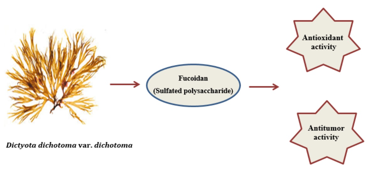
Chemical Composition, Antioxidant, and Antitumor Activity of Fucoidan from the Brown Alga Dictyota dichotoma

1
Department of Botany, Faculty of Science, Tanta University, Tanta 31527, Egypt
2
Department of Botany and Microbiology, Faculty of Science, Damietta University, New Damietta City 34511, Egypt
3
Department of Biological Science, College of Science and Arts at Rabigh, King Abdulaziz University, Rabigh 25732, Saudi Arabia
4
Faculty of Women for Ats, Science and Education, Ain Shams University, Cairo 11757, Egypt
*
Author to whom correspondence should be addressed.
Molecules 2023, 28(20), 7175; https://doi.org/10.3390/molecules28207175
Submission received: 13 September 2023 / Revised: 8 October 2023 / Accepted: 16 October 2023 / Published: 19 October 2023

Abstract

:
Brown macroalgae are a rich source of fucoidans with many pharmacological uses. This research aimed to isolate and characterize fucoidan from Dictyota dichotoma var. dichotoma (Hudson) J.V. Lamouroux and evaluate in vitro its antioxidant and antitumor potential. The fucoidan yield was 0.057 g/g algal dry wt with a molecular weight of about 48.6 kDa. In terms of fucoidan composition, the sulfate, uronic acid, and protein contents were 83.3 ± 5.20 mg/g fucoidan, 22.5 ± 0.80 mg/g fucoidan, and 26.1 ± 1.70 mg/g fucoidan, respectively. Fucose was the primary sugar component, as were glucose, galactose, mannose, xylose, and glucuronic acid. Fucoidan exhibited strong antioxidant potential that increased by more than 3 times with the increase in concentration from 0.1 to 5.0 mg/mL. Moreover, different concentrations of fucoidan (0.05–1 mg/mL) showed their ability to decrease the viability of Ehrlich ascites carcinoma cells in a time-dependent manner. These findings provided a fast method to obtain an appreciable amount of natural fucoidan with established structural characteristics as a promising compound with pronounced antioxidant and anticancer activity.

Graphical Abstract

1. Introduction

The Red Sea is a rich and highly productive ecosystem for marine organisms due to the unique coral reefs extending along the coastline and water temperature fluctuations. The Red Sea is a favorite aqua system for bioactive macroalgal growth along Egypt’s coasts. Approximately 500 seaweed species have been recorded in the Red Sea. Marine macroalgae gain significant importance due to their content of potent bioactive substances such as polysaccharides, fatty acids, heterocyclic carbons, alkaloids, cyclic peptides, and amino acids [1,2]. Brown algae are the largest group of algae, including 1500–2000 species, and are mostly marine and macroscopic. Dictyota dichotoma is a brown seaweed belonging to the family Dictyotaceae and the order Dictyotales. D. dichotoma has a flattened, dichotomously branched thallus without midribs and veins and is anchored to the substrate by rhizoids that terminate as an adhesive disc. D. dichotoma represents one of the most important algae in coral reef ecosystems [3].
In brown algae, Fucoidan, laminarin, alginate, and mannitol are the major storage carbon compounds that exhibit many novel physiological, biological, metabolic, and ecological characteristics. The reproductive phase and collection site play an important role in the content of these active principles and the biological activity of brown seaweed in vitro anti-inflammatory activities of fucoidans from five species of brown seaweeds [4]. Fucoidan is a sulfated polysaccharide extracted from brown algal cell walls such as those of Dictyota menstrualis, Padina boryana, Kjellmaniella crassifolia, and Fucus vesiculosus [5]. The extraction of fucoidan from some brown macroalgae involves multi-extractions, usually by using hot hydrochloric acid, and perhaps includes adding calcium to precipitate alginate during purification [6]. Still, no standardized extraction method is known for fucoidans at present. The extraction method significantly influences the yield and composition of algal polysaccharides [7].
Species of algae and their habitats greatly affect the structure of fucoidan and its chemical composition. Moreover, extraction methods significantly affect the structure and bioactivity of fucoidan [8,9,10,11]. Its bioactivity is due to its molecular characteristics, such as sugar types, sulfate content, linkages, and molecular geometry. Fucoidan could be identified based on its molecular weight by dividing it into low and high molecular weights. Specific Dictyota metabolites, such as fucoidans or polyphenols revealed antiviral, anti-nociceptive, antitumor, and anti-inflammatory activities [7,11]. Additionally, fucoidans obtained from Cystoseira crinite, Laminaria hyperborea, Fucus, F. evanescens, F. vesiculosus, F. distichus, and Ascophyllum nodosum showed antioxidant, antiviral, anti-inflammatory, anti-hyperglycemic, anti-coagulant, antidiabetic, anticancer, antiradical, and antibacterial activities [12,13,14,15,16,17,18,19]. Moreover, sulfated polysaccharides extracted from D. dichotoma var. velutricata, Dictyopteris polypodioides, and Turbinaria ornata showed excellent antioxidant activity [20,21].
Exposure to environmental dangers such as shortwave radiation, contamination, smoking, and herbicides can enhance the generation of free radicals that can destroy DNA, lipids, and essential proteins and cause various human disorders such as cancer, liver injury, and rheumatism. These disorders arise due to “oxidative stress”, which is a difference between the oxidant and antioxidant activities of the body. Antioxidants delay the autoxidation of cellular compartments by reducing the free radical’s formation or interrupting the propagation of the free radical chain with scavenging species and chelating metal ions, thus avoiding peroxide formation and/or reducing oxygen levels. Therefore, the antioxidant ability to scavenge free radicals may greatly prevent and remedy diseases arising from oxidants or free radicals. Hence, antioxidants are vital in diseases such as cancer, aging, inflammation, and Alzheimer’s [22,23]. Brown macroalgae are an excellent source of antioxidants due to their biodiversity and large amounts of various antioxidant compounds. Powerful antioxidant compounds in brown seaweeds include proteins, some pigments (chlorophyll and carotenoids), alkaloids, some vitamins (E and C), glutathione, sulfated polysaccharides, amino acids, amines, and phenolics as flavonoids and coumarins [24,25,26].
Cancer is a major disease burden worldwide and can arise in many body parts. Chemotherapy treats many types of cancer effectively. But like other chemical treatments, it often causes side effects, such as headaches, muscle pain, hair loss, and stomach pain. Marine bioactive products with novel structures can treat some diseases. Till now, cancer treatments do not have safe medicine as the currently available drugs are causing side effects such as vomiting, diarrhea, fatigue, and nausea. Hence, exploring and identifying new safe, cheap, and less toxic anticancer agents from natural sources is important. Nowadays, most pharmaceutical products are derived from various microorganisms, herbal plants, and seaweeds. Fucoidan is a non-toxic compound with therapeutic potential for some diseases. Kim and his colleague reported that fucoidan is used to treat cancer by directly activating different pathways of apoptosis [27]. Our study aimed to find new medications regarding the low side effects of natural marine compounds. Therefore, this work is the first aimed at isolating and characterizing fucoidan from D. dichotoma var. dichotoma (Hudson) J.V. Lamouroux and evaluating the extracted fucoidan in vitro antioxidant and antitumor activity.

2. Results

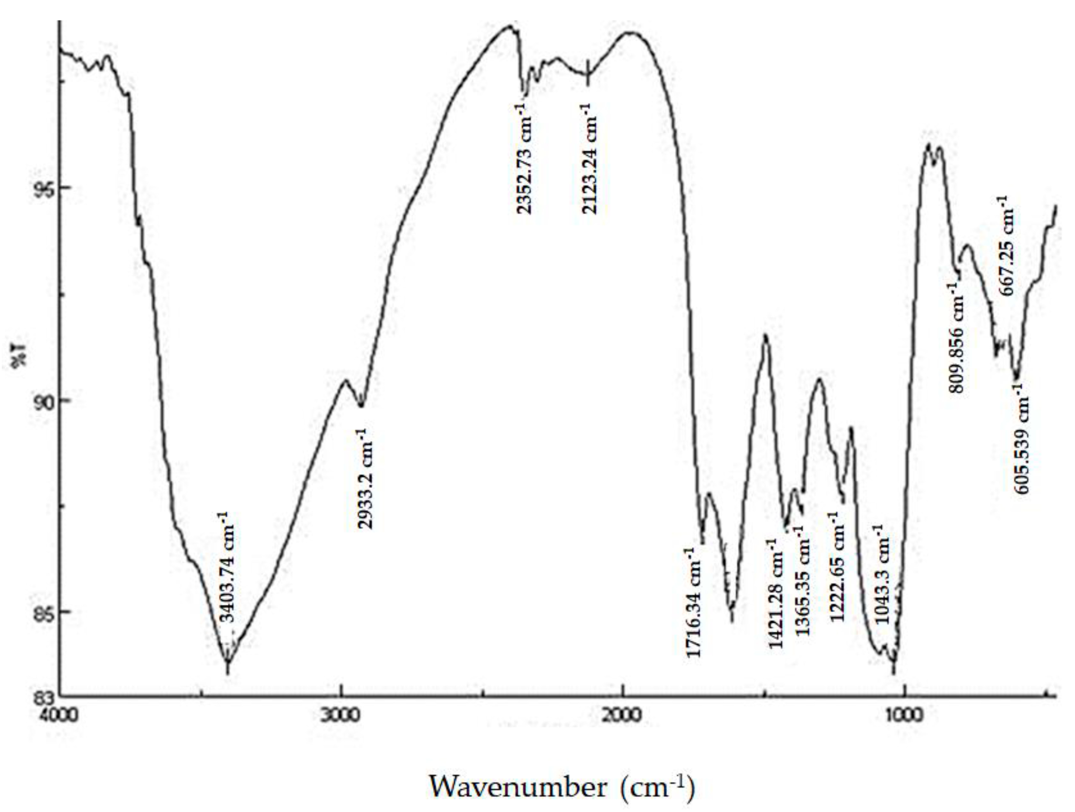
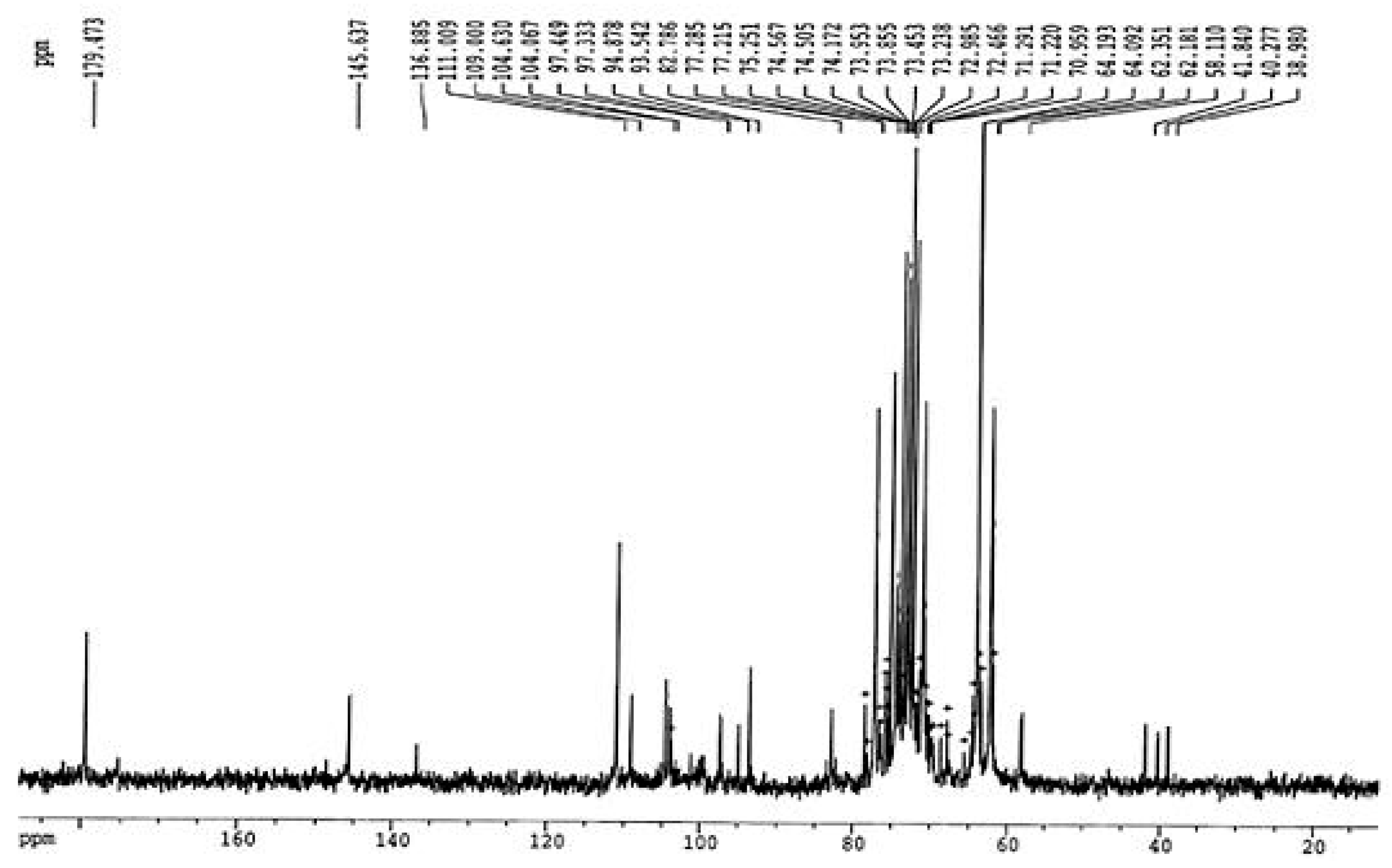
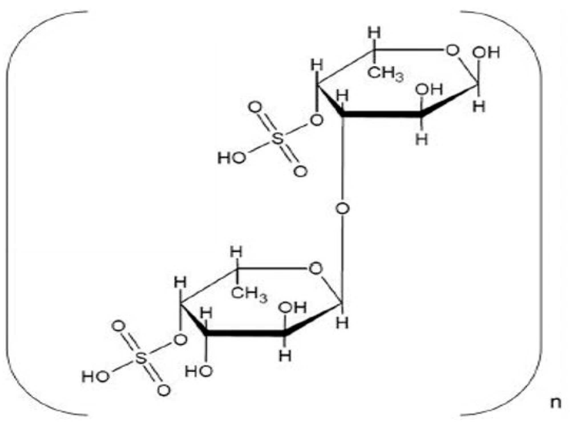
2.1. Structure and Chemical Composition of Isolated Fucoidan

In the current study, the functional groups of the extracted fucoidan powder from D. dichotoma var. dichotoma were analyzed by FTIR spectroscopy, and the spectrum is given in Figure 1. Lyophilized fucoidan powder showed bands in the regions of 1043.3 and 1222.65, confirming that it is an acidic polysaccharide. The stretching of O–C–O vibration (asymmetric carboxylate) was indicated by asymmetric stretching of carboxylate vibration at 1612.2 and 1716.34 cm−1. The bands at 1421.28 and 1365.35 cm−1 were assigned to C–OH with a contribution of O–C–O symmetric stretching vibration of the carboxylate group. One of the most important bands was found at 1043.3 cm−1, indicating D-glucose, and 1222.65 cm−1, corresponding to ester sulfate groups.
The results of 1H NMR spectral analysis of the purified fucoidan extracted from D. dichotoma var. dichotoma are given in Figure 2. 1H NMR showed that signals of protons H-1 arising from α-L-fucose residues and uronic acid residues appeared at alpha-anomeric carbon (5.06 ppm) and C2–C4 hydrogen atoms of a carbohydrate. Signals at 5.69 ppm indicated a 3,4 distribution of α-L-fucose. Signals at 4.87, 4.29, and 4.77 ppm confirmed the shifts of hydrogen atom positions at C2, C3, and C5, respectively. The signal at 1.37 ppm confirmed chemical shifts of hydrogen atoms C6. The double peak at 1.37 ppm corresponded to L-fucopyranose containing two methyl protons at C6. In Figure 3, 13C NMR showed sharp signals at 38.98 and 179.5 ppm, corresponding to important O-acetyl carbon regions. It confirmed the presence of the acetyl group. Anomeric carbon signals were obtained at 104.1 and 93.54 ppm. Both signals at 82.78 and 77.22 ppm show the presence of C4 with a sulfate moiety and the C3 position, respectively. From FTIR and NMR spectra, the structural characterization of the fucoidan compound obtained from D. dichotoma var. dichotoma was presented in Figure 4, which showed a repeated α-(1→3)-linkage. C-4 is always substituted with sulfate groups.
In the current study, the mean sulfate and uronic acid content of the fucoidan extracted from D. dichotoma var. dichotoma were 8.33 ± 0.52% and 2.25 ± 0.08%, respectively. Moreover, the extracted fucoidan contained 2.61 ± 0.17% protein (Table 1).
As shown in Table 1, HPLC analysis revealed the presence of six monosaccharides, including fucose, glucose, galactose, glucuronic acid, mannose, and xylose, in the extracted Fucoidan. Fucose was the main sugar (21.3 ± 1.30%) in fucoidan obtained in this study, followed by glucose (11.9 ± 0.74%). Glucuronic acid comprises 2.09 ± 0.12%, the lowest % in the sugar composition of the extracted Fucoidan.
The fucoidan yield was 0.057 g/g algal dry weight in the present study. In contrast, the average molecular weight measured by gel filtration using dextrans as a standard for fucoidan was 48.6 kDa.

2.2. Physical Characteristics of Fucoidan

The pH value of 1% aqueous fucoidan extracted from D. dichotoma var. dichotoma was 6.5. The solubility of fucoidan from D. dichotoma var. dichotoma in different solvents at room temperature was determined. The results revealed that fucoidan was highly soluble in water and sulfuric acid with a maximum saturation of 250 and 200 mg/mL, respectively, and partially soluble in hydrochloric acid with a maximum saturation of 50 mg/mL.

2.2.1. Antioxidant Activity of Fucoidan

Total antioxidant capacity, DPPH, H2O2, ABTS, nitric oxide, and ferrous ion assays were used as easy, rapid, and sensitive methods to determine the scavenging capacity of the extracted fucoidan. This is followed by the calculation of IC50 values (with 95% confidence intervals). In the present study, a wide range of fucoidan concentrations (0.1–5.0 mg/mL) were screened for their antioxidant potential compared to standard ascorbic acid (positive control). The results in Table 2 indicate that fucoidan from D. dichotoma var. dichotoma expressed appreciable antioxidant potential that increased significantly (p < 0.05) with increased concentration. The extracted fucoidan showed high total antioxidant capacity compared to standard ascorbic acid, with a maximum activity of 71.76 ± 1.5% at 5.0 mg/mL; and low IC50 (1.41 mg/mL). Moreover, fucoidan has strong scavenging activities against DPPH radicals at different concentrations, with an IC50 of 4.59 mg/mL. The maximum activity of fucoidan was recorded as 50.51 ± 1.9% at 5.0 mg/mL. In terms of total antioxidant capacity measured by the other assays, the values reported followed a similar trend to that observed for the DPPH scavenging capacity. Different concentrations of fucoidan obtained from D. dichotoma var. dichotoma exhibited their ability to scavenge hydrogen peroxide, ABTS, and nitric oxide radicals with an IC50 of 4.84 mg/mL, 8.55 mg/mL, and 9.30 mg/mL, respectively. The isolated fucoidan exhibited a low ability to chelate ferrous ions with high IC50 (61.9 mg/mL). The values of all fucoidan concentrations showed higher scavenging activity than standard ascorbic acid (positive control).

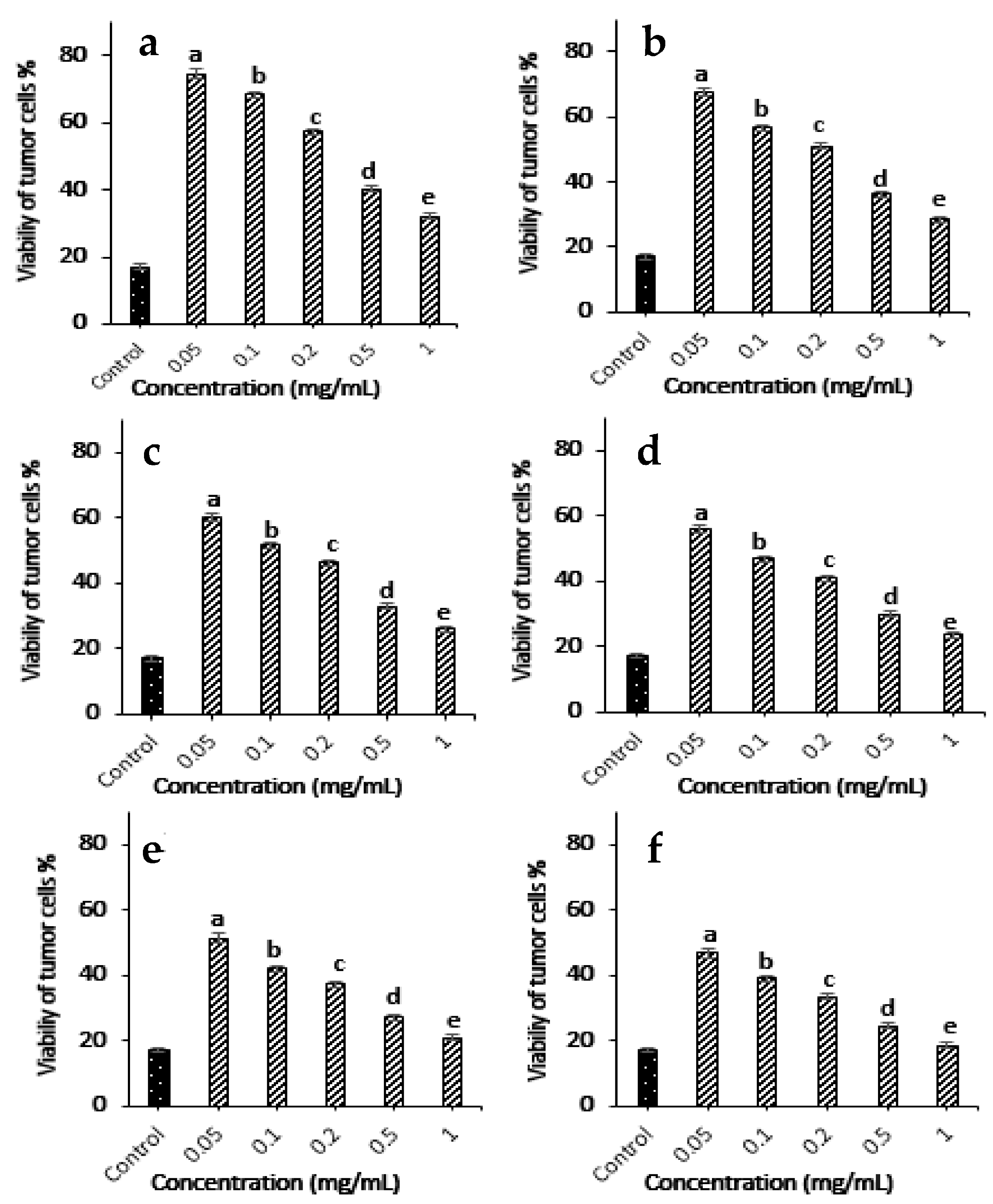
2.2.2. In Vitro Antitumor Activity

The present results reveal that different concentrations of fucoidan extracted from Dictyota dichotoma var. dichotoma in the range (0.05–1 mg/mL) showed their ability to reduce the viability of Ehrlich ascites carcinoma cells, as shown in Figure 5. These results indicated that treatment with different concentrations of fucoidan reduced tumor cell viability significantly in a time-dependent manner (p < 0.05). The high concentration (1 mg/mL) of fucoidan can also reduce tumor cell viability to 18.3% after 30 min.

3. Discussion

FTIR and NMR spectroscopy are important techniques used to deduce the structure of complex polysaccharides. The broadband at 3403.74 cm−1 was assigned to the hydrogen-bonded O–H stretching vibration of the polysaccharide, and that at 2933.2 cm−1 corresponds to the C-6 groups of fucose and galactose units. The characteristic absorption at 809.95 cm−1 indicated the α-configuration of the sugar units and the characteristic C–O–S stretching of the sulfate group [28]. The occurrence of sulfate groups in the extracted polysaccharide is represented by peaks at 800–900 cm−1. The higher sulfate revealed antioxidant activity [29]. The 605.53 and 667.25 cm−1 absorption indicated sulfate ester groups and revealed the sulfate group’s symmetric and asymmetric O=S=O deformation [30]. FTIR and NMR spectra in the present study revealed the presence of fucose-sulfated groups linked via repeated α-(1→3)-linkage, and C-4 is always substituted with sulfate groups, whereas the backbone of fucoidan from Fucus distichus is made up of alternating 2-sulfated 1,3- and 1,4-linked α-L-fucose residues [31]. NMR and monosaccharide analyses showed that fucose content (21.3 ± 1.30%) is higher than galactose (5.14 ± 0.65%). This confirms that more fucose molecules are present in the side chains and branches of the polysaccharide.
As shown in Figure 4, the FTIR and NMR spectra of the extracted fucoidan exhibit notable differences in absorption patterns to those of fucoidans extracted from Sargassum cristaefolium, Sargassum sp., Turbinaria sp., Fucus vesiculosus, and Padina sp. [32,33,34,35]. This confirmed that fucoidan composition is influenced by the extraction method and algal species. This difference in the chemical structure of fucoidan is also perhaps because of complex environmental factors such as temperature, light intensity, light day length, and nutrient concentration, in addition to algal changes such as growth, morphology, and reproduction. Moreover, some previous studies reported that the reproductive phase has a significant impact on the biochemical composition and antioxidant properties of fucoidan, as extracted from Fucus vesiculosus [15].
The average yield of fucoidan extracted from D. dichotoma var. dichotoma in the present work (5.7%) was higher than that obtained for Fucus spiralis (1.35%), Cystoseira crinite (2.80%), C. sedoides (3.30%), Sargassum tenerrimum (3.60%), C. compressa (3.70%), Fucus versiculosus (4.00%), and S. myriocystum (5.52%) [23,36,37], but lower than the yields reported from Sargassum linifolium (13.04%) and Stypopodium schimperi (9.09%), Undaria pinnatifida (8.8%), Fucus distichus (8.69), Sargassum wightii (7.15%), and Sargassum polycystum (7%) [18,38]. The difference in fucoidan yields was possibly due to the differences in algal species, collection area, degree of maturation, and extraction procedures.
The molecular weight of fucoidan differs according to algal species and environmental conditions. This agrees with Hahn [39] and Kawamoto [40]. They found that the molecular weight of fucoidan ranges from 13 to 627 kDa and may reach 3080 kDa.
Fucoidans extracted from brown macroalgae can be low molecular weight fucoidans, medium molecular weight fucoidans, or high molecular weight fucoidans with a polymer size less than ten kDa, ranging from 10 to 10,000 kDa, or more than 10,000 kDa, respectively [40]. The average molecular weight of fucoidan from D. dichotoma var. dichotoma was 48.6 kDa (low molecular weight fucoidan). The molecular weight of fucoidan (48.6 kDa) obtained in this study was smaller than those reported for Undaria pinnatifida (190 kDa), Ascophyllum nodosum (417 kDa), Saccharina longicruris (454 kDa), and Fucus vesiculosus (735 kDa), and higher than those reported for Laminaria japonica (10.5 kDa) and Saccharina longicruris (44 kDa) [41,42,43,44,45,46,47,48].
Fucoidan was insoluble in all tested organic solvents in the present study, including ethanol, methanol, acetone, chloroform, diethyl ether, and petroleum ether. These results were confirmed by Hahn [39], who concluded that fucoidan from brown algae is freely soluble in solvents of higher dielectric constants as water due to isolated shielded opposite groups and isolated from other co-extracted natural compounds by solvents of lower dielectric constants as ethanol.
Fucoidan is a sulfated polysaccharide mainly containing fucose, sulfate, monosaccharides, uronic acids, and protein [45]. It was observed that the sulfate content of the fucoidan obtained from D. dichotoma var. dichotoma was higher than that of fucoidan extracted from Colpomenia sinuosa (5.8%) and lower than that found in Hydroclathrus (18.1%) and F. distichus (38.3%) [31,45]. Yang [33] reports that the bioactivities of fucoidan are strongly related to its sulfate content. The uronic acid content of fucoidan extracted from Sargassum stenophyllum, and Cladosiphon okamuranus is 3.5% and 23.4%, respectively [38,47]. The fucoidan obtained from D. dichotoma var. dichotoma contained 2.61 ± 0.17% protein, which was lower than those reported in fucoidan extracted from Sargassum binderi Sonder (5.5%), and S. longicruris (12.4%) [48]. The difference in protein content was possibly due to the differences in algal species and extraction procedures.
The present study revealed the presence of fucose, glucose, galactose, mannose, xylose, and glucuronic acid in the extracted fucoidan. A similar result has been reported by Dias [49] on fucoidan extracted from Sargassum stenophyllum, which had the same six monosaccharides as the main components. Xylose is one of the common minor carbohydrates in brown seaweeds. Fucose, xylose, and their ratios could be used to characterize brown seaweeds as indicators of fucoidan. The ratio of fucose to xylose in fucoidan extracted in the present study (8.38) was lower than that in fucoidan extracted from F. distichus (10.42) collected from the western coast of Iturup Island in the summer [37]. It can also provide information about the biological activity of brown seaweeds [5]. Previous reports found that the fucose content of fucoidan obtained from Padina tetrastromatica, Fucus evanescens, S. tenerrimum, Cystoseira sedoides, C. compressa, and C. crinite was 54%, 54.9%, 59.3%, 54.5%, 61.5%, and 43.3%, respectively [37], which were more than the content of fucose sugar obtained in this study. Whereas, the fucose:sulfate ratio in fucoidan isolated from D. dichotoma in the present study (2.55:1) differs from that isolated from F. distichus (1:1.21) [31].
In the present study, different concentrations of fucoidan exhibited considerable antioxidant activity that increased with the increase in concentration. Other researchers agreed with the present study that sulfated polysaccharides obtained from some seaweed and fruits showed dose-dependent reducing powers and free radical scavenging effects.
In the phosphomolybdenum method, Mo (VI) is reduced to form a green phosphate–Mo (V) complex at an acid pH. The scavenging activity percentage of the extracted fucoidan (16.26%) at 0.1 mg/mL was lower than those reported for standard ascorbic acid (84%), and fucoidan obtained from Sargassum swartzii (32.34%) [50,51]. DPPH is a stable radical scavenged by an antioxidant compound through hydrogen donation to form a stable yellow DPPH-H molecule. The range of mean values of DPPH (%) in the present study (15.07–51.13%) differs from those reported for algal extracts of Ulva lactuca (4.85–76.0%), Enteromorpha intestinalis (8.66–76%), and Cladophora vagabunda (14.6–78.5%), with a high IC50 value (4.59 mg/mL). This DPPH IC50 value of the extracted fucoidan was also higher than that of alginate extracted from Colpomenia sinuosa (46.2 µg/mL−1) [52]. Hence, the effect of the extracted fucoidan on DPPH radical scavenging could be due to their hydrogen-donating ability or may be related to their high sulfate content. A high percentage of DPPH radical scavenging activity was also reported in the methanol extract of brown seaweed, Turbinaria conoides, Polycladia indica, Turbinaria ornata, and Laurencia obtusa, and red seaweed, Sarconema scinaioides [53].
The ABTS radicals are produced by the reaction of ABTS with H-atom donor antioxidants. These radicals convert into a non-colored form of ABTS, and absorbance decreases. In this study, the maximum activity was found at a concentration of 5 mg/mL to be 50.51%, which was lower than those stated by Tariq [20] for polysaccharides obtained from D. dichotoma var. velutricata (76.87%) and Sargassum variegatum (86.93%). This activity may be related to the fucoidan’s ability to act as a free radical scavenger or by donating a hydrogen atom to the molecule.
Antioxidant compounds can scavenge free radicals, for instance, nitric oxide, by donating protons. This study showed that the extracted fucoidan (at different levels) in sodium nitroprusside solution could decrease the nitrite levels with an IC50 of 9.30 mg/mL by their ability to chelate nitric oxide. The reduction in the nitrite level may be due to competition between fucoidan and oxygen in the reaction with NO [54].
Ferrozine can react with Fe2+ and form a red ferrozine–Fe2+ complex in a ferrous ion chelating assay. Still, in the presence of ion-chelating agents (antioxidant compounds), the complex formation is disrupted, resulting in a decrease in the red color of the complex. The capacity of ferrous ion chelating is expressed by the inhibition percentage of ferrozine–Fe2+ complex formation. Fucoidan obtained from D. dichotoma var. dichotoma exhibits a low chelating ability to ferrous ions with a high IC50 (61.9 mg/mL) at diverse concentrations. The ability (11.70 ± 0.6%) of 1 mg/mL fucoidan obtained in this study was lower than those reported in previous studies [54,55,56]. This difference may be due to alterations in algal species and the fucoidan extraction method. The antioxidant activity of the extracted fucoidan may be due to its effect as a ROS scavenger by removing free hydroxyl radicals and superoxide radicals. There are several factors that determine the antioxidant activity of fucoidan, including concentration, molecular weight, degree of sulphation, substitution groups and their positions, type of sugar, and glycosidation branching [57,58,59,60].
Some existing organisms in the marine ecosystem, such as macroalgae, are rich sources of bioactive substances that exhibit antitumor potential in Ehrlich’s ascites carcinoma cells. It was selected in the present study because it is one of the most common experimental tumor models characterized by high translatability, rapid replication, a short lifespan, and no tumor-specific transplantation antigen. It comprises both tumor cells present as single cells, or spheroids, and stromal cells. Moreover, it was easy to obtain in relatively large amounts at a low cost from the peritoneum of mice. Diverse concentrations of the extracted fucoidan from 0.05 to 1 mg/mL showed their ability to decrease the viability of Ehrlich ascites carcinoma cells in a time-dependent manner. The air-dried powder of Sargassum ringgoldianum, Scytosiphon lomentaria, Lessonia nigrescens, as well as Laminaria japonica showed significant inhibition against Ehrlich carcinoma cells of 46.5%, 69.8%, 60.0%, and 57.6%, respectively. Wang [57] also reported that fucoidan obtained from Sargassum thunbergii inhibits the viability of Ehrlich ascites carcinoma and lung cancer in vivo by inhibiting tumor angiogenesis. Moreover, sulfated polysaccharides from the brown seaweed Dictyota caribaea delayed tumor growth in mice bearing sarcoma in vivo without cytotoxic activity against tumor cells in vitro [59]. Arumugam [61] reported that fucoidan had more significant anticancer activity compared to quercetin. Previous studies reported that fucoidan has a broad range of impacts on cellular functions, including cell cycle regulation, RNA metabolism, protein metabolism, carbohydrate metabolism, bioenergetics, mitochondrial maintenance, and DNA repair pathways. Induction/inhibition of reactive oxygen species (ROS), mitochondrial instability, and caspase and poly (ADP-ribose) polymerase (PARP) cleavage are all aspects of it [62,63].

4. Materials and Methods

4.1. Collection of Algae

Dictyota dichotoma var. dichotoma (Hudson) J.V. Lamouroux was collected at Hurghada, Egypt, on the Red Sea. Its fronds were harvested at the reproductive phase of the vegetatively propagated gametophyte in autumn 2017 and rinsed with seawater to remove epiphytes and debris, then tap and distilled water many times to remove salts. Blotting paper removed surplus water from D. dichotoma fronds. The sample was shade-dried and oven-dried at 60 °C for 4 h. The dried algae were pulverized into 2 mm particles. Dr. M. Deyab recognized the alga samples, and the voucher specimen was placed at the Phycology Laboratory (Damietta University, Faculty of Science).

4.2. Extraction and Purification of Fucoidan

With few changes to the Yang technique [62], fucoidan was isolated from D. dichotoma var. dichotoma (Hudson) J.V. Lamouroux fronds. About 30 g of powdered fronds was shaken in a 400 mL solvent system (ethanol:water:formaldehyde) for 24 h at room temperature to remove pigments and polyphenols. Then the mixture was centrifuged at 4000 rpm for 15 min. The supernatant was withdrawn, and the algal residue was washed several times with 400 mL of acetone, followed by centrifugation at 4000 rpm for 10 min. The supernatant was withdrawn, and the residue dried at room temperature, then extracted with 500 mL of 0.2 N HCl for 24 h and filtered through a nylon cloth. Extraction was repeated with 500 mL of 0.2 N HCl at 70 °C for 2 h, followed by 100 mL of distilled water at 65 °C for 2 h. The combined extracts were neutralized with 0.5 N NaOH and filtered through a nylon cloth. The filtrate was incubated with 1% CaCl2 in a ratio of 2:1 at 4 °C for 24 h to precipitate alginates. After centrifugation at 4000 rpm for 15 min, the supernatant was incubated overnight with 1 L of 99% ethanol at 4 °C. The precipitate (crude fucoidan) was collected after centrifugation at 6000 rpm for 15 min and spread to dry at room temperature overnight [39,64,65].

4.3. Fourier Transforms Infrared (FTIR) Spectroscopy

The extracted powder was identified according to the method described by Kemp [66].

4.4. Hydrolysis of Fucoidan

The extracted fucoidan was hydrolyzed by the method described by Nagaoka [67].

4.5. Nuclear Magnetic Resonance (NMR)

Three milligrams of purified fucoidan powder were dissolved in 0.5 mL of 99.96% D2O (deuterated water) and analyzed by 1H and 13C NMR on a Bruker Biospin Avance 400 NMR spectrometer (Bruker, Billerica, MA, USA) (1H frequency = 400.13 MHz, 13C frequency = 100.62 MHz) at 298 °K using a 5 mm inverse broadband probe head with a shielded z-gradient and XWIN-NMR software version 3.5 with TMS as One pulse sequence produced one-dimensional 1H and 13C spectra. One-dimensional 13C spectra utilizing SEFT and QCD 42 sequences were also used to identify the structure.

4.6. Molecular Weight Analysis

The molecular weight of pure fucoidan was measured using a size-exclusion HPLC column Superdex 200 (300 mm × 10 mm ID) and a SHIMADZU HPLC system (SHIMADZU HPLC, Kyoto, Japan) with a refractive index detector. The chromatographic eluent was 0.2 M NaCl at 0.3 mL/min. The sample concentration was 10 mg/mL. The injection volume was 0.1 mL at 25 °C. Dextrans with molecular weights of 20, 50, 150, 200, and 670 kDa calibrated the column.

4.7. Physical Characteristics of Fucoidan

4.7.1. pH of Fucoidan

The 1% aqueous fucoidan pH was measured using a JENCO 6209 pH meter (JENCO, Scottsdale, AR, USA).

4.7.2. Solubility of Fucoidan

The solubility of fucoidan in different solvents was examined and expressed as mg/mL.

4.7.3. Estimation of Sulfate Content

The extracted fucoidan’s sulfate was estimated turbidimetrically by Dodgson and Price in triplicates [68], and the mean value was reported.

4.7.4. Estimation of Uronic Acid Content

According to Bitter and Muir [69], the uric acid content was determined in triplicates, and the mean value was recorded.

4.7.5. Estimation of Protein Content

Proteins were separated from the other compounds in the extracted fucoidan according to Rajauria et al. [70]. Proteins were precipitated by 100% ethanol overnight at 4 °C and removed by centrifugation. Protein was then determined by the Bradford method using bovine serum albumin as a standard [71]. Protein was added to a 5 mL Coomassie blue dye reagent and mixed well. The optical density was measured at 595 nm against a blank. Protein concentration was determined from a standard curve using bovin in the range of 20–100 µg/mL. Coomassie brilliant blue G-250 was prepared by dissolving 100 mg of the compound in 50 mL of 95% ethanol. To this solution, 100 mL of 85% (W/V) phosphoric acid was added, then diluted to one litter to give a final concentration of 0.01 (W/V) coomassie brilliant blue G-250, 4.7% (W/V) ethanol, and 8.5% (W/V) phosphoric acid [72].

4.7.6. Analysis of Monosaccharides

Reverse-phase HPLC was used to identify monosaccharide content in fucoidan by the method of Wang [72]. The extracted fucoidan was hydrolyzed into monosaccharides with sulfuric acid (12 M, 0.5 mL) for half an hour. Afterward, the hydrolysate was neutralized, filtered, and subjected to a liquid chromatography (HPLC) step using different reference monosaccharides (mannose, glucosamine, rhamnose, ribose, erythrose, glucuronic acid, galacturonic acid, glucose, galactose, xylose, and fucose). The HPLC column (Bio-Rad, Hercules, CA, USA) was operated at 80 °C and coupled with a refractive index detector. The elution system was 0.1 mol/L aqueous ammonium acetate (solvent B) (pH 4.5) and acetonitrile (solvent A). The injection temperature was maintained at 30 °C. Previous steps were repeated in triplicate, and the mean values were recorded.

4.7.7. Evaluation of the Antioxidant Activity of Fucoidan In Vitro

Diverse levels (0.1, 0.5, 1, 2, and 5 mg/mL) of aqueous fucoidan were prepared for the following antioxidant assays:

Total Antioxidant Capacity (TAC)

The total antioxidant activity of fucoidan was estimated by the phosphomolybdenum method, according to Prieto [73].

DPPH Radical Scavenging Activity

The activity of fucoidan at different concentrations of the free radical 1,1-diphenyl-2-picrylhydrazyl (DPPH) was measured by the Mensor method [74].

Hydrogen Peroxide Scavenging Activity

The antioxidant activity of fucoidan at various concentrations was studied using hydrogen peroxide by Ruch [75].

ABTS Scavenging Activity

The antioxidant activity of fucoidan at various concentrations was studied using the 2,2-azino-bis-3-ethylbenzthiazoline-6-sulfonic acid (ABTS) radical cation decolorization assay by Re and his colleague’s method with some modifications [76].

Nitric Oxide Radical Scavenging Assay

The scavenging activity of nitric oxide radicals at different fucoidan concentrations was determined by the Griess reagent [77].

Ferrous Ion Chelating Activity

The ferrous ion chelating activity of fucoidan at various concentrations was determined by the method of Decker and Welch [78]. In all the previous antioxidant assays, ascorbic acid (Sigma-Aldrich Co., St. Louis, MO, USA) was used as a standard substance.

In Vitro Antitumor Activity of Fucoidan

Antitumor activity was assayed for different concentrations of fucoidan obtained from Dictyota dichotoma var. dichotoma. Fucoidan powder was dissolved in phosphate buffered saline to prepare 0.05, 0.1, 0.2, 0.5, and 1 mg/mL, then sterilized using a Millipore filter (GSWP04700—MF-Millipore® Membrane Filter, 0.22 µm pore size, Merck, Darmstadt, Germany) and finally preserved at 4 °C until further usage.

4.8. Source of Tumor Cells

Ehrlich ascites carcinoma cells (EACs) were injected intraperitoneally in Swiss albino mice (their weight was 22–25 g; their age was 70–80 days). The National Cancer Institute, Cairo University, Egypt, provided the parent cell line. This cell line was maintained in mice through serial intraperitoneal transplantation of 1 × 10⁶ viable tumor cells in 0.2 mL of saline. The tumor grows moderately rapidly, which leads to mouse death in 16 to 18 days. These cells were diluted with saline to prepare 5 × 10⁶ viable EAC cells per mL.

4.9. Tumor Cells Viability

Antitumor activity of different concentrations (0.05–1 mg/mL) of fucoidan obtained from Dictyota dichotoma var. dichotoma was detected by measuring tumor cell viability by the dye exclusion test (trypan blue exclusion test) according to the method of Runyang [79]. The control group included normal cells, and the final statistical results were expressed as a percentage of the control group.

4.10. Statistical Analysis

All determinations were carried out in triplicate, and the results are expressed as a mean ± standard deviation. The data were analyzed by one-way analysis of variance. At the same time, Duncan’s multiple comparison tests determined a significant difference between the means of triplicates using the statistical software Statistical Package for the Social Sciences (SPSS) Version 20. The IC50 value was calculated by concentration–response (sigmoidal fitting) with OriginPro 8.0 software.

5. Conclusions

In the present work, a sulfated polysaccharide “fucoidan” purified from Dictyota dichotoma var. dichotoma has been preliminarily characterized by FTIR and NMR spectra for the first time and has been tested for its antioxidant and antitumor activities using various assays. Results of the present study show that the extracted fucoidan contains fucose, glucose, galactose, mannose, xylose, and glucuronic acid. Fucoidan yield is 0.057 g/g algal dry wt, whereas its average molecular weight is 48.6 kDa. Sulfate, uronic acid, and protein contents are 83.3 ± 5.20, 22.5 ± 0.80, and 26.1 ± 1.70 mg/g fucoidan, respectively. The biological tests reveal that the extracted fucoidan is a potent antioxidant and antitumor agent. Based on these findings, future studies should be conducted to explore the mechanisms by which the obtained fucoidan induces antioxidant and antitumor activities and to facilitate the future potential application of this novel compound.

Author Contributions

Conceptualization, M.M.E.-S. and M.A.D.; methodology, F.W.; software, H.E.T.; validation, M.A.-Z. and M.A.D.; data curation, F.W.; writing—original draft preparation, F.W.; writing—review and editing, M.M.E.-S.; visualization, H.E.T.; supervision, M.A.D.; funding acquisition, M.A.-Z.; supervisor, revision, M.M.E.-S. and M.A.D.; writing the manuscript and revision, H.E.T. All authors have read and agreed to the published version of the manuscript.

Funding

This research received no external funding.

Institutional Review Board Statement

Not applicable.

Informed Consent Statement

Sulfated polysaccharides have several industrial and biological uses, which attract scientists. The sulfated polysaccharide “fucoidan” from Dictyota dichotoma var. dichotoma has never been previously isolated, purified, or characterized. Fucoidan contains glucuronic acid, glucose, galactose, mannose, and xylose. It yielded 0.057 g/g algal dry wt and had an average molecular weight of 48.6 kDa. Fucoidan had 26.1 mg protein, 83.3 mg sulfate, and 22.5 mg uronic acid per gram. According to scientific studies, fucoidan is an antioxidant and anticancer agent. These findings recommend more in vitro and in vivo research to understand Fucoidan’s antioxidant and anticancer processes.

Data Availability Statement

Data will be available from the author F.W.

Conflicts of Interest

The authors declare no conflict of interest.

Sample Availability

Not applicable.

References

  1. Gheda, S.; El-Sheekh, M.; Abou-Zeid, A. In vitro anticancer activity of polysaccharide extracted from the red alga Jania rubens against breast and colon cancer cell lines. Asian Pac. J. Trop. Med. 2018, 11, 583–589. [Google Scholar]
  2. El Zawawy, N.; El Shafay, S.; Abomohra, A.E.-F. Macroalgal activity against fungal urinary tract infections: In vitro screening and evaluation study. Rend. Lincei. Sci. Fis. E Nat. 2020, 31, 165–175. [Google Scholar] [CrossRef]
  3. Beach, K.; Walters, L.; Borgeas, H.; Smith, C.; Coyer, J.; Vroom, P. The impact of dictyota spp. On halimeda populations of conch reef, florida keys. J. Exp. Mar. Biol. Ecol. 2013, 297, 141–159. [Google Scholar] [CrossRef]
  4. Obluchinskaya, E.D.; Pozharitskaya, O.N.; Shikov, A.N. In vitro anti-inflammatory activities of fucoidans from five species of brown seaweeds. Mar. Drugs 2022, 20, 606. [Google Scholar] [CrossRef] [PubMed]
  5. Usoltseva, R.V.; Anastyuk, S.D.; Ishina, I.A.; Isakov, V.V.; Zvyagintseva, T.N.; Thinh, P.D.; Zadorozhny, P.A.; Dmitrenok, P.S.; Ermakova, S.P. Structural characteristics and anticancer activity in vitro of fucoidan from brown alga Padina boryana. Carbohydr. Polym. 2018, 184, 260–268. [Google Scholar] [CrossRef] [PubMed]
  6. Sinurat, E.; Peranginangin, R.; Saepudin, E. Purification and characterization of fucoidan from the brown seaweed sargassum binderi sonder. Squalen Bull. Mar. Fish. Postharvest Biotechnol. 2016, 10, 79–87. [Google Scholar] [CrossRef]
  7. Vishchuk, O.S.; Tarbeeva, D.V.; Ermakova, S.P.; Zvyagintseva, T.N. Structural characteristics and biological activity of fucoidans from the brown algae Alaria sp. And Saccharina japonica of different reproductive status. Chem. Biodivers. 2012, 9, 817–828. [Google Scholar] [CrossRef]
  8. Obluchinsksya, E.D.; Makarova, M.N.; Pozharitskaya, O.N.; Shikov, A.N. Effects of ultrasound treatment on the chemical composition and anticoagulant properties of dry Fucus extract. Pharm. Chem. J. 2015, 49, 183–186. [Google Scholar] [CrossRef]
  9. Zayed, A.; El-Aasr, M.; Ibrahim, A.S.; Ulber, R. Fucoidan characterization: Determination of purity and physicochemical and chemical properties. Mar. Drugs 2020, 18, 571. [Google Scholar] [CrossRef]
  10. Zayed, A.; Ulber, R. Fucoidans: Downstream processes and recent applications. Mar. Drugs 2020, 18, 170. [Google Scholar] [CrossRef]
  11. Rabanal, M.; Ponce, N.M.A.; Navarro, D.A.; Gómez, R.M.; Stortz, C.A. The system of fucoidans from the brown seaweed Dictyota dichotoma: Chemical analysis and antiviral activity. Carbohydr. Polym. 2014, 101, 804–811. [Google Scholar] [CrossRef] [PubMed]
  12. Krylova, N.V.; Ermakova, S.P.; Lavrov, V.F.; Leneva, I.A.; Kompanets, G.G.; Iunikhina, O.V.; Nosik, M.N.; Ebralidze, L.K.; Falynskova, I.N.; Silchenko, A.S.; et al. The comparative analysis of antiviral activity of native and modified fucoidans from brown algae Fucus evanescens in vitro and in vivo. Mar. Drugs 2020, 18, 224. [Google Scholar] [CrossRef] [PubMed]
  13. Pozharitskaya, O.N.; Obluchinskaya, E.D.; Shikov, A.N. Mechanisms of bioactivities of fucoidan from the brown seaweed fucus vesiculosus l. Of the barents sea. Mar. Drugs 2020, 18, 275. [Google Scholar] [CrossRef] [PubMed]
  14. Apostolova, E.; Lukova, P.; Baldzhieva, A.; Delattre, C.; Molinié, R.; Petit, E.; Elboutachfaiti, R.; Nikolova, M.; Iliev, I.; Murdjeva, M.; et al. Structural characterization and in vivo anti-inflammatory activity of fucoidan from Cystoseira crinita (desf.) borry. Mar. Drugs 2022, 20, 714. [Google Scholar] [CrossRef] [PubMed]
  15. Obluchinskaya, E.D.; Pozharitskaya, O.N.; Zakharov, D.V.; Flisyuk, E.V.; Terninko, I.I.; Generalova, Y.E.; Smekhova, I.E.; Shikov, A.N. The biochemical composition and antioxidant properties of Fucus vesiculosus from the arctic region. Mar. Drugs 2022, 20, 193. [Google Scholar] [CrossRef] [PubMed]
  16. Li, Y.; McGowan, E.; Chen, S.; Santos, J.; Yin, H.; Lin, Y. Immunopotentiating activity of fucoidans and relevance to cancer immunotherapy. Mar. Drugs 2023, 21, 128. [Google Scholar] [CrossRef]
  17. Venardou, B.; O’Doherty, J.V.; Garcia-Vaquero, M.; Kiely, C.; Rajauria, G.; McDonnell, M.J.; Ryan, M.T.; Sweeney, T. Evaluation of the antibacterial and prebiotic potential of ascophyllum nodosum and its extracts using selected bacterial members of the pig gastrointestinal microbiota. Mar. Drugs 2021, 20, 41. [Google Scholar] [CrossRef]
  18. Obluchinskaya, E.D.; Pozharitskaya, O.N.; Gorshenina, E.V.; Zakharov, D.V.; Flisyuk, E.V.; Terninko, I.I.; Generalova, Y.E.; Shikov, A.N. Arctic edible brown alga Fucus distichus L.: Biochemical composition, antiradical potential and human health risk. Plants 2023, 12, 2380. [Google Scholar] [CrossRef]
  19. Dörschmann, P.; Apitz, S.; Hellige, I.; Neupane, S.; Alban, S.; Kopplin, G.; Ptak, S.; Fretté, X.; Roider, J.; Zille, M.; et al. Evaluation of the effects of fucoidans from Fucus species and Laminaria hyperborea against oxidative stress and iron-dependent cell death. Mar. Drugs 2021, 19, 557. [Google Scholar] [CrossRef]
  20. Tariq, A.; Athar, M.; Ara, J.; Sultana, V.; Ehteshamul-Haque, S.; Ahmad, M. Biochemical evaluation of antioxidant activity in extracts and polysaccharide fractions of seaweeds. Glob. J. Environ. Sci. Manag. 2015, 1, 47–62. [Google Scholar]
  21. El-Sayed, A.A.M.; Abouzeid, F.M.; Ismail, M.M.; ElZokm, G.M. Characterization and utilization of sargassum linifolium and stypopodium schimperi polysaccharides as blue inhibitors for steel electo-polishing. Water Sci. Technol. 2020, 83, 409–424. [Google Scholar] [CrossRef] [PubMed]
  22. Youssif, K.A.; Haggag, E.G.; Elshamy, A.M.; Rabeh, M.A.; Gabr, N.M.; Seleem, A.; Salem, M.A.; Hussein, A.S.; Krischke, M.; Mueller, M.J.; et al. Anti-alzheimer potential, metabolomic profiling and molecular docking of green synthesized silver nanoparticles of Lampranthus coccineus and Malephora lutea aqueous extracts. PLoS ONE 2019, 14, e0223781. [Google Scholar] [CrossRef] [PubMed]
  23. Chauhan, V.; Chauhan, A. Oxidative stress in alzheimer’s disease. Pathophysiology 2006, 13, 195–208. [Google Scholar] [CrossRef]
  24. Ragonese, C.; Tedone, L.; Beccaria, M.; Torre, G.; Cichello, F.; Cacciola, F.; Dugo, P.; Mondello, L. Characterisation of lipid fraction of marine macroalgae by means of chromatography techniques coupled to mass spectrometry. Food Chem. 2014, 145C, 932–940. [Google Scholar] [CrossRef]
  25. El-Shafay, S.; El-Sheekh, M.; Bases, E.; El-shenody, R. Antioxidant, antidiabetic, anti-inflammatory and anticancer potential of some seaweed extracts. Food Sci. Technol. 2021, 42, e20521. [Google Scholar] [CrossRef]
  26. El-Sheekh, M.M.; El-Shenody, R.A.E.K.; Bases, E.A.; El Shafay, S.M. Comparative assessment of antioxidant activity and biochemical composition of four seaweeds, rocky bay of abu qir in alexandria, egypt. Food Sci. Technol. 2021, 41, 29–40. [Google Scholar] [CrossRef]
  27. Kim, E.J.; Park, S.Y.; Lee, J.-Y.; Park, J.H.Y. Fucoidan present in brown algae induces apoptosis of human colon cancer cells. BMC Gastroenterol. 2010, 10, 1–11. [Google Scholar] [CrossRef]
  28. Wang, Q.; Song, Y.; He, Y.; Ren, D.; Kow, F.; Qiao, Z.; Liu, S.; Yu, X. Structural characterisation of algae Costaria costata fucoidan and its effects on ccl4-induced liver injury. Carbohydr. Polym. 2014, 107, 247–254. [Google Scholar] [CrossRef]
  29. Na, Y.; Kim, W.; Kim, S.-M.; Park, J.K.; Lee, S.; Kim, S.; Synytsya, A.; Park, Y. Purification, characterization and immunostimulating activity of water-soluble polysaccharide isolated from capsosiphon fulvescens. Int. Immunopharmacol. 2010, 10, 364–370. [Google Scholar] [CrossRef]
  30. Pielesz, A.; Binias, W. Cellulose acetate membrane electrophoresis and ftir spectroscopy as methods of identifying a fucoidan in Fucusvesiculosus linnaeus. Carbohydr. Res. 2010, 345, 2676–2682. [Google Scholar] [CrossRef]
  31. Nguyen, T.T.; Mikkelsen, M.D.; Tran, V.H.N.; Trang, V.T.D.; Rhein-Knudsen, N.; Holck, J.; Rasin, A.B.; Cao, H.T.T.; Van, T.T.T.; Meyer, A.S. Enzyme-assisted fucoidan extraction from brown macroalgae Fucus distichus subsp. Evanescens and saccharina latissima. Mar. Drugs 2020, 18, 296. [Google Scholar] [CrossRef] [PubMed]
  32. Filote, C.; Lanez, E.; Popa, V.I.; Lanez, T.; Volf, I. Characterization and bioactivity of polysaccharides separated through a (sequential) biorefinery process from Fucus spiralis brown macroalgae. Polymers 2022, 14, 4106. [Google Scholar] [CrossRef] [PubMed]
  33. Yang, C.; Chung, D.; Shin, I.S.; Lee, H.; Kim, J.; Lee, Y.; You, S. Effects of molecular weight and hydrolysis conditions on anticancer activity of fucoidans from sporophyll of Undaria pinnatifida. Int. J. Biol. Macromol. 2008, 43, 433–437. [Google Scholar] [CrossRef] [PubMed]
  34. Isnansetyo, A.; Fikriyah, A.; Kasanah, N.; Murwantoko. Non-specific immune potentiating activity of fucoidan from a tropical brown algae (phaeophyceae), Sargassum cristaefolium in tilapia (Oreochromis niloticus). Aquac. Int. 2015, 24, 465–477. [Google Scholar] [CrossRef]
  35. Isnansetyo, A.; Lutfia, F.; Nursid, M.; Trijoko, T.; Susidarti, R. Cytotoxicity of fucoidan from three tropical brown algae against breast and colon cancer cell lines. Pharmacogn. J. 2016, 9, 14–20. [Google Scholar] [CrossRef]
  36. Singh, S.K.; Castellani, R.; Perry, G. Oxidative stress and alzheimer’s disease. In Inflammation, Aging, and Oxidative Stress; Springer: Berlin/Heidelberg, Germany, 2016; pp. 189–198. [Google Scholar]
  37. Kiseleva, M.I.; Imbs, T.I.; Avilov, S.A.; Bakunina, I.Y. The effects of polyphenolic impurities in fucoidan samples from the brown alga Fucus distichus subsp. Evanescens (c. Agardh) h.T. Powell, 1957 on the embryogenesis in the sea urchin Strongylocentrotus intermedius (a. Agassiz, 1864) and on the embryotoxic action of cucumarioside. Russ. J. Mar. Biol. 2021, 47, 290–299. [Google Scholar]
  38. Ammar, H.H.; Lajili, S.; Ben Said, R.; Le Cerf, D.; Bouraoui, A.; Majdoub, H. Physico-chemical characterization and pharmacological evaluation of sulfated polysaccharides from three species of mediterranean brown algae of the genus Cystoseira. Daru 2015, 23, 1. [Google Scholar] [CrossRef]
  39. Hahn, T.; Lang, S.; Ulber, R.; Muffler, K. Novel procedures for the extraction of fucoidan from brown algae. Process Biochem. 2012, 47, 1691–1698. [Google Scholar] [CrossRef]
  40. Kawamoto, H.; Miki, Y.; Kimura, T.; Tanaka, K.; Nakagawa, T.; Kawamukai, M.; Matsuda, H. Effects of fucoidan from mozuku on human stomach cell lines. Food Sci. Technol. Res. 2006, 12, 218–222. [Google Scholar] [CrossRef]
  41. Van Weelden, G.; Bobiński, M.; Okła, K.; Van Weelden, W.J.; Romano, A.; Pijnenborg, J.M.A. Fucoidan structure and activity in relation to anti-cancer mechanisms. Mar. Drugs 2019, 17, 32. [Google Scholar] [CrossRef]
  42. Rioux, L.E.; Turgeon, S.L.; Beaulieu, M. Characterization of polysaccharides extracted from brown seaweeds. Carbohydr. Polym. 2007, 69, 530–537. [Google Scholar] [CrossRef]
  43. Wang, J.; Zhang, Q.; Zhang, Z.; Zhang, H.; Niu, X. Structural studies on a novel fucogalactan sulfate extracted from the brown seaweed Laminaria japonica. Int. J. Biol. Macromol. 2010, 47, 126–131. [Google Scholar] [CrossRef] [PubMed]
  44. Qi, Y.; Wang, L.; You, Y.; Sun, X.; Wen, C.; Fu, Y.; Song, S. Preparation of low-molecular-weight fucoidan with anticoagulant activity by photocatalytic degradation method. Foods 2022, 11, 822. [Google Scholar] [CrossRef] [PubMed]
  45. Obluchinskaya, E.D.; Pozharitskaya, O.N.; Flisyuk, E.V.; Shikov, A.N. Formulation, optimization and in vivo evaluation of fucoidan-based cream with anti-inflammatory properties. Mar. Drugs 2021, 19, 643. [Google Scholar] [CrossRef] [PubMed]
  46. Holtkamp, A.D.; Kelly, S.; Ulber, R.; Lang, S. Fucoidans and fucoidanases—Focus on techniques for molecular structure elucidation and modification of marine polysaccharides. Appl. Microbiol. Biotechnol. 2009, 82, 1–11. [Google Scholar] [CrossRef]
  47. de Souza, R.M.C.; Marques, C.T.; Guerra Dore, C.M.; Ferreira da Silva, F.R.; Oliveira Rocha, H.A.; Leite, E.L. Antioxidant activities of sulfated polysaccharides from brown and red seaweeds. J. Appl. Phycol. 2007, 19, 153–160. [Google Scholar] [CrossRef]
  48. Cumashi, A.; Ushakova, N.A.; Preobrazhenskaya, M.E.; D’Incecco, A.; Piccoli, A.; Totani, L.; Tinari, N.; Morozevich, G.E.; Berman, A.E.; Bilan, M.I.; et al. A comparative study of the anti-inflammatory, anticoagulant, antiangiogenic, and antiadhesive activities of nine different fucoidans from brown seaweeds. Glycobiology 2007, 17, 541–552. [Google Scholar] [CrossRef]
  49. Dias, P.F.; Siqueira, J.M.; Maraschin, M.; Ferreira, A.G.; Gagliardi, A.R.; Ribeiro-do-Valle, R.M. A polysaccharide isolated from the brown seaweed sargassum stenophyllum exerts antivasculogenic effects evidenced by modified morphogenesis. Microvasc. Res. 2008, 75, 34–44. [Google Scholar] [CrossRef]
  50. Vijayabaskar, P.; Vaseela, N.; Thirumaran, G. Potential antibacterial and antioxidant properties of a sulfated polysaccharide from the brown marine algae Sargassum swartzii. Chin. J. Nat. Med. 2012, 10, 421–428. [Google Scholar] [CrossRef]
  51. Mukherjee, S.; Pawar, N.; Kulkarni, O.; Nagarkar, B.; Thopte, S.; Bhujbal, A.; Pawar, P. Evaluation of free-radical quenching properties of standard ayurvedic formulation Vayasthapana rasayana. BMC Complement. Altern. Med. 2011, 11, 38. [Google Scholar] [CrossRef]
  52. Cui, Y.-Q.; Zhang, L.-J.; Zhang, T.; Luo, D.-Z.; Jia, Y.-J.; Guo, Z.-X.; Zhang, Q.-B.; Wang, X.; Wang, X.-M. Inhibitory effect of fucoidan on nitric oxide production in lipopolysaccharide-activated primary microglia. Clin. Exp. Pharmacol. Physiol. 2010, 37, 422–428. [Google Scholar] [CrossRef] [PubMed]
  53. Cadar, E.; Negreanu-Pirjol, T.; Sirbu, R.; Dragan, A.-M.; Negreanu-Pirjol, B.-S.; Axente, E.; Ionescu, A.-M. Biocompounds from green algae of romanian black sea coast as potential nutraceuticals. Processes 2023, 11, 1750. [Google Scholar] [CrossRef]
  54. Paiva, L.; Lima, E.; Neto, A.I.; Baptista, J. Seasonal variability of the biochemical composition and antioxidant properties of fucus spiralis at two azorean islands. Mar. Drugs 2018, 16, 248. [Google Scholar] [CrossRef] [PubMed]
  55. Farghl, A.; Al-Hasawi, Z.; El-Sheekh, M. Assessment of antioxidant capacity and phytochemical composition of brown and red seaweeds sampled off red sea coast. Appl. Sci. 2021, 11, 11079. [Google Scholar] [CrossRef]
  56. Rodriguez-Jasso, R.M.; Mussatto, S.I.; Pastrana, L.; Aguilar, C.N.; Teixeira, J.A. Microwave-assisted extraction of sulfated polysaccharides (fucoidan) from brown seaweed. Carbohydr. Polym. 2011, 86, 1137–1144. [Google Scholar] [CrossRef]
  57. Wang, L.; Jayawardena, T.U.; Yang, H.W.; Lee, H.G.; Kang, M.C.; Sanjeewa, K.K.A.; Oh, J.Y.; Jeon, Y.J. Isolation, characterization, and antioxidant activity evaluation of a fucoidan from an enzymatic digest of the edible seaweed, Hizikia fusiforme. Antioxidants 2020, 9, 363. [Google Scholar] [CrossRef]
  58. Noda, H.; Amano, H.; Arashima, K.; Hashimoto, S.; Nishizawa, K. Antitumor activity of polysaccharides and lipids from marine algae. Nipon Suisan Gakkaishi 1989, 55, 1265–1271. [Google Scholar] [CrossRef]
  59. Zhen, X.-H.; Quan, Y.-C.; Jiang, H.-Y.; Wen, Z.-S.; Qu, Y.-L.; Guan, L.-P. Fucosterol, a sterol extracted from sargassum fusiforme, shows antidepressant and anticonvulsant effects. Eur. J. Pharmacol. 2015, 768, 131–138. [Google Scholar] [CrossRef]
  60. Assef, A.N.B.; da Costa, B.B.; Moreira, T.A.; do Carmo, L.D.; de Souza, T.d.F.G.; Alencar, N.M.N.; Alves, A.P.N.N.; Cinelli, L.P.; Wilke, D.V. Antitumor and immunostimulating sulfated polysaccharides from brown algae Dictyota caribaea. Carbohydr. Polym. Technol. Appl. 2021, 2, 100142. [Google Scholar] [CrossRef]
  61. Arumugam, P.; Arunkumar, K.; Sivakumar, L.; Murugan, M.; Murugan, K. Anticancer effect of fucoidan on cell proliferation, cell cycle progression, genetic damage and apoptotic cell death in hepg2 cancer cells. Toxicol. Rep. 2019, 6, 556–563. [Google Scholar]
  62. Yang, C.; Chung, D.; You, S. Determination of physicochemical properties of sulphated fucans from sporophyll of undaria pinnatifida using light scattering technique. Food Chem. 2008, 111, 503–507. [Google Scholar] [CrossRef] [PubMed]
  63. Hsu, H.-Y.; Lin, T.-Y.; Hu, C.-H.; Shu, D.; Lu, M.-K. Fucoidan upregulates tlr4/chop-mediated caspase-3 and parp activation to enhance cisplatin-induced cytotoxicity in human lung cancer cells. Cancer Lett. 2018, 432, 112–120. [Google Scholar] [CrossRef] [PubMed]
  64. Muñoz-Castiblanco, T.; Santa Maria de la Parra, L.; Peña-Cañón, R.; Mejía-Giraldo, J.C.; León, I.E.; Puertas-Mejía, M. Anticancer and antioxidant activity of water-soluble polysaccharides from Ganoderma aff. Australe against human osteosarcoma cells. Int. J. Mol. Sci. 2022, 23, 14807. [Google Scholar] [CrossRef] [PubMed]
  65. Ahn, C.-B.; Jeon, Y.-J.; Kang, D.-S.; Shin, T.-S.; Jung, B.-M. Free radical scavenging activity of enzymatic extracts from a brown seaweed Scytosiphon lomentaria by electron spin resonance spectrometry. Food Res. Int. 2004, 37, 253–258. [Google Scholar] [CrossRef]
  66. Kemp, W. Organic Spectroscopy, 3rd ed.; Red Globe Press: London, UK, 1991; p. 393. [Google Scholar] [CrossRef]
  67. Nagaoka, M.; Shibata, H.; Kimura-Takagi, I.; Hashimoto, S.; Kimura, K.; Makino, T.; Aiyama, R.; Ueyama, S.; Yokokura, T. Structural study of fucoidan from cladosiphon okamuranus tokida. Glycoconj. J. 1999, 16, 19–26. [Google Scholar] [CrossRef]
  68. Dodgson, K.S.; Price, R.G. A note on the determination of the ester sulphate content of sulphated polysaccharides. Biochem. J. 1962, 84, 106–110. [Google Scholar] [CrossRef]
  69. Bitter, T.; Muir, H.M. A modified uronic acid carbazole reaction. Anal. Biochem. 1962, 4, 330–334. [Google Scholar] [CrossRef]
  70. Rajauria, G.; Ravindran, R.; García-Vaquero, M.; Rai, D.; Sweeney, T.; O’Doherty, J. Molecular characteristics and antioxidant activity of laminarin extracted from the seaweed species laminaria hyperborea, using hydrothermal-assisted extraction and a multi-step purification procedure. Food Hydrocoll. 2020, 112, 106332. [Google Scholar] [CrossRef]
  71. Bradford, M.M. A rapid and sensitive method for the quantitation of microgram quantities of protein utilizing the principle of protein-dye binding. Anal. Biochem. 1976, 72, 248–254. [Google Scholar] [CrossRef]
  72. Wang, G.Y.; Liang, Z.Y.; Zhang, L.P.; Miao, C.Y.; Zhang, Y.S.; Fei, R. Studies on the structure of js1—The water soluble polysaccharide isolated by alkaline from Hippophae rhamnoides L. Chem. J. Chin. Univ. Chin. Ed. 2001, 22, 1688–1690. [Google Scholar]
  73. Prieto, P.; Pineda, M.; Aguilar, M. Spectrophotometric quantitation of antioxidant capacity through the formation of a phosphomolybdenum complex: Specific application to the determination of vitamin E. Anal. Biochem. 1999, 269, 337–341. [Google Scholar] [CrossRef] [PubMed]
  74. Mensor, L.; Boylan, F.; Leitao, G.; Reis, A.; Santos, T.; Coube, C.; Leitão, S. Screening of brazilian plant extracts for antioxidant activity by the use of dpph free radical method. Phytother. Res. PTR 2001, 15, 127–130. [Google Scholar] [CrossRef] [PubMed]
  75. Ruch, R.J.; Cheng, S.J.; Klauning, J.E. Prevention of cytotoxicity and inhibition of intracellular communication by antioxidant catechins isolated from chinese green tea. Carcinogen 1989, 10, 1003–1008. [Google Scholar] [CrossRef] [PubMed]
  76. Re, R.; Pellegrini, N.; Proteggente, A.; Pannala, A.; Yang, M.; Rice-Evans, C. Antioxidant activity applying an improved abts radical cation decolorization assay. Free Radic. Biol. Med. 1999, 26, 1231–1237. [Google Scholar] [CrossRef]
  77. Gulcin, I. Antioxidant and antiradical activities of l-carnitine. Life Sci. 2006, 78, 803–811. [Google Scholar] [CrossRef]
  78. Decker, E.A.; Welch, B. Role of ferritin as a lipid oxidation catalyst in muscle food. J. Agric. Food Chem. 1990, 38, 674–677. [Google Scholar] [CrossRef]
  79. Runyang, M.; Yang, J.; Wu, E.X.; Lin, S. Instant magnetic labeling of tumor cells by ultrasound in vitro. J. Magn. Magn. Mater. 2011, 323, 2287–2294. [Google Scholar]
Figure 1. FTIR spectrum of purified fucoidan extracted from D. dichotoma var. dichotoma (Hudson) J.V. Lamouroux collected from Hurghada shores along the Red Sea Coast of Egypt.
Molecules 28 07175 g001
Figure 2. 1H NMR spectra of purified fucoidan extracted from D. dichotoma var. dichotoma (Hudson) J.V. Lamouroux collected Hurghada shores along the Red Sea Coast of Egypt.
Molecules 28 07175 g002
Figure 3. 13C NMR spectra of purified fucoidan extracted from D. dichotoma var. dichotoma (Hudson) J.V. Lamouroux collected Hurghada shores along the Red Sea Coast of Egypt.
Molecules 28 07175 g003
Figure 4. Probable structure of fucoidan extracted from D. dichotoma var. dichotoma (Hudson) J.V. Lamouroux collected Hurghada shores along the Red Sea Coast of Egypt.
Molecules 28 07175 g004
Figure 5. Antitumor activity of different concentrations of the extracted fucoidan at 5 min (a), 10 min (b), 15 min (c), 20 min (d), 25 min (e) and 30 min (f). The control group was normal cells without any treatment.
Molecules 28 07175 g005
Table 1. Chemical composition of fucoidan obtained from D. dichotoma var. dichotoma.
Componentsmg/g Fucoidan% Fucoidan
Sulfate83.3 ± 5.208.33 ± 0.52
Uronic acid22.5 ± 0.802.25 ± 0.08
Protein26.1 ± 1.702.61 ± 0.17
Fucose213.0 ± 13.021.3 ± 1.30
Glucose119.0 ± 7.4011.9 ± 0.74
Galactose51.4 ± 6.505.14 ± 0.65
Mannose27.2 ± 0.402.72 ± 0.04
Xylose25.4 ± 1.902.54 ± 0.19
Glucuronic acid20.9 ± 1.202.09 ± 0.12
Each value represents the mean ± SD of triplicate measurements.
Table 2. Antioxidant activity of different concentrations of fucoidan extracted from D. dichotoma var. dichotoma, compared with standard ascorbic acid.
Concentrations (mg/mL)Ascorbic AcidFucoidanFucoidan IC50
(mg/mL)
Total antioxidant capacity (TAC)
0.19.16 ± 0.2716.27 ± 0.731.41
0.516.03 ± 0.4828.63 ± 1.29
127.30 ± 1.0245.17 ± 2.03
234.06 ± 0.9760.83 ± 2.78
540.19 ± 1.2171.76 ± 3.23
DPPH radical scavenging activity
0.18.44 ± 0.2515.07 ± 0.884.59
0.516.82 ± 0.2728.25 ± 1.27
117.34 ± 0.4430.43 ± 1.37
222.96 ± 0.6839.21 ± 1.79
530.63 ± 0.9751.13 ± 2.31
Hydrogen peroxide scavenging activity
0.15.46 ± 0.167.58 ± 0.344.84
0.511.07 ± 0.3315.38 ± 0.69
116.71 ± 0.5023.29 ± 1.05
222.52 ± 0.6831.28 ± 1.42
529.12 ± 0.8740.45 ± 187
ABTS scavenging activity
0.17.48 ± 0.2212.27 ± 0.558.55
0.511.75 ± 0.3519.27 ± 0.87
116.56 ± 0.5227.15 ± 1.22
223.26 ± 0.7038.13 ± 1.72
530.81 ± 1.1250.51 ± 2.27
Nitric oxide radical scavenging assay
0.14.59 ± 0.149.76 ± 0.459.30
0.56.85 ± 0.2114.57 ± 0.67
19.54 ± 0.2920.29 ± 0.91
212.32 ± 0.3726.21 ± 1.18
517.92 ± 0.5438.13 ± 1.72
Ferrous ion chelating activity
0.11.49 ± 0.054.26 ± 0.1961.9
0.52.57 ± 0.087.35 ±0.28
14.10 ± 0.1311.70 ± 0.51
25.44 ± 0.1315.53 ± 0.70
56.53 ± 0.2218.66 ± 0.84
Disclaimer/Publisher’s Note: The statements, opinions and data contained in all publications are solely those of the individual author(s) and contributor(s) and not of MDPI and/or the editor(s). MDPI and/or the editor(s) disclaim responsibility for any injury to people or property resulting from any ideas, methods, instructions or products referred to in the content.

