Article

Characterization and Comparison of Steroidal Glycosides from Polygonatum Species by High-Performance Liquid Chromatography–Electrospray Ionization Mass Spectrometry

Faculty of Pharmaceutical Sciences, Toho University, Miyama 2-2-1, Funabashi 274-8510, Chiba, Japan
*
Authors to whom correspondence should be addressed.
Molecules 2023, 28(2), 705; https://doi.org/10.3390/molecules28020705
Submission received: 24 November 2022 / Revised: 23 December 2022 / Accepted: 6 January 2023 / Published: 10 January 2023

Abstract

:
Polygonatum species have been used as traditional medicines and functional foods in Asia and Europe since ancient times. In this study, a fast and simple method based on liquid chromatography coupled with electrospray ionization mass spectrometry (LC-ESI-MS) was developed to systematically analyze and identify the steroidal glycosides in four major Polygonatum species distributed in Japan, including P. odoratum, P. falcatum, P. macranthum, and P. sibiricum. As a result, 31 steroidal glycosides were tentatively identified, including 18 known and 13 previously unreported glycosides. Their structures were identified by the interpretation of chromatographic behavior and ESI-MS fragmentation patterns. The identification of 31 steroidal glycosides was indicative of a common biogenetic pathway in Polygonatum species. Our study disclosed the chemical profiling of steroidal glycosides in the plants of Polygonatum species, which will benefit better phytochemotaxonomical and phytochemical understanding and quality control for their medicinal usage.

1. Introduction

The plants belonging to the genus Polygonatum (Liliaceae) are perennial herbaceous plants, widely distributed throughout the temperate regions of the Northern Hemisphere, mainly from the Himalayas to Japan [1]. Polygonatum species have a long history of being used as traditional medicines and functional foods both in Europe and Asia. In Europe, a Polygonatum species with the common name “King Solomon’s-seal” or “Solomon’s seal” is used in folk medicines to treat bruising, rheumatism, and black eye [2]. For Āyurveda in India, Polygonatum cirrhifolium Royle and P. verticillatum Allioni are imperative ingredients of “Asthaverga”, a group of eight medicinal plants in Ayurveda medicine mostly used as a tonic and aphrodisiac [3].
In China, Polygonatum species are an important part of traditional Chinese medicines (TCMs) and have been used since ancient times. The rhizomes of P. odoratum (Mill.) Druce are known as “Yuzhu”, which was first documented in Shennong Bencao Jing in about 200 and 250 CE [4]. Meanwhile, the rhizomes of P. sibiricum Red., P. kingianum Coll. et Hemsl. and P. cyrtonema Hua are well-known TCMs named “Huangjing” [4]. Both Yuzhu and Huangjing are traditionally used as a tonic and are included in Chinese Pharmacopoeia (2020) [5], but their medicinal properties have a few differences: Yuzhu (P. odoratum) has a particular emphasis on the treatment of lung disease, while Huangjing (P. sibiricum, etc.) is more likely to be a health-promoting agent [4].
On the other hand, “Huangjing”, but not “Yuzhu”, is also listed in the Japanese pharmacopoeia 18th edition (JP18) and called “Ohsei” [6]. Ohsei was popular in the Edo period (16th to 18th century) as a tonic and aphrodisiac and is now mainly used as an ingredient in energy drinks. However, because of the variety and similarity of Polygonatum species, the original plant of Ohsei (Huangjing) has been not exact for a long time. In fact, the original P. sibiricum plant is not a native species of Japan. The common species of P. falcatum A. Gray and P. macranthum (Maxim.) Koidz. are used as substitutes, as well as P. odoratum (Yuzhu in TCMs) [7].
Previous phytochemical investigations revealed that Polygonatum species contain a variety of chemical constituents including steroidal glycosides, triterpenoid glycosides, homoisoflavonoids, flavonoids, alkaloids, lignins, phenethyl cinnamides, polysaccharides and lectins [8]. Among them, steroidal glycosides are the most characteristic constituents of Polygonatum species [9]. Until now, a total of more than 180 kinds of steroidal glycosides have been isolated from the genus Polygonatum [8] and were mainly classified into furostane- and spirostane-type steroidal glycosides. The furostane-type steroidal glycosides are usually 3,26-O-bidesmosides with an oligosaccharide chain at C-3 and a D-glucosyl residue at C-26, whereas the spirostane-type glycosides are monodesmosides with an oligosaccharide chain attaches to C-3 [9]. However, the chemical information was almost obtained by the results of individual phytochemical investigations on a single species, and, thus, a parallel comparison of the chemical constituents is desirable.
High-Performance Liquid Chromatography–Electrospray Ionization Mass Spectrometry (HPLC-ESI-MS) has been proven to be a considerably effective analysis tool for the rapid determination of steroidal glycosides in plant extracts with the advantage of high sensitivity and low sample requirement, especially when there is a small sample quantity or no reference compounds are available [10]. In the present study, four Polygonatum species (P. odoratum, P. falcatum, P. macranthum, and P. sibiricum, which are the most widely distributed in the native flora of Japan, were chosen [11], and their chemical profiles of steroidal glycosides were characterized and compared by a developed HPLC-ESI-MS method.

2. Results

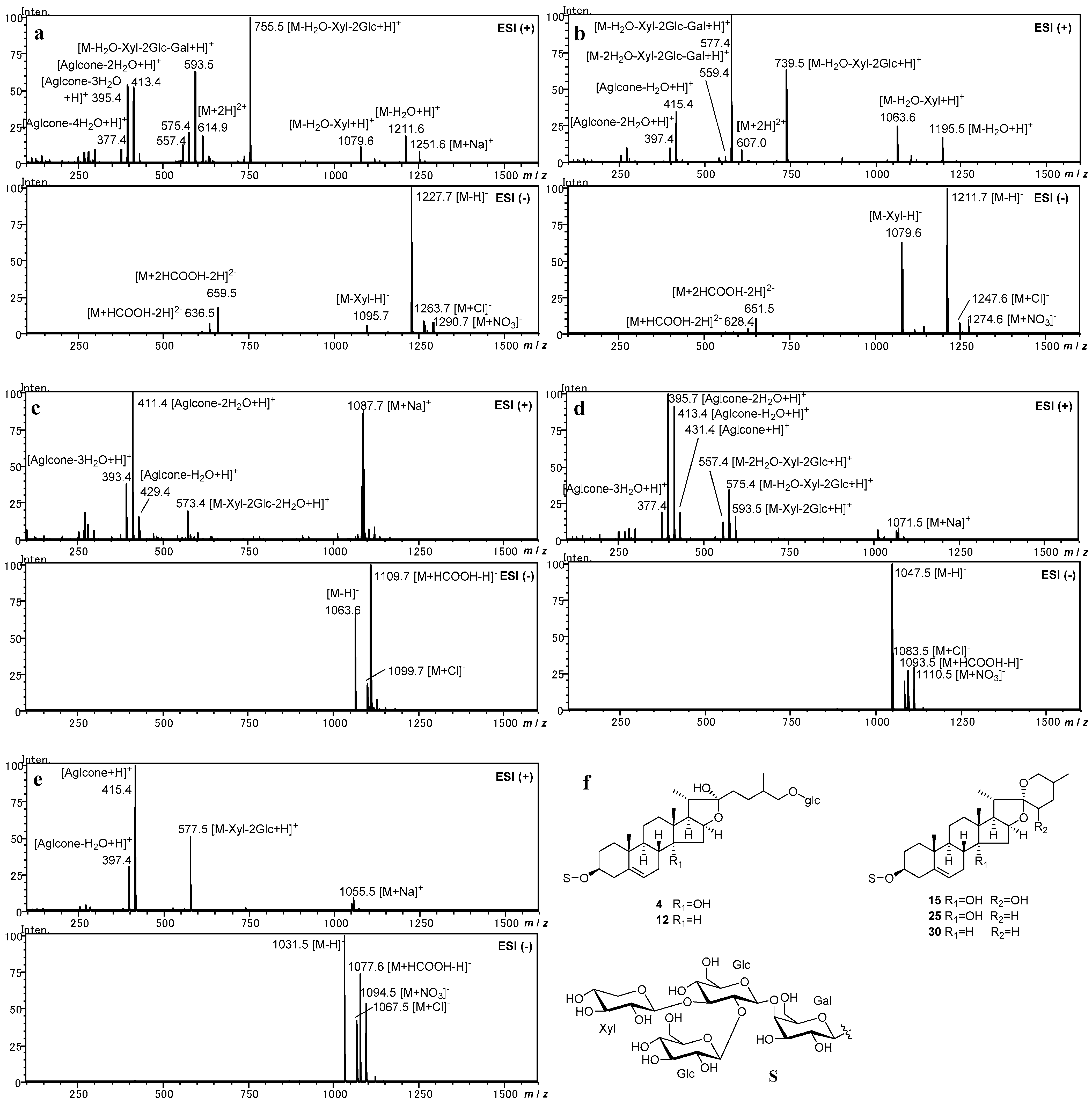
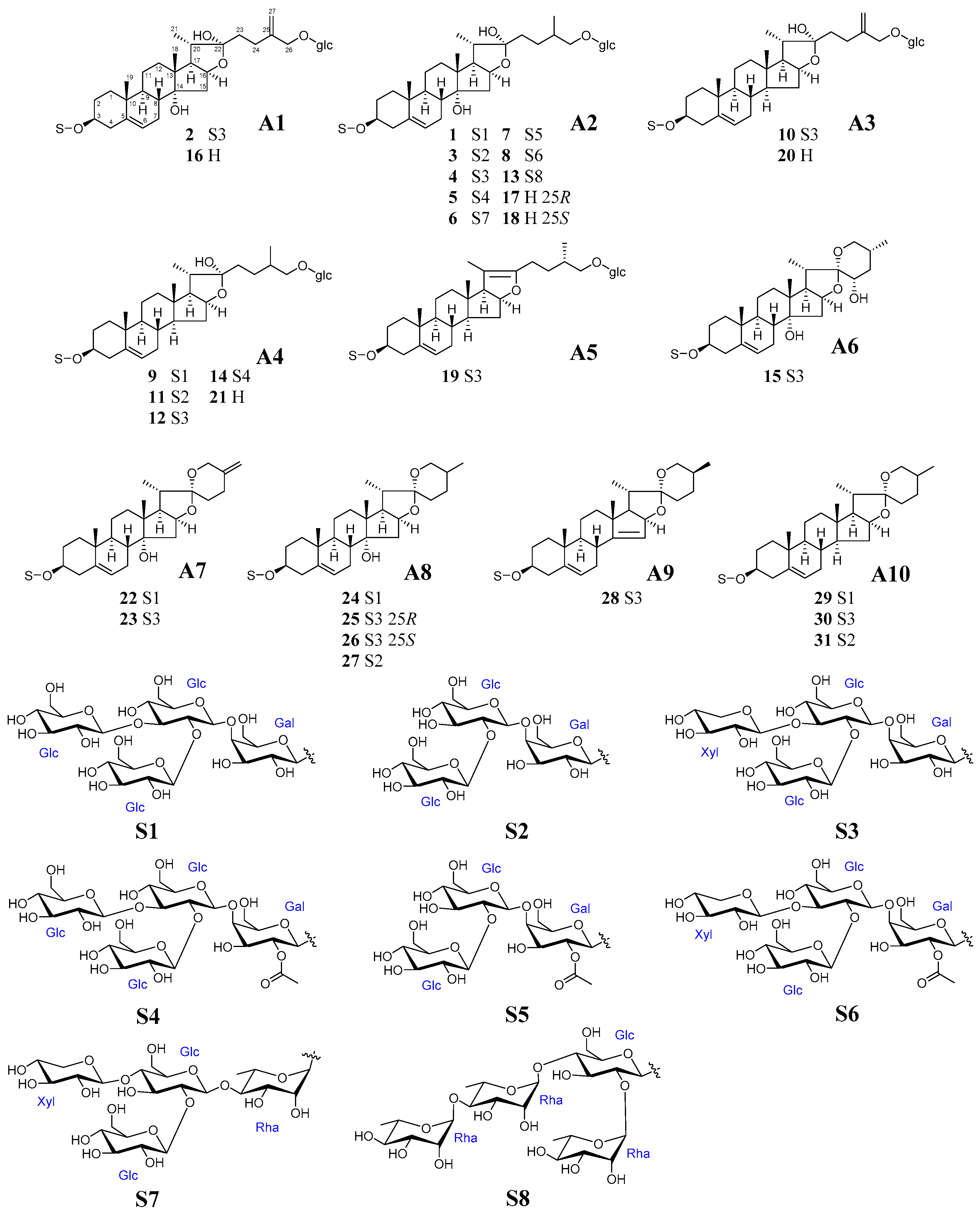
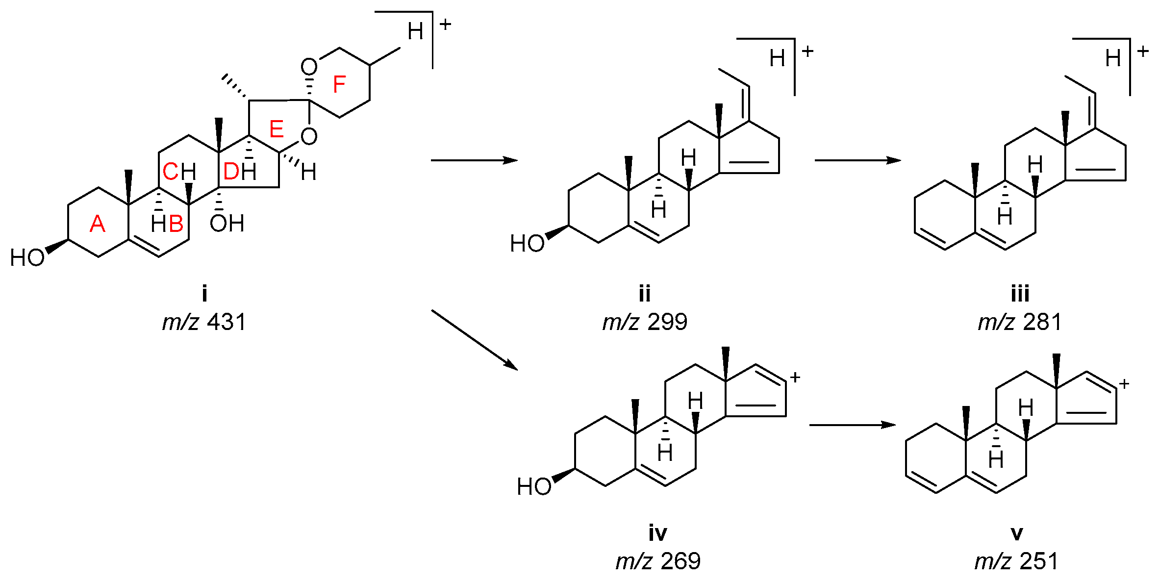
2.1. Investigation of ESI-MS Fragmentation of Authentic Steroidal Glycosides

Five steroidal glycosides [polygonatumoside F (4), timosaponin H1 (12), sibiricoside B (15), (25R)-(3β,14α)-dihydroxy-spirost-5-ene-3-O-β-D-glucopyranosyl-(1→2)-[β-D-xylopyranosyl-(1→3)]-β-D-glucopyranosyl-(1→4)-β-D-galacopyranoside (25), and 25S-aspidistrin (30)] were selected as authentic compounds (Figure 1), among which four (4, 12, 25, and 30) were obtained in our previous phytochemical investigations [9], and one (15) was isolated from P. odoratum in the present study. Incidentally, the 1H- and 13C-NMR spectroscopic data of 15 were fully re-assigned for revising the literature data [12] and given as supporting information in Supplementary Materials Table S1. These authentic compounds possess the same sugar chains at C-3 but are different in the aglycone structures. The authentic compounds provided characteristic mass fragment ions and presented regular mass fragmentation pathways, which were useful to reliably identify the steroidal glycosides from Polygonatum species.
Polygonatumoside F (4) and timosaponin H1 (12) were selected for investigating the mass fragmentation pathway of furostane-type steroidal glycosides. The chemical structures of 4 and 12 were different from each other only in the 14-hydroxy moiety. In the negative ion mode, both 4 and 12 provided deprotonated molecule ions [M-H] as the dominant ion peak. They also provided the fragment ions [M-Xyl-H] at m/z 1095 for 4 and m/z 1079 for 12, respectively. It is noteworthy that divalent ions peaks [M+2HCOOH-2H]2− and [M+HCOOH-2H]2− were observed at m/z 659 and 636 for 4, at m/z 651 and 628 for 12 (Figure 1a,b), respectively. Conversely, the divalent ions were not detected in spirostane-type steroidal glycosides.
In the positive ion mode, they provided characteristic fragment ions [M-H2O+H]+ of furostane-type glycosides, which were generated by dehydration of the hydroxy moiety at C-22. Additionally, divalent ions were detected which were observed at m/z 615 for 4 and at m/z 607 for 12 as [M+2H]2+. Glycosides 4 and 12 possess the same fragmentation pathway for sugar moieties. The fragment ions observed at m/z 1079 for 4 and at m/z 1063 for 12 corresponded to the loss of a xylosyl residue (132 Da) as [M-H2O-Xyl+H]+. The fragment ions at m/z 755 for 4 and m/z 739 for 12 corresponded to the sequential loss of two glucosyl residues (162 Da×2) as [M-H2O-Xyl-2Glc+H]+. They were further fragmented by the loss of a galactosyl residue (162 Da) to generate [M-H2O-Xyl-2Glc-Gal+H]+ at m/z 593 and 577, respectively. The fragmentation was in good agreement with the C-3 sugar moiety to be a lycotetraose (S3) (Figure 2).
The fragment ions, by further loss of the glucosyl residue at C-26, provided key information to determine the structure of aglycone, particularly the presence of 14-hydroxy moiety. The fragment ions derived from the aglycone were observed at m/z 413 [Aglycone-2H2O+H]+ and m/z 395 [Aglycone-3H2O+H]+ for 4, and at m/z 415 [Aglycone-H2O+H]+ and m/z 397 [Aglycone-2H2O+H]+ for 12. Consequently, the observation of these fragment ions by the difference of 2 Da between 4 and 12 was useful to identify the hydroxy substituents on the aglycone with one more hydroxy group [9]. It was suggested that dehydration of the hydroxy moiety at the C-14 position and loss of the glucosyl residue at the C-26 position were simultaneously progressed, since the fragment ion peak assigned to [Aglycone-H2O+H]+ could not have been observed in 4.
For the spirostane-type steroidal glycosides, sibiricoside B (15), (25R)-(3β,14α)-dihydroxy-spirost-5-ene-3-O-β-D-glucopyranosyl-(1→2)-[β-D-xylopyranosyl-(1→3)]-β-D-glucopyranosyl-(1→4)-β-D-galacopyranoside (25), and 25S-aspidistrin (30) were selected as authentic compounds. These glycosides are different from each other in C-14 and C-22 hydroxy substitutions.
As shown in Figure 1c–e, spirostane-type steroidal glycosides 15, 25, and 30 provided deprotonated molecule ion [M-H] in the negative ion mode. However, the divalent ions were not detected, which were different from the furostane-type steroidal glycosides. Instead, the monovalent ions [M+HCOOH-H] were detected. The fragment ions, due to the cleavage of the sugar chain at C-3, were also not detected in the negative ion mode.
In the positive ion mode, the sodium adduct ions [M+Na]+ were detected in 15, 25, and 30, but the fragment ion [M-H2O+H]+ was not detected. The spirostane skeleton contains a spiro-bicyclic acetal at C-22, while the furostane skeleton is usually with a hydroxy moiety at C-22. Thus, observation of the fragment ion [M-H2O+H]+ corresponding to the dehydration of 22-OH is useful to discriminate between the spirostane- and furostane-type steroidal glycosides.
Further fragment ions were detected, corresponding to the cleavage of the sugar chain at C-3 and dehydration of the hydroxy substitutions in the aglycone. Cleavage of the sugar chain was processed in two steps, firstly, the outer trisaccharide of 2-glucosyl-(3-xylosyl)-glucosyl moiety was lost, and then the inner galactosyl was lost. Meanwhile, dehydration also progressed based on the hydroxy substitutions in the aglycone. As a result, the sequential dominant fragment ions were detected at m/z 573 [M-Xyl-2Glc-2H2O+H]+, 411 [Aglycone-2H2O+H]+, and 393 [Aglycone-3H2O+H]+ for 15, at m/z 575 [M-Xyl-2Glc-H2O+H]+, 413 [Aglycone-H2O+H]+, and 395 [Aglycone-2H2O+H]+ for 25, and at m/z 577 [M-Xyl-2Glc+H]+, 415 [Aglycone+H]+, and 397 [Aglycone-H2O+H]+ for 30, respectively.

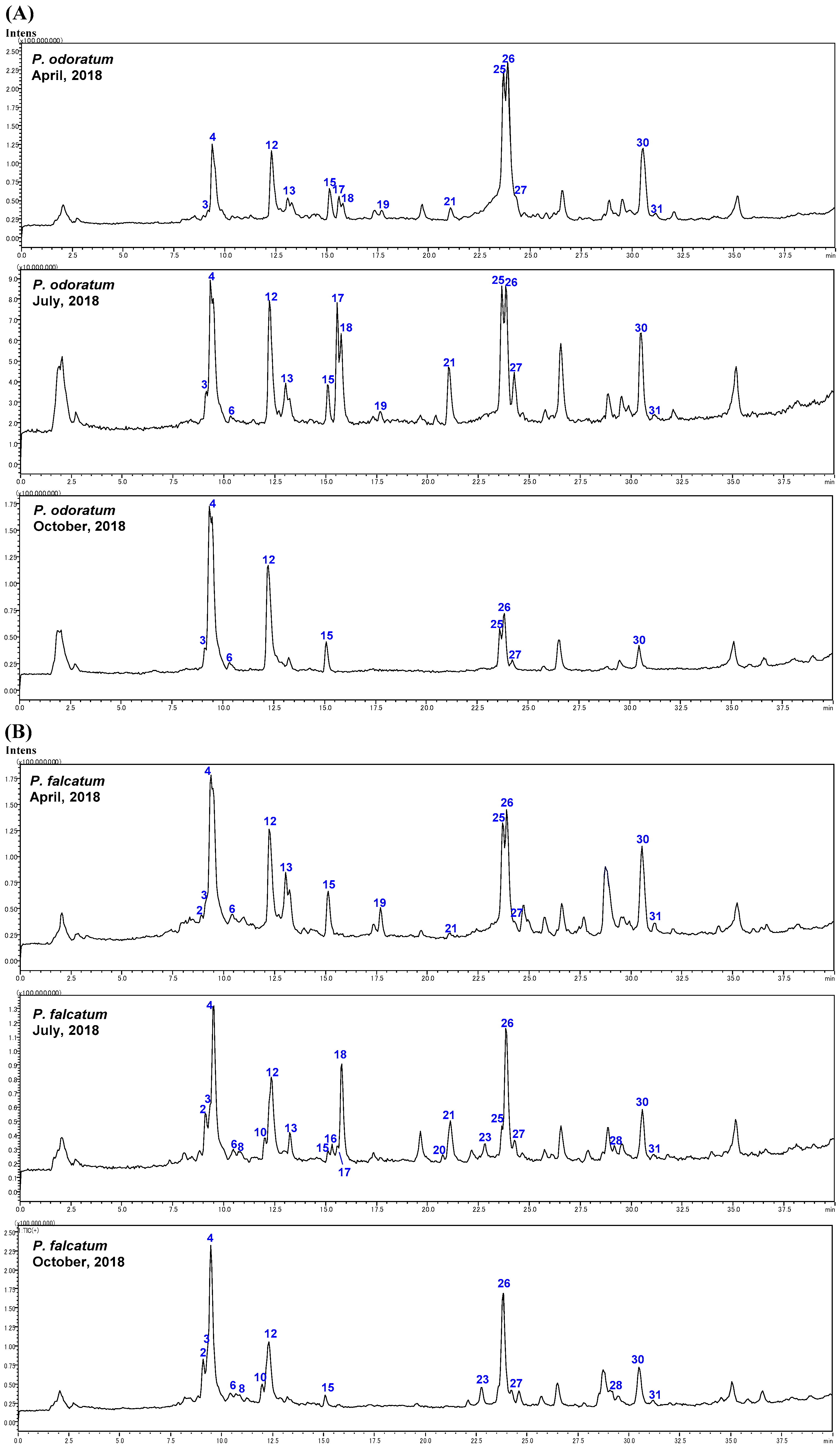
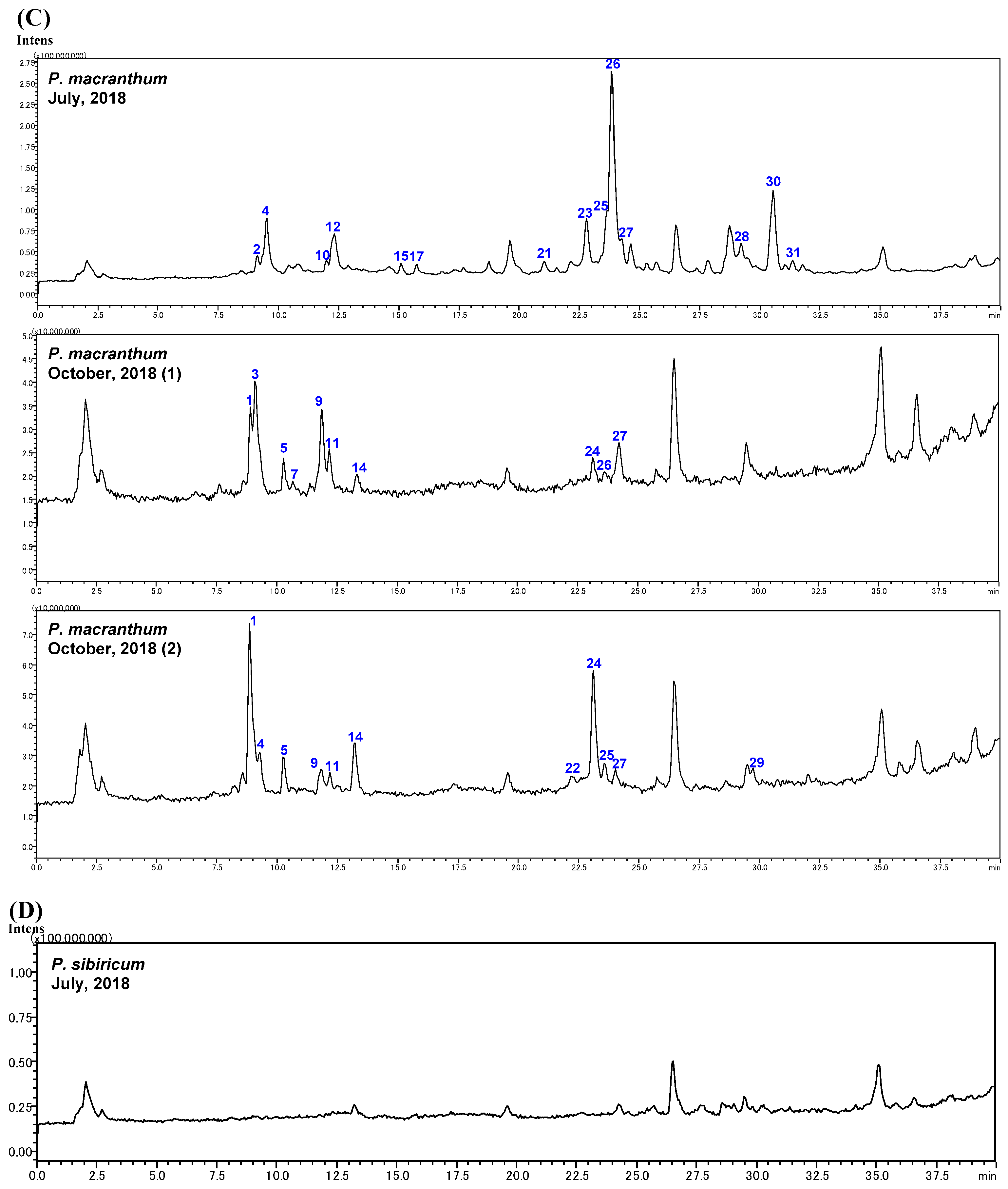
2.2. Identification of Steroidal Glycosides in the Sample Solutions

Four Polygonatum plants (P. odoratum, P. falcatum, P. macranthum, and P. sibiricum) were pre-treated using a C18 cartridge, and their 80% eluted fractions were analyzed by an LC-ESI-MS operated in a full-scan mode in both positive ion mode and negative ion mode to tentatively identify the steroidal glycosides. The structure was elucidated based on fragment patterns found in authentic steroidal glycosides (see Section 2.1). The total ion chromatograms (TICs) of the sample solutions in positive ion mode are shown in Figure 3. According to the ESI-MS fragmentation patterns, a total of 31 glycosides, including five authentic compounds, were manually detected and designated as 1 to 31 according to the retention times sequence. The molecular fragments detected, retention times, and molecular weight of the 31 glycosides were presented in Table 1. The (+) and (−) ESI-MS and the chemical structures for individual glycosides were given in Supplementary Materials Table S1. In the LC-MS analysis of P. sibiricum, steroid glycosides were not detected. Since similar results were obtained from P. sibiricum harvested in April (data not shown) and July (Figure 3D), this is due to differences in plant metabolism, and not an artifact of sample processing.
Peaks 1, 3, 5, 6, 7, and 8 exhibited fragment ions assignable as [M-H2O+H]+ as well as two main fragment ions at m/z 395 and 413, which were the same as peak 4, suggesting that they are frostane-type glycosides and possess the same aglycone as the authentic compound 4 but differ in the structure of C-3 sugar chains (Supplementary Materials Figures S1, S3, S5–S8).
Peak 1 exhibited an [M+Na]+ ion at m/z 1281, and an [M-H] ion at m/z 1257, suggesting the molecular weight of 1 was 30 Da more than that of 4 (Supplementary Materials Figures S1 and S4). Since 1 and 4 produced the same fragment ions at m/z 755, 593, 575, and 557 by cleavage of the C-3 sugar chain, the difference in molecular weight was most like due to the replacement of the terminal xylose moiety in 4 with glucose in 1. The sugar chain of commetetraose (S1) in 1 is common in known steroidal glycosides from Polygonatum plants (Figure 2) [8]. Thus, 1 was identified to be 26-O-(β-D-glucopyranosyl)-furost-5-ene-3β,14α,22α,26-tetraol 3-O-β-D-glucopyranosyl-(1→2)-[β-D-glucopyranosyl-(1→3)]-β-D-glucopyranosyl-(1→4)-β-D-galactopyranoside.
Peak 3 exhibited fragment ions (m/z 917, 755, and 593) generated by sequential losses of three hexose residues (Supplementary Materials Figure S3). Based on comparison with the data of 4, the C-3 sugar chain of 3 was most like to be a trisaccharide of β-D-glucopyranosyl-(1→2)-β-D-glucopyranosyl-(1→4)-β-D-galactopyranose (S2), which lost a xylosyl moiety from S1 (Figure 2). Thus, 3 was identified to be 26-O-(β-D-glucopyranosyl)-furost-5-ene-3β,14α,22α,26-tetraol 3-O-β-D-glucopyranosyl-(1→2)-β-D-glucopyranosyl-(1→4)-β-D-galactopyranoside, which was previously isolated from P. odoratum [13].
As for peaks 5, 7, and 8, two fragment ions were observed at m/z 797 and 593, with a 204 Da (162 Da + 42 Da) mass difference, suggesting the inner galactose was acetylated (Supplementary Materials Figures S5, S7 and S8). Taking account the known steroidal glycosides from Polygonatum plants [14], peak 5 was identified to be 26-O-(β-D-glucopyranosyl)-furost-5-ene-3β,14α,22α,26-tetraol 3-O-β-D-glucopyranosyl-(1→2)-[β-D-glucopyranosyl-(1→3)]-β-D-glucopyranosyl-(1→4)-2″-O-acetyl-β-D-galactopyranoside.
Both 7 and 8 were also considered to have galactosyl residue with an acetyl group in the C-3 sugar chain such as 5. The ions at m/z 959 and 797 from 7 were formed from the sequential losses of two hexose residues from the [M-H2O+H]+ ion at m/z 1121. Therefore, 7 was identified to be 26-O-(β-D-glucopyranosyl)-furost-5-ene-3β,14α,22α,26-tetraol 3-O-β-D-glucopyranosyl-(1→2)-β-D-glucopyranosyl-(1→4)-2″-O-acetyl-β-D-galactopyranoside, a compound that does not have a terminal glucose moiety of sugar chain. Meanwhile, the ion at m/z 797 from 8 was formed from the losses of two hexoses and one pentose residues (456 Da) from the [M-H2O+H]+ ion at m/z 1253. According to the saponin with a β-D-glucopyranosyl-(1→2)-[β-D-xylopyranosyl-(1→3)]-β-D-glucopyranosyl-(1→4)-2″-O-acetyl-β-D-galactopyranose moiety (S5) at C-3 position having been isolated from P. odoratum [15], 8 was identified as shown in Figure 2.
In peak 6 MS, the ion at m/z 593 was 146 Da different than the ion at m/z 739 [M-H2O-Xyl-2Glc+H]+, which corresponded to the loss of a deoxyhexose residue (146 Da) (Supplementary Materials Figure S6). The rhamnose is the usual constituent sugar in the genus Polygonatum [8]. Thus, 6 was determined to be 26-O-(β-D-glucopyranosyl)-furost-5-ene-3β,14α,22α,26-tetraol 3-O-β-D-glucopyranosyl-(1→2)-[β-D-xylopyranosyl-(1→3)]-β-D-glucopyranosyl-(1→4)-α-L-rhamnopyranoside.
Peak 13 exhibited the fragment ions of m/z 901 [M-2Rha+H]+, 755 [M-3Rha+H]+, and 575 [M-Glc-3Rha-H2O+H]+, which were formed from the loss of three deoxyhexose and one pentose moieties (Supplementary Materials Figure S13). In addition, the fragment ions of m/z 413 [Aglycone-H2O+H]+ and 395 [Aglycone-2H2O+H]+ suggested a glucose residue at the C-26 position and the same aglycone as 4. The sugar moiety (S8) of 13 at the C-3 position was reported as those of parrisaponin Pb from P. kingianum [16] and P. zanlanscianense [17], and 13 was identified as shown in Figure 2.
Peak 2 obtained the ions at m/z 753 and 591 by the sequential losses of the sugar residues in the C-3 sugar chain of S3 (Figure 2) from the [M-H2O+H]+ ion (m/z 1209) (Supplementary Materials Figure S2). The ion at m/z 411 from 2 was formed from the loss of the glycosyl residue at C-26, and then the elimination of the hydroxyl groups yielded the ion at m/z 393 [Aglycone-2H2O+H]+ and m/z 375 [Aglycone-3H2O+H]+. These suggested that 2 has two free hydroxyl groups and a double bond on the aglycone, since each of the above fragment ions has a 2 Da mass difference from 4. The double bond was conjectured to be located at C-25 and C-27, since such furostane-type steroids have been reported in Liliaceae plants [18,19]. Thus, 2 was determined to be 26-O-(β-D-glucopyranosyl)-furost-5,25(27)-diene-3β,14α,22α,26-tetraol 3-O-β-D-glucopyranosyl-(1→2)-[β-D-xylopyranosyl-(1→3)]-β-D-glucopyranosyl-(1→4)-β-D-galactopyranoside.
Peaks 17 and 18 provided the same fragment ions and confirmed a pair of isomers. The ions of [M-H2O+H]+ at m/z 593 and [Aglycone-H2O+H]+ at m/z 431 suggested the molecular weight to be 610 Da, and 17 and 18 having a glucosyl moiety at C-26 position as seen in other Polygonatum saponins (Supplementary Materials Figures S17 and S18). By comparing retention times and mass spectra with standard peaks, 17 and 18 could be unequivocally identified as 25R- and 25S-polygonatumoside G, respectively, which have been reported in P. odoratum [9]. Peak 16 showed a similar mass fragmentation pattern with 17 and 18, except in all mass fragment ions have a 2 Da mass difference, which suggested the existence of a double bond in the structure (Supplementary Materials Figure S16). Thus, 16 was identified as furost-5,25(27)-diene-1β,14α,22α,26-tetraol 26-O-β-D-glucopyranoside, the sapogenin of 2 (A2) (Figure 2).
Peak 10 exhibited fragment ions that were less than 2 Da than those of the authentic compound 12 (Supplementary Materials Figures S10 and S12). Then, the ions at m/z 413 and 395 were characterized by the loss of a glucosyl residue at the C-26 position and H2O at the C-3 position and determined the aglycone skeleton with one more double bond than 12. The double bond was conjectured to be located at C-25 and C-27 like 2 and 16. Thus, 10 was inferred as 26-O-(β-D-glucopyranosyl)-furost-5,25(27)-diene-3β,22α,26-triol 3-O-β-D-glucopyranosyl-(1→2)-[β-D-xylopyranosyl-(1→3)]-β-D-glucopyranosyl-(1→4)-β-D-galactopyranoside.
Peaks 20 and 21 followed the same fragmentation pathway with 16, except all mass fragment ions have 16 Da and 18 Da mass differences, respectively (Supplementary Materials Figures S20 and S21). Thus, 20 was considered as a dehydroxy product of 16, and could be assigned as furost-5,25(27)-diene-3β,22α,26-triol 26-O-β-D-glucopyranoside, which has the same aglycone as 10. Meanwhile, 21 was considered as a reduction product of 20, and could be identified as funkioside B (21) [20], since the fragment ions of 21 have more 2 Da than those of peak 20.
Peaks 9, 11, and 14 followed similar fragmentation patterns to the authentic compound 12, and all exhibited the main fragment ions at m/z 577, 415 [Aglycone-H2O+H]+ and 397 [Aglycone-2H2O+H]+ (Supplementary Materials Figures S9, S11 and S14), which suggested that they have the same aglycone as 12 possessing the glucosyl moiety at C-26 position but with differences in C-3 sugar chains. Peak 9 exhibited the fragment ions at m/z 901, 739, and 577, which were formed from the sequential losses of four hexose moieties from the [M-H2O+H]+ ion at m/z 1225, and was conjectured as polyfuroside, which was previously isolated from P. officinale [21], Solanum nigrum L [22] and Allium macrostemon [23] with the C-3 sugar chain of S1 (Figure 2). Peak 11 showed the same fragmentation pattern as 3 with the sugar chain of S2 (Figure 2), and identified as 26-O-(β-D-glucopyranosyl)-furost-5-ene-3β,22α,26-triol 3-O-β-D-glucopyranosyl-(1→2)-β-D-glucopyranosyl-(1→4)-β-D-galactopyranoside. Peak 14 exhibited a similar fragment pattern to 5. Therefore, 14 was identified as 26-O-(β-D-glucopyranosyl)-furost-5-ene-3β,22α,26-triol 3-O-β-D-glucopyranosyl-(1→2)-[β-D-glucopyranosyl-(1→3)]-β-D-glucopyranosyl-(1→4)-2″-O-acetyl-β-D-galactopyranoside.
Peak 19 presented the fragment ions at m/z 739, 577, 415, and 395 same with 12, but the [M-H2O+H]+ ion did not exist, suggesting that there is no hydroxyl group connected to the C-22 position (Supplementary Materials Figure S19). The ions at m/z 739 and 577 were formed from the sequential losses of C-3 sugar chains as S3 (Figure 2) from the ion at m/z 1217. Thus, 19 was identified to be polygodoside G, which was previously isolated from P. odoratum [15].
On the other hand, 11 peaks were identified as spirostane-type steroidal glycosides by no detection of the [M-H2O+H]+ and formation of the [M+HCOOH-H] ions in high abundance.
Peaks 24, 25, 26, and 27 exhibited the same fragment ions at m/z 431, 413, and 395, suggesting that they have the same aglycone(Supplementary Materials Figures S24–S27). In addition, a fragment ion at m/z 575 suggested that the internal galactose in the sugar chain was also common.
Peak 26 was considered to be an isomeric compound of the authentic compound 25 with the same molecular weight of 1049 Da. Peak 26 could be inferred as the stereoisomer of 25 at the C-25 position since almost the same fragment ions were observed in 25.
Peaks 24 and 27 exhibited the almost same fragmentation patterns (Supplementary Materials Figures S24 and S27) as 25 and 26, suggesting that they have the same aglycone skeleton but differ in the sugar moieties. Peak 24 has a molecular weight of 1078 Da, and its fragment ions at m/z 575 and 413 were formed by the sequential losses of sugar residues from S1 (Figure 2). Peak 27 has a molecular weight of 916 Da, differing 132 Da corresponding to a pentose residue from 25, therefore having the same C-3 sugar chains as 3 (S2) (Figure 2). Thus, peak 24 was identified as pod Ⅲ [15] and 27 was identified as polygonatumoside D [24]. Both compounds have been previously reported in P. odoratum.
Both 22 and 23 observed the ions at m/z 573, 411, and 393, suggesting that they have the same aglycone skeleton but different sugar moieties (Supplementary Materials Figures S22 and S23). Peak 23 exhibited the [M+Na]+ ion less 2 Da than authentic 25 (23: m/z 1069; 25: m/z 1071) but the same fragment ions at m/z 299 (ii), 281 (iii), 269 (iv), and 251 (v), which assigned the characteristic fragments of the steroidal nucleus [25], were observed (Figure 4). This suggested that the aglycones of 23 (m/z 429 [Aglycone+H]+) and 25 (m/z 431 [Aglycone+H]+ (i)) have the same A, B, C, D, and E rings and sugar chain, but peak 23 has a carbon-carbon double bond in the F ring [15]. Peak 22 presented an [M+Na]+ ion at m/z 1099 and has a molecular weight of more than 30 Da than 23. The C-3 sugar chain was suggested as S1 (Figure 2). Thus, 23 and 22 were identified as polygodoside A and polygodoside B, respectively [15]. Both compounds have been previously reported in P. odoratum.
Peak 28 produced similar fragment ions to authentic compound 30, except for the 2 Da mass difference of all fragment ions, which suggested that 28 has one more double bond than 30 (Supplementary Materials Figures S28 and S30). Thus, 28 was identified as 25(S)-spirost-5-ene-3β-ol 3-O-β-D-glucopyranosyl-(1→2)-[β-D-xylopyranosyl-(1→3)]-β-D-glucopyranosyl-(1→4)-galactopyranoside, which has been previously reported in P. odoratum [26].
Peaks 29 and 31 exhibited the fragment ions at m/z 577, 415, and 397 (Supplementary Materials Figures S29 and S31), suggesting that they have the same aglycone skeleton as 30 but differ in sugar moieties. Peaks 29 and 31 have a 30 Da (for -CH2O-) and 132 Da (for a pentose moiety) mass difference from 30, respectively. Thus, peak 29 could be assigned as pod Ⅳ, which has been previously reported in P. odoratum [15], and 31 as neosibiricoside D, which has been previously reported in both P. odoratum [14] and P. sibiricum [27].
In general, the retention time of frostane-type steroids was shorter (Rt 8.90–21.08 min) than those of spirostane-types (Rt 22.29 min–31.16 min), except for the spirostane steroid 15 (Supplementary Materials Figure S15) having the hydroxy group in the F-ring (Rt 15.14 min). The retention times, depending on the types of the sugar chain, can be summarized as follows. In the case of the frostane-type steroids, the retention time of compounds having the sugar chain S1 (-Gal(-Glc)-Glc-Glc) is shorter than that of S2 (-Gal(-Glc)-Glc). Meanwhile, the steroids having the sugar chain S3 (-Gal(-Glc)-Glc-Xyl) had a longer retention time than that of S2 (Rt 1 < 3 < 4; 9 < 11 < 12). This retention time relationship is the same as the steroids possessing sugar chains of S5, S6, and S7 which are acetylated at 2-OH of the inner galactose (Rt 5 < 7 < 8). In addition, a steroid in which galactose in the S3 sugar chain of steroidal saponin was replaced with rhamnose (S7) had a longer retention time (Rt 4 < 6). On the other hand, among spirostane glycosides, the one with S1 had the shortest retention time, but the one with S3 had a shorter retention time than the one with S2. (Rt 24 < 25 < 26 < 27; 29 < 30 < 31).

2.3. The Biogenetic Pathway of Steroidal Glycosides in Polygonatum Species

As shown in Figure 3, a total of 15 steroidal glycosides were found in P. odoratum, 22 in P. falcutum, and 23 in P. macranthum, but none in P. sibiricum. All varieties collected in October showed fewer species of steroidal glycosides. On the contrary, the plants collected in July showed the most abundant steroidal glycosides among all the varieties. A possible biogenesis pathway is shown in Figure 5, according to the analysis result of plants collected in July.
Steroidal glycosides in Polygonatum species could be divided into spirostane- and furostane-types base on the aglycone structures (Supplementary Materials Figure S32). Spirostane-types are always the monodesmosides with a sugar chain attached to the C-3 position, and the furostane- types are always the bisdesmosides with an additional glucopyranosyl moiety linked in the C-26 position. Our previous studies about steroidal glycosides [9] and cholestane-type glycosides [24] reported that steroidal glycosides in genus Polygonatum have the same common biosynthetic precursor, (22S)-cholest-5-ene-3β,16β,22-triol. C-22 oxidation of the precursor produced 16,26-dihydroxy-22-keto-cholesterol, then the biosynthesis routes were divided into two classes. One group is characterized by 14-hydroxylation, followed by 3,26-glycosylation, which produces the aglycone polygonatumoside (A2) (Figure S32), while the other group is not, and produces the aglycone A4, a furostane-type aglycone without 14-OH. The furostane-type aglycones A2 and A4 deglycosylated at C-26, followed by a ring closure to 26-OH take place with dehydration of 22-OH, producing the spirostane-type aglycone A8 and A10, respectively. The furostane- and spirostane-type aglycones undergo various modifications in a series of biosynthetic reactions, including hydroxylation and dihydroxylation, to enable further diversification.
The original plants of Huangjing (P. falcutum and P. macranthum) showed a more complex oxidation process than the original plant of Yuzhu (P. odoratum) at the aglycone (Figure 5). Significantly, the original plants of Huangjing (P. falcutum and P. macranthum) have the acetyl groups on hydroxyl groups at the sugar residues (S4, S5, and S6) which were different from Yuzhu (P. odoratum). On the other hand, the steroidal glycosides from Polygonatum species mainly have the sugar moiety as lycotetraose (S3). The main components 4, 12, 15, 25, 26, and 30, which were detected in all varieties with high abundance, all have the C-3 sugar chain as S3.
The results showed a similar chemical composition of steroidal glycosides among the Polygonatum species cultivated in Japan, but it also has some interspecific differences between the two groups of Huangjing and Yuzhu. On the whole, P. falcatum and P. macranthum contained steroidal glycosides with higher oxidation levels, while the sugar moiety also with structural differences from P. odoratum.

3. Materials and Methods

3.1. General Methods

Acetonitrile (MeCN), formic acid (HCOOH), and water (H2O) were LC/MS-grade and obtained from Fujifilm Wako Pure Chemical Industries, Ltd. (Osaka, Japan). The 1H and 13C NMR spectra were measured on a JEOL ECA-500 spectrometer (Tokyo, Japan) with the measuring deuterated solvent as the internal reference, and the chemical shifts were expressed in ppm units. Column chromatography was performed on Diaion HP-20 (Mitsubishi Chemical Corporation, Tokyo, Japan) and ODS (100–200 mesh, Chromatorex DM1020T ODS, Fuji Silysia Chemical Co., Ltd., Aichi, Japan). Semi-preparative HPLC was performed on a Waters 600E HPLC pump (Waters Corp., Milford, MA, USA) with a Shimadzu SPD-10A (Kyoto, Japan) intelligent ultraviolet/visible (UV/vis) detector, a Shodex RI-72 differential refractometer detector (Shoko Science Co., Ltd., Tokyo, Japan), and an RP-C18 silica gel column (YMC Actus Triart C18, 150 × 20 mm I.D., Kyoto, Japan).

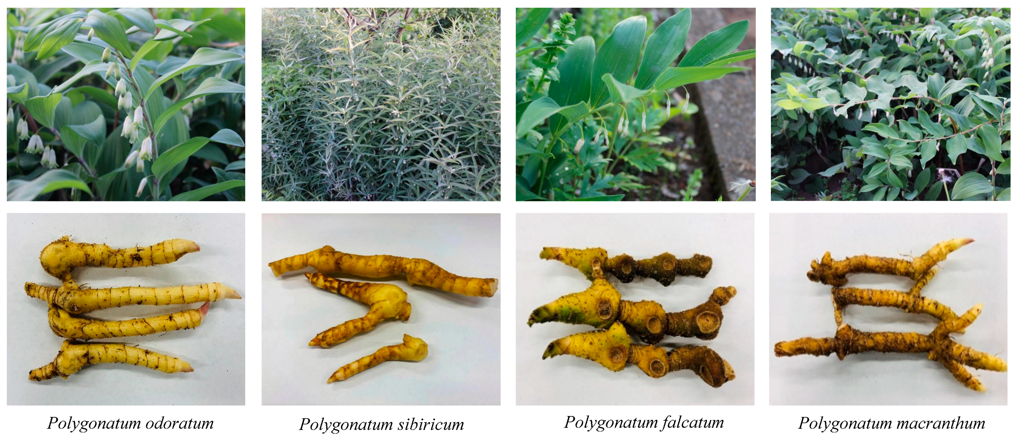
3.2. Plant Materials

Plant materials were shown in Figure 5 and Table 2. The rhizomes of Polygonatum species were cultivated in Toho University Botanical Garden of Medicinal Plants, Japan, and were collected from April to October 2018. All of the plants were identified by one of the authors, WL. The voucher specimens were deposited in the Department of Pharmacognosy, Faculty of Pharmaceutical Sciences, Toho University.

3.3. Preparation of the Sample Solutions

The dried rhizomes were pulverized into homogeneous powders. The powder (0.5 g) was ultrasonically extracted with methanol (MeOH, 10 mL) at room temperature for 1 h. After evaporation, the extracts were ultrasonically dissolved in MeOH (1.5 mL) and H2O (3.5 mL) was added to make a 30% MeOH solution (5 mL). The solution was loaded onto a Sep-Pak C18 plus short cartridge (Waters), eluted with 30% MeOH/H2O solution (v/v) (10 mL), and 80% MeOH/H2O solution (v/v) (10 mL). The 80% MeOH/H2O elution (5 mL) was dried in vacuo and then dissolved in 1 mL MeOH. The extracted solutions were filtered through a 0.45 µm syringe filter and used as sample solutions.

3.4. LC-MS Analysis

LC-MS analysis was performed using an LCMS-8040 triple quadrupole LC/MS/MS mass spectrometer (Shimadzu Co., Ltd., Kyoto, Japan). HPLC was performed on a YMC-Triart C18 column (3.0 μm, 150 × 20 mm I.D.) maintained at 35 °C. The flow rate was 0.2 mL/min. The mobile phase was composed of A (0.1% HCOOH in H2O) and B (0.1% HCOOH in MeCN) with a gradient elution: 0–40 min, 20–70% B; 40–50 min, 100% B. The column was equilibrated for 15 min under the initial conditions. The injection volume was 4 µL for the qualitative analysis.
The mass spectrometer was operated in both positive- and negative-ion modes in the range of m/z 100 to 1600. The ESI parameters were as follows: interface voltage, 4.5 kV in the positive-ion mode and −3.5 kV in the negative-ion mode; dry gas, 15 L/min; and dry temperature, 350 °C.

3.5. Isolation of Sibiricoside B (15)

The air-dried rhizomes of P. odoratum (0.9 kg) were cut into pieces and were ultrasonically extracted with MeOH (2 L) for 1 h at room temperature. After removal of the solvent by evaporation, the extract (138 g) was subjected to Diaion HP-20 column chromatography and eluted with gradients of H2O and 40%, 80%, and 100% MeOH. The 80% MeOH eluate was chromatographed on semi-preparative reversed-phase (RP)-HPLC to afford sibiricoside B (15) (14 mg).

4. Conclusions

In this study, the steroidal glycosides in 4 species of genus Polygonatum were systematically analyzed and identified by a simple, rapid, and sensitive LC-ESI–MS method. The mass fragmentation pattern of spirostane-type and furostane-type steroidal glycosides was also investigated, and 31 compounds were tentatively identified. Our study provided chemical support for further phytochemotaxonomical studies of Polygonatum species and provided evidence for the identification of steroidal glycosides by an LC-ESI–MS method when no reference compounds were available.

Supplementary Materials

The following supporting information can be downloaded at: https://www.mdpi.com/article/10.3390/molecules28020705/s1, Figures S1–S31: Structure and (+) and (−) ESI-MS of 31 compounds; Figure S32: Biogenetic pathway for the steroidal glycoside from Polygonatum species; Table S1: NMR Spectroscopic Data (500 MHz for 1 H and 125 MHz for 13C, in Pyridine-d5) of Compound 15.

Author Contributions

Conceived and designed the experiments: W.L.; Performed the experiments: D.L.; Analyzed the data: D.L., T.K. and W.L.; and wrote the paper: D.L., T.K. and W.L. All authors have read and agreed to the published version of the manuscript.

Funding

This research received no external funding.

Institutional Review Board Statement

Not applicable.

Informed Consent Statement

Not applicable.

Data Availability Statement

The data presented in this study and samples of the compounds are available upon request from the corresponding author.

Conflicts of Interest

The authors declare no conflict of interest.

References

  1. Ohashi, H.; Kadota, Y.; Murata, J.; Yonekura, K.; Kihara, H. Wild Flowers of Japan, 2nd ed.; Heibonsha: Tokyo, Japan, 2015; Volume 1, p. 256. [Google Scholar]
  2. Hatfield, G. Encyclopedia of Folk Medicine: Old World and New World Traditions; ABC-CLIO: Santa Barbara, CA, USA, 2004; p. 150. [Google Scholar]
  3. Dhyani, A.; Nautiyal, B.P.; Nautiyal, M.C. Importance of Astavarga plants in traditional systems of medicine in Garhwal, Indian Himalaya. Int. J. Biodivers. Sci. Ecosyst. Serv. Manag. 2010, 6, 13–19. [Google Scholar] [CrossRef] [Green Version]
  4. Shanghai Scientific and Technical Publishers. Dictionary of Chinese Materia Medica Japanese Edition; Shogakukan: Tokyo, Japan, 1985; Volume 1, pp. 144–147. [Google Scholar]
  5. Pharmacopoeia Committee of P. R. China. Pharmacopoeia of People’s Republic of China; China Medical Science and Technology Press: Beijing, China, 2020; pp. 86, 319. [Google Scholar]
  6. The Committee on the Japanese Pharmacopoeia. The Japanese Pharmacopoeia, 18th ed.; Ministry of Health, Labour and Welfare: Tokyo, Japan, 2021; pp. 2103–2104.
  7. Satake, Y. Wild Flowers of Japan: Herbaceous Plants 1; Heibonsha: Tokyo, Japan, 1982; p. 45. [Google Scholar]
  8. Zhao, P.; Zhao, C.; Li, X.; Gao, Q.; Huang, L.; Xiao, P.; Gao, W. The genus Polygonatum: A review of ethnopharmacology, phytochemistry and pharmacology. J. Ethnopharmacol. 2018, 214, 274–291. [Google Scholar] [CrossRef] [PubMed]
  9. Liu, Q.; Li, W.; Nagata, K.; Fu, H.; Okada, S.; Tanabe, I.; Kobori, Y.; Higai, K.; Norie, W.; Sasaki, T. Isolation, structural elucidation, and liquid chromatography-mass spectrometry analysis of steroidal glycosides from Polygonatum odoratum. J. Agric. Food Chem. 2018, 66, 521–531. [Google Scholar] [CrossRef] [PubMed]
  10. Ahn, M.J.; Kim, J. Identification and quantification of steroidal saponins in Polygonatum species by HPLC/ESI/MS. Arch. Pharm. Res. 2005, 28, 592–597. [Google Scholar] [CrossRef] [PubMed]
  11. Takagi, H. Breaking of two types of dormancy in seeds of Polygonatum odoratum used as vegetables. J. Jpn. Soc. Hortic. Sci. 2001, 70, 416–423. [Google Scholar] [CrossRef]
  12. Son, K.H.; Do, J.C. Steroidal saponins from the rhizomes of Polygonatum sibiricum. J. Nat. Prod. 1990, 53, 333–339. [Google Scholar] [CrossRef] [PubMed]
  13. Kim, S.; Shin, S.Y.; Moon, Y.J.; Seo, J.Y.; Kim, H.K.; Whang, W.K. Content analysis and classification for Polygonati Odorati Rhizoma and Polygonati Rhizoma by steroidal saponin. Yakhak Hoechi 2010, 54, 441–448. [Google Scholar]
  14. Ahn, M.J.; Kim, C.Y.; Yoon, K.D.; Ryu, M.Y.; Cheong, J.H.; Chin, Y.W.; Kim, J. Steroidal Saponins from the Rhizomes of Polygonatum sibiricum. J. Nat. Prod. 2006, 69, 360–364. [Google Scholar] [CrossRef]
  15. Zhang, H.; Chen, L.; Kou, J.P.; Zhu, D.N.; Qi, J.; Yu, B.Y. Steroidal sapogenins and glycosides from the fibrous roots of Polygonatum odoratum with inhibitory effect on tissue factor (TF) procoagulant activity. Steroids 2014, 89, 1–10. [Google Scholar] [CrossRef]
  16. Yu, H.S.; Ma, B.P.; Song, X.B.; Kang, L.P.; Zhang, T.; Fu, J.; Zhao, Y.; Xiong, C.Q.; Tan, D.W.; Zhang, L.J.; et al. Two New Steroidal Saponins from the Processed Polygonatum kingianum. Helv. Chim. Acta 2010, 93, 1086–1092. [Google Scholar] [CrossRef]
  17. Jin, J.M.; Zhang, Y.J.; Li, H.Z.; Yang, C.R. Cytotoxic Steroidal Saponins from Polygonatum zanlanscianense. J. Nat. Prod. 2004, 67, 1992–1995. [Google Scholar] [CrossRef] [PubMed]
  18. Yang, Q.X.; Zhang, Y.J.; Li, H.Z.; Yang, C.R. Polyhydroxylated steroidal constituents from the fresh rhizomes of Tupistra yunnanensis. Steroids 2005, 70, 732–737. [Google Scholar] [CrossRef] [PubMed]
  19. Mari, A.; Napolitano, A.; Perrone, A.; Pizza, C.; Piacente, S. An analytical approach to profile steroidal saponins in food supplements: The case of Ruscus aculeatus. Food Chem. 2012, 134, 461–468. [Google Scholar] [CrossRef]
  20. Kintya, P.K.; Mashchenko, N.E.; Kononova, N.I.; Lazurevskii, G.V. Steroid saponins. XII. Preliminary characterization of steroid glycosides from Funkia ovata. Chem. Nat. Compd. 1976, 12, 241–242. [Google Scholar] [CrossRef]
  21. Janeczko, Z.; Jansson, P.E.; Sendra, J. A new steroidal saponin from Polygonatum officinale. Plant. Med. 1987, 53, 52–54. [Google Scholar] [CrossRef] [PubMed]
  22. Wang, Y.; Xiang, L.; Yi, X.; He, X. Potential Anti-inflammatory Steroidal Saponins from the Berries of Solanum nigrum L. (European Black Nightshade). J. Agric. Food Chem. 2017, 65, 4262–4272. [Google Scholar] [CrossRef]
  23. Qin, Z.F.; Dai, Y.; Yao, Z.H.; He, L.L.; Wang, Q.Y.; Geng, J.L.; Chen, H.F.; Yao, X.S. Study on chemical profiles and metabolites of Allii Macrostemonis Bulbus as well as its representative steroidal saponins in rats by ultra-performance liquid chromatography coupled with quadrupole time-of-flight tandem mass spectrometry. Food Chem. 2016, 192, 499–515. [Google Scholar] [CrossRef]
  24. Bai, H.; Li, W.; Zhao, H.; Anzai, Y.; Li, H.; Guo, H.; Kato, F.; Koike, K. Isolation and structural elucidation of novel cholestane glycosides and spirostane saponins from Polygonatum odoratum. Steroids 2014, 80, 7–14. [Google Scholar] [CrossRef]
  25. Wang, Y.; Liu, E.; Li, P. Chemotaxonomic studies of nine Paris species from China based on ultra-high performance liquid chromatography tandem mass spectrometry and Fourier transform infrared spectroscopy. J. Pharm. Biomed. Anal. 2017, 140, 20–30. [Google Scholar] [CrossRef] [PubMed]
  26. Wang, D.; Li, D.; Zhu, W.; Zhang, J.; Peng, P. Steroidal saponins from the rhizomes of Polygonatum odoratum. Nat. Prod. Res. 2009, 23, 940–947. [Google Scholar] [CrossRef]
  27. Quan, L.T.; Wang, S.C.; Zhang, J. Chemical constituents from Polygonatum odoratum. Biochem. Syst. Ecol. 2015, 58, 281–284. [Google Scholar] [CrossRef]
Figure 1. ESI-MS spectra and structure of authentic steroidal glycosides: (a) ESI-MS spectra of polygonatumoside F (4); (b) ESI-MS spectra of timosaponin H1 (12); (c) ESI-MS spectra of sibiricoside B (15); (d) ESI-MS spectra of (25R)-(3β,14α)-dihydroxy-spirost-5-ene-3-O-β-D-glucopyranosyl-(1→2)-[β-D-xylopyranosyl-(1→3)]-β-D-glucopyranosyl-(1→4)-β-D-galacopyranoside (25); (e) ESI-MS spectra of 25S-aspidistrin (30); (f) Structure of authentic steroidal glycosides.
Molecules 28 00705 g001
Figure 2. Chemical structures of the steroidal glycosides in Polygonatum species.
Molecules 28 00705 g002
Figure 3. Total ion chromatograms of the extracts from the rhizomes of four Polygonatum species in the positive-ionization mode. (A) P. odoratum. (B) P. falcatum. (C) P. macranthum. (D) P. sibiricum.
Molecules 28 00705 g003aMolecules 28 00705 g003b
Figure 4. Proposed fragmentation pathway for the aglycone of compound 25.
Molecules 28 00705 g004
Figure 5. Photographs of plants from genus Polygonatum.
Molecules 28 00705 g005
Table 1. Steroidal saponins detected from Polygonatum plants by LC-ESI-MS analysis.
PeaksStructureRt (min)FormulaMWPositive Ion (m/z)Negative Ion (m/z)Occurrence
AglyconeSugar [M+Na]+[M-H2O+H]+[Aglycone-H2O+H]+[Aglycone-
2H2O+H]+
[Aglycone-
3H2O+H]+
Others[M-H][M+Cl][M+HCOOH-H]
1A2S18.90C57H94O301259.41281.61241.6 413.4395.4755.5575.4 1257.71293.7 PM
2A1S39.11C56H90O291227.31249.61209.6 411.4393.4753.5591.4 1225.71261.7 PF, PM
3 bA2S29.18C51H84O251097.21119.61079.6 413.5395.3917.6755.5593.5 1095.5 PO, PF, PM
4 a,bA2S39.38C56H92O291229.31251.61211.6 413.3395.4755.5593.5575.4557.41227.71263.7 PO, PF, PM
5A2S410.28C59H96O311301.41323.61283.6 413.4395.4797.5593.5 1299.71335.7 PM
6A2S710.31C56H92O281213.31235.61195.6 413.4395.5739.5593.5 1211.71247.5 PO, PF
7A2S510.66C53H86O261139.21161.61121.6 413.7395.6959.5797.7593.5 1137.71173.8 PM
8A2S610.78C58H94O301271.41293.61253.5 413.4395.4797.6755.4593.6 1269.81305.7 PF
9 bA4S111.87C57H94O291243.41265.61225.6415.4397.4 901.6739.5577.4 1241.71277.7 PM
10A3S312.01C56H90O281211.31233.61193.6413.3395.3 1031.5899.4737.6575.41209.71245.5 PF, PM
11A4S212.18C51H84O241081.21103.61063.6415.5397.5 901.6739.6577.4 1079.61115.6 PM
12 a,bA4S312.28C56H92O281213.31235.61195.7415.4397.4 1033.6901.6739.6577.51211.71247.7 PO, PF, PM
13A2S813.08C57H94O271211.41233.6 413.4395.4901.6755.4575.5 1209.71245.61255.7PO, PF
14A4S413.32C59H96O301285.41307.61267.6415.5397.4 943.7781.5577.4 1283.71319.7 PM
15 a,bA6S315.14C50H80O241065.21087.6 411.4393.4573.4 1063.61109.6 PO, PF, PM
16A1H15.32C33H52O10608.8631.4591.5 607.4 653.4PF
17 bA2H15.59C33H54O10610.8633.5593.5431.4 609.4 655.5PO, PF, PM
18 bA2H15.78C33H54O10610.8633.4593.5431.2 609.4 655.5PO, PF, PM
19 bA5S317.70C56H90O271195.31217.7 397.4 739.5577.5415.4 1193.71229.81239.8PO, PF
20A3H20.74C33H52O9592.8615.4575.4 413.5 637.5PF
21 bA4H21.08C33H54O9594.8617.5577.5415.3 639.5PO, PF, PM
22 bA7S122.29C51H80O241077.21099.6 411.4393.4 573.6 1075.6 1121.6PM
23 bA7S322.81C50H78O231047.21069.6 411.4393.3 573.4 1045.6 1091.6PF, PM
24 bA8S123.12C51H82O241079.21101.6 413.4395.4 575.4431.4 1077.7 1123.6PM
25 a,bA8S323.69C50H80O231049.21071.6 413.4395.7 575.2557.2431.4 1047.6 1093.6PO, PF, PM
26 bA8S323.89C50H80O231049.21071.5 413.4395.4 575.4557.4431.4 1047.6 1093.6PO, PF, PM
27 bA8S224.26C45H72O19917.1939.5 413.4395.4 575.4431.4 915.5 961.6PO, PF, PM
28 bA9S329.21C50H78O221031.21053.6 395.4 575.4413.4 1029.6 1075.6PF
29 bA10S129.71C51H82O231063.21085.6 397.3 577.4415.3 1061.5 1107.7PM
30 a,bA10S330.52C50H80O221033.21055.6 397.4 577.5415.4 1031.6 1077.6PO, PF, PM
31 bA10S231.16C45H72O18901.1923.6 397.4 577.7415.4 899.6 945.6PO, PF, PM
a Structurally confirmed by comparison with reference compound. b Reference compound.
Table 2. Plant materials.
No.SpeciesSampleMedicinal Parts
1P. odoratumPO4Rhizomes
2P. odoratumPO7Rhizomes
3P. odoratumPO10Rhizomes
4P. falcatumPF4Rhizomes
5P. falcatumPF7Rhizomes
6P. falcatumPF10Rhizomes
7P. macranthumPM7Rhizomes
8P. macranthumPM10Rhizomes
9P. macranthumPM10-2Rhizomes
10P. sibiricumOS7Rhizomes
Disclaimer/Publisher’s Note: The statements, opinions and data contained in all publications are solely those of the individual author(s) and contributor(s) and not of MDPI and/or the editor(s). MDPI and/or the editor(s) disclaim responsibility for any injury to people or property resulting from any ideas, methods, instructions or products referred to in the content.

