Article

Novel 7-Chloro-4-aminoquinoline-benzimidazole Hybrids as Inhibitors of Cancer Cells Growth: Synthesis, Antiproliferative Activity, in Silico ADME Predictions, and Docking

1
Department of Chemistry and Biochemistry, Faculty of Veterinary Medicine, University of Zagreb, HR-10000 Zagreb, Croatia
2
Department of Medicinal Chemistry, Biochemistry and Clinical Chemistry, Faculty of Medicine Osijek, Josip Juraj Strossmayer University of Osijek, HR-31000 Osijek, Croatia
3
Department of Agroecology and Environmental Protection, Faculty of Agrobiotechnical Sciences Osijek, Josip Juraj Strossmayer University of Osijek, HR-31000 Osijek, Croatia
4
R&D, Pliva Croatia Ltd., TEVA Group Member, HR-10000 Zagreb, Croatia
*
Authors to whom correspondence should be addressed.
Molecules 2023, 28(2), 540; https://doi.org/10.3390/molecules28020540
Submission received: 27 November 2022 / Revised: 29 December 2022 / Accepted: 2 January 2023 / Published: 5 January 2023
(This article belongs to the Special Issue Heterocycles: Design, Synthesis and Biological Evaluation)

Abstract

:
In this study, new 7-chloro-4-aminoquinoline-benzimidazole compounds were synthesized and characterized by NMR, MS, and elemental analysis. These novel hybrids differ in the type of linker and in the substituent on the benzimidazole moiety. Their antiproliferative activities were evaluated on one non-tumor (MDCK1) and seven selected tumor (CaCo-2, MCF-7, CCRF-CEM, Hut78, THP-1, and Raji) cell lines by MTT test and flow cytometry analysis. The compounds with different types of linkers and an unsubstituted benzimidazole ring, 5d, 8d, and 12d, showed strong cytotoxic activity (the GI50 ranged from 0.4 to 8 µM) and effectively suppressed the cell cycle progression in the leukemia and lymphoma cells. After 24 h of treatment, compounds 5d and 12d induced the disruption of the mitochondrial membrane potential as well as apoptosis in HuT78 cells. The drug-like properties and bioavailability of the compounds were calculated using the Swiss ADME web tool, and a molecular docking study was performed on tyrosine-protein kinase c-Src (PDB: 3G6H). Compound 12d showed good solubility and permeability and bound to c-Src with an energy of −119.99 kcal/mol, forming hydrogen bonds with Glu310 and Asp404 in the active site and other residues with van der Waals interactions. The results suggest that compound 12d could be a leading compound in the further design of effective antitumor drugs.

1. Introduction

Cancer is a leading disease worldwide, and the projections indicate that the global incidence of cancer will continue to rise [1,2]. The conventional anti-cancer agents are losing their efficiency [3], highlighting the need to introduce new anti-cancer entities [4,5,6]. One strategy to address the issues with existing chemotherapeutics is molecular hybridization: a strategy that generates more efficient therapeutics by combining two (or more) pharmacophores or two (or more) entire drugs in a single hybrid molecule that addresses one or multiple targets [7,8,9]. The presence of two or more pharmacophores in a single unit leads to a pharmacological potency greater than the sum of each individual moiety’s potency and may have the potential to overcome drug resistance [10]. This approach has prompted us to synthesize and evaluate new hybrid molecules composed of 7-chloroquinoline and benzimidazole structures.
Benzimidazoles are nitrogen-containing heterocyclic aromatic compounds that have a wide variety of chemically therapeutic functions and a significant biological activity profile [11,12]. Over the years, extensive research has shown that the benzimidazole nucleus is also an important core of various agents that are approved or under clinical evaluation as anti-cancer therapies (Figure 1). The anticancer therapeutic Bendamustine, shown in Figure 1, is a multifunctional DNA cross-linking alkylating agent composed of a mechlorethamine group, a butyric acid side chain, and a benzimidazole. It has demonstrated clinical activity against hematologic malignancies when used as a monotherapy or in combination with other chemotherapeutics. It is currently indicated for the treatment of chronic lymphocytic leukemia (CLL) and indolent B cell non-Hodgkin’s lymphoma (NHL) [13,14]. The next important antitumor drug with a benzimidazole moiety is Binimetinib (Figure 1), whose mechanism of action is related to the inhibition of mitogen-activated protein kinases (MEK-1 and 2). Binimetinib has been approved by the FDA for the treatment of melanoma with BRAF mutations [15,16] and has been under clinical investigation as part of a combined therapy for the treatment of colorectal cancers [17,18]. Furthermore, Crenolanib is a benzimidazole that contains a quinoline moiety, among others. It acts as an inhibitor of FMS-like tyrosine kinase 3 (FLT3) and has been evaluated in various clinical trials against acute myeloid leukemia [19].
On the other hand, the quinoline fragment is a part of various biologically active compounds with antiviral, antimicrobial, antiparasitic, and anti-fungal activity [20]. There have also been numerous publications describing the anti-cancer activity of quinoline-based derivatives and their different mechanisms of action [21,22]. The quinolone anti-cancer therapeutic Lenvatinib, shown in Figure 1, is a kinase inhibitor against various growth factor receptors and is approved for the treatment of thyroid and hepatocellular cancer [23,24]. Bosutinib is another quinoline-based anti-cancer agent; it is a Bcr-Abl kinase inhibitor used to treat chronic myelogenous leukemia [25]. In recent years, established antimalarial 7-chloroquinolines such as chloroquine and hydroxychloroquine have been investigated in clinical trials as promising anticancer drugs [26,27].
Considering the antiproliferative activity of the compounds with the aforementioned moieties and the continuation of our work on quinoline-benzimidazole hybrids [28,29], the aim of this study is to identify the physicochemical properties and structural attributes for the anticancer activity, as well as the interactions of 7-chloroquinoline-benzimidazole hybrids on the c-SRC by molecular docking, based on the binding mode of the DSA inhibitor. In addition, in silico physicochemical and pharmacokinetic/ADMET (absorption, distribution, metabolism, excretion, and toxicity) studies were undertaken in order to theoretically predict their behavior as drug candidates.

2. Results and Discussion

2.1. Chemistry

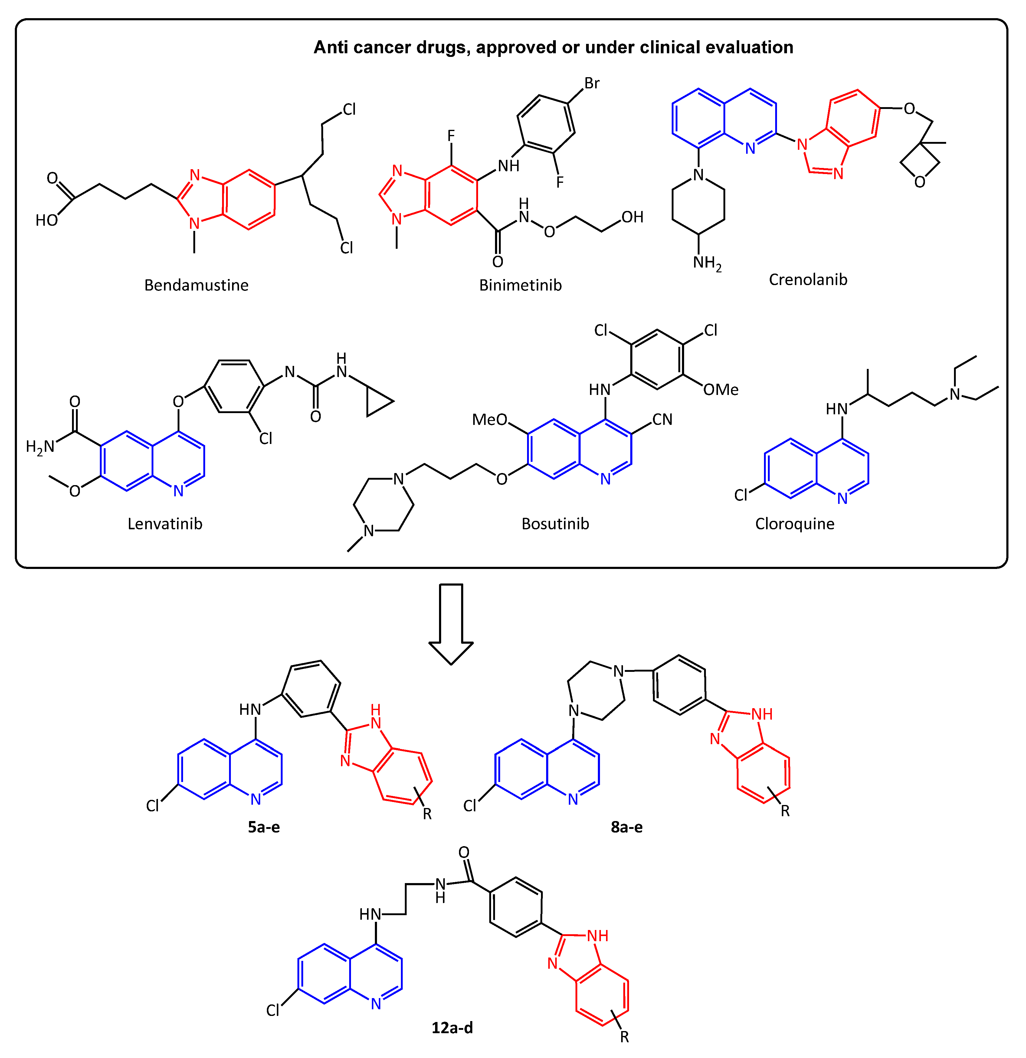
Design and Synthesis of Novel Benzimidazole Derivatives

The synthesis of novel benzimidazole derivatives of 7-chloro-4-aminoquinoline 5a–e, 8a–e, and 12a–d was carried out as shown in Scheme 1 and Scheme 2. With the aim of exploring the influence of the linkers between two pharmacophores, as well as the effect of the substituent at the benzimidazole ring, benzimidazoles with three different amidine substituents or chlorine or without a substituent at the C-5 position and 7-chloroquinoline were joined through three different linkers: 3-phenyl, 4-phenyl piperazine, and ethyl benzamidyl.
Alcohol 2 and aldehyde 3 were prepared using the methods reported by Medlen et al. [30]. 7-chloroquinoline and 3-aminobenzyl alcohol were refluxed in ethanol to yield (3-(7-chloroquinolin-4-ylamino) phenyl)methanol 2. The latter compound was transferred to 3-(7-chloroquinolin-4-ylamino)benzaldehyde 3 by stirring in DMSO with trimethylamine and sulfur trioxide complex in pyridine at room temperature. Quinoline benzimidazoles 5a–e were prepared by employing a condensation reaction between aldehyde 3 and benzene-1,2-diamines 4a–e in the presence of sodium metabisulfite Na2S2O5 in DMSO at 165 °C [31]. The synthesis of the precursors 4a–c was reported earlier [32,33]. All the analytical data of the earlier synthesized compounds correspond with the information previously given and will not be shown in this paper. The synthesis of 7-chloro-4-(piperazin-1-yl)quinoline 6 was also reported earlier [34]. The latter compound, via the nucleophilic substitution of fluorine in 4-fluorobenzaldehyde, resulted in 4-(4-(7-chloroquinolin-4-yl)piperazin-1-yl)benzaldehyde 7. Quinoline benzimidazoles 8a–e were synthesized by the condensation of aldehyde 7 and diamine precursors 4a–e with Na2S2O5.
Scheme 2 illustrates the synthetic route for the compounds with the ethyl benzamide linker 12a–d. Precursors with a carboxylic moiety 10a–d were prepared by the aforementioned condensation with Na2S2O5 from 4-formylbenzoic acid (9) and diamines 4a–d. The previously reported [35] N-(aminomethyl)-7-chloroquinolin-4-amine 11 was linked with the precursors 10a–d in the presence of trimethylamine, 1-hydroxybenzotriazole (HOBt), and N-(3-Dimethylaminopropyl)-N′-ethylcarbodiimide hydrochloride (EDCxHCl) in dichloromethane, a method previously reported by Pešić et al. [36]. We were not able to synthesize a chlorine derivative from this series. Although we prepared a chlorine precursor with a carboxylic moiety, the latter compound in the reaction with amine 11 did not yield the desired product. The NMR spectra of the material isolated from this reaction showed signals which correspond to both the starting amine 11 and the starting chloro benzimidazole benzoic acid derivative. In the NMR spectra, we did not find signals which would clearly indicate that the reaction actually took place. We tried using column cromatography to seperate the reaction mixture components, but we did not isolate any component which did not correspond to the starting compounds. Our attempts to modify the reaction conditions, which were employed for the synthesis of the corresponding compounds 12a–d, were also unsuccessful. The only conclusion we draw is that under these conditions the chlorine group, on the benzimidazole moiety, had a deactivating effect.
In our previous efforts to synthesize new benzimidazoles by the condensation of benzene-1,2-diamines and aryl aldehydes, we readily employed a method with sodium bisulfite NaHSO3, [37,38]. Utilizing Na2S2O5 in the present paper was advantageous because of the shorter reaction time (15 min. vs. 6 h for NaHSO3). In addition, in the case of the compounds 8a–e and 10a–d, the reactions with Na2S2O5 yielded pure products with no need for further purification; this was not the case when we tried to prepare corresponding compounds with NaHSO3.

2.2. Biological Activity

2.2.1. Evaluation of Antiproliferative Activity of the Novel Compounds

The newly synthesized benzimidazole derivatives of 7-chloro-4-aminoquinoline were tested for their effects on the growth of the non-tumor MDCK1 (Madine–Darby canine kidney) cell line, the leukemia and lymphoma cell lines: CCRF-CEM (human acute lymphoblastic leukemia), Hut78 (T-cell lymphoma), THP-1 (acute monocytic leukemia), Burkitt lymphoma (Raji), and the carcinoma cell lines: HeLa (human cervical adenocarcinoma), CaCo-2 (human colorectal adenocarcinoma), and MCF-7 (human breast adenocarcinoma). The obtained results, presented as the concentration achieving 50% of cell growth inhibition (GI50 value), show that the investigated compounds differently influenced tumor cell growth, depending on the cell line and on the dose applied.
As shown in Table 1, the GI50 values for all the investigated compounds ranged from 0.6 to more than 100 μM. The carcinoma cell lines were more resistant to the investigated compounds compared to the leukemia and lymphoma cells. The novel quinoline benzimidazoles 5a–c, in which the pharmacophores are connected with a 3-phenyl linker and the benzimidazole ring carries one of the three different amidine substituents (Scheme 1), exhibited insignificant inhibitory potential against normal and tumor cells. Compounds 5d (containing an unsubstituted benzimidazole ring) and 5e (containing a benzimidazole ring substituted with chlorine) showed mild to strong inhibitory potential against the normal (GI50 ranging from 11.4 to 20.4 μM) and the tumor cell lines (GI50 values ranging from 0.4 to 15.6 μM). In comparison to the MDCK1 and the carcinoma cell lines, compound 5d exhibited significant antiproliferative activity against the lymphoma (Raji: GI50 = 4.3 μM; HuT78: GI50 = 0.4 μM) and leukemia cells (CCRF CEM: GI50 = 8.2 μM; THP1: GI50 = 0.6 μM). With respect to the inhibition potential of 5d against the MDCK1 cells, the tumor selectivity index (SI) of the HuT78 cells is 51 and of the THP1 cells it is 34. The replacement of hydrogen with chlorine at the benzimidazole ring in 5e caused strong antiproliferative activity on all the tested cell lines.
Furthermore, in the group of compounds containing the 4-phenyl piperazine linker (8a–e), a mild to strong, albeit not selective, antiproliferative activity was observed (Table 1). Compounds 8a–c, in which the benzimidazole ring carries one of the three different amidine substituents (Scheme 1), showed moderate effects on the growth of normal and tumor cells, with GI50 values ranging from 20.7 to more than 100 μM. Compound 8d, which possesses an unsubstituted benzimidazole ring, significantly influenced the growth of the lymphoma and leukemia cells (CCRF CEM: GI50 = 5.0 μM, SI = 20; THP1: GI50 = 3.2 μM, SI = 31.5; Raji: GI50 = 3.8 μM, SI = 26.3; HuT78: GI50 = 8.1 μM, SI = 12.3). At the same time, 8d had a negligible effect on the growth of normal and carcinoma cells, with GI50 values of more than 100 μM. The replacement of hydrogen with chlorine at the benzimidazole ring in 8e caused a significant antiproliferative effect on the normal MDCK1 cells (GI50 = 2.7 μM). Compound 8e expressed less cytotoxicity effects against the carcinoma cells (GI50 ranging from 84.0 to >100 μM) and modest effects on the growth of the leukemia and lymphoma cells (GI50 ranging from 13.4 to 41 μM) in comparison to the effects on the MDCK1 cells.
In the group of quinoline benzimidazoles, in which the pharmacophores are connected with the ethyl benzamidyl linker (Scheme 2), a significant distinction in the antiproliferative effects between the normal and the most tumorous cell lines for compounds 12a–c was not observed (Table 1). An exception was the HuT78 cell line, which was significantly sensitive to the effects of the compounds 12a–d (12a: GI50 = 4.1 μM, SI = 24.4; 12b: GI50 = 5.6 μM, SI = 17.9; 12c: GI50 = 4.4 μM, SI = 22.7; 12d: GI50 = 3.5 μM, SI = 28.5).
The antiproliferative affinities of compounds 5d (against the THP1 and HuT 78 cells), 8d (against the CCRF-CEM, THP1, Raji and HuT78 cells), and 12d (against the THP1 and HuT78 cells) were significantly higher (p < 0.001, two-way ANOVA with Bonferroni post-test) compared to the healthy cell line, indicating therapeutic potential.
An insight into the effect of the different substituents at the benzimidazole moiety and the roles of the varied linkers is shown in Figure 2. Non-amidine compounds 5d and 5e with a 3-phenyl linker demonstrated the highest antiproliferative activity against the tumor cell lines, but they were not selective. The introduction of non-amidine substituents into compounds 8d (-H), 8e (-Cl), and 12d (-H) resulted in a higher activity against most of the leukemia and lymphoma cell lines. Compounds 5d, 8d, and 12d, which do not have a substituent at the C-5 position of the benzimidazole, demonstrated a general increase in selectivity compared to the compounds substituted with amidine.

2.2.2. Cell Cycle Redistribution

The delimited cell cycle progression and the evasion of apoptosis are the common events in the development of most tumors. The cell cycle checkpoints fortify the dividing cells against DNA damage and ensure the maintenance of genomic integrity. To assess whether a cell cycle disturbance underlines the antiproliferative activity that some compounds exhibited against lymphoma and leukemia cells in micromolar and submicromolar concentrations, we tested the cell cycle distribution of the HuT-78 and THP-1 cells treated with 5d, 8d, and 12d. As is shown in Figure 3, the compounds 5d and 12d induced a statistically significant redistribution in the cell cycles of the treated HuT78 and THP-1 cells compared to the control (untreated) cells. Compound 5d caused a significant increase in the aggregation of cells in the subG0/G1 phase in both the treated cell lines; a significant increase in the aggregation of cells in the subG0/G1 phase (HuT78: 13 times, p < 0.002; THP-1: (2.9 times, p < 0.001); and a significant decrease in the G0/G1 phase (HuT78: 5 times, p < 0.00; THP-1: 4.5 times, p < 0.004), with a consequent increase in the G2/M phase (HuT78: 2 times, p < 0.003; THP-1: 62.8%, p < 0.034) of the cell cycle.
Furthermore, compound 12d induced a statistically significant enrichment of the subG0/G1 fraction in the treated HuT78 cells (46 times, p < 0.004) and a significant decrease in the G0/G1 phase (for 63.8%, p < 0.004) compared to the non-treated cells. After treatment with 12d, the THP-1 cells showed an increased percentage of cells in the subG0/G1 phase (3.4 times, p < 0.006), while the changes in the other phases of the cell cycle were not statistically significant. These results indicate the prevention of the entry of the treated cells into a new cell cycle and are in accordance with the results published by Zuo at al. and Zhang at al. [39,40]. The subG0/G1 arrest suggests that the initiation of cell cycle arrest may be responsible for the antiproliferative potential. The subG0/G1 peak is indicative of DNA fragmentation and suggests that the cells are dying, most likely through apoptosis. The arrest of the cell cycle in the G2/M phase in the cells treated with the various benzimidazole derivatives has also been observed in several recently published studies [41].
An increased proportion of polyploid giant cancer cells was found in the HuT78 cells treated with compounds 5d and 12d and in the THP-1 cells treated with compound 5d. The increase in the number of polyploids could be explained by the cellular stress due to the activity of the applied compounds, as suggested by previous studies [42,43], or it could be the way in which the cells try to avoid the therapy [44,45].
As shown in Figure 3, no significant changes in the cell cycle were observed in the HuT78 and THP-1 cells treated with compound 8d in comparison with the non-treated cells, indicating a different mechanism of the antiproliferative effect compared to the effects of compounds 5d and 12d.

2.2.3. Apoptosis Induction

The induction of cell cycle arrest at a specific checkpoint and the initiation of apoptosis are mechanisms which are often used for treating cancer with cytotoxic agents [46,47,48]. To prove apoptosis as a mechanism of the treated cell death of HuT78 cells exposed to 5d and 12d, we used two methods for apoptosis detection: changes in the mitochondrial membrane potential (∆Ψm) and the phosphatidylserine externalization on the plasma membrane as typical features of apoptotic cell death.
Changes in the (∆Ψm) were measured using TMRE (tetramethylrhodamine, ethyl ester, and perchlorate) dye. Flow cytometry analysis showed that compound 12d caused a statistically significant mitochondrial membrane disruption in more than 85% of the HuT78 cells. Compound 5d also induced a change in the mitochondrial membrane potential in 23.9% of cells (Figure 4). Complex mitochondrial changes and alterations in the mitochondrial membrane potential may be an early event in the apoptotic process or, conversely, may be a consequence of the apoptotic signaling pathway [49,50]. In response to multiple distinct intracellular stress conditions, mitochondrial membranes can become permeabilized because of the pore-forming activity of the proapoptotic members of the Bcl-2 protein family. Alternatively, mitochondria can lose their structural integrity after the mitochondrial permeability transition, a phenomenon that is initiated at the mitochondrial inner membrane [50,51]. In both cases, permeabilized mitochondria allow the release of proapoptotic proteins into the cellular cytoplasm. The obtained results are consistent with the results of the previously published studies, which showed that some benzothiazole derivatives have the potential to induce the apoptosis of B and T lymphoma cells through the intrinsic pathway by the disruption of the mitochondrial membranes [52].
The results of the flow cytometry analysis of the Annexin V binding on the HuT78 cell membrane after 24 h treatment with 5d and 12d showed that 12d is a potent inducer of cell death (Figure 5). After treatment with 12d, a high percentage of cells were in the late stage of apoptosis/necrosis (83.75%), while the proportion of cells in early apoptosis was negligible (3.97%). The high proportion of cells observed in late apoptosis, 24 h after treatment, may indicate the rapid effect of 12d on Hut78 cells, likely inducing apoptosis soon after the application.
The proapoptotic effect of compound 5d is less dramatic compared to that of compound 12d. After 24 h of the treatment of the HuT78 cells with this compound, about 64% of the cells were alive, 24.75% of the cells were in early apoptosis, and only 8.3% of the cells were in late apoptosis. (Figure 5).

2.3. Absorption, Distribution, Metabolism, Excretion (ADME), and Toxicity Properties

The pharmacokinetic behavior of a drug compound determines its fate in the organism, and it is exploited to predict the ADME properties of that drug compound [53,54]. The physicochemical profiles of potential drugs tend to take into account molecular weight, ability to form hydrogen bonds, and lipophilicity. The octanol-water partition ratio is a common way of expressing the lipophilicity of a compound. There are many methods for the calculation of the logarithmic value of the octanol/water partition coefficient (logP). The Lipinski rule uses the Moriguchi-based algorithm to calculate logP (MLOGP). The advantages of the Moriguchi method are the ease of programming in any language and that this method does not require a large database of parameter values [55]. The in silico ADME properties for the novel 14 compounds were investigated and are recorded in Table 2. According to the calculated ADME parameters, compound 5e, which was found to be one of the most active compounds against cancer cell growth, could be problematic as an effective oral drug since its MLOGP is higher than 4.15 and the log S is lower than −6, which means that is highly lipophilic and poorly soluble in water. Additionally, its fraction of carbons in the sp3 hybridization (saturation) is zero since its structure is missing saturated carbon-carbon bonds [55]. Compound 5d also exhibited a strong inhibitory potential against the growth of all the tested cancer cell lines. Although its Csp3 fraction is zero and the log S is <−6, other ADME properties indicate that this compound could have good oral bioavailability. Thus, it fully satisfies the Lipinski rule. Compound 12d showed good inhibition activity against most of the cell lines. Except for the inappropriate fraction of Csp3, the other ADME properties suggest that molecule 12d has good solubility and permeability and could be effective as an antitumor drug.
The pharmacokinetic behavior of a set of compounds was evaluated using the program pkCSM [56]. This program uses graph-based signatures to develop predictive models of the central ADMET properties, which are important factors to consider when developing a new drug. These properties include human intestinal absorption (HIA/% abs), volume of distribution at steady state in humans (VDss), blood–brain barrier permeability (BBB per.), CNS (central nervous system) permeability, total clearance, oral rat chronic toxicity (LOAEL), and flathead minnow toxicity. The results of this evaluation are presented in Table 3. The compounds 5a and 8a had the highest HIA values, at 95.43% and 95.68%, respectively. HIA is a crucial ADME property because it is one of the key steps in the transport of drugs to their targets in the body [57]. Compound 12b has the highest VDss (0.35 log L kg−1). This means that a higher dose of this compound would be required to achieve a given plasma concentration. The lowest values of VDss were found for compounds 8d and 8e (−0.13, −0.12, respectively), which corresponds to their highest blood-brain barrier permeability (0.53, 0.49, respectively), and it corresponds to their highest CNS permeability (−0.54, −0.59, respectively). This indicated that these compounds have the possibility to easily leave the plasma and enter the extravascular compartments of the body, and after passing the blood–brain barrier, they can easily target the central nervous system [58]. Total clearance is an important pharmacokinetic parameter that describes the rate at which a drug is removed from the plasma. It is calculated by dividing the rate of drug removal from the plasma (mg/min) by the concentration of the drug in the plasma (mg/mL). In this case, the compound with the highest clearance was 5c, at 1.01 log mL min−1 kg−1 [59]. The highest aquatic toxicity evaluated against the fish model organism flathead minnow was estimated for compound 8e (−2.92 log mM), while the most toxic, for the rats, was shown to be compound 5b (1.25 log mg−1 kgbw per day). Both of these compounds are very lipophilic and poorly soluble in water, as indicated in Table 3.

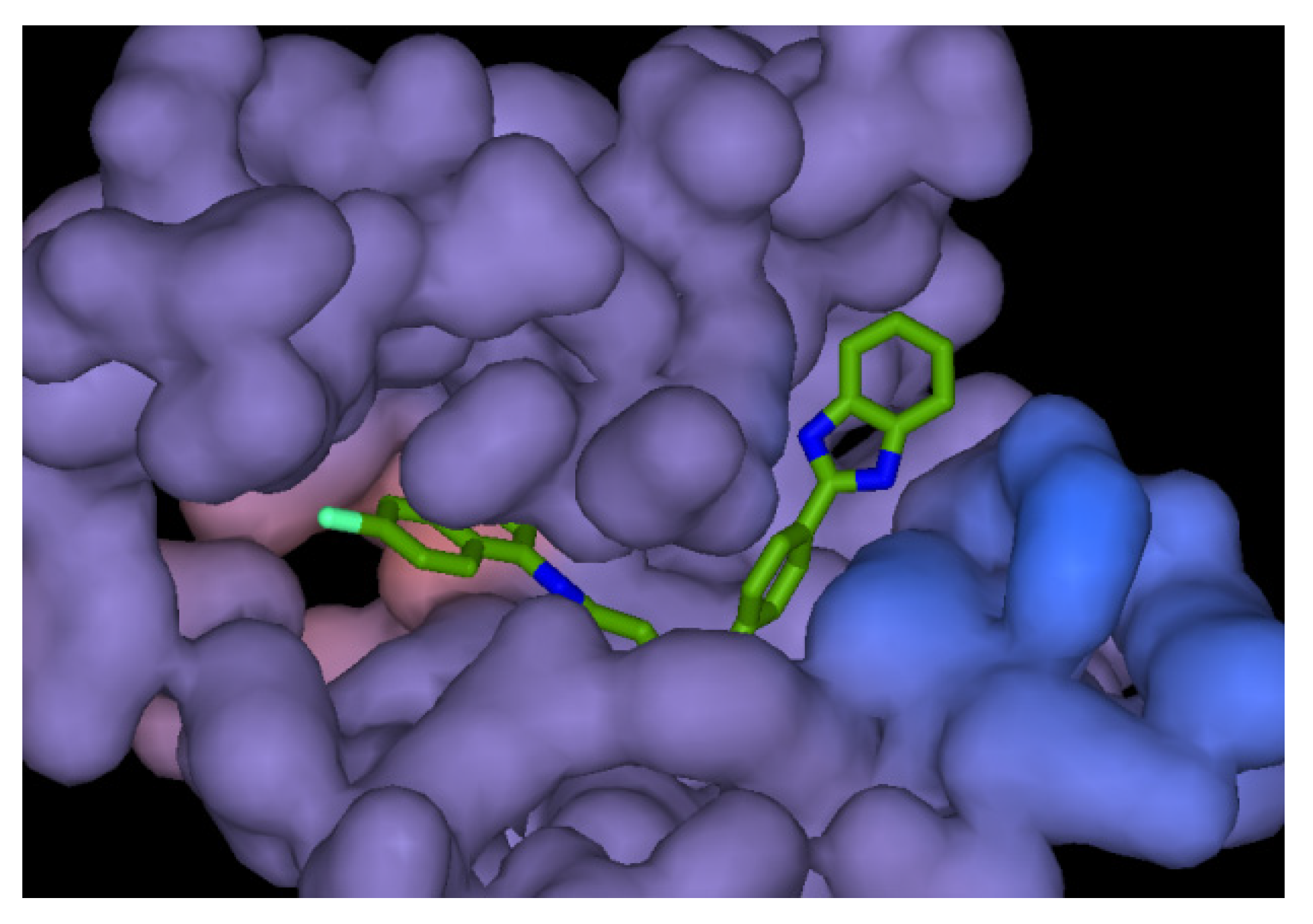
2.4. Molecular Docking

Tyrosine kinases are enzymes that phosphorylate tyrosine residues in specific substrates, and they are activated when a ligand binds to their extracellular domain. Inhibitors of the low molecular weight epidermal growth factor receptor (EGFR) can block extracellular signaling molecules (EGF) and prevent cell proliferation [60]. Src kinases play a crucial role in the assembly and disassembly of tight junctions in CaCo-2 and MDCK1 cell monolayers. It has been shown that the oxidative stress-induced disruption of tight junctions, which form a barrier to the diffusion of toxins, allergens, and pathogens across epithelial tissue, is mediated by the activation of c-Src [61]. The ATP binding pocket of Src is located in the cleft between the two lobes of the enzyme, the small amino-terminal (N) lobe made up of residues 267–337, and the large carboxyl-terminal (C) lobe made up of residues 341,520. The two lobes can move relative to each other, opening and closing the cleft. In the inactive conformation of the kinase, the Asp-Phe-Gly (DFG) motif is flipped by about 180° relative to the active state conformation. The flipped DFG motif moves the Asp away from the ATP binding site, leading to a “DFG”-out state. In the active conformation, the Asp side chain faces into the ATP binding pocket and coordinates with a Mg2+ ion, resulting in a “DFG”-in state [62,63].
As most of the compounds have shown inhibitory activity against the leukemia and lymphoma cell lines (CCRF-CEM, THP1, Raji, and HuT78), tyrosine-protein kinase c-Src (PDB: 3G6H) was chosen as a receptor for the molecular binding studies. These studies aim to predict the ability and mode of interaction of the synthesized molecules with the c-Src enzyme, and the results are compared to the docking results of the bonded ligand pyridinyl-triazine (DSA1, PDB ID: G6H) [62]. The ranking of the screened compounds based on the energies of their interactions is presented in Table 4. These molecular binding studies can provide insights into the potential of the synthesized compounds as ligands for c-Src and their potential as therapeutic agents.
The highest negative total energy (−150.08 kcal mol−1) overtakes compound 12c, ahead of the original ligand, compound DSA1. Compound 12c fits best into the active site of c-Src, as demonstrated by its significant effect on the growth of the HuT78 cell line (Table 1). However, this compound proved to be toxic to the normal, MDCK1 cells.
Compound DSA1, a bonded ligand (G6H), demonstrated slightly weaker binding energy (−133.74 kcal mol−1) than compound 12c. These compounds have the structure scaffold of Imatinib, the effective drug for chronic myeloid leukemia (Gleevec, Novartis), which binds with a higher potency to c-Src in the DFG-flipped conformation. Compound 12b appears in the ranking with the total energy of −122.66 kcal mol−1. This is consistent with the fact that compound 12b is also the significant active compound against the leukemia and lymphoma cells: Raji and HuT78. Compound 12b belongs to the group of compounds (12a–d) with an extension of the molecules’ central linker between the chloroquinoline and benzimidazole parts by the ethyl group, which contains two amino groups. This is in agreement with the previous QSAR findings on the anticancer activities of the CQArA hybrids that the 2-aminoethanol linker enhances their anticancer activity [29]. Unfortunately, compound 12b also exhibited an inhibitory potential against the normal cell line (Table 1). One of the compounds that demonstrated the best toxicity ratio in normal and tumor cells, especially leukemia and lymphoma cells, is 12d. This compound binds to the active site of the c-Src, releasing energy of 119.99 kcal mol−1 (Figure 6). The energies of the interactions between the protein residue and ligand 12d in docked pose 1 are tabulated in Table 5. The interactions of compound 12d with c-Src in the binding site are presented in Figure 6, which shows compound 12d docked in the binding site of c-Src, presented as a hydrophobic surface, while Figure 7 represents the 2D interactions of the ligand 12d with the residual amino acid of receptor c-Src. Like DS1, compound 12d is anchored into a hydrophobic pocket composed of the side chains of Val313, Leu317, Leu322, Val377, and Tyr382 [64]. Compound 12d forms hydrogen bonds with Glu310 (2.77 Å) and Asp404 (3.34 Å) via an amino group from the central linker, N-(2-aminoethyl)benzamide. The same group generates a carbon hydrogen bond with residue Thr338, which is the gatekeeper that controls the access of the inhibitor to the active site in c-Src [64]. The other key van der Waals interactions are between the quinoline ring and Val323 (3.71 Å and 4.45 Å); Met314 (4.84 and 5.02 Å); and Ala403 (4.50 Å). The phenyl ring of benzamide generates π-π interactions with Ala293 (4.43 Å) and Lys295 (5.27 Å), as well as a π-σ interaction with Val281 (5.00 Å). Significant π-π T-shaped interaction creates a benzimidazole ring with Phe405 (4.66 Å), as well as two π-π interactions with Lue273 (5.07 and 5.27 Å). For comparison, Imatinib and the inhibitor triazine derivative DSA8 form a hydrogen bond with Asp404 in the Asp-Phe-Gly (DFG) motif [62]. The results of our previous docking study of quinoline-arylamidine hybrids with c-Src revealed that the key interactions are the H-bonds between the amino groups from the 2-aminoethanol linker, Asp404 and the quinoline nitrogen atom, and Tyr382 and with Tyr340 via nitrogen from the nitrile group [29]. Apigenin glycoside isolated from Cupressus sempervirens creates a hydrogen bond via the hydroxyl group of a glucopyranose ring with Val402 and Val323, while the second hydroxyl group creates hydrogen bonds with Tyr382 and Asp404 [65]. A docking study of azaacridine derivatives revealed that the amido moiety also formed two hydrogen bonds with Glu310 and Asp404 and three π-π interaction bonds and one cation–π bond with Leu 273, Phe 405, Tyr 382, and Lys 295, respectively [66].

3. Materials and Methods

3.1. Chemistry

All the solvents and reagents were used without purification from commercial sources. To monitor the progress of a reaction and for comparison purposes, thin layer chromatography (TLC) was performed on pre-coated Merck silica gel 60F-254 plates using an appropriate solvent system, and the spots were detected under UV light (254 nm). For column chromatography, silica gel (Fluka, 0.063e0.2 mm) was employed; the glass column was slurry-packed under gravity. The melting points (uncorrected) were determined with the Buchi 510 melting point apparatus. The 1H and 13C NMR spectra were acquired on a Bruker 300 and 600 MHz NMR spectrometer. All the data were recorded in DMSO-d6 at 298 K. The chemical shifts were referenced to the residual solvent signal of DMSO at d 2.50 ppm for 1H and d 39.50 ppm for 13C. Individual resonances were assigned on the basis of their chemical shifts, signal intensities, multiplicity of resonances, and H_H coupling constants. High-performance liquid chromatography was performed on an Agilent 1100 series system with UV detection (photodiode array detector) using the Zorbax C18 reverse-phase analytical column (2.1_30 mm, 3.5 mm). All the compounds used for biological evaluation showed >95% purity in this HPLC system. The 4800 Plus MALDI TOF/TOF analyzer (Applied Biosystems Inc., Foster City, CA, USA), equipped with a 200 Hz, 355 nm Nd:YAG laser, was used for mass accuracy analysis of the compounds. Acquisition was performed in positive ion reflector mode. Elemental analyses were performed with the Perkin Elmer 2400 Series Organic Elemental analyzer.
4,7-dichloroquinoline 1, benzene-1,2-diamine 4d, 4-chlorobenzene-1,2-diamine 4e, and 4-formylbenzoic acid 9 are commercially available; the synthesis of compounds 2,3 [30], 4a–c [32,33], 6 [34], and 11 [35] was reported earlier. All the spectroscopic and analytical data of the above-mentioned compounds correspond with information previously reported and will not be shown in this paper. The procedures for the preparation of the remaining compounds illustrated in Scheme 1 and Scheme 2 are presented below.

3.1.1. General Procedure for the Synthesis of Compounds 5a–e

The solution of compound 3 (1 mmol) with the appropriate benzene-1,2-diamine 4a–e (1 mmol) and Na2S2O5 (0.5 mmol) in DMSO (15 mL) is heated at 165 °C for 15 min. The mixture is then cooled to room temperature and water (5 mL) is added, resulting in the precipitation of a crude residue. The crude residues of compounds 5a–c are collected by filtration, dissolved in methanol, and purified using column chromatography (CH2Cl2/MeOH 3:1; MeOH/NH4OH 3:1. The resulting products are converted to hydrochloride salts using anhydrous methanol saturated with HCl (g). The residues of compounds 5d and 5e are collected by filtration and purified by recrystallization using methanol.

2-(3-(7-chloroquinolin-4-ylamino)phenyl)-1H-benzo[d]imidazole-5-carboximidamide trihydrochloride (5a)

Compound 5a was prepared using the above-described method from 3 (190 mg, 0.67 mmol), diamine amidine 4a (126 mg, 0.67 mmol), and Na2S2O5 (64 mg, 0.34 mmol) as a dark brown product (95 mg, 25%); mp > 250 °C. 1H NMR (300 MHz, DMSO) δ 13.80 (s, 1H, NH), 10.69 (s, 1H, NH), 9.35 (s, 2H, NH), 9.03 (s, 2H, NH), 8.78 (d, J = 9.1 Hz, 1H, ArH), 8.58 (d, J = 5.9 Hz, 1H, ArH), 8.37 (s, 1H, ArH), 8.23 (d, J = 7.3 Hz, 2H, ArH), 8.10 (s, 1H, ArH), 7.92–7.59 (m, 5H, ArH), 7.01 (d, J = 6.4 Hz, 1H, ArH). 13C NMR (150 MHz, DMSO) δ 166.44, 152.92, 146.98, 143.65, 139.43, 139.32, 137.57, 131.39, 131.19, 127.30, 126.89, 126.03, 125.06, 122.95, 122.62, 122.27, 121.75, 120.10, 117.36, 112.55, 101.73. Anal. calcd. for C23H17ClN6 × 3HCl × 2H2O (Mr = 558.29): C 49.48, H 4.33, N 15.05; found: C 49.31, H 4.41, N 15.22. HRMS: calcd. for C23H18ClN6 (M + H)+: 413.1281; found: 412.1283.

2-(3-(7-chloroquinolin-4-ylamino)phenyl)-N-isopropyl-1H-benzo[d]imidazole-5-carboximidamide trihydrochloride (5b)

Compound 5b was prepared using the above-described method from 3 (259 mg, 0.92 mmol), diamine amidine 4b (210 mg, 0.92 mmol), and Na2S2O5 (87 mg, 0.46 mmol) as a light brown product (160 mg, 28%); mp > 250 °C. 1H NMR (300 MHz, DMSO-d6) δ/ppm 13.85 (d, J = 35.2 Hz, 1H, NH), 10.04 (s, 1H, NH), 9.48 (dd, J = 39.1, 13.2 Hz, 2H, NH), 9.01 (s, 1H, NH), 8.67 (d, J = 9.1 Hz, 1H, ArH), 8.56 (d, J = 5.8 Hz, 1H, ArH), 8.33 (s, 1H, ArH), 8.21–7.81 (m, 4H, ArH), 7.58 -7.73 (m, 4H, ArH), 7.04 (d, J = 5.9 Hz, 1H), 4.09 (m, 1H, CH), 1.31 (d, J = 6.3 Hz, 6H). 13C NMR (75 MHz, DMSO) δ 162.89, 154.39, 153.77, 150.25, 147.39, 143.52, 140.66, 138.88, 135.91, 135.04, 131.22, 130.95, 126.38, 125.57, 123.70, 123.07, 122.37, 121.73, 120.09, 119.45, 118.26, 112.84, 112.31, 102.37, 45.47, 21.78. Anal. calcd. for C26H23ClN6 × 3HCl × 3.5H2O (Mr = 627.39): C 49.77, H 5.30, N 13.40; found: C 49.69, H 5.05, N 13.22. HRMS: calcd. for C26H24ClN6 (M + H)+: 455.1751; found: 454.1750.

7-chloro-N-(3-(5-(4,5-dihydro-1H-imidazol-2-yl)-1H-benzo[d]imidazol-2-yl)phenyl)quinolin-4-amine trihydrochloride (5c)

Compound 5c was prepared using the above-described method from 3 (265 mg, 0.94 mmol), diamine amidine 4c (200 mg, 0.94 mmol), and Na2S2O5 (0.47 mmol, 89 mg) as a brown product (140 mg, 26%); mp > 250 °C. 1H NMR (300 MHz, DMSO-d6) δ/ppm 14.78 (s, 1H, NH), 11.36 (s, 1H, NH), 10.65 (s, 2H, NH), 8.93 (d, J = 9.2 Hz, 1H, ArH), 8.60 (d, J = 7.0 Hz, 1H, ArH), 8.42 (d, J = 6.9 Hz, 2H, ArH), 8.35 (d, J = 7.9 Hz, 1H, ArH), 8.20 (d, J = 1.9 Hz, 1H, ArH), 7.94 (d, J = 9.0 Hz, 1H, ArH), 7.90–7.64 (m, 4H, ArH), 6.96 (d, J = 7.0 Hz, 1H, ArH), 4.03 (s, 4H, CH2). 13C NMR (75 MHz, DMSO-d6) δ/ppm 165.66, 155.28, 153.88, 144.07, 139.50, 139.00, 138.35, 131.37, 130.78, 128.36, 128.03, 126.73, 126.57, 124.47, 123.47, 119.72, 116.53, 116.48, 115.74, 101.15, 44.72.
Anal. calcd. for C25H19ClN6 × 3HCl × 1.5H2O (Mr = 575.32): C 52.19, H 4.38, N 14.61; found: C 51.97, H 4.25, N 14.22. HRMS: calcd. for C25H20ClN6 (M + H)+: 439.1438; found: 439.1437.

N-(3-(1H-benzo[d]imidazol-2-yl)phenyl)-7-chloroquinolin-4-amine (5d)

Compound 5d was prepared using the above-described method from 3 (250 mg, 0.9 mmol), diamine 4d (95 mg, 0.9 mmol), and Na2S2O5 (0.45 mmol, 85 mg) as a yellow product (130 mg, 36%); mp 248–250 °C. 1H NMR (300 MHz, DMSO-d6) δ/ppm 13.03 (s, 1H, NH), 10.17 (s, 1H, NH), 8.62 (d, J = 9.1 Hz, 1H, ArH), 8.57 (d, J = 6.2 Hz, 1H, ArH), 8.28 (s, 1H, ArH), 8.08 (d, J = 7.8 Hz, 1H, ArH), 8.00 (d, J = 2.1 Hz, 1H, ArH), 7.79 (dd, J = 9.1, 2.1 Hz, 1H, ArH), 7.74–7.52 (m, 4H, ArH), 7.22 (dd, J = 6.0, 3.1 Hz, 2H, ArH), 7.02 (d, J = 6.2 Hz, 1H, ArH). 13C NMR (151 MHz, DMSO-d6) δ/ppm 151.57, 151.24, 149.01, 148.81, 141.09, 135.00, 131.88, 130.70, 127.20, 125.90, 125.09, 124.49, 122.68, 120.97, 118.67, 102.64. Anal. calcd. for C22H15ClN4 × 1.5H2O (Mr = 397.86): C 66.41, H 4.56, N 14.08; found: C 66.21, H 4.33, N 14.19. HRMS: calcd. for C22H16ClN4 (M + H)+: 371.1063; found: 371.1056.

7-chloro-N-(3-(5-chloro-1H-benzo[d]imidazol-2-yl)phenyl)quinolin-4-amine (5e)

Compound 5e was prepared using the above-described method from 3 (296 mg, 1.05 mmol), diamine 4e (150 mg, 1.05 mmol), and Na2S2O5 (0.525 mmol, 100 mg) as a yellow product (160 mg, 33%); mp 244–246 °C. 1H NMR (300 MHz, DMSO-d6) δ/ppm 13.20 (s, 1H, NH), 10.09 (s, 1H, NH), 8.64–8.54 (m, 2H, ArH), 8.26 (s, 1H, ArH), 8.06 (d, J = 7.9 Hz, 1H, ArH), 7.99 (d, J = 2.1 Hz, 1H, ArH), 7.77 (dd, J = 9.1, 2.1 Hz, 1H, ArH), 7.73–7.51 (m, 4H, ArH), 7.25 (d, J = 8.4 Hz, 1H, ArH), 7.03 (d, J = 6.1 Hz, 1H, ArH). 13C NMR (151 MHz, DMSO-d6) δ/ppm 152.44, 151.26, 149.00, 145.33, 140.09, 136.52, 131.60, 130.98, 126.75, 125.70, 125.50, 124.59, 123.90, 123.05, 122.03, 117.89, 102.12, 65.36. Anal. calcd. for C22H14Cl2N4 × 2.75H2O (Mr = 454.82): C 58.11, H 4.32, N 12.32; found: C 57.79, H 4.31, N 12.22. HRMS: calcd. for C22H15Cl2N4 (M + H)+: 405.0674; found: 404.0669.

3.1.2. 4-(4-(7-chloroquinolin-4-yl)piperazin-1-yl)benzaldehyde (7)

A suspension of compound 6 (1.9 g, 7.71 mmol), 4-fluorbenzaldehyde (2.86 mg 23 mmol), and K2CO3 (3.17 mg, 23 mmol) in DMF (40 mL) was heated at 120 °C for 24 h. The mixture was cooled down to room temperature and the reaction suspension was diluted with dichloromethane (100 mL) and washed three times with water (100mL). The organic layer was dried on sodium sulfate, filtered, and concentrated on a rotary evaporator. The obtained yellow oil was diluted with methanol (2 mL), and water (5 mL) was added; the resulting yellow solid was filtered off (2 g, 78%); mp 182–183 °C. 1H NMR (600 MHz, DMSO-d6) δ/ppm 9.76 (s, 1H, CHO), 8.74 (d, J = 5.0 Hz, 1H, ArH), 8.13 (d, J = 9.0 Hz, 1H, ArH), 8.01 (d, J = 2.2 Hz, 1H, ArH), 7.77 (d, J = 8.9 Hz, 2H, ArH), 7.58 (dd, J = 9.0, 2.2 Hz, 1H), 7.14 (d, J = 8.9 Hz, 2H), 7.06 (d, J = 5.0 Hz, 1H, ArH), 3.72–3.64 (m, 4H, CH2), 3.36–3.32 (m, 4H, CH2). 13C NMR (151 MHz, DMSO-d6) δ/ppm 190.84, 156.42, 155.08, 152.70, 150.14, 134.14, 131.97, 128.60, 127.14, 126.53, 126.36, 121.79, 113.99, 110.01, 51.77, 46.89. HRMS: calcd. for C20H19ClN3O (M + H)+: 352.1216; found: 352.1220.

3.1.3. General Procedure for the Synthesis of Compounds 8a–e

A solution of compound 7 (1 mmol) with the appropriate benzene-1,2-diamine 4a–e (1 mmol) and Na2S2O5 (0.5 mmol) in DMSO (15 mL) was heated at 165 °C for 15 min. The mixture was cooled down to room temperature. The addition of water (5 mL) resulted in precipitation. The resulting residue for compounds 8a–c was collected with filtration and dissolved in methanol saturated with HCl; the addition of diethyl ether resulted in the precipitation of the products as hydrochloride salts. The residues of compounds 8d and 8e were collected with filtration, and the products were obtained by recrystallization using methanol.

2-(4-(4-(7-chloroquinolin-4-yl)piperazin-1-yl)phenyl)-1H-benzo[d]imidazole-5-carboxamidine dihydrochloride (8a)

Compound 8a was prepared using the above-described method from 7 (300 mg, 0.86 mmol), diamine amidine 4a (160 mg, 0.86 mmol), and Na2S2O5 (82 mg, 0.43 mmol) as a light brown product (245 mg, 46%); mp > 250 °C. 1H NMR (600 MHz, DMSO-d6) δ/ppm 9.58 (s, 2H, NH), 9.27 (s, 2H, NH), 8.65 (d, J = 7.0 Hz, 1H, ArH), 8.41 (d, J = 8.8 Hz, 2H, ArH), 8.32 (d, J = 9.2 Hz, 1H, ArH), 8.22 (d, J = 2.1 Hz, 1H, ArH), 8.18 (s, 1H, ArH), 7.88–7.68 (m, 2H, ArH), 7.69 (dd, J = 9.2, 2.1 Hz, 1H, ArH), 7.15 (d, J = 7.1 Hz, 1H), 7.13 (d, J = 9.0 Hz, 2H), 4.12 (s, 4H, CH2), 3.81 (s, 4H, CH2). 13C NMR (151 MHz, DMSO-d6) δ/ppm 165.99, 159.94, 153.06, 152.61, 142.22, 140.68, 138.17, 130.51, 129.57, 126.56, 124.96, 124.32, 119.59, 117.50, 114.55, 114.17, 113.43, 105.13, 50.71, 45.32. Anal. calcd. for C27H24ClN7 × 2HCl × 4H2O (Mr = 626.96): C 51.72, H 5.47, N 15.64; found: C 51.68, H 5.49, N 15.32. HRMS: calcd. for C27H25ClN7 (M + H)+: 482.1860; found: 482.1860.

2-(4-(4-(7-chloroquinolin-4-yl)piperazin-1-yl)phenyl)-N-isopropyl-1H-benzo[d]imidazole-5-carboxamidine dihydrochloride (8b)

Compound 8b was prepared using the above-described method from 7 (350 mg, 0.99 mmol), diamine amidine 4b (227 mg, 0.99 mmol), and Na2S2O5 (94 mg, 0.5 mmol) as an orange product (240 mg, 36%); mp 258 °C decomp. 1H NMR (600 MHz, DMSO-d6) δ/ppm 9.83 (d, J = 7.9 Hz, 1H, NH), 9.66 (s, 1H, NH), 9.28 (s, 1H, NH), 8.67 (d, J = 7.0 Hz, 1H, ArH), 8.52 (d, J = 8.7 Hz, 2H, ArH), 8.34 (d, J = 9.2 Hz, 1H, ArH), 8.29 (d, J = 2.1 Hz, 1H, ArH), 8.09 (d, J = 0.9 Hz, 1H, ArH), 7.89 (d, J = 8.5 Hz, 1H, ArH), 7.78 (dd, J = 8.5, 1.2 Hz, 1H, ArH), 7.72 (dd, J = 9.2, 2.1 Hz, 1H, ArH), 7.16 (dd, J = 10.8, 8.2 Hz, 3H, ArH), 4.15 (s, 5H, CH and CH2), 3.84 (s, 4H, CH2), 1.32 (d, J = 6.4 Hz, 6H, CH3). 13C NMR (151 MHz, DMSO-d6) δ/ppm 162.05, 159.92, 153.17, 152.03, 142.13, 140.68, 138.12, 135.85, 132.45, 130.72, 129.56, 126.55, 125.88, 125.48, 119.56, 117.50, 114.47, 113.91, 113.36, 110.83, 105.11, 50.67, 45.72, 45.26, 21.73. Anal. calcd. for C30H30ClN7 × 2HCl × 3.5H2O (Mr = 660.03): C 54.59, H 5.96, N 14.85; found: C 54.68, H 5.83, N 14.72. HRMS: calcd. for C30H31ClN7 (M + H)+: 524.2329; found: 524.2326.

7-chloro-4-(4-(4-(5-(4,5-dihydro-1H-imidazol-2-yl)-1H-benzo[d]imidazol-2-yl)phenyl)piperazin-1-yl)quinoline dihydrochloride (8c)

Compound 8c was prepared using the above-described method from 7 (280 mg, 0.8 mmol), diamine amidine 4c (170 mg, 0.8 mmol), and Na2S2O5 (76 mg, 0.4 mmol) as an orange product (230 mg, 45%); mp 250 °C decomp. 1H NMR (300 MHz, DMSO-d6) δ/ppm 10.72 (s, 2H, NH), 8.70 (d, J = 6.9 Hz, 1H, ArH), 8.37–8.18 (m, 5H, ArH), 7.90 (d, J = 8.4 Hz, 1H, ArH), 7.80 (d, J = 8.4 Hz, 1H, ArH), 7.72 (dd, J = 9.2, 1.9 Hz, 1H, ArH), 7.20 (d, J = 7.0 Hz, 1H, ArH), 7.12 (d, J = 8.9 Hz, 2H, ArH), 4.08 (s, 4H, CH2), 4.03 (s, 4H, CH2), 3.74 (s, 4H, CH2). 13C NMR (151 MHz, DMSO-d6) δ/ppm 165.72, 159.87, 152.15, 143.09, 137.94, 129.30, 126.63, 123.25, 120.30, 117.93, 113.97, 105.73, 51.06, 46.05, 44.72. Anal. calcd. for C29H26ClN7 × 2HCl × 3H2O (Mr = 634.98): C 54.85, H 5.40, N 15.44; found: C 54.76, H 5.38, N 15.22. HRMS: calcd. for C29H27ClN7 (M + H)+: 508.2016; found: 508.2008.

4-(4-(4-(1H-benzo[d]imidazol-2-yl)phenyl)piperazin-1-yl)-7-chloroquinoline (8d)

Compound 8d was prepared using the above-described method from 7 (400 mg, 1.14 mmol), diamine 4d (123 mg, 1.14 mmol), and Na2S2O5 (108 mg, 0.57 mmol) as an orange product (310 mg, 57%); mp 254 °C decomp.1H NMR (300 MHz, DMSO-d6) δ/ppm 8.74 (d, J = 5.2 Hz, 1H, ArH), 8.15 (d, J = 9.0 Hz, 1H, ArH), 8.08 (d, J = 8.8 Hz, 2H, ArH), 8.02 (d, J = 2.1 Hz, 1H, ArH), 7.62–7.55 (m, 3H, ArH), 7.21–7.18 (m, 4H, ArH), 7.10 (d, J = 5.2 Hz, 1H, ArH), 3.60 (s, 4H, CH2), 3.41 (s, 4H, CH2). 13C NMR (151 MHz, DMSO-d6) δ/ppm 156.84, 152.25, 151.98, 151.90, 149.36, 139.13, 134.48, 128.23, 127.89, 126.75, 126.39, 122.44, 121.50, 119.79, 115.20, 114.91, 109.67, 51.89, 47.64. Anal. calcd. for C26H22ClN5 × 2.25H2O (Mr = 480.47): C 64.99, H 5.56, N 14.58; found: C 65.11, H 5.39, N 14.21. HRMS: calcd. for C26H23ClN5 (M + H)+: 440.1642; found: 440.1637.

7-chloro-4-(4-(4-(5-chloro-1H-benzo[d]imidazol-2-yl)phenyl)piperazin-1-yl)quinoline (8e)

Compound 8e was prepared using the above-described method from 7 (400 mg, 1.14 mmol), diamine 4e (162 mg, 1.14 mmol), and Na2S2O5 (108 mg, 0.57 mmol) as a brown product (290 mg, 48%); mp 206–208 °C.1H NMR (300 MHz, DMSO-d6) δ/ppm 8.74 (d, J = 5.3 Hz, 1H, ArH), 8.16 (d, J = 9.0 Hz, 1H, ArH), 8.06 (d, J = 8.9 Hz, 2H, ArH), 8.02 (d, J = 2.2 Hz, 1H, ArH), 7.61 (dd, J = 9.0, 2.2 Hz, 1H, ArH), 7.57 (d, J = 1.9 Hz, 1H, ArH), 7.53 (d, J = 8.5 Hz, 1H, ArH), 7.18 (dd, J = 8.1, 1.5 Hz, 3H, ArH), 7.11 (d, J = 5.3 Hz, 1H, ArH), 3.61 (s, 4H, CH2), 3.45 (s, 5H, CH2). 13C NMR (151 MHz, DMSO-d6) δ/ppm 157.29, 153.60, 152.20, 150.69, 148.22, 134.99, 128.27, 127.12, 126.80, 126.43, 126.41, 122.29, 120.99, 119.79, 115.04, 109.13, 56.49, 51.79, 47.43, 19.01. Anal. calcd. for C26H21Cl2N5 × 3H2O (Mr = 528.43): C 59.1, H 5.15, N 13.25; found: C 58.78, H 4.96, N 13.12. HRMS: calcd. for C26H22Cl2N5 (M + H)+: 474.1252; found: 474.1250

3.1.4. General Procedure for the Synthesis of Compounds 10a–d

A solution of compound 9 (1 mmol) with the appropriate benzene-1,2-diamine 4a–d (1 mmol) and Na2S2O5 (0.5 mmol) in DMSO (15 mL) was heated at 165 °C for 15 min. The mixture was cooled down to room temperature. The addition of water (5 mL) resulted in precipitation. The resulting residue for compounds 10a-c was collected with filtration and dissolved in methanol saturated with HCl; the addition of diethyl ether resulted in the precipitation of the products as hydrochloride salts. The residue of compound 8d was collected with filtration, and the product was obtained by recrystallization using methanol.

4-(5-carbamimidoyl-1H-benzo[d]imidazol-2-yl)benzoic acid dihydrochloride (10a)

Compound 10a was prepared using the above-described method from 9 (300 mg, 2 mmol), diamine amidine 4a (373 mg, 2 mmol), and Na2S2O5 (190 mg, 1 mmol) as a light brown product (445 mg, 60%); mp > 250 °C. 1H NMR (600 MHz, DMSO-d6) δ/ppm 9.50 (s, 2H, NH), 9.24 (s, 2H, NH), 8.48 (d, J = 8.4 Hz, 2H, ArH), 8.27 (d, J = 1.3 Hz, 1H, ArH), 8.15 (d, J = 8.5 Hz, 2H, ArH), 7.89 (d, J = 8.5 Hz, 1H, ArH), 7.81 (d, J = 1.7 Hz, 1H, ArH). 13C NMR (151 MHz, DMSO-d6) δ/ppm 166.56, 165.88, 152.38, 133.06, 131.24, 129.95, 127.59, 123.25, 122.68, 116.3, 114.93. HRMS: calcd. for C15H13N4O2 (M + H)+: 281.1038; found: 281.1031.

4-(5-(N-isopropylcarbamimidoyl)-1H-benzo[d]imidazol-2-yl)benzoic acid dihydrochloride (10b)

Compound 10b was prepared using the above-described method from 9 (300 mg, 2 mmol), diamine amidine 4b (457 mg, 2 mmol), and Na2S2O5 (190 mg, 1 mmol) as a grey product (520 mg, 63%); mp > 250 °C. 1H NMR (300 MHz, DMSO-d6) δ/ppm 9.74 (d, J = 8.0 Hz, 1H, NH), 9.60 (s, 1H, NH), 9.24 (s, 1H, NH), 8.56 (d, J = 8.4 Hz, 2H, ArH), 8.18 (d, J = 8.6 Hz, 3H, ArH), 7.92 (d, J = 8.5 Hz, 1H, ArH), 7.73 (dd, J = 8.6, 1.5 Hz, 1H, ArH), 4.22–4.09 (m, 1H, CH), 1.32 (d, J = 6.4 Hz, 6H, CH3). 13C NMR (75 MHz, DMSO-d6) δ/ppm 166.98, 162.33, 152.04, 139.08, 136.27, 133.96, 130.64, 130.47, 128.41, 125.20, 124.66, 116.46, 115.21, 45.65, 21.76. HRMS: calcd. for C18H19N4O2 (M + H)+: 323.1508; found: 322.1515.

4-(5-(4,5-dihydro-1H-imidazol-2-yl)-1H-benzo[d]imidazol-2-yl)benzoic acid hydrochloride (10c)

Compound 10c was prepared using the above-described method from 9 (300 mg, 2 mmol), diamine amidine 4c (425 mg, 2 mmol), and Na2S2O5 (190 mg, 1 mmol) as a grey product (515 mg, 67%); mp > 250 °C.1H NMR (300 MHz, DMSO-d6) δ/ppm 10.88 (s, 2H, NH), 8.52–8.40 (m, 3H, ArH), 8.14 (d, J = 8.4 Hz, 2H, ArH), 8.00 (dd, J = 8.6, 1.3 Hz, 1H, ArH), 7.89 (d, J = 8.5 Hz, 1H, ArH), 4.03 (s, 4H, CH2). 13C NMR (75 MHz, DMSO-d6) δ/ppm 167.08, 165.41, 153.28, 141.44, 138.29, 133.45, 131.85, 130.40, 128.07, 124.06, 117.56, 117.09, 115.73, 44.73. HRMS: calcd. for C17H15N4O2 (M + H)+: 307.1195; found: 307.1206.

4-(1H-benzo[d]imidazol-2-yl)benzoic acid (10d)

Compound 10d was prepared using the above-described method from 9 (250 mg, 1.66 mmol), diamine 4d (180 mg, 1.66 mmol), and Na2S2O5 (157 mg, 0.83 mmol) as a grey product (350 mg, 82%); mp 215–217 °C. 1H NMR (600 MHz, DMSO-d6) δ/ppm 8.31 (d, J = 8.5 Hz, 2H, ArH), 8.15 (d, J = 8.5 Hz, 2H, ArH), 7.70 (dd, J = 6.0, 3.2 Hz, 2H, ArH), 7.33 (dd, J = 6.1, 3.1 Hz, 2H, ArH). 13C NMR (151 MHz, DMSO-d6) δ/ppm δ 167.20, 150.12, 138.17, 132.73, 130.48, 127.35, 127.35, 123.88, 115.59. HRMS: calcd. for C14H11N2O2 (M + H)+: 239.0820; found: 239.0824.

3.1.5. General Procedure for the Synthesis of Compounds 12a–d

To a suspension of the appropriate benzoic acid 10a–d (1 mmol) in dichloromethane (30 mL), triethylamine (Et3N) (6.75 mmol), and 1-hydroxybenzotriazole (HOBt) (1.1 mmol), compound 11 (1 mmol) and N-(3-Dimethylaminopropyl)-N′-ethylcarbodiimide hydrochloride (EDCxHCl) (1.4 mmol) were added. The reaction mixture was stirred at room temperature for 48 h. An ammonium hydroxide solution (pH 8.0) (30 mL) was added to reaction mixture. The resulting sticky residue was separated with decantation. The residue for compounds 12a–c was dissolved in methanol saturated with HCl; the addition of diethyl ether resulted in the precipitation of the products as hydrochloride salts. The residue of compound 12d was obtained by recrystallization using methanol.

4-(5-carbamimidoyl-1H-benzo[d]imidazol-2-yl)-N-(2-(7-chloroquinolin-4-ylamino)ethyl)benzamide trihydrochloride (12a)

Compound 12a was prepared using the above-described method from 10a (371 mg, 1 mmol), Et3N (688 mg, 6.75 mmol), HOBt (149 mg, 1.1 mmol), compound 11 (222 mg, 1 mmol), and EDCxHCl (267 mg, 1.4 mmol) in dichloromethane (30 mL) as a grey product (290 mg, 45%); mp > 250 °C 1H NMR (600 MHz, DMSO-d6) δ/ppm 14.33 (s, 1H, NH), 9.81 (t, J = 5.7 Hz, 1H, NH), 9.41 (s, 2H, NH), 9.15 (t, J = 5.6 Hz, 1H, NH), 9.12 (d, J = 18.2 Hz, 2H, NH), 8.72 (d, J = 9.2 Hz, 1H, ArH), 8.56 (dd, J = 11.9, 6.7 Hz, 1H, ArH), 8.39 (d, J = 8.4 Hz, 2H, ArH), 8.21 (d, J = 1.2 Hz, 1H, ArH), 8.07 (dd, J = 9.4, 5.3 Hz, 3H, ArH), 7.84 (d, J = 8.5 Hz, 1H, ArH), 7.76 (dd, J = 9.0, 2.1 Hz, 1H, ArH), 7.73 (dd, J = 8.5, 1.7 Hz, 1H, ArH), 7.00 (d, J = 7.2 Hz, 1H, ArH), 3.76 (dd, J = 12.0, 6.0 Hz, 2H, CH2), 3.63 (dd, J = 11.9, 6.0 Hz, 2H, CH2). 13C NMR (151 MHz, DMSO-d6) δ/ppm 166.43, 166.38, 156.26, 153.35, 143.25, 138.97, 138.45, 136.57, 131.10, 128.51, 127.66, 127.33, 126.47, 124.24, 123.34, 122.73, 119.45, 116.06, 99.07, 56.47, 43.07, 38.31, 19.01. Anal. calcd. for C26H22ClN7O × 3HCl × 3H2O (Mr = 647.38): C 48.24, H 4.83, N 15.15; found: C 48.01, H 4.47, N 15.23. HRMS: calcd. for C26H23ClN7O (M + H)+: 484.1653; found: 484.1640.

4-(5-(N-isopropylcarbamimidoyl)-1H-benzo[d]imidazol-2-yl)-N-(2-(7-chloroquinolin-4-ylamino)ethyl)benzamide trihydrochloride (12b)

Compound 12b was prepared using the above-described method from 10b (413 mg, 1 mmol), Et3N (688 mg, 6.75 mmol), HOBt (149 mg, 1.1 mmol), compound 11 (222 mg, 1 mmol), and EDCxHCl (267 mg, 1.4 mmol) in dichloromethane (30 mL) as a light brown product (266 mg, 39%); mp > 250 °C. 1H NMR (600 MHz, DMSO-d6) δ/ppm 14.50 (s, 1H, NH), 9.90 (t, J = 5.7 Hz, 1H, NH), 9.68 (d, J = 8.1 Hz, 1H, NH), 9.54 (s, 1H, NH), 9.26 (t, J = 5.6 Hz, 1H, NH), 9.15 (s, 1H, NH), 8.79 (d, J = 9.2 Hz, 1H, ArH), 8.56 (s, 1H, ArH), 8.48 (d, J = 8.4 Hz, 2H, ArH), 8.15–8.12 (m, 3H, ArH), 8.11 (d, J = 2.1 Hz, 1H, ArH), 7.88 (d, J = 8.5 Hz, 1H, ArH), 7.75 (dd, J = 9.1, 2.1 Hz, 1H, ArH), 7.69 (dd, J = 8.5, 1.6 Hz, 1H, ArH), 7.01 (d, J = 7.2 Hz, 1H, ArH), 4.13 (m, 1H, CH), 3.77 (dd, J = 11.8, 5.9 Hz, 2H, CH2)., 1.31 (d, J = 6.4 Hz, 6H, CH3). 13C NMR (151 MHz, DMSO-d6) δ/ppm 166.27, 162.46, 156.22, 152.47, 143.16, 138.97, 138.39, 137.03, 129.73, 128.57, 128.02, 127.27, 126.57, 124.84, 124.29, 119.40, 116.47, 116.06, 115.20, 99.05, 45.62, 43.03, 38.32, 21.76. Anal. calcd. for C29H28ClN7O × 3HCl × 1.75H2O (Mr = 666.94): C 52.23, H 5.21, N 14.70; found: C 52.02, H 5.57, N 14.53. HRMS: calcd. for C29H29ClN7O (M + H)+: 526.2122; found: 526.2122.

N-(2-(7-chloroquinolin-4-ylamino)ethyl)-4-(5-(4,5-dihydro-1H-imidazol-2-yl)-1H-benzo[d]imidazol-2-yl)benzamide trihydrochloride (12c)

Compound 12c was prepared using the above-described method from 10c (383 mg, 1 mmol), Et3N (688 mg, 6.75 mmol), HOBt (149 mg, 1.1 mmol), compound 11 (222 mg, 1 mmol), and EDCxHCl (267 mg, 1.4 mmol) in dichloromethane (30 mL) as a brown product (304 mg, 44%); mp > 250 °C.1H NMR (600 MHz, DMSO-d6) δ/ppm 14.40 (s, 1H, NH), 10.76 (s, 2H, NH), 9.84 (t, J = 5.7 Hz, 1H, NH), 9.17 (t, J = 5.6 Hz, 1H, NH), 8.74 (d, J = 9.2 Hz, 1H, ArH), 8.60–8.52 (m, 1H, ArH), 8.45 (d, J = 1.0 Hz, 1H, ArH), 8.40 (d, J = 8.4 Hz, 2H, ArH), 8.10–8.06 (m, 3H, ArH), 7.93 (dd, J = 8.5, 1.4 Hz, 1H, ArH), 7.86 (d, J = 8.5 Hz, 1H, ArH), 7.75 (dd, J = 9.1, 2.1 Hz, 1H, ArH), 7.00 (d, J = 7.2 Hz, 1H, ArH), 4.01 (s, 4H, CH2), 3.75 (dd, J = 11.9, 6.0 Hz, 2H, CH2), 3.63 (dd, J = 11.8, 5.9 Hz, 2H, CH2). 13C NMR (151 MHz, DMSO-d6) δ/ppm 166.40, 165.62, 156.24, 153.74, 143.22, 138.97, 138.42, 136.61, 131.02, 128.50, 127.74, 127.30, 126.50, 123.72, 119.44, 116.77, 116.06, 99.06, 44.75, 43.06, 38.30. Anal. calcd. for C28H24ClN7O × 3HCl × 4H2O (Mr = 691.43): C 48.64, H 5.10, N 14.18; found: C 48.33, H 5.26, N 14.04. HRMS: calcd. for C28H25ClN7O (M + H)+: 510.1809; found: 510.1807.

4-(1H-benzo[d]imidazol-2-yl)-N-(2-(7-chloroquinolin-4-ylamino)ethyl)benzamide (12d)

Compound 12d was prepared using the above-described method from 10d (256 mg, 1 mmol), Et3N (688 mg, 6.75 mmol), HOBt (149 mg, 1.1 mmol), compound 11 (222 mg, 1 mmol), and EDCxHCl (267 mg, 1.4 mmol) in dichloromethane (30 mL) as a light yellow product (245 mg, 51%); mp 195 °C decomp. 1H NMR (600 MHz, DMSO-d6) δ/ppm 13.02 (s, 1H, NH), 8.83 (t, J = 5.4 Hz, 1H, NH), 8.45 (d, J = 5.8 Hz, 1H, NH), 8.28 (d, J = 9.1 Hz, 1H, ArH), 8.25 (dd, J = 6.0, 4.2 Hz, 2H, ArH), 8.00(m, 2H, ArH), 7.93 (s, 1H, ArH), 7.81 (d, J = 2.2 Hz, 1H, ArH), 7.67 (s, 1H, ArH), 7.53 (dd, J = 9.0, 2.2 Hz, 2H, ArH), 7.21 (s, 2H, ArH), 6.72 (d, J = 5.8 Hz, 1H, ArH), 3.66–3.44 (m, 4H, CH2). 13C NMR (151 MHz, DMSO-d6) δ/ppm 166.21, 151.27, 150.30, 150.01, 146.83, 143.75, 142.06, 135.04, 134.37, 132.57, 127.83, 126.23, 125.73, 124.73, 124.29, 122.69, 121.99, 118.99, 117.05, 111.45, 98.63, 42.10, 37.91. Anal. calcd. for C25H20ClN5O × 2H2O (Mr = 477.94): C 62.82, H 5.06, N 14.65; found: C 62.49, H 5.27, N 14.33. HRMS: calcd. for C25H21ClN5O (M + H)+: 442.1435; found: 442.1426.

3.2. Biological Activity

3.2.1. Evaluation of the Antiproliferative Effect

The growth inhibition activity was assessed according to the slightly modified procedure performed at the National Cancer Institute, Developmental Therapeutics Program [67].

Cell Lines

The examined compounds were dissolved in DMSO (1 × 10⁻² M). The experiments were carried out on seven human tumor cell lines and a normal cell line. The following cell lines were used: HeLa (human cervical adenocarcinoma; purchased from ATCC), CaCo-2 (human colorectal adenocarcinoma), MCF-7 (human breast adenocarcinoma), CCRF-CEM (human acute lymphoblastic leukemia), Hut78 (T-cell lymphoma), THP-1 (acute monocytic leukemia), Burkitt lymphoma (Raji), and MDCK1 (Madine–Darby canine kidney). The MDCK1 cells were used between 24 and 26 passages.

Cell Culturing

The adherent cells were cultured in Dulbecco’s modified Eagle medium—DMEM (Gibco, Termo Fisher Scientific Inc., Leicestershire, England, UK), supplemented with 10% heat-inactivated fetal bovine serum (FBS, Gibco, Termo Fisher Scientific Inc., Leicestershire, England, UK), 2 mM glutamine, and 100U/0.1mg penicillin/streptomycin. The cells in suspension were cultured in RPMI 1640 (Gibco, Termo Fisher Scientific Inc., Leicestershire, England, UK) medium, supplemented with 10% FBS (Gibco, Termo Fisher Scientific Inc., Leicestershire, England, UK), 2 mM glutamine, 1 mM sodium pyruvate, and 10 mM HEPES. The cells were grown in a humidified atmosphere under the conditions of 37 °C/5% of CO2 gas in the CO2 incubator (IGO 150 CELLlifeTM, JOUAN, Thermo Fisher Scientific, Waltham, MA, USA).

Proliferation Assay

The adherent cells (HeLa, CaCo-2, MCF-7, and MDCK-1) were plated in 96-well flat bottom plates (Greiner, Frickenhausen, Austria) at a concentration of 2 × 10⁴ cells/mL. The suspension cells (Raji, THP-1, CCRF-CEM and Hut78) were plated in 96-well microtiter plates (Sarstead, Newton, NC, USA) at a concentration of 1 × 10⁵ cells/mL. Twenty-four hours later, the cells were treated with test agents in five 10-fold dilutions (10−7 to 10−4 M) and incubated for a further 72 h. The working dilutions were freshly prepared on the day of testing. The solvent was also tested for eventual inhibitory activity by adjusting its concentration to be the same as in the working concentrations. After 72 h of incubation, the cell growth rate was evaluated by performing the MTT assay, which detects dehydrogenase activity in viable cells [68]. For this purpose, upon completion of the incubation period, the growth medium was discarded and 50 μL of MTT was added to each well at a concentration of 5 mg/mL. After four hours of incubation at 37 °C, the water insoluble MTT-formazan crystals were dissolved in 150 μL of dimethyl-sulfoxide (DMSO) for the adherent cells and in 10% SDS with 0.01 mol/L HCl for the cells grown in suspension. The absorbance (OD, optical density) was measured on a microplate reader (iMark, BIO RAD, Hercules, CA, USA) at 595 nm.
The percentage of live cells was calculated as follows: % = OD (sample) − OD (background)/OD (control) − OD (background) × 100.
The optical density (OD) of the background for the adherent cells is the OD of the MTT solution and DMSO; the OD (background) for the suspension cells is the OD of the culture medium with MTT and 10% SDS with 0.01 mol/L HCl; the OD (control) is the OD of the cells grown without the tested compounds.
The results were expressed as GI50, a concentration necessary for 50% inhibition. The calculation of the GI50 value curves and QC analysis was performed using the Excel tools and GraphPadPrism software (La Jolla, CA, USA), v. 5.03. Briefly, individual concentration effect curves are generated by plotting the logarithm of the concentration of the tested compounds (X) vs. the corresponding percent inhibition values (Y) using the least squares fit. The best-fit GI50 values are calculated using log (inhibitor) versus normalized response—variable slope equation, where Y Ľ 100/(1 ţ 10 ((LogIC50 _ X) × HillSlope)). The QC criteria parameters (Z0, S:B, R2, HillSlope) were checked for every GI50 curve.

3.2.2. Flow Cytometry Analysis of Cell Cycle

To analyze the effects of the selected compounds (5d, 8d, and 12d) on the cell cycle of the HuT78 and THP-1 cells, the cells were seeded in 6-well plates (5 × 10⁵ cells per well) and treated for 24 h with the selected compounds 5d and 12d (5 μM). The cells were then collected and fixed in cold 70% ethanol. The fixed samples were stained with 15 µg/mL of propidium iodide (PI) at room temperature and analyzed by flow cytometry (FACS Canto II, BD Biosciences). The results were analyzed using FlowLogic software.
One-way ANOVA analysis of variance was performed in the MedCalc statistical program for the analysis of the cell cycle [69]. p-values of less than 0.05 were considered to be statistically significant.

3.2.3. Measurement of Mitochondrial Membrane Potential (∆Ψm)

Changes in the (∆Ψm) were measured using TMRE (tetramethylrhodamine, ethyl ester and perchlorate) dye. In brief, the tested cells (HuT 78) were plated in 6-well plates at a concentration of 5 × 10⁵ cells per well and treated with 5 μM of the compounds 5d and 12d. After 24 h of treatment, the cells were collected, centrifuged for 6 min at 1100 rpm, and stained with 200 nM of TMRE dye according to the kit protocol (TMRE Mitochondrial Membrane Potential Assay Kit, abcam, Cambridge, UK). The positive control cells were treated with 20 μM FCCP (carbonyl cyanide-p-trifluoromethoxyphenylhydrazone) for 10 min. The cells were analyzed by flow cytometry (BD FACSCalibur, Becton Dickinson, San Jose, CA, USA) and FlowJo software (FlowJo, LLC, Ashland, OR, USA).

3.2.4. Determination of Apoptosis

The proapoptotic potential of compounds 5d and 12d was tested on the HuT78 cells using the Annexin V-FITC Apoptosis Staining/Detection Kit (ab14085; abcam, UK). The cells were plated in 6-well plates at a concentration of 5 × 10⁵ cells/well and treated for 24 h with 12d at a concentration of 5 μM. After the incubation period, the cells were collected and centrifuged at 1100 rpm for 6 min, stained according to the manufacturer‘s protocol, and analyzed by flow cytometry (BD FACSCalibur, Becton Dickinson, San Jose, CA, USA) using FlowJo software (FlowJo, LLC, Ashland, OR, USA).

3.3. Assessment of Absorption, Distribution, Metabolism, and Excretion (ADME) Properties

After the input molecules as SMILES, the structural, physicochemical, pharmacokinetics, and drug-likeness properties were calculated with the SwissADME web tool (http://www.swissadme.ch, accessed on 21 July 2020). Six physicochemical properties were taken into account for bioavailability: lipophilicity, size, polarity, solubility, flexibility, and saturation. SwissADME also provides water solubility in mol/L and mg/mL. “Drug-likeness” assesses qualitatively the chance for a molecule to become an oral drug with respect to bioavailability. Thus, the molecular weight (MW) should be between 150 and 500 g/mol; polarity and total polar surface area (TPSA) between 20 and 130 Å2; solubility in water and log S, not lower than −6; saturation: the ratio of sp3 hybridized carbons over the total carbon count of the molecule (not less than 0.25, and flexibility, no more than 9 rotatable bonds [70]. In this paper, we presented the drug likeness results according to the Lipinski (Pfizer) rule-based filter. The rule of 5′ predicts that there is good absorption or permeation if a molecule has: MW ≤ 500; H-bond donors ≤ 5; H-bond acceptors ≤ 10; and MLOGP ≤ 4.15 [55].

3.4. Molecular Docking Study

The molecular docking of the compounds (5a–e, 8a–e, and 12a–d), including the DSA compound (PDB ID: G6H) as a standalone ligand, was performed with i GEMDOCK (BioXGEM, Taiwan). The crystal coordinates of the c-Src (PDB ID: 3G6H) in the complex with the DSA compound were downloaded from the Protein Data Bank (PDB, https://www.rcsb.org/, accessed on 21 July 2020). In the first step, the structure of the c-Src was prepared, including the removal of the water molecules, and the protein structure was optimized using BIOVIA Discovery Studio 2017 R2 v(Dassault Systèmes, France). The binding site of the protein was defined according to the bonded ligand (PDB ID: G6H). After preparing the protein target and the setting of the optimized structures of 14 compounds as ligands, the genetic parameters were set (population size: 200; generations: 70; number of poses: 3). Each compound in the library was docked into the binding site and the protein–compound interaction profiles of the electrostatic (Elec), hydrogen-bonding (H-bond), and van der Waals (vdW) interactions were generated. Finally, the compounds were ranked by combining the pharmacological interactions and the energy-based scoring function. The energy-based scoring function or total energy (E/kcal mol−1) is: E = vdW + H-bond + Elec. [71].

4. Conclusions

Target amidine- and non-amidine-substituted 7-chloro-4-aminoquinoline-benzimidazole hybrids 5a–e, 8a–e, and 12a–d were designed and prepared using various synthetic methods. The compounds 5a–e and 8a–e were synthesized by the oxidative coupling of o-phenylenediamines with 7-chloro-4-minoquinoline benzaldehydes 3 and 7, and the compounds 12a–d were prepared by the amidation of benzimidazole benzoic acids 10a–d with 7-chloroquinoline diamine 11.
The results of the in vitro studies of the antiproliferative activity toward one normal and seven cancer cell lines suggest that the amidine substituents on the benzimidazole ring decrease the activity of the novel compounds against normal and tumor cells. It should be highlighted that the compounds with unsubstituted benzimidazole rings (5d, 8d, and 12d) showed the highest antiproliferative activity against leukemia and lymphoma cell lines at micromolar or submicromolar concentrations. The potent antiproliferative activities of 5d and 12d are associated with statistically significant redistribution in the cell cycles and with the potential disruption of the mitochondrial membrane, accompanied by apoptosis induction. The ADME properties of compound 12d suggest a good potential as an oral drug with respect to bioavailability. The molecular docking study indicates that an amino group from the N-(2-aminoethyl)benzamide central linker of compound 12d is important for major interactions with the binding site of tyrosine-protein kinase c-Src.

Author Contributions

Conceptualization, L.G.-O. and L.K.; synthesis, L.K., K.S. and K.L.; antiproliferative evaluations, M.L., M.J., I.J. and L.G.-O.; ADME and molecular docking analysis, V.R. and M.K.; writing—original draft preparation, L.K., L.G.-O. and V.R.; writing—review and editing, L.G.-O., L.K., M.B. and V.R.; supervision, L.G.-O., L.K. and V.R.; project administration, L.G.-O.; funding acquisition, L.G.-O. All authors have read and agreed to the published version of the manuscript.

Funding

The biological part of this research was funded by the Faculty of Medicine, Josip Juraj Strossmayer University of Osijek (Institutional project No. IP-11-2021).

Institutional Review Board Statement

Not applicable.

Informed Consent Statement

Not applicable.

Data Availability Statement

Not applicable.

Acknowledgments

We greatly appreciate the financial support of the Faculty of Medicine, University of Osijek (project No. IP-11-2021).

Conflicts of Interest

The authors declare no conflict of interest.

References

  1. Sung, H.; Ferlay, J.; Siegel, R.L.; Laversanne, M.; Soerjomataram, I.; Jemal, A.; Bray, F. Global Cancer Statistics 2020: GLOBOCAN Estimates of Incidence and Mortality Worldwide for 36 Cancers in 185 Countries. CA A Cancer J. C. 2021, 71, 209–249. [Google Scholar] [CrossRef] [PubMed]
  2. Siegel, R.L.; Miller, K.D.; Fuchs, H.E.; Jemal, A. Cancer Statistics, 2022. CA A Cancer J. C. 2022, 72, 7–33. [Google Scholar] [CrossRef] [PubMed]
  3. Nussinov, R.; Tsai, C.J.; Jang, H. Anticancer Drug Resistance: An Update and Perspective. Drug Resist. Updat. 2021, 59, 100796:1–100796:12. [Google Scholar] [CrossRef] [PubMed]
  4. Mao, J.; Wang, D.; Xu, P.; Wang, Y.; Zhang, H.; Wang, S.; Xu, F.; Wang, J.; Zhang, F. Structure-Based Drug Design and Synthesis of Novel N-Aryl-2,4-bithiazole-2-amine CYP1B1-Selective Inhibitors in Overcoming Taxol Resistance in A549 Cells. J. Med. Chem. 2022, 65, 16451–16480. [Google Scholar] [CrossRef] [PubMed]
  5. Labozzetta, M.; Barreca, M.; Spano, V.; Raimondi, M.V.; Poma, P.; Notarbartolo, M.; Barraja, P.; Montalbano, A. Novel insights on [1,2]oxazolo [5,4-e]isoindoles on multidrug resistant acute myeloid leukemia cell line. Drug. Dev. Res. 2022, 83, 1239–1466. [Google Scholar] [CrossRef]
  6. Moreno, S.; Fickl, M.; Bauer, I.; Brunner, M.; Razkova, A.; Dietmar, R.; Delazer, I.; Micura, R.; Lusser, A. 6-Thioguanosine Monophosphate Prodrugs Display Enhanced Performance against Thiopurine-Resistant Leukemia and Breast Cancer Cells. J. Med. Chem. 2022, 65, 15165–15173. [Google Scholar] [CrossRef]
  7. Kumar, S.; Kumar, V. Have molecular hybrids delivered effective anti-cancer treatments and what should future drug discovery focus on? Expert Opin. Drug Discov. 2021, 16, 335–363. [Google Scholar] [CrossRef]
  8. Soltan, O.M.; Shoman, M.E.; Abdel-Aziz, S.A.; Narumi, A.; Konno, H.; Abdel-Aziz, M. Molecular hybrids: A five-year survey on structures of multiple targeted hybrids of protein kinase inhibitors for cancer therapy. Eur. J. Med. Chem. 2021, 225, 113768:1–113768:32. [Google Scholar] [CrossRef]
  9. Szumilak, M.; Wiktorowska-Owczarek, A.; Stanczak, A. Hybrid Drugs—A Strategy for Overcoming Anticancer Drug Resistance? Molecules 2021, 26, 2601. [Google Scholar] [CrossRef]
  10. Singh, A.K.; Kumar, A.; Singh, H.; Sonawane, P.; Paliwal, H.; Thareja, S.; Pathak, P.; Grishina, M.; Jaremko, M.; Emwas, A.H.; et al. Concept of Hybrid Drugs and Recent Advancements in Anticancer Hybrids. Pharmaceuticals 2022, 15, 1071. [Google Scholar] [CrossRef]
  11. Guo, Y.; Hou, X.; Fang, H. Recent Applications of Benzimidazole as a Privileged Scaffold in Drug Discovery. Mini Rev. Med. Chem. 2021, 21, 1367–1379. [Google Scholar] [CrossRef] [PubMed]
  12. Alzhrani, Z.M.M.; Alam, M.M.; Nazreen, S. Recent Advancements on Benzimidazole: A Versatile Scaffold in Medicinal Chemistry. Mini Rev. Med. Chem. 2022, 22, 365–386. [Google Scholar] [CrossRef] [PubMed]
  13. Lalic, H.; Aurer, I.; Batinic, D.; Visnjic, D.; Smoljo, T.; Babic, A. Bendamustine: A review of pharmacology, clinical use and immunological effects. Oncol. Rep. 2022, 47, 114. [Google Scholar] [CrossRef] [PubMed]
  14. Bogeljic-Patekar, M.; Milunović, V.; Mišura-Jakobac, K.; Perica, D.; Mandac-Rogulj, I.; Kursar, M.; Planinc-Peraica, A.; Ostojić-Kolonić, S. Bendamustine: An Old Drug in the New Era for Patients with Non-Hodgkin Lymphomas and Chronic Lymphocytic Leukemia. Acta Clin. Croat. 2018, 57, 542–553. [Google Scholar] [CrossRef] [Green Version]
  15. Tran, B.; Cohen, M.S. The discovery and development of binimetinib for the treatment of melanoma. Expert Opin. Drug Discov. 2020, 15, 745–754. [Google Scholar] [CrossRef] [PubMed]
  16. Sun, J.; Zager, J.S.; Eroglu, Z. Encorafenib/binimetinib for the treatment of BRAF-mutant advanced, unresectable, or metastatic melanoma: Design, development, and potential place in therapy. OncoTargets Ther. 2018, 11, 9081–9089. [Google Scholar] [CrossRef] [Green Version]
  17. Roviello, G.; D’Angelo, A.; Petrioli, R.; Roviello, F.; Cianchi, F.; Nobili, S.; Mini, E.; Lavacchi, D. Encorafenib, Binimetinib, and Cetuximab in BRAF V600E-Mutated Colorectal Cancer. Transl. Oncol. 2020, 13, 100795. [Google Scholar] [CrossRef]
  18. Cutsem, E.V.; Huijberts, S.; Grothey, A.; Yaeger, R.; Cuyle, P.; Elez, E.; Fakih, M.; Montagut, C.; Peeters, M.; Yoshino, T.; et al. Binimetinib, Encorafenib, and Cetuximab Triplet Therapy for Patients With BRAF V600E–Mutant Metastatic Colorectal Cancer: Safety Lead-In Results From the Phase III BEACON Colorectal Cancer Study. J. Clin. Oncol. 2019, 37, 1460–1469. [Google Scholar] [CrossRef]
  19. Zhang, H.; Savage, S.; Reister Schultz, A.; Bottomly, D.; White, L.; Segerdell, E.; Wilmot, B.; McWeeney, S.K.; Eide, C.A.; Nechiporuk, T.; et al. Clinical resistance to crenolanib in acute myeloid leukemia due to diverse molecular mechanisms. Nat. Commun. 2019, 10, 244. [Google Scholar] [CrossRef] [Green Version]
  20. Senerovic, L.; Opsenica, D.; Moric, I.; Aleksic, I.; Spasić, M.; Vasiljevic, B. Quinolines and Quinolones as Antibacterial, Antifungal, Anti-virulence, Antiviral and Anti-parasitic Agents. In Advances in Microbiology, Infectious Diseases and Public Health; Donelli, G., Ed.; Springer: Cham, Switzerland, 2019; Volume 1282, ISBN 978-3-030-53646-6. [Google Scholar] [CrossRef]
  21. Musiol, R. An overview of quinoline as a privileged scaffold in cancer drug discovery. Expert. Opin. Drug. Discov. 2017, 12, 583–597. [Google Scholar] [CrossRef]
  22. Basavarajaiah, S.M. The Versatile Quinoline and Its Derivatives as anti-Cancer Agents: An Overview. Polycycl. Aromat. Compd. 2022, 1–13. [Google Scholar] [CrossRef]
  23. Al-Salama, Z.T.; Syed, Y.Y.; Scott, L.J. Lenvatinib: A Review in Hepatocellular Carcinoma. Drugs 2019, 79, 665–674. [Google Scholar] [CrossRef] [PubMed]
  24. Yan, Z.; Yang, M.; Ching-Lung, L. Clinical efficacy of lenvatinib for the treatment of radioiodine-refractory thyroid carcinoma: A systematic review and meta-analysis of clinical trials. Clin. Endocrin. 2021, 95, 478–488. [Google Scholar] [CrossRef] [PubMed]
  25. Gambacorti-Passerini, C.; Le Coutre, P.; Piazza, R. The role of bosutinib in the treatment of chronic myeloid leukemia. Future Oncol. 2019, 16, 4395–4408. [Google Scholar] [CrossRef] [Green Version]
  26. Abdel-Aziz, A.K.; Saadeldin, M.K.; Salem, A.H.; Ibrahim, S.A.; Shouman, S.; Abdel-Naim, A.B.; Orecchia, R. A Critical Review of Chloroquine and Hydroxychloroquine as Potential Adjuvant Agents for Treating People with Cancer. Future Pharmacol. 2022, 2, 28. [Google Scholar] [CrossRef]
  27. Fereira, P.M.P.; de Sousa, R.W.R.; de Oliviera Fereira, J.R.; Militao, G.C.G.; Bezerra, D.P. Chloroquine and hydroxychloroquine in antitumor therapies based on autophagy-related mechanisms. Pharmacol. Res. 2021, 168, 105582. [Google Scholar] [CrossRef]
  28. Krstulović, L.; Stolić, I.; Jukić, M.; Opačak-Bernardi, T.; Starčević, K.; Bajić, M.; Glavaš-Obrovac, L. New quinoline-arylamidine hybrids: Synthesis, DNA/RNA binding and antitumor activity. Eur. J. Med. Chem. 2017, 137, 196–210. [Google Scholar] [CrossRef]
  29. Rastija, V.; Jukić, M.; Opačak-Bernardi, T.; Krstulović, L.; Stolić, I.; Glavaš-Obrovac, L.J.; Bajić, M. Investigation of the structural and physicochemical requirements of quinoline-arylamidine hybrids for the growth inhibition of K562 and Raji leukemia cells. Turk. J. Chem. 2019, 43, 251–265. [Google Scholar] [CrossRef]
  30. Medlen, C.E.; Chibale, K.; Soares de Melo, C. Inhibition of the Growth of Tumour Cells. International Patent WO 2008/135886 A2, November 2008. [Google Scholar]
  31. Abou-Elkhair, R.A.I.; Hassan, A.E.A.; Boykin, D.W.; Wilson, W.D. Lithium Hexamethyldisilazane Transformation of Transiently Protected 4-Aza/Benzimidazole Nitriles to Amidines and their Dimethyl Sulfoxide Mediated Imidazole Ring Formation. Org. Lett. 2016, 18, 4714–4717. [Google Scholar] [CrossRef]
  32. Hranjec, M.; Starčević, K.; Zamola, B.; Mutak, S.; Đerek, M.; Karminski-Zamola, G. New amidino-benzimidazolyl derivatives of tylosin and desmycosin. J. Antibiot. 2002, 55, 308–314. [Google Scholar] [CrossRef] [Green Version]
  33. Fairley, T.A.; Tidwell, R.R.; Donkor, I.; Naiman, V.; Ohemeng, K.A.; Lombardy, R.J.; Bentley, J.A.; Cory, M. Structure, DNA minor groove binding, and base pair specificity of alkyl- and aryl-linked bis(amidinobenzimidazoles) and bis(amidinoindoles). J. Med. Chem. 1993, 36, 1746–1753. [Google Scholar] [CrossRef] [PubMed]
  34. Salahuddin, A.; Inam, A.; van Zyl, R.L.; Heslop, D.C.; Chen, C.T.; Avecilla, F.; Agarwal, S.M.; Azam, A. Synthesis and evaluation of 7-chloro-4-(piperazin-1-yl)quinoline-sulfonamide as hybrid antiprotozoal agents. Bioorg. Med. Chem. 2013, 21, 3080–3089. [Google Scholar] [CrossRef]
  35. Solomon, V.R.; Puri, S.K.; Srivastava, K.; Katti, S.B. Design and synthesis of new antimalarial agents from 4-aminoquinoline. Bioorg. Med. Chem. 2005, 13, 2157–2165. [Google Scholar] [CrossRef] [PubMed]
  36. Pešić, D.; Starčević, K.; Toplak, A.; Herreros, E.; Vidal, J.; Almela, M.J.; Jelić, D.; Alihodžić, S.; Spaventi, R.; Perić, M. Design, Synthesis, and in Vitro Activity of Novel 2′-O-Substituted 15-Membered Azalides. J. Med. Chem. 2012, 55, 3216–3227. [Google Scholar] [CrossRef]
  37. Bistrović, A.; Krstulović, L.; Harej, A.; Grbčić, P.; Sedić, M.; Koštrun, S.; Pavelić, S.K.; Bajić, M.; Raić-Malić, S. Design, synthesis and biological evaluation of novel benzimidazole amidines as potent multi-target inhibitors for the treatment of non-small cell lung cancer. Eur. J. Med. Chem. 2018, 1, 1616–1634. [Google Scholar] [CrossRef]
  38. Bistrović, A.; Krstulović, L.; Stolić, I.; Drenjančević, D.; Talapko, J.; Taylor, M.C.; Kelly, J.M.; Bajić, M.; Raić-Malić, S. Synthesis, anti-bacterial and anti-protozoal activities of amidinobenzimidazole derivatives and their interactions with DNA and RNA. J. Enzyme Inhib. Med. Chem. 2018, 33, 1323–1334. [Google Scholar] [CrossRef]
  39. Zuo, D.; Jiang, X.; Han, M.; Shen, J.; Lang, B.; Guan, Q.; Bai, Z.; Han, C.; Li, Z.; Zhang, W.; et al. Methyl 5-[(1H-indol-3-yl)selanyl]-1H-benzoimidazol-2-ylcarbamate (M-24), a novel tubulin inhibitor, causes G2/M arrest and cell apoptosis by disrupting tubulin polymerization in human cervical and breast cancer cells. Toxicol. Vitr. 2017, 42, 139–149. [Google Scholar] [CrossRef]
  40. Zhang, L.; Bochkur Dratver, M.; Yazal, T.; Dong, K.; Nguyen, A.; Yu, G.; Dao, A.; Bochkur Dratver, M.; Duhacek-Muggy, S.; Bhat, K.; et al. Mebendazole Potentiates Radiation Therapy in Triple-Negative Breast Cancer. Int. J. Radiat. Oncol. Biol. Phys. 2019, 103, 195–207. [Google Scholar] [CrossRef]
  41. Feng, L.; Su, W.; Cheng, J.; Xiao, T.; Li, H.; Chen, D.; Zhang, Z. Benzimidazole Hybrids as Anticancer Drugs: An Updated Review on Anticancer Properties, Structure–Activity Relationship, and Mechanisms of Action (2019–2021). Arch. Pharm. 2022, 355, 2200051. [Google Scholar] [CrossRef]
  42. Amend, S.R.; Torga, G.; Lin, K.; Kostecka, L.G.; de Marzo, A.; Austin, R.; Austin, R.H.; Pienta, K.J. Polyploid giant cancer cells: Unrecognized actuators of tumorigenesis, metastasis, and resistance. Prostate 2019, 79, 1489–1497. [Google Scholar] [CrossRef]
  43. Niu, N.; Zhang, J.; Zhang, N.; Mercado-Uribe, I.; Tao, F.; Han, Z.; Pathak, S.; Multani, A.S.; Kuang, J.; Yao, J.; et al. Linking genomic reorganization to tumor initiation via the giant cell cycle. Oncogenesis 2016, 5, e281. [Google Scholar] [CrossRef] [Green Version]
  44. Matsumoto, T.; Wakefield, L.; Peters, A.; Peto, M.; Spellman, P.; Grompe, M. Proliferative Polyploid Cells Give Rise to Tumors via Ploidy Reduction. Nat. Commun. 2021, 12, 646. [Google Scholar] [CrossRef]
  45. Was, H.; Borkowska, A.; Olszewska, A.; Klemba, A.; Marciniak, M.; Synowiec, A.; Kieda, C. Polyploidy Formation in Cancer Cells: How a Trojan Horse Is Born. Semin. Cancer Biol. 2022, 81, 24–36. [Google Scholar] [CrossRef]
  46. Carneiro, B.A.; El-Deiry, W.S. Targeting apoptosis in cancer therapy. Nat. Rev. Clin. Oncol. 2020, 17, 395–417. [Google Scholar] [CrossRef]
  47. Pfeffer, C.M.; Singh, A.T.K. Apoptosis: A Target for Anticancer Therapy. Int. J. Mol. Sci. 2018, 19, 448. [Google Scholar] [CrossRef] [Green Version]
  48. Nazreen, S.; Almalki, A.S.A.; Elbehairi, S.E.I.; Shati, A.A.; Alfaifi, M.Y.; Elhenawy, A.A.; Alsenani, N.I.; Alfarsi, A.; Alhadhrami, A.; Alqurashi, E.A.; et al. Cell Cycle Arrest and Apoptosis-Inducing Ability of Benzimidazole Derivatives: Design, Synthesis, Docking, and Biological Evaluation. Molecules 2022, 27, 6899. [Google Scholar] [CrossRef]
  49. Ma, K.; Chen, G.; Li, W.; Kepp, O.; Zhu, Y.; Chen, Q. Mitophagy, Mitochondrial Homeostasis, and Cell Fate. Front. Cell Dev. Biol. 2020, 8, 467. [Google Scholar] [CrossRef]
  50. Gharbaran, R.; Shi, C.; Onwumere, O.; Redenti, S. Plumbagin Induces Cytotoxicity via Loss of Mitochondrial Membrane Potential and Caspase Activation in Metastatic Retinoblastoma. Anticancer Res. 2021, 41, 4725–4732. [Google Scholar] [CrossRef]
  51. Li, M.H.; Yang, P.; Yang, T.; Zhang, K.; Liu, Y.; Liu, J.; Li, L.M.; Luo, X.Y.; Yang, S.X.; Zou, Q.; et al. A Novel Water-Soluble Benzothiazole Derivative BD926 Triggers ROS-Mediated B Lymphoma Cell Apoptosis via Mitochondrial and Endoplasmic Reticulum Signaling Pathways. Int. J. Oncol. 2016, 49, 2127–2134. [Google Scholar] [CrossRef]
  52. Pathak, N.; Rathi, E.; Kumar, N.; Kini, S.G.; Rao, C.M. A Review on Anticancer Potentials of Benzothiazole Derivatives. Mini-Rev. Med. Chem. 2020, 20, 12–23. [Google Scholar] [CrossRef]
  53. Doogue, M.P.; Polasek, T.M. The ABCD of clinical pharmacokinetics. Ther. Adv. Drug Saf. 2013, 4, 5–7. [Google Scholar] [CrossRef] [Green Version]
  54. Kassel, D.B. Applications of high-throughput ADME in drug discovery. Curr. Opin. Chem. Biol. 2004, 8, 339–345. [Google Scholar] [CrossRef]
  55. Lipinski, C.A.; Lombardo, F.; Dominy, B.W.; Feeney, P.J. Experimental and computational approaches to estimate solubility and permeability in drug discovery and development settings. Adv. Drug Deliv. Rev. 2001, 46, 3–26. [Google Scholar] [CrossRef]
  56. Pires, D.E.V.; Blundell, T.L.; Ascher, D.B. pkCSM: Predicting small-molecule pharmacokinetic and toxicity properties using graph-based signatures. J. Med. Chem. 2015, 58, 4066–4072. [Google Scholar] [CrossRef]
  57. Yan, A.; Cai, Z. Prediction of human intestinal absorption by GA feature selection and support vector machine regression. Int. J. Mol. Sci. 2008, 10, 1961–1976. [Google Scholar] [CrossRef]
  58. Carpenter, T.S.; Kirshner, D.A.; Lau, E.Y.; Wong, S.E.; Nilmeier, J.P.; Lightstone, F.C. A Method to predict blood-brain barrier permeability of drug-like compounds using molecular dynamics simulations. Biophys. J. 2014, 107, 630–641. [Google Scholar] [CrossRef] [Green Version]
  59. Toutain, P.L.; Bousquet-Mélou, A. Plasma clearance. J. Vet. Pharmacol. Ther. 2004, 27, 415–425. [Google Scholar] [CrossRef]
  60. Paul, M.K.; Mukhopadhyay, A.K. Tyrosine kinase—Role and significance in cancer. Int. J. Med. Sci. 2004, 1, 101–115. [Google Scholar] [CrossRef] [Green Version]
  61. Basuroy, S.; Sheth, P.; Kuppuswamy, D.; Balasubramanian, S.; Ray, R.M.; Rao, R.K. Expression of kinase-inactive c-Src delays oxidative stress-induced disassembly and accelerates calciummediated reassembly of tight junctions in the Caco-2 cell monolayer. J. Biol. Chem. 2003, 278, 11916–11924. [Google Scholar] [CrossRef]
  62. Seeliger, M.A.; Ranjitkar, P.; Kasap, C.; Shan, Y.; Shaw, D.E.; Shah, N.P.; Kuriyan, J.; Maly, D.J. Equally Potent Inhibition of c-Src and Abl by Compounds that Recognize Inactive Kinase Conformations. Cancer Res. 2009, 69, 2384–2392. [Google Scholar] [CrossRef] [Green Version]
  63. Roskoski, R. Src protein–tyrosine kinase structure and regulation. Biochem. Biophys. Res. Commun. 2004, 324, 1155–1164. [Google Scholar] [CrossRef] [PubMed]
  64. Azam, M.; Seeliger, M.A.; Gray, N.S.; Kuriyan, J.K.; Daley, G.Q. Activation of tyrosine kinases by mutation of the gatekeeper threonine. Nat. Struct. Mol. Biol. 2008, 15, 1109–1118. [Google Scholar] [CrossRef] [PubMed] [Green Version]
  65. El-Sharkawy, E.R.; Almalki, F.; Hadda, T.B.; Rastija, V.; Lafridi, H.; Zgou, H. DFT calculations and POM analyses of cytotoxicity of some flavonoids from aerial parts of Cupressus sempervirens: Docking and Identification of Pharmacophore Sites. Bioorg. Chem. 2020, 100, 103850. [Google Scholar] [CrossRef] [PubMed]
  66. Cui, Z.; Chen, S.; Wang, Y.; Gao, C.; Chen, Y.; Tan, C.; Jiang, Y. Design, synthesis and evaluation of azaacridine derivatives as dual-target EGFR and Src kinase inhibitors for antitumor treatment. Eur. J. Med. Chem. 2016, 136, 372–381. [Google Scholar] [CrossRef] [PubMed]
  67. Boyd, M.R.; Paull, K.D. Some practical considerations and applications of the national cancer institute in vitro anticancer drug discovery screen. Drug Dev. Res. 1995, 34, 91–109. [Google Scholar] [CrossRef]
  68. Mosmann, T. Rapid colorimetric assay for cellular growth and survival: Application to proliferation and cytotoxicity assays. J. Immunol. Meth. 1983, 65, 55–63. [Google Scholar] [CrossRef]
  69. MedCalc® Statistical Software version 20.112, MedCalc Software Ltd.: Ostend, Belgium, 2022. Available online: https://www.medcalc.org(accessed on 20 July 2022).
  70. Daina, A.; Michielin, O.; Zoete, V. SwissADME: A free web tool to evaluate pharmacokinetics, druglikeness and medicinal chemistry friendliness of small molecules. Sci. Rep. 2017, 7, 42717. [Google Scholar] [CrossRef] [Green Version]
  71. Hsu, K.C.; Chen, Y.F.; Lin, S.R.; Yang, J.M. iGEMDOCK: A graphical environment of enhancing GEMDOCK using pharmacological interactions and post-screening analysis. BMC Bioinformatics 2011, 12, S33. [Google Scholar] [CrossRef]
Figure 1. Design of the novel 7-chloro-4-aminoquinoline-benzimidazole hybrids.
Molecules 28 00540 g001
Scheme 1. Reagents and conditions: (i) 3-aminobenzyl alcohol, ethanol, reflux, 4 h; (ii) Et3N, sulfur trioxide pyridine complex, room temperature, 72 h; (iii) Na2S2O5, DMSO, 165 °C, 15 min; (iv) piperazine, ethanol, reflux, 24 h; (v) 4-fluorobenzaldehyde, K2CO3, DMF, 110 °C, 20 h; (vi) Na2S2O5, DMSO, 165 °C.
Molecules 28 00540 sch001
Scheme 2. Reagents and conditions: (i) Na2S2O5, DMSO, 165 °C, 15 min; (ii) Et3N, HOBt, EDC×HCl), room temperature, 44 h.
Molecules 28 00540 sch002
Figure 2. Structure activity relationship for antiproliferative activity of the 7-chloro-4-aminoquinoline–benzimidazole compounds.
Molecules 28 00540 g002
Figure 3. Flow cytometry analysis of the cell cycle distribution of HuT-78 and THP-1 cells exposed to compounds 5d, 8d, and 12d (5 × 10⁻⁶ M) for 24 h. Data are presented as the mean values ± standard deviations. One-way ANOVA analysis of variance was performed in MedCalc statistical program. p-values less than 0.05 were considered to be statistically significant. An asterisk (*) denotes values statistically significantly different when compared to the control (p < 0.05).
Molecules 28 00540 g003
Figure 4. Detection of changes in the mitochondrial membrane potential (∆Ψm) in HuT78 cells after 24 h treatment with 5 µmol dm−3 5d and 12d. Control cells were non-treated cells. (a) Changes in the (∆Ψm) were measured using TMRE (tetramethylrhodamine, ethyl ester, and perchlorate) dye. Positive control cells were treated with 20 μM FCCP (carbonyl cyanide-p-trifluoromethoxyphenylhydrazone). (b) Quantitative data are reported as average value ±SD. An asterisk (*) denotes values statistically significantly different when compared to the control (p < 0.05).
Molecules 28 00540 g004
Figure 5. Detection of apoptosis in HuT78 cells after 24 h treatment with 5 µmol dm−3 5d and 12d. Control cells were non-treated cells. Apoptosis was assessed by Alexa Fluor 488 annexin V/propidium iodide staining. (a) Representative scatter plots of control and treated samples. In each panel, quadrant Q4 shows live cells negative for both PI and Annexin V-FITC, whereas Q1 shows PI positive necrotic cells. Q3 presents early apoptotic cells positive for Annexin V-FITC, and Q2 shows late apoptotic cells positive for both Annexin V-FITC and PI. (b) Percentage of viable, early apoptotic and late apoptotic cells. Quantitative data are reported as average value ±SD. An asterisk (*) denotes values statistically significantly different when compared to the control (p < 0.05).
Molecules 28 00540 g005
Figure 6. Segment of c-Src active site cleft with docked compound 12d.
Molecules 28 00540 g006
Figure 7. (a) Three-dimensional representation of interactions of compound 12d into the biding site of c-Src (represented as hydrophobic surface). Hydrophobicity: dark brown (3.0), dark blue (−3.0). (b) Two-dimensional representation of interactions of compound 12d with c-Src in binding site (dark green = conventional hydrogen bond; green = van der Waals; light green = carbon hydrogen bond; purple = π-σ interactions; light purple = π-π interactions; pink = π-π T-shaped interactions).
Molecules 28 00540 g007
Table 1. Sensitivity of human tumor and normal cells to investigated compounds expressed as GI50 a value.
Molecules 28 00540 i001
GI50 (μM)
Compd.RMDCK1HeLaCaCo-2MCF-7CCRF-CEMTHP1RajiHuT78
5aMolecules 28 00540 i002>100>100>100>100>100>100>100>100
5bMolecules 28 00540 i003>100>100>100>100>100>100>10041.8 ± 8.2
5cMolecules 28 00540 i004>100>100>100>100>100>100>10066.2 ± 14.6
5dH20.4 ± 2.80.7 ± 0.11.0 ± 0.133.6 ± 8.38.2 ± 6.20.6 ± 0.34.3 ± 1.20.4 ± 0.3
5eCl11.4 ± 6.211.8 ± 1.21.1 ± 0.111.1 ± 1.615.6 ± 3.42.1 ± 0.95.4 ± 2.06.7 ± 0.5
8aMolecules 28 00540 i00542.7 ± 10.836.3 ± 9.6>10020.7 ± 8.632.1 ± 3.8>10037.7 ± 8.620.2 ± 6.1
8bMolecules 28 00540 i00658.8 ± 9.821.0 ± 1.910030.9 ± 2.739.6 ± 9.2>10042.9 ± 8.826.2 ± 1.6
8cMolecules 28 00540 i00730.1 ± 5.041.8 ± 8.410024.8 ± 2.225.4 ± 11.571.3 ± 11.443.9 ± 16.227.6 ± 4.7
8dH>100>100>100>1005.0 ± 2.63.2 ± 1.13.8 ± 0.58.1 ± 4.7
8eCl2.7 ± 0.3100>10084.0 ± 7.741.0 ± 0.522.3 ± 7.413.4 ± 2.615.6 ± 9.8
12aMolecules 28 00540 i00821.6 ± 3.211.1 ± 3.824.7 ± 6.239.1 ± 5.620.0 ± 6.8>1008.9 ± 3.34.1 ± 2.0
12bMolecules 28 00540 i00942.9 ± 5.110038.7 ± 12.410043.3 ± 17.510012.8 ± 3.75.6 ± 2.7
12cMolecules 28 00540 i01039.6 ± 2.233.2 ± 5.852.0 ± 9.110056.3 ± 9.0>10020.8 ± 1.84.4 ± 1.0
12dH10010016.4 ± 5.620.0 ± 7.217.2 ± 7.74.0 ± 0.857.7 ± 23.83.5 ± 1.5
5-FU 74.0 ± 3.18.2 ± 1.95.9 ± 0.730.2 ± 4.7>5076.3 ± 0.5>100>100
a GI50—compound concentration that inhibited cell growth by 50%. Data represent mean IC50 (μM) values ± standard deviation (SD) of three independent experiments. Exponentially growing cells were treated with compounds for 72 h. Cytotoxicity was analyzed using MTT survival assay. 5-FU: 5-Fluorouracil.
Table 2. ADME properties for 14 7-chloroquinoline-benzimidazole hybrids.
ID Com.MW *Rot. b.H-b.acc.H-b. DonorsFraction Csp3TPSAMLOGPlog SGIBBB
5a412.874340103.473.7−5.62HighNo
5b454.956340.1289.484.31−6.45LowNo
5c438.914330.0877.994.11−5.74HighNo
5d370.83322053.64.12−6.15HighNo
5e405.28322053.64.6−6.73HighNo
8a481.984330.1597.923.71−6.07HighNo
8b524.066330.2383.934.29−6.92HighNo
8c508.024320.2172.444.1−6.2HighYes
8d439.943210.1548.054.11−6.58HighYes
8e474.383210.1548.054.58−7.17HighYes
12a483.958450.08132.572.53−5.34LowNo
12b526.0310450.17118.583.11−6.19LowNo
12c509.998440.14107.093.46−5.47HighNo
12d441.917330.0882.73.44−5.85HighNo
* MW = molecular weight; Rot. b. = rotatable bonds; H-b. acc. = H-bond acceptors; H-b. don. = H-bond donors; MR = molar refractivity; TPSA = topological polar surface area; water solubility = log S; GI = gastrointestinal absorption; BBB = blood–brain permeant; shadowed cells indicate a violation of the criteria for the effectiveness of a drug.
Table 3. Pharmacokinetic properties calculated by pkCSM program.
HIAVDssBBB Per.CNS Per.Total Clear.Oral RatMinnow Tox.
5a95.430.08−1.20−1.810.871.682.09
5b76.620.30−0.94−1.530.861.252.42
5c84.810.080.25−0.851.012.66−0.47
5d82.53−0.030.33−1.421.001.90−1.66
5e81.58−0.020.28−1.300.941.81−2.05
8a95.680.25−0.97−1.880.883.98−1.67
8b86.290.29−0.97−1.610.883.96−2.53
8c87.350.02−0.71−1.770.882.62−1.95
8d85.08−0.130.53−0.540.871.98−2.49
8e84.13−0.120.49−0.590.811.86−2.92
12a78.140.33−1.36−2.340.953.680.89
12b80.060.35−1.36−2.070.903.56−0.17
12c86.160.15−1.11−2.230.962.320.40
12d80.03−0.03−1.06−1.900.951.820.08
HIA (human intestinal absorption/% absorption); VDss (volume of distribution at steady state in human/log L kg−1); BBB per. (blood-brain barrier permeability/the logarithm value of brain to plasma concentration ratio, log BB); CNS per. (CNS permeability/the logarithm value of permeability surface-area product, log PS); total clear. (total clearance/log mL min−1 kg−1); oral rat (oral rat chronic toxicity-lowest observed adverse effect/log mg−1 kgbw per day); minnow tox. (flathead minnow toxicity/log mM).
Table 4. Ranking of the screened compounds with the energies (kcal mol−1) of pharmacological interactions with tyrosine-protein kinase c-Src (PDB: 3G6H) (kcal mol−1).
ID Com. (Pose Numb.)Total EnergyVan der Waals InteractionH-BondElectrostatic Interactions
12c (1)−150.08−137.48−12.590.00
G6H (2)−133.74−120.60−13.140.00
5b (2)−130.47−119.97−10.500.00
5c (0)−129.58−119.08−10.500.00
12b (2)−122.66−103.09−19.570.00
8b (1)−121.09−107.15−13.930.00
12d (1)−119.99−107.95−12.040.00
5a (1)−119.86−108.54−10.36−0.97
12a (1)−114.60−106.34−8.260.00
8d (2)−114.42−114.420.000.00
5d (2)−113.67−103.65−10.030.00
8c (2)−112.40−104.21−8.190.00
5e (1)−108.71−101.71−7.000.00
8a (1)−104.96−88.97−15.990.00
8e (1)−103.62−94.59−9.030.00
Table 5. Interaction energies between the active site residue c-Src and compound 12d (M = main chain; S = side chain).
H-BondEnergy/kcal mol−1
(Distance, Å)
H-BondEnergy/kcal mol−1
(Distance, Å)
H-M-LYS-295−3.50 (3.10)H-M-VAL-402−3.26 (3.10)
H-S-GLU-310−3.50 (2.77)H-M-ASP-404−1.79 (3.34)
van der Waals int.Energy/kcal mol−1van der Waals int.Energy/kcal mol−1
V-LEU-273−5.03S-THR-338−4.71
S-VAL-281−7.97M-GLY-344−2.12
M-ALA-293−3.29S-SER-345−0.88
M-ALA-293−3.29M-VAL-383−0.34
S-LYS-295−1.92M-HIS-384−0.46
S-GLU-310−5.08S-HIS-384−1.55
S-VAL-313−0.36S-LEU-393−3.42
S-MET-314−8.07M-ALA-403−6.65
S-LEU-317−0.62M-ASP-404−10.65
S-LEU-322−2.67S-ASP-404−5.95
M-VAL-323−4.06M-PHE-405−0.55
S-VAL-323−0.70S-PHE-405−10.41
Disclaimer/Publisher’s Note: The statements, opinions and data contained in all publications are solely those of the individual author(s) and contributor(s) and not of MDPI and/or the editor(s). MDPI and/or the editor(s) disclaim responsibility for any injury to people or property resulting from any ideas, methods, instructions or products referred to in the content.

