
Overview on the Development of Alkaline-Phosphatase-Linked Optical Immunoassays

Abstract
:1. Introduction
2. Fluorescence Immunoassays
2.1. Direct Generation of Fluorescent Molecules or Quenchers
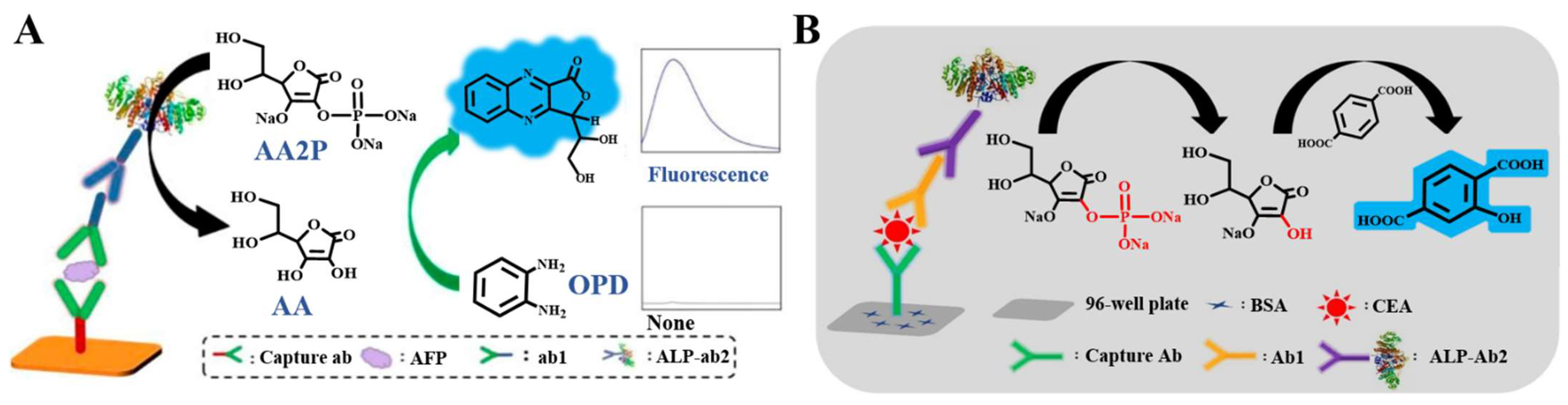
2.2. Generation of Fluorescent Molecules through Chemical Reaction or Enzymatic Cascade Reaction
2.3. Enzymatic-Product-Regulated Fluorescence of Nanomaterials
2.4. Enzymatic-Product-Induced In Situ Generation of Fluorescent Nanomaterials
2.5. Enzymatic-Product-Triggered AIE Phenomenon

| Detection Principle | ALP Substrates | Fluorescence Reporters | Target | Linear Range | LOD | Reference |
|---|---|---|---|---|---|---|
| Direct generation of fluorescent molecules or quenchers | DDAO phosphate | DDAO | C-reactive protein | 0.1–1000 ng/mL | 58 pg/mL | [54] |
| DDAO phosphate | DDAO | AIV H5-HA | 0.23–100 ng/mL | 0.23 ng/mL | [57] | |
| 4-MUP | 4-MU | Anti-T-gondii IgG antibodies | 0–200 U/mL | 0.39 mU/mL | [59] | |
| PNPP | G4/NMM | Zearalenone | 7.5–17.5 ng/mL | 36 pg/mL | [64] | |
| Generation of fluorescent molecules through chemical reaction or enzymatic cascade reaction | AAP | N-heterocyclic fluorophore | AFP | 0.5–40 ng/mL | 0.21 ng/mL | [67] |
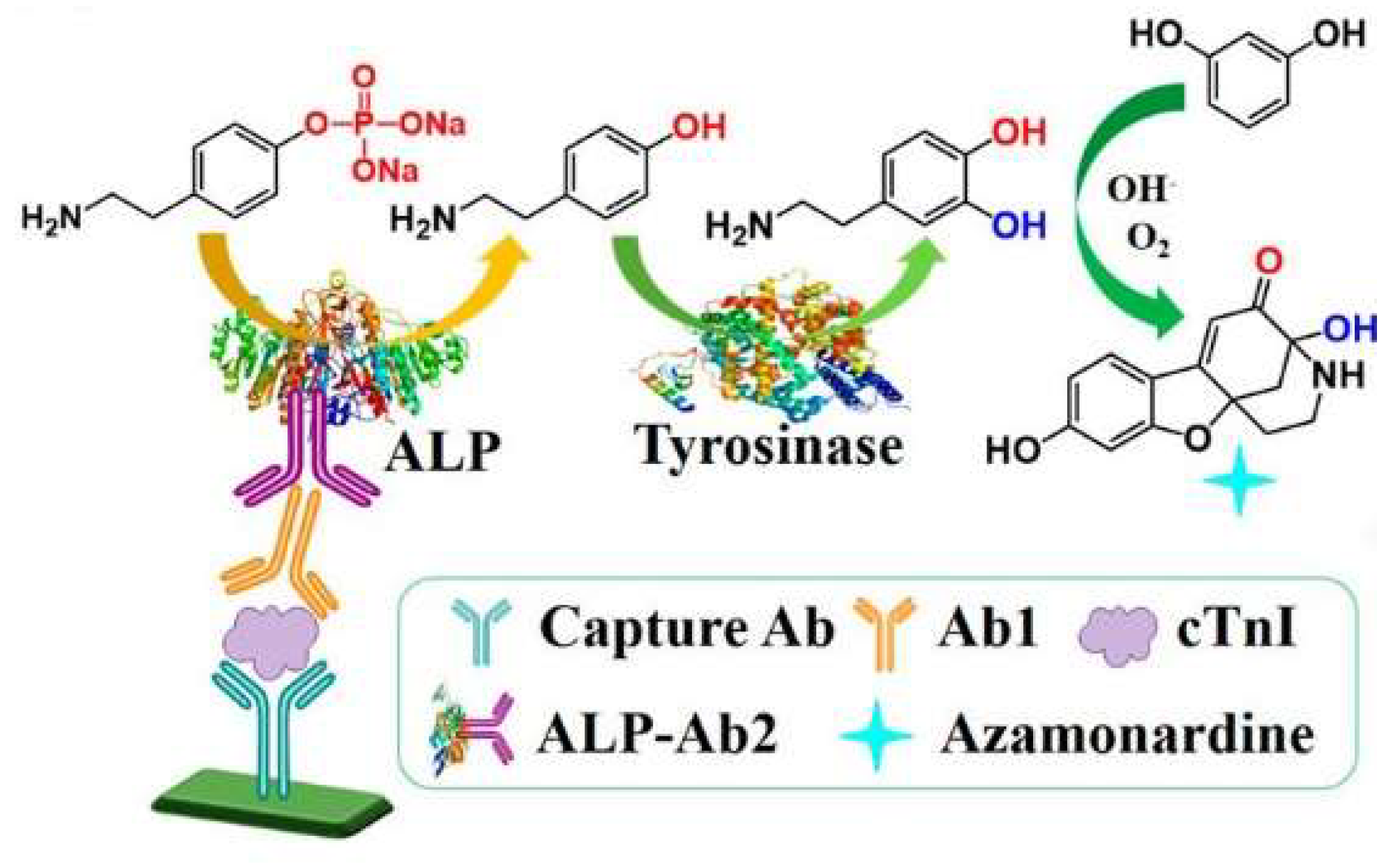
| m-HPP | Azamonardine | cTnI | 0.125–8 ng/mL | 40 pg/mL | [68] | |
| AAP | PTA-OH | CEA | 0.25–30 ng/mL | 0.08 ng/mL | [69] | |
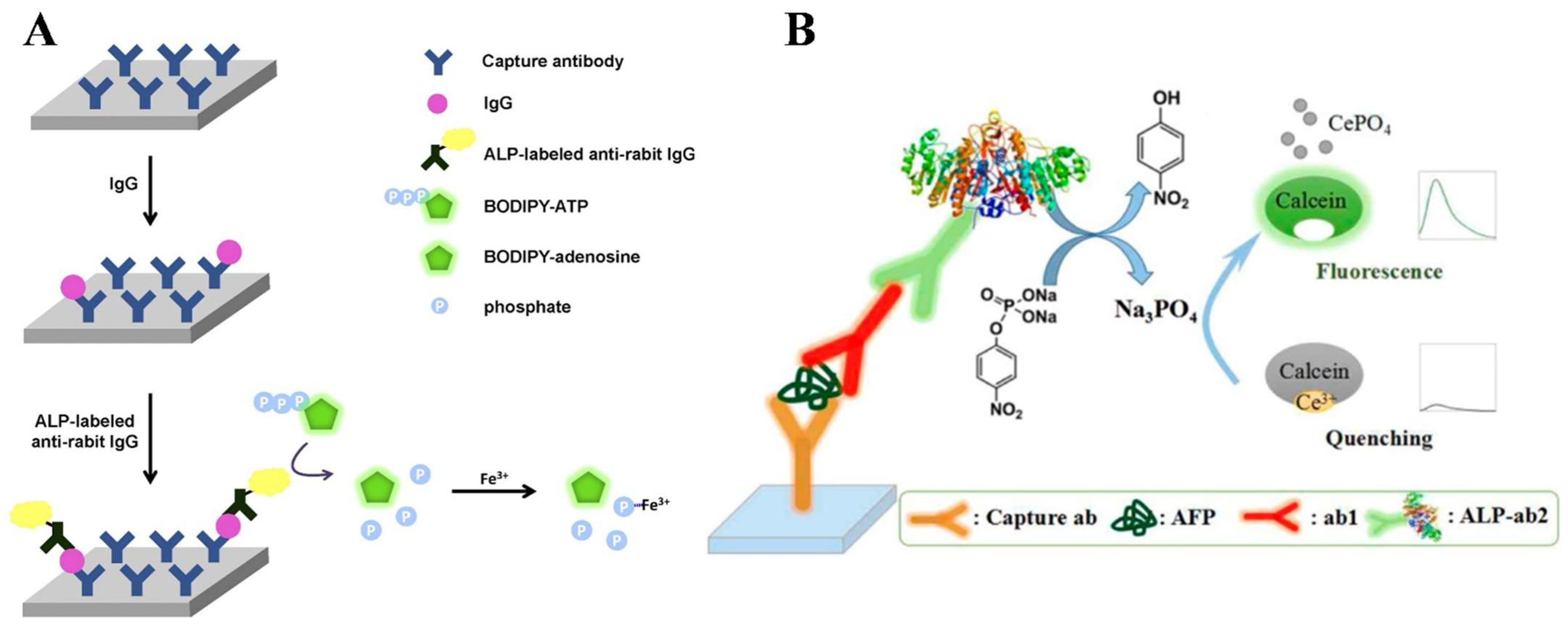
| BODIPY-ATP | BODIPY | IgG | 0–200 ng/mL | 5 ng/mL | [72] | |
| PNPP | Calcein | AFP | 0.2–1 ng/mL | 41 pg/mL | [74] | |
| PAPP | Azamonardine | cTnI | 0.05–4 ng/mL | 15 pg/mL | [75] | |
| Enzymatic-product-regulated fluorescence of nanomaterials | GMP | ThT@GMP/Eu | Mouse IgG | 0.8–100 ng/mL | 0.16 ng/mL | [82] |
| PNPP | CDs | Aflatoxin M1 | 0.003–0.81 ng/mL | 18.6 pg/mL | [86] | |
| AAP | AuNCs | Escherichia coli O157:H7 | 3.3 × 10³–3.3 × 10⁶ cfu/mL | 920 cfu/mL | [87] | |
| AAP | CdTe QDs | HIV-1 p24 antigen | 1–100 pg/mL | 0.2 pg/mL | [93] | |
| AAP | CDs | Human IgG | 40 ng/mL–4 μg/mL | 150 pg/mL | [94] | |
| AAP | CdSe QDs | Ethyl carbamate | 100 ng/mL–10 μg/mL | 24.3 ng/mL | [95] | |
| AAP | AuNCs | Mouse IgG | 0.005–50 ng/mL | 1.5 pg/mL | [97] | |
| AAP | CDs | Aflatoxin B1 | 1 ng/kg–1 μg/kg | 0.69 ng/kg | [109] | |
| Enzymatic-product-induced in situ generation of fluorescent nanomaterials | PAPP | Si CNPs | PSA | 0.02–20 ng/mL | 4.1 pg/mL | [112] |
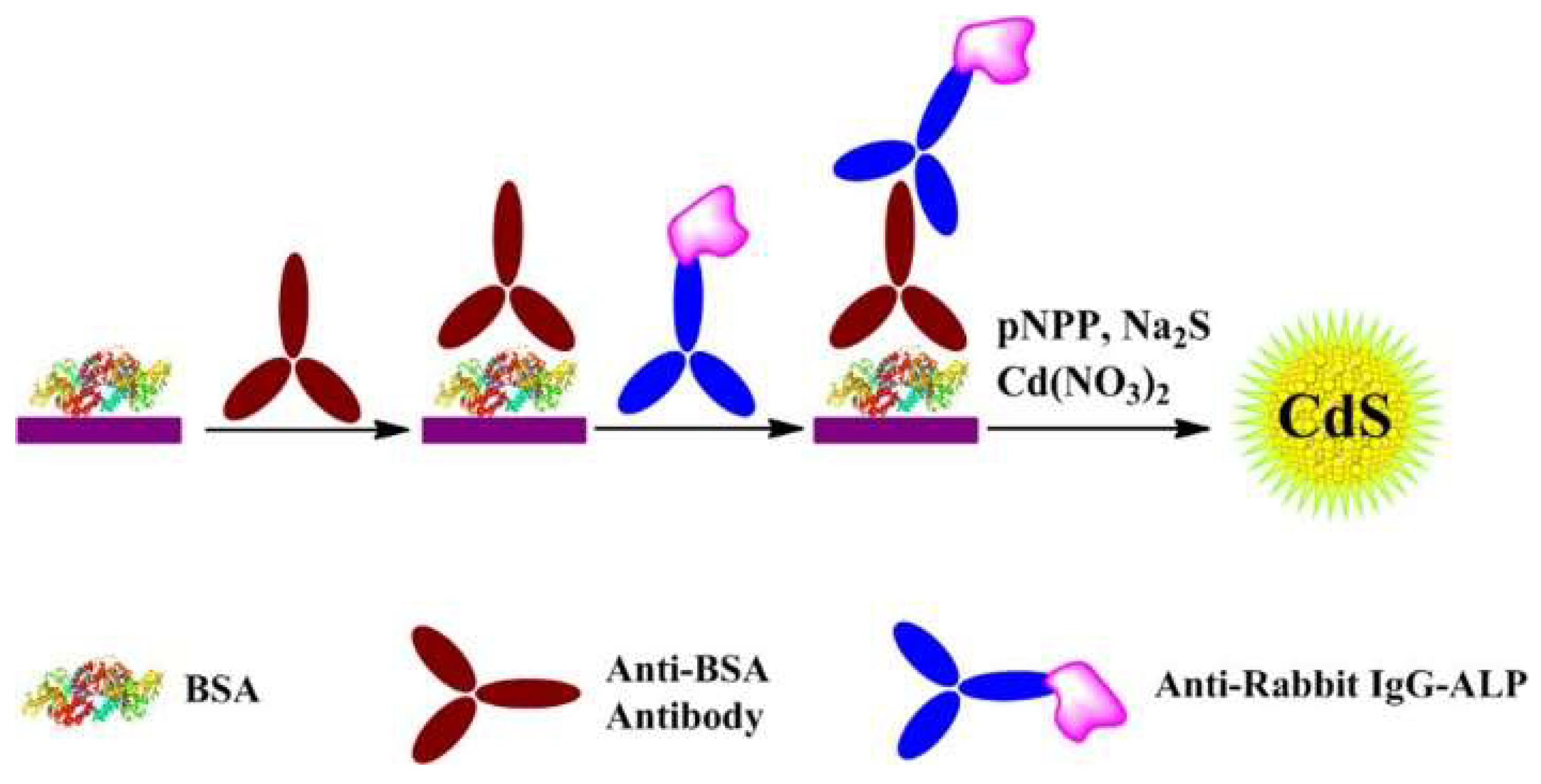
| PNPP | CdS QDs | Anti-BSA Antibody | 0–500 ng/mL | 0.4 ng/mL | [115] | |
| Enzymatic-product-triggered AIE phenomenon | AAP | Self-clickable TPE-based AIEgens | Rabbit anti-human IgG | 0–50 ng/mL | 1.2 ng/mL | [121] |
| AAP | AuNCs | Ochratoxin A | 0–500 ng/mL | 0.62 ng/mL | [122] |
3. Colorimetric Immunoassays
3.1. ALP-Catalyzed Production of Chromogenic Product
3.2. Enzymatic-Product-Triggered Chromogenic Reaction
3.3. Enzymatic-Product-Triggered Plasmonic Phenomenon
3.3.1. Enzymatic-Product-Induced Aggregation of Plasmonic NPs

3.3.2. Enzymatic-Product-Induced In Situ Metallization or Bioetching of Plasmonic NPs
3.4. Enzymatic-Product-Mediated Activity Change of Artificial Enzymes or Nanozymes

| Detection Principle | ALP Substrates | Chromogenic Substrates/Reactions | Target | Linear Range | LOD | Reference |
|---|---|---|---|---|---|---|
| ALP-catalyzed production of chromogenic product | PNPP | PNPP | IgG | 0.5–400 ng/mL | 62.5 pg/mL | [126] |
| PNPP | PNPP | TNF-α | 0–10 ng/mL | 120 pg/mL | [127] | |
| 3-IP | 3-IP | Mouse IgG | 0.3–250 ng/mL | 0.3 ng/mL | [128] | |
| PNPP | PNPP | 2-Deoxycytidine | 10–1000 μM | Not reported | [129] | |
| Enzymatic-product-triggered chromogenic reaction | PAPP | The reaction between diethanolamine and PAP | AFP | 0.1–20 ng/mL | 0.1 ng/mL | [131] |
| AAP | Cu(I)-bicinchoninic complex | Rabbit IgG | 0.1–25 ng/mL | 0.05 ng/mL | [133] | |
| AAP | Fe(III)-phenanthroline complex | CEA | 0.05–100 ng/mL | 21.1 pg/mL | [134] | |
| AAP | Fe(III)- tris-(bathophenanthroline) complex | AFP | 0.01–5 ng/mL | 5 pg/mL | [135] | |
| APP | In situ formation of Prussian blue | PSA | 1–800 ng/mL | 1.2 ng/mL | [136] | |
| APP | In situ formation of Prussian blue | Fenitrothion | 4.7–11.6 ng/mL | 3 ng/mL | [137] | |
| Enzymatic-product-induced aggregation of plasmonic NPs | AAP | Mn2+-mediated aggregation of AuNPs | Fumonisin B1 | 6.25–200 ng/mL | 0.15 ng/mL | [142] |
| ATP | Zn2+-mediated aggregation of AuNPs | Respiratory syncytial virus | 0.1–30 pg/mL | 21 fg/mL | [143] | |
| AAP | AuNPs-based click reaction | Norfloxacin | 3.18 × 10⁻²–6.88 × 10³ pg/mL | 10fg/mL | [144] | |
| Peptide | AuNPs | PCT, IL-6, CRP | 0.2–25 ng/mL, 50–1600 pg/mL, 3.15–100 μg/mL | 0.24 ng/mL, 12.5 pg/mL, 1.15 μg/mL | [146] | |
| Enzymatic-product-induced in situ metallization or bioetching of plasmonic NPs | AAP | Ag growth on SiO2@AuNPs | IgG | 0.7–70 pM | 0.14 pM | [147] |
| PAPP | Ag growth on AuNPs | H9N2 AIV | 0.02–1 ng/mL | 17.5 pg/mL | [148] | |
| AAP | Growth of AuNPs | Tyramine | 0.313–20 mg/L | 0.246 mg/L | [149] | |
| AAP | Growth of AuNPs | HER2 ECD | 1–7 ng/mL | 0.05 ng/mL | [151] | |
| AAP | Ag growth on AuNRs | Xanthylacrylamide | 0.3–17.2 ng/mL | 0.06 ng/mL | [152] | |
| AAP | Iodine-mediated etching of AuNRs | Human IgG | 0.1–10 ng/mL | 100 pg/mL | [162] | |
| Enzymatic-product-mediated activity change of artificial enzymes or nanozymes | GTP | GTP-accelerated TMB oxidation | AFP | 1–100 ng/mL | 0.5 ng/mL | [164] |
| AAP | In situ generated CHNPs to catalyze ABTS oxidation | Aflatoxin B1 | 1 pg/mL–20 ng/mL | 0.73 pg/mL | [170] | |
| AAP | In situ generated PdNPs to catalyze TMB oxidation | PSA | 5–50 ng/mL | 1 ng/mL | [168] |
4. Chemiluminescence Immunoassays
5. SERS Immunoassays
6. Conclusions
Author Contributions
Funding
Institutional Review Board Statement
Informed Consent Statement
Data Availability Statement
Conflicts of Interest
References
- Gan, S.D.; Patel, K.R. Enzyme immunoassay and enzyme-linked immunosorbent assay. J. Investig. Dermatol. 2013, 133, e12–e14. [Google Scholar] [CrossRef] [PubMed]
- Xiong, Y.; Leng, Y.; Li, X.; Huang, X.; Xiong, Y. Emerging strategies to enhance the sensitivity of competitive ELISA for detection of chemical contaminants in food samples. TrAC-Trend. Anal. Chem. 2020, 126, 115861–115879. [Google Scholar] [CrossRef]
- Xia, N.; Huang, Y.; Zhao, Y.; Wang, F.; Liu, L.; Sun, Z. Electrochemical biosensors by in situ dissolution of self-assembled nanolabels into small monomers on electrode surface. Sens. ActuatorsB Chem. 2020, 325, 128777. [Google Scholar] [CrossRef]
- Yi, Z.; Ren, Y.; Li, Y.; Li, Y.; Long, F.; Zhu, A. Optical biosensors for microbial toxin detection: Recent advances and future trends. Microchem. J. 2023, 191, 108894–108911. [Google Scholar] [CrossRef]
- Fu, X.; Chen, L.; Choo, J. Optical nanoprobes for ultrasensitive immunoassay. Anal. Chem. 2017, 89, 124–137. [Google Scholar] [CrossRef]
- Rizzo, F. Optical immunoassays methods in protein analysis: An overview. Chemosensors 2022, 10, 326. [Google Scholar] [CrossRef]
- Lee, D.; Hwang, J.; Seo, Y.; Gilad, A.A.; Choi, J. Optical immunosensors for the efficient detection of target biomolecules. Biotechnol. Bioprocess Eng. 2018, 23, 123–133. [Google Scholar] [CrossRef]
- Wang, Y.; Xianyu, Y. Nanobody and nanozyme-enabled immunoassays with enhanced specificity and sensitivity. Small Methods 2022, 6, e2101576–e2101599. [Google Scholar] [CrossRef]
- Banno, Y.; Nomiyama, T.; Okuno, S.; Ide, S.; Kaji, N. Quantitative evaluation of interleukin-4 by immunowall devices made of gelatin methacryloyl hydrogel. Molecules 2023, 28, 4635. [Google Scholar] [CrossRef]
- Radha, R.; Shahzadi, S.K.; Al-Sayah, M.H. Fluorescent immunoassays for detection and quantification of cardiac troponin I: A short review. Molecules 2021, 26, 4812. [Google Scholar] [CrossRef]
- Chang, J.F.; Yu, L.; Hou, T.; Hu, R.X.; Li, F. Direct and specific detection of glyphosate using a phosphatase-like nanozyme-mediated chemiluminescence strategy. Anal. Chem. 2023, 95, 4479–4485. [Google Scholar] [CrossRef]
- Lee, J.; Takemura, K.; Park, E.Y. Plasmonic nanomaterial-based optical biosensing platforms for Virus detection. Sensors 2017, 17, 2332. [Google Scholar] [CrossRef]
- Niu, X.; Cheng, N.; Ruan, X.; Du, D.; Lin, Y. Review—Nanozyme-based immunosensors and immunoassays: Recent developments and future trends. J. Electrochem. Soc. 2019, 167, 037508–037518. [Google Scholar] [CrossRef]
- Pei, X.; Tao, G.; Wu, X.; Ma, Y.; Li, R.; Li, N. Nanomaterial-based multiplex optical sensors. Analyst 2020, 145, 4111–4123. [Google Scholar] [CrossRef] [PubMed]
- Sang, P.; Hu, Z.; Cheng, Y.; Yu, H.; Yang, F.; Xie, Y.; Yao, W.; Guo, Y.; Qian, H. Exonuclease III-assisted nucleic acid amplification fluorescence immunoassay for the ultrasensitive detection of chloramphenicol in milk. Sens. Actuators B Chem. 2021, 347, 130564–130569. [Google Scholar] [CrossRef]
- Yang, W.; Shen, Y.; Zhang, D.; Xu, W. Protein-responsive rolling circle amplification as a tandem template to drive amplified transduction of fluorescence signal probes for highly sensitive immunoassay. Chem. Commun. 2018, 54, 10195–10198. [Google Scholar] [CrossRef]
- Guo, J.; Yu, H.; Cui, T. Applications of fluorescent materials in the detection of alkaline phosphatase activity. J. Biomed. Mater. Res. Part B 2021, 109, 214–226. [Google Scholar] [CrossRef]
- Han, Y.; Chen, J.; Li, Z.; Chen, H.; Qiu, H. Recent progress and prospects of alkaline phosphatase biosensor based on fluorescence strategy. Biosens. Bioelectron. 2020, 148, 111811–111821. [Google Scholar] [CrossRef]
- Huang, J.; Wei, F.; Cui, Y.; Hou, L.; Lin, T. Fluorescence immunosensor based on functional nanomaterials and its application in tumor biomarker detection. RSC Adv. 2022, 12, 31369–31379. [Google Scholar] [CrossRef]
- Sadiq, Z.; Safiabadi Tali, S.H.; Hajimiri, H.; Al-Kassawneh, M.; Jahanshahi-Anbuhi, S. Gold nanoparticles-based colorimetric assays for environmental monitoring and food safety evaluation. Crit. Rev. Anal. Chem. 2023, 53, 1–36. [Google Scholar] [CrossRef]
- Lee, J.H.; Cho, H.Y.; Choi, H.K.; Lee, J.Y.; Choi, J.W. Application of gold nanoparticle to plasmonic biosensors. Int. J. Mol. Sci. 2018, 19, 2021. [Google Scholar] [CrossRef] [PubMed]
- Liu, L.; Deng, D.; Wu, D.; Hou, W.; Wang, L.; Li, N.; Sun, Z. Duplex-specific nuclease-based electrochemical biosensor for the detection of microRNAs by conversion of homogeneous assay into surface-tethered electrochemical analysis. Anal. Chim. Acta 2021, 1149, 338199. [Google Scholar] [CrossRef] [PubMed]
- Xia, N.; Wu, D.; Sun, T.; Wang, Y.; Ren, X.; Zhao, F.; Liu, L.; Yi, X. Magnetic bead-based electrochemical and colorimetric methods for the detection of poly(ADP-ribose) polymerase-1 with boronic acid derivatives as the signal probes. Sens. Actuators B Chem. 2021, 327, 128913. [Google Scholar] [CrossRef]
- Xia, N.; Wu, D.; Yu, H.; Sun, W.; Yi, X.; Liu, L. Magnetic bead-based electrochemical and colorimetric assays of circulating tumor cells with boronic acid derivatives as the recognition elements and signal probes. Talanta 2021, 221, 121640. [Google Scholar] [CrossRef]
- Shao, Y.; Zhou, H.; Wu, Q.; Xiong, Y.; Wang, J.; Ding, Y. Recent advances in enzyme-enhanced immunosensors. Biotechnol. Adv. 2021, 53, 107867–107883. [Google Scholar] [CrossRef] [PubMed]
- Noji, H.; Minagawa, Y.; Ueno, H. Enzyme-based digital bioassay technology-key strategies and future perspectives. Lab Chip 2022, 22, 3092–3109. [Google Scholar] [CrossRef]
- Sun, J.; Ning, X.; Cui, L.; Ling, M.; Xu, X.; He, S. Assembly of “carrier free” enzymatic nano-reporters for improved ELISA. Analyst 2020, 145, 6541–6548. [Google Scholar] [CrossRef]
- Xia, N.; Sun, T.; Liu, L.; Tian, L.; Sun, Z. Heterogeneous sensing of post-translational modification enzymes by integrating the advantage of homogeneous analysis. Talanta 2022, 237, 122949. [Google Scholar] [CrossRef]
- Grigorenko, V.G.; Andreeva, I.P.; Rubtsova, M.Y.; Egorov, A.M. Recombinant horseradish peroxidase: Production and analytical applications. Biochemistry 2015, 80, 408–416. [Google Scholar] [CrossRef]
- Shi, D.; Sun, Y.; Lin, L.; Shi, C.; Wang, G.; Zhang, X. Naked-eye sensitive detection of alkaline phosphatase (ALP) and pyrophosphate (PPi) based on a horseradish peroxidase catalytic colorimetric system with Cu(II). Analyst 2016, 141, 5549–5554. [Google Scholar] [CrossRef]
- Xianyu, Y.; Zhu, K.; Chen, W.; Wang, X.; Zhao, H.; Sun, J.; Wang, Z.; Jiang, X. Enzymatic assay for Cu(II) with horseradish peroxidase and its application in colorimetric logic gate. Anal. Chem. 2013, 85, 7029–7032. [Google Scholar] [CrossRef]
- Yuan, H.; Liu, L.; Lv, F.; Wang, S. Bioluminescence as a light source for photosynthesis. Chem. Commun. 2013, 49, 10685–10687. [Google Scholar] [CrossRef] [PubMed]
- Abucayon, E.; Ke, N.; Cornut, R.; Patelunas, A.; Miller, D.; Nishiguchi, M.K.; Zoski, C.G. Investigating catalase activity through hydrogen peroxide decomposition by bacteria biofilms in real time using scanning electrochemical microscopy. Anal. Chem. 2014, 86, 498–505. [Google Scholar] [CrossRef] [PubMed]
- Zhou, Y.; Zhuo, Y.; Liao, N.; Chai, Y.; Yuan, R. Ultrasensitive immunoassay based on a pseudobienzyme amplifying system of choline oxidase and luminol-reduced Pt@Au hybrid nanoflowers. Chem. Commun. 2014, 50, 14627–14630. [Google Scholar] [CrossRef] [PubMed]
- Manes, T.; Hoylaerts, M.F.; Müller, R.; Lottspeich, F.; Holke, W.; Millán, J.L. Genetic complexity, structure, and characterization of highly active bovine intestinal alkaline phosphatases. J. Biol. Chem. 1998, 273, 23353–23360. [Google Scholar] [CrossRef]
- Li, S.-J.; Li, C.-Y.; Li, Y.-F.; Fei, J.; Wu, P.; Yang, B.; Ou-Yang, J.; Nie, S.-X. Facile and sensitive near-infrared fluorescence probe for the detection of endogenous akaline phosphatase activity in vivo. Anal. Chem. 2017, 89, 6854–6860. [Google Scholar] [CrossRef]
- Zhang, S.; Garcia-D’Angeli, A.; Brennan, J.P.; Huo, Q. Predicting detection limits of enzyme-linked immunosorbent assay (ELISA) and bioanalytical techniques in general. Analyst 2014, 139, 439–445. [Google Scholar] [CrossRef]
- Tang, Z.; Chen, H.; He, H.; Ma, C. Assays for alkaline phosphatase activity: Progress and prospects. TrAC-Trend. Anal. Chem. 2019, 113, 32–43. [Google Scholar] [CrossRef]
- Wang, K.; Wang, W.; Zhang, X.-Y.; Jiang, A.-Q.; Yang, Y.-S.; Zhu, H.-L. Fluorescent probes for the detection of alkaline phosphatase in biological systems: Recent advances and future prospects. TrAC-Trend. Anal. Chem. 2021, 136, 116189–116217. [Google Scholar] [CrossRef]
- Shaban, S.M.; Byeok Jo, S.; Hafez, E.; Ho Cho, J.; Kim, D.-H. A comprehensive overview on alkaline phosphatase targeting and reporting assays. Coord. Chem. Rev. 2022, 465, 214567–214604. [Google Scholar] [CrossRef]
- Zherdev, A.V.; Dzantiev, B.B. Detection limits of immunoanalytical systems: Limiting factors and methods of reduction. J. Anal. Chem. 2022, 77, 391–401. [Google Scholar] [CrossRef]
- Zhao, Q.; Lu, D.; Zhang, G.; Zhang, D.; Shi, X. Recent improvements in enzyme-linked immunosorbent assays based on nanomaterials. Talanta 2021, 223, 121722–121737. [Google Scholar] [CrossRef]
- Campuzano, S.; Pedrero, M.; Yáñez-Sedeño, P.; Pingarrón, J.M. Nanozymes in electrochemical affinity biosensing. Microchim. Acta 2020, 187, 423–438. [Google Scholar] [CrossRef] [PubMed]
- Ou, X.; Liu, Y.; Zhang, M.; Hua, L.; Zhan, S. Plasmonic gold nanostructures for biosensing and bioimaging. Microchim. Acta 2021, 188, 304–318. [Google Scholar] [CrossRef] [PubMed]
- Yu, T.; Wei, Q. Plasmonic molecular assays: Recent advances and applications for mobile health. Nano Res. 2018, 11, 5439–5473. [Google Scholar] [CrossRef] [PubMed]
- Guo, L.; Jackman, J.A.; Yang, H.-H.; Chen, P.; Cho, N.-J.; Kim, D.-H. Strategies for enhancing the sensitivity of plasmonic nanosensors. Nano Today 2015, 10, 213–239. [Google Scholar] [CrossRef]
- Feng, K.; Kang, Y.; Zhao, J.J.; Liu, Y.L.; Jiang, J.H.; Shen, G.L.; Yu, R.Q. Electrochemical immunosensor with aptamer-based enzymatic amplification. Anal. Biochem. 2008, 378, 38–42. [Google Scholar] [CrossRef]
- Jones, A.; Dhanapala, L.; Kankanamage, R.N.T.; Kumar, C.V.; Rusling, J.F. Multiplexed immunosensors and immunoarrays. Anal. Chem. 2020, 92, 345–362. [Google Scholar] [CrossRef]
- Gil Rosa, B.; Akingbade, O.E.; Guo, X.; Gonzalez-Macia, L.; Crone, M.A.; Cameron, L.P.; Freemont, P.; Choy, K.L.; Guder, F.; Yeatman, E.; et al. Multiplexed immunosensors for point-of-care diagnostic applications. Biosens. Bioelectron. 2022, 203, 114050–114066. [Google Scholar] [CrossRef]
- Tsumuraya, T.; Sato, T.; Hirama, M.; Fujii, I. Highly sensitive and practical fluorescent sandwich ELISA for ciguatoxins. Anal. Chem. 2018, 90, 7318–7324. [Google Scholar] [CrossRef]
- Pirsaheb, M.; Mohammadi, S.; Salimi, A. Current advances of carbon dots based biosensors for tumor marker detection, cancer cells analysis and bioimaging. TrAC-Trend. Anal. Chem. 2019, 115, 83–99. [Google Scholar] [CrossRef]
- Zhou, J.; Gui, Y.; Lv, X.; He, J.; Xie, F.; Li, J.; Cai, J. Nanomaterial-based fluorescent biosensor for food safety analysis. Biosensors 2022, 12, 1072. [Google Scholar] [CrossRef] [PubMed]
- Zhang, H.; Ju, Q.; Pang, S.; Wei, N.; Zhang, Y. Recent progress of fluorescent probes for the detection of alkaline phosphatase (ALP): A review. Dyes. Pigments 2021, 194, 109569–109582. [Google Scholar] [CrossRef]
- Nishiyama, K.; Kasama, T.; Nakamata, S.; Ishikawa, K.; Onoshima, D.; Yukawa, H.; Maeki, M.; Ishida, A.; Tani, H.; Baba, Y.; et al. Ultrasensitive detection of disease biomarkers using an immuno-wall device with enzymatic amplification. Analyst 2019, 144, 4589–4595. [Google Scholar] [CrossRef]
- Zhang, Y.Q.; Xu, Z.L.; Wang, F.; Cai, J.; Dong, J.X.; Zhang, J.R.; Si, R.; Wang, C.L.; Wang, Y.; Shen, Y.D.; et al. Isolation of bactrian camel single domain antibody for parathion and development of one-step dc-FEIA method using VHH-alkaline phosphatase Fusion protein. Anal. Chem. 2018, 90, 12886–12892. [Google Scholar] [CrossRef]
- Huo, J.; Li, Z.; Wan, D.; Li, D.; Qi, M.; Barnych, B.; Vasylieva, N.; Zhang, J.; Hammock, B.D. Development of a highly sensitive direct competitive fluorescence enzyme immunoassay based on a nanobody-alkaline phosphatase Fusion protein for detection of 3-phenoxybenzoic acid in urine. J. Agric. Food. Chem. 2018, 66, 11284–11290. [Google Scholar] [CrossRef]
- Chavez Ramos, K.; Nishiyama, K.; Maeki, M.; Ishida, A.; Tani, H.; Kasama, T.; Baba, Y.; Tokeshi, M. Rapid, sensitive, and selective detection of H5 hemagglutinin from avian influenza virus using an immunowall device. ACS Omega 2019, 4, 16683–16688. [Google Scholar] [CrossRef]
- Liu, X.; Xu, Y.; Wan, D.B.; Xiong, Y.H.; He, Z.Y.; Wang, X.X.; Gee, S.J.; Ryu, D.; Hammock, B.D. Development of a nanobody-alkaline phosphatase fusion protein and its application in a highly sensitive direct competitive fluorescence enzyme immunoassay for detection of ochratoxin A in cereal. Anal. Chem. 2015, 87, 1387–1394. [Google Scholar] [CrossRef]
- Medawar-Aguilar, V.; Jofre, C.F.; Fernandez-Baldo, M.A.; Alonso, A.; Angel, S.; Raba, J.; Pereira, S.V.; Messina, G.A. Serological diagnosis of Toxoplasmosis disease using a fluorescent immunosensor with chitosan-ZnO-nanoparticles. Anal. Biochem. 2019, 564–565, 116–122. [Google Scholar] [CrossRef]
- Obayashi, Y.; Iinobc, R.; Noji, H. A single-molecule digital enzyme assay using alkaline phosphatase with a cumarin-based fluorogenic substrate. Analyst 2015, 140, 5065–5073. [Google Scholar] [CrossRef]
- Mahato, K.; Chandra, P. Paper-based miniaturized immunosensor for naked eye ALP detection based on digital image colorimetry integrated with smartphone. Biosens. Bioelectron. 2020, 128, 9–16. [Google Scholar] [CrossRef]
- Tsaloglou, M.-N.; Jacobs, A.; Morgan, H. A fluorogenic heterogeneous immunoassay for cardiac muscle troponin cTnI on a digital microfluidic device. Anal. Bioanal. Chem. 2014, 406, 5967–5976. [Google Scholar] [CrossRef] [PubMed]
- Kahveci, Z.; Martinez-Tome, M.J.; Mallavia, R.; Mateo, C.R. Fluorescent biosensor for phosphate determination based on immobilized polyfluorene-liposomal nanoparticles coupled with alkaline phosphatase. ACS Appl. Mater. Interfaces 2017, 9, 136–144. [Google Scholar] [CrossRef] [PubMed]
- Ma, L.; Zhang, X.; Xiao, Y.; Fang, H.; Zhang, G.; Yang, H.; Zhou, Y. Fluorescence and colorimetric dual-mode immunoassay based on G-quadruplex/N-methylmesoporphyrin IX and p-nitrophenol for detection of zearalenone. Food Chem. 2023, 401, 134190–134195. [Google Scholar] [CrossRef]
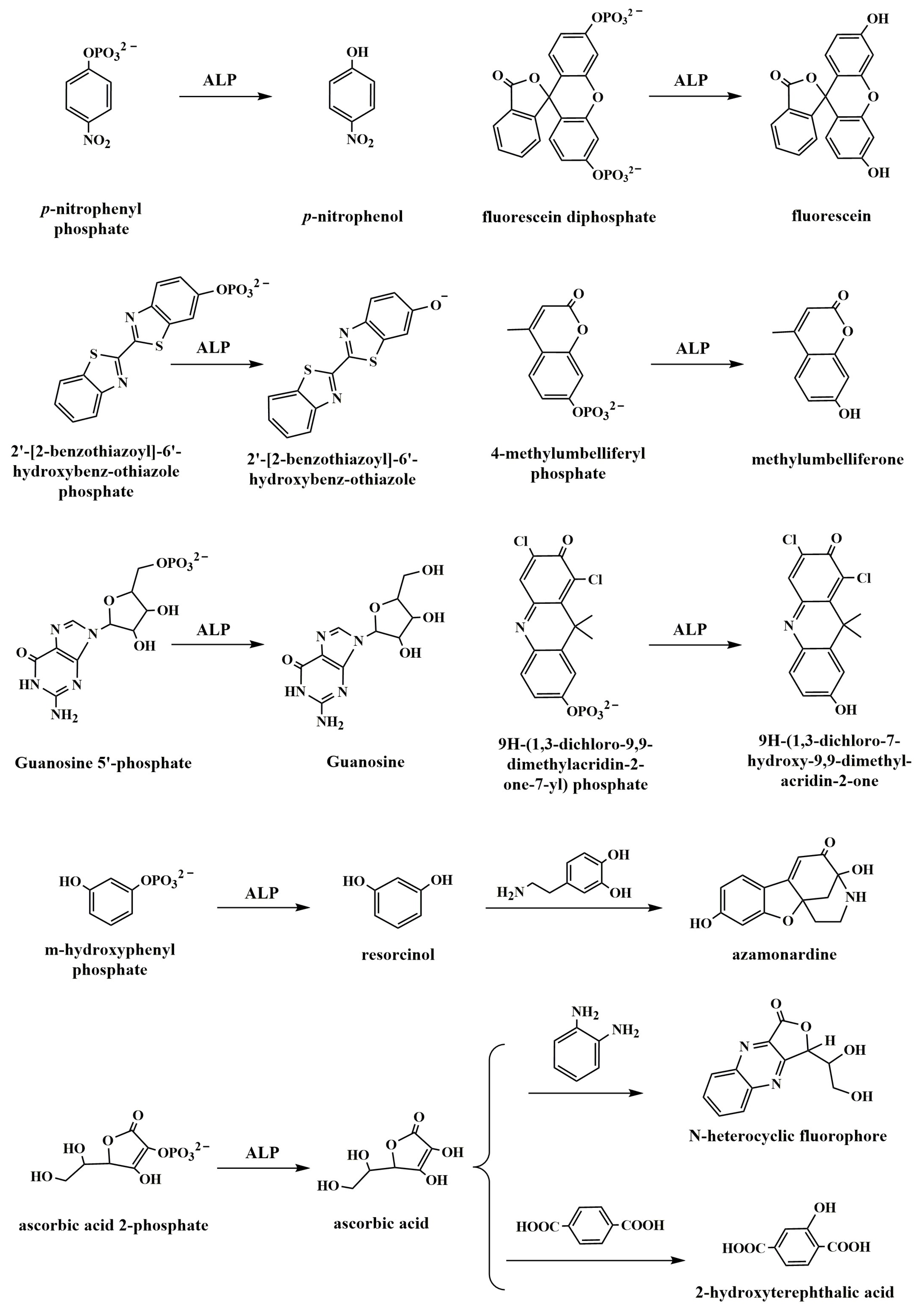
- Pérez-Ruiz, T.; Martínez-Lozano, C.; Tomás, V.; Fenol, J. Fluorimetric determination of total ascorbic acid by a stopped-flow mixing technique. Analyst 2001, 126, 1436–1439. [Google Scholar] [CrossRef] [PubMed]
- Wu, X.; Diao, Y.; Sun, C.; Yang, J.; Wang, Y.; Sun, S. Fluorimetric determination of ascorbic acid with ophenylenediamine. Talanta 2003, 59, 95–99. [Google Scholar] [CrossRef] [PubMed]
- Zhao, D.; Li, J.; Peng, C.; Zhu, S.; Sun, J.; Yang, X. Fluorescence immunoassay based on the alkaline phosphatase triggered in situ fluorogenic reaction of o-phenylenediamine and ascorbic acid. Anal. Chem. 2019, 91, 2978–2984. [Google Scholar] [CrossRef]
- Zhao, J.; Wang, S.; Lu, S.; Liu, G.; Sun, J.; Yang, X. Fluorometric and colorimetric dual-readout immunoassay based on an alkaline phosphatase-triggered reaction. Anal. Chem. 2019, 91, 7828–7834. [Google Scholar] [CrossRef]
- Fan, Y.; Lv, M.; Xue, Y.; Li, J.; Wang, E. In situ fluorogenic reaction generated via ascorbic acid for the construction of universal sensing platform. Anal. Chem. 2021, 93, 6873–6880. [Google Scholar] [CrossRef]
- Wang, Y.; Liu, Y.; Deng, X.; Cong, Y.; Jiang, X. Peptidic β-sheet binding with Congo Red allows both reduction of error variance and signal amplification for immunoassays. Biosens. Bioelectron. 2016, 86, 211–218. [Google Scholar] [CrossRef]
- Geng, F.; Liu, X.; Wei, T.; Wang, Z.; Liu, J.; Shao, C.; Liu, G.; Xu, M.; Feng, L. An alkaline phosphatase-induced immunosensor for SARS-CoV-2 N protein and cardiac troponin I based on the in situ fluorogenic self-assembly between N-heterocyclic boronic acids and alizarin red S. Sens. Actuators B Chem. 2023, 378, 133121–133129. [Google Scholar] [CrossRef]
- Lin, J.H.; Yang, Y.C.; Shih, Y.C.; Hung, S.Y.; Lu, C.Y.; Tseng, W.L. Photoinduced electron transfer between Fe(III) and adenosine triphosphate-BODIPY conjugates: Application to alkaline-phosphatase-linked immunoassay. Biosens. Bioelectron. 2016, 77, 242–248. [Google Scholar] [CrossRef]
- Mu, X.; Jiang, X.; Zhang, Y.; Liu, X.; Zhang, S.; Wang, W.; Huang, Y.; Ma, P.; Song, D. Sensitive ratiometric fluorescence probe based on chitosan carbon dots and calcein for alkaline phosphatase detection and bioimaging in cancer cells. Anal. Chim. Acta 2021, 1188, 339163–339170. [Google Scholar] [CrossRef]
- Chen, C.; Zhao, J.; Lu, Y.; Sun, J.; Yang, X. Fluorescence immunoassay based on the phosphate-triggered fluorescence turn-on detection of alkaline phosphatase. Anal. Chem. 2018, 90, 3505–3511. [Google Scholar] [CrossRef]
- Zhao, J.; Wang, S.; Lu, S.; Bao, X.; Sun, J.; Yang, X. An enzyme cascade-triggered fluorogenic and chromogenic reaction applied in enzyme activity assay and immunoassay. Anal. Chem. 2018, 90, 7754–7760. [Google Scholar] [CrossRef]
- Sun, C.; Shi, Y.; Tang, M.; Hu, X.; Long, Y.; Zheng, H. A signal amplification strategy for prostate specific antigen detection via releasing oxidase-mimics from coordination nanoparticles by alkaline phosphatase. Talanta 2020, 213, 120827–120833. [Google Scholar] [CrossRef] [PubMed]
- Hu, X.; Wei, Z.; Tang, M.; Long, Y.; Zheng, H. Reducing background absorbance via a double-lock strategy for detection of alkaline phosphatase and α-fetoprotein. Microchim. Acta 2020, 187, 489–497. [Google Scholar] [CrossRef]
- Sund, H.; Blomberg, K.; Meltola, N.; Takalo, H. Design of novel, water soluble and highly luminescent Europium labels with potential to enhance immunoassay sensitivities. Molecules 2017, 22, 1807. [Google Scholar] [CrossRef] [PubMed]
- Li, Y.; Wang, P.; Huang, L.; Jia, C.; Gao, X.; Liu, S.; Wang, S.; Zhao, P.; Sun, J.; Zhang, D.; et al. Schiff-base chemistry-coupled catechol oxidase-like nanozyme reaction as a universal sensing mode for ultrasensitive biosensing. Anal. Chem. 2023, 95, 3769–3778. [Google Scholar] [CrossRef] [PubMed]
- Zeng, H.-H.; Liu, F.; Peng, Z.-Q.; Yu, K.; Rong, L.-Q.; Wang, Y.; Wu, P.; Liang, R.-P.; Qiu, J.-D. Lanthanide phosphate nanoparticle-based one-step optical discrimination of alkaline phosphatase activity. ACS Appl. Nano Mater. 2020, 3, 2336–2345. [Google Scholar] [CrossRef]
- Wang, F.; Hu, X.; Hu, J.; Peng, Q.; Zheng, B.; Du, J.; Xiao, D. Fluorescence assay for alkaline phosphatase activity based on energy transfer from terbium to europium in lanthanide coordination polymer nanoparticles. J. Mater. Chem. B 2018, 6, 6008–6015. [Google Scholar] [CrossRef]
- Li, S.; Hu, X.; Li, Y.; Tan, H. Fluorescent enzyme-linked immunosorbent assay based on alkaline phosphatase-responsive coordination polymer composite. Microchim. Acta 2021, 188, 263–272. [Google Scholar] [CrossRef]
- Wang, M.; Wang, S.; Li, L.; Wang, G.; Su, X. β-Cyclodextrin modified silver nanoclusters for highly sensitive fluorescence sensing and bioimaging of intracellular alkaline phosphatase. Talanta 2020, 207, 120315–120322. [Google Scholar] [CrossRef]
- Luo, L.; Jia, B.-Z.; Wei, X.-Q.; Xiao, Z.-L.; Wang, H.; Sun, Y.-M.; Shen, Y.-D.; Lei, H.-T.; Xu, Z.-L. Development of an inner filter effect-based fluorescence immunoassay for the detection of acrylamide using 9-xanthydrol derivatization. Sens. Actuators B Chem. 2021, 332, 129561–129568. [Google Scholar] [CrossRef]
- Tang, C.; Qian, Z.; Huang, Y.; Xu, J.; Ao, H.; Zhao, M.; Zhou, J.; Chen, J.; Feng, H. A fluorometric assay for alkaline phosphatase activity based on β-cyclodextrin-modified carbon quantum dots through host-guest recognition. Biosens. Bioelectron. 2016, 83, 274–280. [Google Scholar] [CrossRef]
- Li, G.; Liu, C.; Zhang, X.; Luo, P.; Lin, G.; Jiang, W. Highly photoluminescent carbon dots-based immunosensors for ultrasensitive detection of aflatoxin M1 residues in milk. Food Chem. 2021, 355, 129443–129450. [Google Scholar] [CrossRef]
- Fang, B.; Peng, J.; Zhang, G.; Xing, K.; Chen, W.; Liu, D.; Shan, S.; Xiong, Y.; Lai, W. I2/I--mediated fluorescence quenching of an Ag+-doped gold nanocluster-based immunoassay for sensitive detection of Escherichia coli O157:H7 in milk. J. Dairy Sci. 2022, 105, 2922–2930. [Google Scholar] [CrossRef]
- Jie, Z.; Qi, G.; Xu, C.; Jin, Y. Enzymatic preparation of plasmonic-fluorescent quantum dot-gold hybrid nanoprobes for sensitive detection of glucose and alkaline phosphatase and dual-modality cell imaging. Anal. Chem. 2019, 91, 14074–14079. [Google Scholar]
- Lu, H.-F.; Zhang, M.-M.; Wu, D.; Huang, J.-L.; Zhu, L.-L.; Wang, C.-M.; Zhang, Q.-L. Colorimetric and fluorescent dual-mode sensing of alkaline phosphatase activity in L-02 cells and its application in living cell imaging based on in-situ growth of silver nanoparticles on graphene quantum dots. Sens. Actuators B Chem. 2018, 258, 461–469. [Google Scholar] [CrossRef]
- Zhu, N.; Zhu, Y.; Wang, J.; Gyimah, E.; Hu, X.; Zhang, Z. A novel fluorescence immunoassay based on AgNCs and ALP for ultrasensitive detection of sulfamethazine (SMZ) in environmental and biological samples. Talanta 2019, 199, 72–79. [Google Scholar] [CrossRef]
- Ni, P.; Chen, C.; Jiang, Y.; Zhang, C.; Wang, B.; Cao, B.; Li, C.; Lu, Y. Gold nanoclusters-based dual-channel assay for colorimetric and turn-on fluorescent sensing of alkaline phosphatase. Sens. Actuators B Chem. 2019, 301, 127080–127086. [Google Scholar] [CrossRef]
- Chen, P.; Yan, S.; Sawyer, E.; Ying, B.; Wei, X.; Wu, Z.; Geng, J. Rapid and simple detection of ascorbic acid and alkaline phosphatase via controlled generation of silver nanoparticles and selective recognition. Analyst 2019, 144, 1147–1152. [Google Scholar] [CrossRef]
- Tang, Z.; Wei, Z.; Huang, K.; Wei, Y.; Li, D.; Yan, S.; Huang, J.; Geng, J.; Tao, C.; Chen, P.; et al. Fluorescence and visual immunoassay of HIV-1 p24 antigen in clinical samples via multiple selective recognitions of CdTe QDs. Microchim. Acta 2021, 188, 422–430. [Google Scholar] [CrossRef]
- Song, P.; Liu, Q.; Zhang, Y.; Liu, W.; Meng, M.; Yin, Y.; Xi, R. The chemical redox modulated switch-on fluorescence of carbon dots for probing alkaline phosphatase and its application in an immunoassay. RSC Adv. 2018, 8, 162–169. [Google Scholar] [CrossRef]
- Zhou, K.; Wang, Z.L.; Luo, L.; Dong, Y.Z.; Yang, J.Y.; Lei, H.T.; Wang, H.; Shen, Y.D.; Xu, Z.L. Development of Cu(II)/Cu(I)-induced quantum dot-mediated fluorescence immunoassay for the sensitive determination of ethyl carbamate. Microchim. Acta 2020, 187, 533–542. [Google Scholar] [CrossRef]
- Xie, W.; Tian, M.; Luo, X.; Jiang, Y.; He, N.; Liao, X.; Liu, Y. A dual-mode fluorescent and colorimetric immunoassay based on in situ ascorbic acid-induced signal generation from metal-organic frameworks. Sens. Actuators B Chem. 2020, 302, 127180–127186. [Google Scholar] [CrossRef]
- Hu, X.L.; Wu, X.M.; Fang, X.; Li, Z.J.; Wang, G.L. Switchable fluorescence of gold nanoclusters for probing the activity of alkaline phosphatase and its application in immunoassay. Biosens. Bioelectron. 2016, 77, 666–672. [Google Scholar] [CrossRef]
- Fang, X.; Li, X.-Q.; Wang, H.; Wu, X.-M.; Wang, G.-L. Tuning surface states to achieve the modulated fluorescence of carbon dots for probing the activity of alkaline phosphatase and immunoassay of α-fetoprotein. Sens. Actuators B Chem. 2018, 257, 620–628. [Google Scholar] [CrossRef]
- Zhu, R.; Huang, W.; Ma, X.; Zhang, Y.; Yue, C.; Fang, W.; Hu, Y.; Wang, J.; Dang, J.; Zhao, H.; et al. Nitrogen-doped carbon dots-V2O5 nanobelts sensing platform for sensitive detection of ascorbic acid and alkaline phosphatase activity. Anal. Chim. Acta 2019, 1089, 131–143. [Google Scholar] [CrossRef]
- Deng, R.; Xie, X.; Vendrell, M.; Chang, Y.T.; Liu, X. Intracellular glutathione detection using MnO2-nanosheet-modified upconversion nanoparticles. J. Am. Chem. Soc. 2011, 133, 20168–20171. [Google Scholar] [CrossRef]
- Xiao, T.; Sun, J.; Zhao, J.; Wang, S.; Liu, G.; Yang, X. FRET effect between fluorescent polydopamine nanoparticles and MnO2 nanosheets and its application for sensitive sensing of alkaline phosphatase. ACS Appl. Mater. Interfaces 2018, 10, 6560–6569. [Google Scholar] [CrossRef] [PubMed]
- Huang, H.; Wang, B.; Chen, M.; Liu, M.; Leng, Y.; Liu, X.; Li, Y.; Liu, Z. Fluorescence turn-on sensing of ascorbic acid and alkaline phosphatase activity based on graphene quantum dots. Sens. Actuators B Chem. 2016, 235, 356–361. [Google Scholar] [CrossRef]
- Li, N.; Li, Y.; Han, Y.; Pan, W.; Zhang, T.; Tang, B. A highly selective and instantaneous nanoprobe for detection and imaging of ascorbic acid in living cells and in vivo. Anal. Chem. 2014, 86, 3924–3930. [Google Scholar] [CrossRef]
- Liang, M.Y.; Zhao, B.; Xiong, Y.; Chen, W.X.; Huo, J.Z.; Zhang, F.; Wang, L.; Li, Y. A “turn-on” sensor based on MnO2 coated UCNPs for detection of alkaline phosphatase and ascorbic acid. Dalton Trans. 2019, 48, 16199–16210. [Google Scholar] [CrossRef] [PubMed]
- Hiremath, S.D.; Banerjee, M.; Chatterjee, A. Review of 2D MnO2 nanosheets as FRET-based nanodot fluorescence quenchers in chemosensing applications. ACS Appl. Nano Mater. 2022, 5, 17373–17412. [Google Scholar] [CrossRef]
- Lu, H.; Xu, S. CDs-MnO2-TPPS ternary system for ratiometric fluorescence detection of ascorbic acid and alkaline phosphatase. ACS Omega 2021, 6, 16565–16572. [Google Scholar] [CrossRef]
- Luo, L.; Lin, S.Q.; Wu, Z.Y.; Wang, H.; Chen, Z.J.; Deng, H.; Shen, Y.D.; Zhang, W.F.; Lei, H.T.; Xu, Z.L. Nanobody-based fluorescent immunoassay using carbon dots anchored cobalt oxyhydroxide composite for the sensitive detection of fenitrothion. J. Hazard. Mater. 2022, 439, 129701. [Google Scholar] [CrossRef] [PubMed]
- Li, H.; Jin, R.; Kong, D.; Zhao, X.; Liu, F.; Yan, X.; Lin, Y.; Lu, G. Switchable fluorescence immunoassay using gold nanoclusters anchored cobalt oxyhydroxide composite for sensitive detection of imidacloprid. Sens. Actuators B Chem. 2019, 283, 207–214. [Google Scholar] [CrossRef]
- Tang, D.; Lin, Y.; Zhou, Q. Carbon dots prepared from Litchi chinensis and modified with manganese dioxide nanosheets for use in a competitive fluorometric immunoassay for aflatoxin B1. Microchim. Acta 2018, 185, 476–484. [Google Scholar] [CrossRef]
- Dong, B.; Li, H.; Sun, J.; Mari, G.M.; Yu, X.; Ke, Y.; Li, J.; Wang, Z.; Yu, W.; Wen, K.; et al. Development of a fluorescence immunoassay for highly sensitive detection of amantadine using the nanoassembly of carbon dots and MnO2 nanosheets as the signal probe. Sens. Actuators B Chem. 2019, 286, 214–221. [Google Scholar] [CrossRef]
- Li, R.; Liu, Q.; Jin, Y.; Li, B. Fluorescent enzyme-linked immunoassay strategy based on enzyme-triggered in-situ synthesis of fluorescent copper nanoclusters. Sens. Actuators B Chem. 2019, 281, 28–33. [Google Scholar] [CrossRef]
- Chen, C.; Zhao, D.; Wang, B.; Ni, P.; Jiang, Y.; Zhang, C.; Yang, F.; Lu, Y.; Sun, J. Alkaline phosphatase-triggered in situ formation of silicon-containing nanoparticles for a fluorometric and colorimetric dual-channel immunoassay. Anal. Chem. 2020, 92, 4639–4646. [Google Scholar] [CrossRef]
- Liu, G.; Zhao, J.; Wang, S.; Lu, S.; Sun, J.; Yang, X. Enzyme-induced in situ generation of polymer carbon dots for fluorescence immunoassay. Sens. Actuators B Chem. 2020, 306, 127583–127590. [Google Scholar] [CrossRef]
- Sun, J.; Hu, T.; Chen, C.; Zhao, D.; Yang, F.; Yang, X. Fluorescence immunoassay system via enzyme-enabled in situ synthesis of fluorescent silicon nanoparticles. Anal. Chem. 2016, 88, 9789–9795. [Google Scholar] [CrossRef]
- Malashikhina, N.; Garai-Ibabe, G.; Pavlov, V. Unconventional application of conventional enzymatic substrate: First fluorogenic immunoassay based on enzymatic formation of quantum dots. Anal. Chem. 2013, 85, 6866–6870. [Google Scholar] [CrossRef]
- Ouyang, J.; Sun, L.; Zeng, F.; Wu, S. Biomarker-activatable probes based on smart AIEgens for fluorescence and optoacoustic imaging. Coord. Chem. Rev. 2022, 458, 214438–214459. [Google Scholar] [CrossRef]
- Li, H.Y.; Lin, H.Y.; Lv, W.X.; Gai, P.P.; Li, F. Equipment-free and visual detection of multiple biomarkers via an AIE luminogen-based paper biosensor. Biosens. Bioelectron. 2020, 165, 112336. [Google Scholar] [CrossRef]
- Li, H.Y.; Wang, C.F.; Hou, T.; Li, F. Amphiphile-mediated ultrasmall AIE dots for ultrasensitive fluorescence biosensing. Anal. Chem. 2017, 89, 9100–9107. [Google Scholar] [CrossRef]
- Dou, L.; Li, Q.; Wang, Z.; Shen, J.; Yu, W. AIEgens: Next generation signaling source for immunoassays? ACS Sens. 2022, 7, 3243–3257. [Google Scholar] [CrossRef]
- Liu, W.; Yu, W.; Li, X.; Zhao, X.; Zhang, Y.; Song, P.; Yin, Y.; Xi, R.; Meng, M. Pyrophosphate-triggered intermolecular cross-linking of tetraphenylethylene molecules for multianalyte detection. Sens. Actuators B Chem. 2018, 266, 170–177. [Google Scholar] [CrossRef]
- Yuan, Y.; Wu, W.; Xu, S.; Liu, B. A biosensor based on self-clickable AIEgen: A signal amplification strategy for ultrasensitive immunoassays. Chem. Commun. 2017, 53, 5287–5290. [Google Scholar] [CrossRef]
- Chen, W.; Zhang, X.; Zhang, Q.; Zhang, G.; Wu, S.; Yang, H.; Zhou, Y. Cerium ions triggered dual-readout immunoassay based on aggregation induced emission effect and 3,3’,5,5’-tetramethylbenzidine for fluorescent and colorimetric detection of ochratoxin A. Anal. Chim. Acta 2022, 1231, 340445–340453. [Google Scholar] [CrossRef] [PubMed]
- Xia, N.; Deng, D.; Mu, X.; Liu, A.; Xie, J.; Zhou, D.; Yang, P.; Xing, Y.; Liu, L. Colorimetric immunoassays based on pyrroloquinoline quinone-catalyzed generation of Fe(II)-ferrozine with tris(2-carboxyethyl)phosphine as the reducing reagent. Sens. Actuators B Chem. 2020, 306, 127571. [Google Scholar] [CrossRef]
- Zheng, W.; Jiang, X. Integration of nanomaterials for colorimetric immunoassays with improved performance: A functional perspective. Analyst 2016, 141, 1196–1208. [Google Scholar] [CrossRef] [PubMed]
- Liu, H.; Yang, X.; Liu, L.; Dang, J.; Xie, Y.; Zhang, Y.; Pu, J.; Long, G.; Li, Y.; Yuan, Y.; et al. Spectrophotometric-dual-enzyme-simultaneous assay in one reaction solution: Chemometrics and experimental models. Anal. Chem. 2013, 85, 2143–2154. [Google Scholar] [CrossRef] [PubMed]
- Tang, J.B.; Tang, Y.; Yang, H.M. Development of an efficient signal amplification strategy for label-free enzyme immunoassay using two site-specific biotinylated recombinant proteins. Anal. Chim. Acta 2015, 859, 66–71. [Google Scholar] [CrossRef]
- Kim, D.; Seo, H.D.; Ryu, Y.; Kim, H.S. Functionalized gold nanoparticles with zinc finger-fused proteins as a colorimetric immunoassay platform. Anal. Chim. Acta 2020, 1126, 154–162. [Google Scholar] [CrossRef]
- Panferov, V.G.; Safenkova, I.V.; Varitsev, Y.A.; Zherdev, A.V.; Dzantiev, B.B. Enhancement of lateral flow immunoassay by alkaline phosphatase: A simple and highly sensitive test for potato virus X. Microchim. Acta 2017, 185, 25–33. [Google Scholar] [CrossRef] [PubMed]
- Darwish, I.; Emara, S.; Askal, H.; El-Rabbat, N.; Akizawa, T.; Yoshiokab, M. Enzyme-linked immunosorbent assay for 2-deoxycytidine. Anal. Chim. Acta 2000, 404, 179–186. [Google Scholar] [CrossRef]
- Jiang, W.; Wang, Z.; Beier, R.C.; Jiang, H.; Wu, Y.; Shen, J. Simultaneous determination of 13 fluoroquinolone and 22 sulfonamide residues in milk by a dual-colorimetric enzyme-linked immunosorbent assay. Anal. Chem. 2013, 85, 1995–1999. [Google Scholar] [CrossRef]
- Sun, J.; Zhao, J.; Bao, X.; Wang, Q.; Yang, X. Alkaline phosphatase assay based on the chromogenic interaction of diethanolamine with 4-aminophenol. Anal. Chem. 2018, 90, 6339–6345. [Google Scholar] [CrossRef]
- Hu, Q.; Zhou, B.; Dang, P.; Li, L.; Kong, J.; Zhang, X. Facile colorimetric assay of alkaline phosphatase activity using Fe(II)-phenanthroline reporter. Anal. Chim. Acta 2017, 950, 170–177. [Google Scholar] [CrossRef]
- Lei, L.; Xie, W.; Chen, Z.; Jiang, Y.; Liu, Y. Metal ion chelation-based color generation for alkaline phosphatase-linked high-performance visual immunoassays. Sens. Actuators B Chem. 2018, 273, 35–40. [Google Scholar] [CrossRef]
- Wu, S.; Tan, H.; Wang, C.; Wang, J.; Sheng, S. A Colorimetric immunoassay based on coordination polymer composite for the detection of carcinoembryonic antigen. ACS Appl. Mater. Interfaces 2019, 11, 43031–43038. [Google Scholar] [CrossRef] [PubMed]
- Chen, Z.; Wang, H.; Zhang, Z.; Chen, L. Chemical redox-cycling for improving the sensitivity of colorimetric enzyme-linked immunosorbent assay. Anal. Chem. 2019, 91, 1254–1259. [Google Scholar] [CrossRef]
- Wei, Y.Y.; Zhang, Y.Z.; Song, D.; Li, J.; Xu, Z.R. Alkaline phosphatase-regulated in situ formation of chromogenic probes for multicolor visual sensing of biomarkers. Talanta 2021, 228, 122222. [Google Scholar] [CrossRef]
- Liu, M.-L.; Zeng, X.; Deng, H.; Wang, Y.; Zhang, Y.-F.; Shen, Y.-D.; Luo, L.; Wang, H.; Chen, Z.-J.; Xu, Z.-L. Phosphate-triggered ratiometric multicolor immunosensor based on nanobody-alkaline phosphatase fusion protein for sensitive detection of fenitrothion. Sens. Actuators B Chem. 2022, 373, 132734–132742. [Google Scholar] [CrossRef]
- Shang, C.; Li, Y.; Zhang, Q.; Tang, S.; Tang, X.; Ren, H.; Hu, P.; Lu, S.; Li, P.; Zhou, Y. Alkaline phosphatase-triggered dual-signal immunoassay for colorimetric and electrochemical detection of zearalenone in cornmeal. Sens. Actuators B Chem. 2022, 358, 131525–131532. [Google Scholar] [CrossRef]
- Tang, Y.; Lai, W.; Zhang, J.; Tang, D. Competitive photometric and visual ELISA for aflatoxin B1 based on the inhibition of the oxidation of ABTS. Microchim. Acta 2017, 184, 2387–2394. [Google Scholar] [CrossRef]
- Hu, J.; Wang, Z.; Li, J. Gold nanoparticles with special shapes: Controlled synthesis, surface-enhanced Raman scattering, and the application in biodetection. Sensors 2007, 7, 3299–3311. [Google Scholar] [CrossRef]
- Li, C.M.; Zhen, S.J.; Wang, J.; Li, Y.F.; Huang, C.Z. A gold nanoparticles-based colorimetric assay for alkaline phosphatase detection with tunable dynamic range. Biosens. Bioelectron. 2013, 43, 366–371. [Google Scholar] [CrossRef] [PubMed]
- Yu, Y.; Li, Y.; Zhang, Q.; Zha, Y.; Lu, S.; Yang, Y.; Li, P.; Zhou, Y. Colorimetric immunoassay via smartphone based on Mn2+-mediated aggregation of AuNPs for convenient detection of fumonisin B1. Food Control 2022, 132, 108481–108488. [Google Scholar] [CrossRef]
- Zhan, L.; Wu, W.B.; Yang, L.; Huang, C.Z. Sensitive detection of respiratory syncytial virus based on a dual signal amplified plasmonic enzyme-linked immunosorbent assay. Anal. Chim. Acta 2017, 962, 73–79. [Google Scholar] [CrossRef] [PubMed]
- Han, Z.; Xia, C.; Ning, B.A.; Xu, Z.; Liu, X.; Zuo, H.; Cai, L.; Sun, T.; Liu, Y. Fluorescent and colorimetric detection of Norfloxacin with a bifunctional ligand and enzymatic signal amplification system. Microchem. J. 2022, 179, 107660–107666. [Google Scholar] [CrossRef]
- Xianyu, Y.; Wang, Z.; Jiang, X. A plasmonic nanosensor for immunoassay via enzyme-triggered click chemistry. ACS Nano 2014, 8, 12741–12747. [Google Scholar] [CrossRef]
- Ran, B.; Zheng, W.; Dong, M.; Xianyu, Y.; Chen, Y.; Wu, J.; Qian, Z.; Jiang, X. Peptide-Mediated Controllable Cross-Linking of Gold Nanoparticles for Immunoassays with Tunable Detection Range. Anal. Chem. 2018, 90, 8234–8240. [Google Scholar] [CrossRef]
- Pham, X.H.; Hahm, E.; Kim, T.H.; Kim, H.M.; Lee, S.H.; Lee, Y.S.; Jeong, D.H.; Jun, B.H. Enzyme-catalyzed Ag growth on Au nanoparticle-assembled structure for highly sensitive colorimetric immunoassay. Sci. Rep. 2018, 8, 6290–6296. [Google Scholar] [CrossRef]
- Zhou, C.H.; Zhao, J.Y.; Pang, D.W.; Zhang, Z.L. Enzyme-induced metallization as a signal amplification strategy for highly sensitive colorimetric detection of avian influenza virus particles. Anal. Chem. 2014, 86, 2752–2759. [Google Scholar] [CrossRef]
- Luo, L.; Luo, S.Z.; Jia, B.Z.; Zhang, W.F.; Wang, H.; Wei, X.Q.; Shen, Y.D.; Lei, H.T.; Xu, Z.L.; Yang, J.Y. A high-resolution colorimetric immunoassay for tyramine detection based on enzyme-enabled growth of gold nanostar coupled with smartphone readout. Food Chem. 2022, 396, 133729–133736. [Google Scholar] [CrossRef]
- Wang, Z.; Li, X.; Zhang, F.; Gao, Y.; Cheng, J.; Fu, F. Regulating the growth rate of gold nanobipyramids via a HClNADH-ascorbic acid system toward a dual-channel multicolor colorimetric immunoassay for simultaneously screening and detecting multiple sulfonamides. Anal. Chem. 2023, 95, 10438–10447. [Google Scholar] [CrossRef]
- Wang, Z.; Chen, Q.; Zhong, Y.; Yu, X.; Wu, Y.; Fu, F. A multicolor immunosensor for sensitive visual detection of breast cancer biomarker based on sensitive NADH-ascorbic-acid-mediated growth of gold nanobipyramids. Anal. Chem. 2020, 92, 1534–1540. [Google Scholar] [CrossRef]
- Fu, H.-J.; Luo, L.; Wang, Y.; Wang, C.-L.; Wang, H.; Shen, Y.-D.; Lei, H.-T.; Hildebrandt, N.; Xu, Z.-L. Enzyme-induced silver deposition on gold nanorods for naked-eye and Smartphone detection of acrylamide in food. ACS Appl. Nano Mater. 2022, 5, 12915–12925. [Google Scholar] [CrossRef]
- Liu, Y.; Pan, M.; Wang, W.; Jiang, Q.; Wang, F.; Pang, D.W.; Liu, X. Plasmonic and photothermal immunoassay via enzyme-triggered crystal growth on gold nanostars. Anal. Chem. 2019, 91, 2086–2092. [Google Scholar] [CrossRef] [PubMed]
- Kim, C.Y.; Shaban, S.M.; Cho, S.Y.; Kim, D.H. Detection of periodontal disease marker with geometrical transformation of Ag nanoplates. Anal. Chem. 2023, 95, 2356–2365. [Google Scholar] [CrossRef] [PubMed]
- Guo, Y.; Wu, J.; Li, J.; Ju, H. A plasmonic colorimetric strategy for biosensing through enzyme guided growth of silver nanoparticles on gold nanostars. Biosens. Bioelectron. 2016, 78, 267–273. [Google Scholar] [CrossRef] [PubMed]
- Luo, S.-Z.; Yang, J.-Y.; Jia, B.-Z.; Wang, H.; Chen, Z.-J.; Wei, X.-Q.; Shen, Y.-D.; Lei, H.-T.; Xu, Z.-L.; Luo, L. Multicolorimetric and fluorometric dual-modal immunosensor for histamine via enzyme-enabled metallization of gold nanorods and inner filter effect of carbon dots. Food Control 2022, 137, 108941–108949. [Google Scholar] [CrossRef]
- He, S.; Huang, Q.; Zhang, Y.; Zhang, H.; Xu, H.; Li, X.; Ma, X. Magnetic beads-based multicolor colorimetric immunoassay for ultrasensitive detection of aflatoxin B1. Chin. Chem. Lett. 2021, 32, 1462–1465. [Google Scholar] [CrossRef]
- Yang, X.; Gao, Z. Enzyme-catalysed deposition of ultrathin silver shells on gold nanorods: A universal and highly efficient signal amplification strategy for translating immunoassay into a litmus-type test. Chem. Commun. 2015, 51, 6928–6931. [Google Scholar] [CrossRef]
- Zha, Y.; Lu, S.; Hu, P.; Ren, H.; Liu, Z.; Gao, W.; Zhao, C.; Li, Y.; Zhou, Y. Dual-modal immunosensor with functionalized gold nanoparticles for ultrasensitive detection of chloroacetamide herbicides. ACS Appl. Mater. Interfaces 2021, 13, 6091–6098. [Google Scholar] [CrossRef]
- Xianyu, Y.; Lin, Y.; Chen, Q.; Belessiotis-Richards, A.; Stevens, M.M.; Thomas, M.R. Iodide-mediated rapid and sensitive surface etching of gold nanostars for biosensing. Angew. Chem. Int. Ed. 2021, 60, 9891–9896. [Google Scholar] [CrossRef]
- Singh, M.M.; Satija, J. Enzyme-assisted metal nanoparticles etching based plasmonic ELISA: Progress and insights. Anal. Biochem. 2022, 654, 114820–114828. [Google Scholar] [CrossRef]
- Zhang, Z.; Chen, Z.; Wang, S.; Cheng, F.; Chen, L. Iodine-mediated etching of gold nanorods for plasmonic ELISA based on colorimetric detection of alkaline phosphatase. ACS Appl. Mater. Interfaces 2015, 7, 27639–27645. [Google Scholar] [CrossRef] [PubMed]
- Gai, P.P.; Pu, L.; Wang, C.; Zhu, D.Q.; Li, F. CeO2@NC nanozyme with robust dephosphorylation ability of phosphotriester: A simple colorimetric assay for rapid and selective detection of paraoxon. Biosens. Bioelectron. 2023, 220, 114841. [Google Scholar] [CrossRef] [PubMed]
- Shi, Y.; Yang, M.; Liu, L.; Pang, Y.; Long, Y.; Zheng, H. GTP as a peroxidase-mimic to mediate enzymatic cascade reaction for alkaline phosphatase detection and alkaline phosphatase-linked immunoassay. Sens. Actuators B Chem. 2018, 275, 43–49. [Google Scholar] [CrossRef]
- Zhou, J.; Tian, F.; Fu, R.; Yang, Y.; Jiao, B.; He, Y. Enzyme–nanozyme cascade reaction-mediated etching of gold nanorods for the detection of Escherichia coli. ACS Appl. Nano Mater. 2020, 3, 9016–9025. [Google Scholar] [CrossRef]
- Chen, W.; Li, M.; Chen, Z.; Yan, Z.; Li, J.; Guo, L.; Ding, C.; Huang, Y. Dual enzyme induced colorimetric sensor for simultaneous identifying multiple pathogens. Biosens. Bioelectron. 2023, 234, 115344–115351. [Google Scholar] [CrossRef]
- Zhang, H.; Yang, D.N.; Li, Y.; Yang, F.Q. Enzyme-regulated in situ formation of copper hexacyanoferrate nanoparticles with oxidase-mimetic behaviour for colorimetric detection of ascorbate oxidase. Biosensors 2023, 13, 344. [Google Scholar] [CrossRef]
- Gao, Z.; Hou, L.; Xu, M.; Tang, D. Enhanced colorimetric immunoassay accompanying with enzyme cascade amplification strategy for ultrasensitive detection of low-abundance protein. Sci. Rep. 2014, 4, 3966–3973. [Google Scholar] [CrossRef]
- Zhang, Z.; Su, B.; Xu, H.; He, Z.; Zhou, Y.; Chen, Q.; Sun, Z.; Cao, H.; Liu, X. Enzyme cascade-amplified immunoassay based on the nanobody-alkaline phosphatase fusion and MnO2 nanosheets for the detection of ochratoxin A in coffee. RSC Adv. 2021, 11, 21760–21766. [Google Scholar] [CrossRef] [PubMed]
- Lai, W.; Guo, J.; Wang, Y.; Lin, Y.; Ye, S.; Zhuang, J.; Tang, D. Enzyme-controllable just-in-time production system of copper hexacyanoferrate nanoparticles with oxidase-mimicking activity for highly sensitive colorimetric immunoassay. Talanta 2022, 247, 123546–123553. [Google Scholar] [CrossRef]
- Jin, L.Y.; Dong, Y.M.; Wu, X.M.; Cao, G.X.; Wang, G.L. Versatile and amplified biosensing through enzymatic cascade reaction by coupling alkaline phosphatase in situ generation of photoresponsive nanozyme. Anal. Chem. 2015, 87, 10429–10436. [Google Scholar] [CrossRef]
- Xie, W.; Lei, L.; Tian, M.; Zhang, Z.; Liu, Y. A high-resolution colorimetric immunoassay platform realized by coupling enzymatic multicolor generation with smartphone readout. Analyst 2018, 143, 2901–2907. [Google Scholar] [CrossRef]
- Wang, X.; Lin, J.M.; Ying, X. Evaluation of carbohydrate antigen 50 in human serum using magnetic particle-based chemiluminescence enzyme immunoassay. Anal. Chim. Acta 2007, 598, 261–267. [Google Scholar] [CrossRef] [PubMed]
- Fu, X.; Meng, M.; Zhang, Y.; Yin, Y.; Zhang, X.; Xi, R. Chemiluminescence enzyme immunoassay using magnetic nanoparticles for detection of neuron specific enolase in human serum. Anal. Chim. Acta 2012, 722, 114–118. [Google Scholar] [CrossRef] [PubMed]
- Shu, M.; Xu, Y.; Liu, X.; Li, Y.; He, Q.; Tu, Z.; Fu, J.; Gee, S.J.; Hammock, B.D. Anti-idiotypic nanobody-alkaline phosphatase fusion proteins: Development of a one-step competitive enzyme immunoassay for fumonisin B1 detection in cereal. Anal. Chim. Acta 2016, 924, 53–59. [Google Scholar] [CrossRef]
- Lee, Y.; Kim, S.S.; Lee, J.H. Chemiluminescent dual-enzyme immunoassays capable of simultaneously quantifying carbohydrate antigen 19-9 and carcinoma embryonic antigen in a sample. Anal. Chim. Acta 2019, 1060, 88–96. [Google Scholar] [CrossRef]
- Liu, R.; Wang, C.; Jiang, Q.; Zhang, W.; Yue, Z.; Liu, G. Magnetic-particle-based, ultrasensitive chemiluminescence enzyme immunoassay for free prostate-specific antigen. Anal. Chim. Acta 2013, 801, 91–96. [Google Scholar] [CrossRef] [PubMed]
- Nie, R.; Huang, J.; Xu, X.; Yang, L. Immunoassays using optical-fiber sensor with all-directional chemiluminescent collection. Anal. Chem. 2020, 92, 6257–6262. [Google Scholar] [CrossRef]
- Zhao, L.; Wang, D.; Shi, G.; Lin, L. Dual-labeled chemiluminescence enzyme immunoassay for simultaneous measurement of total prostate specific antigen (TPSA) and free prostate specific antigen (FPSA). Luminescence 2017, 32, 1547–1553. [Google Scholar] [CrossRef]
- Sasamoto, H.; Maeda, M.; Tsuji, A. Chemiluminescent assay of alkaline phosphatase using phenacyl phosphate. Anal. Chim. Acta 1995, 306, 161–166. [Google Scholar] [CrossRef]
- Kokado, A.; Tsuji, A.; Maeda, M. Chemiluminescence assay of alkaline phosphatase using cortisol-21-phosphate as substrate and its application to enzyme immunoassays. Anal. Chim. Acta 1997, 337, 335–340. [Google Scholar] [CrossRef]
- Li, H.; Cao, Z.; Zhang, Y.; Lau, C.; Lu, J. Combination of quantum dot fluorescence with enzyme chemiluminescence for multiplexed detection of lung cancer biomarkers. Anal. Methods 2010, 2, 1236–1243. [Google Scholar] [CrossRef]
- Fu, Z.; Liu, H.; Ju, H. Flow-through multianalyte chemiluminescent immunosensing system with designed substrate zone-resolved technique for sequential detection of tumor markers. Anal. Chem. 2006, 78, 6999–7005. [Google Scholar] [CrossRef] [PubMed]
- Hu, D.; Yang, L.; Deng, S.; Hao, Y.; Zhang, K.; Wang, X.; Liu, Y.; Liu, H.; Chen, Y.; Xie, M. Development of nanosensor by bioorthogonal reaction for multi-detection of the biomarkers of hepatocellular carcinoma. Sens. Actuators B Chem. 2021, 334, 129653–129661. [Google Scholar] [CrossRef]
- Chen, Y.; Xianyu, Y.; Wu, J.; Dong, M.; Zheng, W.; Sun, J.; Jiang, X. Double-enzymes-mediated bioluminescent sensor for quantitative and ultrasensitive point-of-care testing. Anal. Chem. 2017, 89, 5422–5427. [Google Scholar] [CrossRef]
- Granger, J.H.; Schlotter, N.E.; Crawford, A.C.; Porter, M.D. Prospects for point-of-care pathogen diagnostics using surface-enhanced Raman scattering (SERS). Chem. Soc. Rev. 2016, 45, 3865–3882. [Google Scholar] [CrossRef]
- Siddhanta, S.; Kuzmin, A.N.; Pliss, A.; Baev, A.S.; Khare, S.K.; Chowdhury, P.K.; Ganguli, A.K.; Prasad, P.N. Advances in Raman spectroscopy and imaging for biomedical research. Adv. Opt. Photonics 2023, 15, 318–384. [Google Scholar] [CrossRef]
- Harper, M.M.; McKeating, K.S.; Faulds, K. Recent developments and future directions in SERS for bioanalysis. Phys. Chem. Chem. Phys. 2013, 15, 5312–5328. [Google Scholar] [CrossRef]
- Liu, H.; Wei, L.; Hua, J.; Chen, D.; Meng, H.; Li, Z.; Xiao, L. Enzyme activity-modulated etching of gold nanobipyramids@MnO2 nanoparticles for ALP assay using surface-enhanced Raman spectroscopy. Nanoscale 2020, 12, 10390–10398. [Google Scholar] [CrossRef]
- Ruan, C.; Wang, W.; Gu, B. Detection of alkaline phosphatase using surface-enhanced Raman spectroscopy. Anal. Chem. 2006, 78, 3379–3384. [Google Scholar] [CrossRef]
- Campbell, F.M.; Ingram, A.; Monaghan, P.; Cooper, J.; Sattar, N.; Eckersall, P.D.; Graham, D. SERRS immunoassay for quantitative human CRP analysis. Analyst 2008, 133, 1355–1357. [Google Scholar] [CrossRef]
- Chen, Y.; Cheng, H.; Tram, K.; Zhang, S.; Zhao, Y.; Han, L.; Chen, Z.; Huan, S. A paper-based surface-enhanced resonance Raman spectroscopic (SERRS) immunoassay using magnetic separation and enzyme-catalyzed reaction. Analyst 2013, 138, 2624–2631. [Google Scholar] [CrossRef] [PubMed]
- Cao, F.; Wang, M.; Yi, X.; Sun, D. Enzyme-triggered click chemistry combined with surface-enhanced Raman spectroscopy for the simple and sensitive detection of alkaline phosphatase activity from complex biological samples. Analyst 2022, 147, 2494–2499. [Google Scholar] [CrossRef]
- Chen, J.; Luo, Y.; Liang, Y.; Jiang, J.; Shen, G.; Yu, R. Surface-enhanced Raman scattering for immunoassay based on the biocatalytic production of silver nanoparticles. Anal. Sci. 2009, 25, 347–352. [Google Scholar] [CrossRef]
- Yang, L.; Gao, M.X.; Zhan, L.; Gong, M.; Zhen, S.J.; Huang, C.Z. An enzyme-induced Au@Ag core-shell nanoStructure used for an ultrasensitive surface-enhanced Raman scattering immunoassay of cancer biomarkers. Nanoscale 2017, 9, 2640–2645. [Google Scholar] [CrossRef] [PubMed]
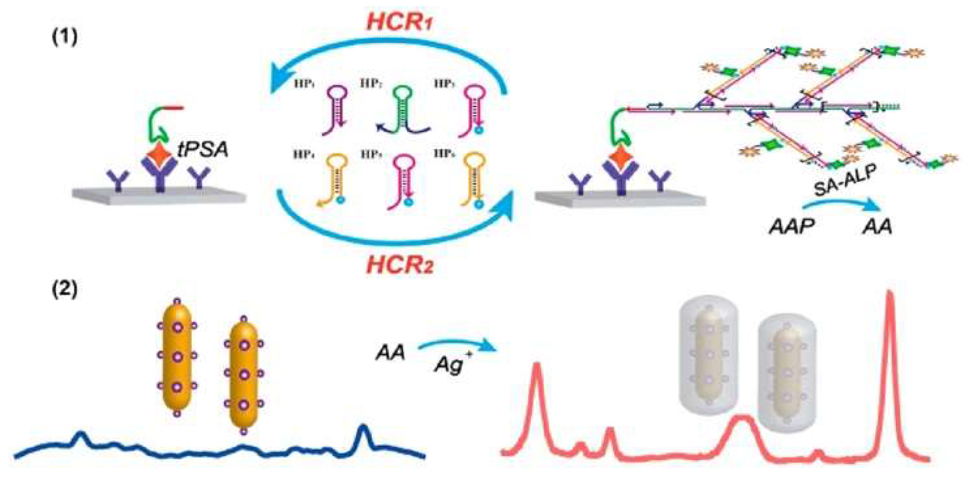
- Cun, F.; Huang, Z.; Lin, Q.; Yu, G.; Chen, H.; Kong, J.; Weng, W. Hybridized chain reaction-amplified alkaline phosphatase-induced Ag-shell nanostructure for the sensitive and rapid surface-enhanced Raman scattering immunoassay of exosomes. Anal. Chem. 2023, 95, 10025–10033. [Google Scholar] [CrossRef] [PubMed]
- Wang, J.R.; Xia, C.; Yang, L.; Li, Y.F.; Li, C.M.; Huang, C.Z. DNA nanofirecrackers assembled through hybridization chain reaction for ultrasensitive SERS immunoassay of prostate specific antigen. Anal. Chem. 2020, 92, 4046–4052. [Google Scholar] [CrossRef] [PubMed]


Disclaimer/Publisher’s Note: The statements, opinions and data contained in all publications are solely those of the individual author(s) and contributor(s) and not of MDPI and/or the editor(s). MDPI and/or the editor(s) disclaim responsibility for any injury to people or property resulting from any ideas, methods, instructions or products referred to in the content. |
© 2023 by the authors. Licensee MDPI, Basel, Switzerland. This article is an open access article distributed under the terms and conditions of the Creative Commons Attribution (CC BY) license (https://creativecommons.org/licenses/by/4.0/).
Share and Cite
Liu, L.; Chang, Y.; Lou, J.; Zhang, S.; Yi, X. Overview on the Development of Alkaline-Phosphatase-Linked Optical Immunoassays. Molecules 2023, 28, 6565. https://doi.org/10.3390/molecules28186565
Liu L, Chang Y, Lou J, Zhang S, Yi X. Overview on the Development of Alkaline-Phosphatase-Linked Optical Immunoassays. Molecules. 2023; 28(18):6565. https://doi.org/10.3390/molecules28186565
Chicago/Turabian StyleLiu, Lin, Yong Chang, Jiaxin Lou, Shuo Zhang, and Xinyao Yi. 2023. "Overview on the Development of Alkaline-Phosphatase-Linked Optical Immunoassays" Molecules 28, no. 18: 6565. https://doi.org/10.3390/molecules28186565