3.5. Examples from Literature
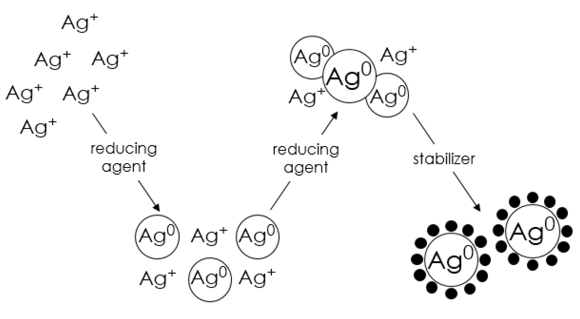
As has been mentioned several times, the method for the synthesis of metal nanoparticles by chemical reduction allows the production of nanomaterials with controlled morphology. To illustrate how changing various synthesis parameters affects the final nanoparticles, selected examples from the literature are described below.
Researchers from the Suriati research group [
63] have synthesized silver nanoparticles by chemical reduction, which turned out to be a simple, inexpensive, and partly green method as they used ascorbic acid (C
6H
8O
6), which is one form of vitamin C, as a surfactant. The metal precursor was silver nitrate (AgNO
3), while the reducing agent was sodium citrate (C
6H
5O
7Na
3). The concentrations of sodium citrate and ascorbic acid were varied to observe the effects of these parameters, particularly on the size and morphology of the silver nanoparticles.
At the reducing agent concentrations of 4.0–8.0 mM, all the nanoparticles produced had a quasi-spherical shape. In contrast, it was observed that the nanoparticle sizes showed a decreasing trend with increasing concentration of the reducer, from 38.53 nm at 4.0 mM C6H5O7Na3 to 36.32 at 8.0 mM C6H5O7Na3. Moreover, with increasing concentration of the reducer, a narrowing of the particle size distribution was observed from 20–65 nm to 20–50 nm. A possible reason for this phenomenon could be that the rate of the reaction is directly proportional to the concentration of the reactant according to the law of action of masses, from which it follows that as the concentration of trisodium citrate increased, the rate of the reaction increased. It has been concluded that as the reaction rate increased, the silver ions were consumed faster, leaving less room for particle size growth.
The TEM observations have shown that the average size of silver nanoparticles increased as the concentration of ascorbic acid increased from 1.0 mM to 4.0 mM. The average sizes of silver nanoparticles produced at 1.0 mM, 2.0 mM, 3.0 mM, and 4.0 mM ascorbic acid were 37.24 nm, 43.04 nm, 45.85 nm, and 47.28 nm, respectively. It seemed likely that ascorbic acid was able to kinetically control the growth rate of various surfaces by selectively adsorbing on these surfaces. Because of the acidic properties of ascorbic acid, the addition of a high concentration of ascorbic acid subsequently lowered the pH of the solution.
The work reported by Chou and co-workers [
94] investigated the synthesis of silver nanoparticles by the chemical reduction method using silver nitrate (AgNO
3) as a metal precursor, formaldehyde (CH
2O) as a reducing agent, and polyvinylpyrrolidone/poly(vinyl alcohol), (PVP/PVA) as stabilizing agents. A solution of either sodium carbonate (Na
2CO
3) or sodium hydroxide (NaOH) was used to determine the preferred pH. The effect of the amount of alkaline solution on the morphology of the final nanoparticles was examined.
Although high pH is preferred in nanoparticle synthesis due to its higher reducing power, Chou’s research group has shown an adverse effect of high pH on particle size. When more NaOH was added to the reaction system, the silver colloids settled at the bottom of the solution. Thus, NaOH was replaced with a weak base, Na
2CO
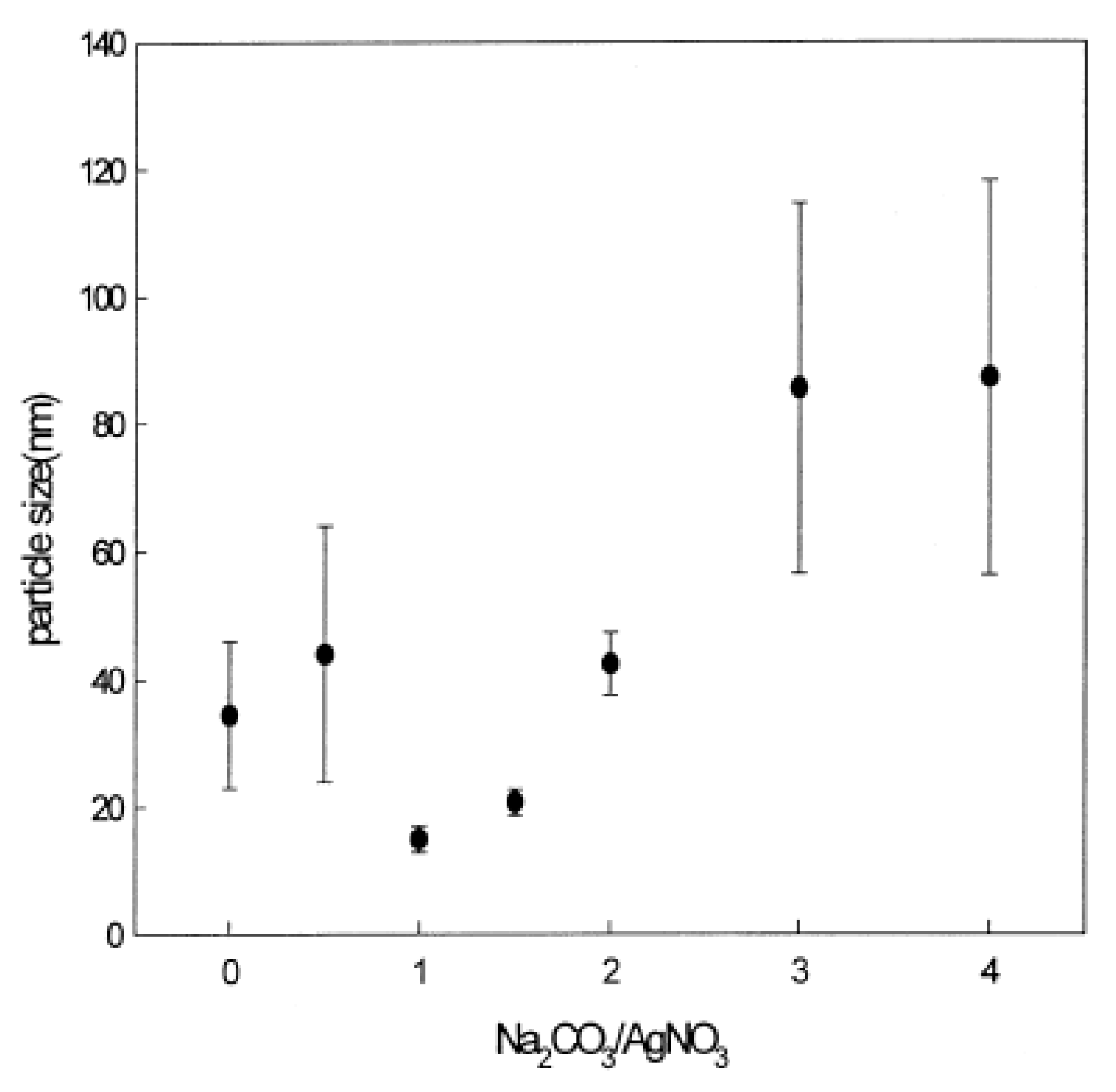
3, in order to release hydroxyl ions only when the pH fell below certain values. The effect of the amount of sodium carbonate on the average particle size and also the standard deviation of the particle size distribution is shown in
Figure 8. As shown in this figure, the optimal conditions were at a ratio of Na
2CO
3/AgNO
3 between 1.0 and 1.5, at which smaller nanoparticle sizes were obtained. When more Na
2CO
3 was added, the pH of the solution increased, which adversely affected the stability of the silver colloids; moreover, the size of the nanoparticles increased from 10–20 nm to 80–100 nm, as did the size distribution, as indicated by the bar shown in the figure below.
Liguo’s research group [
95] prepared silver nanoparticles by the chemical reduction method, using hexadecyltrimethylammonium bromide (CTAB) to prevent nanoparticle agglomeration, silver nitrate (AgNO
3) as the metal source and formaldehyde (CH
2O) as the reducing agent. The pH of the solution was adjusted by adding nitric acid (V) (HNO
3) or sodium hydroxide (NaOH) to the solution mixture at the nanoparticle synthesis stage.
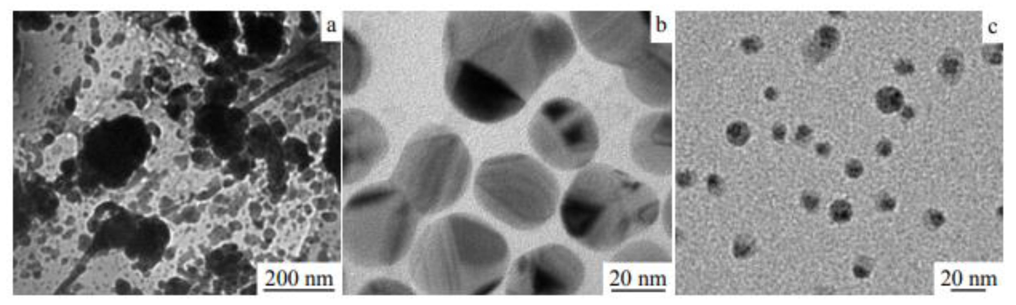
The addition of a small amount of CTAB to the solution resulted in the formation of particles of large size, irregular shape, and significant agglomeration degree (
Figure 9a). As the amount of CTAB increased, the particle sizes were better dispersed. When the CTAB/AgNO
3 ratio was 0.8, nanosilver particles of 20–40 nm were obtained (
Figure 9b), while when this ratio reached 1.2, then particles smaller than 10 nm were formed (
Figure 9c).
It has been shown that CTAB has an inhibitory effect on the process of particle growth and agglomeration, which was explained by the effect of long carbon chains of CTAB, which may reduce the possibility of collisions between silver particles. In addition, CTAB, due to its structure, exhibits a strong steric effect. Thus, at low concentrations, it had little effect on the particle size and agglomeration, and as its concentration increased, better dispersion was achieved. On the other hand, an excessive amount of CTAB retards the growth of particles, as they are wrapped inside CTAB molecules, which prevents the growth of nuclei resulting in the formation of particles of several nanometers in size.
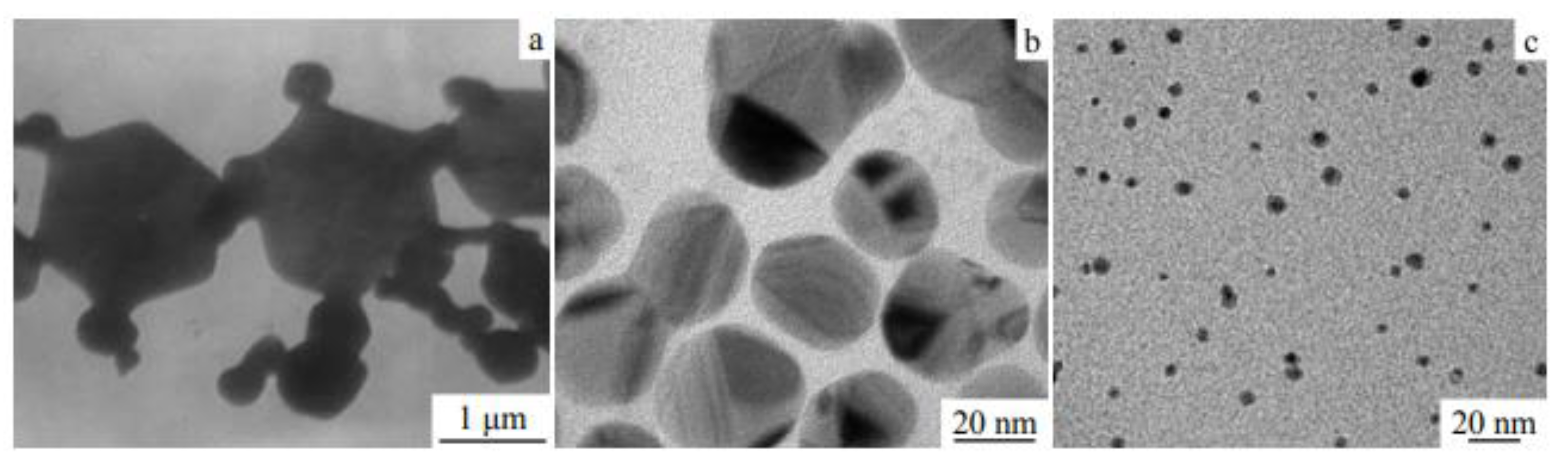
As established by Liguo et al., the reaction temperature affects the morphology of silver particles in two ways. On the one hand, the rate of reduction in silver nitrate depends on temperature, while on the other hand, temperature affects the interactions, in this case, between CTAB and Ag
+. As a result, both temperature-dependent processes ultimately affect the morphology of the nanosilver particles. The morphological response of particles prepared at different temperatures was examined. TEM images of the obtained nanoparticles at the reaction temperatures of 20 °C, 40 °C, and 60 °C are presented in
Figure 10a, b, and c, respectively. When the reaction temperature was 20 °C, heterogeneous particles of about 100 nm in size were obtained. With increasing reaction temperature, the particles became smaller—their size was close to 30 nm at 40 °C, while at 60 °C, it was about 10 nm.
These observations were interpreted as follows. When the reaction temperature is low (20 °C), CTAB dissolves poorly, and in addition, precipitation is often easier, which ultimately leads to a small amount of CTAB participating in the reaction, further resulting in a reduced effect on inhibiting particle growth. On the other hand, the reducing ability of formaldehyde is very weak at low temperatures. As a result, the reaction is slow, and the initial nuclei can then consume most of the reduced silver atoms. The number of further forming nuclei is smaller and large particles with a wide size distribution are eventually formed. At 40 °C, CTAB is fully dissolved, and the amount of silver nitrate decreases at a moderate rate. The nanosilver particles obtained exhibit a flake morphology. When the temperature reaches 60 °C, the reduction in silver cations is faster. A large number of silver atoms are generated in a short time, and the nucleation rate is greatly increased. The nucleation process consumes most of the silver reserves, which inhibits the growth of newly formed particles, so small particles are formed.
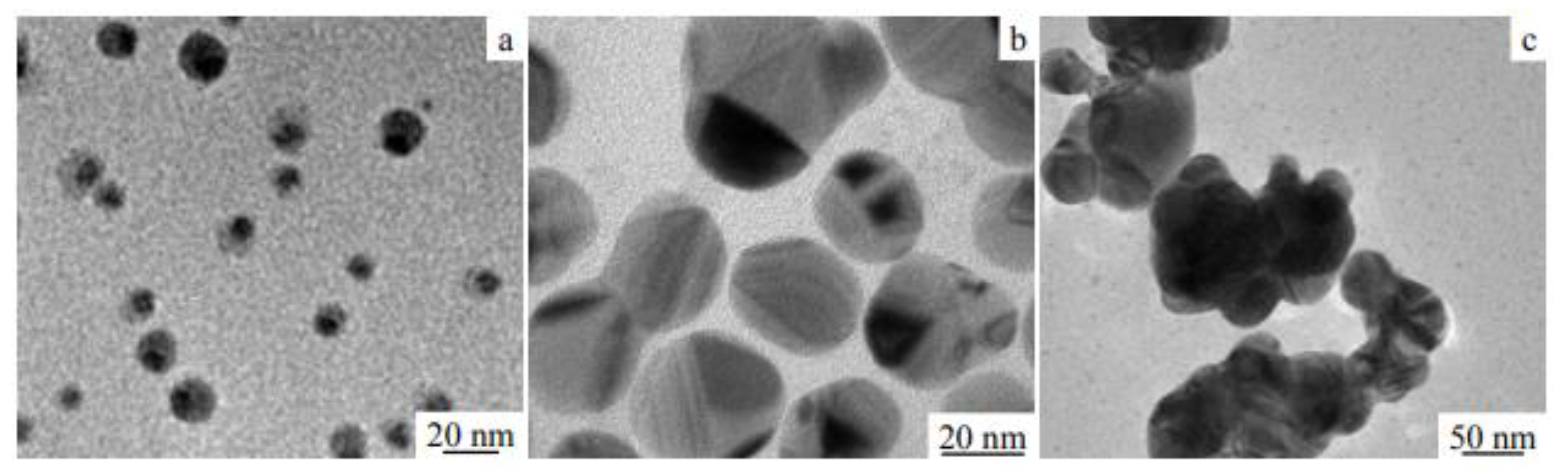
The morphology of silver nanoparticles at different pH was also examined. As shown in
Figure 11, at pH of 3, the particle size was less than 20 nm; at pH increased to 9, the agglomeration of the particles formed was advanced; while at pH of 6, the particles showed a flaky structure and good dispersion. The explanation proposed was that at a low pH value, the reduction ability of formaldehyde is weak, resulting in a low reaction rate. On the other hand, the presence of a large number of H
+ in the solution inhibits the reaction and causes an incomplete reaction of silver cations.
In addition, after the reactions were complete, the contents of Ag+ with Cl− at different pH were checked. At pH of 3, the reduction in Ag+ was incomplete because fewer silver atoms existing in the solution participated in the nucleation and particle growth processes, eventually leading to smaller nanosilver particle formation. At weakly acidic pH, a small amount of H+ in solution is beneficial for maintaining the stability of the bilayer surface, which favors the formation of stable nanoparticles with good dispersion.
Guzmán and co-workers [
91] have synthesized silver nanoparticles by the chemical reduction method using silver nitrate (AgNO
3) as the metal precursor, hydrazine hydrate (N
2H
4), and/or sodium citrate (Na
3C
6H
5O
7) as the reducing agent and sodium dodecyl sulfate (SDS) and/or sodium citrate (Na
3C
6H
5O
7) for stabilizing the whole system. The effect of the concentration of sodium citrate and the type of reducing agent used on the morphology of the final particles was investigated.
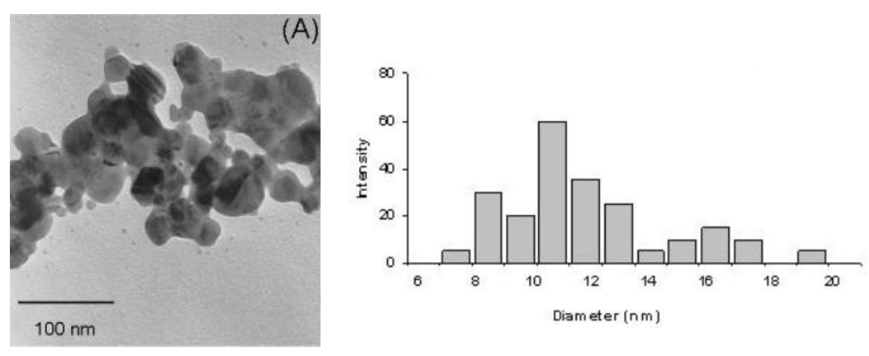
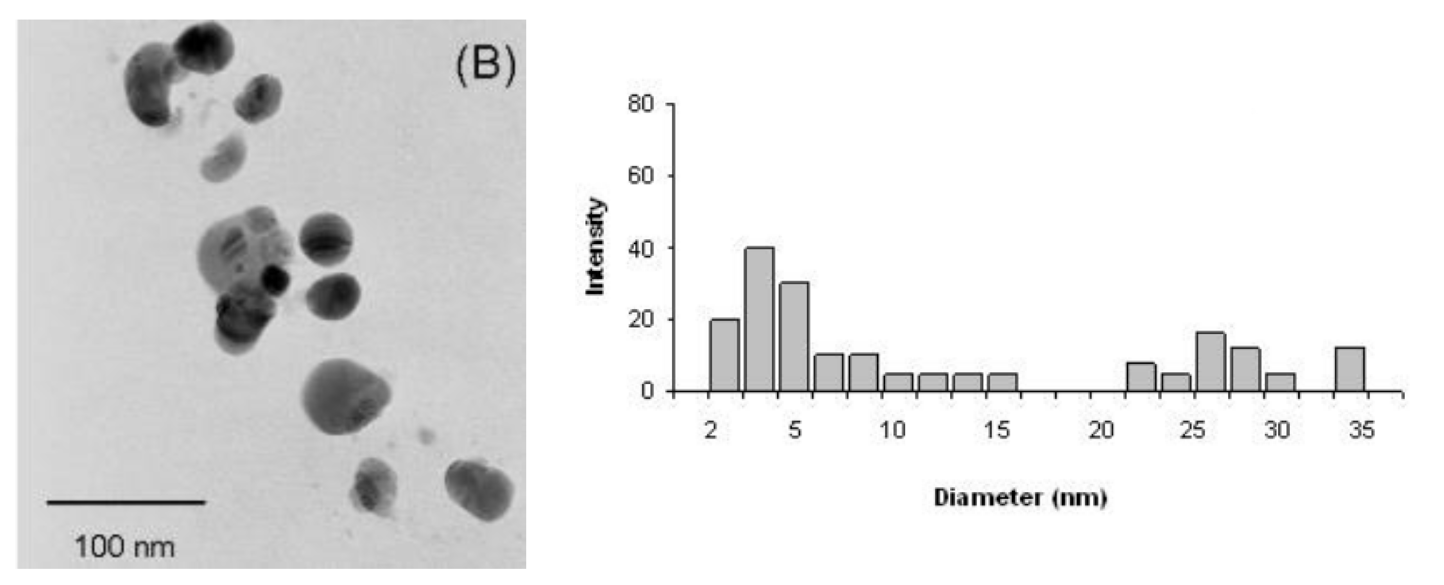
TEM studies indicated a correlation between the concentration of sodium citrate and the morphology of the final nanoparticles. For hydrazine at a concentration of 2.0 mM and sodium citrate at 1.0 mM, the silver nanoparticles were obtained in the form of small, significantly agglomerated grains (
Figure 12A, left). In addition, the obtained particle size histogram showed the size range of the silver nanoparticles from 7 to 20 nm with an average diameter of 9 nm (
Figure 12A, right). In contrast, the nanoparticles obtained at twice the concentration of sodium citrate and the same concentration of hydrazine assumed a spherical shape and were additionally characterized by good dispersion (
Figure 12B, left). From the histogram, it was deduced that, in this case, the silver particles assumed sizes ranging from 7 to 20 nm and 22 to 35 nm, with an average diameter of 11 nm (
Figure 12B, right). The described relationship was also confirmed by UV-Vis studies, which revealed typical plasmonic absorption maxima at 405 nm and 406 nm when the sodium citrate solution of 1.0 mM and 2.0 mM, respectively, was used. The observed differences in the position and shape of plasmonic absorption are due to differences in the particle size, shape, and dielectric constant of the surrounding medium.
In addition, the effect of the type of reducing agent on the particle size was examined. When hydrazine was used as a reducing agent, the average diameter of the particles obtained was about 30 nm. On the other hand, when a mixture of hydrazine and sodium citrate was used as a reducing agent, the average particle diameter was in the range of 15–48 nm, which, compared to the particle size obtained with hydrazine alone, indicates a slight increase in the average particle diameter.
Song and co-workers [
89] have synthesized silver nanoparticles using silver nitrate (AgNO
3) as the precursor while sodium borohydride (NaBH
4) and sodium dodecyl sulfate (SDS) as the reducing agent and stabilizing agents, respectively. The effects of several variables, i.e., the concentration of AgNO
3, NaBH
4, and SDS, on the final silver nanoparticles were examined.
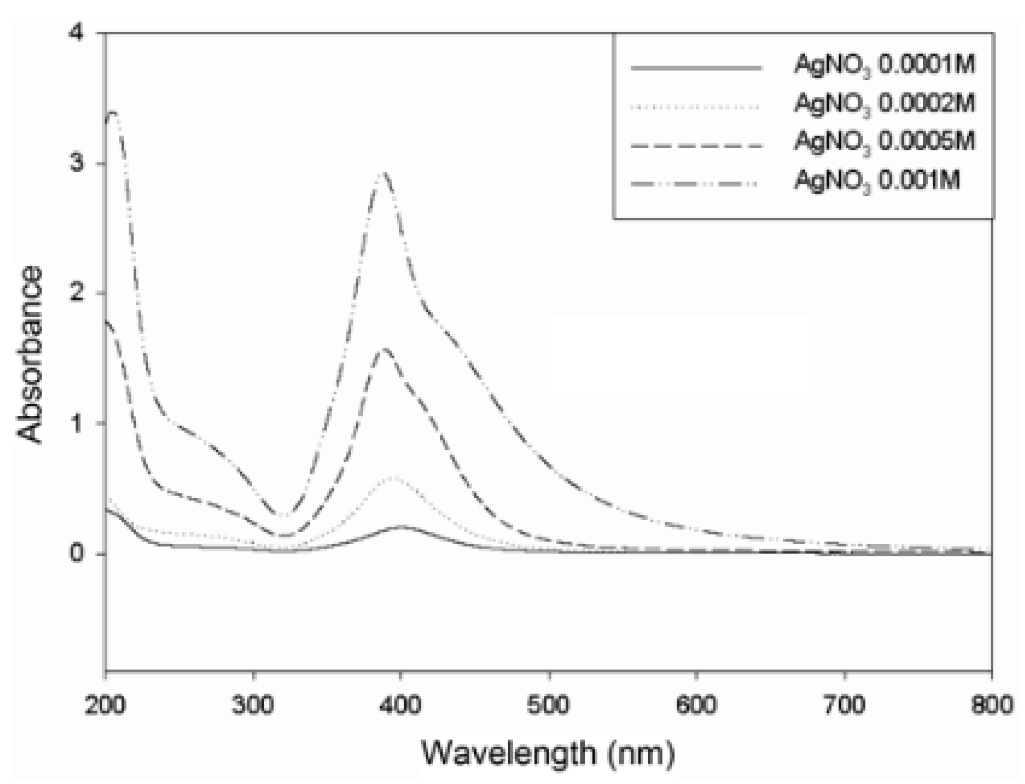
Figure 13 shows the UV-Vis spectra of colloidal silver nanoparticles prepared at different initial concentrations of AgNO
3 (0.0001 M, 0.0002 M, 0.0005 M, and 0.001 M). The nanoparticles were synthesized at the NaBH
4/AgNO
3 molar ratio of 10 and SDS/AgNO
3 weight ratio of 2. The color of the solutions depended on the concentration of AgNO
3 added. As the initial concentration of AgNO
3 increased, the color of the solution changed from yellow to brown. The absorption peak at about 400 nm was attributed to plasmonic excitation by silver nanospheres, indicating the formation of silver nanoparticles. At low concentrations of AgNO
3, the maximum weak absorption of surface plasmon peaks was observed at 400 nm, indicating that silver nanoparticles were produced at relatively low concentrations. As the AgNO
3 concentration increased, the intensity of the maximum plasmonic peak increased, indicating that higher concentrations of silver nanoparticles were formed.
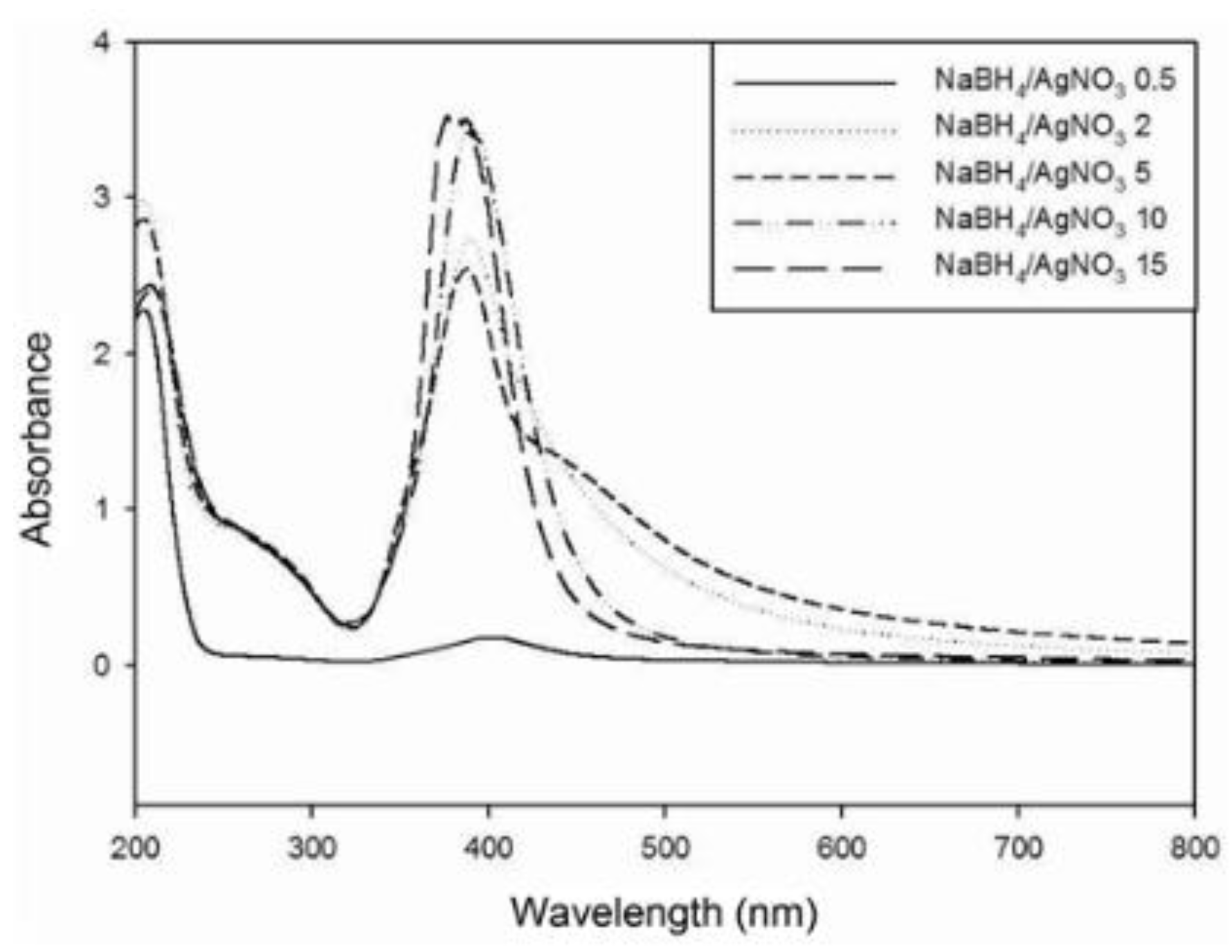
To understand the effect of NaBH
4 concentration, the reduction reaction was studied at different NaBH
4/AgNO
3 molar ratios (0.5–15), at the initial AgNO
3 concentration of 0.001 M, and the SDS/AgNO
3 weight ratio of 2. The corresponding UV-Vis spectra are shown in
Figure 14. At the lowest NaBH
4/AgNO
3 molar ratio, a weak plasmonic peak was observed at 400 nm, indicating a relatively low concentration of silver nanoparticles formed, the reason being insufficient reduction. It is known that the UV-Vis absorption peak can also provide information about the degree of dispersion of silver nanoparticles. The narrower it is, the better the dispersion degree of nanoparticles is obtained. At molar ratios of 2 and 5, the absorption peak at 400 nm was broad, indicating that the silver nanoparticles were aggregated, while when the molar ratios were 10 and 15, narrow absorption peaks were obtained, indicating that the silver nanoparticles were well dispersed. The reason for this, according to Song et al., was the use of too little NaBH
4 so that boron hydroxide B(OH)
3 (produced by hydrolysis of NaBH
4, see Equation (2)) was absorbed into the silver nanoparticles, reducing the electron density and causing deep aggregation. On the other hand, when an excessive amount of NaBH
4 was used, a thick layer of BH
4− prevented the absorption of boron hydroxide onto the surfaces of silver nanoparticles, resulting in well-dispersed nanoparticles. These results indicate that NaBH
4 acted not only as a reducing agent but also as a stabilizer protecting against the aggregation of silver nanoparticles.
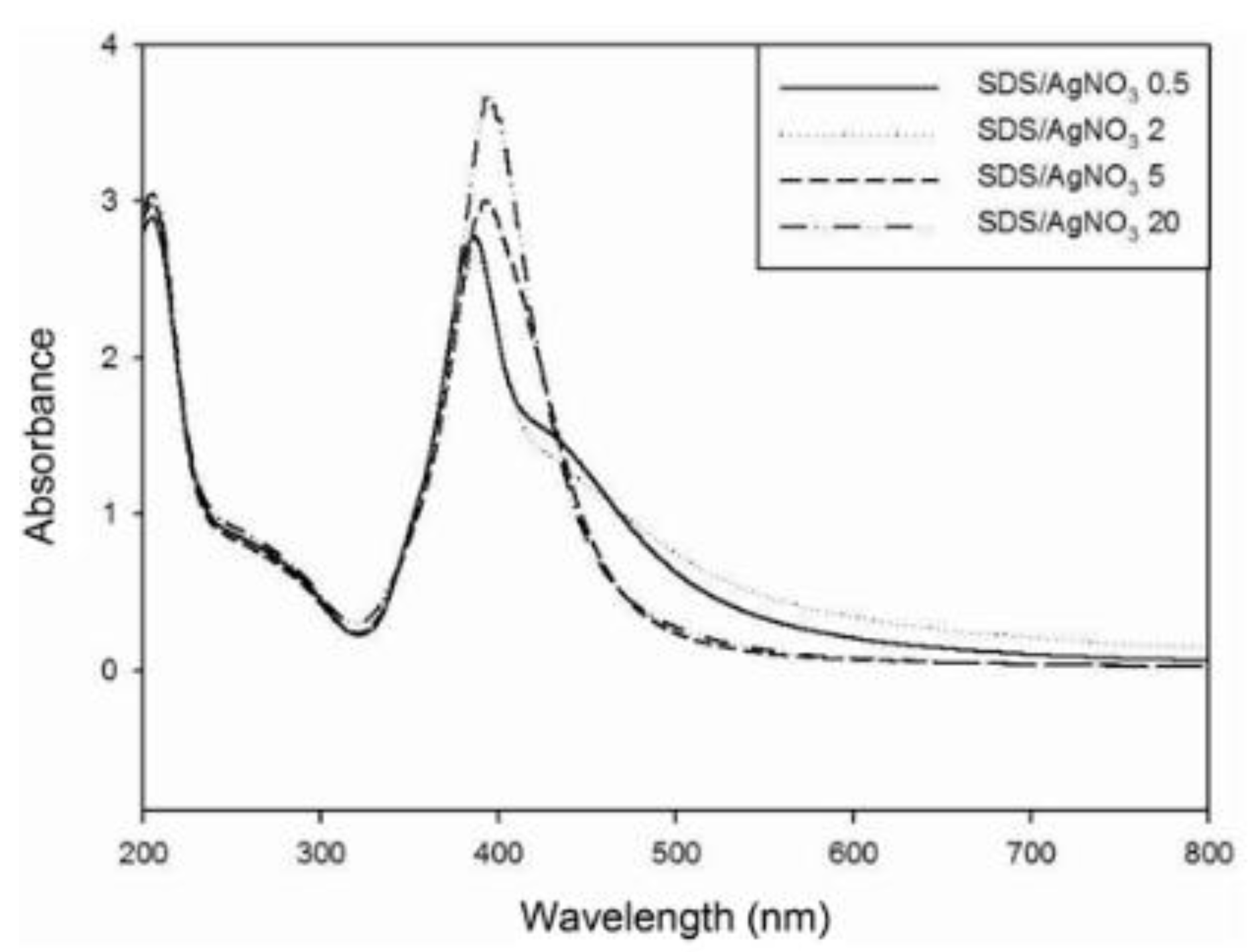
The main purpose of introducing SDS into the solution was to prevent the growth and aggregation of silver nanoparticles.
Figure 15 shows UV-Vis spectra of silver nanoparticles with different SDS/AgNO
3 weight ratios (0.5–20). The nanoparticles were synthesized under conditions of an initial AgNO
3 concentration (0.001 M) and a NaBH
4/AgNO
3 molar ratio of 4. As the SDS concentration increased, the color of the solutions changed from brown to yellow. At high SDS/AgNO
3 weight ratios (5, 20), narrow plasmonic absorption peaks were observed at 400 nm, confirming the nanocrystalline nature and well-dispersed state of the silver particles. However, when the weight ratios were low (0.5, 2), the absorption peaks became broad, indicating that the silver nanoparticles were aggregated. These results imply that with the right amount of SDS, it absorbs the surface of silver nanoparticles and protects them from steric growth and aggregation.
Researchers from the Alqadi research group [
96] synthesized silver nanoparticles, where they controlled their size by changing the pH value of the reaction system. For the synthesis, they used silver nitrate (AgNO
3), which was reduced with ascorbic acid (C
6H
8O
6), while sodium citrate (Na
3C
6H
5O
7) was used as a stabilizer. The pH was manipulated with the addition of either sodium hydroxide (NaOH) or citric acid (C
6H
8O
7).
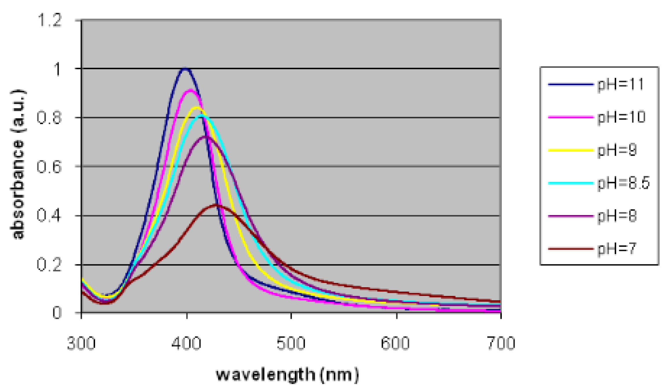
The changes in the size of silver nanoparticles in response to a change in the pH of the solution were monitored. Absorption spectra recorded at different pH values are shown in
Figure 16. Based on the plasmon resonance peaks obtained, it was found that at high pH, silver nanoparticles of smaller sizes were obtained (the plasmon resonance peak shifted towards the short wavelength region as well as increasingly narrow peaks) compared to those obtained at low pH values. The difference was attributed to the different rates of precursor reduction.
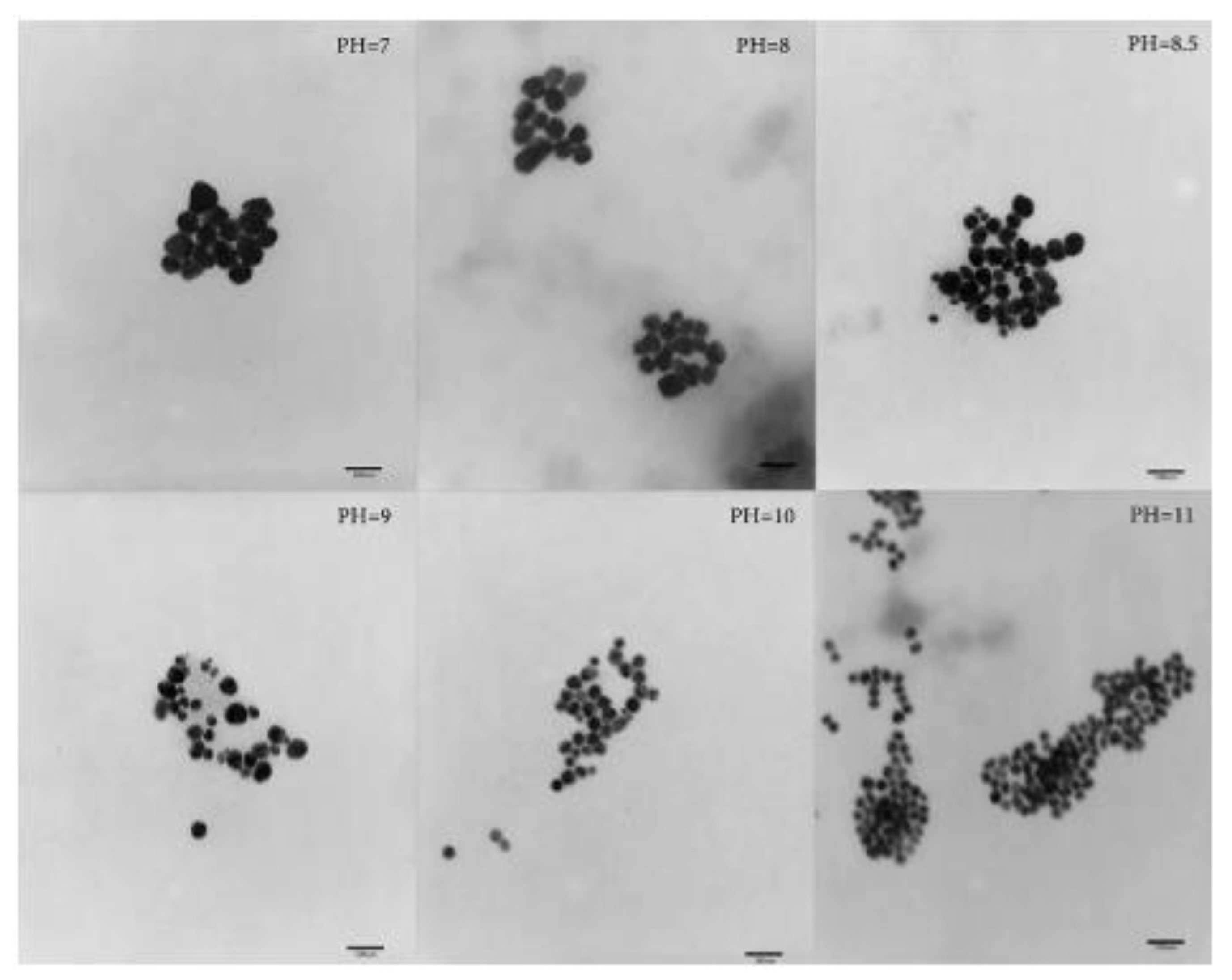
In addition to the inverse proportionality between particle size and pH value, it is clear that increasing the pH value yields spherical nanoparticles, while at low pH, rods and triangular particle shapes were formed, as shown in
Figure 17. The irregularity of the particle shape was attributed to the slow reduction rate of the precursor, as well as the poor balance between nucleation and growth processes.
Beyribey and co-workers [
83] have synthesized platinum nanoparticles by the chemical reduction in hexachloroplatinic acid (H
2PtCl
6) with hydrazine (N
2H
4). The purpose of the experiment was to study the effect of temperature and pH on the structure of platinum particles.
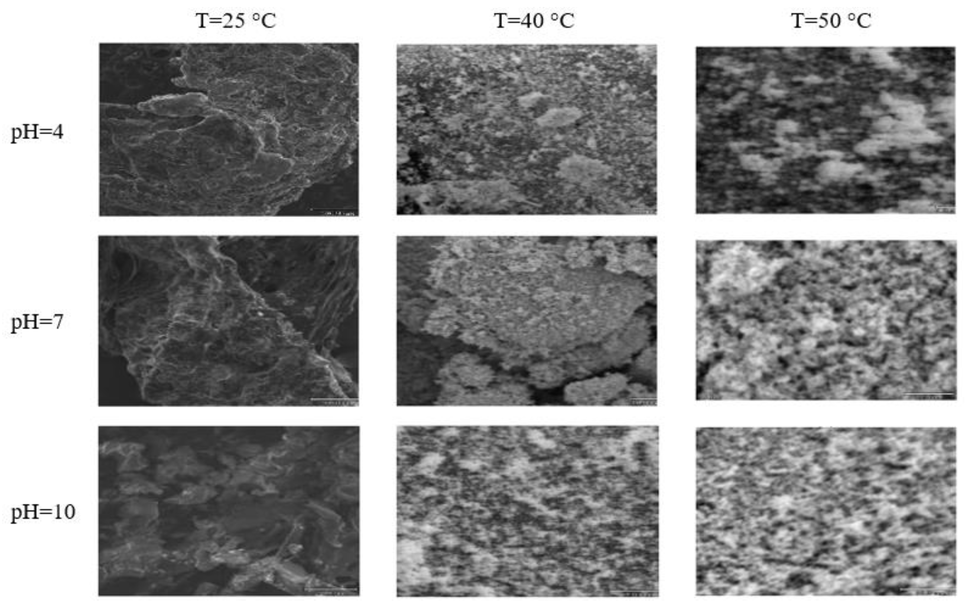
The synthesis of platinum nanoparticles was carried out at pH equal to 4, 7, or 10 and at 25 °C, 40 °C, or 50 °C. At the low pH of the solution, no characteristic structures of platinum nanoparticles formed; they were only observed when the pH of the solution was 10. A significant difference in the morphological distribution of platinum particles at high temperatures and pH was also observed compared to that of the nanoparticles obtained at lower temperatures and lower pH values, as shown in the photographs below (
Figure 18).
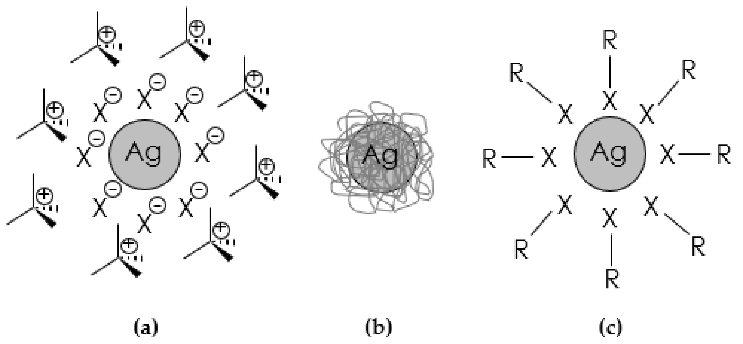
The zeta potential, informing about the physical stability of emulsions and suspensions, was also measured. If all the particles in a suspension have a high negative or positive zeta potential, then they will repel each other and will not tend to flocculate. However, if the particles have low zeta potential values, then there is no force to prevent the particles from clumping together. The general dividing line between stable and unstable suspensions is usually taken at +30 mV or −30 mV. Particles with a zeta potential more positive than +30 mV or more negative than −30 mV are usually considered stable. It has been shown that platinum particles synthesized at 25 °C in the pH range of 4–7 are unstable (
Table 6).
An equally interesting study was conducted by scientists from the Patharkar research group [
105], who synthesized ruthenium nanoparticles by the chemical reduction in ruthenium chloride (RuCl
3) using sodium borohydride (NaBH
4) as a reducing agent and sodium dodecyl sulfate (SDS) as a stabilizer (other stabilizers such as PVP, CTAB, and AOT were also used). The influence of changes in such parameters as the molar ratio (MR) of SDS/RuCl
3, NaBH
4/RuCl
3, or the type of stabilizer used on the Ru nanoparticles size and their size distribution was established.
The SDS/RuCl
3 molar ratio was changed from 1 to 40, keeping the RuCl
3 concentration at 0.2 mM and the NaBH
4/RuCl
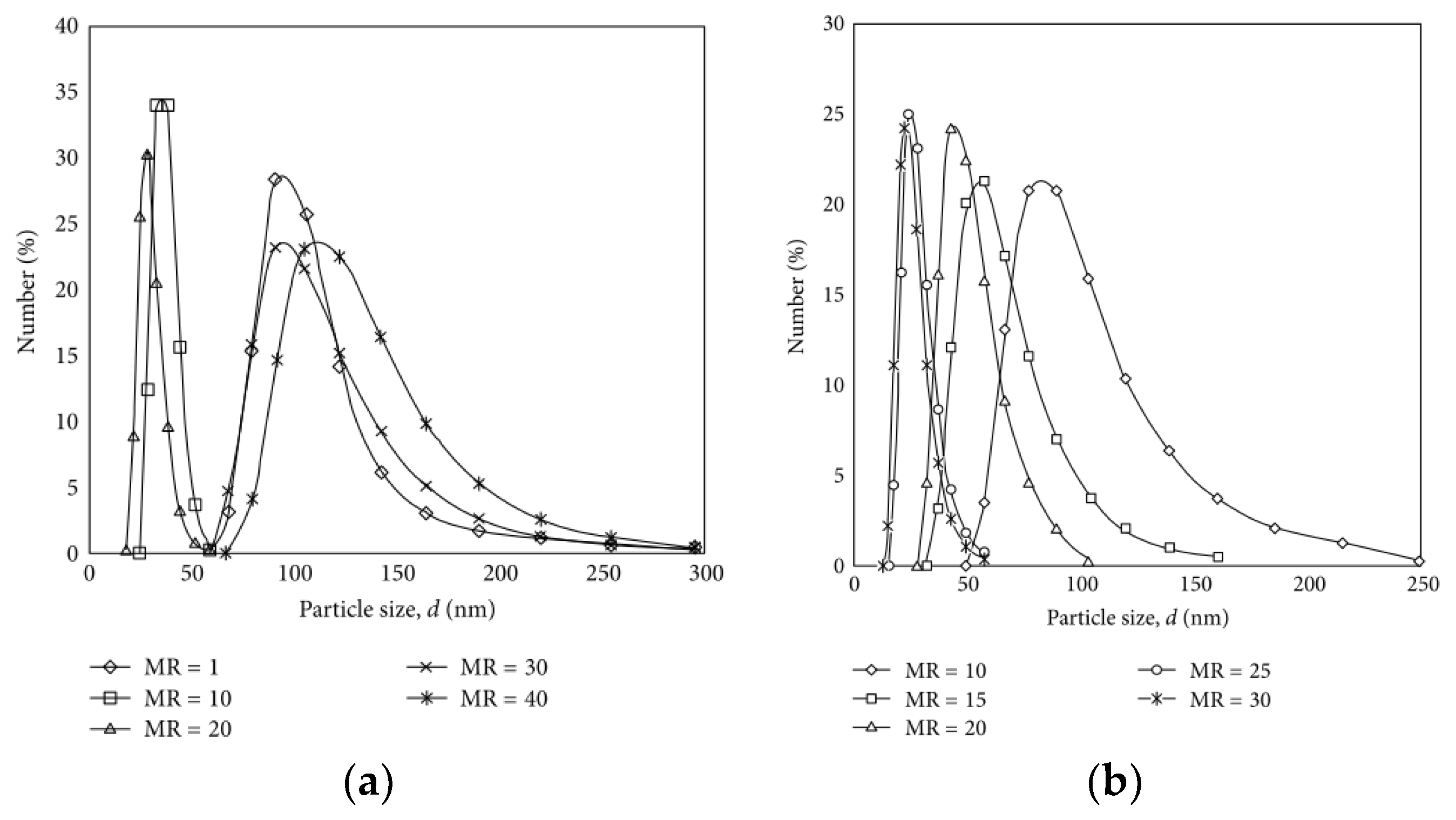
3 molar ratio at 30. As shown in
Figure 19a, the particle size decreased as the MR of SDS/RuCl
3 increased to 20. The diameter of Ru nanoparticles was found to be 90 nm at MR = 1 and 20 nm at MR = 20. At MR > 20, the particle size increased as the SDS/RuCl
3 ratio increased. The large size of the ruthenium nanoparticles formed at MR = 1 was interpreted as due to a higher degree of agglomeration, which was the result of an insufficient amount of stabilizing agent in the system. On the other hand, an increasingly higher surfactant concentration increased the viscosity of the system, which led to a decrease in the migration rate of the surfactant and/or a decrease in the diffusion rate of the micelles and a decrease in electrostatic repulsion, which had the effect of promoting the agglomeration process of the particles and so larger nanoparticles were ultimately formed.
The researchers decided to study the effect of NaBH
4 concentration (MR NaBH
4/RuCl
3 = 10–30) on the size of Ru nanoparticles, holding other parameters constant (RuCl
3 = 0.2 mM, MR SDS/RuCl
3 = 20). The scientists observed that at a lower NaBH
4/RuCl
3 molar ratio (MR = 10), the size of the nanoparticles was larger due to insufficient reduction in RuCl
3 (
Figure 19b). However, as the molar ratio increased from 15 to 30, narrow peaks were obtained, suggesting that the Ru nanoparticles produced were smaller in size. Based on the results, the researchers concluded that a lower concentration of NaBH
4 produces boron hydroxide through hydrolysis of NaBH
4. The boron hydroxide was then absorbed into the Ru nanoparticles, reducing the electron density of the surface and causing the Ru nanoparticles to aggregate, resulting in a larger nanoparticle size. On the other hand, a higher concentration of NaBH
4 increased the concentration of boron hydroxide, which formed a thick BH
4− layer, preventing boron hydroxide from being absorbed into the surface of Ru nanoparticles, resulting in well-dispersed yet smaller nanoparticles.
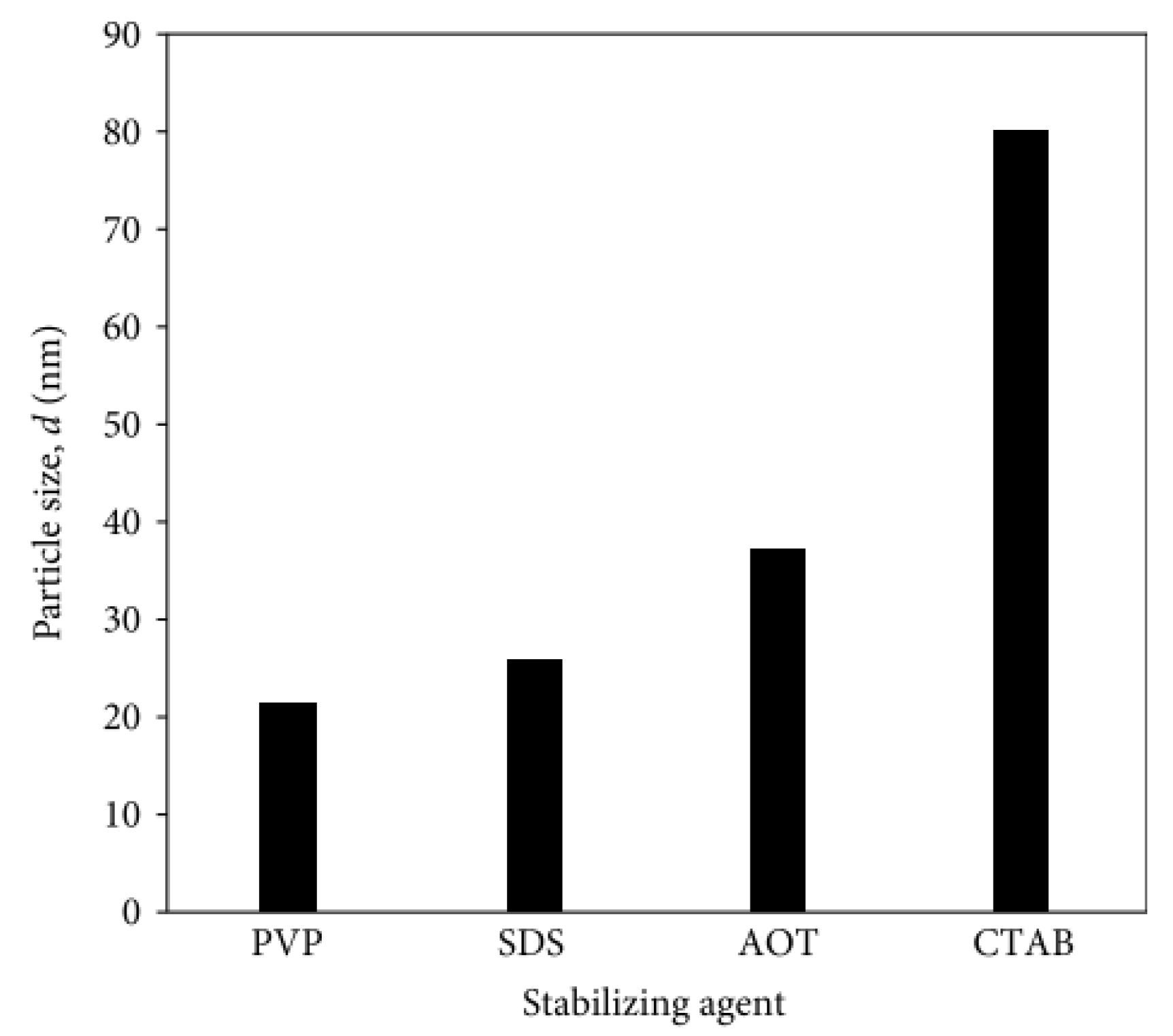
In order to establish the effect of different types of stabilizing agents on nanoparticle size, a series of syntheses were performed in which PVP, SDS, CTAB, or AOT were used as stabilizers, and the final materials were subjected to particle size testing (RuCl
3 concentration = 0.2 mM, MR surfactant/RuCl
3 = 20, MR NaBH
4/RuCl
3 = 30). The smallest particle size was obtained when using PVP (~20 nm) and SDS (~25 nm), which was much smaller than that measured when AOT and CTAB were used as stabilizers (
Figure 20). The explanation was that PVP, due to its structure, can act as both a stabilizer and a reducing agent, which resulted in the formation of particles of small size. In contrast, in the presence of CTAB, which is a cationic surfactant, the Ru nanoparticles were attracted to the positive charge of the surfactant, where they agglomerated near the outer surface of the micelles, resulting in the formation of larger nanoparticles.


