
Novel Cerium(IV) Coordination Compounds of Monensin and Salinomycin

 , , , , and
Abstract
:
1. Introduction
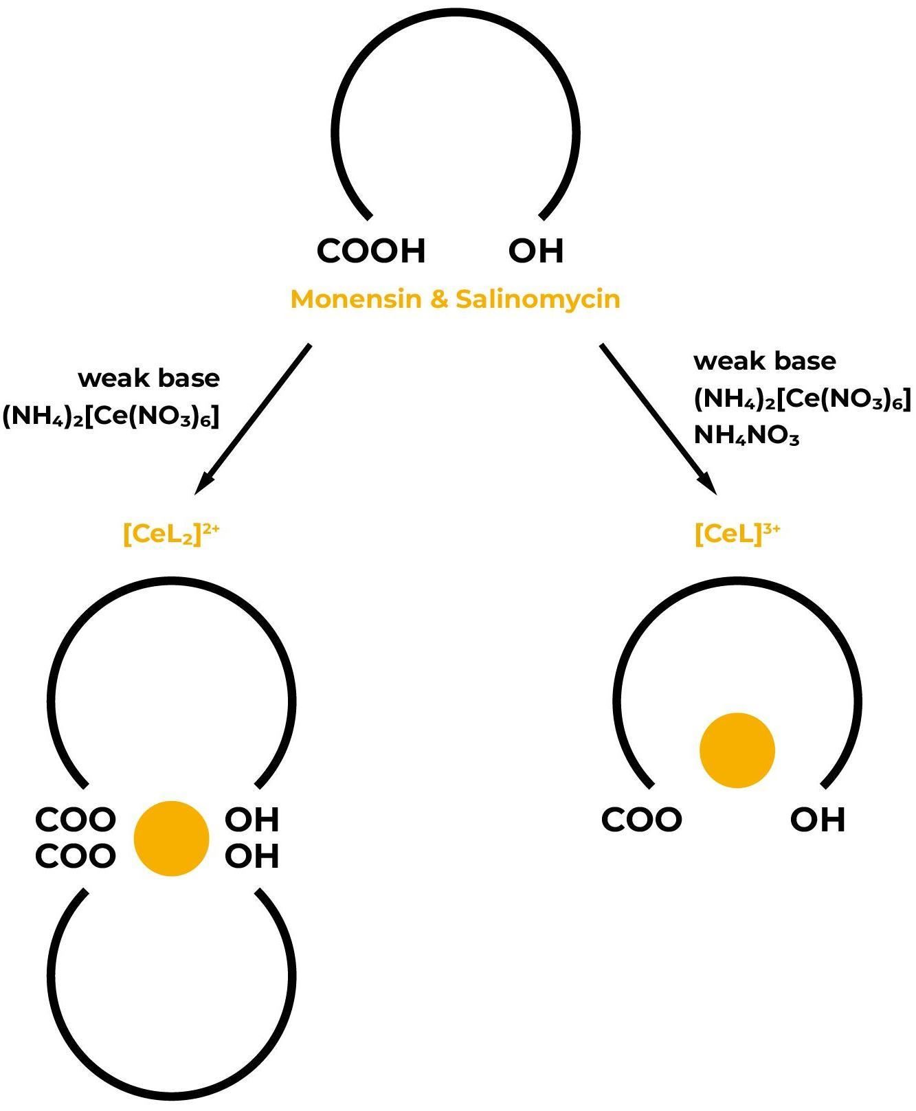
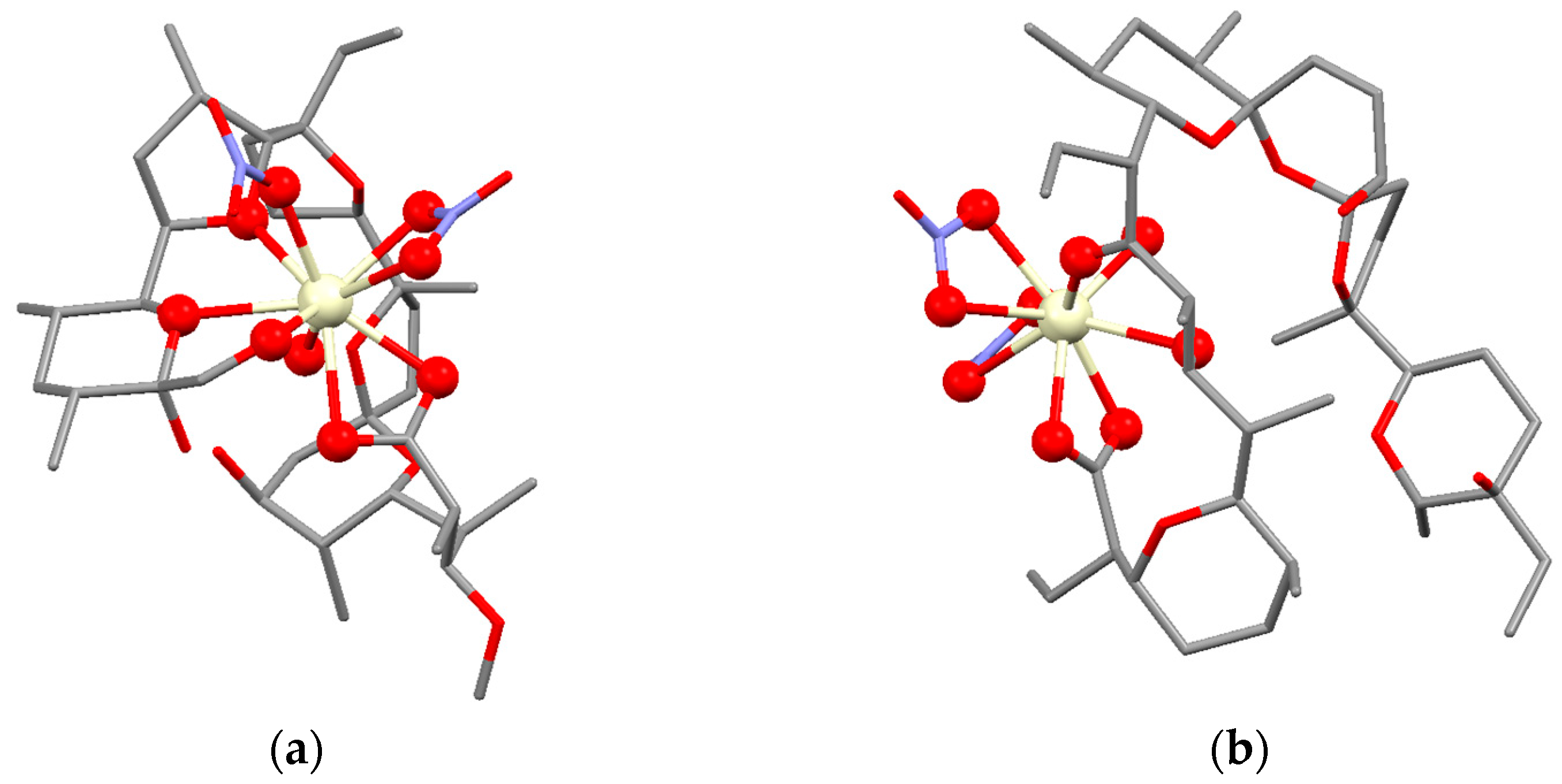
2. Results and Discussion
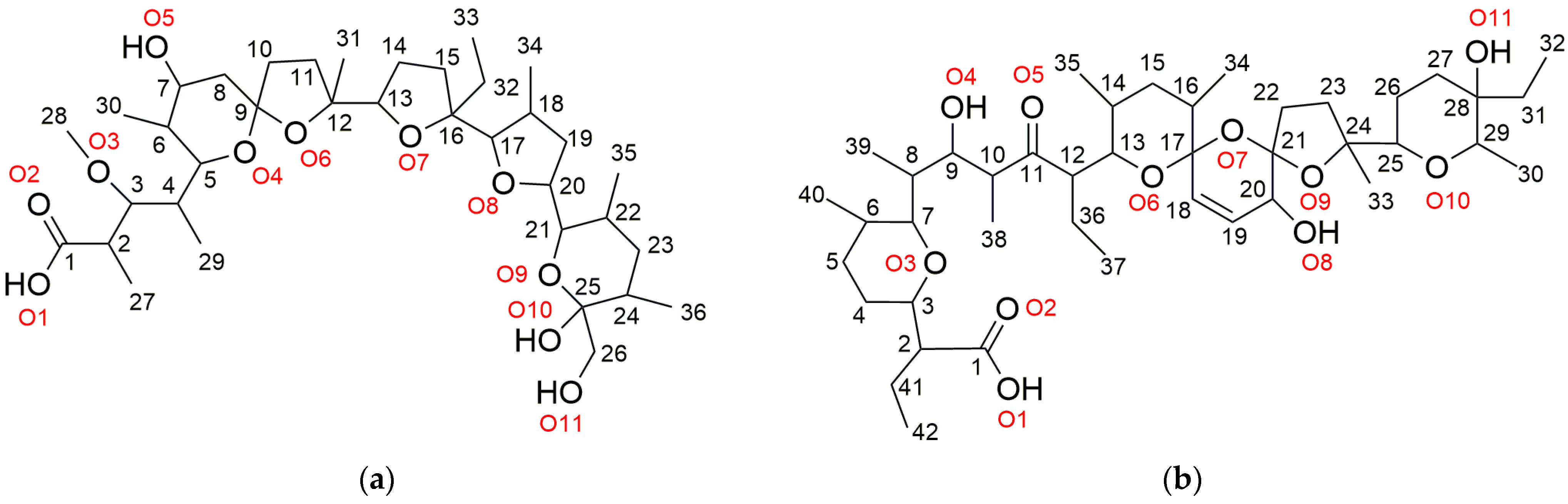
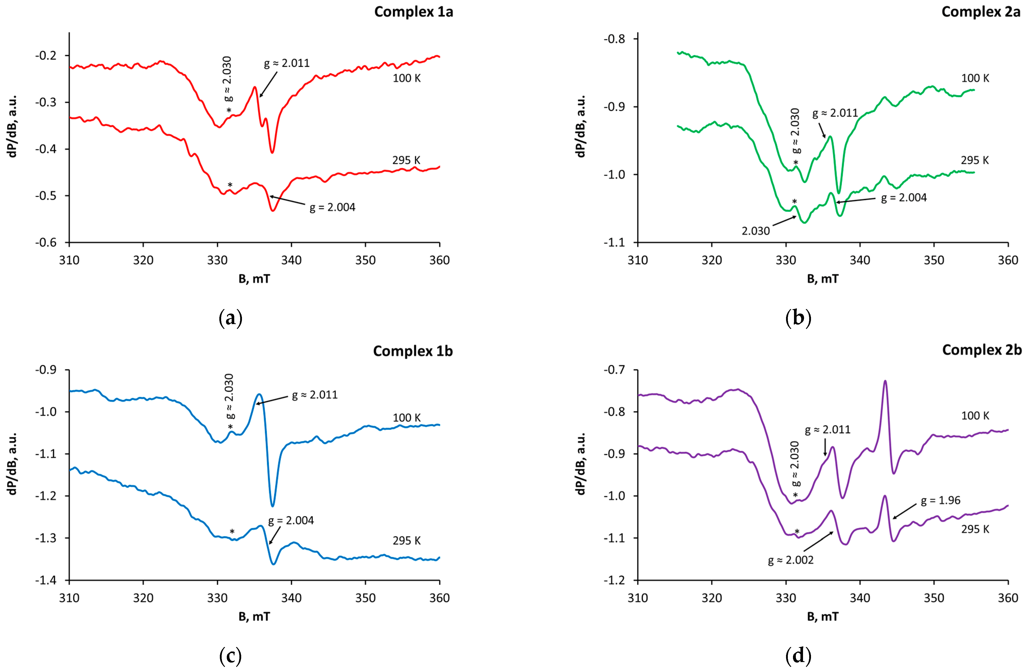
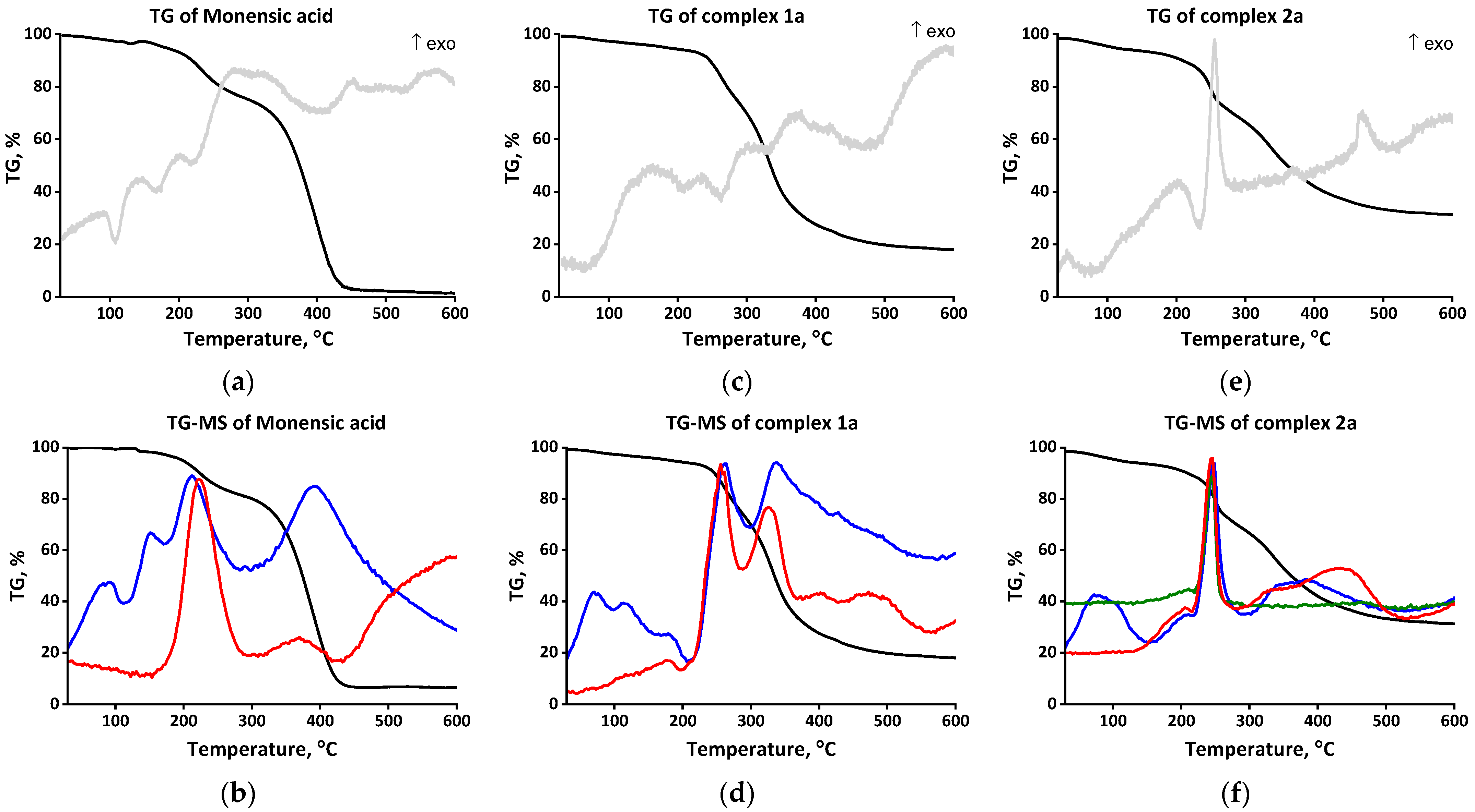
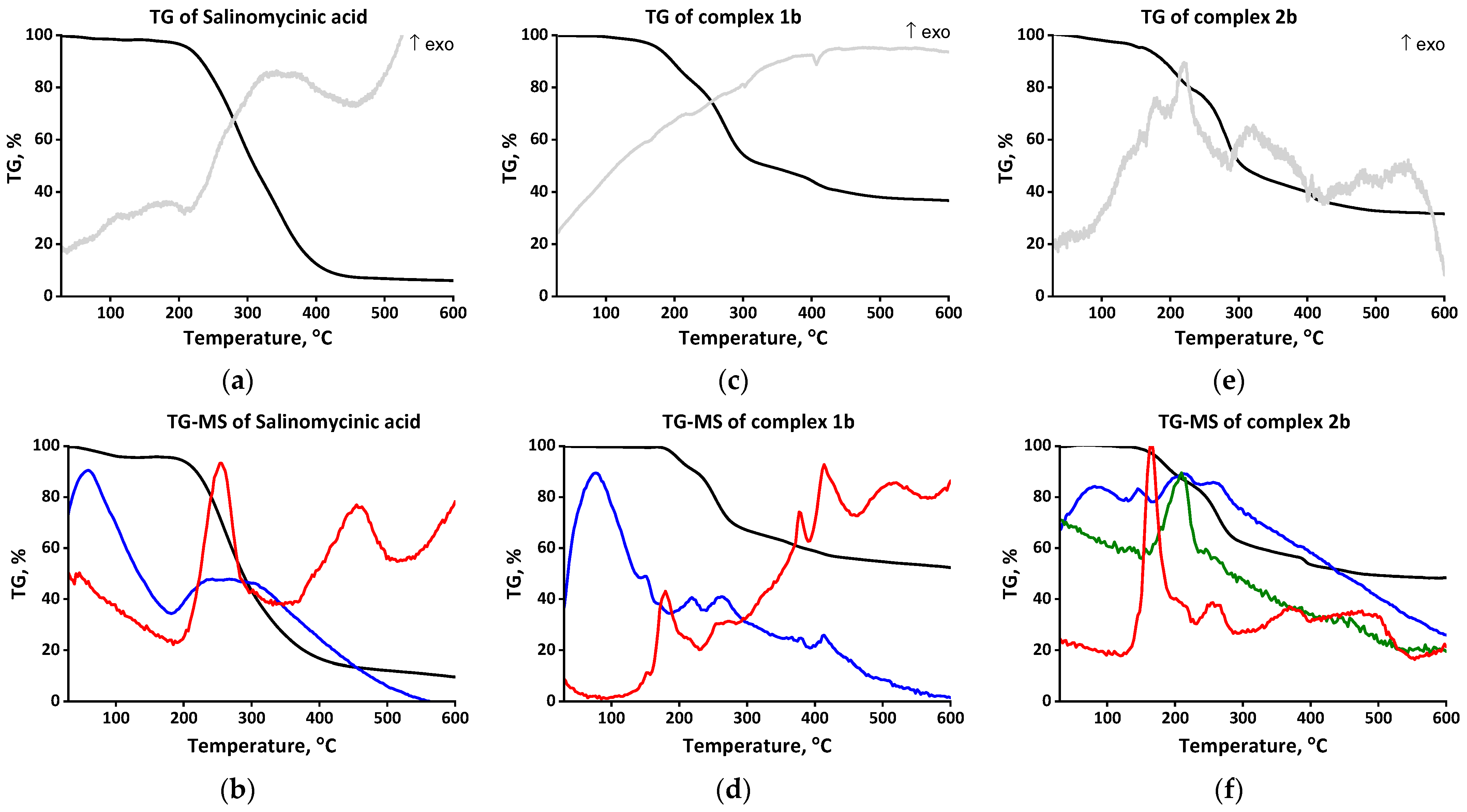
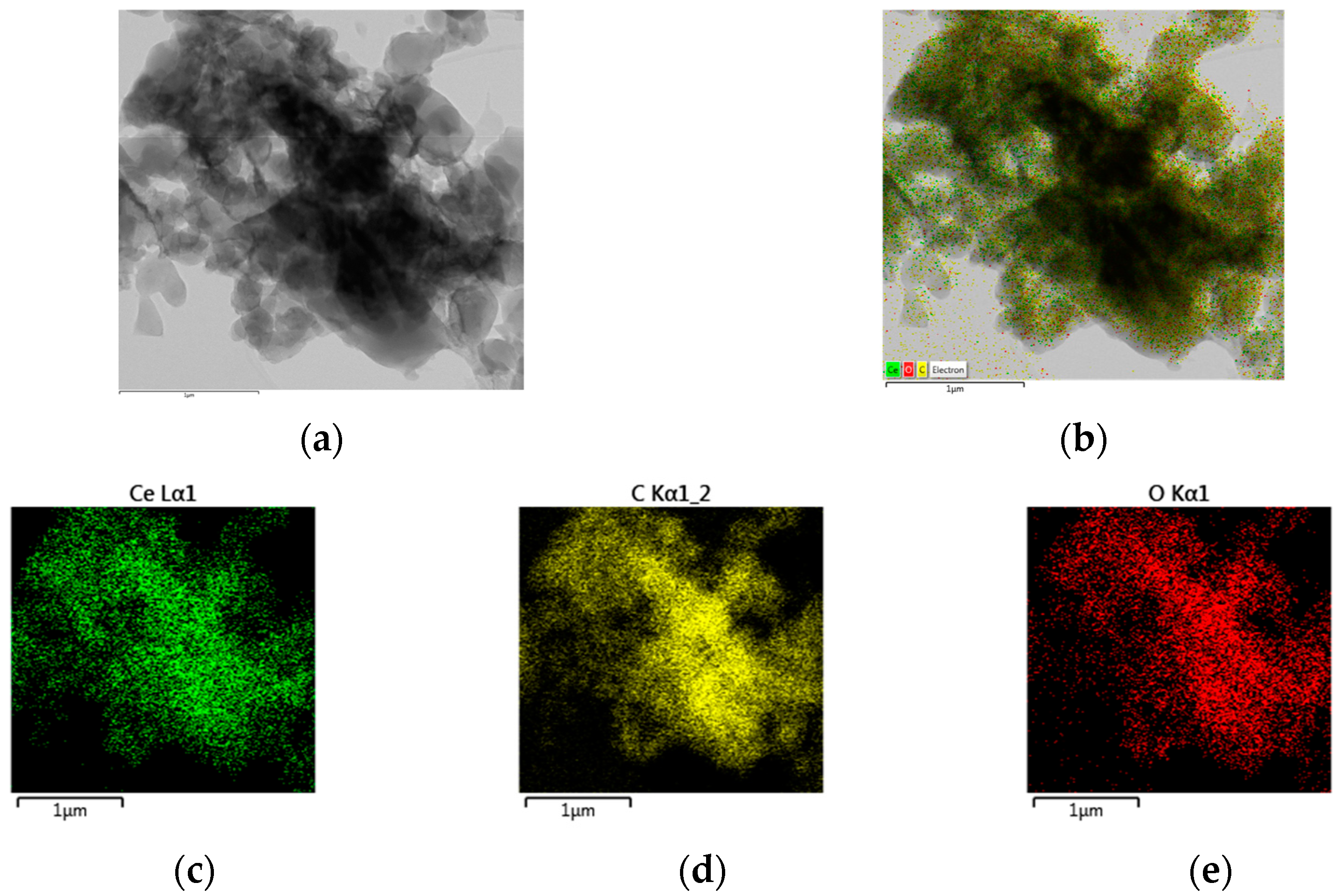
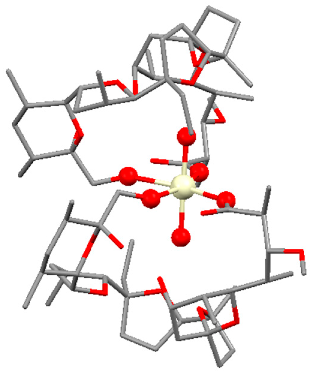
2.1. Characterization of Complexes 1a–b
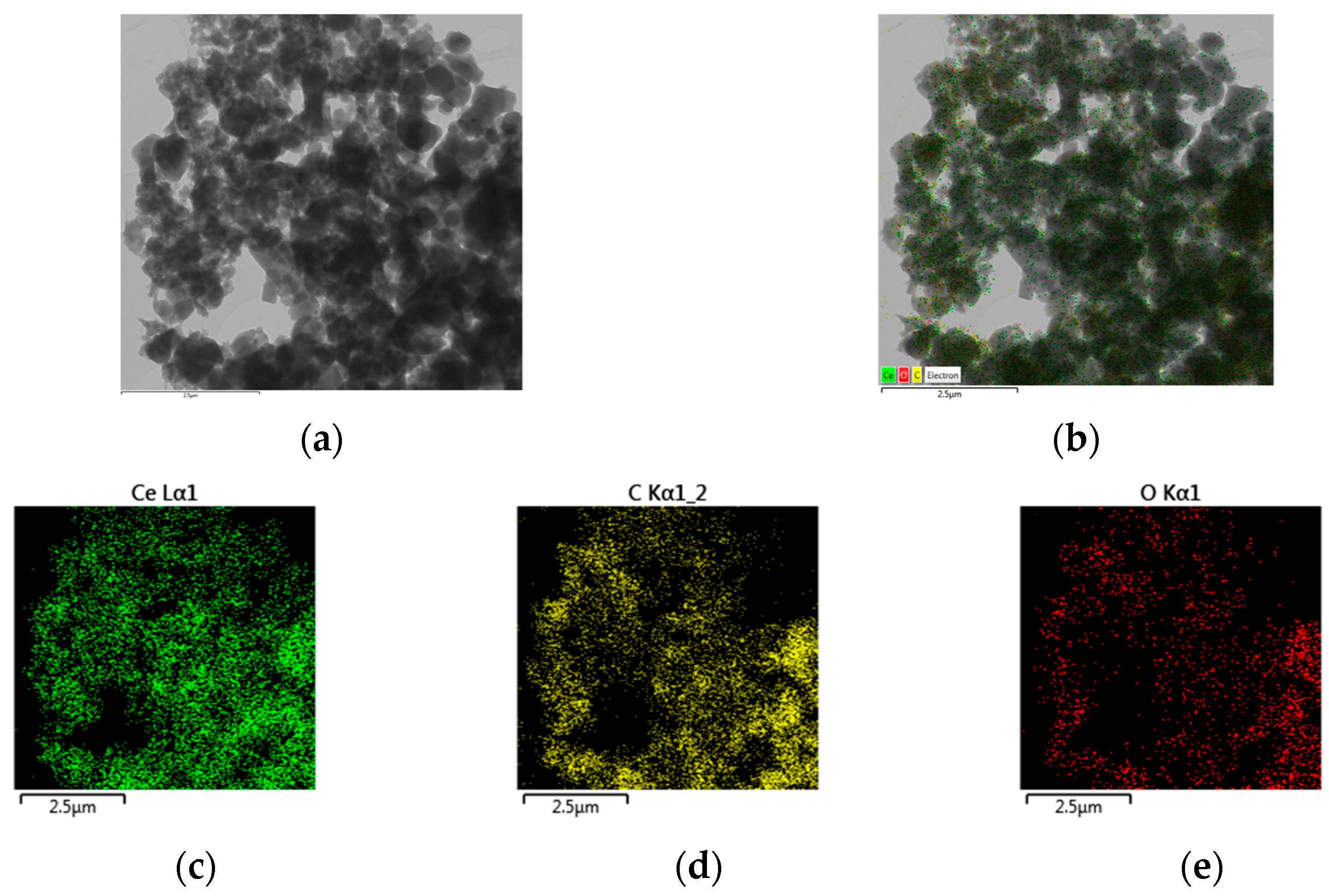
2.2. Characterization of Complexes 2a–b
2.3. Biological Activity of Polyether Ionophores and Their Ce(IV) Coordination Species 1–2
3. Experimental
3.1. Materials
3.2. Physico-Chemical Methods
3.3. Synthesis of Complexes 1–2
3.4. Computational Studies
3.5. Antibacterial Test
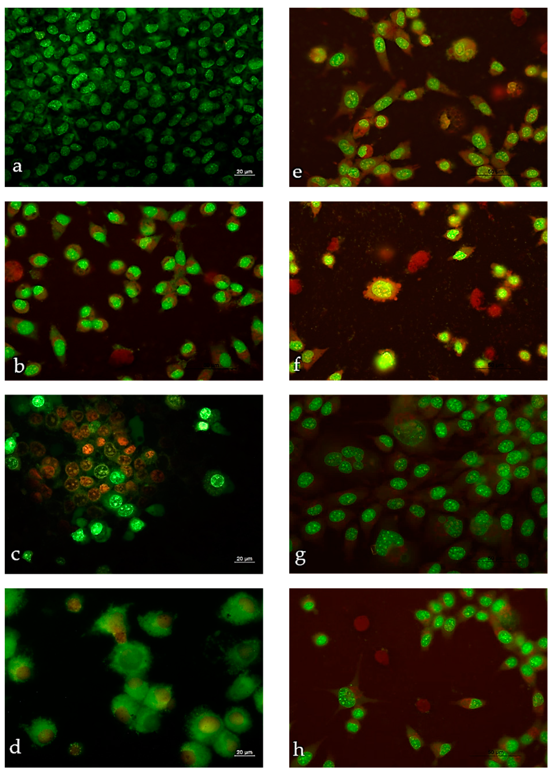
3.6. Cytotoxicity Assays
3.6.1. MTT Test
3.6.2. Double Staining with Acridine Orange (AO) and Propidium Iodide (PI)
3.6.3. Colony Forming Method
3.6.4. Statistical Analysis
4. Conclusions
Supplementary Materials
Author Contributions
Funding
Institutional Review Board Statement
Informed Consent Statement
Data Availability Statement
Conflicts of Interest
Sample Availability
References
- Available online: https://www.who.int/news/item/22-06-2022-22-06-2022-lack-of-innovation-set-to-undermine-antibiotic-performance-and-health-gains (accessed on 16 March 2023).
- Mohammed, H.S.; Tripathi, V.D. Medicinal applications of coordination complexes. J. Phys. Conf. Ser. 2020, 1664, 012070. [Google Scholar] [CrossRef]
- Gasser, G. Metal complexes and cedicine: A successful combination. Chimia 2015, 69, 442–446. [Google Scholar] [CrossRef] [Green Version]
- Habala, L.; Valentová, J. Metal complexes in medicine and pharmacy-the past and the present III. Ceska Slov. Farm. 2020, 69, 121–129. [Google Scholar] [PubMed]
- Jurca, T.; Marian, E.; Vicaş, L.G.; Mureşan, M.E.; Fritea, L. Metal complexes of pharmaceutical substances. In Spectroscopic Analyses-Developments and Applications; Sharmin, E., Zafar, F., Eds.; IntechOpen, Ltd.: London, UK, 2017. [Google Scholar] [CrossRef] [Green Version]
- Dabrowiak, J.C. Metals in Medicine, 1st ed.; John Wiley & Sons, Inc.: Hoboken, NJ, USA, 2017; pp. 285–327. [Google Scholar] [CrossRef]
- Bao, G. Lanthanide complexes for drug delivery and therapeutics. J. Luminesc. 2020, 228, 117622. [Google Scholar] [CrossRef]
- Kontoghiorghes, G.J. Advances on chelation and chelator metal complexes in medicine. Int. J. Mol. Sci. 2020, 21, 2499. [Google Scholar] [CrossRef] [PubMed] [Green Version]
- Kostova, I. Lanthanides as anticancer agents. Curr. Med. Chem. Anticancer Agents 2005, 5, 591–602. [Google Scholar] [CrossRef] [PubMed]
- Trudu, F.; Amato, F.; Vaňhara, P.; Pivetta, T.; Peña-Méndez, M.; Josef Havel, J. Coordination compounds in cancer: Past, present and perspectives. J. Appl. Biomed. 2015, 13, 79–403. [Google Scholar] [CrossRef]
- Teo, R.D.; Termini, J.; Gray, H.B. Lanthanides: Applications in cancer diagnosis and therapy. J. Med. Chem. 2016, 59, 6012–6024. [Google Scholar] [CrossRef] [Green Version]
- Ndagi, U.; Mhlongo, N.; Soliman, M.E. Metal complexes in cancer therapy-an update from drug design perspective. Drug Des. Devel. Ther. 2017, 11, 599–616. [Google Scholar] [CrossRef] [Green Version]
- Singh, A.K.; Kumar, A.; Singh, H.; Sonawane, P.; Pathak, P.; Grishina, M.; Yadav, J.P.; Verma, A.; Kumar, P. Metal Complexes in cancer treatment: Journey so far. Chem. Biodivers. 2023, 20, e202300061. [Google Scholar] [CrossRef]
- Zhou, S.; Wang, F.; Wong, E.T.; Fonkem, E.; Hsieh, T.C.; Wu, J.M.; Wu, E. Salinomycin: A novel anti-cancer agent with known anti-coccidial activities. Curr. Med. Chem. 2013, 20, 4095–4101. [Google Scholar] [CrossRef] [PubMed] [Green Version]
- Dewangan, J.; Srivastava, S.; Rath, S.K. Salinomycin: A new paradigm in cancer therapy. Tumor Biol. 2017, 39, 1010428317695035. [Google Scholar] [CrossRef] [PubMed] [Green Version]
- Wang, X.; Wu, X.; Zhang, Z.; Ma, C.; Wu, T.; Tang, S.; Zeng, Z.; Huang, S.; Gong, C.; Yuan, C.; et al. Monensin inhibits cell proliferation and tumor growth of chemo-resistant pancreatic cancer cells by targeting the EGFR signaling pathway. Sci. Rep. 2018, 8, 17914. [Google Scholar] [CrossRef] [PubMed] [Green Version]
- Jiang, J.; Li, H.; Qaed, E.; Zhang, J.; Song, Y.; Wu, R.; Bu, X.; Wang, Q.; Tang, Z. Salinomycin, as an autophagy modulator—A new avenue to anticancer: A review. J. Exp. Clin. Cancer Res. 2018, 37, 26. [Google Scholar] [CrossRef] [PubMed] [Green Version]
- Verma, S.P.; Das, P. Monensin induces cell death by autophagy and inhibits matrix metalloproteinase 7 (MMP7) in UOK146 renal cell carcinoma cell line. Vitr. Cell. Dev. Biol.-Anim. 2018, 54, 208736–208742. [Google Scholar] [CrossRef]
- Markowska, A.; Kaysiewicz, J.; Markowska, J.; Huczyński, A. Doxycycline, salinomycin, monensin and ivermectin repositioned as cancer drugs. Bioorg. Med. Chem. Lett. 2019, 29, 1549–1554. [Google Scholar] [CrossRef]
- Yusenko, M.V.; Trentmann, A.; Andersson, M.K.; Ghani, L.A.; Jakobs, A.; Arteaga Paz, M.F.; Mikesch, J.H.; Peter von Kries, J.; Stenman, G.; Klempnauer, K.H. Monensin, a novel potent MYB inhibitor, suppresses proliferation of acute myeloid leukemia and adenoid cystic carcinoma cells. Cancer Lett. 2020, 479, 61–70. [Google Scholar] [CrossRef]
- Wang, H.; Zhang, H.; Zhu, Y.; Wu, Z.; Cui, C.; Cai, F. Anticancer mechanisms of salinomycin in breast cancer and Its clinical applications. Front. Oncol. 2021, 11, 654428. [Google Scholar] [CrossRef]
- Yao, S.; Wang, W.; Zhou, B.; Cui, X.; Yang, H.; Zhang, S. Monensin suppresses cell proliferation and invasion in ovarian cancer by enhancing MEK1 SUMOylation. Exp. Ther. Med. 2021, 22, 1390. [Google Scholar] [CrossRef]
- Li, Y.; Sun, Q.; Chen, S.; Yu, X.; Jing, H. Monensin inhibits anaplastic thyroid cancer via disrupting mitochondrial respiration and AMPK/mTOR signaling. Anticancer Agents Med. Chem. 2022, 22, 2539–2547. [Google Scholar] [CrossRef]
- Zhou, Y.; Deng, Y.; Wang, J.; Yan, Z.; Wei, Q.; Ye, J.; Zhang, J.; He, T.-C.; Qiao, M. Effect of antibiotic monensin on cell proliferation and IGF1R signaling pathway in human colorectal cancer cells. Ann. Med. 2023, 55, 954–964. [Google Scholar] [CrossRef] [PubMed]
- Colombatti, M.; Dosio, F. Synthesis of monensin derivatives and their effect on the activity of ricin A-chain immunotoxins. In Immunotoxin Methods and Protocols, Methods in Molecular Biology; Hall, W.A., Ed.; Humana: Totowa, NJ, USA, 2001; Volume 166, pp. 55–70. [Google Scholar] [CrossRef]
- Aowicki, D.; Huczyński, A. Structure and antimicrobial properties of monensin A and its derivatives: Summary of the achievements. BioMed Res. Int. 2013, 2013, 742149. [Google Scholar] [CrossRef] [PubMed] [Green Version]
- Antoszczak, M. A comprehensive review of salinomycin derivatives as potent anticancer and anti-CSCs agents. Eur. J. Med. Chem. 2019, 166, 48–64. [Google Scholar] [CrossRef] [PubMed]
- Antoszczak, M.; Huczyński, A. Salinomycin and its derivatives—A new class of multiple-targeted “magic bullets”. Eur. J. Med. Chem. 2019, 176, 208–227. [Google Scholar] [CrossRef]
- Versini, A.; Colombeau, L.; Hienzsch, A.; Gaillet, C.; Retailleau, P.; Debieu, S.; Müller, S.; Cañeque, T.; Rodriguez, R. Salinomycin derivatives kill breast cancer stem cells by lysosomal iron targeting. Chemistry 2020, 26, 7416–7424. [Google Scholar] [CrossRef]
- Naujokat, C.; Steinhart, R. Salinomycin as a drug for targeting human cancer stem cells. Biomed. Res. Intern. 2012, 2012, 950658. [Google Scholar] [CrossRef]
- Klose, J.; Trefz, S.; Wagner, T.; Steffen, L.; Charrier, A.P.; Radhakrishnan, P.; Volz, C.; Schmidt, T.; Ulrich, A.; Dieter, S.M.; et al. Salinomycin: Anti-tumor activity in a pre-clinical colorectal cancer model. PLoS ONE 2019, 14, e0211916. [Google Scholar] [CrossRef] [Green Version]
- Zhou, J.; Liu, S.; Wang, Y.; Dai, W.; Zou, H.; Wang, S.; Zhang, J.; Pan, J. Salinomycin effectively eliminates cancer stem-like cells and obviates hepatic metastasis in uveal melanoma. Mol. Cancer 2019, 18, 159. [Google Scholar] [CrossRef]
- Naujokat, C.; Laufer, S. Salinomycin, a candidate drug for the elimination of cancer stem cells. In Role of Cancer Stem Cells in Cancer Biology and Therapy, 1st ed.; Dittmar, T., Zänker, K.S., Eds.; CRC Press: Boca Raton, FL, USA, 2013; Chapter 9; pp. 228–257. [Google Scholar] [CrossRef]
- Ivanova, J.; Pantcheva, I.N.; Mitewa, M.; Simova, S.; Tanabe, M.; Osakada, K. Cd(II) and Pb(II) complexes of the polyether ionophorous antibiotic salinomycin. Chem. Centr. J. 2011, 5, 52. [Google Scholar] [CrossRef] [Green Version]
- Ivanova, J.; Pantcheva, I.N.; Zhorova, R.; Momekov, G.; Simova, S.; Stoyanova, R.; Zhecheva, E.; Ivanova, S.; Mitewa, M. Synthesis, spectral properties, antibacterial and antitumor activity of salinomycin complexes with the transition metal ions Co(II), Ni(II), Cu(II) and Zn(II). J. Chem. Chem. Eng. David Publ. 2012, 6, 551–562. [Google Scholar]
- Pantcheva, I.; Petkov, N.; Simova, S.; Zhorova, R.; Dorkov, P. Alkaline-earth metal(II) complexes of salinomycin-spectral properties and antibacterial activity. In Chemical Sciences for New Decade; Ramasami, P., Ed.; De Gruyter: Berlin, Germany; Boston, MA, USA, 2022; Volume 2, pp. 65–78. [Google Scholar] [CrossRef]
- Pashkunova-Martic, I.; Kukeva, R.; Stoyanova, R.; Pantcheva, I.; Dorkov, P.; Friske, J.; Hejl, M.; Jakupec, M.; Hohagen, M.; Legin, A.; et al. Novel salinomycin-based paramagnetic complexes-first evaluation of their potential theranostic properties. Pharmaceutics 2022, 14, 2319. [Google Scholar] [CrossRef] [PubMed]
- Pantcheva, I.N.; Mitewa, M.I.; Sheldrick, W.S.; Oppel, I.M.; Zhorova, R.; Dorkov, P. First divalent metal complexes of the polyether ionophore monensin A: X-ray structures of [Co(Mon)2(H2O)2] and [Mn(Mon)2(H2O)2] and their properties. Curr. Drug Discov. Technol. 2008, 5, 154–161. [Google Scholar] [CrossRef]
- Pantcheva, I.N.; Zhorova, R.; Mitewa, M.; Simova, S.; Mayer-Figge, H.; Sheldrick, W.S. First solid state alkaline-earth complexes of monensic A acid: X-ray crystal structure of [M(Mon)2(H2O)2] (M = Mg, Ca), spectral properties and cytotoxicity against Gram-positive bacteria. BioMetals 2010, 23, 59–70. [Google Scholar] [CrossRef]
- Pantcheva, I.N.; Ivanova, J.; Zhorova, R.; Mitewa, M.; Simova, S.; Mayer-Figge, H.; Sheldrick, W.S. Nickel(II) and zinc(II) dimonensinates: Crystal structure, spectral properties and bactericidal activity. Inorg. Chim. Acta 2010, 363, 1879–1886. [Google Scholar] [CrossRef]
- Pantcheva, I.; Dimitrova, R.; Nedzhib, A.; Dorkov, P.; Stoyanova, R.; Zhecheva, E. Coordination compounds of polyether ionophore monensin with gadolinium(III) ions. In Nanoscience & Nanotechnology; Balabanova, E., Mileva, E., Eds.; BPS Ltd.: Sofia, Bulgaria, 2019; Volume 19, pp. 40–46. [Google Scholar]
- Pantcheva, I.; Dimitrova, R.; Ivanova, V.; Nedzhib, A.; Dorkov, P.; Dinev, D.; Spasov, R.; Alexandrova, R. Spectral properties and biological activity of La(III) and Nd(III) monensinates. Open Chem. 2019, 17, 1423–1434. [Google Scholar] [CrossRef]
- Alexandrova, R.I.; Zhivkova, T.; Pantcheva, I.N.; Mitewa, M.I. Cytotoxic and antiproliferative activities of monensic acid and its metal(II) complexes against drug sensitive and multidrug resistant human tumor cell lines. Intern. J. Biol. Biomed. Eng. 2011, 5, 93–101. [Google Scholar]
- Alexandrova, R.I.; Zhivkova, T.; Alexandrov, M.; Miloshev, G.; Georgieva, M.; Pantcheva, I.N.; Mitewa, M.I. Cytostatic and cytotoxic properties of monensic acid and its biometal(II) complexes against human tumor/non-tumor cell lines. Cent. Eur. J. Chem. 2012, 10, 1464–1474. [Google Scholar] [CrossRef]
- Pantcheva, I.N.; Alexandrova, R.I.; Zhivkova, T.; Mitewa, M.I. In vitro activity of biometal(II) complexes of monensin against virus-induced transplantable animal tumors. Biotechn. Biotechn. Equipmn. 2013, 27, 3703–3708. [Google Scholar] [CrossRef]
- Zhorova, R.; Marina, M.; Radeva, A.; Mitewa, M.I.; Pantcheva, I.N. Cytotoxicity of monensic acid and its biometal(II) complexes against anaerobic bacterial strain Clostridium perfringens spp. Biotechn. Biotechn. Equipmn. 2013, 27, 4308–4310. [Google Scholar] [CrossRef]
- Alexandrova, R.I.; Zhivkova, T.D.; Ivanov, D.; Andonova-Lilova, B.D.; Dyakova, L.V.; Pantcheva, I.N.; Dorkov, P. Study on in vitro toxicity of biometal(II) monensinates against rat Zajdela liver tumour. Chem. Didact. Ecol. Metrol. 2020, 25, 125–132. [Google Scholar] [CrossRef]
- Wason, M.S.; Zhao, J. Cerium oxide nanoparticles: Potential applications for cancer and other diseases. Am. J. Transl. Res. 2013, 5, 126–131. [Google Scholar] [PubMed]
- Gao, Y.; Chen, K.; Ma, J.L.; Gao, F. Cerium oxide nanoparticles in cancer. OncoTargets Ther. 2014, 7, 835–840. [Google Scholar] [CrossRef] [PubMed] [Green Version]
- Rajeshkumar, S.; Naik, P. Synthesis and biomedical applications of cerium oxide nanoparticles—A review. Biotechnol. Rep. 2017, 17, 1–5. [Google Scholar] [CrossRef] [PubMed]
- Datta, A.; Mishra, S.; Manna, K.; Saha, K.D.; Mukherjee, S.; Roy, S. Pro-oxidant therapeutic activities of cerium oxide nanoparticles in colorectal carcinoma cells. ACS Omega 2020, 5, 9714–9723. [Google Scholar] [CrossRef] [PubMed] [Green Version]
- Asgharzadeh, F.; Hashemzadeh, A.; Rahmani, F.; Yaghoubi, A.; Nazari, S.E.; Avan, A.; Mehr, S.M.H.; Soleimanpour, S.; Khazaei, M. Cerium oxide nanoparticles acts as a novel therapeutic agent for ulcerative colitis through anti-oxidative mechanism. Life Sci. 2021, 278, 119500. [Google Scholar] [CrossRef]
- Piro, N.A.; Robinson, J.R.; Walsh, P.J.; Schelter, E.J. The electrochemical behavior of cerium(III/IV) complexes: Thermodynamics, kinetics and applications in synthesis. Coord. Chem. Rev. 2014, 260, 21–36. [Google Scholar] [CrossRef]
- So, Y.-M.; Leung, W.-H. Recent advances in the coordination chemistry of cerium(IV) complexes. Coord. Chem. Rev. 2017, 340, 172–197. [Google Scholar] [CrossRef]
- Chen, Z.F.; Wei, J.H.; Liu, Y.C.; Liu, M.; Gu, Y.O.; Huang, K.B.; Wang, M.; Liang, H. High antitumor activity of 5,7-dihalo-8-quinolinolato cerium complexes. Eur. J. Med. Chem. 2013, 68, 454–462. [Google Scholar] [CrossRef]
- Ravi, M.; Chennam, K.P.; Ushaiah, B.; Eslavath, R.K.; Perugu, S.; Ajumeera, R.; Devi, C.S. A study on spectroanalytical aspects, DNA-interaction, photo-cleavage, radical scavenging, cytotoxic activities, antibacterial and docking properties of 3-(1-(6-methoxybenzo[d] thiazol-2-ylimino) ethyl)-6-methyl-3H-pyran-2, 4-dione and its metal complexes. J. Fluoresc. 2015, 25, 1279–1296. [Google Scholar] [CrossRef]
- Zohrevandi, M.; Abdolmaleki, S.; Ghadermazi, M.; Gholiee, Y.; Aliabadi, A.; Motieiyan, E.; Hakimi, M.; Marabello, D. Synthesis, characterization, crystallographic structure, theoretical studies, and in vitro cytotoxicity assessment of two Gd(III) and Ce(IV) complexes containing pyridine-2,6-dicarboxylate. Polyhedron 2021, 211, 115561. [Google Scholar] [CrossRef]
- Nakamoto, K. Infrared and Raman Spectra of Inorganic and Coordination Compounds: Part A: Theory and Applications in Inorganic Chemistry, 6th ed.; John Wiley & Sons, Inc.: Hoboken, NJ, USA, 2008; pp. 149–354. [Google Scholar]
- Trubetskaya, A.; Jensen, P.A.; Jensen, A.D.; Glarborg, P.; Larsen, F.H.; Andersen, M.L. Characterization of free radicals by electron spin resonance spectroscopy in biochars from pyrolysis at high heating rates and athigh temperatures. Biomass Bioenergy 2016, 94, 117–129. [Google Scholar] [CrossRef] [Green Version]
- Murphy, D.M.; Chiesa, M. EPR of paramagnetic centers on solid surfaces. In Electron Paramagnetic Resonance; Gilbert, B.C., Davies, M.J., Murphy, D.M., Eds.; RSC Publishing: Cambridge, UK, 2008; Volume 21, pp. 105–130. [Google Scholar]
- Dorkov, P.; Pantcheva, I.N.; Sheldrick, W.S.; Mayer-Figge, H.; Petrova, R.; Mitewa, M. Synthesis, structure and antimicrobial activity of manganese(II) and cobalt(II) complexes of the polyether ionophore antibiotic sodium monensin A. J. Inorg. Biochem. 2008, 102, 26–32. [Google Scholar] [CrossRef] [PubMed]
- Dinev, D.; Popova, K.B.; Zhivkova, T.; Dyakova, L.; Abudalleh, A.; Alexandrova, R.; Culita, D.C.; Mocanu, T.; Maxim, C.; Marinescu, G. Synthesis, structural characterization, and cytotoxic activity in tumor cells of Cu(II) and Co(II) complexes with o-vanillin amino acids Schiff bases. Appl. Organomet. Chem. 2022, 36, e6862. [Google Scholar] [CrossRef]
- Qi, D.; Liu, Y.; Li, J.; Huang, J.H.; Hu, X.; Wu, E. Salinomycin as a potent anticancer stem cell agent: State of the art and future directions. Med. Res. Rev. 2022, 42, 1037–1063. [Google Scholar] [CrossRef]
- Pfeffer, C.M.; Singh, A.T.K. Apoptosis: A target for anticancer therapy. Int. J. Mol. Sci. 2018, 19, 448. [Google Scholar] [CrossRef] [Green Version]
- Carneiro, B.A.; El-Deiry, W.S. Targeting apoptosis in cancer therapy. Nat. Rev. Clin. Oncol. 2020, 17, 395–417. [Google Scholar] [CrossRef]
- Smalley, K.S.; Lioni, M.; Herlyn, M. Life isn’t flat: Taking cancer biology to the next dimension. Vitr. Cell Dev. Biol. Anim. 2006, 42, 242–247. [Google Scholar] [CrossRef]
- Lee, J.; Lilly, G.D.; Doty, R.C.; Podsiadlo, P.; Kotov, N.A. In vitro toxicity testing of nanoparticles in 3D cell culture. Small 2009, 5, 1213–1221. [Google Scholar] [CrossRef] [Green Version]
- Gertenbach, P.G.; Popov, A.I. Solution chemistry of monensin and its alkali-metal ion complexes-potentiometric and spectroscopic studies. J. Am. Chem. Soc. 1975, 97, 4738–4744. [Google Scholar] [CrossRef]
- Becke, A.D. Density-functional exchange-energy approximation with correct asymptotic behavior. Phys. Rev. A At. Mol. Opt. Phys. 1988, 38, 3098–3100. [Google Scholar] [CrossRef]
- Becke, A.D. Density functional thermochemistry. III. The role of exact exchange. J. Chem. Phys. 1993, 98, 5648–5652. [Google Scholar] [CrossRef] [Green Version]
- Lee, C.; Yang, W.; Parr, R.G. Development of the Colle-Salvetti correlation-energy formula into a functional of the electron density. Phys. Rev. B 1988, 37, 785–789. [Google Scholar] [CrossRef] [PubMed] [Green Version]
- Miehlich, B.; Savin, A.; Stoll, H.; Preuss, H. Results obtained with the correlation energy density functionals of Becke and Lee, Yang and Parr. Chem. Phys. Lett. 1989, 157, 200–206. [Google Scholar] [CrossRef]
- Grimme, S.; Antony, J.; Ehrlich, S.; Krieg, H. A consistent and accurate ab initio parameterization of density functional dispersion correction (DFT-D) for the 94 elements H-Pu. J. Chem. Phys. 2010, 132, 154104. [Google Scholar] [CrossRef] [PubMed] [Green Version]
- Pitchard, B.P.; Altarawy, D.; Didier, B.; Gibson, T.D.; Windus, T.L. A new basis set exchange: An open, up-to-date resource for the molecular sciences community. J. Chem. Inf. Model. 2019, 59, 4814–4820. [Google Scholar] [CrossRef]
- Hay, P.J.; Martin, R.L.; Uddin, J.; Scuseria, G.E. Theoretical study of CeO2 and Ce2O3 using a screened hybrid density functional. J. Chem. Phys. 2006, 125, 034712. [Google Scholar] [CrossRef]
- NIST Standard Reference Database Number 101; NIST Computational Chemistry Comparison and Benchmark Database. The National Institute of Standards and Technology (NIST): Gaithersburg, MD, USA, 2022. [CrossRef]
- Iliev, S.; Gocheva, G.; Ivanova, N.; Atanasova, B.; Petrova, J.; Madjarova, G.; Ivanova, A. Identification and computational characterization of isomers with cis and trans amide bonds in folate and its analogues. Phys. Chem. Chem. Phys. 2018, 20, 28818–28831. [Google Scholar] [CrossRef]
- Jarvis, R.A.; Patrick, E.A. Clustering using a similarity measure based on shared near neighbors. IEEE Trans. Comput. 1973, C-22, 1025–1034. [Google Scholar] [CrossRef]
- Bayly, C.I.; Cieplak, P.; Cornell, W.; Kollman, P.A. A well-behaved electrostatic potential based method using charge restraints for deriving atomic charges: The RESP model. J. Chem. Phys. 1993, 97, 10269–10280. [Google Scholar] [CrossRef]
- Cornell, W.D.; Cieplak, P.; Bayly, C.I.; Kollman, P.A. Application of RESP charges to calculate conformational energies, hydrogen bond energies, and free energies of solvation. J. Am. Chem. Soc. 1993, 115, 9620–9631. [Google Scholar] [CrossRef]
- Cieplak, P.; Cornell, W.D.; Bayly, C.; Kollman, P.A. Application of the multimolecule and multiconformational RESP methodology to biopolymers: Charge derivation for DNA, RNA, and proteins. J. Comput. Chem. 1995, 16, 1357–1377. [Google Scholar] [CrossRef]
- Lutz, W.K.; Winkler, F.K.; Dunitz, J.D. Crystal structure of the antibiotic monensin. Similarities and differences between free acid and metal complex. Helv. Chim. Acta 1971, 54, 1103–1108. [Google Scholar] [CrossRef] [PubMed]
- Paulus, E.F.; Kurz, M.; Matter, H.; Vertesy, L. Solid-state and solution structure of the salinomycin-sodium complex: Stabilization of different conformers for an ionophore in different environments. J. Am. Chem. Soc. 1998, 120, 8209–8221. [Google Scholar] [CrossRef]
- van Veggel, F.C.J.M.; Reinhoudt, D.N. New, Accurate Lennard-Jones parameters for trivalent lanthanide ions, tested on [18]Crown-6. Chem. Eur. J. 1999, 5, 90–95. [Google Scholar] [CrossRef]
- Jorgensen, W.L.; Tirado-Rives, J. Potential energy functions for atomic-level simulations of water and organic and biomolecular systems. J. Proc. Natl. Acad. Sci. USA 2005, 102, 6665–6670. [Google Scholar] [CrossRef] [Green Version]
- Caleman, C.; van Maaren, P.J.; Hong, M.; Hub, J.S.; Costa, L.T.; van der Spoel, D. Force field benchmark of organic liquids: Density, enthalpy of vaporization, heat Ccapacities, surface tension, isothermal compressibility, volumetric expansion coefficient, and dielectric constant. J. Chem. Theor. Comput. 2012, 8, 61–74. [Google Scholar] [CrossRef]
- Jorgensen, W.L.; Maxwell, D.S.; Tirado-Rives, J. Development and testing of the OPLS all-atom force field on conformational energetics and properties of organic liquids. J. Am. Chem. Soc. 1996, 118, 11225–11236. [Google Scholar] [CrossRef]
- Berendsen, H.J.C.; Postma, J.P.M.; van Gunsteren, W.F.; Di Nola, A.; Haak, J.R. Molecular dynamics with coupling to an external bath. J. Chem. Phys. 1984, 81, 3684–3690. [Google Scholar] [CrossRef] [Green Version]
- Hypercube. HyperChem 7.0; Hypercube: Gainesville, FL, USA, 2001. [Google Scholar]
- Abraham, M.J.; Murtola, T.; Schulz, R.; Páll, S.; Smith, J.C.; Hess, B.; Lindahl, E. GROMACS: High performance molecular simulations through multi-level parallelism from laptops to supercomputers. Software X 2015, 1–2, 19–25. [Google Scholar] [CrossRef] [Green Version]
- Case, D.A.; Darden, T.A.; Cheatham, T.E., III; Simmerling, C.L.; Wang, J.; Duke, R.E.; Luo, R.; Merz, K.M.; Wang, B.; Pearlman, D.A.; et al. AMBER 8; University of California: San Francisco, CA, USA, 2004. [Google Scholar]
- Frisch, M.J.; Trucks, G.W.; Schlegel, H.B.; Scuseria, G.E.; Robb, M.A.; Cheeseman, J.R.; Scalmani, G.; Barone, V.; Petersson, G.A.; Nakatsuji, H.; et al. Gaussian 16, Revision, C.01; Gaussian, Inc.: Wallingford, CT, USA, 2016. [Google Scholar]
- Andrews, J.M. Determination of minimum inhibitory concentrations. J. Antimicrob. Chemother. 2001, 48 (Suppl. S1), 5–16. [Google Scholar] [CrossRef] [Green Version]
- Mosmann, T. Rapid colorimetric assay for cellular growth and survival: Application to proliferation and cytotoxicity assays. J. Immunol. Methods 1983, 65, 55–63. [Google Scholar] [CrossRef] [PubMed]
- Wahab, S.I.A.; Abdul, A.B.; Alzubairi, A.S.; Elhassan, M.M.; Mohan, S. In vitro ultramorphological assessment of apoptosis induced by zerumbone on (HeLa). J. Biomed. Biotechmol. 2009, 2009, 769568. [Google Scholar] [CrossRef] [Green Version]


| Vibration | MonH × H2O | 1a | 2a | |
|---|---|---|---|---|
| KBr | CHCl3 | KBr/CHCl3 | KBr/CHCl3 | |
| , H2O | 3531 | 3518 | - | - |
| , H2O | 3461 | 3467 | - | - |
| , OH | 3336 | 3293 | 3454 | 3454 |
| , COOH | 1709 | 1707 | - | - |
| , COO− | - | - | 1552 (1571, 1564) | 1505 (1498) |
| , COO− | - | - | 1414 (1385) | 1414 (1419) |
| δHOH, H2O | 1627 | 1621 | - | - |
| , NO3− | - | - | - | 1555 (1586, 1521) |
| , NO3− | - | - | - | 1289 (1282, 1222) |
| νMO | - | - | 550 | 500 |
| δONO | - | - | - | 378 |
| Vibration | SalH | 1b | 2b |
|---|---|---|---|
| KBr/CHCl3 | KBr/CHCl3 | KBr/CHCl3 | |
| , OH | 3506/3497 | 3456 | 3452 |
| νC=O | 1710 | 1710 | 1710 (1687) |
| , COOH | 1710 | - | - |
| , COO− | - | 1552 | 1512 (1484) |
| , COO− | - | 1411 | 1411 (1423) |
| νC=C | 1638 | 1632 | 1637 |
| , NO3− | - | - | 1552 (1579, 1551) |
| , NO3− | - | - | 1288 (1253) |
| νMO | - | 530 | 540 |
| δONO | - | - | 380 |
| M-O Bond | Populated Conformers of Complex 2a | M-O Bond | Populated Conformers of Complex 2b | ||
|---|---|---|---|---|---|
| 39.3% | 25.9% | 47.7% | 32.7% | ||
| O1-Ce | 2.42 | 2.40 | O1-Ce | 2.40 | 2.40 |
| O2-Ce | 2.54 | 2.45 | O2-Ce | 2.42 | 2.40 |
| O8-Ce | 2.55 | 2.68 | O4-Ce | 2.67 | 2.64 |
| O9-Ce | 2.61 | 2.76 | O5-Ce | 2.58 | 2.55 |
| O11-Ce | 2.46 | 2.45 | OH−-Ce | 2.04 | 2.08 |
| OH−-Ce | 2.05 | 2.07 | η2-NO3−-Ce | 2.44 2.50 | 2.49 2.49 |
| NO3−-Ce | 2.48 | 2.45 | |||
| η2-NO3−-Ce | 2.40 2.52 | 2.45 2.48 | η2-NO3−-Ce | 2.46 2.55 | 2.46 2.53 |
| Compound | K. rhizophila ATCC 9341 | B. subtilis ATCC 6633 | B. cereus ATCC 11778 | |||
|---|---|---|---|---|---|---|
| µg/mL | µM | µg/mL | µM | µg/mL | µM | |
| MonH × H2O | 125 | 181.4 | 15.63 | 22.7 | 3.91 | 5.7 |
| 1a, [Ce(Mon)2(OH)2] | 500 | 326.4 | 15.63 | 10.2 | 7.81 | 5.1 |
| 2a, [Ce(Mon)(η2-NO3)(NO3)(OH)] | 500 | 525.8 | 62.5 | 65.7 | 15.63 | 16.4 |
| SalH | 62.5 | 83.2 | 31.25 | 41.6 | 7.81 | 10.4 |
| 1b, [Ce(Sal)2(OH)2] | 62.5 | 37.3 | 31.25 | 18.7 | 7.81 | 4.7 |
| 2b, [Ce(Sal)(η2-NO3)2(OH)] | 62.5 | 60.6 | 31.25 | 30.3 | 7.81 | 7.6 |
| (NH4)2[Ce(NO3)6] | >1000 | >1824 | >1000 | >1824 | >1000 | >1824 |
| Compound | MM, g/mol | HeLa | Lep-3 | SI | ||
|---|---|---|---|---|---|---|
| CC50, µg/mL | CC50, µM | CC50, µg/mL | CC50, µM | |||
| MonH × H2O | 688.87 | 0.52 | 0.75 | 3.56 | 5.17 | 6.89 |
| 2a, [Ce(Mon)(η2-NO3)(NO3)OH] | 951.00 | <0.50 | <0.53 | 6.00 | 6.31 | >12.62 |
| SalH | 751.01 | 1.43 | 1.90 | >10 | >13.32 | >7.02 |
| 2b, [Ce(Sal)(η2-NO3)2OH] | 1031.13 | 2.81 | 2.73 | >10 | >9.71 | >3.56 |
| cisplatin [62] | 300.05 | 8.46 | 28 | 0.49 | 1.63 | 0.058 |
| oxaliplatin [62] | 397.29 | 6.12 | 15 | 0.95 | 2.39 | 0.159 |
| epirubicin [62] | 543.53 | 17.38 | 32 | 0.74 | 1.36 | 0.043 |
Disclaimer/Publisher’s Note: The statements, opinions and data contained in all publications are solely those of the individual author(s) and contributor(s) and not of MDPI and/or the editor(s). MDPI and/or the editor(s) disclaim responsibility for any injury to people or property resulting from any ideas, methods, instructions or products referred to in the content. |
© 2023 by the authors. Licensee MDPI, Basel, Switzerland. This article is an open access article distributed under the terms and conditions of the Creative Commons Attribution (CC BY) license (https://creativecommons.org/licenses/by/4.0/).
Share and Cite
Petkov, N.; Pantcheva, I.; Ivanova, A.; Stoyanova, R.; Kukeva, R.; Alexandrova, R.; Abudalleh, A.; Dorkov, P. Novel Cerium(IV) Coordination Compounds of Monensin and Salinomycin. Molecules 2023, 28, 4676. https://doi.org/10.3390/molecules28124676
Petkov N, Pantcheva I, Ivanova A, Stoyanova R, Kukeva R, Alexandrova R, Abudalleh A, Dorkov P. Novel Cerium(IV) Coordination Compounds of Monensin and Salinomycin. Molecules. 2023; 28(12):4676. https://doi.org/10.3390/molecules28124676
Chicago/Turabian StylePetkov, Nikolay, Ivayla Pantcheva, Anela Ivanova, Radostina Stoyanova, Rositsa Kukeva, Radostina Alexandrova, Abedullkader Abudalleh, and Petar Dorkov. 2023. "Novel Cerium(IV) Coordination Compounds of Monensin and Salinomycin" Molecules 28, no. 12: 4676. https://doi.org/10.3390/molecules28124676