Article

Ultrastructural, Energy-Dispersive X-ray Spectroscopy, Chemical Study and LC-DAD-QToF Chemical Characterization of Cetraria islandica (L.) Ach

1
National Center for Natural Products Research, School of Pharmacy, The University of Mississippi, University, MS 38677, USA
2
S.D. Asfendiyarov Kazakh National Medical University, School of Pharmacy, Almaty 050012, Kazakhstan
3
Department of Pharmacognosy, Faculty of Pharmacy, Minia University, Minia 61519, Egypt
4
Division of Pharmacognosy, Department of BioMolecular Sciences, School of Pharmacy, The University of Mississippi, University, MS 38677, USA
*
Author to whom correspondence should be addressed.
Molecules 2023, 28(11), 4493; https://doi.org/10.3390/molecules28114493
Submission received: 4 May 2023 / Revised: 27 May 2023 / Accepted: 29 May 2023 / Published: 1 June 2023

Abstract

:
The lichen Cetraria islandica (L.) Ach. has been used in traditional and modern medicines for its many biological properties such as immunological, immunomodulating, antioxidant, antimicrobial, and anti-inflammatory activities. This species is gaining popularity in the market, with interest from many industries for selling as medicines, dietary supplements, and daily herbal drinks. This study profiled the morpho-anatomical features by light, fluorescence, and scanning electron microscopy; conducted an elemental analysis using energy-dispersive X-ray spectroscopy; and phytochemical analysis was performed using high-resolution mass spectrometry combined with a liquid chromatography system (LC-DAD-QToF) of C. islandica. In total, 37 compounds were identified and characterized based on comparisons with the literature data, retention times, and their mass fragmentation mechanism/s. The identified compounds were classified under five different classes, i.e., depsidones, depsides, dibenzofurans, aliphatic acids, and others that contain simple organic acids in majority. Two major compounds (fumaroprotocetraric acid and cetraric acid) were identified in the aqueous ethanolic and ethanolic extracts of C. islandica lichen. This detailed morpho-anatomical, EDS spectroscopy, and the developed LC-DAD-QToF approach for C. islandica will be important for correct species identification and can serve as a useful tool for taxonomical validation and chemical characterization. Additionally, chemical study of the extract of C. islandica led to isolation and structural elucidation of nine compounds, namely cetraric acid (1), 9′-(O-methyl)protocetraric acid (2), usnic acid (3), ergosterol peroxide (4), oleic acid (5), palmitic acid (6), stearic acid (7), sucrose (8), and arabinitol (9).

1. Introduction

Lichens are complex organisms composed of fungus, the mycobiont, and one or more photosynthetic algae (green alga or cyanobacteria). The photobiont lives in a symbiotic relationship, occurring in temperate habitats to the most extreme environmental conditions in the arctic and subarctic areas of Europe and North America [1,2,3]. Recently, studies showed that the lichens live in more complex structures, such as tripartite lichens or those with more partners [4]. This organism was considered as lichenized fungi based on differentiated characteristics, such as superficial thallus, slow growth rate, and longevity of thalli and fruit bodies [5]. Over 20,000 species of lichens are known worldwide [6], and it is estimated that around 1000 of these known species are from central Asia, mainly including Kazakhstan [7]. The flora of Kazakhstan is known for its rich diversity of all kinds of plant species, including lichens. The sample Cetraria islandica (L.) Ach. (Family: Parmeliaceae) was collected from the Karaganda region, Karkaraly National Park, located at 1200 m above sea level. The climate of this area is moderately continental, the average annual air temperature is 2 °C, in winter −15 °C, and in summer 20 °C. The global distribution of this species is reported in the west and north of Iceland, northern Wales, England, Scotland, Southwestern Ireland, and other parts of the world [8]. This species is commonly known as true Iceland lichen or Iceland moss. C. islandica is used in treating and prophylaxis of chronic obstructive pulmonary disease (COPD), mainly tuberculosis [9]. C. islandica is also used in the food industry as an additive in sweet dishes [10], and traditionally it is used for preparing herbal tea and homemade decoctions for treating dry cough, a temporary loss of appetite, and oral mucosal problems [11]. The scientific recommendation made by the Committee on Herbal Medicinal Products (HMPC) states that C. islandica can be used in the preparations of demulcent (smoothing agents) for mouth and throat irritation and the associated dry cough, and to treat the temporary loss of appetite only in adults, adolescents, and children over 6 years and above [12]. The micromorphology and chemical variation in C. islandica and C. ericetorum were studied earlier [13]. The reported chemical compounds of C. islandica are chitin; lichenin; isolichenin; sucrose; mannitol galactomannan; lichen acids: fumarprotocetraric acid, cetraric acid, fumaric acid, citric acid, usnic acid, and others; and minerals: calcium, sodium, iron, bromine, etc. [9,14]. Earlier pharmacological studies using crude extracts of C. islandica have shown immunological, immunomodulating, and bacteriostatic properties. The lichenin-types β-(1-3) and -(1-4)-glucan (lichenan) are the only β glucans found in Parmeliaceae proven to have antiviral activity [14,15]. The lichen acids, such as fumaroprotocetraric acid (2.6–11.5%), protocetraric acid (0.2–0.3%), protolichesterinic acid (0.1–1.5%), and usnic acid (0.04% of trace amount) were reported in C. islandica [16,17,18,19,20]. Fumaroprotocetraric acid has shown potential neuroprotective properties [21] and usnic acid was proven to act as an antibiotic. The importance of this lichen is very high in traditional and modern therapeutic usage, especially as dietary supplements and pharmaceutical products. Thus, this manuscript emphasizes profiling the macro and microscopic characters for authentication purposes.
Apart from morphological studies, spot tests on the thallus/medulla and the application of chromatography followed by high-resolution mass-spectrometry-based methods or direct spectrometric methods are useful in the chemotaxonomic identification of the species along with dereplication strategies and secondary metabolite characterization [22,23,24]. Exploration of the secondary metabolites—including metabolites in minor composition—using high-resolution mass-spectrometry-based methods leads to novel molecule isolation [25]. This current method for C. islandica analysis employs different solvent extractions to understand the extraction efficiency of the solvents in relation to the number of secondary metabolites. In addition, this paper will aim to establish the fragmentation mechanisms of major secondary metabolites in order to understand their class of chemistry and fragmentation pathways for dereplication strategies. As a result, isolated major chemical constituents are presented in this study.

2. Results

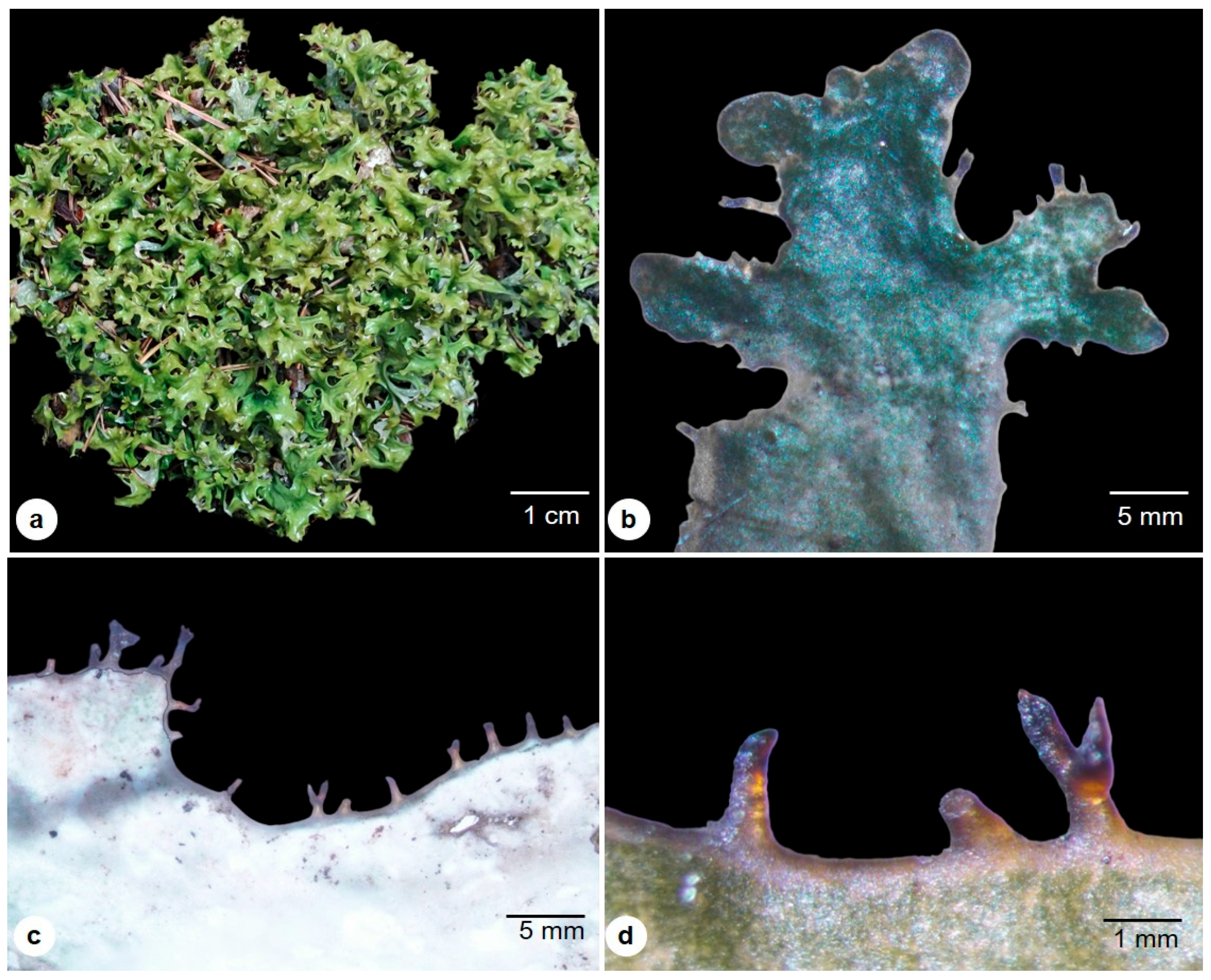
2.1. Macro-Morphological Description

This fruticose lichen is small to medium, with a heteromerous foliose thallus of 12–15 cm tall, upright, growing loose, irregular in structure, and attached to the substrate by the help of short filamentous rhizoids or its aggregate hyphae of the lower cortical layer of the thallus (Figure 1a). The thallus is glabrous, flat, or grooved, with brittle bands on blades of the thallus that are dichotomously branched, narrow at the base, curled ribbon-like at the top, with a width of 0.5 to 2 cm and less than 1 to 1.5 mm in diameter (Figure 1b,c). The fresh thallus is soft and leathery, pale white to silver on the lower surface and dark shiny green on the upper surface. In maturity, it turns dark greenish-brown on the upper surface and pale white to light gray on the lower surface (Figure 1c). The thallus is folded, wavy, and pitted in texture. Pycnidia are dark brown, ciliated at the margin, and uncommonly on the base of the branches (Figure 1d).

2.2. Chemical Spot-Test Analysis

The spot test suggests the presence of lichen acids, such as fumaroprotocetraric acid and protocetraric acid, by the K (potassium in the form of KOH) & I (Lugol’s solution) tests [16,18,20]. The K test gives the reddish-brown color changes in the upper and lower surface of the thallus, and the Iodine turns the thallus to bluish-black (Figure 2). These color changes are characteristic features of C. islandica due to its lichen acids.

2.3. Micro-Morphological Description

The thallus is heteromeric, dorsoventral, and composed of dead, collapsed, gelatinized hyphae on both surfaces and the middle photobiont layer. The cortical upper region shows the surface opening, where the cortical hyphae emerge and help in gas exchanges (Figure 3a). The lower surface is uneven and grooved in many places, phycobiont and mycobiont hyphae are visible, and they are gonidial and cortical as well (Figure 3b). At the lower thallus, hyphae extend through the hydrophilic cortex layer and form a patch of pseudocyphella, showing many respiratory macules for air-breathing (Figure 3c); the ascomata are absent. The margin of the thallus with regular lobes of teeth-like structures, called pycnidia, is an asexual fruiting body. The transverse section is composed of an outer epicortex, 0.5–1.5 μm thick, and covered with biofilm (generally bacterial) (Figure 3d). The upper cortex, 30.05–50.6 μm wide, is formed into tight collectively packed cortical cells with fungal hyphae (Figure 3d,e). The medullary region measures 70–100 μm wide and consists of loosely interwoven mycobiont hyphae that run horizontally and provide large airspaces for the phycobiont. The algal cell, 3.25–12.5 μm in diameter, forms a bi-stratified structure. These phycobiont cells are located immediately below the upper cortex region of the medulla and are absent or lack abundance in the lower medulla region (Figure 3e). The binary division of reproduction is observed in the algal cell in the phycobiont region (Figure 3f). The internal chloroplast stroma is visible in the algal cell (Figure 3g,h), producing energy for its fungus counterpart. Mycobiont hyphae are 1.5–2 μm in width and are thick unicellular, forming a network of hydrated hyphae providing the water and growth environment to the algal cells. The unstained traverse section of hyphae shows the autofluorescence of phycobiont under the excitation band range of 340 to 390 nm, indicating the presence of chlorophyll pigments in the cells; and mycobiont visible under the excitation band range of 400 to 450 nm, due to the fact of the chitinous cell wall (Figure 3i,j) and the cross end of the hyphae visible with the outer cell wall (yellow) and inner plasma (in bluish green) under fluorescence (Figure 3k). The Pycnidia projected on the surface of lobes and the ostiole openings are visible under SEM observations (Figure 3l–n). The lower cortex region is about 25.05–33.05 μm in width, composed of tightly packed fungal hyphae.

2.4. EDS Mapping of Nano-Elemental Particles

Elemental mapping on the surface of C. islandica with the SEM micrograph shows the presence of some common elemental composition of calcium (Ca), and trace nano-sized crystalline silica (Si), bromine (Br), sodium (Na), iron (Fe) on the upper surface of the thallus (Figure 4a,d). These are in some homogeneous shapes with nanoparticle sizes. The lower surface showed the presence of silica (Si), sodium (Na), iron (Fe), nickel (Ni), potassium (K), and also calcium (Ca) (Figure 4b,c,e). In many lichens, it forms the calcium crystalline on the surface of the hyphal cell wall; this is not observed in this species. Secondly, sodium is present on the upper surface in trace amounts. Iron was detected on both surfaces of the thallus (Figure 4). C. islandica collected from Finland by Airaksinen et al. [17] noted minerals such as lead, cadmium, mercury, arsenic, calcium, magnesium, and iron in earlier studies. Most lichens have high metal accumulation due to their habitat; for example, the lichen collected from Iceland shows the maximum concentration of Co and P and the minimum of As, Cd, and Pb compared to other samples studied by Giordani et al. [26].

2.5. Identification of the Isolated Compounds from C. islandica

Chemical investigation of the extract of C. islandica led to the isolation and structural elucidation of nine compounds (Figure 5). The isolated compounds were identified as cetraric acid (1) [27], 9′-(O-methyl) protocetraric acid (2) [28], usnic acid (3) [29], ergosterol peroxide (4) [30], oleic acid (5), palmitic acid (6), stearic acid (7) [31], sucrose (8) [32], and D-arabinitol (9) [33]. The structures of the isolated compounds were determined using various spectroscopic analyses such as 1D NMR (1H NMR, 13C NMR, and DEPT), 2D NMR (1H–1H COSY, HSQC and HMBC), HR-ESI-MS, and GC-MS (Supplementary Material Figures S1–S28), and by comparing their chemical shifts with those reported in the literature.

2.6. Identification and Characterization of Major Lichen Acids and Other Compounds

Chemical constituents of C. islandica were analyzed by reverse phase LC using a gradient elution of the mobile phase. Further identification of chromatographic peaks using electrospray ionization–high-resolution mass-spectrometry (ESI–QToF), was carried out for identification and characterization of chemical constituents including depsidones (115), depsides (16), dibenzofuran (17), aliphatic acids or lipids (1832), and other acids, etc., (3337). Data was obtained using both positive and negative ESI modes and suggested that the negative mode performed better than the positive mode. The identified compounds are summarized in Table 1, including the retention time, molecular formulas, m/z values in both positive and negative modes, and major HRMS mass fragment ions. A total of 61 metabolites were detected, and among them, 37 compounds were tentatively characterized based on accurate mass spectrum and fragment ions, while 24 peaks remained unidentified (Supplementary Material Figure S29). DAD chromatograms of various extracts (Aq. EtOH, EtOH, MeOH, and acetone) of C. islandica are represented in Figure 6 at 210 nm, 254 nm, and 280 nm wavelengths. The representative LC-MS total current chromatograms of C. islandica in the negative mode of ionization are presented in Figure 7. Further, we compared and analyzed the distribution of the secondary metabolites in different extracts of the C. islandica sample. Structures were tentatively assigned to secondary metabolites of C. islandica.

2.6.1. Depsidones (Compounds 115)

Twelve depsidones were tentatively identified corresponding to the peaks 115. Gudjonsdottir and Ingólfsdóttir reported fumaroprotocetraric acid as the major compound, belonging to the depsidone class [20]. Protocetraric acid derivatives are the major compounds observed among various C. islandica extracts. Protocetraric acid (compound 7, m/z 373.0560, C18H13O9), commonly found in the Ramalina genus, was detected in C. islandica samples [34]. Major product ion/s correspond to m/z 329.0666 [M-H-CO2], 311.0561 [M-H-CO2-H2O], 285.0765 [M-H-2CO2], and 255.0665 [M-H-2CO2-OCH2] indicate the sequential loss of -CO2, -H2O, and -OCH2 groups. This explains the presence of protocetraric acid with precursor ion 373.0570 [M − H] in the negative mode ionization. Because of its acidic nature, compounds favor the negative mode of ionization rather than the positive mode. Two major compounds, at 14.2 min and 16.5 min, were identified in this study assigned as fumaroprotocetraric acid (compound 10) and cetraric acid (compound 14), respectively. The detailed fragmentation mechanism (Figure 8 and Figure 9) is based on the observed product ions that are shown in Table 1. Based on this, above the fragmentation mechanism, the chemical structures of compounds 115 were tentatively assigned [22].

2.6.2. Depside/s (Compound 16)

Depsides are lichenic aromatic compounds formed by the esterification of two orcinol units [35]. Consequently, depside fragmentation usually gives rise to characteristic fragment ions due to the cleavage of the ester bond. Compound 16, with a [M − H] pseudomolecular ion at m/z 387.1449, was identified as divaricatic acid, which showed diagnostic fragment ions at m/z 209.0822, 195.0662 [M-H-C11H12O3], 177.0556 [M-H-C11H12O3-H2O], and 151.0765 [M-H-C11H12O3-CO2]. The fragment ions at m/z 209.0822 and 195.0662 are due to the cleavage of the ester bond.

2.6.3. Dibenzofurans (Compound 17)

Usnic acid (Compound 17) is the only compound belonging to dibenzofurans in this study. The precursor ion at m/z 343.0823 [M − H] belongs to usnic acid, which was confirmed by the MS/MS fragments observed, as shown in Table 1. Characteristic fragments of usnic acid show m/z 328.0586 [M-H-CH3], m/z 259.0606 generated from a precursor ion through Retro Diels Alder (RDA) cleavage of C1–12 and C2–3 bonds, accompanied by the loss of characteristic fragments at m/z 84 Da, which subsequently leads to the loss of a -CO moiety produced fragment at m/z 231.0660 [23].

2.6.4. Aliphatic Acids/Lipids (Compounds 1832)

Gudjonsdottir et al. and Xu et al. reported that aliphatic acids are another major class of compounds present in C. islandica [20,34]. The fragmentation pathway and profiles of product ions for compounds 1832 were similar in each case. Compounds 1829 are polyhydroxylated lipids, which are reported previously [23,36] from various lichens. Precursor ions—[M − H] with 403.3069 (C22H44O6), 417.3233 (C23H46O6), 431.3384 (C24H48O6), 459.3696 (C26H52O6), 251.1020 (C16H28O2), and 385.2595 (C21H38O6)—were identified as ventosic acid, tetrahydroxytrocosanoic acid, 6-ethyl-6-n-pentylpentadecan-4,5,7,8,15-pentol-15-acetate [37], tetrahydroxyhexasanoic acid, hexadecadienoic acid, and rangiformic acid, respectively, based on the exact mass and their fragment ions observed (Table 1). Since the compound structures were tentatively proposed as per Reddy et al., compounds 19, 2124, and 26 were mentioned as unreported compounds based on their molecular formula and fragment ions observed [23]. Further, some of these aliphatic acids contain long carbon chains attached to a furan ring with electron-withdrawing groups (i.e., -COOH, -C=O) (compounds 3032) were identified as roccellaric acid, lichesterinic acid/protolichesterinic acid, respectively. Aliphatic compounds follow the sequential loss of CO2 and cleavage of carbon linkage.

2.6.5. Others (Compounds 3337)

Apart from depsidones, depsides, dibenzofurans, and aliphatic acid, few other compounds were identified in the C. islandica extracts. Most of these compounds are citric acid, pyroglutamic acid, fumaric acid, and benzoic acid. Identified compounds along with their exact mass and fragment ions are provided in Table 1.

3. Discussion

The thalli consist of densely anastomosing hyphae in the upper and lower cortex and loosely formed hyphae around the phycobiont in the medullary region. The phycobiont in this species is the unicellular green algae Trebouxia erici [38], while the mycobiont fungus belongs to the genus Aspicilia [39]. The fungal partner is more responsible for the nutritional supply to the algal partner and maintains their hydration. Thus, this lichen behaves poikilohydric to survive unfavorable conditions [40,41,42]. Morphologically, the main characteristic to distinguish C. islandica from closely related lichen is the presence of distinct pseudocyphellae on the lower surface; this observation is similar to the earlier description by Honegger and Haisch [43], this characteristic is also used to distinguish C. islandica var. islandica and C. islandica var. tenuifolia by the region of presence [44]. Most lichens produce a wide range of polyphenols, which crystallize at hypha surfaces in the medullary layer or within the peripheral cortex, giving the thalli a characteristic coloration [45]; however, these features are not observed in this species. This lichen is attached to the substrate mainly by the clusters of agglutinated parallel hyphae at the basal region. The EDS spectra reveal that calcium and sodium are present on the upper surface alone. Iron elements are common on both surfaces. Due to their capability of accumulating inorganic minerals from the environment, lichens are widely used as indicators of air pollution or air quality. The analysis of heavy metals on the samples collected from different regions needs to be taken into consideration to avoid the risk factors in humans [46]. These elemental studies also significantly help to know the spatial relationship between the lichen and its habitat as well as its environmental condition.
LC-MS is a powerful tool for the qualitative analysis of secondary metabolites from lichens [47]. Structures of lichen secondary metabolites are characterized by the presence of carboxyl or phenyl groups, which could be ionized by ESI [22,23]. In this present study, 37 compounds were clearly tentatively identified and characterized from C. islandica by LC-QToF-MS/MS by accurate mass, molecular formula, and fragmentation pattern.
The most abundant secondary metabolites from the Cetraria species are polyketides and aliphatic acids. Dibenzofuran derivatives such as usnic acid, depsidones such as fumarprotocetraric and protocetraric acids, and fatty acids such as lichesterinic and protolichesterinic acids, are present in Cetraria species [48].
Protocetraric acid derivatives are the major compounds observed among various C. islandica extracts. Protocetraric acid (Compound 7) was commonly found in the Ramalina genus [27] and C. islandica [48]. Two major compounds were identified in this study, assigned as fumaroprotocetraric acid (Compound 10) and cetraric acid (Compound 14), found in C. islandica [48]. On the other hand, usnic acid (Compound 17) is the only compound belonging to dibenzofurans that was identified in this study and found in C. islandica [48,49].

4. Materials and Methods

4.1. General Experimental Procedures

1H and 13C NMR spectra were recorded on Bruker Avance 400 MHz instrument. HR-ESI-MS was taken on BrukerBioApex-FTMS with electron spray ionization. Solvents used in this work, e.g., n-hexane, dichloromethane (DCM), ethyl acetate (EtOAc), methanol (MeOH), and ethanol (EtOH), were purchased from Fisher Scientific, USA. Deuterated solvents including chloroform-d (CDCl3), methanol-d4 (CD3OD), and dimethyl sulfoxide-d6 (DMSO-d6)) were used for nuclear magnetic resonance (NMR) spectroscopic analyses purchased from Cambridge Isotope Laboratories, Inc., Tewksbury, MA, USA. Column chromatography (CC) was performed using silica gel 60 (Merck, Darmstadt, Germany; 70–230 mesh) and sephadex LH-20 (0.25–0.1 mm, Sorbent Technologies, Norcross, GA, USA). Thin-layer chromatography (TLC) analyses were carried out using pre-coated silica G plates w/UV254 (Sorbent Technologies, Norcross, GA, USA; 20 cm × 20 cm, 200 µm in thickness). Ultraviolet lamp (Spectroline ENF-240C, Specrtonics Corporation, New York, NY, USA) was used for visualization of spots on thin-layer chromatograms at 254 and/or 365 nm. Spots were visualized by spraying with 2% vanillin (Tokyo Chemical Industry Co. Ltd., Tokyo, Japan) in sulfuric acid-ethanol followed by heating at 110 °C.

4.2. Sample Collection

Cetraria islandica was collected in the region of the Republic of Kazakhstan, Karagandy Province, Karkaraly National Park, during the summer of 2021. The sample was assigned with NCNPR #24269 and deposited in the Botanical Repository of the National Center for Natural Product Research at the University of Mississippi. This specimen was found near the famous lake in this region—Shaitankol—growing on the fallen pine needles litter in the pine forest. C. islandica was authenticated by botanist Dr. Sebastian John Adams, National Center for Natural Products Research (NCNPR), University of Mississippi, USA, and also identified by Prof. Bruce McCune, Alumni Association Distinguished Professor, Department of Botany and Plant Pathology, Oregon State University, Corvallis, OR, USA.

4.3. Spot Test Procedure

The sample was subject to spot tests using sodium hypochlorite (bleach), potassium hydroxide (K-Test), and iodine (I-Test) on the surface of the C. islandica, and the color changes were recorded. These general procedures were followed as Microchemical Methods for identifying lichens by Orange [50].

4.4. Preparation of Samples for Light Microscopy

Microtome and hand-sections of fresh and dried samples were used for studies. Transverse sections of thalli by microtome were processed and stained with toluidine blue for histology observations and the hand sections for autofluorescence observations [51]. The micrographs were prepared using an Olympus BX53 fluorescence microscope equipped with DP74 camera systems and CellSens standard version imaging software (Olympus Corp., Tokyo, Japan). External morphology was photographed using a Nikon SMZ-U equipped with Nikon DSFiv Camera systems and Nikon Element BR Imaging Software (Nikon Inc., Tokyo, Japan).

4.5. Preparation of Samples for Scanning Electron Microscopy (SEM) & Energy-Dispersive X-ray Spectroscopy (EDS)

The samples were rehydrated with water in an oven at 60 °C, fixed in 2.5% glutaraldehyde for two days, washed in sodium cacodylate buffer, and then passed through 30%, 50%, 70%, 90%, and 100% ethanol solutions. The samples were then dried in a Leica CPD300 critical point dryer (Leica Microsystems, Wetzlar, Germany) supplied with liquid CO2. The dried samples were mounted on aluminum stubs with double-sided adhesive carbon tapes and coated with platinum using a Desk V TSC sputter coater (Denton Vacuum, Moorestown, NJ, USA) supplied with argon gas. The samples were imaged using a JSM-7200F field-emission SEM (JEOL Ltd., Tokyo, Japan). Mineral elements were mapped and analyzed using an EDS detector (Oxford Instruments, Oxford, UK) attached to the SEM.

4.6. Extraction and Isolation

The dried lichen material (570 g) was extracted by maceration with 95% ethanol three times at room temperature. The combined extracts were concentrated under reduced pressure to yield a residue (27.41 g). The ethanolic extract (18.73 g) was fractionated by using silica gel VLC technique, in which it was eluted initially with DCM, DCM-MeOH (5% and 10%), and then with EtOAc-DCM-MeOH-H2O (15:8:4:1, 10:6:4:1, 6:4:4:1) and finally washed with MeOH affording 29 fractions. Fraction 7 (975.6 mg) was subjected to Sephadex LH-20 CC using MeOH, yielding 8 subfractions. The second subfraction (860.8 mg) was purified through silica gel CC using hexane-EtOAc (20, 25, 30, and 35%), then DCM and DCM-MeOH (5%), giving a mixture of compounds 5, 6, and 7 (53.8 mg), and compound 4 (82.3 mg). Fraction 14 (1.42 g) was rechromatographed over silica gel CC using EtOAc-DCM-MeOH-H2O (15:8:4:1 and 10:6:4:1) and finally washed with MeOH, producing compound 1 (47.6 mg) and compound 3 (22.5 mg). Fraction 22 (2.38 g) was subjected to silica gel CC using EtOAc-DCM-MeOH-H2O (15:8:4:1, 10:6:4:1, and 6:4:4:1), then DCM-MeOH-H2O (65:35:10), and finally washed with MeOH, yielding compound 5 (18 mg) and compound 2 (34.8 mg). Compounds 8 and 9 were obtained from fraction 27 (4.90 g) and fraction 29 (1.58 g), respectively.

4.7. Liquid Chromatography-Diode Array Detector-Quadrupole Time-of-Flight Mass Spectrometry (LC-DAD-QToF)

4.7.1. Chemical Used for LC-DAD-QToF Analysis

Acetonitrile, methanol, formic acid used are of HPLC-certified grade and water was purified using a Milli-Q system (Millipore, Bedford, MA, USA).

4.7.2. Sample Preparation for LC-DAD-QToF Analysis

One gram of shade-dried C. islandica lichen material was weighed and extracted with aqueous ethanol, ethanol, methanol, and acetone solvents individually using 3 mL respective solvent followed by centrifugation at 10,000 rpm for 15 min. The extraction procedure was repeated for total of three times to ensure the maximum amount to be extracted from lichen samples. Further final volume was adjusted to 10 mL using respective solvents used for extraction. In continuation, the solvent was evaporated under vacuum conditions. From this, 10 mg of dried extract was weighed and dissolved in methanol solvent followed by filtration using 0.20 µ PTFE syringe filters and placed into LC vials prior to analysis.

4.7.3. Instrumentation Setup for Liquid Chromatography Diode Array Detector-Quadrupole Time-of-Flight Mass Spectrometry (LC-DAD-QToF)

The liquid chromatographic system was an Agilent Series 1290 and the separation was achieved on an Acquity UPLCTM HSS C18 column (100 mm × 2.1 mm I.D., 1.8 µm). The mobile phase consisted of water with 0.1% formic acid (A) and acetonitrile with 0.1% formic acid (B) at a flow rate of 0.2 mL/min. Analysis was performed using the following gradient elution: 95% A/5% B to 85%A/15%B in 3 min; in next 17 min to 85 % B; and finally in next 3 min to 100% B. Each run was followed by a 3 min wash with 100% B and an equilibration period of 5 min with 95% A/5% B. Two microliters of sample were injected. The column temperature was 40 °C.
The mass spectrometric analysis was performed with a QToF-MS-MS (Model #G6545B, Agilent Technologies, Santa Clara, CA, USA) equipped with an ESI source with Jet Stream technology using the following parameters: drying gas (N2) flow rate, 13 L/min; drying gas temperature, 300 °C; nebulizer pressure, 20 psig; sheath gas temperature, 300 °C; sheath gas flow, 12 L/min; capillary voltage, 4000 V; nozzle voltage, 0 V; skimmer, 65 V; Oct RF V, 750 V; and fragmentor voltage, 150 V. All the operations, acquisition, and analysis of data were controlled by Agilent MassHunter Acquisition Software Ver. A.10.1 and processed with MassHunter Qualitative Analysis software Ver. B.10.00. Each sample was analyzed in positive mode over the range of m/z = 100–1100 and extended dynamic range (flight time to m/z 1700 at 2 GHz acquisition rate). Accurate mass measurements were obtained by means of reference ion correction using reference masses at m/z 121.0509 (protonated purine) and 922.0098 [protonated hexakis (1H, 1H, 3H-tetrafluoropropoxy) phosphazine or HP-921] in positive ion mode. Samples were analyzed in all-ion MS–MS mode, where experiment 1 was carried out with collision energy of zero and experiment two with fixed collision energy of 45 eV. The compounds were confirmed in each spectrum.

5. Conclusions

The morpho-anatomical features were described and this compliance of key diagnostic features will help the correct identification of raw materials in commerce and also serve the taxonomical and quality control in preparing the monograph. Additionally, nine compounds were isolated from C. islandica, including two depsidones, one benzofuran, two fatty acids, one sterol, and two sugars. On the other hand, metabolic profiling of C. islandica using LC-QToF led to the chemical characterization and identification of 37 compounds, of which depsidones were the major secondary metabolites, followed by aliphatic acids or lipids.

Supplementary Materials

The following supporting information can be downloaded at: https://www.mdpi.com/article/10.3390/molecules28114493/s1, Figures S1–S28: NMR, HR-ESI-MS, and EI-MS spectra of the isolated compounds 19; Figure S29: HR-MS/MS spectra of the identified compounds 137.

Author Contributions

Conceptualization, N.M. and S.A.R.; methodology, N.M., B.A., S.J.A., K.K. and V.R.; formal analysis, N.M., B.A., S.J.A. and K.K.; writing—original draft preparation, N.M., B.A., S.J.A. and K.K.; writing—review and editing, M.N.S., U.D., I.A.K. and S.A.R.; supervision, S.A.R. All authors have read and agreed to the published version of the manuscript.

Funding

This research received no external funding.

Institutional Review Board Statement

Not applicable.

Informed Consent Statement

Not applicable.

Data Availability Statement

Not applicable.

Acknowledgments

The authors would like to thank S.D. Asfendiyarov Kazakh National Medical University, Republic of Kazakhstan, and the National Center for Natural Products Research, School of Pharmacy, The University of Mississippi, USA for their support.

Conflicts of Interest

The authors declare no conflict of interest.

Sample Availability

Samples of the compounds are not available from the authors.

References

  1. Honegger, R. Functional Aspects of the Lichen Symbiosis. Annu. Rev. Plant Physiol. 1991, 42, 5553–5578. [Google Scholar] [CrossRef]
  2. Wichtl, M. Herbal Drugs and Phytopharmaceuticals, 3rd ed.; Medpharm GmbH Scientific Publishers: Stuttgart, Germany, 1994. [Google Scholar]
  3. Nash, T.H. Lichen Biology, 2nd ed.; Cambridge University Press: Cambridge, UK, 2008. [Google Scholar]
  4. Spribille, T.; Tuovinen, V.; Resl, P.; Vanderpool, D.; Wolinski, H.; Aime, M.C.; Schneider, K.; Stabentheiner, E.; Toome-Heller, M.; Thor, G.; et al. Basidiomycete yeasts in the cortex of ascomycete macrolichens. Science 2016, 353, 488–492. [Google Scholar] [CrossRef]
  5. Sipman, H.J.M.; Aptroot, A. Where are the missing lichens. Mycol. Res. 2001, 105, 1433–1439. [Google Scholar] [CrossRef]
  6. Hawksworth, D.L.; Lücking, R. Fungal Diversity Revisited: 2.2 to 3.8 Million Species. Microbiol. Spectr. 2017, 5. [Google Scholar] [CrossRef] [PubMed]
  7. Norkulov, M.; Khaydarov, K.; Umurzakova, Z. Taxonomy and Ecology of the Lichens of the Ohaliksai River Basin. Am. J. Plant Sci. 2001, 12, 1380–1386. [Google Scholar] [CrossRef]
  8. Angier, B. Field Guide to Edible Wild Plants; Stackpole Books: Harrisburg, PA, USA, 1974; p. 106. [Google Scholar]
  9. Vladimirova, I.N.; Georgiyants, V.A. Extracted compounds from Cetraria islandica . Chem. Nat. Compd. 2013, 49, 347–348. [Google Scholar] [CrossRef]
  10. Bryukhova, S.V. Substantiation of the Use of Icelandic Cetraria in the Technology of Boiled Sausages. Candidate/Doctor of Sciences in Technology Dissertation, East Siberia State University of Technology and Management, Ulan-Ude, Russia, 2013; p. 117. [Google Scholar]
  11. Committee on Herbal Medicinal Products (HMPC). European Union Herbal Monograph on Cetraria islandica (L.) Acharius s.l., thallus (No. EMA/HMPC/678891/2013); European Medicines Agency: London, UK, 2014; Available online: http://www.ema.europa.eu/docs/en_GB/document_library/Herbal_Community_herbal_monograph/2015/02/WC500182211.pdf (accessed on 8 January 2022).
  12. Committee on Herbal Medicinal Products (HMPC). European Union Herbal Medicine: Summary for the Public (No. EMA/812331/2016); European Medicines Agency: London, UK, 2017; Available online: https://www.ema.europa.eu/en/documents/herbal-summary/iceland-moss-summary-public_en.pdf (accessed on 8 January 2022).
  13. Kristinsson, H. Chemical and Morphological Variation in the Cetraria islandica Complex in Iceland. Bryologist 1969, 72, 344–357. [Google Scholar] [CrossRef]
  14. Freysdottir, J.; Omarsdottir, S.; Ingόlfsdόttir, K.; Vikingsson, A.; Olafsdottir, E.S. in vitro and in vivo Immunomodulating Effects of Traditionally Prepared Extract and Purified Compounds from Cetraria islandica . Int. immunopharmacol. 2008, 8, 423–430. [Google Scholar] [CrossRef]
  15. Olafsdottir, E.S.; Ingolfsdottir, K.; Barsett, H.; Paulsen, B.S.; Jurcic, K.; Wagner, H. Immunologically Active (1→3)-(1→4)-α-D-Glucan from Cetraria islandica . Phytomedicine 1999, 6, 33–39. [Google Scholar] [CrossRef]
  16. Sticher, O. Über die Antibakterielle Wirksamkeit von Lichen islandicus mit besonderer Berücksichtigung der Inhaltsstoffe. 1. Pharm. Acta Helv. 1965, 40, 385–394. [Google Scholar]
  17. Airaksinen, M.M.; Peura, P.; Antere, S. Toxicity of Iceland Lichen and Reindeer Lichen. Arch. Toxicol. 1986, 9, 406–409. [Google Scholar]
  18. Huovinen, K.; Härmälä, P.; Hiltunen, R.; Schantz, M.V. Variation of Fumaroprotocetraric and Protocetraric Acids in Cetraria islandica and C. ericetorum . Planta Med. 1986, 52, 508. [Google Scholar] [CrossRef] [PubMed]
  19. Ingólfsdóttir, K.; Jurcic, K.; Fischer, B.; Wagner, H. Immunologically Active Polysaccharide from Cetraria islandica . Planta Med. 1994, 60, 527–531. [Google Scholar] [CrossRef]
  20. Gudjónsdóttir, G.A.; Ingólfsdóttir, K. Quantitative Determination of Protolichesterinic- and Fumaroprotocetraric Acid in Cetraria islandica by High-Performance Liquid Chromatography. J. Chromatogr. A 1997, 757, 303–306. [Google Scholar] [CrossRef]
  21. Fernández-Moriano, C.; Divakar, P.K.; Crespo, A.; Gómez-Serranillos, M.P. In vitro Neuroprotective Potential of Lichen Metabolite Fumarprotocetraric Acid via Intracellular Redox Modulation. Toxicol. Appl. Pharmacol. 2017, 3, 83–94. [Google Scholar] [CrossRef]
  22. Kumar, K.; Siva, B.; Sarma, V.U.M.; Mohabe, S.; Reddy, A.M.; Boustie, J.; Tiwari, A.K.; Rao, N.R.; Babu, K.S. UPLC-MS/MS quantitative analysis and structural fragmentation study of five Parmotrema lichens from the Eastern Ghats. J. Pharm. Biomed. Anal. 2018, 156, 45–57. [Google Scholar] [CrossRef] [PubMed]
  23. Reddy, S.D.; Siva, B.; Kumar, K.; Babu, V.S.P.; Sravanthi, V.; Boustie, J.; Nayak, V.L.; Tiwari, A.K.; Rao, C.H.V.; Sridhar, B.; et al. Comprehensive Analysis of Secondary Metabolites in Usnea longissima (Lichenized Ascomycetes, Parmeliaceae) Using UPLC-ESI-QTOF-MS/MS and Pro-Apoptotic Activity of Barbatic Acid. Molecules. 2019, 24, 2270. [Google Scholar] [CrossRef] [PubMed]
  24. Le Pogam, P.; Le Lamer, A.C.; Legouin, B.; Boustie, J.; Rondeau, D. In situ DART-MS as a Versatile and Rapid Dereplication Tool in Lichenology: Chemical Fingerprinting of Ophioparma ventosa. Phytochem . Anal. 2016, 27, 354–363. [Google Scholar]
  25. Le Pogam, P.; Le Lamer, A.C.; Siva, B.; Legouin, B.; Bondon, A.; Graton, J.; Jacquemin, D.; Rouaud, I.; Ferron, S.; Obermayer, W.; et al. Minor Pyranonapthoquinones from the Apothecia of the Lichen Ophiparma ventosa . J. Nat. Prod. 2016, 79, 1005–1011. [Google Scholar] [CrossRef]
  26. Giordani, P.; Minganti, V.; Brignole, D.; Malaspina, P.; Cornara, L.; Drava, G. Is There a Risk of Trace Element Contamination in Herbal Preparations? A Test Study on the Lichen Cetraria islandica . Chemosphere 2017, 181, 778–785. [Google Scholar] [CrossRef]
  27. Stepanenko, L.S.; Skirina, I.F.; Dmitrenok, P.S.; Khotimchenko, S.V. Characteristics of the Far-Eastern Lichen Cetraria islandica. Chem. Nat. Compd. 1996, 32, 66–70. [Google Scholar] [CrossRef]
  28. Bézivin, C.; Tomasi, S.; Rouaud, I.; Delcros, J.-G.; Boustie, J. Cytotoxic Activity of Compounds from the Lichen: Cladonia convoluta. Planta Med. 2004, 70, 874–877. [Google Scholar] [CrossRef]
  29. Eifler-Lima, V.L.; Sperry, A.; Sinbandhit, S.; Boustie, J.; Tomasi, S.; Schenkel., E. NMR Spectral Data of Salazinic Acid Isolated from Some Species of Parmotrema . Magn. Reson. Chem. 2000, 38, 472–474. [Google Scholar] [CrossRef]
  30. Nowak, R.; Drozd, M.; Mendyk, E.; Lemieszek, M.; Krakowiak, O.; Kisiel, W.; Rzeski, W.; Szewczyk, K. A New Method for the Isolation of Ergosterol and Peroxyergosterol as Active Compounds of Hygrophoropsis aurantiaca and in Vitro Antiproliferative Activity of Isolated Ergosterol Peroxide. Molecules 2016, 21, 946. [Google Scholar] [CrossRef]
  31. Di Pietro, M.E.; Mannu, A.; Mele, A. NMR Determination of Free Fatty Acids in Vegetable Oils. Processes 2020, 8, 410. [Google Scholar] [CrossRef]
  32. De Bruyn, A. The identification by 1H- and 13C-NMR Spectroscopy of Sucrose, I-Kestose, and Neokestose in Mixtures Present in Plant Extracts. Carbohydr. Res. 1991, 211, 131–136. [Google Scholar] [CrossRef] [PubMed]
  33. Jones, C.; Aguilera, B.; van Boom, J.H.; Buchanan, J.G. Confirmation of the D Configuration of the 2-Substituted Arabinitol 1-Phosphate Residue in the Capsular Polysaccharide from Streptococcus pneumoniae Type 17F. Carbohydr. Res. 2002, 337, 2353–2358. [Google Scholar] [CrossRef] [PubMed]
  34. Xu, M.; Heidmarsson, S.; Thorsteinsdottir, M.; Kreuzer, M.; Hawkins, J.; Omarsdottir, S.; Olafsdottir, E.S. Authentication of Iceland Moss (Cetraria islandica) by UPLCQToFMS Chemical Profiling and DNA Barcoding. Food Chem. 2018, 245, 989996. [Google Scholar] [CrossRef]
  35. Nguyen, K.H.; Chollet-Krugler, M.; Gouault, N.; Tomasi, S. UV-Protectant Metabolites from Lichens and their Symbiotic Partners. Nat. Prod. Rep. 2013, 30, 1490–1508. [Google Scholar] [CrossRef]
  36. Solberg, Y.J. Studies on Chemistry of Lichens. III. Long Chain Tetrahydroxy Fatty Acids from Some Norwegian lichens. Acta Chem. Scand. 1960, 14, 2152–2160. [Google Scholar] [CrossRef]
  37. Latkowska, E.; Bober, B.; Chrapusta, E.; Adamski, M.; Kaminski, A.; Bialczyk, J. Secondary Metabolites of the Lichen Hypogymnia physodes (L.) Nyl. and their presence in spruce (Picea abies (L.) H. Karst.) Bark. Phytochemistry 2015, 118, 116–123. [Google Scholar] [CrossRef]
  38. Bačkor, M.; Zetikova, J. Effects of Copper Cobalt and Mercury on the Chlorophyll Contents of Lichens Cetraria islandica and Flavocetraria cucullata . J. Hattori Bot. Lab. 2003, 93, 175–187. [Google Scholar]
  39. Ahmadjian, V.; Hale, M.E. The Lichens; Academic Press: New York, NY, USA, 1973; p. 697. [Google Scholar]
  40. Fink, B. The Lichen Flora of the United States; University of Michigan Press: Ann Arbor, MI, USA, 1935; p. 426. [Google Scholar]
  41. Hale, M.E. Lichen Handbook: A Guide to the Lichens of Eastern North America; Smithsonian Institution Press: Washington, DC, USA, 1961; p. 178. [Google Scholar]
  42. Sanders, W.B. Lichens: The Interface Between Mycology and Plant Morphology: Whereas Most Other Fungi Live as an Absorptive Mycelium Inside Their Food Substrate, the Lichen Fungi Construct a Plant-Like Body Within Which Photosynthetic Algal Symbionts are Cultivated. Bioscience 2001, 51, 1025–1035. [Google Scholar] [CrossRef]
  43. Honegger, R.; Haisch, A. Immunocytochemical Location of the (1→3) (1→4)-β-Glucan Lichenin in the Lichen-Forming Ascomycete Cetraria islandica (Icelandic moss). New Phytol. 2001, 150, 739–746. [Google Scholar] [CrossRef]
  44. Wade, A. Cetraria islandica (L.) Ach. var. tenuifolia (Rate) Wain.–C. crispa (Ach.) Nyl. Lichenologist 1958, 1, 40. [Google Scholar]
  45. Huneck, S.; Yoshimura, I. Identification of Lichen Substances; Springer: Berlin/Heidelberg, Germany, 1996. [Google Scholar]
  46. European Food Safety Authority. Compendium of Botanicals Reported to Contain Naturally Occurring Substances of Possible Concern for Human Health When Used in Food and Food Supplements. EFSA J. 2012, 10, 2663. [Google Scholar]
  47. Patriche, S.; Ghinea, I.O.; Adam, G.; Gurau, G.; Furdui, B.; Dinica, R.M.; Rebegea, L.F.; Lupoae, M. Characterization of Bioactive Compounds from Romanian Cetraria islandica (L). Ach. Rev. Chim. 2019, 70, 2186–2191. [Google Scholar] [CrossRef]
  48. Musharraf, S.G.; Kanwal, N.; Vinitha, M.; Thadhani, M.; Choudhary, I. Rapid Identification of Lichen Compounds Based on the Structure–Fragmentation Relationship Using ESI–MS/MS Analysis. Anal. Methods 2015, 7, 6066–6076. [Google Scholar] [CrossRef]
  49. Sánchez, M.; Ureña-Vacas, I.; González-Burgos, E.; Divakar, P.K.; Gómez-Serranillos, M.P. The Genus Cetraria s. str.-A Review of Its Botany, Phytochemistry, Traditional Uses and Pharmacology. Molecules 2022, 27, 4990. [Google Scholar] [CrossRef] [PubMed]
  50. Orange, A.; James, P.W.; White, F.J. Microchemical Methods for the Identification of Lichens, 2nd ed.; British Lichen Society: London, UK, 2010. [Google Scholar]
  51. Adams, S.J.; Kumar, T.S.; Muthuraman, G.; Majeed, A. Distribution, Morphology, Anatomy and Histochemistry of Crepidium acuminatum . Mod. Phytomorphol. 2018, 12, 15–32. [Google Scholar]
Figure 1. External morphology of C. islandica: (a) Habit; (b) Upper surface view; (c) Lower surface; (d) Brittle bands, Pycnidia.
Molecules 28 04493 g001
Figure 2. Chemical spot test of C. islandica, showing the color reaction after bleach (a,b), K (c,d), and I (e,f) tests. Scale bar: (a,c,e)—1 cm; (b,d,f)—0.5 mm.
Molecules 28 04493 g002
Figure 3. C. islandica thallus observed under light and scanning electron microscopy; (a) Upper surface, arrowhead indicating the opening of outer biofilm layer, cortical hyphae; (b,c) patches of pseudocyphella in lower surface (arrowhead); (d,e) Transverse section of thallus, showing the heteromerous structure of Mycobiont hyphae (red arrowhead) associated with algal cells (white & black arrowhead); (f) colony formation of daughter cells by asexual reproduction (yellow asterisk) after binary division; (g) The magnified view of phycobiont; (h) The mycelium (red arrowhead) and the chloroplast stroma (white asterisk) visible in open algal cells; (i,j) Autofluorescence of phycobiont (red) and mycobiont (yellow), highly differential with the specific emission of spectra of algal and fungal components; (k) Hyphal plasma autofluorescence in bluish-green and the cell wall in yellow; (ln) Pycnidia; (n) Showing the ostioles (yellow arrowhead) at the apex. bf—biofilm; lc—lower cortex; mb—mycobiont; md—medulla; pb—phycobiont; ps—pseudocyphella; uc—upper cortex; Scale bars: 10 μm—(a,c,e,ln); 20 μm—(f,g); 50 μm—(i,j); 100 μm—(b); 200 μm—(d).
Molecules 28 04493 g003
Figure 4. The EDS spectrum and elemental mapping of upper surface (a,d), lower surface (b,e), and transverse section of the C. islandica thallus (c).
Molecules 28 04493 g004
Figure 5. Structures of the isolated compounds from C. islandica (19).
Molecules 28 04493 g005
Figure 6. LC–DAD chromatograms for Cetraria islandica lichen extracts: (A) Aqueous EtOH; (B) EtOH; (C) MeOH; (D) Acetone at different wavelengths.
Molecules 28 04493 g006
Figure 7. TCC (LC-QToF-ESI-MS) chromatograms for Cetraria islandica lichen extracts ((A) Aqueous EtOH; (B) EtOH; (C) MeOH; (D) Acetone).
Molecules 28 04493 g007
Figure 8. MS/MS fragmentation mechanism of fumaroprotocetraric acid (Compound 10).
Molecules 28 04493 g008
Figure 9. MS/MS fragmentation mechanism of cetraric acid (Compound 14).
Molecules 28 04493 g009
Table 1. LC-QToF-MS data for compounds from different extracts of Cetraria islandica lichen.
#RT
(min)
Compound NameMol.
Formula
Error
(ppm)
[M − H]Fragment Ions
(−ve Mode)
Extraction Solvent
Aq.
EtOH
EtOHMeOHAcetone
Depsidones
19.4Dihydroprotocetraric acidC18H16O90.53375.0724357.0622 [M-H-H2O]; 313.0721 [M-H-H2O-CO2]; 295.0612 [M-H-2H2O-CO2]; 239.0716 [M-H-2H2O-CO2-2CO]; 213.0557 [M-H-2H2O-CO2-2CO-CH2];++++
211.1Dihydrosubpsoramic acidC17H14O80.02345.0616327.0508 [M-H-H2O]; 283.0612 [M-H-H2O-CO2], 239.0711 [M-H-H2O-2CO2];++++
311.2Dihydrofumaroproto-cetraric acidC22H18O120.63473.0728357.0620 [M-H-C4H4O4]; 313.0722 [M-H-C4H4O4-CO2]; 115.0040 [C4H4O4-H];++++
411.43,9-Dihydroxy-10-(hydroxymethyl)-4-(methoxymethyl)-1,7-dimethyl-6-oxobenzo[b][1,4]benzodioxepine-2-carboxylic acidC19H18O90.00389.0878371.0774 [M-H-H2O]; 357.0617 [M-H-H2O-CH2]; 327.0873 [M-H-H2O-CO2]; 313.0717 [M-H-H2O-CH2-CO2]; 295.0615 [M-H-2H2O-CH2-CO2]; 251.0714 [M-H-2H2O-CH2-2CO2];ND+++
512.0Methyl derivative of 3,9-Dihydroxy-10-(hydroxymethyl)-4-(methoxymethyl)-1,7-dimethyl-6-oxobenzo[b][1,4]benzodioxepine-2-carboxylic acidC20H20O90.99403.1039385.0931 [M-H-H2O]; 357.0618 [M-H-H2O-CH2-CH3]; 313.0722 [M-H-H2O-CH2-CH3-CO2];++NDND
612.5++NDND
712.8Protocetraric acidC18H14O91.33373.0570355.0464 [M-H2O-H], 329.0666 [M-CO2-H]; 311.0561 [M-H2O-CO2-H], 267.0664 [M-H2O-2CO2-H];++++
813.0Physodalic acidC20H16O100.24415.0672373.0567 [M-H-CH2-CO];++NDND
913.6Succinprotocetraric acidC22H18O120.42473.0727355.0461[M-H-C4H4O4]; 311.0564 [M-H-C4H4O4-CO2]; 239.0711 [M-H-C4H4O4-2CO2-CO]; 117.0197 [C4H6O4-H];++++
1014.2Fumarprotocetraric acidC22H16O120.85471.0573355.0464 [M-H-C4H4O4]; 311.0565 [M-H-C4H4O4-CO2]; 267.0665 [M-H-C4H4O4-2CO2]; 239.0708 [M-H-C4H4O4-2CO2-CO]; 115.0039 [C4H4O4-H];++++
1115.1Subpsoromic acidC17H12O8−1.16343.0465299.0566 [M-H-CO2]; 255.0667 [M-H-2CO2]; 229.0512 [M-H-2CO2-2CO]; 213.0563 [M-H-C4H2O5]; 201.0563 [M-H-C5H2O5];++++
1215.3Methylprotocetraric acidC19H16O90.25387.0723343.0823 [M-H-CO2]; 311.0562 [M-H-CO2-H2O-CH2]; 267.0664 [M-H-2CO2-H2O-CH2]; 255.0663 [M-H-2CO2-H2O-CH2-O]; 239.0712 [M-H-2CO2-H2O-CH2-2O];++++
1316.3Vesuvianic acidC21H18O9−0.24413.0877355.0456 [M-H-C3H6O]; 311.0560 [M-H-C3H6O-CO2];NDNDND+
1416.5Cetraric acidC20H18O9−1.24401.0883357.0976 [M-H-CO2]; 313.1076 [M-H-2CO2]; 311.0561 [M-H-CO2-CH3-O-CH3]; 267.0663 [M-H-2CO2-CH3-O-CH3]; 239.0712 [M-H-2CO2-CH3-O-CH3-CO]; 229.0508[M-H-2CO2-CH3-O-CH3-C3H2]; 213.0558 [M-H-2CO2-CH3-O-CH3-CO-C2H2]; 187.0400 [M-H-2CO2-CH3-O-CH3-CO-2C2H2];++++
1517.1Virensic acidC18H14O81.40357.0621313.0718 [M-H-CO2]; 269.0820 [M-H-2CO2];++++
Depsides
1620.7Divaricatic acidC21H24O70.00387.1449209.0822 [M-H-C10H10O3]; 195.0662 [M-H-C11H12O3]; 177.0556 [M-H-C11H14O4]; 151.0765 [M-H-C11H12O3-CO2]; 133.0657 [M-H-C11H14O4-CO2];++++
Dibenzofuran/s
1722.0Usnic acidC18H16O70.00343.0823328.0586 [M-H-CH3]; 259.0608 [M-H-C4H4O2]; 231.0660 [M-H-C4H4O2-CO];++++
Aliphatic acids/Lipids
1814.6Ventosic acidC22H44O60.99403.3069215.1288 [M-H-C11H24O2]; 185.1183 [M-H-C11H24O2-OCH2]; 169.1232 [M-H-C11H24O2-O-OCH2]; 157.1233 [M-H-C11H24O2-C-O-OCH2];++++
1914.89Unreported compoundC26H50O81.43489.3440429.3224 [M-H-AcOH]; 197.1548 [M-H-AcOH-C12H24O4]; 167.1440 [M-H-AcOH-C12H24O4-CH2O]; 157.1235 [M-H-AcOH-C12H24O4-3CH2]; 127.1127 [M-H-AcOH-C12H24O4-3CH2-CH2O];++++
2015.4Tetrahydroxy
tricosanoic acid
C23H46O62.63417.3233229.1448 [M-H-C11H24O2]; 199.1341 [M-H-C11H24O2-CH2O]; 183.1391 [M-H-C11H24O2-O-CH2O]; 157.1235 [M-H-C11H24O2- CH2O-C2H2O]; 127.1131 [M-H-C11H24O2- 2CH2O-C2H2O];++++
2116.0Unreported compoundC27H52O80.39503.3591443.3380 [M-H-AcOH]; 293.1790 [M-H-AcOH-C8H22O2]; 265.1478 [M-H-AcOH-C8H22O2-2CH2];++++
2216.6++++
2317.4++++
2417.8++++
2516.26-Ethyl-6-n-pentylpentadecan-4,5,7,8,15-pentol-15-acetateC24H48O61.39431.3384243.1602 [M-H-C11H24O2]; 213.1498 [M-H-C11H24O2-CH2O]; 197.1545 [M-H-C11H24O2-O-CH2O]; 167.1440 [M-H-C11H24O2-O-2CH2O]; 157.1234 [M-H-C11H24O2-CH2O-C3H4O]; 127.1130 [M-H-C11H24O2-2CH2O-C3H4O];++++
2617.1Unreported compoundC28H54O80.58517.3749457.3537 [M-H-AcOH]; 241.1445 [M-H-AcOH-C13H28O2]; 197.1528 [M-H-AcOH-C13H28O2-CO2]; 185.1547 [M-H-AcOH-C13H28O2-C-CO2]; 167.1441[M-H-AcOH-C13H28O2-C-CO2-H2O]; 155.1442 [M-H-AcOH-C13H28O2-C-C-CO2-H2O];++++
2718.4Tetrahydroxy
hexacosanoic acid
C26H52O61.09459.3696441.3579 [M-H-H2O]; 351.2172 [M-H-H2O-C6H18];++++
2822.5Hexadecadienoic acidC16H28O21.19251.2020 ND+++
2923.8Rangiformic acidC21H38O6−0.26385.2595353.2330 [M-H-CH3OH], 309.2499 [M-H-CH3OH-CO2], 265.2536 [M-H-CH3OH-2CO2];++++
3024.2Roccellaric acidC19H34O40.61325.2386281.2483 [M-H-CO2];++++
3124.3Lichesterinic acid/Protolichesterinic acidC19H32O4−0.31323.2227279.2326 [M-H-CO2];++++
3224.5++++
Others
332.0Citric acidC6H8O72.09191.0201111.0091 [M-CO2-2H2O];++++
342.2Pyroglutamic acidC5H7NO32.34128.0356-ND+++
352.8Fumaric acidC4H4O40.00115.0037-++++
367.9Benzoic acidC7H6O20.00121.0295-++++
379.7Diethylmethyl succinateC9H16O40.53187.0977-++++
Note: + indicates presence of compound; ND—not detected. -CO2 = 43.9898 Da; -CH3OH = 32.0262 Da; -CH3 = 15.0235 Da; -CH2 = 14.0157 Da; -H2O = 18.0016 Da; -OCH2 = 30.0106 Da; -CO = 27.9949 Da; -C-CO = 39.9949 Da; -CH3-CH-CO = 56.0262 Da; -AcOH = 60.0211 Da.
Disclaimer/Publisher’s Note: The statements, opinions and data contained in all publications are solely those of the individual author(s) and contributor(s) and not of MDPI and/or the editor(s). MDPI and/or the editor(s) disclaim responsibility for any injury to people or property resulting from any ideas, methods, instructions or products referred to in the content.

