Article

Phenolic-Rich Extracts from Circular Economy: Chemical Profile and Activity against Filamentous Fungi and Dermatophytes

1
Department of Agriculture and Forest Sciences (DAFNE), University of Tuscia, Via San Camillo de Lellis, 01100 Viterbo, Italy
2
Phytolab, Department of Statistics, Informatics, Applications “G. Parenti”, DiSIA, University of Florence, Via Ugo Schiff 6, 50019 Sesto Fiorentino, Italy
3
Bioricerche S.r.l., Loc. Ferro di Cavallo, 58034 Castell’Azzara, Italy
*
Author to whom correspondence should be addressed.
Molecules 2023, 28(11), 4374; https://doi.org/10.3390/molecules28114374
Submission received: 29 April 2023 / Revised: 23 May 2023 / Accepted: 25 May 2023 / Published: 26 May 2023
(This article belongs to the Special Issue Advances in Research on Food Bioactive Molecules and Health)

Abstract

:
Fungal infections represent a relevant issue in agri-food and biomedical fields because they could compromise quality of food and humans’ health. Natural extracts represent a safe alternative to synthetic fungicides and in the green chemistry and circular economy scenario, agro-industrial wastes and by-products offer an eco-friendly source of bioactive natural compounds. In this paper, phenolic-rich extracts from Olea europaea L. de-oiled pomace, Castanea sativa Mill. wood, Punica granatum L. peel, and Vitis vinifera L. pomace and seeds were characterized by HPLC-MS-DAD analysis. Finally, these extracts were tested as antimicrobial agents against pathogenic filamentous fungi and dermatophytes such as Aspergillus brasiliensis, Alternaria sp., Rhizopus stolonifer, and Trichophyton interdigitale. The experimental results evidenced that all extracts exhibited a significant growth inhibition for Trichophyton interdigitale. Punica granatum L., Castanea sativa Mill., and Vitis vinifera L. extracts showed a high activity against Alternaria sp. and Rhizopus stolonifer. These data are promising for the potential applications of some of these extracts as antifungal agents in the food and biomedical fields.

1. Introduction

Fungal infections pose a major challenge to academia and industry operating in agri-food and biomedical fields. Annually, fungal pathogens can lead to losses of up to 30% in crops and orchards, endangering the quality and safety of food and feed even through the production of mycotoxins such as aflatoxins by Aspergillus, which represent a serious threat for humans due to their carcinogenicity and large diffusion among different food matrices [1,2,3].
Fungi are major concerns throughout the entire food supply chain, including post-harvest phases, where losses can lead to resources depletion, increased safety risks for consumers, and waste production [4]. The occurrence across supply chains of microbial pathogens, including fungi and the consequent insurgence of food-borne diseases, could be mitigated and prevented through the application of food safety principles according to the Hazard Analysis and Critical Control Point (HACCP) methodology and ISO 22000:2018 standard [5,6,7]. The presence of yeasts and fungi may be investigated in food and feed by certified laboratories according to standard horizontal ISO methods [8,9].
Aside from food contamination, humans can be affected by fungi even by inhalation, leading to the onset of life-threatening illness, especially in immunocompromised subjects, such as aspergillosis, allergies, and asthma [10,11]. In addition to invasive diseases, cutaneous infections could be caused by fungal species and dermatophytes responsible for a series of human disorders of nails, hair, and skin due to their ability to degrade keratin [12]. The increased tolerance and resistance to synthetic antifungal agents are reflected in the enhancement of morbidity rates [13].
Within this framework, natural compounds could offer concrete opportunities for the development of novel antifungal agents to fight fungal pathogens affecting humans, animals, and foods in compliance with the principles of sustainability and green chemistry [14,15,16,17]. Among natural compounds, polyphenols represent an interesting group of secondary metabolites widely diffused in the plant world. They include several thousand compounds with a huge variability of molecular weight as phenolic acids, stilbenes, flavonoids, lignans, and tannins deriving from the acetate and shikimate pathways [18]. These compounds have multiple physiological activities: they act as defense agents against various abiotic and biotic stresses and in response to pathogen attacks, are signaling compounds, attract pollinating insects, are responsible for the color of flowers and some fruits, protect against UV-Vis radiation, and show structural functions [18]. They are also well known for their beneficial effects on human health in the prevention and treatment of chronic diseases such as diabetes, cancer, and cardiovascular and neurodegenerative diseases for their strong antioxidant, anti-inflammatory, antiaging, antiproliferative and antimicrobial activities [19,20,21,22,23].
Polyphenols are also found in food and agro-industrial by-products and wastes [24,25,26,27]. According to the green chemistry and circular economy concept, strategies aimed at the valorization of these materials should be designed and developed for the sustainability of production processes and the environment [28].
Olive (Olea europaea L.), chestnut (Castanea sativa Mill.), pomegranate (Punica granatum L.), and vine (Vitis vinifera L.) are typical cultivated plants in the Mediterranean area. Their stems, branches, leaves, and fruits contain polyphenols. Processing them produces large amounts of by-products and wastes that represent a cost to agri-business because they require proper disposal to ensure environmental sustainability. Alternatively, they can be valorized by recovering the active ingredients for use in various applications with significant economic and environmental benefits [28].
The production of extra-virgin olive oil from Olea europaea L. results in olive leaves, wastewaters, and pomace. These by-products and wastes contain phenolic alcohols and acids, and secoiridoids and flavonoids. Only 2% of the total polyphenols is found in olive oil; 53% and 45% are in wastewaters and pomace, respectively [29]. Chestnut (Castanea sativa Mill.) is mainly utilized for wood and fiber production. The corresponding wastes, rich in hydrolysable tannins with antioxidant and antimicrobial activity, are used as tanning agents for leather; mordants for textiles, paper, and wood; and as natural agents to clarify wine and stabilize the organoleptic characteristics [30,31]. Pomegranate juice production generates waste consisting mainly of peels and seeds. Peels constitute 50% of the total fruit and ellagitannins account for >99% of the total polyphenolic content of pomegranate. These compounds have shown promising therapeutic properties as anti-inflammatory and antibacterial agents [32,33]. Grape (Vitis vinifera L.) processing produces stalks, lees, marc, and grape seeds. The pomace accounts for about 20–30% of the original weight of the grapes, and the grape seeds for about 38–52% of the solid waste [34]. These by-products are valuable raw materials due to their high polyphenol content and antioxidant activity [35,36].
In this work, phenolic-rich extracts deriving from Olea europaea L. de-oiled pomace, Castanea sativa Miller wood, Punica granatum L. peel, and Vitis vinifera L. pomace and seeds were characterized by HPLC-DAD-MS to define the qualitative and quantitative profile of polyphenols. Finally, they were tested against a panel of pathogenic filamentous fungi and dermatophytes such as Aspergillus brasiliensis (ex A. niger), Alternaria sp., Rhizopus stolonifer, and Trichophyton interdigitale. The advantages of using these natural extracts over synthetic antifungal agents relate to the sustainability of the agri-food chain from a biorefinery perspective, according to green chemistry methodologies and the circular economy model.

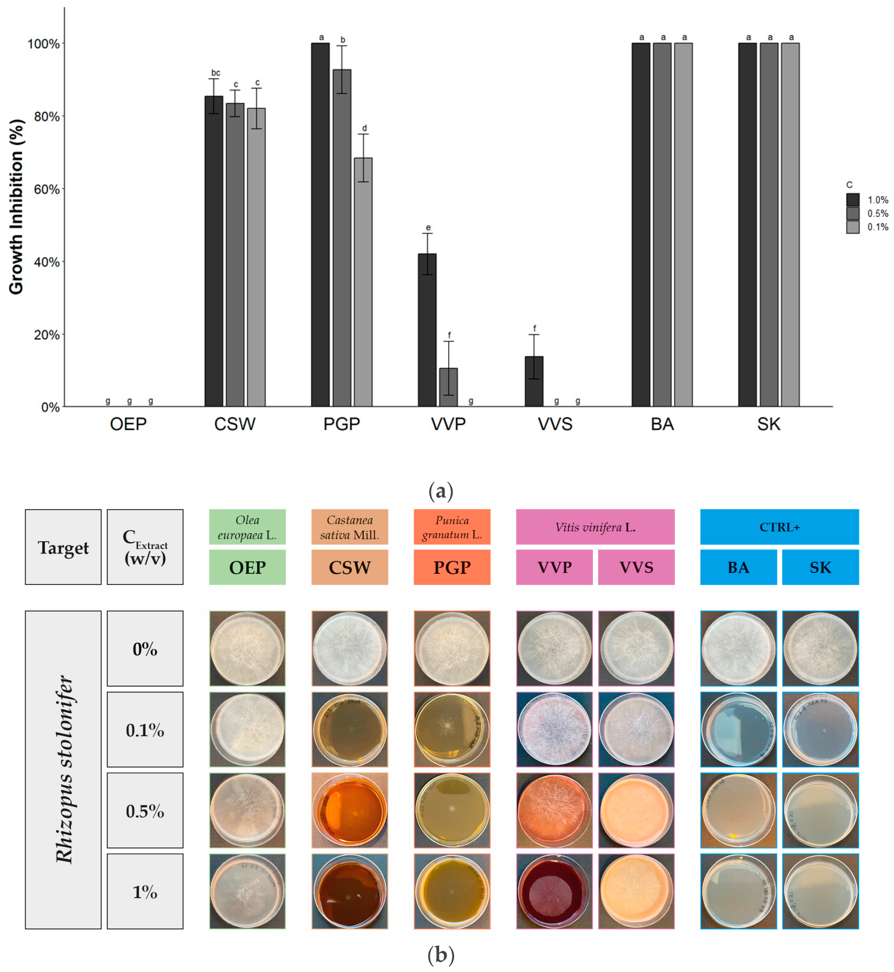
2. Results and Discussion

2.1. Phenolic-Rich Extracts

Based on our experience on the valorization of agro-industrial by-products and wastes, Olea europaea L. de-oiled pomace, Castanea sativa Mill. wood, Punica granatum L. peel, and Vitis vinifera L. pomace and seeds were selected as starting materials for the corresponding phenolic-rich extracts named OEP, CSW, PGP, VVP, and VVS, respectively. OEP, CSW, and VVS were commercial industrial fractions obtained by sustainable processes through water or water–ethanol extraction and purification/concentration by membrane technology [35]. The low percentage of ethanol, in combination with water, increased the yield of extraction of polyphenols, avoiding high temperatures which would cause their degradation. This methodology allows for making the production processes sustainable even on an industrial scale, avoiding the use of pollutants or toxic solvents for both the extraction and refining steps. In particular, membrane technologies are currently an interesting example of a sustainable separation and concentration technology, often integrated in biorefineries to rationalize the processes and the structure of the plants, and to reduce environmental and economic impacts whilst maintaining the quality of the products. For the extracts under examination, several refining steps were applied from ultrafiltration up to reverse osmosis to obtain the concentrated solutions to be spray-dried and the purified water to be reused for subsequent extraction batches [37].
PGP and VVP were prepared in a laboratory as described in the Section 3 Materials and Methods.
OEP, CSW, PGP, VVP, and VVS were analyzed by HPLC-DAD-MS to define the qualitative and quantitative content of polyphenols (see Table 1 and Table 2 and Figures S1–S5; Tables S1–S5 in Supplementary Materials). As reported in Table 1, OEP, CSW, and PGP are characterized by a total polyphenol content of 173 ± 5 mg/g, 260 ± 3 mg/g, and 115 ± 2 mg/g, respectively. OEP contains mainly hydroxytyrosol (138 ± 4.0 mg/g) and tyrosol in lower amount (35.0 ± 0.8) [38]. The most representative phenolic compounds found in CSW and PGP are hydrolysable tannins such as vescalagin (47.6 ± 0.5) and castalagin (97.7 ± 0.9) in CSW, and α-punicalagin (27.1 ± 0.3) and β-punicalagin (58.5 ± 0.6) in PGP, together to low amounts of vescalin (9.3 ± 0.2), castalin (8.1 ± 0.2), α-punicalin (1.25 ± 0.04), and β-punicalin (1.32 ± 0.02) obtained by hydrolysis during water extraction [39,40,41,42].
As reported in Table 2, VVP and VVS are characterized by a total polyphenol content of 425 ± 8 mg/g and 686 ± 20 mg/g, respectively. They are represented by condensed tannins with different degrees of polymerization, which are stable under water extraction conditions. The main components are procyanidin tetramers with 293 ± 4 mg/g and 315 ± 9 mg/g in VVP and VVS, respectively. The monomers catechin and epicatechin were identified in both samples, although in different amounts (0.414 ± 0.008 mg/g and 0.320 ± 0.008 mg/g in VVP; 45 ± 1 mg/g and 30.3 ± 0.8 mg/g in VVS). The study of the chromatographic profile of VVP at 520 nm also allows for the identification and quantification of anthocyanins retained within the plant material after the winemaking process (total: 3.14 ± 0.07 mg/g). The absence of aglycones and large amounts of degradation products suggests that the transformation processes undergone by the plant material were able to keep most of the less stable compounds intact. However, the low amount of these compounds in a by-product such as grape marc is plausible, given that they are largely transferred into the wine during winemaking, and due to their low stability.

2.2. Antifungal Activity

As already reported in the Introduction, polyphenols showed antimicrobial activity and several phenolic-rich extracts have been tested against pathogens [43,44,45].
In this work, the selected fungi in relation to food and biomedical issues were Alternaria sp., Aspergillus brasiliensis, Rhizopus stolonifer, and Trichophyton interdigitale. Alternaria is a ubiquitous genus that includes about 300 species between saprophytes and pathogens [46]. Because of its wide diffusion in plants, it could represent a threat during pre- and post-harvest phases, even through the production of mycotoxins [47]. In particular, the Alternaria species can produce more than 70 different toxins, which led the European Food Safety Authority (EFSA) to reveal the exposure levels of the European population to Alternaria by-toxins, which were troubling owing to higher levels found on toddlers [48,49]. Aspergillus brasiliensis is one of the 18 species included in the black aspergilli group, Aspergillus section Nigri [50]. Generally confused with A. niger, this biseptate species is responsible for the production of several secondary metabolites such as deyhidrocarolic acid, funalenone, and malformins [51]. Aspergillus brasiliensis ATCC 16,404 is widely used as a reference microorganism in several European standard ENs for chemical disinfectants and antiseptics in the medical area [52]. Moreover, the food industry and international organizations use Aspergillus brasiliensis ATCC 16,404 as a test microorganism in bio validation assays, i.e., to investigate the effects of UV sterilization treatment on food packaging [53]. Rhizopus stolonifer is a Zygomycete which affects a wide range of fruits and vegetables, as well as bread. The etiological agent of “soft rot” and “black bread mold” is characterized by a fast penetration ability and the rapid growth of a hairy gray mycelium, resulting in one of the most uncontrollable postharvest pathogens [54]. Trichophyton interdigitale is a clonal anthropophilic line of T. mentagrophytes involved in human dermatophytosis such as athlete’s foot “tinea pedis” and onychomycosis [55,56]. As other Trichophyton sp. it may produce biofilms as a virulence factor, reducing the effectiveness of locally applied antifungal agents or making it necessary for them to be used at high concentrations [57].
In the experimental design of this work, for each pathogen three different concentrations of OEP, CSW, PGP, VVP, and VVS were tested (1.0%, 0.5% and 0.1% w/v) using a diffusion assay [58,59]. Even though, in the literature, higher concentrations are tested by diffusion assays (up to 5–10–15% w/v) [60,61,62], the maximum concentration evaluated in this work was 1%, both for technical reasons related to the influence of the extract on the technological properties of the PDA medium and for economic evaluation in view of the potential industrial application of extracts that should prove to be active. Benzoic acid (BA, E210) and potassium sorbate (SK, E202) were used as positive controls. These compounds are largely employed as food additives with antifungal properties. BA, in the limit of 0.1%, and SK are Generally Recognized as Safe (GRAS) by the Food and Drug Administration (FDA), and their use in Europe is regulated by EC 1333/2008 [63,64]. Moreover, BA is a component of Whitfield’s ointment, a topical treatment of tinea dermatophytosis still used today in developing countries [65,66].
The data of the antifungal activity of OEP, CSW, PGP, VVP, and VVS against Alternaria sp., Aspergillus brasiliensis, Rhizopus stolonifer, and Trichophyton interdigitale are reported in Table 3, Table 4 and Table 5 and Figure 1, Figure 2, Figure 3 and Figure 4.
As showed in Figure 1, OEP, CSW, PGP, and VVS did not reveal any inhibitory activity against Aspergillus brasiliensis also at 1.0% (w/v). The only extract exhibiting an activity, albeit modest, was VVP, with a growth inhibition of 48.0 ± 3.9% at 1.0% w/v, but a dramatic decrease in activity was observed at 0.5 and 0.1% w/v. Similar to other dark septate endophytes, some Aspergillus species, including A. niger, can degrade tannins using tannase enzymes. In particular, the Aspergillus niger GH1 strain showed the ability to degrade ellagitannins from pomegranate peel due to the activity of the ellagitannase enzyme, releasing ellagic acid from punicalagin [67,68,69]. The growth inhibition observed with VVP could be related to the presence of anthocyanins [70].
Table 3 and Figure 2 report the antifungal activity of OEP, CSW, PGP, VVP, and VVS against Alternaria sp. At 1.0% w/v, all extracts inhibited the growth of the pathogen, even if some differences of activity were observed. The lowest growth inhibition was evidenced for VVS (17.6 ± 8.0%) and the highest for CSW and PGP (100%). The inhibitory effect of CSW drastically decreased starting from 0.5% w/v while PGP retained the maximum activity also at 0.1% w/v. Due to this high effect, PGP was tested at a lower concentration (0.01% w/v), evidencing a significant decrease in activity, with an EC50 value of 0.026%, corresponding to 260 mg/L of extract and 29.9 mg/L of polyphenols. These data are in accordance with the literature [71,72]. In fact, sweet chestnut offers different waste matrices with antimicrobial activity against Alternaria. An aqueous extract from burs rich in hydrolysable tannins affects mycelial growth and spore germination against Alternaria alternata [71]. The main phenolic component of PGP, punicalagin, has been proved the most efficient pomegranate peel compound against Alternaria alternata AL19, with an inhibitory activity starting from 92.9 µM [72]. Satisfactory activity was observed for VVP at 1.0% w/v (62.6 ± 9.9%), which decreased proportionally with the concentration up to 34.7 ± 8.8% at 0.1% w/v. This activity was quantified to an EC50 value of 0.37% (3.7 g/L of the extract, 1.6 g/L of polyphenols), lower than CSW (0.54%, 5.4 g/L of the extract, 1.4 g/L of polyphenols). As expected, both BA and SK completely inhibited fungal growth from 1.0% to 0.1% w/v. The results of OEP at 0.1% w/v were influenced by guttation, which consists of the production of fungal exudates composed by liquid droplets. It caused a reduced growth of the mycelium, resulting in a fake enhanced antimicrobial activity and a high variability of data (18.2 ± 14.4%). However, during guttation, fungal species could produce secondary metabolites, including phenolic compounds and toxins involved in several key ecological roles [73].
As depicted in Table 4 and Figure 3, the antifungal activity of OEP, CSW, PGP, VVP, and VVS against Rhizopus stolonifer depended on the plant materials. At 1.0% w/v, OEP did not inhibit the growth; VVS and VVP produced a modest effect (13.7 ± 6.2 and 42.0 ± 5.7%, respectively); CSW showed significant activity (82.0 ± 8.7%); and PGP a total inhibitory effect. These last extracts also retained the activity at lower concentrations (0.5 and 0.1% w/v) with 83.4 ± 3.7% and 82.1 ± 5.5%; 92.8 ± 6.6% and 68.4 ± 6.6% of growth inhibition, respectively. The antifungal activity of pomegranate aqueous peel extract from the “shishe kab” Iranian cultivar against Rhizopus stolonifer was previously recorded using the poisoned food technique [74].
Table 5 and Figure 4 evidenced that all extracts exhibited a relevant growth inhibition against Trichophyton interdigitale. Except for OEP, which showed a growth inhibition of 66.5 ± 2.9% at 1.0% w/v [75,76], the extracts evidenced total inhibition. To the best of our knowledge, this is the first work investigating the antimicrobial activities of extracts of Castanea sativa Mill. against dermatophytes. No growth was observed using CSW at concentrations ranging from 1.0% to 0.1%, showing the best performance compared to both extracts and controls. With this extract, the test was carried out at lower concentrations. Only at 0.005% w/v was a drastic decay of antimicrobial activity recorded, resulting in an EC50 value of 0.0063%, corresponding to 63 mg/L of extract and 16.38 mg/L of polyphenols. PGP completely inhibited fungal growth until 0.1%, with an EC50 value of 0.014%, corresponding to 140 mg/L of extract 16.1 mg/L of polyphenols. In the literature, it was already reported that hydrolysable tannins possess antifungal activity against dermatophytes [77]. Crude extracts of pomegranate peels, as well as isolated punicalagin, showed inhibitory activity on conidia germination and mycelium growth of dermatophytes [78]. A strong performance was revealed even for VVS, in accordance with other works that highlighted the inhibitory effects of flavan-3-ols from different Vitis vinifera L. matrices against dermatophytes [79].

3. Materials and Methods

3.1. Chemicals

All solvents for HPLC-DAD-MS analyses (HPLC grade), formic acid, and epigallocatechin gallate (analytical grade) were purchased from Sigma Aldrich Chemical Company Inc. (Milwaukee, WI, USA). Tyrosol, gallic acid, ellagic acid, catechin, and malvidin 3-O-glucoside were supplied by Extrasynthèse S.A. (Lyon, Nord-Genay, France). Hydroxytyrosol was synthetized in the laboratory [80]. HPLC-grade water was obtained via distillation and purification with a Labconco Water Pro PS polishing station (Labconco Corporation, Kansas City, MO, USA). Potassium sorbate, benzoic acid, potassium hydroxide, and hydrochloric acid 37% were purchased from Carlo Erba Reagents Srl (Cornaredo, Milan, Italy), Tween 20 from Biolife Italiana Srl (Milan, Italy), and Potato Dextrose Agar from Oxoid Ltd. (Basingstoke, Hampshire, UK). Sterile plasticware and cotton swabs were purchased from Unifo Srl (Zero Branco, Treviso, Italy).

3.2. Phenolic-Rich Extracts

All extracts were derived from circular economy processes and were obtained by sustainable extraction methodologies. OEP was furnished by Bionap Srl (Piano Tavola Belpasso, Italy) and CSW was a gift of Gruppo Mauro Saviola Srl (Viadana, Italy). The commercially available Saviotan® Feed was produced in the plant operating in Radicofani (Italy) by a sustainable, green process of hot water extraction and concentration/purification by membrane technology, described by us in a previous paper [37]. PGP was obtained from pomegranate peel (Punica granatum L., cv. Wonderful) collected in Grosseto, Italy. The peels were separated from the fresh fruits, finely chopped, put in a polypropylene filter bag, and then extracted for 1 h in boiling water (10% w/v) under magnetic stirring. The extraction mixture was left to cool down to room temperature and kept under maceration for 24 h; then, the extract was filtered under vacuum, frozen at −20 °C and lyophilized to obtain the final powder (yield: 9.2%). VVP was obtained from dried grape pomace furnished by Cantina Cesarini Sartori (Loc. Purgatorio, Gualdo Cattaneo, Italy). The powder (60 g) was extracted with ethanol/water = 70:30 (300 mL) adjusted at pH 2.5 by adding HCOOH for 24 h, under mechanical stirring. After filtration under vacuum, the solvent was evaporated; finally, the extract was rinsed with distilled water, frozen at −20 °C and lyophilized to obtain the final powder (yield: 0.9%). VVS was furnished by Consulente Enologica Srl (Pietraia di Cortona, Italy).

3.3. Characterization of Phenolic-Rich Extracts

OEP, CSW, PGP, VVP, and VVS were analyzed by HPLC-DAD-MS using a HP-1260 liquid chromatograph equipped with a DAD detector and an MSD API-electrospray (Agilent Technologies, Santa Clara, CA, USA) operating in negative and positive ionization mode. Mass spectrometer operating conditions were the following: gas temperature 350 °C at a flow rate of 10.0 L/min, nebulizer pressure 30 psi, quadrupole temperature 30 °C, and capillary voltage 3500 V. The fragmentor was set at 120 eV. For CSW and PGP, a Luna, C18 250 × 4.60 mm, 5 μm column (Phenomenex, Torrance, CA, USA) operating at 26 °C was used. The eluents were H2O (adjusted to pH 3.2 with HCOOH) and CH3CN. A four-step linear solvent gradient starting from 100% H2O up to 100% CH3CN was performed with a flow rate of 0.8 mL/min over a 55 min period, as previously described [37,45]. Gallic acid, flavanols, and procyanidins of VVP, VVS, and OEP were analyzed by using a column Lichrosorb RP18 250 × 4.60 mm i.d, 5 µm (Merck Darmstadt, Germany). The eluents were H2O adjusted to pH 3.2 with HCOOH and CH3CN. A four-step linear solvent gradient was used, starting from 100% H2O up to 100% CH3CN, for 117 min at a flow rate of 0.8 mL/min [25]. For anthocyanins of VVP, a Luna, C18 250 × 4.60 mm, 5 μm column (Phenomenex, Torrance, CA, USA) operating at 26 °C was used. The eluents were H2O (adjusted to pH 1.8 with HCOOH) and CH3CN. A multi-step linear solvent gradient was used, starting from 95% H2O up to 100% CH3CN, for 26 min at a flow rate of 0.8 mL/min.
Polyphenols present in the extracts were identified by using their chromatographic, spectrophotometric, and spectrometric data. Their retention times and data from HPLC-DAD and HPLC-MS were compared with those of the available specific commercial standards, also taking into account our previous results obtained by LC-MS-MS and/or LC-MS-TOF analysis of the same matrices, and data in the literature [81,82]. Each compound was quantified by HPLC-DAD using a five-point regression curve built with the available standards. Calibration curves with r2 ≥ 0.9998 were considered. The concentrations of the individual compounds were calculated by applying the appropriate corrections for changes in molecular weight. Ellagic acid and ellagitannins were calibrated at 254 nm with ellagic acid; gallic acid and gallotannins at 280 nm with gallic acid; epicatechin gallate (ECG) and epigallocatechin gallate (EGCG) at 280 nm with EGCG; catechin, epicatechin, and procyanidins at 280 nm with catechin hydrate; tyrosol and hydroxytyrosol at 280 nm with pure standards; and anthocyanins at 520 nm using malvidin-3-O-glucoside as reference. The evaluation of the polyphenol content was carried out in triplicate and the results were recorded as mean values with standard deviations ≤5%.

3.4. Fungal Pathogens

Extracts were tested against filamentous fungi Aspergillus brasiliensis (ex A. niger) derived from ATCC 16404, Alternaria sp. derived from ATCC 20,084 (Microbiologics, St. Cloud, MN, USA), Rhizopus stolonifer derived from ATCC 14,037, and Trichophyton interdigitale derived from ATCC 9533 (Kairosafe, Trieste, Italy). Fungi were maintained on Potato Dextrose Agar (PDA) at 25 °C.

3.5. In Vitro Antifungal Activity Assay

The fungal inoculum was prepared from fresh culture of about 4 days for Rhizopus stolonifer, 7 days for Alternaria sp. and Aspergillus brasiliensis, and 15 days for Trichophyton interdigitale following a procedure based on the EUCAST E.DEF 9.4 [83] with minor changes. Briefly, 5 mL of sterile water with Tween 20 (0.1% v/v) was added to the culture. To promote conidial suspension, the culture was gently scraped using a sterile cotton swab. The obtained suspension was recovered, shaken for about 15 s with a vortex and filtered to remove hyphae and clumps. The inoculum was spectrophotometrically adjusted to an equivalent final concentration of McFarland 0.5 (approximately 1–5 × 10⁶ CFU/mL).
Testing media were obtained by adding different amounts of extracts to PDA to obtain final concentrations of 1.0%; 0.5%; and 0.1% (w/v). As control, two food and cosmetic preservative SK and BA were used. After solubilization, pH was adjusted to 5.6 ± 0.2 using KOH 1 M or HCl 1 M. The media were then sterilized at 121 °C for 15 min and transferred to 55 mm petri dishes.
The antifungal activity assay was performed based on a diffusion method according to the literature with slight modifications [58,59]. A total of 10 μL of conidial suspension was inoculated in the center of the agar plate. Plates were incubated at 25 °C in darkness and growth was observed daily until the mycelium of the negative control (PDA only) touched the edge of the plate. Growth inhibition (GI), expressed as a percentage, was calculated by measuring the colony diameter and using the following equation:
GI(%) = [(dcdt)/dc] × 100
where dc is the mean diameter of the negative control (PDA only) and dt is the mean diameter of the treatment.

3.6. Statistical Analysis

Data analysis was performed using RStudio Desktop (version 2023.13.0+386, Posit Software, PBC, Boston, MA, USA). To determine the differences between treatments, a one-way analysis of variance (ANOVA) was performed with significance level set at p = 0.05. Means separation was carried out using Tukey’s HSD test. EC50 was calculated using the “LL.2” function of the “drc” package, corresponding to a log-logistic model, where the lower limit was fixed at 0 (negative control dc for PDA only) and the upper limit was fixed at 1 (corresponding to total inhibition) [84].

4. Conclusions

In this paper, five phenolic-rich extracts (OEP, CSW, PGP, VVP, and VVS) derived from Olea europaea L. de-oiled pomace, Castanea sativa Mill. wood, Punica granatum L. peel, and Vitis vinifera L. pomace and seeds were characterized by HPLC-MS-DAD analysis to define their qualitative and quantitative phenolic profiles. Based on these analyses, OEP was found to be rich in hydroxytyrosol; CSW and PGP in hydrolysable tannins (vescalagin, castalagin, α-punicalagin, and β-punicalagin); and VVP and VVS in condensed tannins (procyanidins, trimers, and tetramers).
All extracts were tested against selected pathogenic filamentous fungi and dermatophytes such as Aspergillus brasiliensis, Alternaria sp., Rhizopus stolonifer, and Trichophyton interdigitale at three different concentrations (1.0%, 0.5%, and 0.1% w/v) using a diffusion assay. OEP, CSW, PGP, and VVS did not reveal any inhibitory activity against Aspergillus brasiliensis also at 1.0% (w/v). On the contrary, at the same concentration, VVP showed a (modest) growth inhibition. CSW and PGP exhibited a total growth inhibition against Alternaria sp. at 1.0% w/v, but VVP was also very effective at the same concentration. PGP retained the activity until 0.1% w/v with an EC50 value of 0.026%, corresponding to 260 mg/L of extract and 29.9 mg/L of polyphenols. Against Rhizopus stolonifer, PGP showed a total growth inhibition at 1.0% w/v, but strong performances were also observed at lower concentrations (up 0.1% w/v); CSW behaved similarly. Interestingly, CSW, PGP, VVS, and VVP showed a complete growth inhibition of Trichophyton interdigitale at 1.0% (w/v) and the activity was retained at lower concentrations. CSW was active until 0.005% w/v corresponding to an EC50 value of 0.0063% (63 mg/L of extract, 16.38 mg/L of polyphenols). PGP completely inhibited fungal growth until 0.1%, with an EC50 value of 0.014% (140 mg/L of extract, 16.1 mg/L of polyphenols).
Based on these data, we could conclude that the chemical composition is crucial for the biological activity of the extracts against the selected pathogens. In particular, hydrolysable and condensed tannins play a relevant role in the activity. The data obtained from this research seem promising for the potential application of some of these extracts as antifungal agents in the food and biomedical fields according to the green chemistry and circular economy concepts.

Supplementary Materials

The following supporting information can be downloaded at: https://www.mdpi.com/article/10.3390/molecules28114374/s1. Figure S1. Chromatographic profile of OEP at 280 nm; Table S1. Quali-quantitative analysis of OEP; Figure S2. Chromatographic profile of CSW at 254 and 280 nm; Table S2. Quali-quantitative analysis of CSW; Figure S3. Chromatographic profile of PGP at 254 and 280 nm; Table S3. Quali-quantitative analysis of PGP; Figure S4. Chromatographic profile of VVP at 520 and 280 nm; Table S4. Quali-quantitative analysis of VVP; Figure S5. Chromatographic profile of VVS acquired at 280 nm; Table S5. Quali-quantitative analysis of VVS.

Author Contributions

Conceptualization, A.L. and R.B.; methodology, A.L., M.P. (Mirco Pizzetti) and R.B.; data curation, A.L., M.C., P.V., M.P. (Mirco Pizzetti) and R.B.; writing—original draft preparation, A.L., M.C., P.V. and R.B.; writing—review and editing, A.L., M.C., P.V., M.P. (Mirco Pizzetti), M.P. (Marco Papalini) and R.B.; supervision, R.B. All authors have read and agreed to the published version of the manuscript.

Funding

This research received no external funding.

Institutional Review Board Statement

Not applicable.

Informed Consent Statement

Not applicable.

Data Availability Statement

The data presented in this study are available on reasonable request from the corresponding authors. OPPURE The authors confirm that the data supporting the findings of this study are available within the article and Supplementary Materials.

Acknowledgments

The authors acknowledge the Italian Ministry of University and Research (MUR), University of Tuscia (Viterbo, Italy), and Bioricerche Srl (Castell’Azzara, Grosseto, Italy) for a grant attributed to Andrea Lombardi in the frame of DM 1061/2021 (Dottorato di Ricerca, PON Ricerca e Innovazione 2014–2020). The authors acknowledge Innovation Ecosystem Rome Technopole, National Recovery and Resilience Plan (NRRP), Mission 4, Component 2 Investment 1.5, funded from the European Union—Next Generation EU (Code project: ECS00000024). The authors are grateful to the technical staff of Bioricerche Srl for their support.

Conflicts of Interest

The authors declare no conflict of interest.

Sample Availability

OEP, CSW, PGP, VVP, and VVS are available from the authors.

References

  1. Avery, S.V.; Singleton, I.; Magan, N.; Goldman, G.H. The fungal threat to global food security. Fungal Biol. 2019, 123, 555–557. [Google Scholar] [CrossRef] [PubMed]
  2. Fisher, M.C.; Gurr, S.J.; Cuomo, C.A.; Blehert, D.S.; Jin, H.; Stukenbrock, E.H.; Stajich, J.E.; Kahmann, R.; Boone, C.; Denning, D.W.; et al. Threats posed by the fungal kingdom to humans, wildlife, and agriculture. mBio 2020, 11, e00449-20. [Google Scholar] [CrossRef] [PubMed]
  3. Mahato, D.K.; Lee, K.E.; Kamle, M.; Devi, S.; Dewangan, K.N.; Kumar, P.; Kang, S.G. Aflatoxins in food and feed: An overview on prevalence, detection and control strategies. Front. Microbiol. 2019, 10, 2266. [Google Scholar] [CrossRef] [PubMed]
  4. Alkan, N.; Fortes, A.M. Insights into molecular and metabolic events associated with fruit response to post-harvest fungal pathogens. Front. Plant Sci. 2015, 6, 889. [Google Scholar] [CrossRef] [PubMed]
  5. Hoffmann, S.; Scallan, E. Chapter 2-Epidemiology, cost, and risk analysis of foodborne disease. In Foodborne Diseases, 3rd ed.; Dodd, C.E.R., Aldsworth, T., Stein, R.A., Cliver, D.O., Riemann, H.P., Eds.; Academic Press: Cambridge, MA, USA, 2017; pp. 31–63. [Google Scholar]
  6. Granja, N.; Domingues, P.; Cabecinhas, M.; Zimon, D.; Sampaio, P. ISO 22000 Certification: Diffusion in Europe. Resources 2021, 10, 100. [Google Scholar] [CrossRef]
  7. ISO 22000:2018; Food Safety Management Systems—Requirements for Any Organization in the Food Chain. International Organization for Standardization: Geneva, Switzerland, 2018. Available online: https://www.iso.org/standard/65464.html (accessed on 24 March 2023).
  8. ISO 21257-1:2008; Microbiology of Food and Animal Feeding Stuffs—Horizontal Method for the Enumeration of Yeasts and Moulds—Part 1: Colony Count Technique in Products with Water Activity Greater than 0,95. International Organization for Standardization: Geneva, Switzerland, 2008. Available online: https://www.iso.org/standard/38275.html (accessed on 24 March 2023).
  9. ISO 21257-2:2008; Microbiology of Food and Animal Feeding Stuffs—Horizontal Method for the Enumeration of Yeasts and Moulds—Part 2: Colony Count Technique in Products with Water Activity Less than or Equal to 0,95. International Organization for Standardization: Geneva, Switzerland, 2008. Available online: https://www.iso.org/standard/38276.html (accessed on 24 March 2023).
  10. Kousha, M.; Tadi, R.; Soubani, A.O. Pulmonary Aspergillosis: A clinical review. Eur. Respir. Rev. 2011, 20, 156–174. [Google Scholar] [CrossRef]
  11. Lehmann, S.; Sprünken, A.; Wagner, N.; Tenbrock, K.; Ott, H. Clinical relevance of IgE-mediated sensitization against the mould Alternaria alternata in children with asthma. Ther. Adv. Respir. Dis. 2017, 11, 30–39. [Google Scholar] [CrossRef]
  12. Segal, E.; Elad, D. Human and zoonotic dermatophytoses: Epidemiological aspects. Front. Microbiol. 2021, 12, 713532. [Google Scholar] [CrossRef]
  13. Garvey, M.; Rowan, N.J. Pathogenic drug resistant fungi: A review of mitigation strategies. Int. J. Mol. Sci. 2023, 24, 1584. [Google Scholar] [CrossRef]
  14. Bernini, R.; Mincione, E.; Barontini, M.; Fabrizi, G.; Pasqualetti, M.; Tempesta, S. Convenient oxidation of alkylated phenols and methoxytoluenes to antifungal 1,4-benzoquinones with hydrogen peroxide/methyltrioxorhenium catalytic system in neutral ionic liquid. Tetrahedron 2006, 62, 7733–7737. [Google Scholar] [CrossRef]
  15. Bernini, R.; Mincione, E.; Provenzano, G.; Fabrizi, G.; Tempesta, S.; Pasqualetti, M. Obtaining new flavanones exhibiting antifungal activities by methyltrioxorhenium-catalyzed epoxidation-methanolysis of flavones. Tetrahedron 2008, 64, 7561–7566. [Google Scholar] [CrossRef]
  16. Bernini, R.; Pasqualetti, M.; Provenzano, G.; Tempesta, S. Ecofriendly synthesis of halogenated flavonoids and evaluation of their antifungal activity. New J. Chem. 2015, 39, 2980–2987. [Google Scholar] [CrossRef]
  17. Lombardi, A.; Fochetti, A.; Vignolini, P.; Campo, M.; Durazzo, A.; Lucarini, M.; Puglia, D.; Luzi, F.; Papalini, M.; Renzi, M.; et al. Natural active ingredients for poly (lactic acid)-based materials: State of the art and perspectives. Antioxidants 2022, 11, 2074. [Google Scholar] [CrossRef]
  18. Dewick, P.M. Medicinal Natural Products: A Biosynthetic Approach, 3rd ed.; Dewick, P.M., Ed.; John Wiley & Sons, Ltd.: Chichester, UK, 2009. [Google Scholar]
  19. Sun, W.; Shahrajabian, M.H. Therapeutic potential of phenolic compounds in medicinal plants. Natural health products for human health. Molecules 2023, 28, 1845. [Google Scholar] [CrossRef] [PubMed]
  20. Roma, E.; Mattoni, E.; Lupattelli, P.; Moeini, S.S.; Gasperi, T.; Bernini, R.; Incerpi, S.; Tofani, D. New dihydroxytyrosyl esters from dicarboxylic acids. Synthesis and evaluation of the antioxidant activity in vitro (ABTS) and in cell-cultures (DCF assay). Molecules 2020, 25, 3135. [Google Scholar] [CrossRef]
  21. Bernini, R.; Velotti, F. Natural polyphenols as immunomodulators to rescue immune response homeostasis: Quercetin as a research model against severe COVID-19. Molecules 2021, 26, 5803. [Google Scholar] [CrossRef] [PubMed]
  22. Luzi, F.; Pannucci, E.; Clemente, M.; Grande, E.; Urciuoli, S.; Romani, A.; Torre, L.; Puglia, D.; Bernini, R.; Santi, L. Hydroxytyrosol and oleuropein-enriched extracts obtained from olive oil wastes and by-products as active antioxidant ingredients for poly(vinyl alcohol)-based films. Molecules 2021, 26, 2104. [Google Scholar] [CrossRef]
  23. Laghezza Masci, V.; Bernini, R.; Villanova, N.; Clemente, M.; Cicaloni, V.; Tinti, L.; Salvini, L.; Taddei, A.R.; Tiezzi, A.; Ovidi, E. In vitro anti-proliferative and apoptotic effects of hydroxytyrosyl oleate on SH-SY5Y human neuroblastoma cells. Int. Mol. J. Sci. 2022, 23, 12348. [Google Scholar] [CrossRef]
  24. Pinto, G.; De Pascale, S.; Aponte, M.; Scaloni, A.; Addeo, F.; Caira, S. Polyphenol profiling of chestnut pericarp, integument and curing water extracts to qualify these food by-products as a source of Antioxidants. Molecules 2021, 26, 2335. [Google Scholar] [CrossRef] [PubMed]
  25. Romani, A.; Ieri, F.; Urciuoli, S.; Noce, A.; Marrone, G.; Nediani, C.; Bernini, R. Health Effects of Phenolic Compounds Found in Extra-Virgin Olive Oil, By-Products, and Leaf of Olea europaea L. Nutrients 2019, 11, 1776. [Google Scholar] [CrossRef]
  26. Lucarini, M.; Durazzo, A.; Bernini, R.; Campo, M.; Vita, C.; Souto, E.B.; Lombardi-Boccia, G.; Ramadan, M.F.; Santini, A.; Romani, A. Fruit wastes as valuable source of value-added compounds: A collaborative perspective. Molecules 2021, 26, 6338. [Google Scholar] [CrossRef]
  27. Goufo, P.; Singh, R.K.; Cortez, I. A Reference list of phenolic compounds (including stilbenes) in grapevine (Vitis vinifera L.) roots, woods, canes, stems, and leaves. Antioxidants 2020, 9, 398. [Google Scholar] [CrossRef]
  28. Stahel, W.R. The circular economy. Nature 2016, 531, 435–438. [Google Scholar] [CrossRef] [PubMed]
  29. Rodis, P.S.; Karathanos, V.T.; Mantzavinou, A. Partitioning of olive oil antioxidants between oil and water phases. J. Agric. Food Chem. 2002, 50, 596–601. [Google Scholar] [CrossRef] [PubMed]
  30. Aliaño-González, M.J.; Gabaston, J.; Ortiz-Somovilla, V.; Cantos-Villar, E. Wood Waste from Fruit Trees: Biomolecules and Their Applications in Agri-Food Industry. Biomolecules 2022, 12, 238. [Google Scholar] [CrossRef]
  31. Echegaray, N.; Gómez, B.; Barba, F.J.; Franco, D.; Estévez, M.; Carballo, J.; Marszałek, K.; Lorenzo, J.M. Chestnuts and by-products as source of natural antioxidants in meat and meat products: A review. Trends Food Sci. Technol. 2018, 82, 110–121. [Google Scholar] [CrossRef]
  32. Jurenka, J. Therapeutic applications of pomegranate (Punica granatum L.): A review. Altern. Med. Rev. 2008, 13, 128–144. [Google Scholar]
  33. Nuamsetti, T.; Dechayuenyong, P.; Tantipaibulvut, S. Antibacterial activity of pomegranate fruit peels and arils. Sci. Asia 2012, 38, 319–322. [Google Scholar] [CrossRef]
  34. Dwyer, K.; Hosseinian, F.; Rod, M. The market potential of grape waste alternatives. J. Food Res. 2014, 3, 91–96. [Google Scholar] [CrossRef]
  35. Lucarini, M.; Durazzo, A.; Romani, A.; Campo, M.; Lombardi-Boccia, G.; Cecchini, F. Bio-based compounds from grape seeds: A biorefinery approach. Molecules 2018, 23, 1888. [Google Scholar] [CrossRef]
  36. Ky, I.; Teissedre, P.-L. Characterisation of Mediterranean Grape Pomace Seed and Skin Extracts: Polyphenolic Content and Antioxidant Activity. Molecules 2015, 20, 2190–2207. [Google Scholar] [CrossRef]
  37. Campo, M.; Pinelli, P.; Romani, A. Hydrolyzable tannins from sweet chestnut fractions obtained by a sustainable and eco-friendly industrial process. Nat. Prod. Commun. 2016, 11, 1934578X1601100. [Google Scholar] [CrossRef]
  38. Romani, A.; Campo, M.; Urciuoli, S.; Marrone, G.; Noce, A.; Bernini, R. An industrial and sustainable platform for the production of bioactive micronized powders and extracts enriched in polyphenols from Olea europaea L. and Vitis vinifera L. wastes. Front. Nutr. 2020, 7, 120. [Google Scholar] [CrossRef] [PubMed]
  39. Feldman, K.S.; Smith, R.S. Ellagitannin chemistry. First total synthesis of the 2, 3-and 4, 6-coupled ellagitannin pedunculagin. J. Org. Chem. 1996, 61, 2606–2612. [Google Scholar] [CrossRef]
  40. Yamada, H.; Wakamori, S.; Hirokane, T.; Ikeuchi, K.; Matsumoto, S. Structural revisions in natural ellagitannins. Molecules 2018, 23, 1901. [Google Scholar] [CrossRef]
  41. Karaseva, V.; Bergeret, A.; Lacoste, C.; Ferry, L.; Fulcrand, H. Influence of extraction conditions on chemical composition and thermal properties of chestnut wood extracts as tannin feedstock. ACS Sustain. Chem. Eng. 2019, 7, 17047–17054. [Google Scholar] [CrossRef]
  42. Moccia, F.; Flores-Gallegos, A.C.; Chávez-González, M.L.; Sepúlveda, L.; Marzorati, S.; Verotta, L.; Panzella, L.; Ascacio-Valdes, J.A.; Aguilar, C.N.; Napolitano, A. Ellagic acid recovery by solid state fermentation of pomegranate wastes by Aspergillus niger and Saccharomyces cerevisiae: A comparison. Molecules 2019, 24, 3689. [Google Scholar] [CrossRef]
  43. Funatogawa, K.; Hayashi, S.; Shimomura, H.; Yoshida, T.; Hatano, T.; Ito, H.; Hirai, Y. Antibacterial activity of hydrolyzable tannins derived from medicinal plants against Helicobacter pylori. Microbiol. Immunol. 2004, 48, 251–261. [Google Scholar] [CrossRef]
  44. Côté, J.; Caillet, S.; Doyon, G.; Sylvain, J.F.; Lacroix, M. Bioactive compounds in cranberries and their biological properties. Crit. Rev. Food Sci. Nutr. 2010, 50, 666–679. [Google Scholar] [CrossRef]
  45. Romani, A.; Campo, M.; Pinelli, P. HPLC/DAD/ESI-MS analyses and anti-radical activity of hydrolyzable tannins from different vegetal species. Food Chem. 2012, 130, 214–221. [Google Scholar] [CrossRef]
  46. Lee, H.B.; Patriarca, A.; Magan, N. Alternaria in Food: Ecophysiology, Mycotoxin Production and Toxicology. Mycobiology 2015, 43, 93–106. [Google Scholar]
  47. Patriarca, A. Alternaria in Food Products. Curr. Opin. Food Sci. 2016, 11, 1–9. [Google Scholar] [CrossRef]
  48. Tralamazza, S.M.; Piacentini, K.C.; Iwase, C.H.T.; Rocha, L.d.O. Toxigenic Alternaria Species: Impact in Cereals Worldwide. Curr. Opin. Food Sci. 2018, 23, 57–63. [Google Scholar] [CrossRef]
  49. Arcella, D.; Eskola, M.; Gómez Ruiz, J.A.; EFSA (European Food Safety Authority). Scientific report on the dietary exposure assessment to Alternaria toxins in the European population. EFSA J. 2016, 14, 4654. [Google Scholar]
  50. Varga, J.; Kocsubé, S.; Tóth, B.; Frisvad, J.C.; Perrone, G.; Susca, A.; Meijer, M.; Samson, R.A. Aspergillus brasiliensis sp. nov., a biseriate black Aspergillus species with world-wide distribution. Int. J. Syst. Evol. Microbiol. 2007, 57, 1925–1932. [Google Scholar] [CrossRef] [PubMed]
  51. Nielsen, K.F.; Mogensen, J.M.; Johansen, M.; Larsen, T.O.; Frisvad, J.C. Review of secondary metabolites and mycotoxins from the Aspergillus niger group. Anal. Bioanal. Chem. 2009, 395, 1225–1242. [Google Scholar] [CrossRef]
  52. Tyski, S.; Bocian, E.; Laudy, A.E. Application of normative documents for determination of biocidal activity of disinfectants and antiseptics dedicated to the medical area: A narrative review. J. Hosp. Infect. 2022, 125, 75–91. [Google Scholar] [CrossRef]
  53. Racchi, I.; Scaramuzza, N.; Hidalgo, A.; Cigarini, M.; Berni, E. Sterilization of food packaging by UV-C irradiation: Is Aspergillus brasiliensis ATCC 16404 the best target microorganism for industrial bio-validations? Int. J. Food Microbiol. 2021, 357, 109383. [Google Scholar] [CrossRef]
  54. Bautista-Baños, S.; Bosquez-Molina, E.; Barrera-Necha, L.L. Rhizopus stolonifer (soft rot). In Postharvest Decay Control Strategies, 1st ed.; Bautista-Baños, S., Ed.; Elsevier Inc.: London, UK, 2014; Chapter 1; pp. 1–37. [Google Scholar]
  55. Tang, C.; Kong, X.; Ahmed, S.A.; Thakur, R.; Chowdhary, A.; Nenoff, P.; Uhrlass, S.; Verma, S.B.; Meis, J.F.; Kandemir, H.; et al. Taxonomy of the Trichophyton mentagrophytes/T. interdigitale Species Complex Harboring the Highly Virulent, Multiresistant Genotype T. indotineae. Mycopathologia 2021, 186, 315–326. [Google Scholar] [CrossRef]
  56. Jazdarehee, A.; Malekafzali, L.; Lee, J.; Lewis, R.; Mukovozov, I. Transmission of onychomycosis and dermatophytosis between household members: A Scoping Review. JoF 2022, 8, 60. [Google Scholar] [CrossRef]
  57. Yazdanpanah, S.; Sasanipoor, F.; Khodadadi, H.; Rezaei-Matehkolaei, A.; Jowkar, F.; Zomorodian, K.; Kharazi, M.; Mohammadi, T.; Nouripour-Sisakht, S.; Nasr, R.; et al. Quantitative analysis of in vitro biofilm formation by clinical isolates of dermatophyte and antibiofilm activity of common antifungal drugs. Int. J. Dermatol. 2023, 62, 120–127. [Google Scholar] [CrossRef] [PubMed]
  58. Kinay, P.; Mansour, M.F.; Mlikota Gabler, F.; Margosan, D.A.; Smilanick, J.L. Characterization of fungicide-resistant isolates of Penicillium digitatum collected in California. Crop Prot. 2007, 26, 647–656. [Google Scholar] [CrossRef]
  59. Achar, P.N.; Quyen, P.; Adukwu, E.C.; Sharma, A.; Msimanga, H.Z.; Nagaraja, H.; Sreenivasa, M.Y. Investigation of the Antifungal and Anti-Aflatoxigenic Potential of Plant-Based Essential Oils against Aspergillus flavus in Peanuts. J. Fungi 2020, 6, 383. [Google Scholar] [CrossRef] [PubMed]
  60. Touba, E.P.; Zakaria, M.; Tahereh, E. Anti-fungal activity of cold and hot water extracts of spices against fungal pathogens of Roselle (Hibiscus sabdariffa) in vitro. Microb. Pathog. 2012, 52, 125–129. [Google Scholar] [CrossRef]
  61. Sharma, R.L.; Ahir, R.R.; Yadav, S.L.; Sharma, P.; Ghasolia, R.P. Effect of nutrients and plant extracts on Alternaria blight of tomato caused by Alternaria alternata. J. Plant. Dis. Prot. 2021, 128, 951–960. [Google Scholar] [CrossRef]
  62. Rafiq, S.; Wagay, N.A.; Elansary, H.O.; Malik, M.A.; Bhat, I.A.; Kaloo, Z.A.; Hadi, A.; Alataway, A.; Dewidar, A.Z.; El-Sabrout, A.M.; et al. Phytochemical screening, antioxidant and antifungal activities of Aconitum Chasmanthum Stapf ex Holmes wild rhizome extracts. Antioxidants 2022, 11, 1052. [Google Scholar] [CrossRef]
  63. US FDA. Electronic Code of Federal Regulations (eCFR). Substances Generally Recognized as Safe Title 21, Chapter I, Subchapter E, Part 582. Available online: https://www.ecfr.gov/current/title-21/chapter-I/subchapter-E/part-582?toc=1 (accessed on 20 March 2023).
  64. Commission Regulation (EC) No 1333/2008 of 16 December 2008 on Food Additives (Text with EEA Relevance). Available online: https://eur-lex.europa.eu/legal-content/IT/ALL/?uri=celex%3A32008R1333 (accessed on 28 March 2023).
  65. Fonseka, S.; Bandara, D.D.J.; Wickramaarachchi, D.C.; Narankotuwa, N.G.K.H.H.; Kumarasiri, P.V.R. Treatment of difficult-to-treat-dermatophytosis: Results of a randomized, double-blind, placebo-controlled study. J. Infect. Dev. Ctries. 2021, 15, 1731–1737. [Google Scholar] [CrossRef]
  66. El-Gohary, M.; van Zuuren, E.J.; Fedorowicz, Z.; Burgess, H.; Doney, L.; Stuart, B.; Moore, M.; Little, P. Topical antifungal treatments for tinea cruris and tinea corporis. Cochrane Database Syst. Rev. 2014, 8, CD009992. [Google Scholar] [CrossRef]
  67. Van Diepeningen, A.D.; Debets, A.J.M.; Varga, J.; Van Der Gaag, M.; Swart, K.; Hoekstra, R.F. Efficient degradation of tannic acid by black Aspergillus species. Mycol. Res. 2004, 108, 919–925. [Google Scholar] [CrossRef]
  68. Kernaghan, G.; Griffin, A.; Gailey, J.; Hussain, A. Tannin tolerance and resistance in dark septate endophytes. Rhizosphere 2022, 23, 100574. [Google Scholar] [CrossRef]
  69. Ascacio-Valdés, J.A.; Aguilera-Carbó, A.F.; Buenrostro, J.J.; Prado-Barragán, A.; Rodríguez-Herrera, R.; Aguilar, C.N. The complete biodegradation pathway of ellagitannins by Aspergillus niger in solid-state fermentation. J. Basic Microbiol. 2016, 56, 329–336. [Google Scholar] [CrossRef] [PubMed]
  70. Sampaio, S.L.; Lonchamp, J.; Dias, M.I.; Liddle, C.; Petropoulos, S.A.; Glamoclija, J.; Alexopoulos, A.; Santos-Buelga, C.; Ferreira, I.C.F.R.; Barros, L. Anthocyanin-rich extracts from purple and red potatoes as natural colourants: Bioactive properties, application in a soft drink formulation and sensory analysis. Food Chem. 2021, 342, 128526. [Google Scholar] [CrossRef] [PubMed]
  71. Esposito, T.; Celano, R.; Pane, C.; Piccinelli, A.L.; Sansone, F.; Picerno, P.; Zaccardelli, M.; Aquino, R.P.; Mencherini, T. Chestnut (Castanea Sativa Miller.) Burs Extracts and Functional Compounds: UHPLC-UV-HRMS Profiling, Antioxidant Activity, and Inhibitory Effects on Phytopathogenic Fungi. Molecules 2019, 24, 302. [Google Scholar] [CrossRef]
  72. Brighenti, V.; Iseppi, R.; Pinzi, L.; Mincuzzi, A.; Ippolito, A.; Messi, P.; Sanzani, S.M.; Rastelli, G.; Pellati, F. Antifungal Activity and DNA Topoisomerase Inhibition of Hydrolysable Tannins from Punica granatum L. Int. J. Mol. Sci. 2021, 22, 4175. [Google Scholar] [CrossRef] [PubMed]
  73. Krain, A.; Siupka, P. Fungal guttation, a source of bioactive compounds, and its ecological role—A Review. Biomolecules 2021, 11, 1270. [Google Scholar] [CrossRef] [PubMed]
  74. Tehranifar, A.; Selahvarzi, Y.; Kharrazi, M.; Bakhsh, V.J. High potential of agro-industrial by-products of pomegranate (Punica granatum L.) as the powerful antifungal and antioxidant substances. Ind. Crop. Prod. 2011, 34, 1523–1527. [Google Scholar] [CrossRef]
  75. Markin, D.; Duek, L.; Berdicevsky, I. In vitro antimicrobial activity of olive leaves. Antimikrobielle Wirksamkeit von Olivenblättern in vitro. Mycoses 2003, 46, 132–136. [Google Scholar] [CrossRef]
  76. Zoric, N.; Horvat, I.; Kopjar, N.; Vucemilovic, A.; Kremer, D.; Tomic, S.; Kosalec, I. Hydroxytyrosol expresses antifungal activity in vitro. Curr. Drug Targets 2013, 14, 992–998. [Google Scholar] [CrossRef]
  77. Latté, K.P.; Kolodziej, H. Antifungal effects of hydrolysable tannins and related compounds on dermatophytes, mould fungi and yeasts. Z. Naturforsch. C J. Biosci. 2000, 55, 467–472. [Google Scholar] [CrossRef] [PubMed]
  78. Foss, S.R.; Nakamura, C.V.; Ueda-Nakamura, T.; Cortez, D.A.; Endo, E.H.; Dias Filho, B.P. Antifungal activity of pomegranate peel extract and isolated compound punicalagin against dermatophytes. Ann. Clin. Microbiol. Antimicrob. 2014, 13, 32. [Google Scholar] [CrossRef]
  79. Simonetti, G.; Brasili, E.; Pasqua, G. Antifungal activity of phenolic and polyphenolic compounds from different matrices of Vitis vinifera L. against human pathogens. Molecules 2020, 25, 3748. [Google Scholar] [CrossRef] [PubMed]
  80. Bernini, R.; Mincione, E.; Barontini, M.; Crisante, F. Convenient synthesis of hydroxytyrosol and its lipophilic derivatives from tyrosol or homovanillyl alcohol. J. Agric. Food Chem. 2008, 56, 8897–8904. [Google Scholar] [CrossRef] [PubMed]
  81. Crupi, P.; Coletta, A.; Milella, R.A.; Perniola, R.; Gasparro, M.; Genghi, R.; Antonacci, D. HPLC-DAD-ESI-MS Analysis of flavonoids compounds in in 5 seedless table grapes grown in Apulian region. J. Food Sci. 2012, 77, C174–C181. [Google Scholar] [CrossRef]
  82. Wong-Paz, J.E.; Guyot, S.; Aguilar-Zárate, P.; Muñiz-Márquez, D.B.; Contreras-Esquivel, J.C.; Aguilar, C.N. Structural characterization of native and oxidized procyanidins (condensed tannins) from coffee pulp (Coffea arabica) using phloroglucinolysis and thioglycolysis-HPLC-ESI-MS. Food Chem. 2021, 340, 127830. [Google Scholar] [CrossRef]
  83. Guinea, J.; Meletiadis, J.; Arikan-Akdagli, S.; Muehlethaler, K.; Kahlmeter, G.; Arendrup, M.C.; Subcommittee on Antifungal Susceptibility Testing (AFST) of the ESCMID European Committee for Antimicrobial Susceptibility Testing (EUCAST). EUCAST Definitive Document E.DEF 9.4. Method for the Determination of Broth Dilution Minimum Inhibitory Concentrations of Antifungal Agents for Conidia Forming Moulds. Available online: https://www.eucast.org/fileadmin/src/media/PDFs/EUCAST_files/AFST/Files/EUCAST_EDef_9.4_method_for_susceptibility_testing_of_moulds.pdf (accessed on 10 February 2023).
  84. Ritz, C.; Baty, F.; Streibig, J.C.; Gerhard, D. Dose-Response Analysis Using R. PLoS ONE 2015, 10, e0146021. [Google Scholar] [CrossRef] [PubMed]
Figure 1. Antifungal activity of phenolic-rich extracts against Aspergillus brasiliensis.
Molecules 28 04374 g001
Figure 2. Antifungal activity of phenolic-rich extracts against Alternaria sp. (a) Histogram reporting the growth inhibitory activity of the extracts at different concentrations. Data expressed by mean ± SD (n = 6). Bars with different letters are significantly different according to Tukey’s HSD test (p < 0.05). (b) Graphical representation of results with pictures of petri dishes at the end of the diffusion assay.
Molecules 28 04374 g002
Figure 3. Antifungal activity of phenolic-rich extracts against Rhizopus stolonifer. (a) Histogram reporting the growth inhibitory activity of the extracts at different concentrations. Data expressed by mean ± SD (n = 6). Bars with different letters are significantly different according to Tukey’s HSD test (p < 0.05). (b) Graphical representation of results with pictures of petri dishes at the end of the diffusion assay.
Molecules 28 04374 g003
Figure 4. Antifungal activity of phenolic-rich extracts against Trichophyton interdigitale. (a) Histogram reporting the growth inhibitory activity of the extracts at different concentrations. Data expressed by mean ± SD (n = 6). Bars with different letters are significantly different according to Tukey’s HSD test (p < 0.05). (b) Graphical representation of results with pictures of petri dishes at the end of the diffusion assay.
Molecules 28 04374 g004
Table 1. Quali-quantitative HPLC-DAD-MS analysis of OEP, CSW, and PGP.
ExtractIdentificationRT (min)λmax (nm)[M − H] (m/z)mg/g
OEPHydroxytyrosol20.6280153138 ± 4.0
Tyrosol27.027613735.0 ± 0.8
Total polyphenols 173 ± 5
CSWVescalin6.9246, 276 sh6319.3 ± 0.2
Castalin8.8246, 280 sh6318.1 ± 0.2
Pedunculagin I11.5258, 378 sh78310.0 ± 0.2
Monogalloyl glucose14.12743313.81 ± 0.08
Gallic acid15.427216916.2 ± 0.3
Vescalagin18.4245, 280 sh93347.6 ± 0.5
Dehydrated tergallic-C-glucoside20.8250, 3746139.3 ± 0.2
Castalagin21.9248, 280 sh93397.7 ± 0.9
Digalloyl glucose24.127448319.6 ± 0.2
Trigalloyl glucose32.427663520.6 ± 0.2
Tetragalloyl glucose38.02767877.7 ± 0.1
Ellagic acid39.6254, 3703016.1 ± 0.2
Pentagalloyl glucose40.82749394.26 ± 0.08
Total polyphenols 260 ± 3
PGPHHDP glucose 110.4slope4810.75 ± 0.01
HHDP glucose 211.1slope4810.437 ± 0.009
HHDP glucose 312.5slope4810.71 ± 0.01
Gallic acid15.42721691.25 ± 0.02
Monogalloyl glucose15.52743310.106 ± 0.005
α-Punicalin17.0258, 3787811.25 ± 0.04
β-Punicalin17.2258, 3807811.32 ± 0.02
Punicalagin isomer 118.4258, 37810836.90 ± 0.09
Pedunculagin I18.7258, 378 sh7831.16 ± 0.06
Punicalagin isomer 219.6258, 37810836.91 ± 0.08
Pedunculagin III21.0260, 3789330.69 ± 0.01
α-Punicalagin23.7258, 378108327.1 ± 0.3
β-Punicalagin25.9258, 380108358.5 ± 0.6
Ellagic acid hexoside31.7254, 3624632.10 ± 0.08
Vanoleic acid bilactone34.7258, 3664690.45 ± 0.01
Ellagitannin m/z 95135.9264, 3649511.02 ± 0.02
Ellagic acid rhamnoside37.0254, 3604470.61 ± 0.03
Ellagic acid pentoside37.4254, 3624330.87 ± 0.04
Ellagic acid39.1254, 3683012.60 ± 0.08
Total polyphenols 115 ± 2
Results are expressed as mg of each compound per g of extract. Retention times (RT), wavelengths of maximum UV absorbance (λmax), and the m/z values for the ESI-MS molecular ions after negative ionization of each compound are reported.
Table 2. Quali-quantitative HPLC-DAD-MS analysis of polyphenols in VVP and VVS.
ExtractIdentificationRT (min)λmax (nm)[M + H]+ (m/z)mg/g
VVPDelphinidin-3-glucoside8.25224650.262 ± 0.007
Cyanidin-3-glucoside9.05144490.0097 ± 0.0003
Petunidin-3-glucoside9.35244790.365 ± 0.008
Peonidin-3-glucoside10.65181630.089 ± 0.003
Malvidin-3-glucoside11.05264931.30 ± 0.02
Delphinidin-3-coumaroyl glucoside15.85306110.130 ± 0.004
Cyanidin-3-acetyl glucoside17.65244910.0100 ± 0.0005
Petunidin-3-coumaroyl glucoside18.05326250.173 ± 0.005
Malvidin-3-coumaroyl glucoside20.05326390.80 ± 0.02
Gallic acid16.0272169 [M − H]2.37 ± 0.06
Procyanidin dimer B330.62805797.0 ± 0.2
Catechin33.92802910.414 ± 0.008
Procyanidin trimers57.42808672.01 ± 0.05
Procyanidin dimer B659.02805792.85 ± 0.08
Procyanidin dimer B264.028057910.2 ± 0.3
Epicatechin76.52802910.320 ± 0.008
Procyanidin trimer77.028086750 ± 2
Epicatechin gallate dimers79.02808830.85 ± 0.02
Procyanidin tetramers90.92801155293 ± 4
Epicatechin gallate dimers104.428088353 ± 1
Total polyphenols 425 ± 8
VVSGallic acid16.0272169 [M − H]1.50 ± 0.02
Procyanidin dimer B330.628057926 ± 1
Catechin33.928029145 ± 1
Procyanidin trimer57.42808678.8 ± 0.2
Procyanidin dimer B659.028057911.2 ± 0.3
Procyanidin dimer B264.028057913.6 ± 0.3
Epicatechin76.528029130.3 ± 0.8
Procyanidin dimers gallate88.328073120.1 ± 0.5
Procyanidin trimers digallate89.72801171315 ± 9
Procyanidin tetramers (I)90.0280115554.7 ± 0.16
Epicatechin gallate92.22804436.24 ± 0.08
Procyanidin tetramers (II)95.0280115511.6 ± 0.5
Procyanidin dimers digallate98.5280883142 ± 5
Total polyphenols 686 ± 20
Results are expressed as mg of each compound per g of extract. Retention times (RT), wavelengths of maximum UV absorbance (λmax), and the m/z values for the ESI-MS molecular ions after positive or negative ionization of each compound are reported.
Table 3. Antifungal activity of phenolic-rich extracts against Alternaria sp.
Extract/CompoundGrowth Inhibition (%) at Different Extract Concentration (w/v)
1.0%0.5%0.1%
OEP48.3 ± 2.7 c35.8 ± 8.7 bc18.2 ± 14.4 c
CSW100 a32.1 ± 7.9 c4.7 ± 4.4 c
PGP100 a100 a100 a
VVP62.6 ± 9.9 b46.3 ± 13.1 b34.7 ± 8.8 b
VVS17.6 ± 8.0 d24.1 ± 1.8 c5.0 ± 3.6 c
BA100 a100 a100 a
SK100 a100 a100 a
Sign. code*********
Mean ± SD (n = 6). Values within each column followed by different letters are significantly different according to Tukey’s HSD test (p < 0.05). Sign. code expresses results of ANOVA analysis (*** corresponding to p < 0.001).
Table 4. Antifungal activity of phenolic-rich extracts against Rhizopus stolonifer.
Extract/CompoundGrowth Inhibition (%) at Different Extract Concentration (w/v)
1.0%0.5%0.1%
OEPNo effectNo effectNo effect
CSW82.0 ± 8.7 b83.4 ± 3.7 c82.1 ± 5.5 b
PGP100 a92.8 ± 6.6 b68.4 ± 6.6 c
VVP42.0 ± 5.7 c7.0 ± 7.9 deNo effect
VVS13.7 ± 6.2 dNo effectNo effect
BA100 a100 a100 a
SK100 a100 a100 a
Sign. code*********
Mean ± SD (n = 6). Values within each column followed by different letters are significantly different according to Tukey’s HSD test (p < 0.05). Sign. code expresses results of ANOVA analysis (*** corresponding to p < 0.001).
Table 5. Antifungal activity of phenolic-rich extracts against Trichophyton interdigitale.
Extract/CompoundGrowth Inhibition (%) at Different Extract Concentration (w/v)
1.0%0.5%0.1%
OEP 66.5 ± 2.9 b 25.3 ± 1.2 b 14.0 ± 1.0 d
CSW 100 a 100 a 100 a
PGP 100 a 100 a 100 a
VVP 100 a 100 a 45.3 ± 4.9 c
VVS 100 a 100 a 52.2 ± 4.9 b
BA 100 a 100 a 100 a
SK 100 a 100 a 100 a
Sign. code *** *** ***
Mean ± SD (n = 6). Values within each column followed by different letters are significantly different according to Tukey’s HSD test (p < 0.05). Sign. code expresses results of ANOVA analysis (*** corresponding to p < 0.001).
Disclaimer/Publisher’s Note: The statements, opinions and data contained in all publications are solely those of the individual author(s) and contributor(s) and not of MDPI and/or the editor(s). MDPI and/or the editor(s) disclaim responsibility for any injury to people or property resulting from any ideas, methods, instructions or products referred to in the content.

