
Recent Progress in the Rational Design of Biothiol-Responsive Fluorescent Probes
 and
and
Abstract
:1. Introduction
2. Reaction Mechanisms of Biothiol-Responsive Fluorescent Probes
2.1. Biothiol Fluorescent Probes Based on Thiolysis Reactions
2.2. Biothiol Fluorescent Probes Based on Michael Addition Reactions
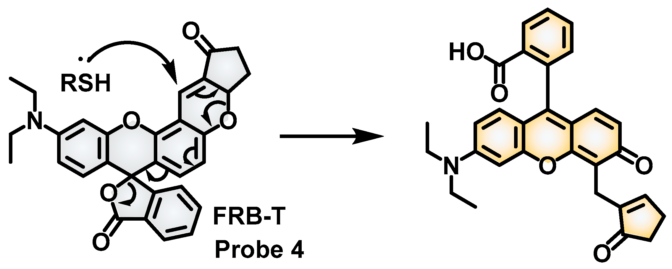
2.3. Biothiol Fluorescent Probes Based on Reduction Reactions
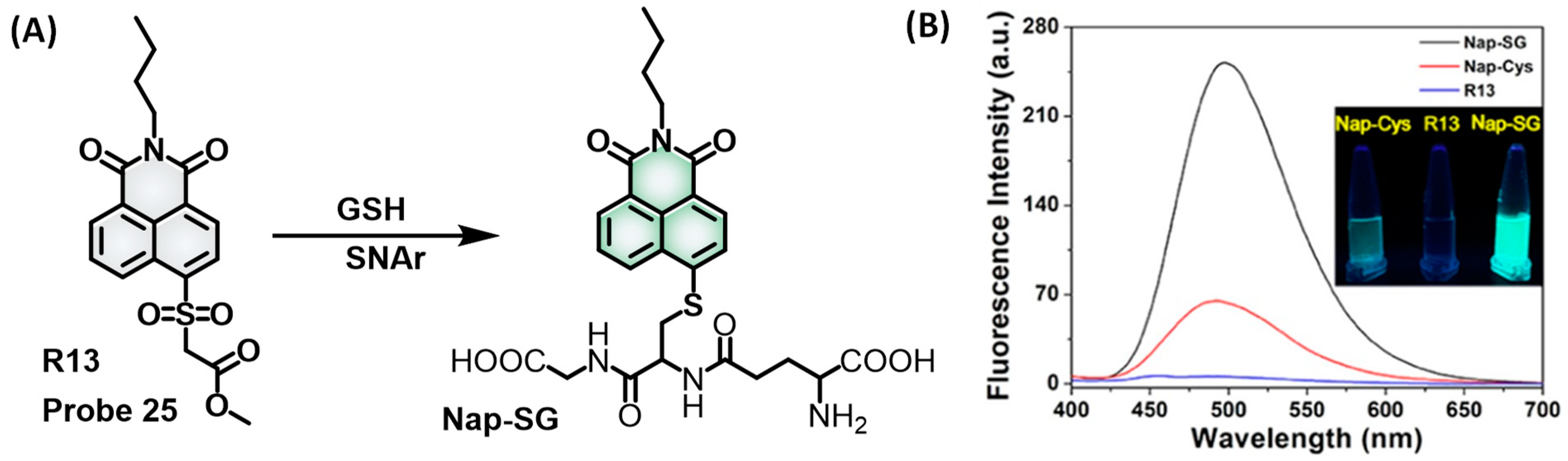
2.4. Biothiol Fluorescent Probes Based on SNAr Reactions
3. Design Strategies for Specific Fluorescent Probes for Intracellular Biothiols
3.1. Specific Fluorescent Probes for Intracellular Cys
3.2. Specific Fluorescent Probes for GSH
3.3. Mitochondria-Specific Fluorescent Probes for Biothiols
4. Design Strategies for Reversible Fluorescent Probes for Biothiols
5. Design Strategies of Fluorescent Probes for Discriminated Detection of Biothiols
5.1. Discriminated Probes with a Single Reactive Site for Biothiols
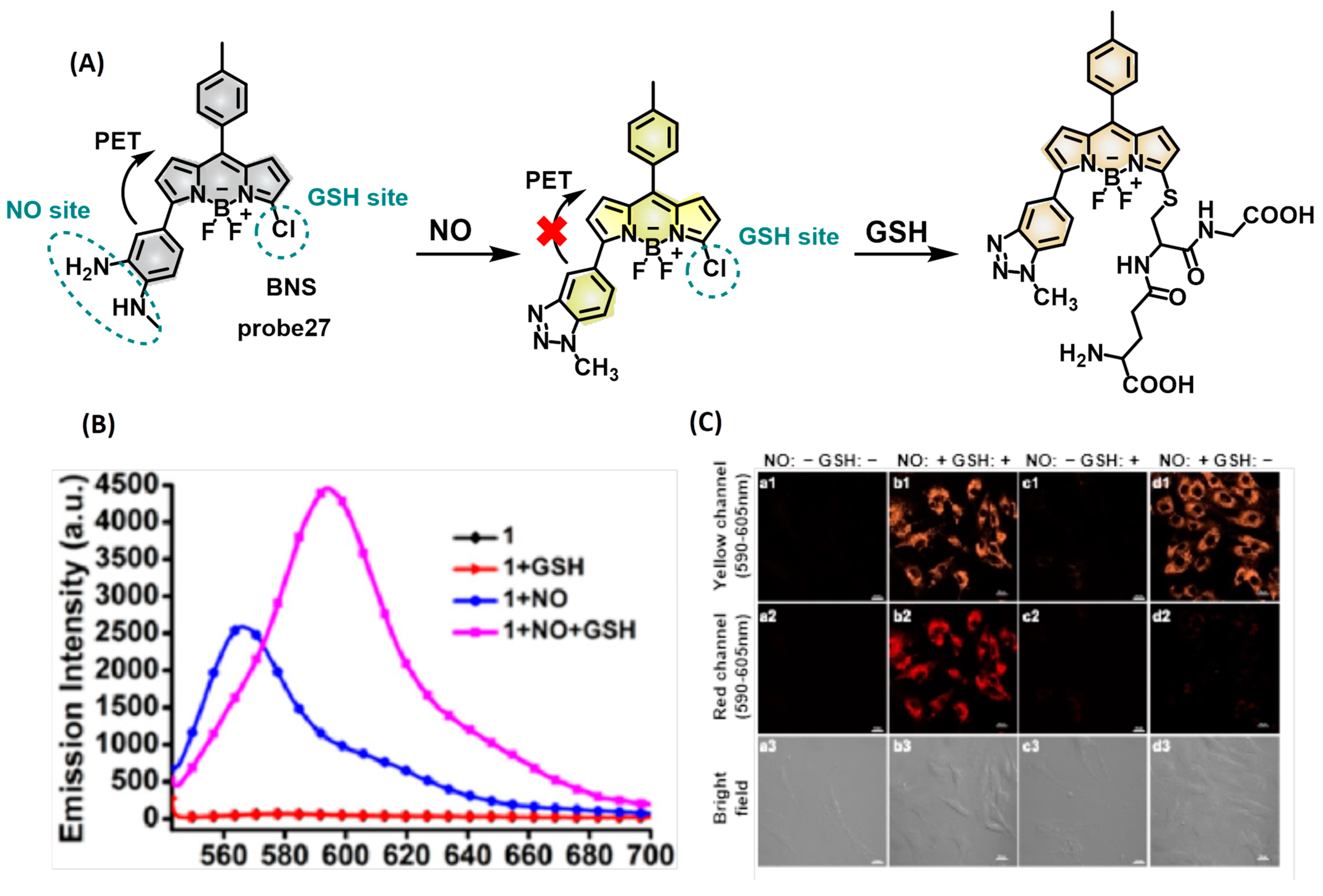
5.2. Discriminated with Multiple Reactive Sites
6. Conclusions and Outlook
Author Contributions
Funding
Institutional Review Board Statement
Informed Consent Statement
Acknowledgments
Conflicts of Interest
References
- Han, H.; Wang, F.; Chen, J.J.; Li, X.X.; Fu, G.Q.; Zhou, J.W.; Zhou, D.S.; Wu, W.; Chen, H.M. Changes in biothiol levels are closely associated with alzheimer’s disease. J. Alzheimers Dis. 2021, 82, 527–540. [Google Scholar] [CrossRef] [PubMed]
- Suzuki, Y.J.; Shi, S.S.; Day, R.M.; Blumberg, J.B. Differential regulation of MAP kinase signaling by pro- and antioxidant biothiols. Ann. N. Y. Acad. Sci. 2000, 899, 159–167. [Google Scholar] [CrossRef]
- Dalton, T.P.; Chen, Y.; Schneider, S.N.; Nebert, D.W.; Shertzer, H.G. Genetically altered mice to evaluate glutathione homeostasis in health and disease. Free Radic. Biol. Med. 2004, 37, 1511–1526. [Google Scholar] [CrossRef]
- Lu, S.C. Dysregulation of glutathione synthesis in liver disease. Liver Res. 2020, 4, 64–73. [Google Scholar] [CrossRef]
- Vairetti, M.; Di Pasqua, L.G.; Cagna, M.; Richelmi, P.; Ferrigno, A.; Berardo, C. Changes in glutathione content in liver diseases: An update. Antioxidants 2021, 10, 364. [Google Scholar] [CrossRef] [PubMed]
- Travaglio, M.; Michopoulos, F.; Yu, Y.Z.; Popovic, R.; Foster, E.; Coen, M.; Martins, L.M. Increased cysteine metabolism in PINK1 models of Parkinson’s disease. Dis. Model Mech. 2023, 16, dmm049727. [Google Scholar] [CrossRef] [PubMed]
- Bonifácio, V.D.; Pereira, S.A.; Serpa, J.; Vicente, J.B. Cysteine metabolic circuitries: Druggable targets in cancer. Brit. J. Cancer 2021, 124, 862–879. [Google Scholar] [CrossRef]
- Mani, S.; Yang, G.D.; Wang, R. A critical life-supporting role for cystathionine γ-lyase in the absence of dietary cysteine supply. Free Radic. Biol. Med. 2011, 50, 1280–1287. [Google Scholar] [CrossRef]
- Hasan, T.; Arora, R.; Bansal, A.K.; Bhattacharya, R.; Sharma, G.S.; Singh, L.R. Disturbed homocysteine metabolism is associated with cancer. Exp. Mol. Med. 2019, 51, 1–13. [Google Scholar] [CrossRef]
- Koklesova, L.; Mazurakova, A.; Samec, M.; Biringer, K.; Samuel, S.M.; Büsselberg, D.; Kubatka, P.; Golubnitschaja, O. Homocysteine metabolism as the target for predictive medical approach, disease prevention, prognosis, and treatments tailored to the person. EPMA J. 2021, 12, 477–505. [Google Scholar] [CrossRef]
- Pi, T.T.; Liu, B.; Shi, J.S. Abnormal homocysteine metabolism: An insight of Alzheimer’s disease from DNA methylation. Behav. Neurol. 2020, 2020, 8438602. [Google Scholar] [CrossRef] [PubMed]
- Rehman, T.; Shabbir, M.A.; Inam Ur Raheem, M.; Manzoor, M.F.; Ahmad, N.; Liu, Z.W.; Ahmad, M.H.; Siddeeg, A.; Abid, M.; Aadil, R.M. Cysteine and homocysteine as biomarker of various diseases. Food Sci. Nutr. 2020, 8, 4696–4707. [Google Scholar] [CrossRef] [PubMed]
- Guidara, W.; Messedi, M.; Naifar, M.; Maalej, M.; Grayaa, S.; Omri, S.; Thabet, J.B.; Maalej, M.; Charfi, N.; Ayadi, F. Predictive value of oxidative stress biomarkers in drug-free patients with schizophrenia and schizo-affective disorder. Psychiat. Res. 2020, 293, 113467. [Google Scholar] [CrossRef]
- Ivanov, A.V.; Popov, M.A.; Aleksandrin, V.V.e.; Kozhevnikova, L.M.; Moskovtsev, A.A.; Kruglova, M.P.; Vladimirovna, S.E.; Aleksandrovich, S.V.; Kubatiev, A.A. Determination of glutathione in blood via capillary electrophoresis with pH-mediated stacking. Electrophoresis 2022, 43, 1859–1870. [Google Scholar] [CrossRef]
- Xie, J.W.; Cheng, D.; Li, P.P.; Xu, Z.J.; Zhu, X.H.; Zhang, Y.Y.; Li, H.T.; Liu, X.Y.; Liu, M.L.; Yao, S.Z. Au/Metal–organic framework nanocapsules for electrochemical determination of glutathione. ACS Appl. Nano Mater. 2021, 4, 4853–4862. [Google Scholar] [CrossRef]
- Jîtcă, G.; Fogarasi, E.; Ősz, B.E.; Vari, C.E.; Fülöp, I.; Croitoru, M.D.; Rusz, C.M.; Dogaru, M.T. Profiling the concentration of reduced and oxidized glutathione in rat brain using HPLC/DAD chromatographic system. Molecules 2021, 26, 6590. [Google Scholar] [CrossRef]
- Forgacsova, A.; Galba, J.; Mojzisova, J.; Mikus, P.; Piestansky, J.; Kovac, A. Ultra-high performance hydrophilic interaction liquid chromatography–triple quadrupole tandem mass spectrometry method for determination of cysteine, homocysteine, cysteinyl-glycine and glutathione in rat plasma. J. Pharm. Biomed 2019, 164, 442–451. [Google Scholar] [CrossRef] [PubMed]
- Li, X.C.; Zhao, S.J.; Li, B.L.; Yang, K.; Lan, M.H.; Zeng, L.T. Advances and perspectives in carbon dot-based fluorescent probes: Mechanism, and application. Coord. Chem. Rev. 2021, 431, 213686. [Google Scholar] [CrossRef]
- Jun, J.V.; Chenoweth, D.M.; Petersson, E.J. Rational design of small molecule fluorescent probes for biological applications. Org. Biomol. Chem. 2020, 18, 5747–5763. [Google Scholar] [CrossRef] [PubMed]
- Cao, D.X.; Liu, Z.Q.; Verwils, P.; Koo, S.; Jangjili, P.; Kim, J.S.; Lin, W.Y. Coumarin-based small-molecule fluorescent chemosensors. Chem. Rev. 2019, 119, 10403–10519. [Google Scholar] [CrossRef]
- Yin, J.L.; Huang, L.; Wu, L.L.; Li, J.F.; James, T.D.; Lin, W.Y. Small molecule based fluorescent chemosensors for imaging the microenvironment within specific cellular regions. Chem. Soc. Rev. 2021, 50, 12098–12150. [Google Scholar] [CrossRef] [PubMed]
- Gao, L.Q.; Wang, W.; Wang, X.; Yang, F.; Xie, L.X.; Shen, J.; Brimble, M.A.; Xiao, Q.C.; Yao, S.Q. Fluorescent probes for bioimaging of potential biomarkers in Parkinson’s disease. Chem. Soc. Rev. 2021, 50, 1219–1250. [Google Scholar] [CrossRef] [PubMed]
- Chen, L.; Li, J.B.; Chen, D.G. Recent advances in fluorescent probes for biothiols. Chin. J. Org. Chem. 2021, 41, 611–623. [Google Scholar] [CrossRef]
- Liu, W.J.; Chen, J.; Xu, Z.C. Fluorescent probes for biothiols based on metal complex. Coord. Chem. Rev. 2021, 429, 213638. [Google Scholar] [CrossRef]
- Dai, J.A.; Ma, C.G.; Zhang, P.; Fu, Y.Q.; Shen, B.X. Recent progress in the development of fluorescent probes for detection of biothiols. Dye. Pigment. 2020, 177, 108321. [Google Scholar] [CrossRef]
- Dong, J.N.; Lu, G.W.; Tu, Y.Y.; Fan, C.B. Recent research progress of red-emitting/near-infrared fluorescent probes for biothiols. New J. Chem. 2022, 46, 10995–11020. [Google Scholar] [CrossRef]
- Zhang, R.; Yong, J.X.; Yuan, J.L.; Xu, Z.P. Recent advances in the development of responsive probes for selective detection of cysteine. Coord. Chem. Rev. 2020, 408, 213182. [Google Scholar] [CrossRef]
- Yue, Y.K.; Huo, F.J.; Yin, C.X. The chronological evolution of small organic molecular fluorescent probes for thiols. Chem. Sci. 2020, 12, 1220–1226. [Google Scholar] [CrossRef]
- Tarai, A.; Li, Y.; Liu, B.; Zhang, D.; Li, J.; Yan, W.; Zhang, J.; Qu, J.; Yang, Z. A review on recognition of tri-/tetra-analyte by using simple organic colorimetric and fluorometric probes. Coord. Chem. Rev. 2021, 445, 214070. [Google Scholar] [CrossRef]
- Wang, C.; Chi, W.; Qiao, Q.L.; Tan, D.; Xu, Z.C.; Liu, X.G. Twisted intramolecular charge transfer (TICT) and twists beyond TICT: From mechanisms to rational designs of bright and sensitive fluorophores. Chem. Soc. Rev. 2021, 50, 12656–12678. [Google Scholar] [CrossRef]
- Cao, D.X.; Zhu, L.L.; Liu, Z.Q.; Lin, W.Y. Through bond energy transfer (TBET)-based fluorescent chemosensors. J. Photochem. Photobiol. C 2020, 44, 100371. [Google Scholar] [CrossRef]
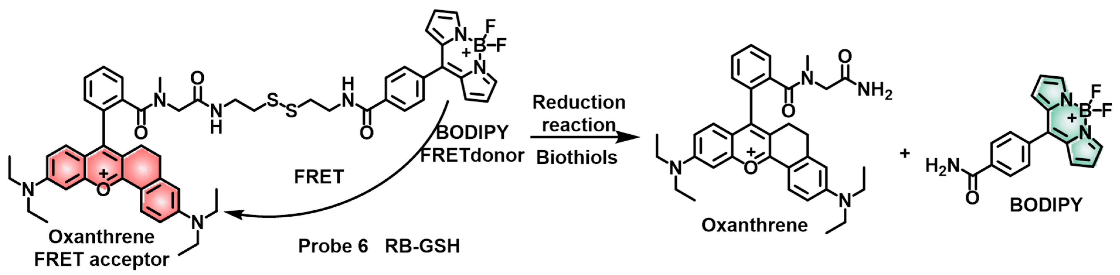
- Wu, L.L.; Huang, C.S.; Emery, B.P.; Sedgwick, A.C.; Bull, S.D.; He, X.P.; Tian, H.; Yoon, J.J.; Sessler, J.L.; James, T.D. Förster resonance energy transfer (FRET)-based small-molecule sensors and imaging agents. Chem. Soc. Rev. 2020, 49, 5110–5139. [Google Scholar] [CrossRef] [PubMed]
- Pal, A.; Karmakar, M.; Bhatta, S.R.; Thakur, A. A detailed insight into anion sensing based on intramolecular charge transfer (ICT) mechanism: A comprehensive review of the years 2016 to 2021. Coord. Chem. Rev. 2021, 448, 214167. [Google Scholar] [CrossRef]
- Gu, H.; Wang, W.J.; Wu, W.Y.; Wang, M.L.; Liu, Y.R.; Jiao, Y.J.; Wang, F.; Wang, F.; Chen, X.Q. Excited-state intramolecular proton transfer (ESIPT)-based fluorescent probes for biomarker detection: Design, mechanism, and application. Chem. Commun. 2023, 59, 2056–2071. [Google Scholar] [CrossRef] [PubMed]
- Cai, X.L.; Liu, B. Aggregation-induced emission: Recent advances in materials and biomedical applications. Angew. Chem. Int. Edit. 2020, 59, 9868–9886. [Google Scholar] [CrossRef]
- Niu, H.Y.; Liu, J.W.; O’Connor, H.M.; Gunnlaugsson, T.; James, T.D.; Zhang, H. Photoinduced electron transfer (PeT) based fluorescent probes for cellular imaging and disease therapy. Chem. Soc. Rev. 2023, 52, 2322–2357. [Google Scholar] [CrossRef] [PubMed]
- Sun, Q.; Ren, R.; Wu, P.P.; Zhuo, L.S.; Dong, H.; Peng, H.T.; Cao, Y.F.; Luo, X.G.; She, N.F. A 2, 7-naphthyridine-based fluorescent turn-on probe for detection of biothiols in vitro and in vivo. Dye. Pigment. 2020, 182, 108702. [Google Scholar] [CrossRef]
- Luo, J.D.; Xie, Z.L.; Lam, J.W.Y.; Cheng, L.; Chen, H.Y.; Qiu, C.F.; Kwok, H.S.; Zhan, X.W.; Liu, Y.Q.; Zhu, D.B.; et al. Aggregation-induced emission of 1-methyl-1,2,3,4,5-pentaphenylsilole. Chem. Commun. 2001, 1740–1741. [Google Scholar] [CrossRef] [PubMed]
- Yang, J.L.; Wei, J.M.; Luo, F.; Dai, J.; Hu, J.J.; Lou, X.D.; Xia, F. Enzyme-responsive peptide-based AIE bioprobes. Top. Curr. Chem. (Cham) 2020, 378, 47. [Google Scholar] [CrossRef]
- Alam, P.; Leung, N.L.C.; Zhang, J.; Kwok, R.T.K.; Lam, J.W.Y.; Tang, B.Z. AIE-based luminescence probes for metal ion detection. Coord. Chem. Rev. 2021, 429, 213693. [Google Scholar] [CrossRef]
- Dai, F.F.; Zhao, M.; Yang, F.Z.; Wang, T.T.; Wang, C. An ESIPT coupled AIE fluorescent probe for biothiols detection and imaging based on a chalcone fluorophore. Dye. Pigment. 2020, 183, 108627. [Google Scholar] [CrossRef]
- Yuan, D.; Pan, K.X.; Xu, S.Y.; Wang, L.Y. Dual-channel recognition of human serum albumin and glutathione by fluorescent probes with site-dependent responsive features. Anal. Chem. 2022, 94, 12391–12397. [Google Scholar] [CrossRef] [PubMed]
- Zheng, S.Y.; Peng, J.Q.; Jiang, L.; Gu, H.; Wang, F.; Wang, C.Q.; Lu, S.; Chen, X.Q. A rhodol-derived probe for intracellular biothiols imaging and rapid labelling of sulfhydryl-containing proteins. Sens. Actuators B Chem. 2022, 367, 132148. [Google Scholar] [CrossRef]
- Ma, K.Q.; Zhao, L.L.; Yue, Y.K.; Huo, F.J.; Chao, J.B.; Yin, C.X. Thiol “click” chromene ring opening and subsequent cascade nucleophilic cyclization NIR fluorescence imaging reveal high levels of thiol in drug-resistant cells. Anal. Chem. 2020, 92, 15936–15942. [Google Scholar] [CrossRef] [PubMed]
- Wang, L.; Wang, J.B.; Xia, S.; Wang, X.X.; Yu, Y.T.; Zhou, H.W.; Liu, H.Y. A FRET-based near-infrared ratiometric fluorescent probe for detection of mitochondria biothiol. Talanta 2020, 219, 121296. [Google Scholar] [CrossRef]
- Kang, Y.F.; Niu, L.Y.; Yang, Q.Z. Fluorescent probes for detection of biothiols based on “aromatic nucleophilic substitution-rearrangement” mechanism. Chin. Chem. Lett. 2019, 30, 1791–1798. [Google Scholar] [CrossRef]
- Cheng, W.; Xue, X.Q.; Zhang, F.; Zhang, B.X.; Li, T.H.; Peng, L.; Cho, D.H.; Chen, H.L.; Fang, J.G.; Chen, X.G. A novel AIEgen-based probe for detecting cysteine in lipid droplets. Anal. Chim. Acta 2020, 1127, 20–28. [Google Scholar] [CrossRef]
- Xu, Z.Y.; Si, S.F.; Zhang, Z.J.; Tan, H.Y.; Qin, T.Y.; Wang, Z.L.; Wang, D.; Wang, L.; Liu, B. A fluorescent probe with dual acrylate sites for discrimination of different concentration ranges of cysteine in living cells. Anal. Chim. Acta 2021, 1176, 338763. [Google Scholar] [CrossRef]
- Li, Z.G.; Zhang, Y.; Jiang, Y.H.; Li, H.W.; Chen, C.Y.; Liu, W.S. A ratiometric fluorescent probe based on two-isophorone fluorophore for detecting cysteine. J. Mater. Chem. B 2022, 10, 6207–6213. [Google Scholar] [CrossRef]
- Jiao, S.; He, X.; Xu, L.B.; Ma, P.Y.; Liu, C.M.; Huang, Y.B.; Sun, Y.; Wang, X.H.; Song, D.Q. A red-emitting fluorescence turn-on probe for the discrimination of cysteine from biothiols and its bioimaging applications in living cells. Sens. Actuators B Chem. 2019, 290, 47–52. [Google Scholar] [CrossRef]
- Lu, Z.L.; Lu, Y.N.; Fan, C.H.; Sun, X.; Shao, W.Q.; Jiang, N.; Gong, X.Y.; Lu, Y.Z.; Sun, G.X.; Jiang, X.C. Ultrafast deep-red emission fluorescent probe for highly selective imaging of endogenous cysteine in living cells and mice. Sens. Actuators B Chem. 2019, 290, 581–590. [Google Scholar] [CrossRef]
- Wang, K.; Wang, W.; Guo, M.Y.; Chen, S.Y.; Yang, Y.S.; Wang, B.Z.; Xu, C.; Zhu, H.L. Design and synthesis of a novel “turn-on” long range measuring fluorescent probe for monitoring endogenous cysteine in living cells and Caenorhabditis elegans. Anal. Chim. Acta 2021, 1152, 338243. [Google Scholar] [CrossRef] [PubMed]
- Zhang, B.X.; Zhang, H.J.; Zhong, M.; Wang, S.; Xu, Q.H.; Cho, D.H.; Qiu, H.D. A novel off-on fluorescent probe for specific detection and imaging of cysteine in live cells and in vivo. Chin. Chem. Lett. 2020, 31, 133–135. [Google Scholar] [CrossRef]
- Ren, H.X.; Huo, F.J.; Zhang, Y.B.; Zhao, S.H.; Yin, C.X. An NIR ESIPT-based fluorescent probe with large stokes shift for specific detection of Cys and its bioimaging in cells and mice. Sens. Actuators B Chem. 2020, 319, 128248. [Google Scholar] [CrossRef]
- Liu, J.; Wang, Z.Q.; Mao, G.J.; Jiang, W.L.; Tan, M.; Xu, F.; Li, C.Y. A near-infrared fluorescent probe with large Stokes shift for imaging Cys in tumor mice. Anal. Chim. Acta 2021, 1171, 338655. [Google Scholar] [CrossRef]
- Liu, Q.C.; Liu, C.; Jiao, X.J.; Cai, S.T.; He, S.; Zhao, L.C.; Zeng, X.S.; Wang, T.H. Lysosome-targeted near-infrared fluorescent dye and its application in designing of probe for sensitive detection of cysteine in living cells. Dye. Pigment. 2021, 190, 109293. [Google Scholar] [CrossRef]
- Dong, B.L.; Lu, Y.R.; Zhang, N.; Song, W.H.; Lin, W.Y. Ratiometric imaging of cysteine level changes in endoplasmic reticulum during H2O2-Induced Redox Imbalance. Anal. Chem. 2019, 91, 5513–5516. [Google Scholar] [CrossRef]
- Liu, D.J.; Lv, Y.; Chen, M.; Cheng, D.; Song, Z.L.; Yuan, L.; Zhang, X.B. A long wavelength emission two-photon fluorescent probe for highly selective detection of cysteine in living cells and an inflamed mouse model. J. Mater. Chem. B 2019, 7, 3970–3975. [Google Scholar] [CrossRef]
- Long, Y.; Liu, J.R.; Tian, D.H.; Dai, F.; Zhang, S.X.; Zhou, B. Cooperation of ESIPT and ICT processes in the designed 2-(2′-hydroxyphenyl)benzothiazole derivative: A near-infrared two-photon fluorescent probe with a large stokes shift for the detection of cysteine and its application in biological environments. Anal. Chem. 2020, 92, 14236–14243. [Google Scholar] [CrossRef]
- Zhou, P.C.; She, M.Y.; Liu, P.; Zhang, S.Y.; Li, J.L. Measuring the distribution and concentration of cysteine by fluorescent sensor for the visual study of paracetamol-induced pro-sarcopenic effect. Sens. Actuators B Chem. 2020, 318, 128258. [Google Scholar] [CrossRef]
- Karaku, E.; Saya, M.; Dartar, S.; Kaya, B.U.; Emrullahoğlu, M. Fluorescein propiolate: A propiolate-decorated fluorescent probe with remarkable selectivity towards cysteine. Chem. Commun. (Camb) 2019, 55, 4937–4940. [Google Scholar] [CrossRef] [PubMed]
- Huang, Z.; Wu, C.Y.; Li, Y.Q.; Zhou, Z.; Xie, R.H.; Pang, X.; Xu, H.; Li, H.T.; Zhang, Y.Y. A fluorescent probe for the specific detection of cysteine in human serum samples. Anal. Methods 2019, 11, 3280–3285. [Google Scholar] [CrossRef]
- Kang, Z.Z.; Jiang, J.J.; Tu, Q.; Liu, S.T.; Zhang, Y.; Wang, D.E.; Wang, J.Y.; Yuan, M.S. Dual-site chemosensor for monitoring ·OH-cysteine redox in cells and in vivo. J. Am. Chem. Soc. 2023, 145, 507–515. [Google Scholar] [CrossRef] [PubMed]
- Zhang, F.; Chen, F.; Shen, R.P.; Chen, Y.X.; Zhao, Z.J.; Zhang, B.X.; Fang, J.G. Naphthalimide fluorescent skeleton for facile and accurate quantification of glutathione. Anal. Chem. 2023, 95, 4301–4309. [Google Scholar] [CrossRef]
- Jing, C.L.; Wang, Y.Z.; Song, X.R.; Li, X.X.; Kou, M.C.; Zhang, G.L.; Dou, W.; Liu, W.S. Dual-fluorophore and dual-site multifunctional fluorescence sensor for visualizing the metabolic process of GHS to SO2 and the SO2 Toxicological mechanism by two-photon imaging. Anal. Chem. 2023, 95, 1376–1384. [Google Scholar] [CrossRef]
- Chen, J.A.; Zhang, Z.Y.; Gao, J.; Tan, J.H.; Gu, X.F. Design of fluorescent probes with optimized responsiveness and selectivity to GSH. Tetrahedron. Lett. 2019, 60, 1226–1230. [Google Scholar] [CrossRef]
- Me, Y.; Li, H.; Song, C.Z.; Chen, X.G.; Son, Q.H. An 8-arylselenium BODIPY fluorescent probe for rapid and sensitive discrimination of biothiols in living cells. Chem. Commun. (Camb) 2021, 57, 10198–10201. [Google Scholar] [CrossRef]
- Lv, F.; Guo, X.; Wu, H.; Li, H.; Tang, B.; Yu, C.J.; Hao, E.H.; Jiao, L.J. Direct sulfonylation of BODIPY dyes with sodium sulfinates through oxidative radical hydrogen substitution at the alpha-position. Chem. Commun. (Camb) 2020, 56, 15577–15580. [Google Scholar] [CrossRef]
- Huang, C.M.; Qian, Y. A highly sensitive two-photon fluorescent probe for glutathione with near-infrared emission at 719 nm and intracellular glutathione imaging. Spectrochim. Acta A Mol. Biomol. Spectrosc. 2019, 217, 68–76. [Google Scholar] [CrossRef]
- Chen, X.X.; Niu, L.Y.; Yang, Q.Z. Visualizing the underlying signaling pathway related to nitric oxide and glutathione in cardiovascular disease therapy by a sequentially activated fluorescent probe. Anal. Chem. 2021, 93, 3922–3928. [Google Scholar] [CrossRef]
- Feng, S.M.; Gong, S.Y.; Zheng, Z.P.; Feng, G.Q. Smart dual-response probe reveals an increase of GSH level and viscosity in Cisplatin-induced apoptosis and provides dual-channel imaging for tumor. Sens. Actuators B Chem. 2022, 351, 130940. [Google Scholar] [CrossRef]
- Ren, H.X.; Huo, F.J.; Shen, T.R.; Liu, X.G.; Yin, C.X. Molecular-dimension-dependent ESIPT break for specific reversible response to GSH and its real-time bioimaging. Anal. Chem. 2021, 93, 12801–12807. [Google Scholar] [CrossRef]
- Lee, H.Y.; Choi, Y.P.; Kim, S.; Yoon, T.J.; Guo, Z.Q.; Lee, S.Y.; Swamy, K.M.K.; Kim, G.; Lee, J.Y.; Shin, I.; et al. Selective homocysteine turn-on fluorescent probes and their bioimaging applications. Chem. Commun. 2014, 50, 6967–6969. [Google Scholar] [CrossRef]
- Barve, A.; Lowry, M.; Escobedo, J.O.; Thainashmuthu, J.; Strongin, R.M. Fluorescein tri-aldehyde promotes the selective detection of homocysteine. J. Fluoresc. 2016, 26, 731–737. [Google Scholar] [CrossRef]
- Luo, X.Z.; Zhang, C.L.; Yuan, F.; Cheng, S.S.; Zhu, Y.X.; Xiang, M.M.; Hu, X.Y.; Xian, Y.Z. Dual-channel fluorescent probe for the detection of peroxynitrite and glutathione in mitochondria: Accurate discrimination of Inflammatory and progressing tumor cells. Anal. Chem. 2022, 94, 15790–15800. [Google Scholar] [CrossRef]
- Zhu, Y.D.; Pan, H.T.; Song, Y.Y.; Jing, C.; Gan, J.A.; Zhang, J.J. Mitochondria-targeted fluorescent probe for rapid detection of thiols and its application in bioimaging. Dye. Pigment. 2021, 191, 109376. [Google Scholar] [CrossRef]
- Yue, L.Z.; Huang, H.W.; Song, W.H.; Lin, W.Y. Research on mitochondrial oxidative stress accompanying the diabetic process under airborne particulate matter pollution by NIR fluorescence imaging of cysteine. Chem. Eng. J. 2022, 441, 135981. [Google Scholar] [CrossRef]
- Zhao, L.; He, X.; Li, D.; Xu, S.M.; Huang, Y.B.; Li, X.L.; Wang, X.H.; Sun, Y.; Ma, P.Y.; Song, D.Q. A novel fluorescence probe for localization of nucleoli developed via chain reaction of endogenous cysteine in cells. J. Mater. Chem. B 2020, 8, 7652–7658. [Google Scholar] [CrossRef] [PubMed]
- Li, S.J.; Wang, P.P.; Ye, M.T.; Yang, K.; Cheng, D.; Mao, Z.Q.; He, L.W.; Liu, Z.H. Cysteine-activatable near-infrared fluorescent probe for dual-channel tracking lipid droplets and mitochondria in epilepsy. Anal. Chem. 2023, 95, 5133–5141. [Google Scholar] [CrossRef] [PubMed]
- Tian, M.; Liu, Y.; Jiang, F.L. On the route to quantitative detection and real-time monitoring of glutathione in living cells by reversible fluorescent probes. Anal. Chem. 2020, 92, 14285–14291. [Google Scholar] [CrossRef]
- Liu, H.; Wang, S.S.; Gao, H.; Shen, Z. Reversible reaction-based fluorescent probes for dynamic sensing and bioimaging. Eur. J. Org. Chem. 2020, 2020, 5647–5663. [Google Scholar] [CrossRef]
- Khatun, S.; Yang, S.; Zhao, Y.Q.; Lu, Y.X.; Podder, A.; Zhou, Y.; Bhuniya, S. Highly chemoselective self-calibrated fluorescent probe monitors Glutathione dynamics in nucleolus in live cells. Anal. Chem. 2020, 92, 10989–10995. [Google Scholar] [CrossRef] [PubMed]
- Tian, M.; Liu, X.Y.; He, H.; Ma, X.Z.; Liang, C.; Liu, Y.; Jiang, F.L. Real-time imaging of intracellular glutathione levels based on a ratiometric fluorescent probe with extremely fast response. Anal. Chem. 2020, 92, 10068–10075. [Google Scholar] [CrossRef] [PubMed]
- Liu, H.Z.; Song, W.T.; Zhang, S.R.; Chan, K.S.; Guo, Z.J.; Shen, Z. A ratiometric fluorescent probe for real-time monitoring of intracellular glutathione fluctuations in response to cisplatin. Chem. Sci. 2020, 11, 8495–8501. [Google Scholar] [CrossRef] [PubMed]
- Li, N.; Wang, T.; Wang, N.; Fan, M.T.; Cui, X.Y. A substituted-rhodamine-based reversible fluorescent probe for in vivo quantification of glutathione. Angew. Chem. Int. Ed. Engl. 2023, 135, e202217326. [Google Scholar] [CrossRef]
- Yue, Y.K.; Huo, F.J.; Pei, X.Y.; Wang, Y.T.; Yin, C.X. Fluorescent imaging of resveratrol induced subcellular cysteine up-regulation. Anal. Chem. 2020, 92, 6598–6603. [Google Scholar] [CrossRef]
- Yin, C.X.; Xiong, K.M.; Huo, F.J.; Salamanca, J.C.; Strongin, R.M. Fluorescent probes with multiple binding sites for the discrimination of Cys, Hcy, and GSH. Angew. Chem. Int. Ed. Engl. 2017, 56, 13188–13198. [Google Scholar] [CrossRef]
- Xu, Z.Y.; Qin, T.Y.; Zhou, X.F.; Wang, L.; Liu, B. Fluorescent probes with multiple channels for simultaneous detection of Cys, Hcy, GSH, and H2S. TrAc.-Tends Anal. Chem. 2019, 121, 115672. [Google Scholar] [CrossRef]
- Wang, W.; Wang, K.; Wang, X.T.; Man, R.J.; Xu, C.; Yang, Y.S.; Zhu, H.L. A novel selective probe for detecting glutathione from other biothiols based on the concept of fluorescence fusion. Anal. Chim. Acta 2021, 1177, 338786. [Google Scholar] [CrossRef]
- Zhu, N.S.; Guo, X.L.; Pang, S.R.; Chang, Y.L.; Liu, X.M.; Shi, Z.; Feng, S.H. A mitochondria-immobilized unimolecular fluorescent probe for multiplexing imaging of living cancer cells. Anal. Chem. 2020, 92, 11103–11110. [Google Scholar] [CrossRef]
- Yang, X.P.; Liu, J.F.; Xie, P.Y.; Han, X.J.; Zhang, D.; Ye, Y.; Zhao, Y. Visualization of biothiols and HClO in cancer therapy via a multi-responsive fluorescent probe. Sens. Actuators B Chem. 2021, 347, 130620. [Google Scholar] [CrossRef]
- Su, P.R.; Zhu, Z.W.; Tian, Y.H.; Liang, L.J.; Wu, W.Y.; Cao, J.; Cheng, B.; Liu, W.S.; Tang, Y. A TAT peptide-based ratiometric two-photon fluorescent probe for detecting biothiols and sequentially distinguishing GSH in mitochondria. Talanta 2020, 218, 121127. [Google Scholar] [CrossRef] [PubMed]
- Zhang, H.; Yue, X.X.; Li, W.X.; Chen, W.Q.; Wang, Y.G.; Li, X.; Ye, Y.; Song, X.Z. Selective and discriminative fluorescence sensing of Cys, Hcy, GSH and H2S with concise and distinct signals. Sens. Actuators B Chem. 2021, 331, 129394. [Google Scholar] [CrossRef]
- Guo, T.Y.; Chen, X.Y.; Qu, W.B.; Yang, B.; Tian, R.W.; Geng, Z.R.; Wang, Z.L. Red and rear-infrared fluorescent probe for distinguishing cysteine and homocysteine through single-wavelength excitation with distinctly dual emissions. Anal. Chem. 2022, 94, 5006–5013. [Google Scholar] [CrossRef]
- Han, S.H.; Zhang, H.; Yue, X.X.; Wang, J.P.; Yang, L.; Wang, B.H.; Song, X.Z. A ratiometric, fast-responsive, and single-wavelength excited fluorescent probe for the discrimination of Cys and Hcy. Anal. Chem. 2021, 93, 10934–10939. [Google Scholar] [CrossRef]
- Yin, G.X.; Niu, T.T.; Yu, T.; Gan, Y.B.; Sun, X.Y.; Yin, P.; Chen, H.M.; Zhang, Y.Y.; Li, H.T.; Yao, S.Z. Simultaneous visualization of endogenous homocysteine, cysteine, glutathione, and their transformation through different fluorescence channels. Angew. Chem. Int. Ed. Engl. 2019, 58, 4557–4561. [Google Scholar] [CrossRef]
- Yang, Y.; Feng, Y.; Li, H.; Shen, R.; Wang, Y.Z.; Song, X.R.; Cao, C.; Zhang, G.L.; Liu, W.S. Hydro-soluble NIR fluorescent probe with multiple sites and multiple excitations for distinguishing visualization of endogenous Cys/Hcy, and GSH. Sens. Actuators B Chem. 2021, 333, 129189. [Google Scholar] [CrossRef]
- Huang, Y.F.; Zhang, Y.B.; Huo, F.J.; Chao, J.B.; Cheng, F.Q.; Yin, C.X. A new strategy: Distinguishable multi-substance detection, multiple pathway tracing based on a new site constructed by the reaction process and its tumor targeting. J. Am. Chem. Soc. 2020, 142, 18706–18714. [Google Scholar] [CrossRef]
- Zhang, H.; Xu, L.Z.; Chen, W.Q.; Huang, J.; Huang, C.S.; Sheng, J.R.; Song, X.Z. Simultaneous discrimination of cysteine, homocysteine, glutathione, and H2S in living cells through a multisignal combination strategy. Anal. Chem. 2019, 91, 1904–1911. [Google Scholar] [CrossRef]
- Kaushik, R.; Nehra, N.; Novakova, V.; Zimcik, P. Near-infrared probes for biothiols (cysteine, homocysteine, and glutathione): A comprehensive review. ACS Omega 2023, 8, 98–126. [Google Scholar] [CrossRef]
- Chen, Z.Z.; Wang, B.Q.; Liang, Y.Q.; Shi, L.; Cen, X.H.; Zheng, L.; Liang, E.; Huang, L.; Cheng, K. Near-infrared fluorescent and photoacoustic dual-mode probe for highly sensitive and selective imaging of cysteine in vivo. Anal. Chem. 2022, 94, 10737–10744. [Google Scholar] [CrossRef]
- Zhang, J.; Zhang, Y.C.; Guo, Q.; Wen, G.H.; Xiao, H.Y.; Qi, S.; Wang, Y.; Zhang, H.T.; Wang, L.D.; Sun, H.Y. Photoacoustic/fluorescence dual-modality probe for biothiol discrimination and tumor diagnosis in cells and mice. ACS Sens. 2022, 7, 1105–1112. [Google Scholar] [CrossRef] [PubMed]
- Ni, X.W.; Chen, K.Z.; Qiao, S.L. A photocontrollable thermosensitive chemical spatiotemporally destabilizes mitochondrial membranes for cell fate manipulation. Biomater. Sci. 2022, 10, 2550–2556. [Google Scholar] [CrossRef] [PubMed]
- Yang, S.; Jiang, J.X.; Zhou, A.X.; Zhou, Y.B.; Ye, W.L.; Cao, D.S.; Yang, R.H. Substrate-photocaged enzymatic fluorogenic probe enabling sequential activation for light-controllable monitoring of intracellular tyrosinase activity. Anal. Chem. 2020, 92, 7194–7199. [Google Scholar] [CrossRef] [PubMed]
- Kim, Y.W.; Kim, J.H.; An, J.M.; Park, C.K.; Kim, D.K. All-nontoxic fluorescent probe for biothiols and its clinical applications for real-time glioblastoma visualization. ACS Sens. 2023, 8, 1723–1732. [Google Scholar] [CrossRef] [PubMed]


| Probe Number | Detection Type | λex/λem (nm) | LOD | Linear Range | Application | Ref. |
|---|---|---|---|---|---|---|
| 1 | Turn on | 355/582 | GSH: 5.46 nM | 0–12 μM | Image GSH in A549 cells. Detect GSH in zebrafish. | [37] |
| 2 | Turn on | 460/600 | GSH: 0.227 mM Cys: 0.283 mM Hcy: 0.182 mM | – – – | Detect biothiols with indicator papers. Image biothiols in PC3 cells and A549 cells. | [41] |
| 3 | Turn on | GSH: 400/575 HAS: 500/660 | GSH: 4.65 mM HAS: 0.035 mM | 5–60 mM 0.5–18 mM | Image drug-induced HSA and endogenous/exogenous GSH in HepG2 cells. | [42] |
| 4 | Turn on | 520/557 | GSH: 49 nM Cys: 70 nM Hcy: 62 nM | – – – | Image endogenous and exogenous biothiols in HeLa cells. Label proteins containing sulfhydryl groups. | [43] |
| 5 | Turn on | 600/698 | GSH: 60 nM Cys: 85 nM | – – | Image endogenous and endogenous biothiols in HeLa cells. Image biothiols in mouse models. | [44] |
| 6 | Ratiometric (I512/I656) | 480/511, 656 | GSH: 0.26 μM | 10–100 μM | Image endogenous and endogenous biothiols in HeLa cells. | [45] |
| 7 | Turn on | 360/560 | Cys: 2.4 μM | 0–100 μM | Image endogenous and endogenous Cys in HepG2 cells. Target lipid drops (LDs) Cys in HepG2 cells, HeLa cells, and RAW 264.7 cells. | [47] |
| 8 | Turn on | 354/449 | Cys: 97 nM | – | Monitor high and low concentration ranges of Cys in 4T1 cells. | [48] |
| 9 | Ratiometric (I568/I540) | 457/540, 568 | Cys: 105.6 nM | 0–300 mM | Image endogenous and exogenous Cys in osteoblasts. | [49] |
| 10 | Turn on | 580/615 | Cys: 0.12 μM | 0–10 μM | Image endogenous and exogenous Cys in BEL-7402. | [50] |
| 11 | Turn on | 580/645 | Cys: 10 nM | 0–200 μM | Image endogenous and exogenous Cys in HeLa cells. Visualize endogenous Cys in nude mouse models. | [51] |
| 12 | Turn on | 709/742 | Cys: 0.11 μM | – | Target mitochondria Cys in HeLa cells. | [52] |
| 13 | Turn on | 570/607 | Cys: 0.04 μM | – | Image endogenous and exogenous Cys in HeLa cells. Image endogenous and exogenous Cys in zebrafish. | [53] |
| 14 | Turn on | 423/686 | Cys: 0.98 mM (pH 7.0), Cys: 0.2 mM (pH 8.0) | – – | Image endogenous and exogenous Cys in HeLa cells. Visualize endogenous Cys in nude mouse models. | [54] |
| 15 | Turn on | 590/770 | Cys: 0.4 μM | 1–70 μM | Image endogenous and exogenous Cys in HCT116 cells. Image endogenous Cys in HCT116-xenograft tumor mice. | [55] |
| 16 | Turn on | 543/633 | Cys: 0.133 μM | 0–300 μM | Image endogenous and exogenous Cys in MCF-7 cells. Image of Cys in Caenorhabditis elegans in various conditions. | [56] |
| 17 | Ratiometric (I550/I440) | 390/440, 550 | Cys: 1.80 μM | 10–80 μM | Target endoplasmic reticulum (ER) Cys in HeLa cells. Image endogenous and exogenous Cys in zebrafishes. Visualize cellular cysteine level changes during H2O2-induced redox imbalance. | [57] |
| 18 | Turn on | 550/610 | Cys: 24.1 nM | – | Image endogenous Cys in HepG2 cells. Visualize Cys in an inflamed mouse model and leg skin slices by two-photon (TP) imaging. | [58] |
| 19 | Turn on | 411/713 | Cys: 102 nM | 1–12.5 μM | Visualize endogenous and exogenous Cys in HeLa cells by TP imaging. Target mitochondria and ER Cys in HeLa cells, HepG2 cells, and PC12 cells. Visualize endogenous and exogenous Cys in zebrafish. Visualize endogenous and exogenous Cys in the mouse brain by TP imaging. | [59] |
| 20 | Turn on | 470/523 | Cys: 0.15 μM | 0–200 μM | Image endogenous and exogenous Cys in living A549 cells. Image Cys in old rats chronically treated with APAP (N-acetyl-p-aminophenol). | [60] |
| 21 | Turn on | 470/523 | Cys: 1.08 μM | 0–200 μM | Image endogenous and exogenous Cys in living A549 cells. | [60] |
| 22 | Turn on | 460/515 | Cys: 182 nM | – | Image endogenous and exogenous Cys in living A549 cells. | [61] |
| 23 | Turn on | 355/440 | Cys: 63 nM | 0–200 μM | Determine Cys in human serum. | [62] |
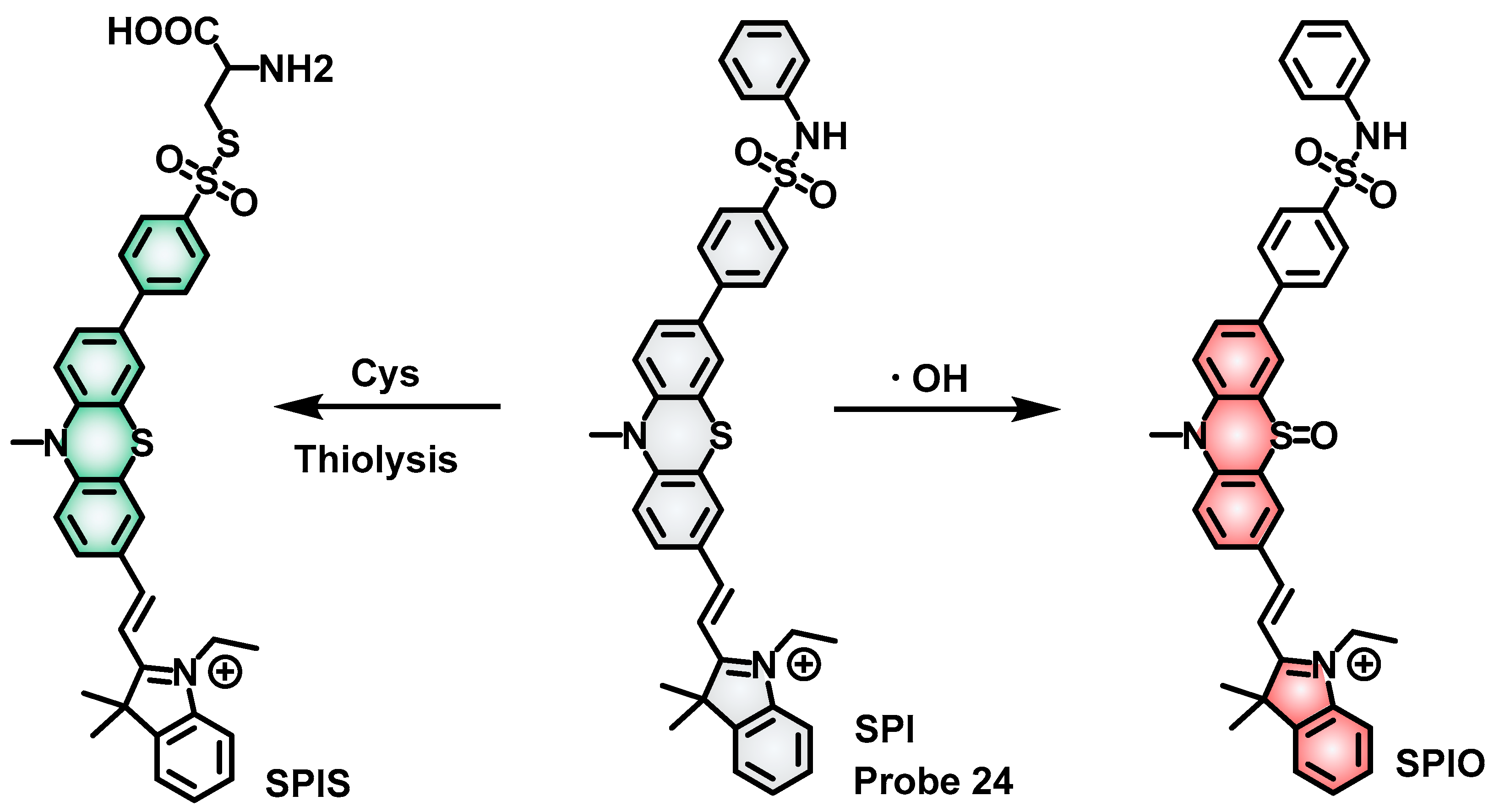
| 24 | Turn on | Cys: 426/538 ·OH: 485/608 | Cys: 30.01 nM ·OH: 69.65 nM | 0–50 μM 0–20 μM | Images endogenous and exogenous Cys in RAW 264.7 cells. Image real-time changes in ·OH and Cys by sulfasalazine (SAS, a GPX4 inhibitor) in RAW 264.7. Monitor ·OH and Cys in living mice and zebrafish models. | [63] |
| 25 | Turn on | GSH: 390/498 | – | – | Determine GSH levels in cells and mouse livers. Quantify GSH content in Parkinson’s mouse brains. | [64] |
| 26 | Cys: Turn on : Ratiometric (I496/I638) | GSH: 410/496 : 410/496, 638 | GSH: 17.43 : 412.45 nM | GSH: 0–1.5 : 0–60 μM | Target mitochondria in living HepG2 cells. Image endogenous GSH of HepG2 cells in real-time. Image exogenous of HepG2 cells in real-time. Image the metabolic process of GSH-SO2 in HepG2 cells. Monitoring of the high-dose SO2 effect on GSH content in cells. | [65] |
| 27 | Turn on | GSH: 532/595 NO: 532/565 | GSH: 29 nM NO: 34 nM | 0–100 μM 0–10 μM | Image NO and GSH in human umbilical vein endothelial cells (HUVECs). Visualize NO-induced GSH enhancement induced by Pravastatin or VC in HUVECs. Image NO and GSH in zebrafish models induced by Pravastatin or VC. | [70] |
| 28 | GSH: Ratiometric (I535/I650) | GSH: 500/650, 450/509 | GSH: 0.2 μM | GSH: 0–20 μM | Target mitochondria in living MCF-7 cells and HeLa cells. Image endogenous and exogenous GSH in MCF-7 cells. Image of viscosity in HeLa cells induced by Nystain (an ionophore that can destroy the structure of mitochondria) and lipopolysaccharide, LPS (an initiator of inflammation). Image of changes in GSH and viscosity in apoptosis induced by cisplatin. Distinguish normal cells from cancer cells (HeLa cells and RAW 264.7). Discriminate the tumor from the normal tissues in H1975-tumor mice. | [71] |
| Viscosity: Turn on | Viscosity: 510/627 | Viscosity: – | Viscosity: 6.582–945 cp | |||
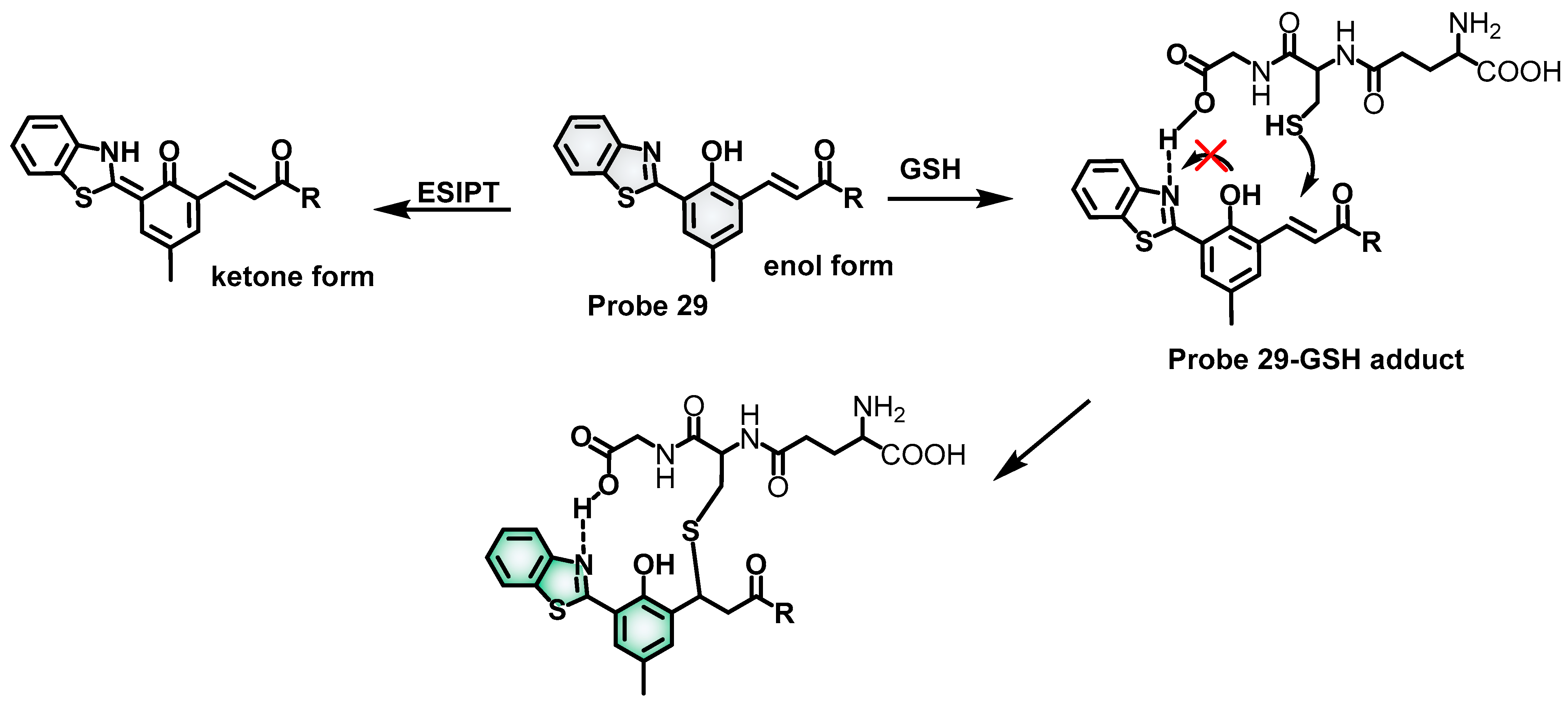
| 29 | Ratiometric (I519/I621) | 415/519, 621 | – | GSH: 0–3 mM | Image endogenous and exogenous GSH in RAW 264.7 cells. Monitor GSH variation in cells treated by LPS or heat. | [72] |
| 30 | Turn on | GSH: 615/670 ONOO−: 450/530 | GSH: 5.36 μM ONOO−: 89.32 nM | 0–500 mM 0–10 μM | Image endogenous GSH and ONOO− in living cells. Distinguish between normal, malignant, and metastatic breast cells. Distinct normal, inflammatory, and cancerous tissues in different zebrafish models. | [75] |
| 31 | Turn on | 545/604 | GSH: 13.8 μM Cys: 13.4 μM Hcy: 22.9 μM | 0–4 μM – – | Target mitochondrial biothiols in HeLa cells. | [76] |
| 32 | Turn on | 495/628 | Cys: 36.8 nM | 0–70 μM | Target mitochondrial Cys in living HeLa cells. Image endogenous and exogenous Cys in HeLa cells. Image endogenous Cys in diabetic cell models is induced by high glucose. Distinguish between normal and diabetic mouse models. | [77] |
| 33 | Turn on | 550/616 | Cys: 27 nm | 0.1–3 μM | Image endogenous and exogenous Cys in HeLa cells. Target mitochondrial Cys and nuclear RNA in living cells. | [78] |
| 34 | Turn on | 430/650 | Cys: 43 nm | 1–100 μM | Target mitochondria and LDs Cys in PC12 cells. Imaging of Cys in LDs and mitochondria during apoptosis induced by lipopolysaccharide (LPS), H2O2, and ultraviolet radiation (UV). Image endogenous Cys in control mice, acute epilepsy mice, chronic epilepsy mice, curcumin-treated acute epilepsy mice, and curcumin-treated chronic epilepsy mice. | [79] |
| 35 | Ratiometric (I460/I510) | 350/460, 510 | – | GSH: 0–6 mM | Image dynamic changes of GSH levels in HeLa cells treated by inhibitors and donors for GSH. Image dynamic of GSH in S-phase in the early stage of the cell cycle. Visualize dynamic changes in the redox state induced by GSH, H2O2, and GSSG in HeLa cells. | [82] |
| 36 | Ratiometric (F505/F587) | 420/505, 530/587 | – | GSH: 0.1–10 mM | Monitor GSH levels in living cells in real time. | [83] |
| 37 | Ratiometric (F608/F544) | 488/544, 543/608 | – | GSH: 1–10 mM | Image intracellular GSH concentrations in response to cisplatin treatment. | [84] |
| 38 | Ratiometric (F736/F675) | 640/675, 736 | – | GSH: 0–20 mM | Quantification of GSH in living cells. Monitor GSH levels in xenografted tumors. Monitor GSH levels in chronic renal failure. Monitor GSH levels in liver fibrosis. | [85] |
| 39 | Turn on | 414/489 | Cys: 1.331 μM | 0–80 μM | Image mitochondrial Cys in 7702 cells. | [86] |
| 40 | Turn on | 414/489 | – | – | HeLa cellular Cys analysis. | [86] |
| 41 | GSH: Turn on Cys: Turn on Hcy: Turn on | GSH: 400/530 Cys: 400/490, 560 Hcy: 400/490, 560 | GSH: 0.20 μM Cys: – Hcy: – | GSH: 10–100 μM Cys: – Hcy: – | Monitor the GSH levels in cells. | [89] |
| 42 | Cys: Turn on Hcy: Turn on HClO: Turn on GSH: Turn on | Cys: 488/550 Hcy: 488/550 HClO: 543/580; 405/450 GSH: 405/450 | Cys: 333 nM Hcy: 352 nM HClO: 167 nM – – | Cys: 0–100 μM Hcy: 0–100 μM HClO: 4–15 μM – – | Image HClO in MCF-7 cells. Simultaneous visualization of Cys/Hcy, HOCl, and mitochondrial status in A549 cells. | [90] |
| 43 | Cys: Turn on | Cys: 488/550; 560/650 | Cys: 0.21 μM, 0.12 μM | Cys: 0–5 μM 1–16 μM | Discriminative detection of biothiols in KYSE-30 cells. Image biothiols and HClO in KYSE-30 cells. Target mitochondria in MCF-7 cells. | [91] |
| Hcy: Turn on | Hcy: 488/550; 560/650 | Hcy: 0.26 μM, 0.19 μM | Hcy: 0–10 μM 0–18 Μm | |||
| GSH: Turn on | GSH: 560/650 | GSH: 0.40 μM | GSH: 0–8 μM | |||
| HClO: Turn on | HClO: 410/490 | HClO: 2.16 μM | HClO: 0–12 μM | |||
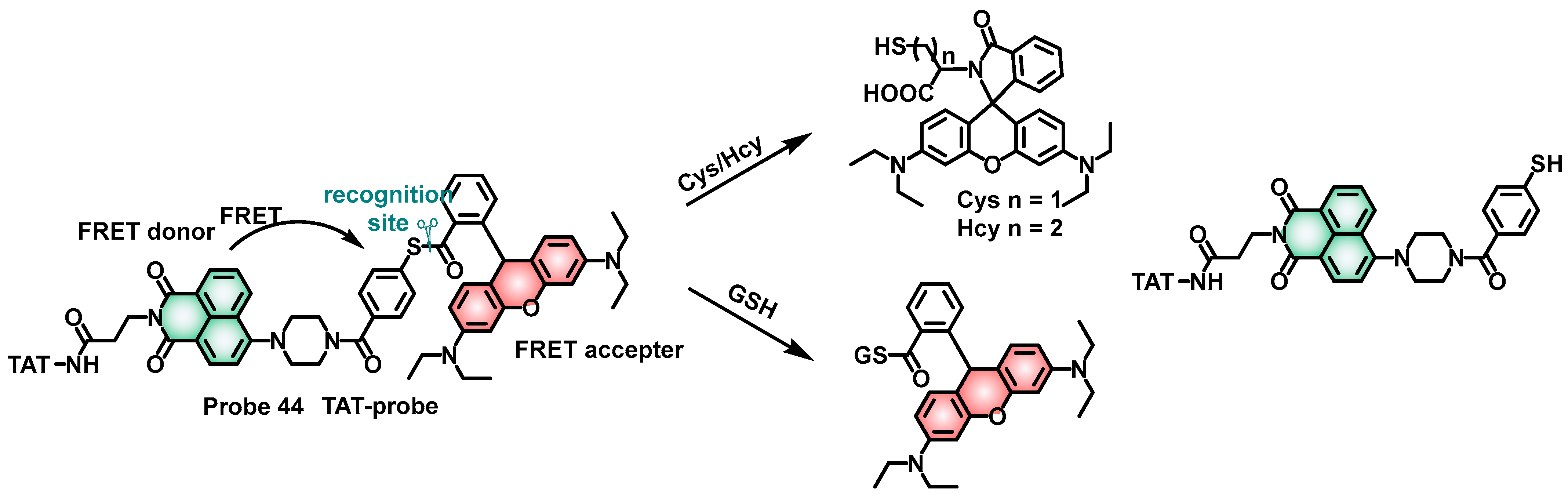
| 44 | Hcy: Ratiometric (I520/I585) | Hcy: 404/520, 585 | Hcy: 6.51 μM | Hcy: 0–12 μM | Detect biothiols and distinguish GSH from Cys and Hcy in the mitochondria of living cells with two-photon excitation. | [92] |
| Cys: Ratiometric (I520/I585) | Cys: 404/520, 585 | Cys: 0.865 μM | Cys: 0–12 μM | |||
| GSH: Ratiometric (I520/I585) | GSH: 404/520, 585 | GSH: 5.15 μM | GSH: 0–12 μM | |||
| 45 | Cys: Turn on Hcy: Turn on GSH: Turn on H2S: Turn on | Cys: 370/450 Hcy: 370/450 GSH: 422/530 H2S: 466/610 | Cys: 0.032 μM Hcy: 0.028 μM GSH: 0.012 μM H2S: 1.2 μM | Cys: 5.0–60.0 μM Hcy: 0–60.0 μM Hcy: 2.0–12.0 μM H2S: 40.0–130.0 μM | Image endogenous four thiols in HeLa cells. Detect these four thiols in zebrafish. | [93] |
| 46 | Cys: Turn on Hcy: Ratiometric (I740/I625) | Cys: 550/625 Hcy: 550/625, 740 | Cys: 0.55 μM Hcy: 0.35 μM | Cys: 0−30 μM Hcy: 0−40 μM | Discrimination of exogenous Cys and Hcy in HepG-2 cells. Target mitochondria in living HepG-2 cells. Visualization of the fluctuation of endogenous Cys in HepG-2 cells. | [94] |
| 47 | Cys: Ratiometric (I485/I608) | Cys: 380/485, 608 | Cys: 0.17 nM | Cys: 0−20.0 μM | Fluorescence imaging of Cys and Hcy in living HeLa cells and zebrafish. | [95] |
| Hcy: Turn on | Hcy: 380/608 | Hcy: 0.19 nM | Hcy: 0−20.0 μM | |||
| 48 | Hcy: Turn on Cys: Turn on GSH: Turn on | Hcy: 375/467 Cys: 400/503 GSH: 500/568 | Hcy: 0.7 nM Cys: 0.2 nM GSH: 1 nM | Hcy: 0−30 μM Cys: 0−30 μM GSH: 0−10 μM | Simultaneous visualizations of endogenous Hcy, Cys, and GSH in BEL-7402, L-02 cells, and RAW 264.7 cells. Dynamic fluorescence imaging for biothiols in BEL-7402. | [96] |
| 49 | Cys: Turn on | Cys: 360/457 517/576 558/611 | Cys: − 2.505 μM − | Cys: − 0−120 μM − | Image endogenous and exogenous Cys/Hcy/GSH in living A375 cells. Measure biothiols in the solutions of fetal bovine serum (FBS). | [97] |
| Hcy: Turn on | Hcy: 480/559 517/576 558/611 | Hcy: − 1.919 μM − | Hcy: − 0−120 μM − | |||
| GSH: Turn on | GSH: 400/529 517/576 558/611 | GSH: − 2.596 μM − | GSH: − 0−120 μM − | |||
| 50 | Cys: Ratiometric (I480/I625) | Cys: 380/480, 625 | Cys: 0.10 μM | Cys: 0−150 μM | Image of Cys/Hcy and GSH in HeLa cells. Image of Cys/GSH metabolism in HeLa cells. Image of Cys/GSH metabolism in HeLa tumor nude mice. Tumor targeting in HeLa tumor-nude mice. | [98] |
| Hcy: Ratiometric (I480/I625) | Hcy: 380/480, 625 | Hcy: 0.02 μM | Hcy: 0−100 μM | |||
| Ratiometric (I545/I625) | GSH: 450/545, 625 | GSH: 0.04 μM | Cys: 0−150 μM | |||
| 51 | Cys: Turn on | Cys: 376/473 424/537, 582/640 | Cys: − − 2.43 μM | Cys: − − 10−60 μM | Distinguish Cys, Hcy, GSH, and H2S from distinct signals in living HeLa cells. | [99] |
| Hcy: Turn on | Hcy: 376/473, 424/537, 582/640 | Hcy: − − 4.78 μM | Hcy: − − 20−100 μM | |||
| GSH: Turn on | GSH: 376/473, 424/537, 582/640 | GSH: − − 2.43 μM | GSH: − − 20−70 μM | |||
| H2S: Turn on | H2S: 582/602 | H2S: 1.14 μM | H2S: 30−100 μM |
Disclaimer/Publisher’s Note: The statements, opinions and data contained in all publications are solely those of the individual author(s) and contributor(s) and not of MDPI and/or the editor(s). MDPI and/or the editor(s) disclaim responsibility for any injury to people or property resulting from any ideas, methods, instructions or products referred to in the content. |
© 2023 by the authors. Licensee MDPI, Basel, Switzerland. This article is an open access article distributed under the terms and conditions of the Creative Commons Attribution (CC BY) license (https://creativecommons.org/licenses/by/4.0/).
Share and Cite
Xie, W.; Jiang, J.; Shu, D.; Zhang, Y.; Yang, S.; Zhang, K. Recent Progress in the Rational Design of Biothiol-Responsive Fluorescent Probes. Molecules 2023, 28, 4252. https://doi.org/10.3390/molecules28104252
Xie W, Jiang J, Shu D, Zhang Y, Yang S, Zhang K. Recent Progress in the Rational Design of Biothiol-Responsive Fluorescent Probes. Molecules. 2023; 28(10):4252. https://doi.org/10.3390/molecules28104252
Chicago/Turabian StyleXie, Wenzhi, Jinyu Jiang, Dunji Shu, Yanjun Zhang, Sheng Yang, and Kai Zhang. 2023. "Recent Progress in the Rational Design of Biothiol-Responsive Fluorescent Probes" Molecules 28, no. 10: 4252. https://doi.org/10.3390/molecules28104252