

Application of Rosmarinic Acid with Its Derivatives in the Treatment of Microbial Pathogens


Abstract
:1. Introduction
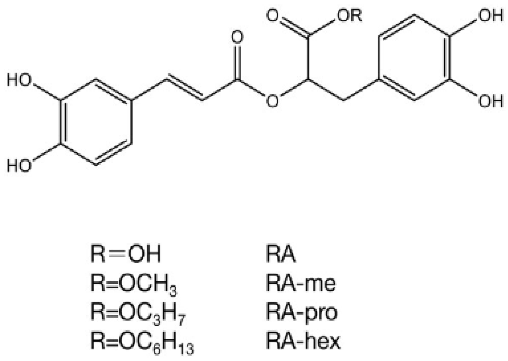
2. Derivates of Rosmarinic Acid

3. Antimicrobial Activity

| Pathogenic Microorganisms | Active Concentrations | References | Pathogenic Microorganisms | Active Concentrations | References |
|---|---|---|---|---|---|
| Staphylococcus epidermidis 5001 Stenotrophomonas maltophilia Enterococcus faecalis C159-6 Staphylococcus lugdunensis T26A3 Pseudomonas aeruginosa ATCC 27583 | MIC (0.3 mg/mL of RA) MIC (0.3 mg/mL of RA) MIC (0.3 mg/mL of RA) MIC (0.6 mg/mL of RA) MIC (2.5 mg/mL of RA) | [77] | Escherichia coli | MIC 0.8 mg/mL of RA; MBC 0.9 mg/mL of RA | [189] |
| Staphylococcus aureus | MIC 1.0 mg/mL of RA; MBC 1.1 mg/mL of RA | ||||
| Salmonella | MIC 0.9 mg/mL of RA; MBC 1.0 mg/mL of RA | ||||
| Bacillus subtilis | MIC 1.0 mg/mL of RA; MBC 1.1 mg/mL of RA | ||||
| Corynebacterium T25-17 Mycobacterium smegmatis 5003 Staphylococcus warneri T12A12 | MIC (2.5 mg/mL of RA) MIC (1.2 mg/mL of RA) MIC (1.2 mg/mL of RA) | Micrococcus luteus | MIC 0.1 mg/mL; MBC 0.2 mg/mL | [193] | |
| Rothia mucilagenosa | MIC 0.1 mg/mL; MBC 0.2 mg/mL | ||||
| Klebsiella sp. | IZ 28 mm at 1 mg/mL of RA | [177] | Streptococcus agalactiae | MIC 0.05 mg/mL; MBC 0.1 mg/mL | |
| Stenotrophomonas maltophela | IZ 19 mm at 1 mg/mL of RA | Streptococcus angiosus | MIC 0.05 mg/mL; MBC 0.1 mg/mL | ||
| Streptomyces sp. | IZ 26 mm at 1 mg/mL of RA | Streptococcus dysgalactie | MIC 0.05 mg/mL; MBC 0.1 mg/mL | ||
| Pantoea agglomerans | IZ 18 mm at 1 mg/mL of RA | Streptococcus oralis | MIC 0.05 mg/mL; MBC 0.1 mg/mL | ||
| Paenibacillus chibensis | IZ < 1 mm at RA-methyl ester IZ 4.4 mm at tannic acid IZ > 2 mm at RA-hexyl ester IZ between 3 mm and 4 mm at RA-propyl ester | [194] | Streptococcus parasanquinis | MIC 0.05 mg/mL; MBC 0.1 mg/mL | |
| Streptococcus pyogenes | MIC 0.1 mg/mL; MBC 0.2 mg/mL | ||||
| Streptococcus salivarius | MIC 0.002 mg/mL; MBC 0.004 mg/mL | ||||
| Staphylococcus waeneri | IZ < 1 mm at RA-methyl ester IZ 5 mm at tannic acid IZ ˃ 2 mm at RA-hexyl ester IZ between 2 mm and 3 mm at RA-propyl ester | Staphylococcus aureus | MIC ˃ 0.8 mg/mL; MBC ˃ 0.8 mg/mL | ||
| Staphylococcus hominis | MIC 0.4 mg/mL; MBC 0.8 mg/mL | ||||
| Bacillus cereus | IZ > 3 mm at RA-methyl ester IZ 6 mm at tannic acid IZ 7.7 mm at RA-hexyl ester IZ 9 mm at RA-propyl ester | Enterobacter cloacae | MIC 0.1 mg/mL; MBC 0.2 mg/mL | ||
| Strenotrophomonas maltophila | MIC0.4 mg/mL; MBC 0.8 mg/mL | ||||
| Bacillus subtilis | MICs 5 ppm of AR | [181] | Candida albicans 475/15 | MIC 0.1 mg/mL of RA MFC 0.2 mg/mL of RA | |
| Bacillus cereus | MICs 10 ppm of AR | Candida albicans 13/15 | MIC 0.1 mg/mL of RA; MFC 0.2 mg/mL of RA | ||
| Bacillus polymyxa | MICs 15 ppm of AR | Candida albicans 17/15 | MIC 0.1 mg/mL of RA; MFC 0.2 mg/mL of RA | ||
| C. butyricum: C. sporogenes | MICs of <20 ppm of RA | [190] | Candida albicans 527/14 | MIC 0.15 mg/mL of RA; MFC 0.3 mg/mL of RA | |
| SARS-CoV-2 | IC50 at 25.47 ng μL−1 of RA | [195] | Candida albicans 10/15 | MIC 0.15 mg/mL of RA; MFC 0.3 mg/mL of RA | |
| Enterovirus A71 (EV-A71) | In vivo 100 mg/kg/day of RA | [196] | Candida albicans 532 | MIC 0.1 mg/mL of RA; MFC 0.2 mg/mL of RA | |
| S. aureus | IZ 22 ± 1.00 mm at 1.33 ± 0.01 mg/g of RA | [183] | Candida albicans ATCC 10231 | MIC 0.2 mg/mL of RA; MFC 0.4 mg/mL of RA | |
| L. monocytogenes | IZ 20 ± 2.00 mm at 1.33 ± 0.01 mg/g of RA | Candia krusei H1/16 | MIC 0.2 mg/mL of RA; MFC 0.4 mg/mL of RA | ||
| E. coli | IZ 8 ± 0.50 mm at 1.33 ± 0.01 mg/g of RA | Candida glabrata 4/6/15 | MIC 0.1 mg/mL of RA; MFC 0.2 mg/mL of RA | ||
| S. typhimurium | IZ 10 ± 0.00 mm at 1.33 ± 0.01 mg/g of RA | Candida tropicalis ATCC 750 | MIC at 0.2 mg/mL of RA MFC at 0.4 mg/mL of RA | ||
| C. albicans | IZ 28 ± 3.00 mm at 1.33 ± 0.01 mg/g of RA | Candida parapsilosis ATCC 22019 | MIC at 0.1 mg/mL of RA MFC at 0.2 mg/mL of RA |
4. Antibiofilm Activity
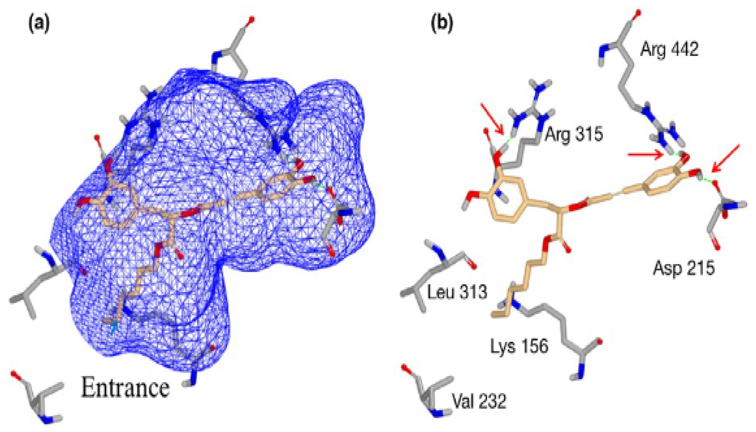
5. Modes of Action


6. Combined Application of Rosmarinic Acid and Derivatives with Other Antimicrobial Agents
| Rosmarinic Acid with | Microorganisms | Synergy | References |
|---|---|---|---|
| Vancomycin | Staphylococcus aureus | + | [182] |
| Ofloxacin | Staphylococcus aureus | + | |
| Amoxicillin | Staphylococcus aureus | + | |
| Vancomycin | MRSA | + | |
| Ofloxacin | MRSA | − | |
| Amoxicillin | MRSA | − | |
| Penicillin | MRSA | + | [220] |
| Methyl rosmarinate | Staphylococcus epidermidis 5001 | − | [77] |
| Stenotrophomonas maltophilia | − | ||
| Enterococcus faecalis C159-6 | − | ||
| Staphylococcus lugdunensis T26A3 | − | ||
| Pseudomonas aeruginosa ATCC 27583 | + | ||
| Corynebacterium T25-17 | − | ||
| Mycobacterium smegmatis 5003 | − | ||
| Staphylococcus warneri T12A12 | − | ||
| Isoquercetin | Staphylococcus epidermidis 5001 | − | |
| Stenotrophomonas maltophilia | − | ||
| Enterococcus faecalis C159-6 | − | ||
| Staphylococcus lugdunensis T26A3 | − | ||
| Pseudomonas aeruginosa ATCC 27583 | − | ||
| Corynebacterium T25-17 | − | ||
| Mycobacterium smegmatis 5003 | − | ||
| Staphylococcus warneri T12A12 | − | ||
| Hyperoside | Staphylococcus epidermidis 5001 | − | |
| Stenotrophomonas maltophilia | − | ||
| Enterococcus faecalis C159-6 | − | ||
| Staphylococcus lugdunensis T26A3 | − | ||
| Pseudomonas aeruginosa ATCC 27583 | − | ||
| Corynebacterium T25-17 | − | ||
| Mycobacterium smegmatis 5003 | + | ||
| Staphylococcus warneri T12A12 | + | ||
| Ulvan | COVID-19 | + | [221] |
| Chitosan | Escherichia coli | + | [223] |
| Polyvlactic acid/layered double hydroxides-Rosmarinic acid | Escherichia coli | + | [224] |
| Staphylococcus aureus | + | ||
| FeIII/MoO42/PO43 | herpes simplex virus | + | [225] |
| VSV-Ebola pseudotypes | + |
7. Cytotoxic Effect of RA
8. Conclusions
Author Contributions
Funding
Institutional Review Board Statement
Informed Consent Statement
Data Availability Statement
Acknowledgments
Conflicts of Interest
References
- Simonsen, G.S. Antimicrobial resistance surveillance in Europe and beyond. Eurosurveillance 2018, 23, 1800560. [Google Scholar] [CrossRef] [PubMed]
- Founou, L.L.; Founou, R.C.; Essack, S.Y. Antibiotic resistance in the food chain: A developing country-perspective. Front. Microbiol. 2016, 7, 1881. [Google Scholar] [CrossRef] [PubMed]
- World Health Organization. Antimicrobial Resistance: Global Report on Surveillance; World Health Organization: Geneva, Switzerland, 2014. [Google Scholar]
- Dadgostar, P. Antimicrobial resistance: Implications and costs. Infect. Drug Resist. 2019, 12, 3903–3910. [Google Scholar] [CrossRef]
- Magiorakos, A.-P.; Srinivasan, A.; Carey, R.B.; Carmeli, Y.; Falagas, M.; Giske, C.; Harbarth, S.; Hindler, J.; Kahlmeter, G.; Olsson-Liljequist, B. Multidrug-resistant, extensively drug-resistant and pandrug-resistant bacteria: An international expert proposal for interim standard definitions for acquired resistance. Clin. Microbiol. Infect. Drug Resist. 2012, 18, 268–281. [Google Scholar] [CrossRef]
- World Health Organization. Global Antimicrobial Resistance and Use Surveillance System (GLASS) Report: 2021; World Health Organization: Geneva, Switzerland, 2021. [Google Scholar]
- Scarpati, M.; Oriente, G. Chicoric acid (dicaffeyltartic acid): Its isolation from chicory (Chicorium intybus) and synthesis. Tetrahedron Lett. 1958, 4, 43–48. [Google Scholar] [CrossRef]
- Satake, T.; Kamiya, K.; Saiki, Y.; Hama, T.; Fujimoto, Y.; Kitanaka, S.; Kimura, Y.; Uzawa, J.; Endang, H.; Umar, M. Studies on the constituents of fruits of Helicteres isora L. Chem. Pharm. Bull. 1999, 47, 1444–1447. [Google Scholar] [CrossRef]
- Kuźma, Ł.; Wysokińska, H. Production of secondary metabolites in shoots of Salvia nemorosa L. cultured in vitro. Biotechnologia 2003, 63, 154–159. [Google Scholar]
- Hörhammer, L.; Wagner, H.; Schilcher, H. On the knowledge of the constituents of Lycopus europaeus. 1. On the constituents of medicinal plants with hormone and antihormone-like action. Arzneim. Forsch. 1962, 12, 1–7. [Google Scholar]
- Kelley, C.J.; Harruff, R.C.; Carmack, M. Polyphenolic acids of Lithospermum ruderale. II. Carbon-13 nuclear magnetic resonance of lithospermic and rosmarinic acids. J. Org. Chem. 1976, 41, 449–455. [Google Scholar] [CrossRef]
- Tanaka, T.; Morimoto, S.; Nonaka, G.-I.; Nishioka, I.; Yokozawa, T.; Chung, H.Y.; Oura, H. Magnesium and ammonium-potassium lithospermates B, the active principles having a uremia-preventive effect from Salvia miltiorrhiza. Chem. Pharm. Bull. 1989, 37, 340–344. [Google Scholar] [CrossRef]
- Jiang, R.-W.; Lau, K.-M.; Hon, P.-M.; Mak, T.C.; Woo, K.-S.; Fung, K.-P. Chemistry and biological activities of caffeic acid derivatives from Salvia miltiorrhiza. Curr. Med. Chem. 2005, 12, 237–246. [Google Scholar] [CrossRef] [PubMed]
- Agata, I.; Hatano, T.; Nishibe, S.; Okuda, T. A tetrameric derivative of caffeic acid from Rabdosia japonica. Phytochem. Lett. 1989, 28, 2447–2450. [Google Scholar] [CrossRef]
- Nishizawa, M.; Tsuda, M.; Hayashi, K. Two caffeic acid tetramers having enantiomeric phenyldihydronaphthalene moieties from Macrotomia euchroma. Phytochem. Lett. 1990, 29, 2645–2649. [Google Scholar] [CrossRef]
- Lu, Y.; Foo, L.Y. Rosmarinic acid derivatives from Salvia officinalis. Phytochem. Lett. 1999, 51, 91–94. [Google Scholar] [CrossRef]
- Lu, Y.; Foo, L.Y.; Wong, H. Sagecoumarin, a novel caffeic acid trimer from Salvia officinalis. Phytochem. Lett. 1999, 52, 1149–1152. [Google Scholar] [CrossRef]
- Tanaka, T.; Nishimura, A.; Kouno, I.; Nonaka, G.-i.; Young, T.-J. Isolation and characterization of yunnaneic acids a–d, four novel caffeic acid metabolites from Salvia yunnanensis. J. Nat. Prod. 1996, 59, 843–849. [Google Scholar] [CrossRef]
- Bulgakov, V.P.; Inyushkina, Y.V.; Fedoreyev, S.A. Rosmarinic acid and its derivatives: Biotechnology and applications. Crit. Rev. Biotechnol. 2012, 32, 203–217. [Google Scholar] [CrossRef]
- Zheng, Q.; Sun, Z.; Zhang, X.; Yuan, J.; Wu, H.; Yang, J.; Xu, X. Clerodendranoic acid, a new phenolic acid from Clerodendranthus spicatus. Molecules 2012, 17, 13656–13661. [Google Scholar] [CrossRef]
- Trennheuser, F.; Burkhard, G.; Becker, H. Anthocerodiazonin an alkaloid from Anthoceros agrestis. Phytochem. Lett. 1994, 37, 899–903. [Google Scholar] [CrossRef]
- Petersen, M.; Simmonds, M.S. Rosmarinic acid. Phytochemistry 2003, 62, 121–125. [Google Scholar] [CrossRef]
- Akhtar, M.S.; Hossain, M.A.; Said, S.A. Isolation and characterization of antimicrobial compound from the stem-bark of the traditionally used medicinal plant Adenium obesum. J. Tradit. Complement. Med. 2017, 7, 296–300. [Google Scholar] [CrossRef] [PubMed]
- Tufa, T.; Damianakos, H.; Zengin, G.; Graikou, K.; Chinou, I. Antioxidant and enzyme inhibitory activities of disodium rabdosiin isolated from Alkanna sfikasiana Tan, Vold and Strid. S. Afr. J. Bot. 2019, 120, 157–162. [Google Scholar] [CrossRef]
- Kuruuzum-Uz, A.; Suleyman, H.; Cadirci, E.; Guvenalp, Z.; Demirezer, L.O. Investigation on anti-inflammatory and antiulcer activities of Anchusa azurea extracts and their major constituent rosmarinic acid. Z. Für Nat. C 2012, 67, 360–366. [Google Scholar] [CrossRef] [PubMed]
- Hu, B.-C.; Liu, Y.; Zheng, M.-Z.; Zhang, R.-Y.; Li, M.-X.; Bao, F.-Y.; Li, H.; Chen, L.-X. Triterpenoids from Anchusa italica and their protective effects on hypoxia/reoxygenation induced cardiomyocytes injury. Bioorg. Chem. 2020, 97, 103714. [Google Scholar] [CrossRef]
- Takeda, R.; Hasegawa, J.; Shinozaki, M. The first isolation of lignans, megacerotonic acid and anthocerotonic acid, from non-vascular plants, Anthocerotae (hornworts). Tetrahedron Lett. 1990, 31, 4159–4162. [Google Scholar] [CrossRef]
- Lasure, A.; Van Poel, B.; Pieters, L.; Claeys, M.; Gupta, M.; Berghe, D.V.; Vlietinck, A. Complement-inhibiting properties of Apeiba tibourbou. Planta Med. 1994, 60, 276–277. [Google Scholar] [CrossRef]
- Olivier, D.K.; van Wyk, B.-E.; van Heerden, F.R. The chemotaxonomic and medicinal significance of phenolic acids in Arctopus and Alepidea (Apiaceae subfamily Saniculoideae). Biochem. Syst. Ecol. 2008, 36, 724–729. [Google Scholar] [CrossRef]
- Yuzbasioglu, M.; Kuruuzum-Uz, A.; Guvenalp, Z.; Simon, A.; Tóth, G.; Harput, U.S.; Kazaz, C.; Bilgili, B.; Duman, H.; Saracoglu, I. Cytotoxic compounds from endemic Arnebia purpurea. Nat. Prod. Commun. 2015, 10, 1934578X1501000415. [Google Scholar] [CrossRef]
- Argoti, J.C.; Linares-Palomino, P.J.; Salido, S.; Ramírez, B.; Insuasty, B.; Altarejos, J. On-line activity screening for radical scavengers from Baccharis chilco. Chem. Biodivers. 2013, 10, 189–197. [Google Scholar] [CrossRef]
- Badem, M.; Sener, S.O.; Kanbolat, S.; Korkmaz, N.; Yildirmiş, S.; Ozgen, U.; Aliyazicioglu, R.; Salva, E.; Kaban, K.; Kandemir, A. Evaluation of biological activities of Barbarea integrifolia and isolation of a new glucosinolate derivated compound. Z. Für Nat. C 2021, 76, 375–382. [Google Scholar] [CrossRef] [PubMed]
- Scognamiglio, M.; Buommino, E.; Coretti, L.; Graziani, V.; Russo, R.; Caputo, P.; Donnarumma, G.; Fiorentino, A. Phytochemical investigation and antimicrobial assessment of Bellis sylvestris leaves. Phytochem. Lett. 2016, 17, 6–13. [Google Scholar] [CrossRef]
- de Mello Andrade, J.M.; dos Santos Passos, C.; Rubio, M.A.K.; Mendonça, J.N.; Lopes, N.P.; Henriques, A.T. Combining in vitro and in silico approaches to evaluate the multifunctional profile of rosmarinic acid from Blechnum brasiliense on targets related to neurodegeneration. Chem.-Biol. Interact. 2016, 254, 135–145. [Google Scholar] [CrossRef] [PubMed]
- Zhang, J.; Wang, Z.-W.; Mi, Q. Phenolic compounds from Canna edulis Ker residue and their antioxidant activity. LWT-Food Sci. Technol. 2011, 44, 2091–2096. [Google Scholar] [CrossRef]
- Ly, T.N.; Shimoyamada, M.; Yamauchi, R. Isolation and characterization of rosmarinic acid oligomers in Celastrus hindsii Benth leaves and their antioxidative activity. J. Agric. Food Chem. 2006, 54, 3786–3793. [Google Scholar] [CrossRef]
- Yoshida, M.; Fuchigami, M.; Nagao, T.; Okabe, H.; Matsunaga, K.; Takata, J.; Karube, Y.; Tsuchihashi, R.; Kinjo, J.; Mihashi, K. Antiproliferative constituents from Umbelliferae plants VII. Active triterpenes and rosmarinic acid from Centella asiatica. Biol. Pharm. Bull. 2005, 28, 173–175. [Google Scholar] [CrossRef] [PubMed]
- Chen, F.-Y. Studies on chemical constituents of Chloranthus fortunei. Chin. Tradit. Herb. Drugs 2020, 24, 1485–1490. [Google Scholar]
- Ma, X.; Huang, M.; Deng, S.; Yang, J.; Ke, R.; Song, P.; Yang, X. Chemical constituents and bioactivity of Chloranthus multistachys Pei. Yunnan Univ. 2017, 39, 124–129. [Google Scholar]
- Sun, Z.; Zheng, Q.; Ma, G.; Zhang, X.; Yuan, J.; Wu, H.; Liu, H.; Yang, J.; Xu, X. Four new phenolic acids from Clerodendranthus spicatus. Phytochem. Lett. 2014, 8, 16–21. [Google Scholar] [CrossRef]
- Murata, T.; Sasaki, K.; Sato, K.; Yoshizaki, F.; Yamada, H.; Mutoh, H.; Umehara, K.; Miyase, T.; Warashina, T.; Aoshima, H. Matrix metalloproteinase-2 inhibitors from Clinopodium chinense var. parviflorum. J. Nat. Prod. 2009, 72, 1379–1384. [Google Scholar] [CrossRef]
- Saltos, M.B.V.; Puente, B.F.N.; Malafronte, N.; Braca, A. Phenolic compounds from clinopodium tomentosum (Kunth) govaerts (Lamiaceae). J. Braz. Chem. Soc. 2014, 25, 2121–2124. [Google Scholar] [CrossRef]
- Wei, X.M.; Cheng, J.K.; Cheng, D.L.; Gao, L.M. Chemical constituents from Clinopodium urticifolium. J. Chin. Chem. Soc. 2004, 51, 1043–1049. [Google Scholar] [CrossRef]
- Kumaran, A.; Karunakaran, R.J. Activity-guided isolation and identification of free radical-scavenging components from an aqueous extract of Coleus aromaticus. Food Chem. 2007, 100, 356–361. [Google Scholar] [CrossRef]
- Petersen, M. Coleus spp.: In vitro culture and the production of forskolin and rosmarinic acid. In Medicinal and Aromatic Plants VI; Springer: Berlin/Heidelberg, Germany, 1994; pp. 69–92. [Google Scholar]
- Tewtrakul, S.; Miyashiro, H.; Nakamura, N.; Hattori, M.; Kawahata, T.; Otake, T.; Yoshinaga, T.; Fujiwara, T.; Supavita, T.; Yuenyongsawad, S. HIV-1 integrase inhibitory substances from Coleus parvifolius. Phytother. Res. 2003, 17, 232–239. [Google Scholar] [CrossRef] [PubMed]
- Li, H.M.; Hwang, S.H.; Kang, B.G.; Hong, J.S.; Lim, S.S. Inhibitory effects of Colocasia esculenta (L.) Schott constituents on aldose reductase. Molecules 2014, 19, 13212–13224. [Google Scholar] [CrossRef] [PubMed]
- Fouseki, M.M.; Damianakos, H.; Karikas, G.A.; Roussakis, C.; Gupta, M.P.; Chinou, I. Chemical constituents from Cordia alliodora and C. colloccoca (Boraginaceae) and their biological activities. Fitoterapia 2016, 115, 9–14. [Google Scholar] [CrossRef]
- Marini, G.; Graikou, K.; Zengin, G.; Karikas, G.A.; Gupta, M.P.; Chinou, I. Phytochemical analysis and biological evaluation of three selected Cordia species from Panama. Ind. Crops Prod. 2018, 120, 84–89. [Google Scholar] [CrossRef]
- Owis, A.I.; Abo-Youssef, A.M.; Osman, A.H. Leaves of Cordia boissieri A. DC. as a potential source of bioactive secondary metabolites for protection against metabolic syndrome-induced in rats. Z. Für Nat. C 2017, 72, 107–118. [Google Scholar] [CrossRef]
- Fatima, M.; Siddiqui, B.; Begum, S. New neolignan glucoside and new biphenyl ether lignan from the fruits of Cordia latifolia. Chem. Nat. Compd. 2017, 53, 432–435. [Google Scholar] [CrossRef]
- Giles-Rivas, D.; Estrada-Soto, S.; Aguilar-Guadarrama, A.B.; Almanza-Pérez, J.; García-Jiménez, S.; Colín-Lozano, B.; Navarrete-Vázquez, G.; Villalobos-Molina, R. Antidiabetic effect of Cordia morelosana, chemical and pharmacological studies. J. Ethnopharmacol. 2020, 251, 112543. [Google Scholar] [CrossRef]
- Al-Musayeib, N.; Perveen, S.; Fatima, I.; Nasir, M.; Hussain, A. Antioxidant, anti-glycation and anti-inflammatory activities of phenolic constituents from Cordia sinensis. Molecules 2011, 16, 10214–10226. [Google Scholar] [CrossRef]
- Ticli, F.K.; Hage, L.I.; Cambraia, R.S.; Pereira, P.S.; Magro, Â.J.; Fontes, M.R.; Stábeli, R.G.; Giglio, J.R.; França, S.C.; Soares, A.M. Rosmarinic acid, a new snake venom phospholipase A2 inhibitor from Cordia verbenacea (Boraginaceae): Antiserum action potentiation and molecular interaction. Toxicon 2005, 46, 318–327. [Google Scholar] [CrossRef] [PubMed]
- Damianakos, H.; Jeziorek, M.; Sykłowska-Baranek, K.; Buchwald, W.; Pietrosiuk, A.; Chinou, I. Pyrrolizidine alkaloids from Cynoglossum columnae ten.(Boraginaceae). Phytochem. Lett. 2016, 15, 234–237. [Google Scholar] [CrossRef]
- Sabrin, M.S.; Selenge, E.; Takeda, Y.; Batkhuu, J.; Ogawa, H.; Jamsransuren, D.; Suganuma, K.; Murata, T. Isolation and evaluation of virucidal activities of flavanone glycosides and rosmarinic acid derivatives from Dracocephalum spp. against feline calicivirus. Phytochem. Lett. 2021, 191, 112896. [Google Scholar] [CrossRef] [PubMed]
- Shi, Q.-Q.; Dang, J.; Wen, H.-X.; Yuan, X.; Tao, Y.-D.; Wang, Q.-L. Anti-hepatitis, antioxidant activities and bioactive compounds of Dracocephalum heterophyllum extracts. Bot. Stud. 2016, 57, 16. [Google Scholar] [CrossRef] [PubMed]
- Olennikov, D.N.; Chirikova, N.K.; Okhlopkova, Z.M.; Zulfugarov, I.S. Chemical composition and antioxidant activity of Tánara Ótó (Dracocephalum palmatum Stephan), a medicinal plant used by the North-Yakutian nomads. Molecules 2013, 18, 14105–14121. [Google Scholar] [CrossRef] [PubMed]
- Zuo, M.; Yang, C.; Tian, Q.; Luo, Y.; Yang, C.; Liu, J. Chemical constituents of Dracoce-phalumtanguticum Maxim of genus Dracocephalum. Yunnan Minzu Univ. 2015, 24, 101–103. [Google Scholar]
- Le, T.T.; Kang, T.K.; Do, H.T.; Nghiem, T.D.; Lee, W.-B.; Jung, S.H. Protection against oxidative stress-induced retinal cell death by compounds isolated from Ehretia asperula. Nat. Prod. Commun. 2021, 16, 1934578X211067986. [Google Scholar] [CrossRef]
- Iqbal, K.; Nawaz, S.A.; Malik, A.; Riaz, N.; Mukhtar, N.; Mohammad, P.; Choudhary, M.I. Isolation and lipoxygenase-inhibition studies of phenolic constituents from Ehretia obtusifolia. Chem. Biodivers. 2005, 2, 104–111. [Google Scholar] [CrossRef]
- Simpol, L.R.; Otsuka, H.; Ohtani, K.; Kasai, R.; Yamasaki, K. Nitrile glucosides and rosmarinic acid, the histamine inhibitor from Ehretia philippinensis. Phytochem. Lett. 1994, 36, 91–95. [Google Scholar] [CrossRef]
- Li, L.; Peng, Y.; Xu, L.-J.; Li, M.-H.; Xiao, P.-G. Flavonoid glycosides and phenolic acids from Ehretia thyrsiflora. Biochem. Syst. Ecol. 2008, 36, 915–918. [Google Scholar] [CrossRef]
- Zhong, J.; Feng, F.; Li, H.; Li, H.; Li, R. Chemical constituents from Elsholtiza bodinieri Vaniot. Kunming Univ. Sci. Technol. 2013, 38, 75–79. [Google Scholar]
- Li, H.; Nakashima, T.; Tanaka, T.; Zhang, Y.-J.; Yang, C.-R.; Kouno, I. Two new maltol glycosides and cyanogenic glycosides from Elsholtzia rugulosa Hemsl. J. Nat. Med. 2008, 62, 75–78. [Google Scholar] [CrossRef]
- Peng, H.; Xing, Y.; Gao, L.; Zhang, L.; Zhang, G. Simultaneous separation of apigenin, luteolin and rosmarinic acid from the aerial parts of the copper-tolerant plant Elsholtzia splendens. Environ. Sci. Pollut. Res. 2014, 21, 8124–8132. [Google Scholar] [CrossRef] [PubMed]
- Devkota, H.P.; Tsushiro, K.; Watanabe, T. Bioactive phenolic compounds from the flowers of Farfugium japonicum (L.) Kitam. var. giganteum (Siebold et Zucc.) Kitam.(Asteraceae). Nat. Prod. Res. 2022, 36, 4036–4039. [Google Scholar] [CrossRef] [PubMed]
- Parejo, I.; Viladomat, F.; Bastida, J.; Schmeda-Hirschmann, G.; Burillo, J.; Codina, C. Bioguided isolation and identification of the nonvolatile antioxidant compounds from fennel (Foeniculum vulgare Mill.) waste. J. Agric. Food Chem. 2004, 52, 1890–1897. [Google Scholar] [CrossRef]
- Hawas, U.W.; Gamal-Eldeen, A.M.; El-Desouky, S.K.; Kim, Y.-K.; Huefner, A.; Saf, R. Induction of caspase-8 and death receptors by a new dammarane skeleton from the dried fruits of Forsythia koreana. Z. Für Nat. C 2013, 68, 29–38. [Google Scholar] [CrossRef]
- Shahat, A.A.; Hidayathulla, S.; Khan, A.A.; Alanazi, A.M.; Al Meanazel, O.T.; Alqahtani, A.S.; Alsaid, M.S.; Hussein, A.A. Phytochemical profiling, antioxidant and anticancer activities of Gastrocotyle hispida growing in Saudi Arabia. Acta Trop. 2019, 191, 243–247. [Google Scholar] [CrossRef]
- Yang, N.-Y.; Duan, J.-A.; Li, P.; Qian, S.-H. Chemical constituents of Glechoma longituba. Acta Pharm. Sin. 2006, 41, 431–434. [Google Scholar]
- Aquino, R.; Ciavatta, M.L.; De Simone, F.; Pizza, C. A flavanone glycoside from Hamelia patens. Phytochem. Lett. 1990, 29, 2359–2360. [Google Scholar] [CrossRef]
- Trute, A.; Nahrstedt, A. Identification and quantitative analysis of phenolic compounds from the dry extract of Hedera helix. Planta Med. 1997, 63, 177–179. [Google Scholar] [CrossRef]
- Yang, X.; Lei, Z.; Yu, Y.; Xiao, L.; Cheng, D.; Zhang, Z. Phytochemical characteristics of callus suspension culture of Helicteres angustifolia L. and its in vitro antioxidant, antidiabetic and immunomodulatory activities. S. Afr. J. Bot. 2019, 121, 178–185. [Google Scholar] [CrossRef]
- Tra, N.T.; Ha, N.T.T.; Cham, B.T.; Anh, L.T.T.; Yen, L.T.H.; Giang, B.L.; Anh, D.T.T.; Tuyen, N.V.; Kiem, P.V. A new benzofuran derivative from the stems of Helicteres hirsuta. Nat. Prod. Commun. 2019, 14, 1934578X19858814. [Google Scholar] [CrossRef]
- Sousa de Lucena, H.F.; Madeiro, S.A.L.; Siqueira, C.D.; Filho, J.M.B.; de Fátima Agra, M.; da Silva, M.S.; Fechine Tavares, J. Hypenol, a new lignan from Hypenia salzmannii. Helv. Chim. Acta 2013, 96, 1121–1125. [Google Scholar] [CrossRef]
- Abedini, A.; Roumy, V.; Mahieux, S.; Biabiany, M.; Standaert-Vitse, A.; Rivière, C.; Sahpaz, S.; Bailleul, F.; Neut, C.; Hennebelle, T. Rosmarinic acid and its methyl ester as antimicrobial components of the hydromethanolic extract of Hyptis atrorubens Poit.(Lamiaceae). Evid.-Based Complement. Altern. Med. 2013, 2013, 604536. [Google Scholar] [CrossRef] [PubMed]
- Almtorp, G.T.; Hazell, A.C.; Torssell, K.B. A lignan and pyrone and other constituents from Hyptis capitata. Phytochem. Lett. 1991, 30, 2753–2756. [Google Scholar] [CrossRef]
- Falcao, R.A.; do Nascimento, P.L.; de Souza, S.A.; da Silva, T.M.; de Queiroz, A.C.; da Matta, C.B.; Moreira, M.S.; Camara, C.A.; Silva, T. Antileishmanial phenylpropanoids from the leaves of Hyptis pectinata (L.) Poit. Evid.-Based Complement. Altern. Med. 2013, 2013, 460613. [Google Scholar] [CrossRef]
- Tang, G.; Liu, X.; Gong, X.; Lin, X.; Lai, X.; Wang, D.; Ji, S. Studies on the chemical compositions of Hyptis suaveolens (L.) Poit. J. Serb. Chem. Soc. 2019, 84, 245–252. [Google Scholar] [CrossRef]
- Kuhnt, M.; Rimpler, H.; Heinrich, M. Lignans and other compounds from the Mixe Indian medicinal plant Hyptis verticillata. Phytochem. Lett. 1994, 36, 485–489. [Google Scholar] [CrossRef]
- Furukawa, M.; Makino, M.; Ohkoshi, E.; Uchiyama, T.; Fujimoto, Y. Terpenoids and phenethyl glucosides from Hyssopus cuspidatus (Labiatae). Phytochem. Lett. 2011, 72, 2244–2252. [Google Scholar] [CrossRef]
- Arif, Z.; Khan, S.; Farheen, S.; Kazmi, M.H.; Fatima, I.; Malik, A.; Ali, M.S.; Inamullah, F.; Afaq, S.; Shaikh, S.A. Turpesteryl ester, a new antibacterial steroid from Ipomoea turpethum. Chem. Nat. Compd. 2020, 56, 270–273. [Google Scholar] [CrossRef]
- Niu, X.-M.; Li, S.-H.; Na, Z.; Mei, S.-X.; Zhao, Q.-S.; Sun, H.-D. Studies on chemical constituents of Isodon eriocalyx var. laxiflora. Chin. Tradit. Herb. Drugs 2003, 34, 300–303. [Google Scholar]
- LI, L.-J. Chemical constituents in ethyl acetate extract from Rabdosia flexicaulis. Chin. Tradit. Herb. Drugs 2015, 46, 339–343. [Google Scholar]
- Zhou, W.; Xie, H.; Xu, X.; Liang, Y.; Wei, X. Phenolic constituents from Isodon lophanthoides var. graciliflorus and their antioxidant and antibacterial activities. J. Funct. Foods 2014, 6, 492–498. [Google Scholar] [CrossRef]
- Huang, H.; Chao, Q.-R.; Tan, R.; Sun, H.-D.; Wang, D.-C.; Ma, J.; Zhao, S.-X. A new rosmarinic acid derivative from Isodon oresbius. Planta Med. 1999, 65, 92–93. [Google Scholar] [CrossRef] [PubMed]
- Xiaoke, Z.; Qin, L.; Weisheng, F. Studies on chemical constituents of phenolic acids in Rabdosia rubescens. Zhongguo Yao Xue Za Zhi 2004, 39, 335–336. [Google Scholar]
- Khan, S.; Taning, C.N.T.; Bonneure, E.; Mangelinckx, S.; Smagghe, G.; Ahmad, R.; Fatima, N.; Asif, M.; Shah, M.M. Bioactivity-guided isolation of rosmarinic acid as the principle bioactive compound from the butanol extract of Isodon rugosus against the pea aphid, Acyrthosiphon pisum. PLoS ONE 2019, 14, e0215048. [Google Scholar] [CrossRef]
- Jiang, B.; Hou, A.-J.; Li, M.-L.; Li, S.-H.; Han, Q.-B.; Wang, S.-J.; Lin, Z.-W.; Sun, H.-D. Cytotoxic ent-kaurane diterpenoids from Isodon sculponeata. Planta Med. 2002, 68, 921–925. [Google Scholar] [CrossRef] [PubMed]
- Murata, T.; Miyase, T.; Yoshizaki, F. Hyaluronidase inhibitors from Keiskea japonica. Chem. Pharm. Bull. 2012, 60, 121–128. [Google Scholar] [CrossRef]
- Dehaghi, N.K.; Gohari, A.; Sadat-Ebrahimi, S.; Badi, H.N.; Amanzadeh, Y. Phytochemistry and antioxidant activity of Lallemantia iberica aerial parts. Res. J. Pharmacogn. 2016, 3, 27–34. [Google Scholar]
- Yadikar, N.; Bobakulov, K.; Li, G.; Aisa, H.A. Seven new phenolic compounds from Lavandula angustifolia. Phytochem. Lett. 2018, 23, 149–154. [Google Scholar] [CrossRef]
- Parejo, I.; Caprai, E.; Bastida, J.; Viladomat, F.; Jáuregui, O.; Codina, C. Investigation of Lepechinia graveolens for its antioxidant activity and phenolic composition. J. Ethnopharmacol. 2004, 94, 175–184. [Google Scholar] [CrossRef] [PubMed]
- Crespo, M.I.; Chabán, M.F.; Lanza, P.A.; Joray, M.B.; Palacios, S.M.; Vera, D.M.A.; Carpinella, M.C. Inhibitory effects of compounds isolated from Lepechinia meyenii on tyrosinase. Food Chem. Toxicol. 2019, 125, 383–391. [Google Scholar] [CrossRef] [PubMed]
- Esteves, P.F.; Kuster, R.M.; Barbi, N.d.S.; Menezes, F.d.S. Chemical composition and cytotoxic activity of Lepechinia speciosa (St. Hill) Epling. Lat. Am. J. Pharm 2010, 29, 38–44. [Google Scholar]
- Revoltella, S.; Baraldo, G.; Waltenberger, B.; Schwaiger, S.; Kofler, P.; Moesslacher, J.; Huber-Seidel, A.; Pagitz, K.; Kohl, R.; Jansen-Duerr, P. Identification of the NADPH oxidase 4 inhibiting principle of Lycopus europaeus. Molecules 2018, 23, 653. [Google Scholar] [CrossRef]
- Woo, E.-R.; Piao, M.S. Antioxidative constituents fromlycopus lucidus. Arch. Pharmacal Res. 2004, 27, 173–176. [Google Scholar] [CrossRef]
- Neamah, S.; Sarhan, I.A.; Al-Shaye’a, O.N. Extraction and evaluation of the anti-inflammatory activity of six compounds of Marrubium vulgare L. Biosci. Res. 2018, 15, 2393–2400. [Google Scholar]
- Murata, T.; Miyase, T.; Yoshizaki, F. Hyaluronidase inhibitory rosmarinic acid derivatives from Meehania urticifolia. Chem. Pharm. Bull. 2011, 59, 88–95. [Google Scholar] [CrossRef]
- Tagashira, M.; Ohtake, Y. A new antioxidative 1,3-benzodioxole from Melissa officinalis. Planta Med. 1998, 64, 555–558. [Google Scholar] [CrossRef]
- Aksit, H.; Çelik, S.M.; Sen, Ö.; Erenler, R.; Demirtas, I.; Telci, İ.; Elmastas, M. Complete isolation and characterization of polar portion of Mentha dumetorum water extract. Rec. Nat. Prod. 2014, 8, 277. [Google Scholar]
- She, G.M.; Xu, C.; Liu, B.; Shi, R.B. Polyphenolic acids from mint (the aerial of Mentha haplocalyx Briq.) with DPPH radical scavenging activity. J. Food Sci. 2010, 75, C359–C362. [Google Scholar] [CrossRef]
- Guvenalp, Z.; Ozbek, H.; Karadayi, M.; Gulluce, M.; Kuruuzum-Uz, A.; Salih, B.; Demirezer, O. Two antigenotoxic chalcone glycosides from Mentha longifolia subsp. longifolia. Pharm. Biol. 2015, 53, 888–896. [Google Scholar] [CrossRef] [PubMed]
- Inoue, T.; Sugimoto, Y.; Masuda, H.; Kamei, C. Antiallergic effect of flavonoid glycosides obtained from Mentha piperita L. Biol. Pharm. Bull. 2002, 25, 256–259. [Google Scholar] [CrossRef] [PubMed]
- Zheng, J.; Gao, H.; Chen, G.; Yang, X.; Wu, B.; Wu, L. Chemical constituents of the active parts of Mentha spicata L.(II). Shenyang Pharm. Univ 2006, 23, 212–216. [Google Scholar]
- Wang, F.; Xiang, R.; Lin, C.; Zhu, C. Chemical constituents from Mesona chinensis. Chin. Med. Mater. 2017, 40, 2839–2843. [Google Scholar]
- Küpeli Akkol, E.; Gürağaç Dereli, F.T.; Ilhan, M. Assessment of antidepressant effect of the aerial parts of micromeria myrtifolia Boiss. & Hohen on mice. Molecules 2019, 24, 1869. [Google Scholar] [CrossRef]
- Liang, C.; Zhou, X.; Wang, P.; Tan, X.; Luo, Q.; Chen, X.; Pan, Z. Chemical constituents from stems and leaves of Microsorium fortunei. Chin. Med. Mater. 2017, 40, 2089–2092. [Google Scholar]
- De Tommasi, N.; De Simone, F.; De Feo, V.; Pizza, C. Phenylpropanoid glycosides and rosmarinic acid from Momordica balsamina. Planta Med. 1991, 57, 201. [Google Scholar] [CrossRef]
- Goldansaz, S.M.; Festa, C.; Pagano, E.; De Marino, S.; Finamore, C.; Parisi, O.A.; Borrelli, F.; Sonboli, A.; D’Auria, M.V. Phytochemical and biological studies of Nepeta asterotricha Rech. f.(Lamiaceae): Isolation of nepetamoside. Molecules 2019, 24, 1684. [Google Scholar] [CrossRef]
- Takeda, Y.; Ooiso, Y.; Masuda, T.; Honda, G.; Otsuka, H.; Sezik, E.; Yesilada, E. Iridoid and eugenol glycosides from Nepeta cadmea. Phytochem. Lett. 1998, 49, 787–791. [Google Scholar] [CrossRef]
- Rabee, M.; Andersen, Ø.M.; Fossen, T.; Enerstvedt, K.H.; Abu Ali, H.; Rayyan, S. Acylated flavone O-glucuronides from the aerial parts of Nepeta curviflora. Molecules 2020, 25, 3782. [Google Scholar] [CrossRef]
- Ruiz-Vargas, J.A.; Morales-Ferra, D.L.; Ramírez-Ávila, G.; Zamilpa, A.; Negrete-León, E.; Acevedo-Fernández, J.J.; Peña-Rodríguez, L.M. α-Glucosidase inhibitory activity and in vivo antihyperglycemic effect of secondary metabolites from the leaf infusion of Ocimum campechianum mill. J. Ethnopharmacol. 2019, 243, 112081. [Google Scholar] [CrossRef] [PubMed]
- Kelm, M.; Nair, M.; Strasburg, G.; DeWitt, D. Antioxidant and cyclooxygenase inhibitory phenolic compounds from Ocimum sanctum Linn. Phytomedicine 2000, 7, 7–13. [Google Scholar] [CrossRef] [PubMed]
- Chatzopoulou, A.; Karioti, A.; Gousiadou, C.; Lax Vivancos, V.; Kyriazopoulos, P.; Golegou, S.; Skaltsa, H. Depsides and other polar constituents from Origanum dictamnus L. and their in vitro antimicrobial activity in clinical strains. J. Agric. Food Chem. 2010, 58, 6064–6068. [Google Scholar] [CrossRef] [PubMed]
- Basli, A.; Delaunay, J.-C.; Pedrot, E.; Bernillon, S.; Madani, K.; Monti, J.-P.; Mérillon, J.-M.; Chibane, M.; Richard, T. New cyclolignans from Origanum glandulosum active against beta-amyloid aggregation. Rec. Nat. Prod. 2014, 8, 208–216. [Google Scholar]
- Erenler, R.; Sen, O.; Aksit, H.; Demirtas, I.; Yaglioglu, A.S.; Elmastas, M.; Telci, I. Isolation and identification of chemical constituents from Origanum majorana and investigation of antiproliferative and antioxidant activities. J. Sci. Food Agric. 2016, 96, 822–836. [Google Scholar] [CrossRef]
- Elmastas, M.; Celik, S.M.; Genc, N.; Aksit, H.; Erenler, R.; Gulcin, İ. Antioxidant activity of an Anatolian herbal tea—Origanum minutiflorum: Isolation and characterization of its secondary metabolites. Int. J. Food Prop. 2018, 21, 374–384. [Google Scholar] [CrossRef]
- Erenler, R.; Meral, B.; Sen, O.; Elmastas, M.; Aydin, A.; Eminagaoglu, O.; Topcu, G. Bioassay-guided isolation, identification of compounds from Origanum rotundifolium and investigation of their antiproliferative and antioxidant activities. Pharm. Biol. 2017, 55, 1646–1653. [Google Scholar] [CrossRef]
- Koukoulitsa, C.; Karioti, A.; Bergonzi, M.C.; Pescitelli, G.; Di Bari, L.; Skaltsa, H. Polar constituents from the aerial parts of Origanum vulgare L. ssp. hirtum growing wild in Greece. J. Agric. Food Chem. 2006, 54, 5388–5392. [Google Scholar] [CrossRef]
- Lee, K.H.; Yang, M.C.; Kim, K.H.; Kwon, H.C.; Choi, S.U.; Lee, K.R. A new phenolic amide from the roots of Paris verticillata. Molecules 2008, 13, 41–45. [Google Scholar] [CrossRef]
- Lim, H.J.; Woo, K.W.; Lee, K.R.; Lee, S.K.; Kim, H.P. Inhibition of proinflammatory cytokine generation in lung inflammation by the leaves of Perilla frutescens and its constituents. Biomol. Ther. 2014, 22, 62. [Google Scholar] [CrossRef]
- Ha, T.J.; Lee, J.H.; Lee, M.-H.; Lee, B.W.; Kwon, H.S.; Park, C.-H.; Shim, K.-B.; Kim, H.-T.; Baek, I.-Y.; Jang, D.S. Isolation and identification of phenolic compounds from the seeds of Perilla frutescens (L.) and their inhibitory activities against α-glucosidase and aldose reductase. Food Chem. 2012, 135, 1397–1403. [Google Scholar] [CrossRef] [PubMed]
- Gu, L.; Wu, T.; Wang, Z. TLC bioautography-guided isolation of antioxidants from fruit of Perilla frutescens var. acuta. LWT-Food Sci. Technol. 2009, 42, 131–136. [Google Scholar] [CrossRef]
- Senol, F.S.; Ślusarczyk, S.; Matkowski, A.; Pérez-Garrido, A.; Girón-Rodríguez, F.; Cerón-Carrasco, J.P.; den-Haan, H.; Peña-García, J.; Pérez-Sánchez, H.; Domaradzki, K. Selective in vitro and in silico butyrylcholinesterase inhibitory activity of diterpenes and rosmarinic acid isolated from Perovskia atriplicifolia Benth. and Salvia glutinosa L. Phytochem. Lett. 2017, 133, 33–44. [Google Scholar] [CrossRef] [PubMed]
- Kubínová, R.; Švajdlenka, E.; Schneiderová, K.; Hanáková, Z.; Dall’Acqua, S.; Farsa, O. Polyphenols and diterpenoids from Plectranthus forsteri ‘Marginatus’. Biochem. Syst. Ecol. 2013, 49, 39–42. [Google Scholar] [CrossRef]
- Ji, H.-S.; Li, H.; Mo, E.-J.; Kim, U.-H.; Kim, Y.-H.; Park, H.-Y.; Jeong, T.-S. Low-density lipoprotein-antioxidant flavonoids and a phenolic ester from Plectranthus hadiensis var. tomentosus. Appl. Biol. Chem. 2019, 62, 58. [Google Scholar] [CrossRef]
- Kubínova, R.; Pořízková, R.; Navrátilová, A.; Farsa, O.; Hanáková, Z.; Bačinská, A.; Čížek, A.; Valentová, M. Antimicrobial and enzyme inhibitory activities of the constituents of Plectranthus madagascariensis (Pers.) Benth. J. Enzym. Inhib. Med. Chem. 2014, 29, 749–752. [Google Scholar] [CrossRef]
- Kubínová, R.; Gazdová, M.; Hanáková, Z.; Jurkaninová, S.; Dall’Acqua, S.; Cvačka, J.; Humpa, O. New diterpenoid glucoside and flavonoids from Plectranthus scutellarioides (L.) R. Br. S. Afr. J. Bot. 2019, 120, 286–290. [Google Scholar] [CrossRef]
- Hu, H.; Wang, G.; Liu, J.; Cao, H.; Zheng, X. Studies on phenolic compounds from Polygonum aviculane. China J. Chin. Mater. Med. 2006, 31, 740–742. [Google Scholar]
- Zhu, J. Depsides from Prunella vulgaris. Chin. Chem. Lett. 2000, 11, 997–1000. [Google Scholar]
- Kim, H.-I.; Quan, F.-S.; Kim, J.-E.; Lee, N.-R.; Kim, H.J.; Jo, S.J.; Lee, C.-M.; Jang, D.S.; Inn, K.-S. Inhibition of estrogen signaling through depletion of estrogen receptor alpha by ursolic acid and betulinic acid from Prunella vulgaris var. lilacina. Biochem. Biophys. Res. Commun. 2014, 451, 282–287. [Google Scholar] [CrossRef]
- Lee, I.K.; Kim, D.H.; Lee, S.Y.; Kim, K.R.; Choi, S.U.; Hong, J.K.; Lee, J.H.; Park, Y.H.; Lee, K.R. Triterpenoic acids of Prunella vulgaris var. lilacina and their cytotoxic activities in vitro. Arch. Pharmacal Res. 2008, 31, 1578–1583. [Google Scholar] [CrossRef] [PubMed]
- Chanu, M.B.; Labala, R.K.; Sheikh, Y.; Borah, J.C.; Ghosh, S.K.; Sahoo, D.; Singh, O.J.; Shakya, A.; Thongam, B. Bioassay guided isolation of alpha-glucosidase inhibitory compound, in vivo postprandial anti hyperglycemia and docking study of the isolated compound from the leaves of the methanolic extract of Quercus serrata. Biosci. Biotech. Res. Commun. 2018, 11, 647–657. [Google Scholar] [CrossRef]
- Hyun, H.B.; Shrestha, S.; Boo, K.H.; Cho, S.K. Evaluation of antioxidant potential of ethyl acetate fraction of Rosmarinus officinalis L. and its major components. J. Korean Soc. Appl. Biol. Chem. 2015, 58, 715–722. [Google Scholar] [CrossRef]
- Koysu, P.; Genc, N.; Elmastas, M.; Aksit, H.; Erenler, R. Isolation, identification of secondary metabolites from Salvia absconditiflora and evaluation of their antioxidative properties. Nat. Prod. Res. 2019, 33, 3592–3595. [Google Scholar] [CrossRef]
- Qu, G.; Yue, X.; An, F.; Dai, S.; Li, G.; Li, B. Chemical constituents contained in Salvia castanea. China J. Chin. Mater. Med. 2012, 37, 1985–1989. [Google Scholar]
- Zhang, H.-J.; Li, L.-N. Salvianolic acid I: A new depside from Salvia cavaleriei. Planta Med. 1994, 60, 70–72. [Google Scholar] [CrossRef]
- Ertaş, A.; Çakırca, H.; Yener, I.; Akdeniz, M.; Fırat, M.; Topçu, G.; Kolak, U. Bioguided isolation of secondary metabolites from Salvia cerino-pruinosa Rech. f. var. cerino-pruinosa. Rec. Nat. Prod. 2021, 15, 585–592. [Google Scholar]
- Gao, J.-F.; Ding, L.; Zhang, P.; Liu, J.-X. Chemical constituents of Salvia chinensis. China J. Chin. Mater. Med. 2013, 38, 1556–1559. [Google Scholar]
- Qian, T.-X.; Li, L.-N. Isosalvianolic acid C, a depside possessing a dibenzooxepin skeleton. Phytochem. Lett. 1992, 31, 1068–1070. [Google Scholar] [CrossRef]
- Wang, X.; Kasimu, R.; Niyazi, Z. Studies on the chemical constituents of the flowers of salvia deserta schang. J. Xinjiang Med. Univ. 2003, 26, 583–585. [Google Scholar]
- Ai, C.-B.; Deng, Q.-H.; Song, W.-Z.; Li, L.-N. Salvianolic acid J, a depside from Salvia flava. Phytochem. Lett. 1994, 37, 907–908. [Google Scholar] [CrossRef]
- Kang, J.; Tang, Y.; Liu, Q.; Guo, N.; Zhang, J.; Xiao, Z.; Chen, R.; Shen, Z. Isolation, modification, and aldose reductase inhibitory activity of rosmarinic acid derivatives from the roots of Salvia grandifolia. Fitoterapia 2016, 112, 197–204. [Google Scholar] [CrossRef] [PubMed]
- Xia, G.-H. Triterpenes and phenolic acids from roots of Salvia kiaometiensis. Chin. Tradit. Herb. Drugs 2019, 24, 1043–1048. [Google Scholar]
- Gohari, A.R.; Saeidnia, S.; Malmir, M.; Hadjiakhoondi, A.; Ajani, Y. Flavones and rosmarinic acid from Salvia limbata. Nat. Prod. Res. 2010, 24, 1902–1906. [Google Scholar] [CrossRef]
- Tung, N.H.; Hung, L.Q.; Van Oanh, H.; Huong, D.T.L.; Thuong, P.T.; Long, D.D.; Hai, N.T. Bioactive phenolic compounds from the roots of Danshen (Salvia miltiorrhiza). Nat. Prod. Commun. 2018, 13, 1934578X1801301018. [Google Scholar] [CrossRef]
- Nugroho, A.; Kim, M.-H.; Choi, J.; Baek, N.-I.; Park, H.-J. In vivo sedative and gastroprotective activities of Salvia plebeia extract and its composition of polyphenols. Arch. Pharmacal Res. 2012, 35, 1403–1411. [Google Scholar] [CrossRef]
- Gong, X.; Yang, S. Isolation, identification and antioxidant properties of flavonoids from Salvia plebeia. Chin. Wild Plant. Resour. 2013, 32, 24–27. [Google Scholar]
- Yang, Y.; Bing, Z.; Sun, L.; Wu, Z.; Chen, W. Chemical constituents of Salvia przewalskii Maxim. Asian J. Chem. 2013, 25, 1747–1748. [Google Scholar]
- Wu, Z.-J.; Ouyang, M.-A.; Yang, C.-R. Polyphenolic constituents of Salvia sonchifolia. Acta Bot. Yunnanica 1999, 21, 393–398. [Google Scholar]
- Moharram, F.A.-e.; Marzouk, M.S.; El-Shenawy, S.M.; Gaara, A.H.; El Kady, W.M. Polyphenolic profile and biological activity of Salvia splendens leaves. J. Pharm. Pharmacol. 2012, 64, 1678–1687. [Google Scholar] [CrossRef]
- Çulhaoğlu, B.; Hatipoğlu, S.D.; Dönmez, A.A.; Topçu, G. Antioxidant and anticholinesterase activities of lupane triterpenoids and other constituents of Salvia trichoclada. Med. Chem. Res. 2015, 24, 3831–3837. [Google Scholar] [CrossRef]
- Rungsimakan, S.; Rowan, M.G. Terpenoids, flavonoids and caffeic acid derivatives from Salvia viridis L. cvar. Blue Jeans. Phytochem. Lett. 2014, 108, 177–188. [Google Scholar] [CrossRef] [PubMed]
- Zhang, Z.-F.; Chen, H.-s.; Li, J.-R.; Jiang, J.-D.; Li, Z.-R. Studies on polyphenolic chemical constitutents from root of Salvia yunnansis. China J. Chin. Mater. Med. 2007, 32, 1886–1890. [Google Scholar]
- Arda, N.; Gören, N.; Kuru, A.; Pengsuparp, T.; Pezzuto, J.M.; Qiu, S.-X.; Cordell, G.A. Saniculoside N from Sanicula europaea L. J. Nat. Prod. 1997, 60, 1170–1173. [Google Scholar] [CrossRef] [PubMed]
- Zhou, L.-Y.; Liu, H.-Y.; Xie, B.-B.; Liu, Z.-H.; Chen, C.-X. Two new glycosides from Sanicula lamelligera. Z. Für Nat. B 2006, 61, 607–610. [Google Scholar] [CrossRef]
- Lee, I.-K.; Kim, M.-A.; Lee, S.-Y.; Hong, J.-K.; Lee, J.-H.; Lee, K.-R. Phytochemical constituents of Schizonepeta tenuifolia Briquet. Nat. Prod. Sci. 2008, 14, 100–106. [Google Scholar] [CrossRef]
- Deveci, E.; Tel-Çayan, G.; Duru, M.E.; Öztürk, M. Phytochemical contents, antioxidant effects, and inhibitory activities of key enzymes associated with Alzheimer’s disease, ulcer, and skin disorders of Sideritis albiflora and Sideritis leptoclada. J. Food Biochem. 2019, 43, e13078. [Google Scholar] [CrossRef]
- García, J.M.; Prieto, L.J.; Guevara, A.; Malagon, D.; Osorio, C. Chemical studies of yellow tamarillo (Solanum betaceum Cav.) fruit flavor by using a molecular sensory approach. Molecules 2016, 21, 1729. [Google Scholar] [CrossRef]
- Taiwo, B.J.; Obuotor, E.; Onawunmi, G.O.; Ogundaini, A.O. Radical scavenging compounds from the aerial parts of Solenostemon monostachys briq (Lamiaceae). Afr. J. Tradit. Complement. Altern. Med. 2015, 12, 140–144. [Google Scholar] [CrossRef]
- Trifan, A.; Skalicka-Woźniak, K.; Granica, S.; Czerwińska, M.E.; Kruk, A.; Marcourt, L.; Wolfender, J.-L.; Wolfram, E.; Esslinger, N.; Grubelnik, A. Symphytum officinale L.: Liquid-liquid chromatography isolation of caffeic acid oligomers and evaluation of their influence on pro-inflammatory cytokine release in LPS-stimulated neutrophils. J. Ethnopharmacol. 2020, 262, 113169. [Google Scholar] [CrossRef]
- Boonyarikpunchai, W.; Sukrong, S.; Towiwat, P. Antinociceptive and anti-inflammatory effects of rosmarinic acid isolated from Thunbergia laurifolia Lindl. Pharmacol. Biochem. Behav. 2014, 124, 67–73. [Google Scholar] [CrossRef] [PubMed]
- Dall’Acqua, S.; Peron, G.; Ferrari, S.; Gandin, V.; Bramucci, M.; Quassinti, L.; Mártonfi, P.; Maggi, F. Phytochemical investigations and antiproliferative secondary metabolites from Thymus alternans growing in Slovakia. Pharm. Biol. 2017, 55, 1162–1170. [Google Scholar] [CrossRef] [PubMed]
- Khouya, T.; Ramchoun, M.; Amrani, S.; Harnafi, H.; Rouis, M.; Couchie, D.; Simmet, T.; Alem, C. Anti-inflammatory and anticoagulant effects of polyphenol-rich extracts from Thymus atlanticus: An in vitro and in vivo study. J. Ethnopharmacol. 2020, 252, 112475. [Google Scholar] [CrossRef] [PubMed]
- Erenler, R.; Sen, O.; Yildiz, I.; Aydin, A. Antiproliferative activities of chemical constituents isolated from Thymus praecox subsp grossheimii (Ronniger) Jalas. Rec. Nat. Prod. 2016, 10, 766–770. [Google Scholar]
- Sevindik, H.G.; Ozgen, U.; Atila, A.; Er, H.O.; Kazaz, C.; Duman, H. Phtytochemical Studies and Quantitative HPLC Analysis of Rosmarinic Acid and Luteolin 5-O-β-D-Glucopyranoside on Thymus praecox subsp. grossheimii var. grossheimii. Chem. Pharm. Bull. 2015, 63, 720–725. [Google Scholar] [CrossRef] [PubMed]
- Lee, I.-C.; Bae, J.-S.; Kim, T.; Kwon, O.J.; Kim, T.H. Polyphenolic constituents from the aerial parts of Thymus quinquecostatus var. japonica collected on Ulleung Island. J. Korean Soc. Appl. Biol. Chem. 2011, 54, 811–816. [Google Scholar] [CrossRef]
- Aziz, S.; Irshad, M. Isolation of a new antibacterial polyphenol from Thymus serpyllum. Chem. Nat. Compd. 2014, 49, 1023–1027. [Google Scholar] [CrossRef]
- Kontogiorgis, C.; Ntella, M.; Mpompou, L.; Karallaki, F.; Athanasios, P.; Hadjipavlou-Litina, D.; Lazari, D. Study of the antioxidant activity of Thymus sibthorpii Bentham (Lamiaceae). J. Enzym. Inhib. Med. Chem. Res. 2016, 31, 154–159. [Google Scholar] [CrossRef]
- Ozgen, U.; Mavi, A.; Terzi, Z.; Kazaz, C.; Asci, A.; Kaya, Y.; Secen, H. Relationship between chemical structure and antioxidant activity of luteolin and its glycosides isolated from Thymus sipyleus subsp sipyleus var. sipyleus. Planta Med. 2010, 5, 12–21. [Google Scholar]
- Engelbertz, J.; Lechtenberg, M.; Studt, L.; Hensel, A.; Verspohl, E.J. Bioassay-guided fractionation of a thymol-deprived hydrophilic thyme extract and its antispasmodic effect. J. Ethnopharmacol. 2012, 141, 848–853. [Google Scholar] [CrossRef]
- Lin, Y.-L.; Chang, Y.-Y.; Kuo, Y.-H.; Shiao, M.-S. Anti-Lipid-Peroxidative Principles from Tournefortia s armentosa. J. Nat. Prod. 2002, 65, 745–747. [Google Scholar] [CrossRef] [PubMed]
- Salehi, B.; Shivaprasad Shetty, M.; Anil Kumar, N.V.; Živković, J.; Calina, D.; Oana Docea, A.; Emamzadeh-Yazdi, S.; Sibel Kılıç, C.; Goloshvili, T.; Nicola, S. Veronica plants—Drifting from farm to traditional healing, food application, and phytopharmacology. Molecules 2019, 24, 2454. [Google Scholar] [CrossRef] [PubMed]
- Benedec, D.; Hanganu, D.; Oniga, I.; Tiperciuc, B.; Olah, N.-K.; Raita, O.; Bischin, C.; Silaghi-Dumitrescu, R.; Vlase, L. Assessment of rosmarinic acid content in six Lamiaceae species extracts and their antioxidant and antimicrobial potential. Pak. J. Pharm. Sci 2015, 28, 2297–2303. [Google Scholar]
- Wang, J.; Pan, X.; Han, Y.; Guo, D.; Guo, Q.; Li, R. Rosmarinic acid from eelgrass shows nematicidal and antibacterial activities against pine wood nematode and its carrying bacteria. Mar. Drugs 2012, 10, 2729–2740. [Google Scholar] [CrossRef]
- Achamlale, S.; Rezzonico, B.; Grignon-Dubois, M. Rosmarinic acid from beach waste: Isolation and HPLC quantification in Zostera detritus from Arcachon lagoon. Food Chem. 2009, 113, 878–883. [Google Scholar] [CrossRef]
- Yesilbag, D.; Eren, M.; Agel, H.; Kovanlikaya, A.; Balci, F. Effects of dietary rosemary, rosemary volatile oil and vitamin E on broiler performance, meat quality and serum SOD activity. Br. Poult. Sci. 2011, 52, 472–482. [Google Scholar] [CrossRef] [PubMed]
- Chung, Y.-C.; Hsieh, F.-C.; Lin, Y.-J.; Wu, T.-Y.; Lin, C.-W.; Lin, C.-T.; Tang, N.-Y.; Jinn, T.-R. Magnesium lithospermate B and rosmarinic acid, two compounds present in Salvia miltiorrhiza, have potent antiviral activity against enterovirus 71 infections. Eur. J. Pharmacol. 2015, 755, 127–133. [Google Scholar] [CrossRef]
- Moreno, S.; Scheyer, T.; Romano, C.S.; Vojnov, A.A. Antioxidant and antimicrobial activities of rosemary extracts linked to their polyphenol composition. Free Radic. Res. 2006, 40, 223–231. [Google Scholar] [CrossRef]
- Ekambaram, S.P.; Perumal, S.S.; Balakrishnan, A.; Marappan, N.; Gajendran, S.S.; Viswanathan, V. Antibacterial synergy between rosmarinic acid and antibiotics against methicillin-resistant Staphylococcus aureus. J. Intercult. Ethnopharmacol. 2016, 5, 358. [Google Scholar] [CrossRef]
- Swarup, V.; Ghosh, J.; Ghosh, S.; Saxena, A.; Basu, A. Antiviral and anti-inflammatory effects of rosmarinic acid in an experimental murine model of Japanese encephalitis. Antimicrob. Agents Chemother. 2007, 51, 3367–3370. [Google Scholar] [CrossRef]
- Myint, K.B.; Sing, L.C.; Wei, Z. Tannic acid as phytochemical potentiator for antibiotic resistance adaptation. APCBEE Procedia 2013, 7, 175–181. [Google Scholar] [CrossRef]
- Wang, T.; Ma, B.; Jin, A.; Li, X.; Zhang, X.; Wang, W.; Cai, Y. Facile loading of Ag nanoparticles onto magnetic microsphere by the aid of a tannic acid—Metal polymer layer to synthesize magnetic disinfectant with high antibacterial activity. J. Hazard. Mater. 2018, 342, 392–400. [Google Scholar] [CrossRef] [PubMed]
- Tintino, S.R.; Morais-Tintino, C.D.; Campina, F.F.; Costa, M.d.S.; Menezes, I.R.; de Matos, Y.M.L.; Calixto-Júnior, J.T.; Pereira, P.S.; Siqueira-Junior, J.P.; Leal-Balbino, T.C. Tannic acid affects the phenotype of Staphylococcus aureus resistant to tetracycline and erythromycin by inhibition of efflux pumps. Bioorganic Chem. 2017, 74, 197–200. [Google Scholar] [CrossRef] [PubMed]
- Suriyarak, S.; Bayrasy, C.; Schmidt, H.; Villeneuve, P.; Weiss, J. Impact of fatty acid chain length of rosmarinate esters on their antimicrobial activity against Staphylococcus carnosus LTH1502 and Escherichia coli K-12 LTH4263. J. Food Prot. 2013, 76, 1539–1548. [Google Scholar] [CrossRef]
- Zhang, J.; Cui, X.; Zhang, M.; Bai, B.; Yang, Y.; Fan, S. The antibacterial mechanism of perilla rosmarinic acid. Biotechnol. Appl. Biochem. 2022, 69, 1757–1764. [Google Scholar] [CrossRef]
- Cetin-Karaca, H. Evaluation of Natural Antimicrobial Phenolic Compounds against Foodborne Pathogens. Master’s Thesis, University of Kentucky, Lexington, KY, USA, 2011. [Google Scholar]
- Bailly, F.; Cotelle, P. Anti-HIV activities of natural antioxidant caffeic acid derivatives: Toward an antiviral supplementation diet. Curr. Med. Chem. 2005, 12, 1811–1818. [Google Scholar] [CrossRef]
- Dubois, M.; Bailly, F.; Mbemba, G.; Mouscadet, J.-F.; Debyser, Z.; Witvrouw, M.; Cotelle, P. Reaction of rosmarinic acid with nitrite ions in acidic conditions: Discovery of nitro-and dinitrorosmarinic acids as new anti-HIV-1 agents. J. Med. Chem. 2008, 51, 2575–2579. [Google Scholar] [CrossRef]
- Lindel, T. Chemistry and biology of the pyrrole–imidazole alkaloids. In The Alkaloids: Chemistry and Biology; Elsevier: Amsterdam, The Netherlands, 2017; Volume 77, pp. 117–219. [Google Scholar]
- Zhu, F.; Wang, J.; Takano, H.; Xu, Z.; Nishiwaki, H.; Yonekura, L.; Yang, R.; Tamura, H. Rosmarinic acid and its ester derivatives for enhancing antibacterial, α-glucosidase inhibitory, and lipid accumulation suppression activities. J. Food Biochem. 2019, 43, e12719. [Google Scholar] [CrossRef]
- Elebeedy, D.; Elkhatib, W.F.; Kandeil, A.; Ghanem, A.; Kutkat, O.; Alnajjar, R.; Saleh, M.A.; Abd El Maksoud, A.I.; Badawy, I.; Al-Karmalawy, A.A. Anti-SARS-CoV-2 activities of tanshinone IIA, carnosic acid, rosmarinic acid, salvianolic acid, baicalein, and glycyrrhetinic acid between computational and in vitro insights. RSC Adv. 2021, 11, 29267–29286. [Google Scholar] [CrossRef]
- Hsieh, C.-F.; Jheng, J.-R.; Lin, G.-H.; Chen, Y.-L.; Ho, J.-Y.; Liu, C.-J.; Hsu, K.-Y.; Chen, Y.-S.; Chan, Y.F.; Yu, H.-M. Rosmarinic acid exhibits broad anti-enterovirus A71 activity by inhibiting the interaction between the five-fold axis of capsid VP1 and cognate sulfated receptors. Emerg. Microbes Infect. 2020, 9, 1194–1205. [Google Scholar] [CrossRef]
- Slobodníková, L.; Fialová, S.; Hupková, H.; Grančai, D. Rosmarinic acid interaction with planktonic and biofilm Staphylococcus aureus. Nat. Prod. Commun. 2013, 8, 1934578X1300801223. [Google Scholar] [CrossRef]
- Chandra, J.; Kuhn, D.M.; Mukherjee, P.K.; Hoyer, L.L.; McCormick, T.; Ghannoum, M.A. Biofilm formation by the fungal pathogen Candida albicans: Development, architecture, and drug resistance. J. Bacteriol. 2001, 183, 5385–5394. [Google Scholar] [CrossRef] [PubMed]
- Rama Devi, K.; Srinivasan, R.; Kannappan, A.; Santhakumari, S.; Bhuvaneswari, M.; Rajasekar, P.; Prabhu, N.M.; Veera Ravi, A. In vitro and in vivo efficacy of rosmarinic acid on quorum sensing mediated biofilm formation and virulence factor production in Aeromonas hydrophila. Biofouling 2016, 32, 1171–1183. [Google Scholar] [CrossRef]
- Ivanov, M.; Kostić, M.; Stojković, D.; Soković, M. Rosmarinic acid–modes of antimicrobial and antibiofilm activities of a common plant polyphenol. S. Afr. J. Bot. 2022, 146, 521–527. [Google Scholar] [CrossRef]
- Raeisi, M.; Tabaraei, A.; Hashemi, M.; Behnampour, N. Effect of sodium alginate coating incorporated with nisin, Cinnamomum zeylanicum, and rosemary essential oils on microbial quality of chicken meat and fate of Listeria monocytogenes during refrigeration. Int. J. Food Microbiol. 2016, 238, 139–145. [Google Scholar] [CrossRef] [PubMed]
- Honório, V.G.; Bezerra, J.; Souza, G.T.; Carvalho, R.J.; Gomes-Neto, N.J.; Figueiredo, R.C.; Melo, J.V.; Souza, E.L.; Magnani, M. Inhibition of Staphylococcus aureus cocktail using the synergies of oregano and rosemary essential oils or carvacrol and 1,8-cineole. Front. Microbiol. 2015, 6, 1223. [Google Scholar] [CrossRef]
- Zhuang, Y.; Jiang, J.; Bi, H.; Yin, H.; Liu, S.; Liu, T. Synthesis of rosmarinic acid analogues in Escherichia coli. Biotechnol. Lett. 2016, 38, 619–627. [Google Scholar] [CrossRef]
- Suriyarak, S.; Gibis, M.; Schmidt, H.; Villeneuve, P.; Weiss, J. Antimicrobial mechanism and activity of dodecyl rosmarinate against Staphylococcus carnosus LTH1502 as influenced by addition of salt and change in pH. J. Food Prot. 2014, 77, 444–452. [Google Scholar] [CrossRef]
- Simoes, M. Antimicrobial strategies effective against infectious bacterial biofilms. Curr. Med. Chem. 2011, 18, 2129–2145. [Google Scholar] [CrossRef]
- Lucarini, R.; Bernardes, W.A.; Ferreira, D.S.; Tozatti, M.G.; Furtado, R.; Bastos, J.K.; Pauletti, P.M.; Januário, A.H.; Silva, M.L.A.E.; Cunha, W.R. In vivo analgesic and anti-inflammatory activities of Rosmarinus officinalis aqueous extracts, rosmarinic acid and its acetyl ester derivative. Pharm. Biol. 2013, 51, 1087–1090. [Google Scholar] [CrossRef]
- Ge, L.; Zhu, M.; Li, X.; Xu, Y.; Ma, X.; Shi, R.; Li, D.; Mu, C. Development of active rosmarinic acid-gelatin biodegradable films with antioxidant and long-term antibacterial activities. Food Hydrocoll. 2018, 83, 308–316. [Google Scholar] [CrossRef]
- Sindhu, S.S.; Sehrawat, A.; Phour, M.; Kumar, R. Nutrient Acquisition and Soil Fertility: Contribution of Rhizosphere Microbiomes in Sustainable Agriculture. In Microbial BioTechnology for Sustainable Agriculture Volume 1; Springer: Berlin/Heidelberg, Germany, 2022; pp. 1–41. [Google Scholar]
- Yang, M.; Zhang, X.; Qiao, O.; Ji, H.; Zhang, Y.; Han, X.; Wang, W.; Li, X.; Wang, J.; Guo, L. Rosmarinic acid potentiates and detoxifies tacrine in combination for Alzheimer’s disease. Phytomedicine 2023, 109, 154600. [Google Scholar] [CrossRef] [PubMed]
- Fernandes, F.; Barroso, M.F.; De Simone, A.; Emriková, E.; Dias-Teixeira, M.; Pereira, J.P.; Chlebek, J.; Fernandes, V.C.; Rodrigues, F.; Andrisano, V. Multi-target neuroprotective effects of herbal medicines for Alzheimer’s disease. J. Ethnopharmacol. 2022, 290, 115107. [Google Scholar] [CrossRef] [PubMed]
- de Oliveira Bispo, M.; Morocho-Jácome, A.L.; Escudeiro, C.C.; Martinez, R.M.; de Oliveira Pinto, C.A.S.; Rosado, C.; Velasco, M.V.R.; Baby, A.R. Photoprotective Efficacy of the Association of Rosmarinic Acid 0.1% with Ethylhexyl Methoxycinnamate and Avobenzone. Cosmetics 2023, 10, 11. [Google Scholar] [CrossRef]
- Radziejewska, I.; Supruniuk, K.; Bielawska, A. Anti-cancer effect of combined action of anti-MUC1 and rosmarinic acid in AGS gastric cancer cells. Eur. J. Pharmacol. 2021, 902, 174119. [Google Scholar] [CrossRef]
- Yehya, A.H.; Asif, M.; Majid, A.M.A.; Oon, C.E. Polymolecular botanical drug of Orthosiphon stamineus extract (C5OSEW5050ESA) as a complementary therapy to overcome gemcitabine resistance in pancreatic cancer cells. J. Tradit. Complement. Med. 2023, 13, 39–50. [Google Scholar] [CrossRef]
- do Nascimento, R.F.; de Oliveira Formiga, R.; Machado, F.D.F.; de Sales, I.R.P.; de Lima, G.M.; Alves Júnior, E.B.; Vieira, G.C.; Pereira, R.F.; de Araújo, A.A.; de Araújo Junior, R.F. Rosmarinic acid prevents gastric ulcers via sulfhydryl groups reinforcement, antioxidant and immunomodulatory effects. Naunyn-Schmiedeberg’s Arch. Pharmacol. 2020, 393, 2265–2278. [Google Scholar] [CrossRef]
- Shahidi, F.; Naczk, M. Phenolics in Food and Nutraceuticals; CRC Press: Boca Raton, FL, USA, 2003. [Google Scholar]
- Araniti, F.; Costas-Gil, A.; Cabeiras-Freijanes, L.; Lupini, A.; Sunseri, F.; Reigosa, M.J.; Abenavoli, M.R.; Sanchez-Moreiras, A.M. Rosmarinic acid induces programmed cell death in Arabidopsis seedlings through reactive oxygen species and mitochondrial dysfunction. PLoS ONE 2018, 13, e0208802. [Google Scholar] [CrossRef]
- Tsukamoto, Y.; Ikeda, S.; Uwai, K.; Taguchi, R.; Chayama, K.; Sakaguchi, T.; Narita, R.; Yao, W.-L.; Takeuchi, F.; Otakaki, Y. Rosmarinic acid is a novel inhibitor for Hepatitis B virus replication targeting viral epsilon RNA-polymerase interaction. PLoS ONE 2018, 13, e0197664. [Google Scholar] [CrossRef]
- Alagawany, M.; Abd El-Hack, M.E.; Farag, M.R.; Gopi, M.; Karthik, K.; Malik, Y.S.; Dhama, K. Rosmarinic acid: Modes of action, medicinal values and health benefits. Anim. Health Res. Rev. 2017, 18, 167–176. [Google Scholar] [CrossRef]
- Savu, M.; Simo, M.K.; Fopokam, G.X.; Olaru, S.M.; Cioanca, O.; Boyom, F.F.; Stefan, M. New Insights into the Antimicrobial Potential of Polyalthia longifolia—Antibiofilm Activity and Synergistic Effect in Combination with Penicillin against Staphylococcus aureus. Microorganisms 2022, 10, 1943. [Google Scholar] [CrossRef] [PubMed]
- Coiai, S.; Campanella, B.; Paulert, R.; Cicogna, F.; Bramanti, E.; Lazzeri, A.; Pistelli, L.; Coltelli, M.-B. Rosmarinic acid and Ulvan from terrestrial and marine sources in anti-microbial bionanosystems and biomaterials. Appl. Sci. 2021, 11, 9249. [Google Scholar] [CrossRef]
- Lu, P.; Sui, M.; Zhang, M.; Wang, M.; Kamiya, T.; Okamoto, K.; Itoh, H.; Okuda, S.; Suzuki, M.; Asakura, T. Rosmarinic acid and sodium citrate have a synergistic bacteriostatic effect against Vibrio species by inhibiting iron uptake. Int. J. Mol. Sci. 2021, 22, 13010. [Google Scholar] [CrossRef] [PubMed]
- Huerta-Madroñal, M.; Caro-León, J.; Espinosa-Cano, E.; Aguilar, M.R.; Vázquez-Lasa, B. Chitosan–Rosmarinic acid conjugates with antioxidant, anti-inflammatory and photoprotective properties. Carbohydr. Polym. 2021, 273, 118619. [Google Scholar] [CrossRef] [PubMed]
- Cicogna, F.; Passaglia, E.; Benedettini, M.; Oberhauser, W.; Ishak, R.; Signori, F.; Coiai, S. Rosmarinic and Glycyrrhetinic Acid-Modified Layered Double Hydroxides as Functional Additives for Poly (Lactic Acid)/Poly (Butylene Succinate) Blends. Molecules 2023, 28, 347. [Google Scholar] [CrossRef]
- Langland, J.; Jacobs, B.; Wagner, C.E.; Ruiz, G.; Cahill, T.M. Antiviral activity of metal chelates of caffeic acid and similar compounds towards herpes simplex, VSV-Ebola pseudotyped and vaccinia viruses. Antivir. Res. 2018, 160, 143–150. [Google Scholar] [CrossRef]
- Romano, C.S.; Abadi, K.; Repetto, V.; Vojnov, A.A.; Moreno, S. Synergistic antioxidant and antibacterial activity of rosemary plus butylated derivatives. Food Chem. 2009, 115, 456–461. [Google Scholar] [CrossRef]
- Madureira, A.R.; Pereira, A.; Castro, P.M.; Pintado, M. Production of antimicrobial chitosan nanoparticles against food pathogens. J. Food Eng. 2015, 167, 210–216. [Google Scholar] [CrossRef]
- Al-Rajhi, A.M.; Qanash, H.; Bazaid, A.S.; Binsaleh, N.K.; Abdelghany, T.M. Pharmacological Evaluation of Acacia nilotica Flower Extract against Helicobacter pylori and Human Hepatocellular Carcinoma In Vitro and In Silico. J. Funct. Biomater. 2023, 14, 237. [Google Scholar] [CrossRef]
- Qanash, H.; Bazaid, A.S.; Aldarhami, A.; Alharbi, B.; Almashjary, M.N.; Hazzazi, M.S.; Felemban, H.R.; Abdelghany, T.M. Phytochemical Characterization and Efficacy of Artemisia judaica Extract Loaded Chitosan Nanoparticles as Inhibitors of Cancer Proliferation and Microbial Growth. Polymers 2023, 15, 391. [Google Scholar] [CrossRef]
- Fuster, M.G.; Carissimi, G.; Montalbán, M.G.; Víllora, G. Antitumor activity of rosmarinic acid-loaded silk fibroin nanoparticles on HeLa and MCF-7 cells. Polymers 2021, 13, 3169. [Google Scholar] [CrossRef]
- Kolettas, E.; Thomas, C.; Leneti, E.; Skoufos, I.; Mbatsi, C.; Sisoula, C.; Manos, G.; Evangelou, A. Rosmarinic acid failed to suppress hydrogen peroxide-mediated apoptosis but induced apoptosis of Jurkat cells which was suppressed by Bcl-2. Mol. Cell. Biochem. 2006, 285, 111–120. [Google Scholar] [CrossRef]
- Ma, Z.-J.; Yan, H.; Wang, Y.-J.; Yang, Y.; Li, X.-B.; Shi, A.-C.; Xu, J.-W.; Lu, Y.-B.; Li, L.; Wang, X.-X. Proteomics analysis demonstrating rosmarinic acid suppresses cell growth by blocking the glycolytic pathway in human HepG2 cells. Biomed. Pharmacother. 2018, 105, 334–349. [Google Scholar] [CrossRef] [PubMed]
- Hashiesh, H.M.; Elkhoely, A.A.; Eissa, A.A.; Youns, M.M. Rosmarinic acid enhances cisplatin cytotoxicity in hepg2 cell line and attenuates its nephrotoxicity in mice. Int. J. Pharm. Sci. Res. 2018, 9, 2731–2743. [Google Scholar]
- Huang, Y.; Cai, Y.; Huang, R.; Zheng, X. Rosmarinic acid combined with adriamycin induces apoptosis by triggering mitochondria-mediated signaling pathway in HepG2 and Bel-7402 Cells. Med. Sci. Monit. Int. Med. J. Exp. Clin. Res. 2018, 24, 7898. [Google Scholar] [CrossRef] [PubMed]
- Murakami, K.; Haneda, M.; Qiao, S.; Naruse, M.; Yoshino, M. Prooxidant action of rosmarinic acid: Transition metal-dependent generation of reactive oxygen species. Toxicol. Vitr. 2007, 21, 613–617. [Google Scholar] [CrossRef] [PubMed]
- Hur, Y.-G.; Yun, Y.; Won, J. Rosmarinic acid induces p56lck-dependent apoptosis in Jurkat and peripheral T cells via mitochondrial pathway independent from Fas/Fas ligand interaction. J. Immunol. 2004, 172, 79–87. [Google Scholar] [CrossRef]
- Ozgun, G.; Ozgun, E. The cytotoxic concentration of rosmarinic acid increases MG132-induced cytotoxicity, proteasome inhibition, autophagy, cellular stresses, and apoptosis in HepG2 cells. Hum. Exp. Toxicol. 2020, 39, 514–523. [Google Scholar] [CrossRef]
Disclaimer/Publisher’s Note: The statements, opinions and data contained in all publications are solely those of the individual author(s) and contributor(s) and not of MDPI and/or the editor(s). MDPI and/or the editor(s) disclaim responsibility for any injury to people or property resulting from any ideas, methods, instructions or products referred to in the content. |
© 2023 by the authors. Licensee MDPI, Basel, Switzerland. This article is an open access article distributed under the terms and conditions of the Creative Commons Attribution (CC BY) license (https://creativecommons.org/licenses/by/4.0/).
Share and Cite
Kernou, O.-N.; Azzouz, Z.; Madani, K.; Rijo, P. Application of Rosmarinic Acid with Its Derivatives in the Treatment of Microbial Pathogens. Molecules 2023, 28, 4243. https://doi.org/10.3390/molecules28104243
Kernou O-N, Azzouz Z, Madani K, Rijo P. Application of Rosmarinic Acid with Its Derivatives in the Treatment of Microbial Pathogens. Molecules. 2023; 28(10):4243. https://doi.org/10.3390/molecules28104243
Chicago/Turabian StyleKernou, Ourdia-Nouara, Zahra Azzouz, Khodir Madani, and Patricia Rijo. 2023. "Application of Rosmarinic Acid with Its Derivatives in the Treatment of Microbial Pathogens" Molecules 28, no. 10: 4243. https://doi.org/10.3390/molecules28104243