Article

An Oligopeptide-Protected Ultrasmall Gold Nanocluster with Peroxidase-Mimicking and Cellular-Imaging Capacities

1
Department of Chemistry and Centre for Atomic Engineering of Advanced Materials, Key Laboratory of Structure and Functional Regulation of Hybrid Materials of Physical Science and Information Technology and Anhui Province Key Laboratory of Chemistry for Inorganic/Organic Hybrid Functionalized Materials, Anhui University, Hefei 230601, China
2
Institute of Energy, Hefei Comprehensive National Science Center, Hefei 230601, China
*
Authors to whom correspondence should be addressed.
Molecules 2023, 28(1), 70; https://doi.org/10.3390/molecules28010070
Submission received: 14 November 2022 / Revised: 12 December 2022 / Accepted: 17 December 2022 / Published: 21 December 2022
(This article belongs to the Special Issue Optical Properties of Metal Complexes)

Abstract

:
Recent decades have witnessed the rapid progress of nanozymes and their high promising applications in catalysis and bioclinics. However, the comprehensive synthetic procedures and harsh synthetic conditions represent significant challenges for nanozymes. In this study, monodisperse, ultrasmall gold clusters with peroxidase-like activity were prepared via a simple and robust one-pot method. The reaction of clusters with H2O2 and 3,3′,5,5′-tetramethylbenzidine (TMB) followed the Michaelis-Menton kinetics. In addition, in vitro experiments showed that the prepared clusters had good biocompatibility and cell imaging ability, indicating their future potential as multi-functional materials.

1. Introduction

In recent years, biomimetic nano-enzymes have attracted increasing research interest [1,2,3] because of their promising catalytic efficiency and capacity to overcome the disadvantages of natural enzymes, such as high separation cost, low stability and storage difficulties. To date, significant progress has been made with metal nanoparticles protected by organic/biological ligands. Gold and its alloy nanoclusters represent one of the most promising categories and have been successfully developed to exhibit peroxidase-like, catalase-like and oxidase-like activities, demonstrating great potential for biosensing and bio clinic applications [4,5]. In this context, horseradish peroxidase (HRP) mimetic represents one of the most attractive subjects because of the diversity of its applications. For example, Li and co-workers recently developed a peroxidase-like nanozyme by organizing gold nanoparticles with single stranded DNA scaffolds and regulating the catalytic activity by changing the scaffold sequence [6]. In addition, peroxidase-like ultrafine gold aerogels have been developed by Shang and co-workers, using D-penicillamine-stabilized gold nanoclusters as the building blocks [7].
Despite the great progress made and highly promising applications, the synthetic procedures for obtaining the current horseradish peroxidase mimetic nano-enzymes generally require relatively harsh conditions. For example, a frequently used strategy for the formation of hybrid materials is to coat the active metal nanoclusters with carriers [8,9,10]. And the one-pot synthesis method always requires strong alkaline conditions, high temperatures (100–200 °C), comprehensive synthetic and separation procedures, or a long reaction time [11,12,13,14,15,16,17]. Consequently, the development of a peroxidase-like nanozyme with easily achievable and mild synthetic and separation procedures is of significant interest.
Compared to plasmonic metal nanoparticles or clusters protected by biological ligands (such as BSA), ultrasmall gold nanoclusters with a sub nanometer (1–2 nm) size-regime have recently emerged as a novel material because of their excellent physicochemical properties (such as ease of functionalization, strong luminescence, and enhanced permeability and retention effects) [18,19]. Specifically, the combination of a cysteine-containing peptide fragment with another biofunctional fragment has been successfully developed as an efficient strategy in the ligand design of metal nanoclusters. In this study, using the DGECGC oligopeptide (note: the DGEA fragment is able to target the overexpressed α2β1-integrins on the surface of human prostate cancer cells [20,21,22], and the thiol group in the GC fragment helps in size control), a monodispersed, ultrasmall gold nanocluster (~1.3 nm) was prepared via a convenient one-pot synthesis under mild conditions (in water solution, 70 °C, 2 h). The synthesis eliminated the necessity for pre-functionalization or postseparation procedures, as shown in Scheme 1. The prepared clusters showed high stability and were brightly emissive after being kept in the dark at 4 °C for over one month. The peroxidase mimetic character was evidenced by the reaction of the as-prepared clusters with H2O2 and 3.3′,5.5′-tetramethylbenzidine (TMB). Additionally, cytotoxicity tests showed the high biocompatibility and cellular-imaging capacity of the prepared clusters. Therefore, these clusters can be used as an easily achievable, low-toxicity and highly sensitive fluorescence imaging material for future studies.

2. Results and Discussion

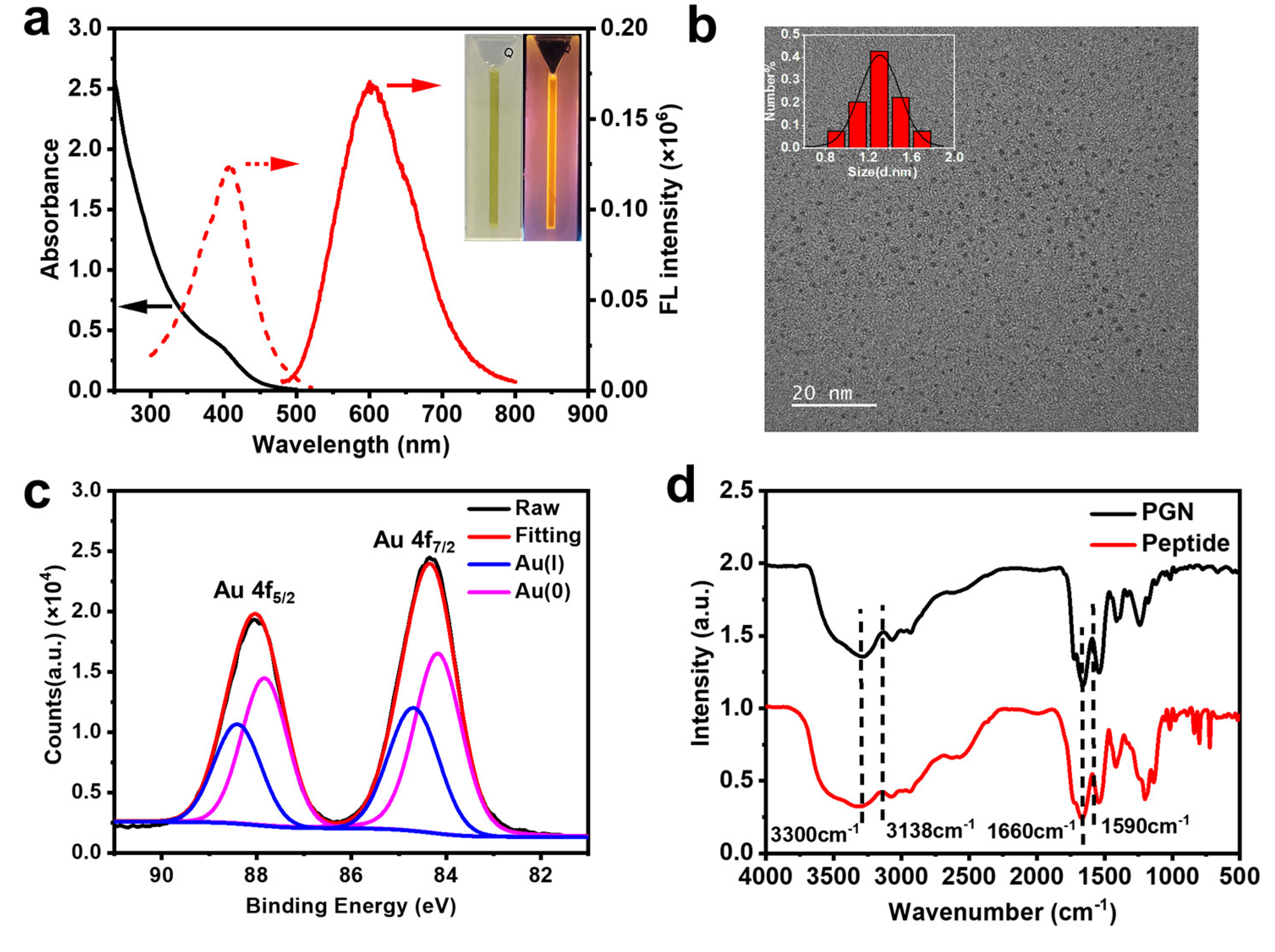
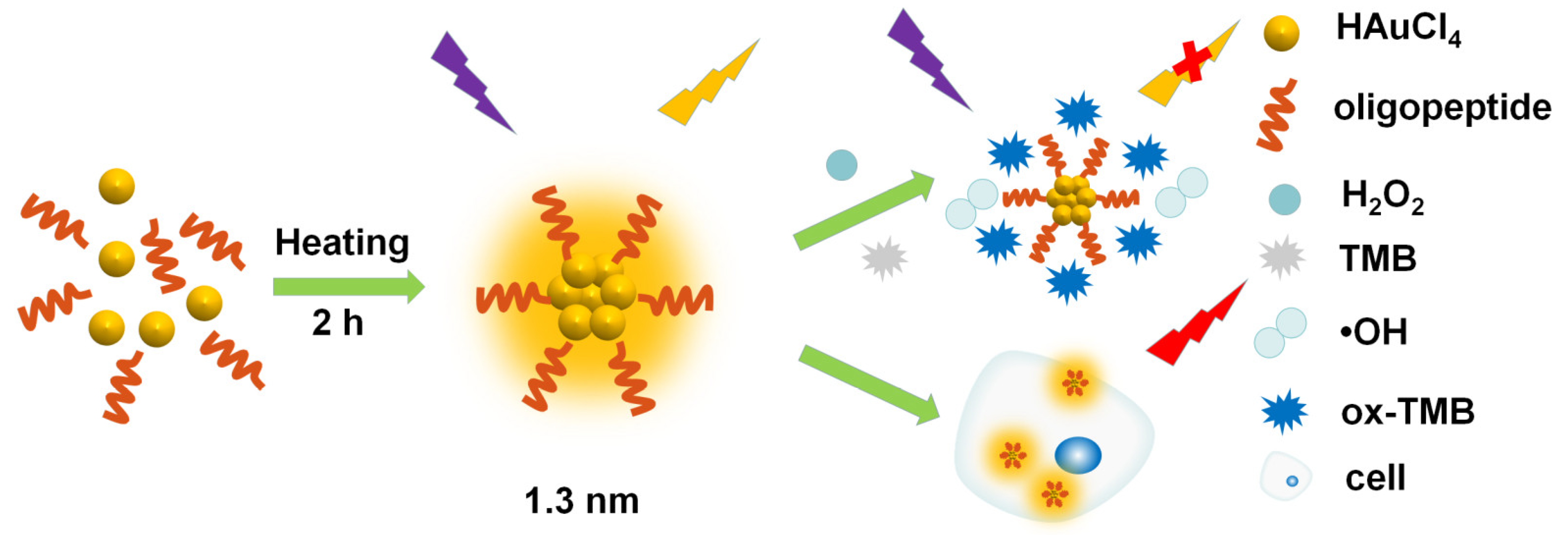
2.1. Characterization of Materials

A reported synthetic method was used to protect the gold nanoclusters with DGEAGC oligopeptide [23]. Briefly, the oligopeptide was mixed with HAuCl4 in ultrapure water, followed by thermal reduction at 70 °C. The color of the solution changed from yellow to colorless within 5 min. A pale yellow solution was obtained after ~2 h, indicating the formation of peptide-protected gold nanoclusters (abbreviated as PGN). As shown in Figure 1a, the absorption of the PGN exhibited a shoulder peak at ~400 nm. The absence of a surface plasmon resonance peak at ~520 nm excludes the formation of Au NPs [24,25]. Strong orange-red fluorescence was observed under 365 nm UV light irradiation (Figure 1a inset). The fluorescence spectra showed maximum excitation and emission peaks at 400 nm and 600 nm, respectively. The consistency of optical absorption and the excitation peak indicated that the fluorescence originated from intrinsic transitions (rather than aggregation induced emission, etc.) (Figure S1 from Supplementary Materials) [26,27]. After purifying the prepared nanoclusters using native polyacrylamide gel electrophoresis (PAGE), only one band with orange-red luminescence was observed (under UV-light irradiation, (Figure S2 from Supplementary Materials). The optical absorption and emission of the fluorescent band components were close to that of the prepared clusters (Figure S3 from Supplementary Materials), evidencing their predominance in the crude products.
Furthermore, HRTEM revealed the uniform size of the clusters with an average size of 1.30 ± 0.01 nm via the Gaussian fitting curve (Figure 1b). In this context, both the optical tests and the TEM analysis indicated the monodispersity of the prepared clusters. In addition, the absolute quantum yields of the PGN were 9.77% and 0.52% in the solid state and aqueous phases, respectively. The average fluorescence lifetime was 6.14 µs (Figure S4 from Supplementary Materials) [28,29,30]. According to X-ray photoelectron spectroscopy analysis (XPS, Figure 1c), the binding energies of Au 4f5/2 and Au 4f7/2 in the PGN were 88.0 and 84.3 eV respectively, corresponding to an Au(I): Au(0) ratio of 67.2%, which indicated the presence of a metallic core structure. The presence of O, C, N, Au, and S elements in the PGN was also verified using XPS (Figure S5 from Supplementary Materials) [31,32].
The FTIR spectrum of the PGN was similar to that of free peptide ligands (Figure 1d). The preserved carbonyl peak at 1590 cm−1, amide I peak at 1660 cm−1, and the carboxylic O-H peaks in the range of 2930–3550 cm−1 implied the maintained structure of the peptide ligands on the cluster surface [33,34].
Interestingly, the fluorescence intensity of the PGN was dependent on the temperature and pH of the aqueous solution. As shown in Figure S6 (from Supplementary Materials), the PGN fluorescence intensity showed a negative linear correlation with temperatures ranging from 20 to 40 °C. With regarding to pH dependency, the solution fluorescence was the strongest at pH = 5 (consistent with the isoelectric point of the C-terminus cysteine amino acid in the oligopeptide ligand).

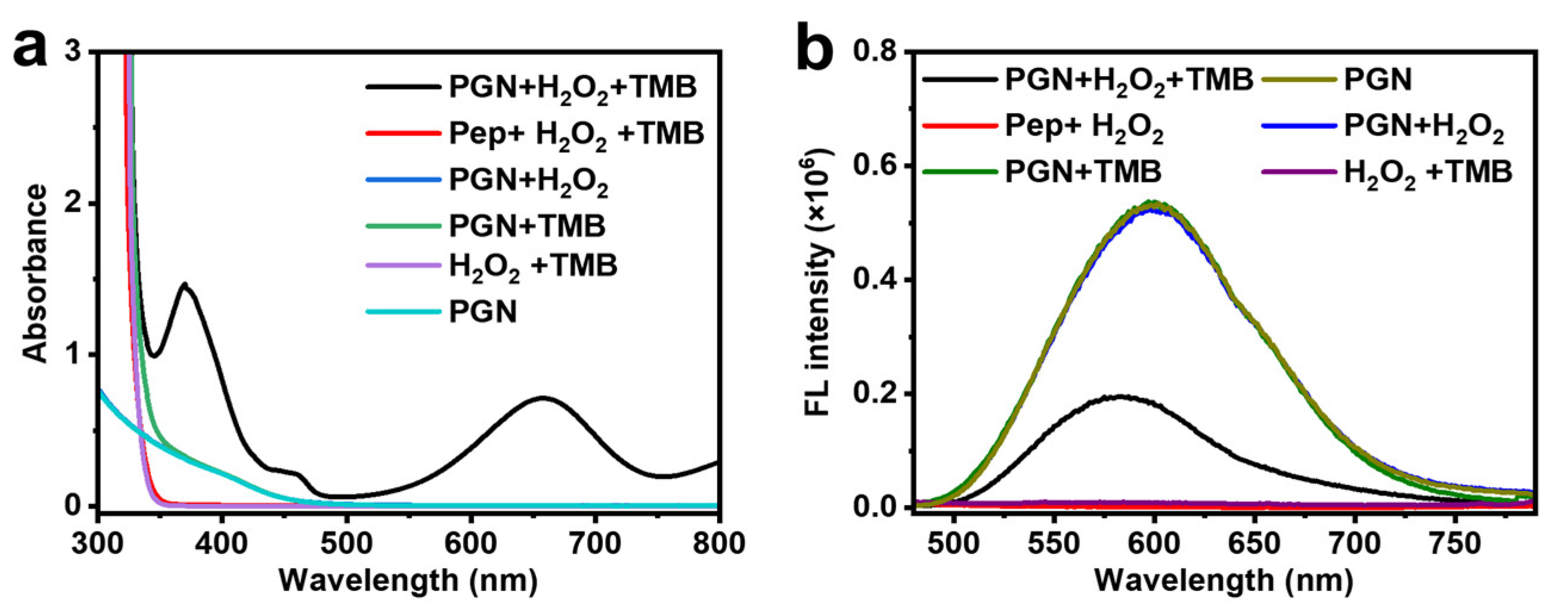
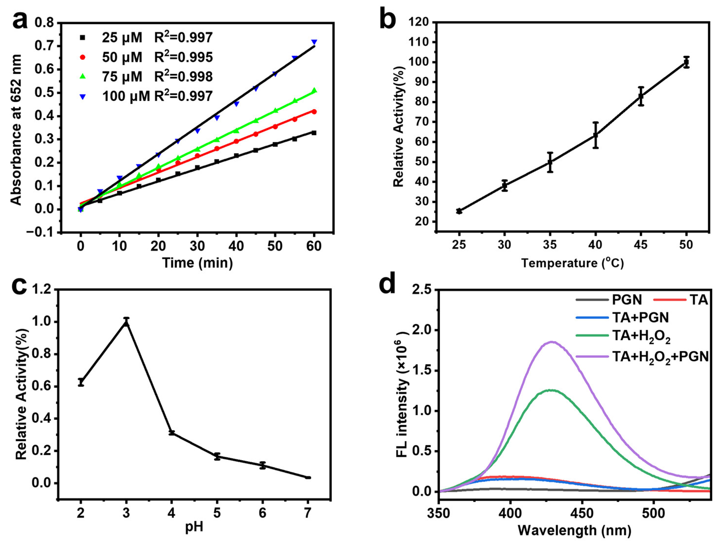
2.2. Peroxidase-like Activity of PGN

As shown in Figure 2a, mixing the aqueous solution of PGN with H2O2 and TMB resulted in remarkably changed UV-Vis spectra [35], and an additional absorption peak at ~652 nm (characteristic peak of ox-TMB). The observation demonstrated the horseradish peroxidase (HRP)-mimicking activity of the prepared clusters. By contrast, the characteristic peak of ox-TMB was invisible when the free oligopeptide ligand was mixed with H2O2 and TMB under otherwise identical conditions. Similarly, removing either TMB or H2O2 from the three-component system resulted in the absence of the ~652 nm peak. The results indicated that the gold nanoclusters, rather than the free ligands, were responsible for the HRP-mimicking activity [36]. It was notable that in addition to H2O2, the prepared PGN was able to oxidize TMB in the presence of dioxygen, but the significantly lower intensity of the ~652 nm peak indicated the inferior activity of dioxygen compared to that of H2O2 (Figure S7 from Supplementary Materials).
Interestingly, the fluorescence of the PGN was quenched in the three-component system. As shown in Figure 2b, the fluorescence of the PGN did not change after the addition H2O2 but decreased significantly after TMB was added. Further studies showed that the fluorescence intensity of the PGN-TMB system decreased regularly with the addition of increased amounts of H2O2 (Figure S8 from Supplementary Materials). These phenomena were mainly caused by fluorescence resonance energy transfer (FRET) between the oxidized form of TMB (with optical absorption at 652 nm) and the PGN (with maximum emission at 600 nm, Figure S9 from Supplementary Materials) [37,38]. Adding GSH into the PGN + H2O2 + TMB system, the characteristic absorption of ox-TMB at 652 nm was significantly diminished (Figure S10 from Supplementary Materials) and the fluorescence intensity was restored and comparable to that of the PGN + H2O2 + TMB (Figure S11 from Supplementary Materials). The results verified the reaction of GSH with ox-TMB. In view of the GSH induced emission enhancement of PGN, we were not able to completely exclude the possibility that the GSH binds with PGN to restore the fluorescence.
Like other Au NC-based peroxidase mimetics and the natural horseradish peroxidase, the peroxidase activity of PGN was dependent on the solution characteristics, such as the PGN concentration, pH, and temperature [39,40]. As shown in Figure 3a, the optical absorption of the reaction system at 652 nm increased with cluster concentration, and each system showed a linear correlation within 60 min. In addition, the catalytic activity of the PGN was sensitive to the pH and temperature of the solution, similar to recently reported nanomaterial-based peroxidases [41]. Based on the results in Figure 3b,c, pH = 3 and temperature 37 °C were selected for the subsequent tests.
Regarding the catalytic mechanism, it has frequently been suggested that the peroxidase-mimic nanozymes undergo a two-step mechanism, i.e., decomposition of H2O2 to generate hydroxyl radicals (•OH), and then oxidation of the colorless TMB to blue ox-TMB. To examine whether our PGN followed the same mechanism, we used terephthalic acid (TA) as a fluorescent probe for •OH, because the formed 2-hydroxyterephthalic acid (if present) exhibits an intense, characteristic emission at ~430 nm [42,43]. As shown in Figure 3d, the fluorescence intensity of the aqueous solution of the PGN-TA-H2O2 system at ~430 nm was stronger than that of the other controls. The result indicated that PGN can generate •OH from H2O2, thereby promoting the oxidation of TMB.

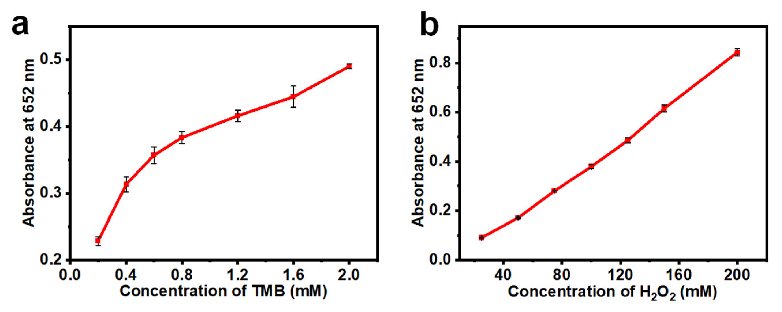
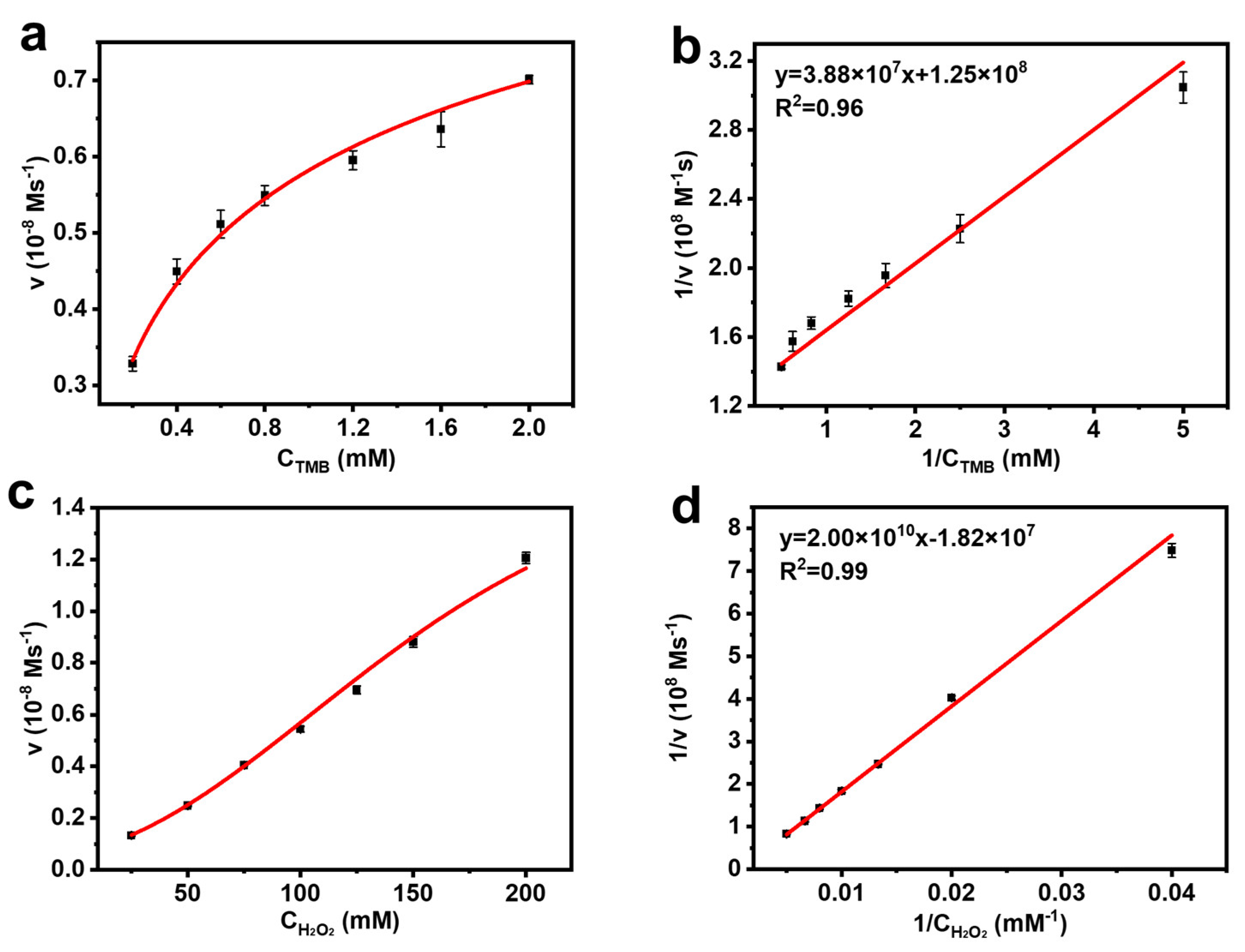
2.3. Kinetic Assay of the Peroxidase-like Activity

Next, we evaluated the kinetics of the peroxidase-like activity of PGN using a typical Michaelis–Menten approach. As shown in Figure 4, in a certain range of H2O2 and TMB concentrations, a typical absorption intensity spectrum was obtained under the reaction conditions of pH = 3 and 37 °C. The typical curves were obtained with both TMB and H2O2 as the substrate by monitoring their absorbance change at 652 nm (Figure 5a,c). Then, the enzyme kinetic parameters, Michaelis–Menten constant (Km) and maximal reaction velocity (Vmax), could be calculated from Lineweaver–Burk plots (Figure 5b,d). The Km value is a measure of the binding affinity between enzymes and substrates: the higher the value of Km, the weaker the affinity. The Kcat value measures the maximum number of colored products generated per enzyme per second [44]. As summarized in Table 1, when using TMB as the substrate, the Km and Kcat values of the PGN were 0.31 mM and 8 × 10⁻⁵ s−1, respectively. When using H2O2 as the substrate, the Km and Kcat values of the PGN were 1069 mM and 5.35 × 10⁻⁴ s−1, respectively. The Km value of the PGN with TMB as the substrate was almost identical to that of HRP, indicating that the affinity of PGN to TMB is like that of HRP. Additionally, the Km value of PGN for H2O2 appeared much higher than that of HRP. The results indicate that the affinity of PGN to H2O2 is lower than that of HRP. Therefore, a higher concentration of H2O2 is required to obtain the maximum reaction rate of the PGN. Then, we tested the potential peroxidase-mimicking activity of the GSH protected gold nanoclusters. According to the detailed kinetic analysis (Figures S12 and S13 from Supplementary Materials), the Vmax, Km and Kcat of the GSH protected clusters closed to that of the oligopeptide protected ones (Table S1 from Supplementary Materials). The result indicated that the incorporation of the bio-functional DGEA fragment did not affect the biomimicking activity of the metal clusters.

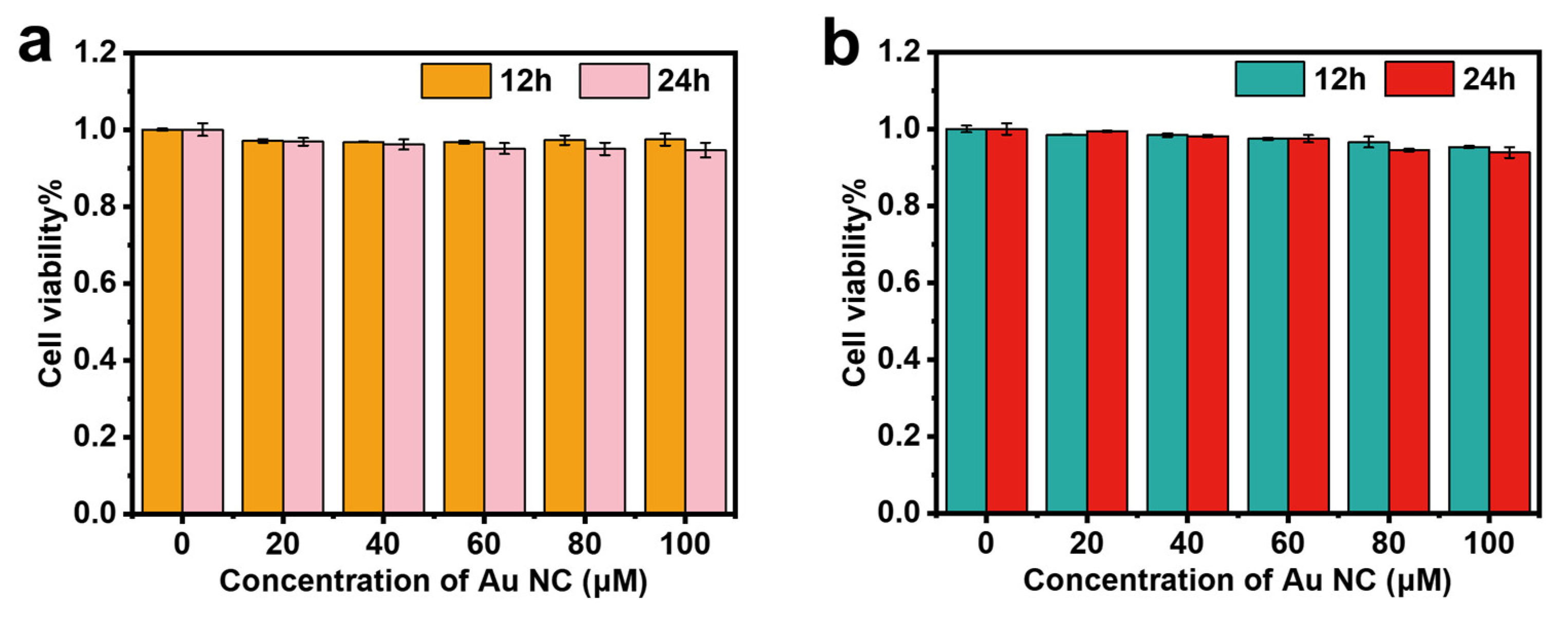
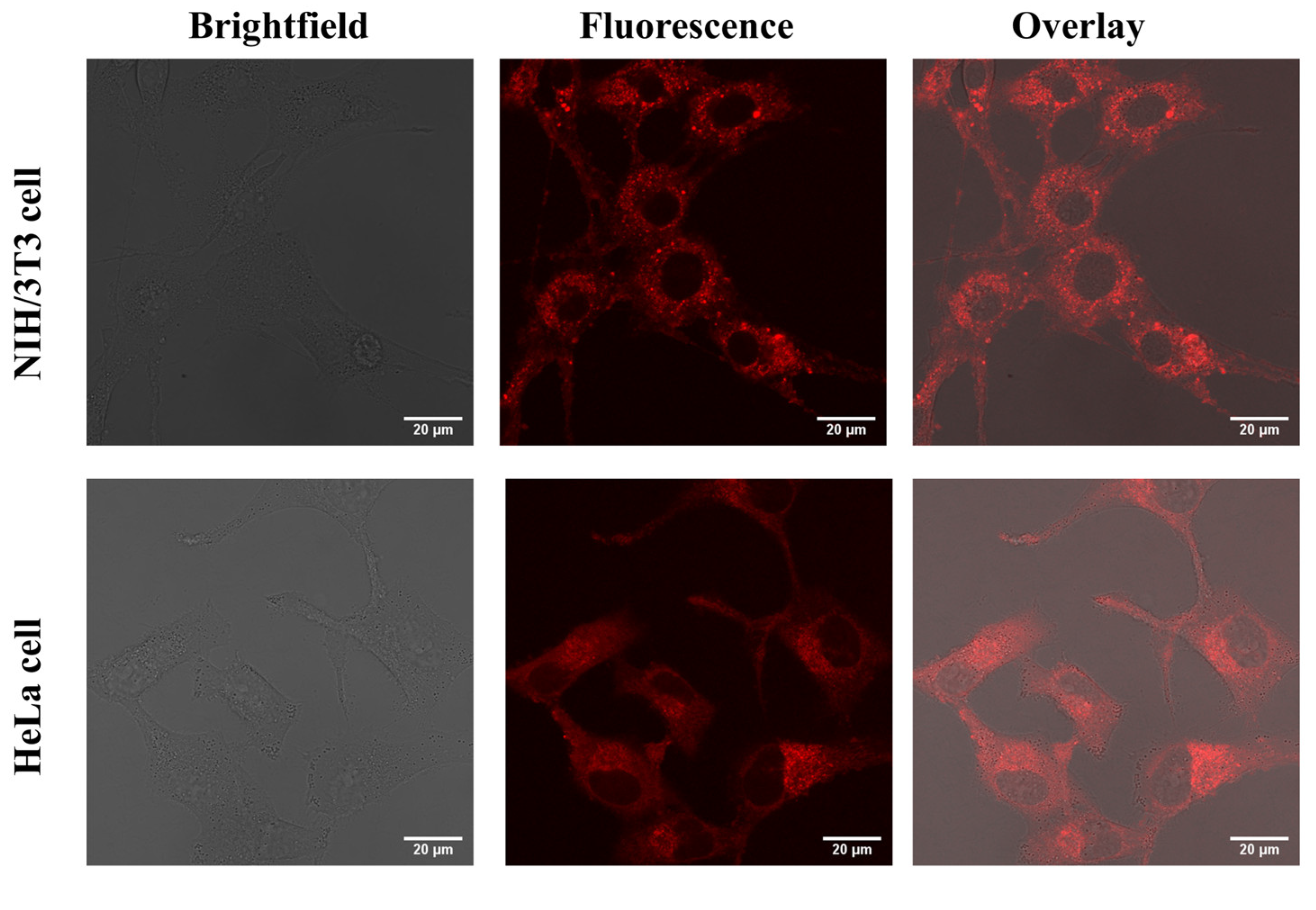
2.4. Cytotoxicity Study and In Vitro Imaging

To evaluate the biocompatibility of PGN, cell viability assays were carried out via an MTT assay (MTT = 3-(4,5-dimethylthiazol-2-yl)-2,5 diphenyltetrazolium bromide) [45,46], using NIH 3T3 and HeLa cells as the test candidates. As shown in Figure 6a, the presence of 0–100 μM PGN produced slight perturbation in the proliferation of the NIH 3T3 and HeLa cells within 24 h, and 95% cell viability was maintained even up to a relatively high dose of PGN of 100 μM after 24 h incubation. The low cytotoxicity to NIH 3T3 and HeLa cells is fundamentally important for cell imaging applications. As shown in Figure 7, after 2 h incubation with the PGN, the NIH 3T3 and HeLa cells showed a bright fluorescent signal in the cytoplasm (using confocal laser scanning microscopy), indicating that PGN have cell uptake and biological imaging capabilities.

3. Materials and Methods

3.1. Materials and Chemicals

All chemicals were commercially available and used without further purification. Custom-made DGEAGC polypeptides were obtained from Bankpeptide Biological Technology Co., Ltd, (Hefei, China). Hydrogen tetrachloroaurate tetrahydrate (HAuCl4·4H2O, ≥99.99%, metals basis) was purchased from Sino-Platinum Metals Co., Ltd. (Shanghai, China). Hydrochloric acid (HCl, ≥98%); and sodium hydroxide (NaOH, ≥97%) were purchased from Sinopharm Group Co., Ltd, (Beijing, China). Hydrogen peroxide solution(H2O2, ≥30%) was purchased from Xilong Science Co., Ltd, (Shanghai, China). The 3,3’,5,5’-tetramethylbenzidine (TMB, ≥99%) was purchased from Shanghai Macklin Biochemical Co., Ltd, (Shanghai, China). Terephthalic acid (TA, ≥98%) was purchased from Shanghai Macklin Biochemical Co., Ltd. All glassware was thoroughly cleaned with aqua regia (HCl/HNO3 3/1 v/v), rinsed with copious amounts of pure water, and then dried in an oven prior to use.

3.2. Equipment

UV−vis absorption spectra were recorded using a UV-6000PC instrument, and the solution samples were prepared using ultrapure water as the solvent. All fluorescence spectra were obtained using a HORIBA FluoroMax-4P fluorescence spectrophotometer. Transmission electron microscopy (TEM) and HRTEM images were obtained from a JEM-F200 microscope. X-ray photoelectron spectroscopy (XPS) measurements were performed on a ESCALAB 250 high-performance electron spectrometer with monochromated Al Kα radiation as the excitation source. The binding energies were corrected by referencing the binding energies of C(1s) arising from the added hydrocarbons as an internal standard. Fourier transform infrared spectroscopy (FT-IR) was recorded using a Bruker Vertex80 + Hyperion2000 apparatus. The gold content in the clusters was measured using Inductively coupled plasma mass spectrometry (ICP-MS) on an Agilent 7800 instrument. The fluorescence quantum yield was measured using an Edinburgh-Steady State/Transient Fluorescence Spectrometer FLS1000. Photoluminescence lifetimes were measured by time-correlated single-photon counting (TCSPC) on a Horiba Fluoro max plus spectrofluorometer with a pulsed light-emitting diode (LED) (400 nm) as the excitation source.

3.3. Synthesis of the PGN

The fluorescent gold nanoclusters were synthesized using DGEAGC as the surface ligand. Briefly, fresh aqueous solutions of HAuCl4 (0.2 g/mL, 68 μL) and the oligopeptide (50 mg/mL, 1.0 mL) were added to 17.6 mL of deionized water. Next, the aqueous solution was vigorously stirred at room temperature for 5 min, and then heated to 70 °C. After 2 h, clusters with strong orange-red fluorescence were formed.
Native polyacrylamide gel electrophoresis (PAGE) was carried out using discontinuous gels (1.5 mm × 80 mm × 70 mm). Resolving and stacking gels were prepared from 30 and 4 wt% acrylamide monomers. 200 μL of raw products was mixed with 20 μL of 5 vol% glycerol and then electrophoresis was run for 2 h at a constant voltage of 150 V at 4 °C. After electrophoresis, the band was cut from the gels and soaked in ultrapure water overnight at 4 °C to obtain the sample solution.

3.4. Peroxidase-like Activity

Quantities of 135 μL of the prepared clusters, 300 μL of 5 mM TMB and 200 μL of 100 mM H2O2 were added into 1.365 mL water. The mixture solution was then incubated in a water bath at 37 °C for 60 min before cooling to room temperature. The absorption spectra were recorded using a UV-Vis spectrophotometer.

3.5. Fluorescence Quenching Experiments with Adding H2O2 into the Cluster-TMB System

Quantities of 135 μL of the prepared clusters, 300 μL of 5 mM TMB and 200 μL of different concentrations of H2O2 were added to 1.365 mL water. The mixture solution was then incubated in a water bath at 37 °C for 60 min before cooling to room temperature. The emission spectra were recorded using a fluorescence analyzer.

3.6. Concentration-Dependent Peroxidase-like Activity of the Prepared Clusters

The concentration-dependent peroxidase-like activity of the gold clusters assays was measured at 37 °C with various concentrations of PGN (25, 50, 75, and 100 μM) in the presence of 100 mM H2O2 and 5 mM TMB at pH 3.0. The reaction was performed in a thermostatic mixer, and the absorbance of the samples at 652 nm was recorded via UV-Vis at different time points.
The pH of the solution containing 135 μL of clusters, 200 μL of 100 mM H2O2 and 300 μL of 5 mM TMB was adjusted from 2 to 7, and the absorbance of each sample at 652nm after retention in a thermomixer at 37 °C for 1h was recorded using a UV-Vis spectrophotometer.
The pH of the solution containing 135 μL of cluster solution, 200 μL of 100 mM H2O2 and 300 μL of 5 mM TMB was adjusted to 3, and the reaction was kept at different temperatures from 25 to 50 °C for 1 h. Then the absorbance of each sample at 652 nm was recorded using a UV-Vis spectrophotometer.

3.7. Determination of the Hydroxyl (·OH) Radical

Briefly, 135 μL of cluster solution was added to 2 mL water at pH 4. In this solution, 200 μL of 100 mM H2O2 and 800 μL of 6.25 mM terephthalic acid were mixed and incubated at 37 °C for 30 min. Then, the fluorescence spectra of the resultant solutions were measured with the excitation wavelength at 315 nm.

3.8. Steady-State Kinetic Assay of the Peroxidase-like Activity of the Clusters

Kinetic measurements of peroxidase-like properties were carried out by monitoring the time-dependent absorbance of ox-TMB at 652 nm using a UV–Vis spectrophotometer. The reaction kinetic data were measured according to the changing substrate concentration of TMB and H2O2. For the kinetic data relating TMB, 135 µL of cluster solution was added to a solution containing 200 µL of H2O2 (100mM); and 300 µL of different concentrations of TMB. Similarly, for the kinetic data relating H2O2, 135 µL of cluster solution was added to a solution containing 300 µL of TMB (1 mM); and 200 µL of different concentrations of H2O2.
The kinetic parameters (Vmax and Km) for peroxidase-like activity of PGN were deduced from Michaelis-Menten equation:
v = V max × [ S ] K m + [ S ]
where v, Vmax, [S] and Km are the initial velocity, the maximal reaction velocity, the concentration of TMB and the Michaelis constant, respectively. Kcat was derived from Kcat = Vmax/[E], where [E] represents the concentration of the clusters (Au based).

3.9. Cytotoxicity Experiment

MTT (5-dimethylthiazol-2-yl-2,5-diphenyltetrazolium bromide) assays were performed. NIH 3T3 and HeLa cells were passed and plated to ca. 70% confluence in 96-well plates 24 h before treatment. Then, DMEM (Dulbecco’s Modified Eagle Medium) with 10% FBS (fetal bovine serum) was removed and replaced with fresh DMEM, and the cluster solution was then added to obtain final concentrations of 0, 20, 40, 60, 80 and 100 μM (Au-based). The treated cells were incubated for 12/24 h at 37 °C under 5% CO2. Then, the cells were treated with 5 mg/mL MTT (10 μL/well) and incubated for another 4 h (37 °C, 5% CO2). The cells were then dissolved in DMSO (150 μL/well), and the absorbance at 570 nm was recorded. Cell viability (%) was analyzed based on the following equation:
Cell viability % = OD570 (sample)/OD570 (control) × 100%
where OD570 (sample) is the optical density of the wells treated with various concentration of clusters and OD570 (control) is that of the wells treated with DMEM containing 10% FBS (fetal bovine serum). The percentage of cell survival values were relative to untreated control cells. Each individual cytotoxicity experiment was repeated three times.

3.10. Cell Culture and Confocal Fluorescence Imaging

NIH 3T3 and HeLa cells were cultured in Dulbecco’s Modified Eagle Medium (DMEM) supplemented with 10% FBS (fetal bovine serum), penicillin (100 μg/mL), and streptomycin (100 μg/mL) at 37 °C in a humidified atmosphere of 5% CO2 and 95% air. Cytotoxicity assays showed that the clusters are biocompatible at low concentrations, so a concentration of 60 μM was chosen for fluorescence imaging in cells. The cells were incubated with 60 μM clusters for 2 h, then washed 3 times with PBS buffer. Then, cell imaging was recorded using a confocal microscope (TCS SP8 DIVE).

4. Conclusions

Nano-enzymes have recently attracted increasing research interest for their high catalytic activity and capacity to overcome the disadvantages of natural enzymes, whereas comprehensive synthetic procedures and harsh reaction conditions (such as high temperature, long reaction time,) significantly restrict the application of these materials. In this study, gold nanoclusters protected by oligopeptides (DGEAGC) were prepared using a mild one-pot synthesis method, i.e., via simply reacting the oligopeptide and HAuCl4 in aqueous solution at 70 °C for only two hours. The UV-Vis, PAGE separation, HRTEM, XPS, and FTIR analysis demonstrated the monodispersity of the prepared clusters. The prepared clusters showed strong orange-red emission and excellent chemical stability. PGN can oxidize TMB to produce blue ox-TMB in the presence of H2O2, indicating peroxidase activity, which conforms to the steady-state kinetic equation of the enzyme. In particular, the good biocompatibility and cell imaging properties indicate the potential of gold nanoclusters as a multifunctional platform.

Supplementary Materials

The following supporting information can be downloaded at: https://www.mdpi.com/article/10.3390/molecules28010070/s1. Figure S1: UV-vis absorption spectrum (black line) and the luminescence excitation (red line) of PGN. Figure S2: Digital photo of the PAGE bands upon UV light excitation. Figure S3: Comparison of (a) UV-Vis and (b) fluorescence intensity of the crude products and the fluorescent band. Figure S4: Fluorescence lifetime decays of PGN. Figure S5: The whole XPS spectra of PGN. Figure S6: (a) Fluorescence spectra of PGN at different temperatures (from 20 to 40 °C). (b) Correlation of fluorescence intensity at 600 nm with temperature ranging from 20 to 40 °C. (c) Fluorescence spectra of PGN at different pH (from 3 to 7). (d) Correlation of fluorescence intensity at 600 nm with pH in the range of 3–7. Figure S7: Comparison of the peroxidase-like (in presence of H2O2) and the oxidase-like (in presence of dioxygen) activity of PGN. Figure S8: Fluorescence changes of the PGN-TMB system after adding H2O2 at different concentrations (0, 5, 10, 20, 30, 40, 50, 60, 70, 80, 90 and 100 mM). Figure S9: Emission spectra of PGN (black line) and UV-Vis absorption spectra of the PGN-TMB-H2O2 system (red line). Figure S10: The UV-Vis spectrum of PGN + H2O2 + TMB before and after adding GSH. Figure S11: Fluorescence spectrum of PGN; PGN + GSH; PGN + H2O2 + TMB and PGN + H2O2 + TMB + [GSH]. Figure S12: (a) The absorption peak at 652 nm in the presence of different concentrations of TMB (0.2, 0.4, 0.6, 1.2, 1.6 mM). (b) The absorption peak at 652 nm in the presence of different concentrations of H2O2 (25, 50, 75, 100, 125 mM). The concentration of the GSH-Au NCs was fixed at 100 μM (Au basis). Figure S13: Steady-state kinetic assay and catalytic mechanism of the synthesized GSH-Au NCs towards various components: (a,b) 1 mM TMB and different concentrations of H2O2, (c,d) 100 mM H2O2 and different concentrations of TMB. Table S1: Comparison of kinetic parameters of Km and Kcat of PGN, and GSH-Au NCs.

Author Contributions

Conceptualization, H.Y., X.M. and M.Z.; methodology, D.F., L.C. and J.O.; data analysis, H.Y., D.F., J.O., Z.Z. and L.Z.; writing—original draft preparation, D.F.; writing—review and editing, H.Y., X.M. and M.Z. All authors have read and agreed to the published version of the manuscript.

Funding

National Science Foundation of Anhui Province (2108085J08, 2008085J08). The University Synergy Innovation Program of Anhui Province (GXXT-2021-023). National Natural Science Foundation of China (22077001).

Institutional Review Board Statement

Not applicable.

Informed Consent Statement

Not applicable.

Data Availability Statement

All data are included in the article and/or Supplementary Materials.

Conflicts of Interest

The authors declare no conflict of interest.

References

  1. Robinson, P.K. Enzymes: Principles and biotechnological applications. Essays Biochem. 2015, 59, 1–41. [Google Scholar] [CrossRef] [PubMed]
  2. Ma, S.; Wang, J.; Yang, G.; Yang, J.; Ding, D.; Zhang, M. Copper(II) ions enhance the peroxidase-like activity and stability of keratin-capped gold nanoclusters for the colorimetric detection of glucose. Mikrochim Acta 2019, 186, 271. [Google Scholar] [CrossRef] [PubMed]
  3. Zhang, Y.; Li, S.; Liu, H.; Long, W.; Zhang, X.D. Enzyme-Like Properties of Gold Clusters for Biomedical Application. Front. Chem. 2020, 8, 219. [Google Scholar] [CrossRef] [PubMed] [Green Version]
  4. Krishna Kumar, A.S.; Tseng, W.-L. Perspective on recent developments of near infrared-emitting gold nanoclusters: Applications in sensing and bio-imaging. Anal. Methods 2020, 12, 1809–1826. [Google Scholar] [CrossRef]
  5. You, J.G.; Lu, C.Y.; Krishna Kumar, A.S.; Tseng, W.L. Cerium(iii)-directed assembly of glutathione-capped gold nanoclusters for sensing and imaging of alkaline phosphatase-mediated hydrolysis of adenosine triphosphate. Nanoscale 2018, 10, 17691–17698. [Google Scholar] [CrossRef] [PubMed]
  6. Chen, X.; Wang, Y.; Dai, X.; Ding, L.; Chen, J.; Yao, G.; Liu, X.; Luo, S.; Shi, J.; Wang, L.; et al. Single-Stranded DNA-Encoded Gold Nanoparticle Clusters as Programmable Enzyme Equivalents. J. Am. Chem. Soc. 2022, 144, 6311–6320. [Google Scholar] [CrossRef]
  7. Xu, J.; Sun, F.; Li, Q.; Yuan, H.; Ma, F.; Wen, D.; Shang, L. Ultrasmall Gold Nanoclusters-Enabled Fabrication of Ultrafine Gold Aerogels as Novel Self-Supported Nanozymes. Small 2022, 18, e2200525. [Google Scholar] [CrossRef]
  8. Qiu, N.; Liu, Y.; Guo, R. Electrodeposition-Assisted Rapid Preparation of Pt Nanocluster/3D Graphene Hybrid Nanozymes with Outstanding Multiple Oxidase-Like Activity for Distinguishing Colorimetric Determination of Dihydroxybenzene Isomers. ACS Appl. Mater. Interfaces 2020, 12, 15553–15561. [Google Scholar] [CrossRef]
  9. Li, Y.; Li, S.; Bao, M.; Zhang, L.; Carraro, C.; Maboudian, R.; Liu, A.; Wei, W.; Zhang, Y.; Liu, S. Pd Nanoclusters Confined in ZIF-8 Matrixes for Fluorescent Detection of Glucose and Cholesterol. ACS Appl. Nano Mater. 2021, 4, 9132–9142. [Google Scholar] [CrossRef]
  10. Qin, F.; Zhang, J.; Zhou, Z.; Xu, H.; Cui, L.; Lv, Z.; Qin, Y. TiO2 Nanoflowers Decorated with FeOx Nanocluster and Single Atoms by Atomic Layer Deposition for Peroxidase-Mimicking Nanozymes. ACS Appl. Nano Mater. 2022, 5, 13090–13099. [Google Scholar] [CrossRef]
  11. Liu, C.; Zhang, X.; Han, X.; Fang, Y.; Liu, X.; Wang, X.; Waterhouse, G.I.N.; Xu, C.; Yin, H.; Gao, X. Polypeptide-Templated Au Nanoclusters with Red and Blue Fluorescence Emissions for Multimodal Imaging of Cell Nuclei. ACS Appl. Bio. Mater. 2020, 3, 1934–1943. [Google Scholar] [CrossRef] [PubMed]
  12. Maity, S.; Bain, D.; Chakraborty, S.; Kolay, S.; Patra, A. Copper Nanocluster (Cu23 NC)-Based Biomimetic System with Peroxidase Activity. ACS Sustain. Chem. Eng. 2020, 8, 18335–18344. [Google Scholar] [CrossRef]
  13. Wang, G.L.; Jin, L.Y.; Dong, Y.M.; Wu, X.M.; Li, Z.J. Intrinsic enzyme mimicking activity of gold nanoclusters upon visible light triggering and its application for colorimetric trypsin detection. Biosens. Bioelectron. 2015, 64, 523–529. [Google Scholar] [CrossRef] [PubMed]
  14. Dehghani, Z.; Hosseini, M.; Mohammadnejad, J.; Ganjali, M.R. New Colorimetric DNA Sensor for Detection of Campylobacter jejuni in Milk Sample Based on Peroxidase-Like Activity of Gold/Platinium Nanocluster. ChemistrySelect 2019, 4, 11687–11692. [Google Scholar] [CrossRef]
  15. Li, M.; Lao, Y.H.; Mintz, R.L.; Chen, Z.; Shao, D.; Hu, H.; Wang, H.X.; Tao, Y.; Leong, K.W. A multifunctional mesoporous silica-gold nanocluster hybrid platform for selective breast cancer cell detection using a catalytic amplification-based colorimetric assay. Nanoscale 2019, 11, 2631–2636. [Google Scholar] [CrossRef] [PubMed]
  16. Wu, T.; Sun, J.; Lei, J.; Fan, Q.; Tang, X.; Zhu, G.; Yan, Q.; Feng, X.; Shi, B. An efficient treatment of biofilm-induced periodontitis using Pt nanocluster catalysis. Nanoscale 2021, 13, 17912–17919. [Google Scholar] [CrossRef]
  17. Zhang, C.-X.; Gao, Y.-C.; Li, H.-W.; Wu, Y. Gold–Platinum Bimetallic Nanoclusters for Oxidase-like Catalysis. ACS Appl. Nano Mater. 2020, 3, 9318–9328. [Google Scholar] [CrossRef]
  18. Han, L.; Li, Y.; Fan, A. Improvement of mimetic peroxidase activity of gold nanoclusters on the luminol chemiluminescence reaction by surface modification with ethanediamine. Luminescence 2018, 33, 751–758. [Google Scholar] [CrossRef]
  19. Dehghani, Z.; Mohammadnejad, J.; Hosseini, M. A new colorimetric assay for amylase based on starch-supported Cu/Au nanocluster peroxidase-like activity. Anal. Bioanal. Chem. 2019, 411, 3621–3629. [Google Scholar] [CrossRef]
  20. Garcia-Lopez, V.; Chen, F.; Nilewski, L.G.; Duret, G.; Aliyan, A.; Kolomeisky, A.B.; Robinson, J.T.; Wang, G.; Pal, R.; Tour, J.M. Molecular machines open cell membranes. Nature 2017, 548, 567–572. [Google Scholar] [CrossRef]
  21. Hu, H.; Zhang, Y.; Shukla, S.; Gu, Y.; Yu, X.; Steinmetz, N.F. Dysprosium-Modified Tobacco Mosaic Virus Nanoparticles for Ultra-High-Field Magnetic Resonance and Near-Infrared Fluorescence Imaging of Prostate Cancer. ACS Nano 2017, 11, 9249–9258. [Google Scholar] [CrossRef] [PubMed] [Green Version]
  22. Zhan, F.-K.; Liu, J.-C.; Cheng, B.; Liu, Y.-C.; Lai, T.-S.; Lin, H.-C.; Yeh, M.-Y. Tumor targeting with DGEA peptide ligands: A new aromatic peptide amphiphile for imaging cancers. Chem. Comm. 2019, 55, 1060–1063. [Google Scholar] [CrossRef]
  23. Luo, Z.; Yuan, X.; Yu, Y.; Zhang, Q.; Leong, D.T.; Lee, J.Y.; Xie, J. From aggregation-induced emission of Au(I)-thiolate complexes to ultrabright Au(0)@Au(I)-thiolate core-shell nanoclusters. J. Am. Chem. Soc. 2012, 134, 16662–16670. [Google Scholar] [CrossRef] [PubMed]
  24. Gao, L.; Liu, M.Q.; Ma, G.F.; Wang, Y.L.; Zhao, L.N.; Yuan, Q.; Gao, F.P.; Liu, R.; Zhai, J.; Chai, Z.F.; et al. Peptide-Conjugated Gold Nanoprobe: Intrinsic Nanozyme-Linked Immunsorbant Assay of Integrin Expression Level on Cell Membrane. ACS Nano 2015, 9, 10979–10990. [Google Scholar] [CrossRef]
  25. Gurunathan, S.; Han, J.; Park, J.H.; Kim, J.H. A green chemistry approach for synthesizing biocompatible gold nanoparticles. Nanoscale Res. Lett. 2014, 9, 11. [Google Scholar] [CrossRef] [Green Version]
  26. Sugiuchi, M.; Maeba, J.; Okubo, N.; Iwamura, M.; Nozaki, K.; Konishi, K. Aggregation-Induced Fluorescence-to-Phosphorescence Switching of Molecular Gold Clusters. J. Am. Chem. Soc. 2017, 139, 17731–17734. [Google Scholar] [CrossRef] [PubMed]
  27. Zheng, J.; Petty, J.T.; Dickson, R.M. High quantum yield blue emission from water-soluble Au8 nanodots. J. Am. Chem. Soc. 2003, 125, 7780–7781. [Google Scholar] [CrossRef]
  28. Yu, H.; Rao, B.; Jiang, W.; Yang, S.; Zhu, M. The photoluminescent metal nanoclusters with atomic precision. Coord. Chem. Rev. 2019, 378, 595–617. [Google Scholar] [CrossRef]
  29. Zheng, Y.; Lai, L.; Liu, W.; Jiang, H.; Wang, X. Recent advances in biomedical applications of fluorescent gold nanoclusters. Adv. Colloid Interface Sci. 2017, 242, 1–16. [Google Scholar] [CrossRef]
  30. Li, Y.; Zhai, T.; Chen, J.; Shi, J.; Wang, L.; Shen, J.; Liu, X. Water-Dispersible Gold Nanoclusters: Synthesis Strategies, Optical Properties, and Biological Applications. Chem. Eur. J. 2022, 28, e202103736. [Google Scholar] [CrossRef]
  31. Shang, L.; Azadfar, N.; Stockmar, F.; Send, W.; Trouillet, V.; Bruns, M.; Gerthsen, D.; Nienhaus, G.U. One-pot synthesis of near-infrared fluorescent gold clusters for cellular fluorescence lifetime imaging. Small 2011, 7, 2614–2620. [Google Scholar] [CrossRef] [PubMed]
  32. Yu, Y.; Luo, Z.; Chevrier, D.M.; Leong, D.T.; Zhang, P.; Jiang, D.E.; Xie, J. Identification of a highly luminescent Au22(SG)18 nanocluster. J. Am. Chem. Soc. 2014, 136, 1246–1249. [Google Scholar] [CrossRef] [PubMed]
  33. Kazemi, M.S.; Rasaeinezhad, S.; Es’haghi, Z. Evaluation of flutamide loading capacity of biosynthesis of plant-mediated glutathione-modified gold nanoparticles by Dracocephalum Kotschyi Boiss extract. Chem. Zvesti 2020, 74, 2041–2048. [Google Scholar] [CrossRef]
  34. Kong, J.; Yu, S. Fourier transform infrared spectroscopic analysis of protein secondary structures. Acta Biochim. Biophys. Sin. 2007, 39, 549–559. [Google Scholar] [CrossRef] [PubMed] [Green Version]
  35. Tao, Y.; Lin, Y.; Huang, Z.; Ren, J.; Qu, X. Incorporating graphene oxide and gold nanoclusters: A synergistic catalyst with surprisingly high peroxidase-like activity over a broad pH range and its application for cancer cell detection. Adv. Mater. 2013, 25, 2594–2599. [Google Scholar] [CrossRef]
  36. Marquez, L.A.; Dunford, H.B. Mechanism of the oxidation of 3,5,3’,5’-tetramethylbenzidine by myeloperoxidase determined by transient- and steady-state kinetics. Biochemistry 1997, 36, 9349–9355. [Google Scholar] [CrossRef]
  37. Du, C.; Qi, L.; Wang, Y.; Pei, K.; Zhang, R.; Wu, D.; Qi, W. Fluorescence dual “turn-on” detection of acid phosphatase via fluorescence resonance energy transfer and dual quenching strategy to improve sensitivity. Dyes Pigm. 2022, 205, 110554. [Google Scholar] [CrossRef]
  38. Zhao, Q.; Chen, S.; Huang, H.; Zhang, L.; Wang, L.; Liu, F.; Chen, J.; Zeng, Y.; Chu, P.K. Colorimetric and ultra-sensitive fluorescence resonance energy transfer determination of H2O2 and glucose by multi-functional Au nanoclusters. Analyst 2014, 139, 1498–1503. [Google Scholar] [CrossRef]
  39. Li, J.J.; Qiao, D.; Yang, S.Z.; Weng, G.J.; Zhu, J.; Zhao, J.W. Colorimetric determination of cysteine based on inhibition of GSH-Au/Pt NCs as peroxidase mimic. Spectrochim. Acta A Mol. Biomol. Spectrosc. 2021, 248, 119257. [Google Scholar] [CrossRef]
  40. Hu, D.; Sheng, Z.; Fang, S.; Wang, Y.; Gao, D.; Zhang, P.; Gong, P.; Ma, Y.; Cai, L. Folate receptor-targeting gold nanoclusters as fluorescence enzyme mimetic nanoprobes for tumor molecular colocalization diagnosis. Theranostics 2014, 4, 142–153. [Google Scholar] [CrossRef]
  41. Zhao, Y.; Zhuang, S.; Liao, L.; Wang, C.; Xia, N.; Gan, Z.; Gu, W.; Li, J.; Deng, H.; Wu, Z. A Dual Purpose Strategy to Endow Gold Nanoclusters with Both Catalysis Activity and Water Solubility. J. Am. Chem. Soc. 2020, 142, 973–977. [Google Scholar] [CrossRef] [PubMed]
  42. Wu, Y.; Gao, Y.; Du, J. Bifunctional gold nanoclusters enable ratiometric fluorescence nanosensing of hydrogen peroxide and glucose. Talanta 2019, 197, 599–604. [Google Scholar] [CrossRef] [PubMed]
  43. Singh, V.K.; Yadav, P.K.; Chandra, S.; Bano, D.; Talat, M.; Hasan, S.H. Peroxidase mimetic activity of fluorescent NS-carbon quantum dots and their application in colorimetric detection of H2O2 and glutathione in human blood serum. J. Mater. Chem. B 2018, 6, 5256–5268. [Google Scholar] [CrossRef] [PubMed]
  44. Li, M.; Yang, J.; Ou, Y.; Shi, Y.; Liu, L.; Sun, C.; Zheng, H.; Long, Y. Peroxidase-like activity of 2’,7’-difluorofluorescein and its application for galactose detection. Talanta 2018, 182, 422–427. [Google Scholar] [CrossRef]
  45. Lewinski, N.; Colvin, V.; Drezek, R. Cytotoxicity of nanoparticles. Small 2008, 4, 26–49. [Google Scholar] [CrossRef]
  46. Gao, P.; Wu, S.; Chang, X.; Liu, F.; Zhang, T.; Wang, B.; Zhang, K.Q. Aprotinin Encapsulated Gold Nanoclusters: A Fluorescent Bioprobe with Dynamic Nuclear Targeting and Selective Detection of Trypsin and Heavy Metal. Bioconjug. Chem. 2018, 29, 4140–4148. [Google Scholar] [CrossRef]
Scheme 1. The synthesis and peroxidase-mimic activity of gold nanoclusters.
Molecules 28 00070 sch001
Figure 1. (a) UV-Vis absorption spectrum (black line), luminescence excitation (broken red line) and emission spectra (solid red line) of the PGN (peptide protected gold nanoclusters); inset: digital photo of PGN in aqueous solution under visible (left) and UV light (right). (b) HRTEM image of the prepared PGN; inset histograms of core sizes. (c) High-resolution Au (4f) peaks of the PGN. (d) FTIR spectrum of the PGN and the free oligopeptide.
Molecules 28 00070 g001
Figure 2. Catalytic properties of the PGN: (a) absorption and (b) fluorescence spectra for the PGN-H2O2-TMB system and the related control systems.
Molecules 28 00070 g002
Figure 3. (a) Time-dependent absorbance change at 652 nm of mixture containing 100 mM H2O2 and 5 mM TMB in the presence of different concentrations of the PGN (Au basis). (b) Temperature-dependent peroxidase-like activity of the PGN. (c) pH-dependent peroxidase-like activity of PGN. (d) Fluorescence spectroscopy monitoring of the oxidation of terephthalic acid to 2-hydroxyterephthalic acid in the presence of the PGN.
Molecules 28 00070 g003
Figure 4. (a) The absorption peak at 652 nm in the presence of different concentrations of TMB (0.2, 0.4, 0.6, 0.8, 1.2, 1.6 and 2.0 mM). (b) The absorption peak at 652 nm in the presence of different concentrations of H2O2 (25, 50, 75, 100, 125, 150 and 200 mM). PGN concentration was fixed at 100 μM.
Molecules 28 00070 g004
Figure 5. Steady−state kinetic assay and catalytic mechanism of the as−synthesized PGN towards various components: (a,b) 1 mM TMB and different concentrations of H2O2; (c,d) 100 mM H2O2 and different concentrations of TMB.
Molecules 28 00070 g005
Figure 6. The viability of (a) NIH 3T3 and (b) HeLa cells after treatment with different concentrations of PGN (0, 20, 40, 60, 80 and 100 μM) for 12 and 24 h. The viability was obtained using the MTT test. The error bar represents the standard error of three trials.
Molecules 28 00070 g006
Figure 7. Fluorescence imaging of HeLa cells after incubation with PGN (60 μM) for 2 h. Scale bar = 20 µm. Brightfield (left), fluorescence (center) and overlay (right) images.
Molecules 28 00070 g007
Table 1. Comparison of kinetic parameters of Km and Kcat of PGN, and HRP.
CatalystSubstrateKm (mM)Kcat (s−1)Reference
PGNTMB0.318 × 10−5This work
H2O210695.35 × 10−4
Peptide-Au NPsTMB0.2773.57[24]
H2O292914.6
HRPTMB0.3015.18 × 103[40]
H2O20.9354.05 × 103
Disclaimer/Publisher’s Note: The statements, opinions and data contained in all publications are solely those of the individual author(s) and contributor(s) and not of MDPI and/or the editor(s). MDPI and/or the editor(s) disclaim responsibility for any injury to people or property resulting from any ideas, methods, instructions or products referred to in the content.

