Article

Investigating the Functional Role of the Cysteine Residue in Dehydrin from the Arctic Mouse-Ear Chickweed Cerastium arcticum

1
Advanced Bio-Resource R&D Center, Kyungpook National University, Daegu 41566, Korea
2
Research Unit of Cryogenic Novel Material, Korea Polar Research Institute, Incheon 21990, Korea
3
Division of Bacterial Diseases, Center for Laboratory Control of Infectious Diseases, Korea Centers for Diseases Control and Prevention, Cheongju-si 28159, Korea
4
New Drug Development Center, Daegu-Gyeongbuk Medical Innovation Foundation, Daegu 41061, Korea
5
Department of Polar Sciences, University of Science and Technology, Incheon 21990, Korea
6
Department of Chemistry, Graduate School of General Studies, Sookmyung Women’s University, Seoul 04310, Korea
*
Author to whom correspondence should be addressed.
These authors contributed equally to this work.
Molecules 2022, 27(9), 2934; https://doi.org/10.3390/molecules27092934
Submission received: 21 March 2022 / Revised: 3 May 2022 / Accepted: 3 May 2022 / Published: 4 May 2022
(This article belongs to the Topic Advances in Enzymes and Protein Engineering)

Abstract

:
The stress-responsive, SK5 subclass, dehydrin gene, CaDHN, has been identified from the Arctic mouse-ear chickweed Cerastium arcticum. CaDHN contains an unusual single cysteine residue (Cys143), which can form intermolecular disulfide bonds. Mutational analysis and a redox experiment confirmed that the dimerization of CaDHN was the result of an intermolecular disulfide bond between the cysteine residues. The biochemical and physiological functions of the mutant C143A were also investigated by in vitro and in vivo assays using yeast cells, where it enhanced the scavenging of reactive oxygen species (ROS) by neutralizing hydrogen peroxide. Our results show that the cysteine residue in CaDHN helps to enhance C. arcticum tolerance to abiotic stress by regulating the dimerization of the intrinsically disordered CaDHN protein, which acts as a defense mechanism against extreme polar environments.

1. Introduction

Arctic plants are exposed to various environmental stresses, such as freezing temperatures, drought and water deficiency, high ultraviolet radiation levels due to ozone depletion, nutrient depletion, salinity, and very short growing seasons [1,2].
Thus, to thrive at sub-zero temperatures, Arctic plants have developed diverse morphological and physiological adaptations for the different stages of growth development, such as long life cycles, extended primordial development of leaves and flowers, well-developed root systems, and efficient growth-development stages [3,4]. Dehydrins (DHNs) are a member of the late embryonic developmental abundance (LEA) protein family, which help plants during stress conditions, such as salinity, drought and low temperatures, which result in cell dehydration [5,6]. This protein was first identified in cottonseed at a later stage of embryonic development and was later found in other tissues and organs as well [7]. LEA genes have been significantly induced by abiotic stress and their overexpression in transgenic plants increased tolerance to a variety of abiotic stress sources [8,9]. A common feature of LEA proteins is their intrinsically disordered structure due to their amino acid composition, such as high glycine content and high hydrophilicity [10]. Based on their sequence similarity, LEA proteins are classified into three major groups (LEA I, LEA II, and LEA III) and each group has specific functions during dehydration [11]. The high accumulation of group II LEA proteins, including DHNs, is one of the prominent plant responses to cellular dehydration generated by abiotic stresses, such as cold, drought, and high salinity [12].
DHNs are a class of hydrophilic, thermostable cell rescue proteins with molecular masses ranging from 9 to 200 kDa [13,14] that are ubiquitous among various plants, algae, and cyanobacteria [12,15]. DHNs are very rich in glycine residues, whereas cysteine and tryptophan residues are lacking or under-represented [12]. A structural feature of DHNs is the existence of highly conserved sequence motifs called the K-, S-, and Y-segments [12]. The K-segment is a highly conserved lysine-rich motif (i.e., EKKGIMDKIKEKLPG) present in 1–11 copies near the C-terminal and it is the only conserved segment present in all DHNs [16,17]. Many DHNs also contain the serine-repeat S-segment formed by a stretch of 4–10 serine residues (LHRSGS4–10(E/D)3) and the tyrosine-rich Y-segment ((V/T)D(E/Q) YGNP) located near the N terminal [18,19]. In addition to the highly conserved domains, DHNs possess φ-segments defined as all the residues located between Y-, S-, and K-segments. The sequences and lengths of the φ-segments are not conserved compared with those in the other segments.
Arctic Cerastium arcticum Lange DHN (CaDHN) has been successfully expressed in yeast and Escherichia coli [20]. The overexpression of CaDHN in transgenic yeast confers tolerance against various forms of abiotic stress by improving redox homeostasis and enhancing fermentation capacity, especially at low temperatures (18 °C). Although most DHNs generally lack cysteine residue, CaDHN exhibits a single cysteine residue. In this study, we aimed to determine the role of the cysteine residue in CaDHN. For more detailed information, we performed a mutational analysis of CaDHN, which revealed its physicochemical properties via in vitro and in vivo assays. To the best of our knowledge, this is the first study that the unusual cysteine residue in CaDHN induces intermolecular disulfide bond-mediated dimerization of CaDHN, which plays an important role in the intrinsic and acquired tolerance of the transgenic yeast by modulating its function.

2. Results & Discussion

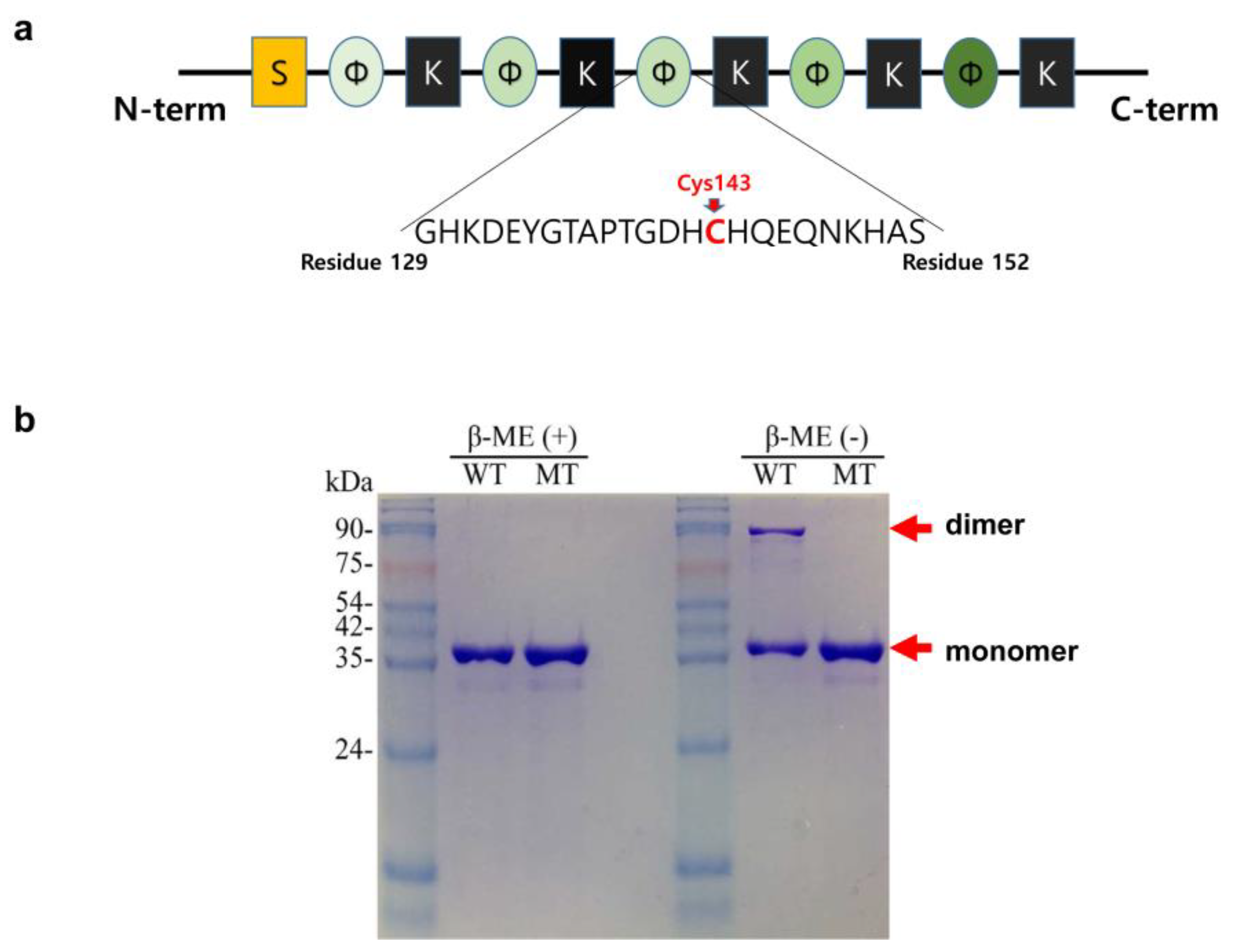
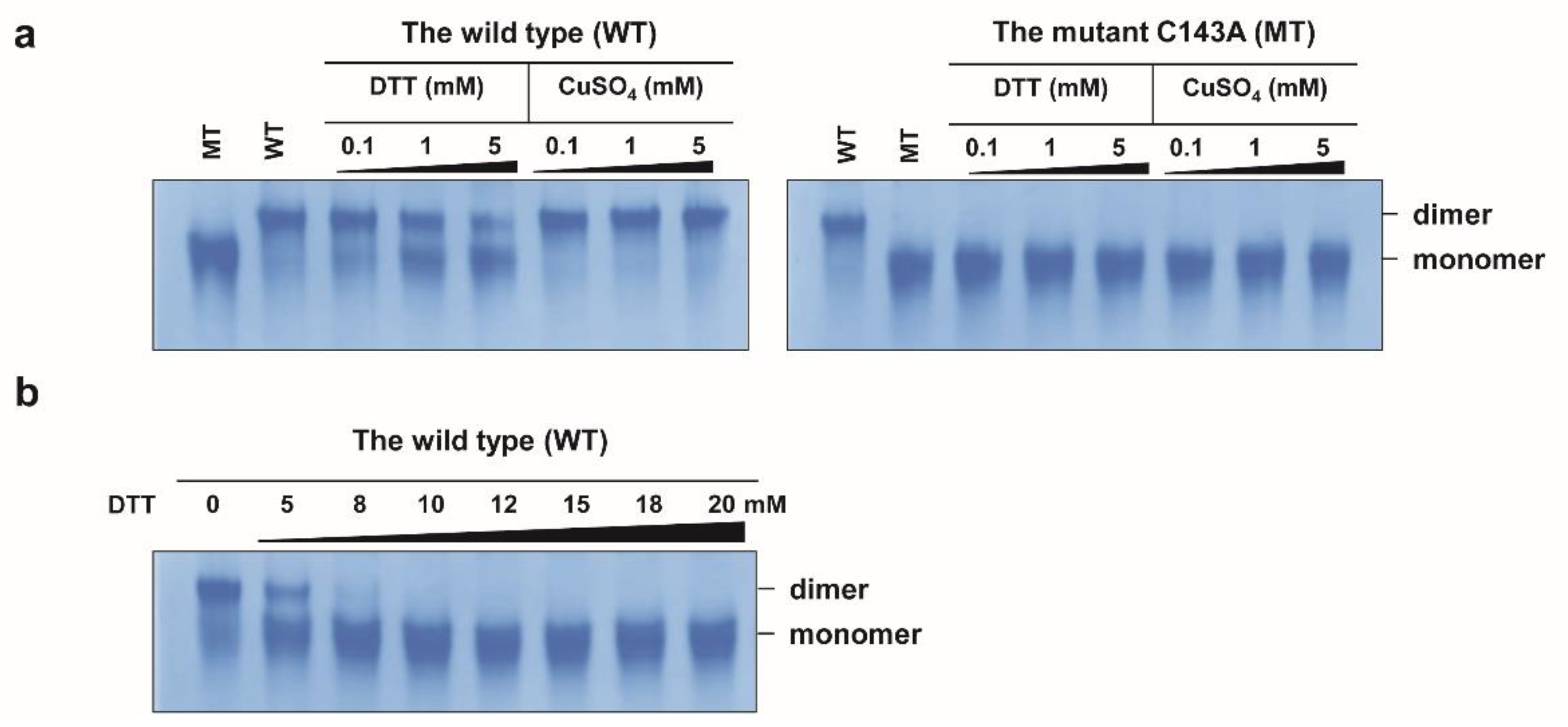
2.1. Disulfide Bond-Mediated Dimerization

CaDHN has five K-segment-like regions and one S-segment-like region [20]. Among the five K-segment-like regions, three were highly conserved relative to those in other DHNs. On the phylogenetic tree, CaDHN belonged to the DHN group close to LEA 2 (Figure S1). Interestingly, CaDHN has an unusual single cysteine residue (Cys143), which is very rare in other reported DHNs [21]. The cysteine residue is positioned at the φ-segment between the second and the third K-segments (Figure 1a). After the sodium dodecyl-sulfate polyacrylamide gel electrophoresis (SDS-PAGE) of the purified wild type (WT) protein, we observed two bands for the protein. The upper band was at approximately 80 kDa and disappeared after treatment with the reducing agent β-mercaptoethanol (β-ME). The lower band, at 40 kDa, did not migrate on the gel upon β-ME treatment (Figure 1b). To check whether the CaDHN formed an intermolecular disulfide bond, we constructed the mutant C143A, wherein an alanine residue was introduced instead of the cysteine. The mutant C143A protein exhibited only a single band in the non-reduced SDS-PAGE gel (Figure 1b) and not the upper band with high molecular weight as shown by the WT protein. On the native gel, both the WT and C143A proteins appeared as molecules of different sizes (Figure 2a). To confirm the existence of the intermolecular disulfide bond, we added different concentrations of dithiothreitol (DTT) and copper ions (as CuSO4) to both proteins and analyzed them on the native gel. DTT is a reducing agent that prevents disulfide bond formation between cysteine residues, and the oxidant copper ion is recognized as a transition metal ion that forces the formation of a disulfide bond between two cysteine residues by oxidation of their thiol groups [22,23]. As the DTT concentration increased, the upper (WT) band decreased in a dose-dependent manner and most of the protein migrated to the lower band at DTT concentrations above 8 mM; this position agrees with that of the C143A protein band on the SDS-PAGE gel. In contrast, increasing the copper concentration in the WT solution resulted in the migration of the small amount of the lower band migrate to the higher molecular weight upper band. This suggested that copper ions could trigger the formation of the disulfide bond in the WT protein, thereby moving to the upper band position due to the intermolecular dimerization of the protein. The C143A protein remained at the position of the lower band even in the presence of copper ions. These results suggested that CaDHN undergoes a molecular conformational change by disulfide bond-mediated dimerization, but not by metal-mediated dimerization between the protein molecules via amino acid residues, such as histidine [24,25,26]. The binding interaction between histidine residues and metal ions has been reported in citrus (Citrus unshiu) DHN [27]. The third non-conserved φ-segment motif containing the cysteine residue contains four histidine residues out of a total of 22 in the whole molecule. As shown in Figure 2a, in the C143A, there is no band shift after the addition of DTT and CuSO4. The upper band of the WT protein shifted completely to the lower band in the presence of 10 mM DTT. Thus, our results confirm that the WT protein could exist in the dimer form resulting from an intermolecular disulfide bond. Dimerization of DHN from plants has also been reported in the TsDHN-2 (Y2SK2 type) protein from Thellungiella salsuginea O.E. Schulz and the OpsDHN1 (SK3 type) from Opuntia streptacantha Lem. Interestingly, both DHNs dimerize differently in the structural interaction between their monomers. The TsDHN-2 protein has a potential dimerization state associated with a hydrophobic surface. However, in the OpsDHN1 protein, the region responsible for the dimer interaction is the histidine-rich motif that is located in the non-conserved segment between the first and the second K-segment [24,28].

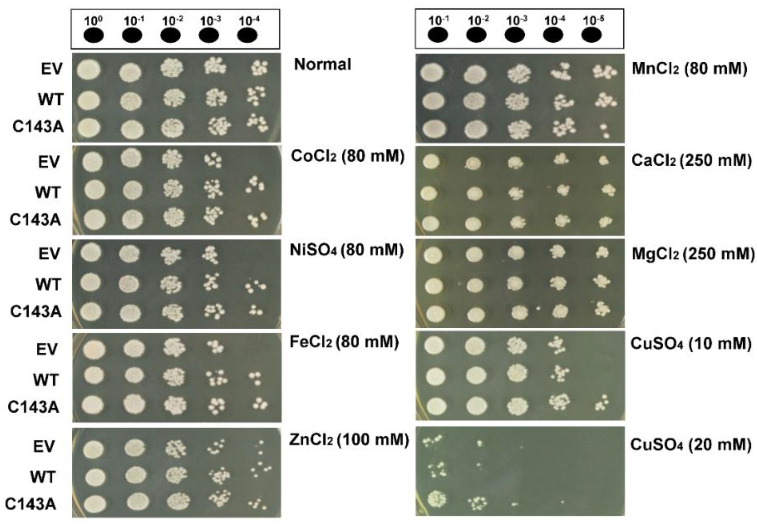
2.2. Metal Binding Activity (MBA)

Some DHNs exhibit metal ion-binding properties, which have radical scavenging activity against cytotoxic hydroxyl radicals caused by metal-catalyzed reactions [27,29,30,31]. To investigate the metal-binding ability of the CaDHN, we tested their semi-quantitative MBA. Both the monomer and the dimer forms of CaDHN exhibited similar features regarding their MBA. As shown in Figure S2a, both proteins were fully bound to the metal ions, Fe3+, Co2+, Ni2+, Cu2+, and Zn2+, but not to Mg2+, Ca2+, and Mn2+. These results were consistent with those obtained previously for citrus (Citrus unshiu) DHN [27]. The histidine residue in DHNs has been known to contribute to their MBA. CaDHN also has more abundant histidine residues than those of other proteins. To investigate DHN dimerization via other metal ions, we also performed native gel electrophoresis of both proteins in the presence of the same kind of metal ions as were used for testing the MBA. As a result, no size shift was observed with all seven tested metals, except for Cu2+ ions (Figure S2b). Therefore, the in vitro assay showed that the CaDHN protein had MBA as mentioned above. Next, we tested in vivo CaDHN-mediated metal ion homeostasis using transgenic yeast. The metal-ion tolerance of CaDHN-expressing cells was improved compared with that of empty vector (EV) cells when yeast cells were challenged by stress induced by various metals including Co2+, Ni2+, Fe2+, and Zn2+ (Figure S2). On the other hand, the survival of CaDHN-expressing cells was similar to that of EV cells when treated with Mg2+, Ca2+, and Mn2+. These results for metal tolerance were almost consistent with the in vitro MBA of the protein (Figure 3). Interestingly, under copper stress, C143A CaDHN cells recovered more rapidly than WT CaDHN cells (Figure 3). According to a previous report, Ni2+, Cu2+, and Zn2+ bind to CuCOR15 protein, but Mg2+, Ca2+, and Mn2+ do not [27]. As seen in KnS-type DHNs, the SK5-type CaDHN binds to various metal ions [25]. Expression of DHN2 and DHN3 from Brassica juncea (L.) Czern. in transgenic tobacco enhanced its tolerance for heavy metals, such as Cd2+ and Zn2+ by attenuating lipid peroxidation and protecting cellular membranes after lower electrolyte leakage [32].

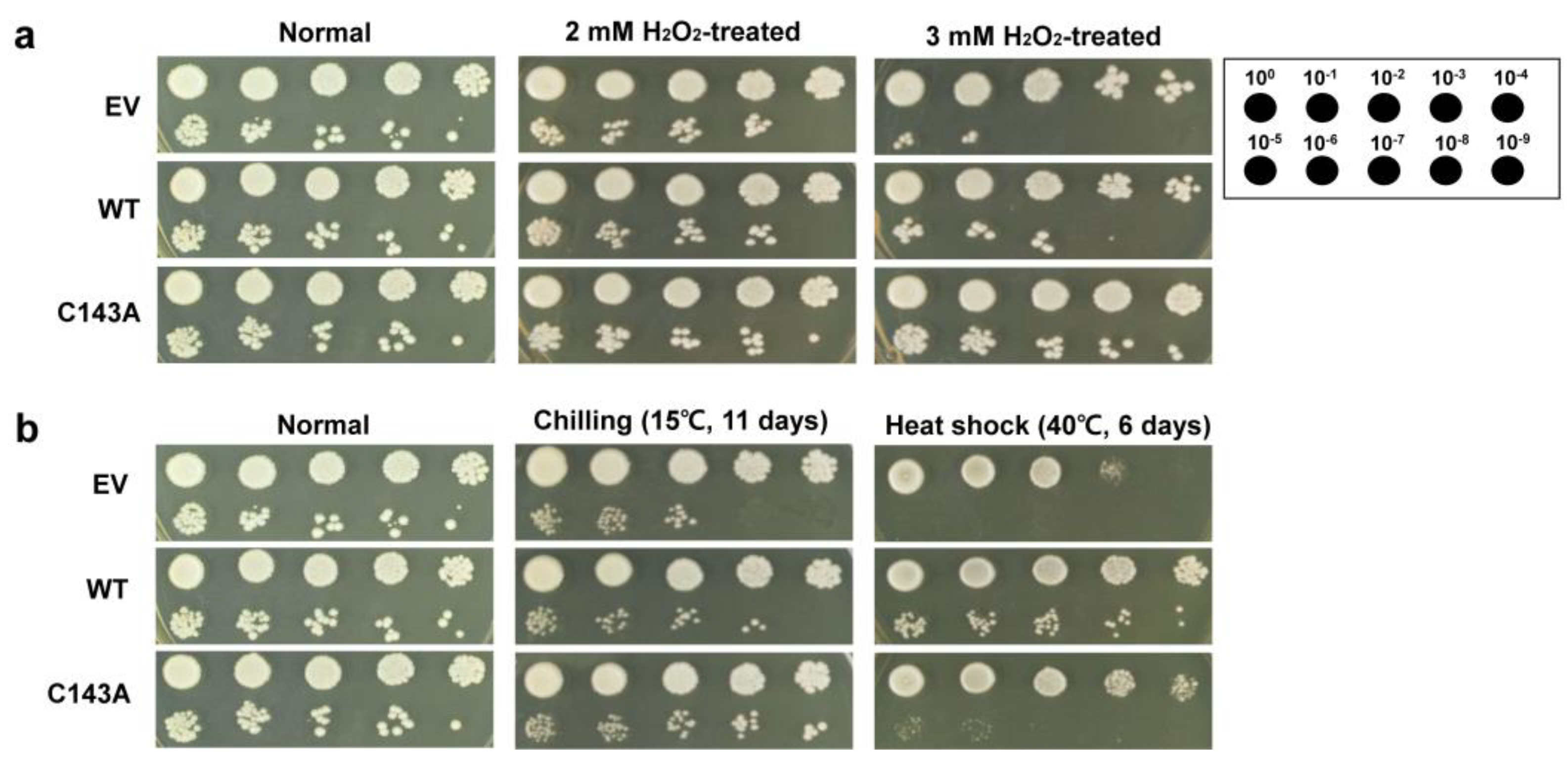
2.3. Yeast Stress Tolerance Assay

To investigate whether the dimerization of CaDHN protein affects its in vivo functional role, CaDHN (WT or C143A)-expressing transgenic yeast cells were developed. In transgenic yeast cells, the expression levels of both CaDHNs (WT and C143A) were identified by immunoblotting analysis, and the results were similar for both cells (Figure S3). Using the same transgenic yeasts, we examined whether CaDHN accumulation affects the acquired tolerance to various kinds of abiotic stress. Both transgenic cells were more tolerant than cells with EV alone when exposed to hydrogen peroxide (H2O2). Cells with C143A CaDHN were more tolerant to oxidative stress than those with WT CaDHN (Figure 4a). In general, although H2O2 induces the formation of disulfide bonds, which can directly oxidize cysteine residue, such oxidative cysteine modification triggers structural alterations in the target protein [33]. An irreversible change in DHN conformation resulted in changes in protein function [34,35]. In the presence of H2O2 during protein production, the CaDHN protein expressed in E. coli was found to be modified by hypo-oxidation [dioxidation (O2) and trioxidation (O3)] and carbamidomethylation (cam) (Figures S4–S8). The modification of cysteine in the CaDHN monomeric state is irreversible. Our results imply that the increase in the amount of CaDHN monomers is not only induced by the modification of its cysteine residue through environmental influence or by exogenous stimuli, but also by a cellular redox protein. At high temperatures, the thermal tolerance of WT CaDHN cells was higher than that of C143A CaDHN cells, while the tolerance of WT CaDHN cells against cold stress was lower than that of C143A CaDHN cells (Figure 4b). The ability of C143A CaDHN cells to tolerate cold was better than that of WT CaDHN cells under various chilling conditions (Figure S9). A positive correlation has been found between the accumulation of DHN and the response of plants to abiotic stresses. For example, overexpression of the Arabidopsis thaliana DHN genes (LTI29 and LTI30) was associated with increased tolerance to stress from freezing, dehydration, and chilling [36]. In addition, overexpression of the C. unshiu COR19 gene in transgenic tobacco reduces ion leakage and lipid oxidation under cold and freezing conditions, thereby increasing cold stress tolerance [37,38]. Therefore, these results indicate that the monomer form of the CaDHN protein leads to enhanced physiological tolerance of the cell for low temperatures and H2O2, but not for high temperatures.
In summary, CaDHN has a unique single cysteine residue, which plays a role in its dimerization via an intermolecular disulfide bond. CaDHN exhibits biological functions including redox and metal ion homeostasis. Our results suggest that regulation of the cysteine-mediated disulfide bond of CaDHN following the cellular redox state could improve the tolerance of polar plants to abiotic stress through conformational changes.

3. Materials and Methods

3.1. Cloning, Expression and Purification

Construction of recombinant CaDHN in E. coli was performed as reported previously [20]. In brief, total RNA from C. arcticum L. (Caryophyllaceae) leaves collected from Brogger-Halvoya, Svalbard was isolated using the RNeasy Plant Mini kit (Qiagen, North Rhine-Westphalia, Germany) according to the manufacturer’s instructions and cDNA was synthesized using reverse transcriptase. The CaDHN gene was amplified by polymerase chain reaction with a specific primer set harboring restriction enzyme sites represented in Table S1. CaDHN gene was introduced between the EcoRI and XhoI sites within the pET30a vector (Novagen, Reno, NV, USA) (Table S1) using the sequence- and ligation-independent cloning method. A tobacco etch virus (TEV) protease recognition sequence was inserted in front of the CaDHN gene. During the construction of recombinant CaDHN protein, we recognized that the N-terminal long tag contained within the pET30a vector was able to enhance the CaDHN overexpression. The N-terminal tag is composed of 52 amino acids corresponding to about 6 kDa. In detail, the sequence of the N-terminal long tag is MHHHHHHSSGLVPRGSGMKETAAAKFERQHMDSPDLGTDDDDKAMADIGSEF. The recombinant plasmid was transformed into E. coli BL21 (DE3) strain. For purification, the pelleted cells were suspended in the lysis buffer containing 20 mM Tris-HCl at pH 8.0, 200 mM NaCl and 10 mM imidazole, and lysed by sonication. After centrifugation, the supernatant was loaded onto nickel-charged affinity resin and the target protein was washed and eluted with the wash buffer containing 20 mM Tris-HCl at pH 8.0, 200 mM NaCl, 30 mM imidazole and the elution buffer containing 20 mM Tris-HCl at pH 8.0, 200 mM NaCl, and 300 mM imidazole, respectively. Next, TEV protease (New England Biolabs, Ipswich, MA, USA) was added to the solution and dialyzed in dialysis buffer (20 mM Tris-HCl at pH 8.0, 200 mM NaCl and 10 mM imidazole) at 4 °C overnight. A second nickel-charged affinity chromatography step was performed to remove uncleaved CaDHN and the N-terminal tag peptide. The mutant gene of C143A was prepared by Site-Directed Mutagenesis using PCR (Table S1). The predicted molecular weight of the CaDHN protein was calculated to be 29,509 Da.

3.2. Native Gel Electrophoresis

Native gel electrophoresis was run on 10% polyacrylamide gels. The electrode buffer contained 25 mM Tris base and 19 mM glycine. All native gels were run in an ice bath to dissipate heat generated during the runs. The protein bands were visualized using Coomassie Brilliant Blue R-250.

3.3. Development of Yeast Expression Vector and Transgenic Yeast

For the development of the yeast expression vector bearing the CaDHN gene, the target gene was subcloned into the digested p426GPD vector using the glyceraldehyde-3-phosphate dehydrogenase (GPD1) promoter (Euroscarf, Oberursel, Germany) (Table S1). The resulting plasmid was named p426GPD:CaDHN_WT. The mutant gene of CaDHN_C143A was prepared by the PCR-dependent Site-Directed Mutagenesis kit (New England Biolabs) using the p426GPD::CaDHN_WT backbone, according to the manufacturer’s protocols (Table S1). The resulting plasmids (p426GPD::CaDHN_WT and p426GPD::CaDHN_C143A) were used to transform a conventional Saccharomyces cerevisiae. Haploid S. cerevisiae BY4741 (Euroscarf) cells grown at 28 °C overnight in a nutrient-rich Yeast Extract-Peptone-Dextrose (YPD) broth medium containing 1% yeast extract, 2% peptone, and 2% dextrose were reinoculated into a fresh YPD broth medium, and then incubated for 4 h at 28 °C with shaking (180 rpm) until the optical density of the culture solution at 600 nm (A600) reached approximately 1.0 (1 × 10⁷ cells/mL) to produce early exponential cells. The resulting yeast cells were then transformed with the two target plasmids (p426GPD::CaDHN_WT, p426GPD::CaDHN_C143A) by using the PEG/LiCl method reported previously [39]. The transformed cells were plated on a minimal nutrient agar medium (0.67% yeast nitrogen base without amino acids and with ammonium sulfate, 0.192 % yeast synthetic drop-out medium supplement without uracil (Sigma-Aldrich, Burlington, MA, USA), 2% glucose, and 1.5% agar). The plates were incubated at 28 °C for 3–4 days. Positive colonies grown in a medium lacking uracil were selected and used for subsequent experiments.

3.4. Stress Response Assay in Yeast Cells

Yeast cells grown in the YPD broth medium until the mid-log phase (A600 = 2.5; 1 × 10⁹ cells/mL) were exposed to different concentrations of H2O2 (2 mM and 3 mM), zinc chloride (100 mM ZnCl2), copper sulfate (10 mM and 20 mM CuSO4), cobalt chloride (80 mM CoCl2), nickel sulfate (80 mM NiSO4), ferric chloride (80 mM FeCl2), manganese chloride (80 mM MnCl2), calcium chloride (250 mM CaCl2), and magnesium chloride (250 mM MgCl2) for 1 h with shaking (180 rpm). They were then serially diluted to 10−4 with fresh YPD broth medium. Five microliters of this diluted solution were then loaded onto YPD agar (YPD plus 1.5% agar) plates, which were incubated for 3 days at 28 °C and then photographed. On the other hand, for extreme temperatures, yeast cells of the same phase were serially diluted to 10−9 with fresh YPD broth, loaded onto YPD agar plates and incubated for 6 days at 40 °C, and for 11 days at 15 °C, respectively.

Supplementary Materials

The following supporting information can be downloaded at: https://www.mdpi.com/article/10.3390/molecules27092934/s1, Table S1. Polymerase chain reaction (PCR) primers used in this study. Figure S1: Phylogenetic tree of dehydrins (DHNs) and late embryogenesis-abundant (LEA) proteins. Figure S2. Investigation of metal binding activity (MBA) of Cerastium arcticum Lange DHN (CaDHN) using metal ion affinity chromatography and native gel analysis; Figure S3. Construction and expression of the Cerastium arcticum Lange DHN (CaDHN). Figure S4. Identification of the cysteine modification using a liquid chromatography tandem mass spectrometer (LC-MS/MS) with a Surveyor nano-flow system, connected to a 7-Tesla Finnigan LTQ-FT mass spectrometer. Figure S5. Liquid chromatography tandem mass spectrometer (LC-MS/MS) spectra of the Cerastium arcticum Lange dehydrin (CaDHN) peptide containing the cysteine sulfenic acid (-SOH). Figure S6. Liquid chromatography tandem mass spectrometer (LC-MS/MS) spectra of the Cerastium arcticum Lange dehydrin (CaDHN) peptide containing the cysteine sulfinic acid (-SO2H). Figure S7. Liquid chromatography tandem mass spectrometer (LC-MS/MS) spectra of the Cerastium arcticum Lange dehydrin (CaDHN) peptide containing the cysteine sulfonic acid (-SO3H). Figure S8. Liquid chromatography tandem mass spectrometer (LC-MS/MS) spectra of the Cerastium arcticum Lange dehydrin (CaDHN) peptide containing the carbamidomethyl cysteine (cam). Figure S9. Cell viability of transgenic yeast against thermal stress [40].

Author Contributions

Conceptualization, I.-S.K., W.C. and H.-W.K.; investigation, I.-S.K., W.C. and A.K.P.; data curation, H.K. and J.S.; writing—original draft preparation, I.-S.K., W.C. and H.-W.K.; visualization, A.K.P.; writing—review and editing, I.-S.K., W.C., J.H.L., S.C.S., T.D.K. and H.-W.K.; funding acquisition, J.H.L. and H.-W.K.; supervision, H.-W.K.; project administration, H.-W.K. All authors have read and agreed to the published version of the manuscript.

Funding

This research was supported by a National Research Foundation of Korea Grant from the Korean Government (MSIT; the Ministry of Science and ICT) NRF-2021M1A5A1075524) (KOPRI-PN22014). This research was also a part of the project titled “Development of potential antibiotic compounds using polar organism resources (15250103, KOPRI Grant PM22030)”, funded by the Ministry of Oceans and Fisheries, Korea.

Data Availability Statement

Data is contained within the article and Supplementary Material.

Conflicts of Interest

The authors declare no potential conflict of interest.

References

  1. Hagen, A.R.; Giese, H.; Brochmann, C. Trans-Atlantic dispersal and phylogeography of Cerastium arcticum (Caryophyllaceae) inferred from RAPD and SCAR markers. Am. J. Bot. 2001, 88, 103–112. [Google Scholar] [CrossRef] [PubMed]
  2. Bokhorst, S.; Bjerke, J.W.; Davey, M.P.; Taulavuori, K.; Taulavuori, E.; Laine, K.; Callaghan, T.V.; Phoenix, G.K. Impacts of extreme winter warming events on plant physiology in a sub-Arctic heath community. Physiol. Plant. 2010, 140, 128–140. [Google Scholar] [CrossRef] [PubMed]
  3. Chew, O.; Lelean, S.; John, U.P.; Spangenberg, G.C. Cold acclimation induces rapid and dynamic changes in freeze tolerance mechanisms in the cryophile Deschampsia antarctica E. Desv. Plant Cell Environ. 2012, 35, 829–837. [Google Scholar] [CrossRef]
  4. Archambault, A.; Stromvik, M.V. PR-10, defensin and cold dehydrin genes are among those over expressed in Oxytropis (Fabaceae) species adapted to the arctic. Funct. Integr. Genomics 2011, 11, 497–505. [Google Scholar] [CrossRef] [PubMed] [Green Version]
  5. Allagulova, C.R.; Gimalov, F.R.; Shakirova, F.M.; Vakhitov, V.A. The plant dehydrins: Structure and putative functions. Biochemistry 2003, 68, 945–951. [Google Scholar] [PubMed]
  6. Garay-Arroyo, A.; Colmenero-Flores, J.M.; Garciarrubio, A.; Covarrubias, A.A. Highly hydrophilic proteins in prokaryotes and eukaryotes are common during conditions of water deficit. J. Biol. Chem. 2000, 275, 5668–5674. [Google Scholar] [CrossRef] [PubMed] [Green Version]
  7. Singh, S.; Cornilescu, C.C.; Tyler, R.C.; Cornilescu, G.; Tonelli, M.; Lee, M.S.; Markley, J.L. Solution structure of a late embryogenesis abundant protein (LEA14) from Arabidopsis thaliana, a cellular stress-related protein. Protein Sci. 2005, 14, 2601–2609. [Google Scholar] [CrossRef] [PubMed] [Green Version]
  8. Babu, R.C.; Zhang, J.X.; Blum, A.; Ho, T.H.D.; Wu, R.; Nguyen, H.T. HVA1, a LEA gene from barley confers dehydration tolerance in transgenic rice (Oryza sativa L.) via cell membrane protection. Plant Sci. 2004, 166, 855–862. [Google Scholar] [CrossRef]
  9. Kim, H.S.; Lee, J.H.; Kim, J.J.; Kim, C.H.; Jun, S.S.; Hong, Y.N. Molecular and functional characterization of CaLEA6, the gene for a hydrophobic LEA protein from Capsicum annuum. Gene 2005, 344, 115–123. [Google Scholar] [CrossRef]
  10. Sasaki, K.; Christov, N.K.; Tsuda, S.; Imai, R. Identification of a novel LEA protein involved in freezing tolerance in wheat. Plant Cell Physiol. 2014, 55, 136–147. [Google Scholar] [CrossRef] [PubMed] [Green Version]
  11. Dure, L.; Crouch, M.; Harada, J.; Ho, T.H.D.; Mundy, J.; Quatrano, R.; Thomas, T.; Sung, Z.R. Common amino-acid sequence domains among the LEA proteins of higher plants. Plant Mol. Biol. 1989, 12, 475–486. [Google Scholar] [CrossRef] [PubMed]
  12. Close, T.J. Dehydrins: A commonality in the response of plants to dehydration and low temperature. Physiol. Plant. 1997, 100, 291–296. [Google Scholar] [CrossRef]
  13. Yang, Y.; He, M.; Zhu, Z.; Li, S.; Xu, Y.; Zhang, C.; Singer, S.D.; Wang, Y. Identification of the dehydrin gene family from grapevine species and analysis of their responsiveness to various forms of abiotic and biotic stress. BMC Plant Biol. 2012, 12, 140. [Google Scholar] [CrossRef] [PubMed] [Green Version]
  14. Takahashi, R.; Joshee, N.; Kitagawa, Y. Induction of chilling resistance by water-stress, and cDNA sequence-analysis and expression of water stress-regulated genes in rice. Plant Mol. Biol. 1994, 26, 339–352. [Google Scholar] [CrossRef] [PubMed]
  15. Campbell, S.A.; Close, T.J. Dehydrins: Genes, proteins, and associations with phenotypic traits. New Phytol. 1997, 137, 61–74. [Google Scholar] [CrossRef]
  16. Koag, M.C.; Wilkens, S.; Fenton, R.D.; Resnik, J.; Vo, E.; Close, T.J. The K-segment of maize DHN1 mediates binding to anionic phospholipid vesicles and concomitant structural changes. Plant Physiol. 2009, 150, 1503–1514. [Google Scholar] [CrossRef] [PubMed] [Green Version]
  17. Hughes, S.; Graether, S.P. Cryoprotective mechanism of a small intrinsically disordered dehydrin protein. Protein Sci. 2011, 20, 42–50. [Google Scholar] [CrossRef] [Green Version]
  18. Alsheikh, M.K.; Svensson, J.T.; Randall, S.K. Phosphorylation regulated ion-binding is a property shared by the acidic subclass dehydrins. Plant Cell Environ. 2005, 28, 1114–1122. [Google Scholar] [CrossRef]
  19. Close, T.J. Dehydrins: Emergence of a biochemical role of a family of plant dehydration proteins. Physiol. Plant. 1996, 97, 795–803. [Google Scholar] [CrossRef]
  20. Kim, I.S.; Kim, H.Y.; Kim, Y.S.; Choi, H.G.; Kang, S.H.; Yoon, H.S. Expression of dehydrin gene from Arctic Cerastium arcticum increases abiotic stress tolerance and enhances the fermentation capacity of a genetically engineered Saccharomyces cerevisiae laboratory strain. Appl. Microbiol. Biotechnol. 2013, 97, 8997–9009. [Google Scholar] [CrossRef]
  21. Graether, S.P.; Boddington, K.F. Disorder and function: A review of the dehydrin protein family. Front. Plant Sci. 2014, 5, 576. [Google Scholar] [CrossRef] [Green Version]
  22. Lumb, I.; Hundal, M.S.; Hundal, G. Copper(II)-catalyzed disulfide scission--stepwise aerobic oxidative cleavage to sulfinate and sulfonate and reductive anaerobic cleavage to thiols. Inorg. Chem. 2014, 53, 7770–7779. [Google Scholar] [CrossRef]
  23. Hiniker, A.; Collet, J.F.; Bardwell, J.C. Copper stress causes an in vivo requirement for the Escherichia coli disulfide isomerase DsbC. J. Biol. Chem. 2005, 280, 33785–33791. [Google Scholar] [CrossRef] [Green Version]
  24. Hernandez-Sanchez, I.E.; Martynowicz, D.M.; Rodriguez-Hernandez, A.A.; Perez-Morales, M.B.; Graether, S.P.; Jimenez-Bremont, J.F. A dehydrin-dehydrin interaction: The case of SK3 from Opuntia streptacantha. Front. Plant Sci. 2014, 5, 520. [Google Scholar] [CrossRef]
  25. Hara, M.; Kondo, M.; Kato, T. A KS-type dehydrin and its related domains reduce Cu-promoted radical generation and the histidine residues contribute to the radical-reducing activities. J. Exp. Bot. 2013, 64, 1615–1624. [Google Scholar] [CrossRef] [Green Version]
  26. Mu, P.Q.; Feng, D.R.; Su, J.B.; Zhang, Y.; Dai, J.R.; Jin, H.L.; Liu, B.; He, Y.M.; Qi, K.B.; Wang, H.B.; et al. Cu2+ triggers reversible aggregation of a disordered His-rich dehydrin MpDhn12 from Musa paradisiaca. J. Biochem. 2011, 150, 491–499. [Google Scholar] [CrossRef]
  27. Hara, M.; Fujinaga, M.; Kuboi, T. Metal binding by citrus dehydrin with histidine-rich domains. J. Exp. Bot. 2005, 56, 2695–2703. [Google Scholar] [CrossRef] [Green Version]
  28. Rahman, L.N.; McKay, F.; Giuliani, M.; Quirk, A.; Moffatt, B.A.; Harauz, G.; Dutcher, J.R. Interactions of Thellungiella salsuginea dehydrins TsDHN-1 and TsDHN-2 with membranes at cold and ambient temperatures-surface morphology and single-molecule force measurements show phase separation, and reveal tertiary and quaternary associations. Biochim. Biophys. Acta 2013, 1828, 967–980. [Google Scholar] [CrossRef] [Green Version]
  29. Alsheikh, M.K.; Heyen, B.J.; Randall, S.K. Ion binding properties of the dehydrin ERD14 are dependent upon phosphorylation. J. Biol. Chem. 2003, 278, 40882–40889. [Google Scholar] [CrossRef] [Green Version]
  30. Kruger, C.; Berkowitz, O.; Stephan, U.W.; Hell, R. A metal-binding member of the late embryogenesis abundant protein family transports iron in the phloem of Ricinus communis L. J. Biol. Chem. 2002, 277, 25062–25069. [Google Scholar] [CrossRef] [Green Version]
  31. Svensson, J.; Palva, E.T.; Welin, B. Purification of recombinant Arabidopsis thaliana dehydrins by metal ion affinity chromatography. Protein Expr. Purif. 2000, 20, 169–178. [Google Scholar] [CrossRef] [PubMed]
  32. Xu, J.; Zhang, Y.X.; Wei, W.; Han, L.; Guan, Z.Q.; Wang, Z.; Chai, T.Y. BjDHNs confer heavy-metal tolerance in plants. Mol. Biotechnol. 2008, 38, 91–98. [Google Scholar] [CrossRef] [PubMed]
  33. Waszczak, C.; Akter, S.; Jacques, S.; Huang, J.J.; Messens, J.; Van Breusegem, F. Oxidative post-translational modifications of cysteine residues in plant signal transduction. J. Exp. Bot. 2015, 66, 2923–2934. [Google Scholar] [CrossRef] [PubMed] [Green Version]
  34. Hanin, M.; Brini, F.; Ebel, C.; Toda, Y.; Takeda, S.; Masmoudi, K. Plant dehydrins and stress tolerance: Versatile proteins for complex mechanisms. Plant Signal. Behav. 2011, 6, 1503–1509. [Google Scholar] [CrossRef]
  35. Mouillon, J.M.; Eriksson, S.K.; Harryson, P. Mimicking the plant cell interior under water stress by macromolecular crowding: Disordered dehydrin proteins are highly resistant to structural collapse. Plant Physiol. 2008, 148, 1925–1937. [Google Scholar] [CrossRef] [Green Version]
  36. Puhakainen, T.; Hess, M.W.; Makela, P.; Svensson, J.; Heino, P.; Palva, E.T. Overexpression of multiple dehydrin genes enhances tolerance to freezing stress in Arabidopsis. Plant Mol. Biol. 2004, 54, 743–753. [Google Scholar] [CrossRef]
  37. Rorat, T. Plant dehydrins--tissue location, structure and function. Cell. Mol. Biol. Lett. 2006, 11, 536–556. [Google Scholar] [CrossRef]
  38. Hara, M.; Terashima, S.; Fukaya, T.; Kuboi, T. Enhancement of cold tolerance and inhibition of lipid peroxidation by citrus dehydrin in transgenic tobacco. Planta 2003, 217, 290–298. [Google Scholar] [CrossRef]
  39. Gietz, R.D.; Woods, R.A. Genetic transformation of yeast. Biotechniques 2001, 30, 816–820, 822–826, 828 passim. [Google Scholar] [CrossRef] [Green Version]
  40. Hara, M.; Shinoda, Y.; Tanaka, Y.; Kuboi, T. DNA binding of citrus dehydrin promoted by zinc ion. Plant Cell Environ. 2009, 32, 532–541. [Google Scholar] [CrossRef]
Figure 1. Dimerization of Cerastium arcticum Lange dehydrin (CaDHN) by the unique cysteine. (a). Scheme of the cysteine position in the CaDHN. The CaDHN protein containing 270 amino acids was composed of five K segments, a conserved S segment and five non-conserved φ-segments. The unique cysteine within the φ-segment is highlighted in red. (b). Purified CaDHNs analyzed on Coomassie blue were stained on an SDS-PAGE gel. WT, the wild type protein; MT, the mutant C143A protein; β-ME(+), protein treated with 10 mM of β-mercaptoethanol. Each gel lane was loaded with 1 μg of the protein.
Molecules 27 02934 g001
Figure 2. Native gel analysis for dimerization via disulfide bond of the purified Cerastium arcticum Lange dehydrins (CaDHNs). (a) Both wild type (WT) and mutant C143A (MT) proteins treated with different concentrations of dithiothreitol (DTT) and copper ions (CuSO4). (b) DTT-treated WT protein (0–20 mM). Each gel lane was loaded with 1.5 μg of the protein.
Molecules 27 02934 g002
Figure 3. A metal-mediated stress response assay of CaDHN (WT and C143A)-expressing transgenic yeast on plates. Cells transformed with an empty vector (EV; p426GPD)) were used as control. Mid-log yeast cells were exposed to stressors for 1 h with shaking (180 rpm), serially diluted with a fresh YPD broth medium and spotted onto YPD agar plates. Five microliters were used for a spotting assay. The plates were incubated for 3–4 days at 28 °C and photographed. The results were representative of at least three independent experiments conducted under identical conditions.
Molecules 27 02934 g003
Figure 4. Cell viability of transgenic yeast against oxidative and thermal stress. An oxidative stress tolerance assay (a); thermal stress tolerance assay (b). Stress responses of Saccharomyces cerevisiae BY4741 cells expressing the CaDHN gene were determined by the spot assay. The cells transformed with an empty vector (EV) were used as a control. Mid-log yeast cells were serially diluted to 10–9 with YPD broth medium and 5 μL of the diluted solution were spotted onto YPD agar medium in the absence and presence of hydrogen peroxide (H2O2). The plates were incubated at 28 °C during the time indicated and then photographed. For H2O2-mediated spotting assay, the plates were incubated for 3–4 days at 28 °C. The results were representative of at least three independent experiments conducted under identical conditions.
Molecules 27 02934 g004
Publisher’s Note: MDPI stays neutral with regard to jurisdictional claims in published maps and institutional affiliations.

