
Phytochemical Profile of Antibacterial Agents from Red Betel Leaf (Piper crocatum Ruiz and Pav) against Bacteria in Dental Caries


Abstract
:1. Introduction
2. Gram-Positive and Negative Bacteria Cause Dental Caries
2.1. Gram-Positive Bacteria
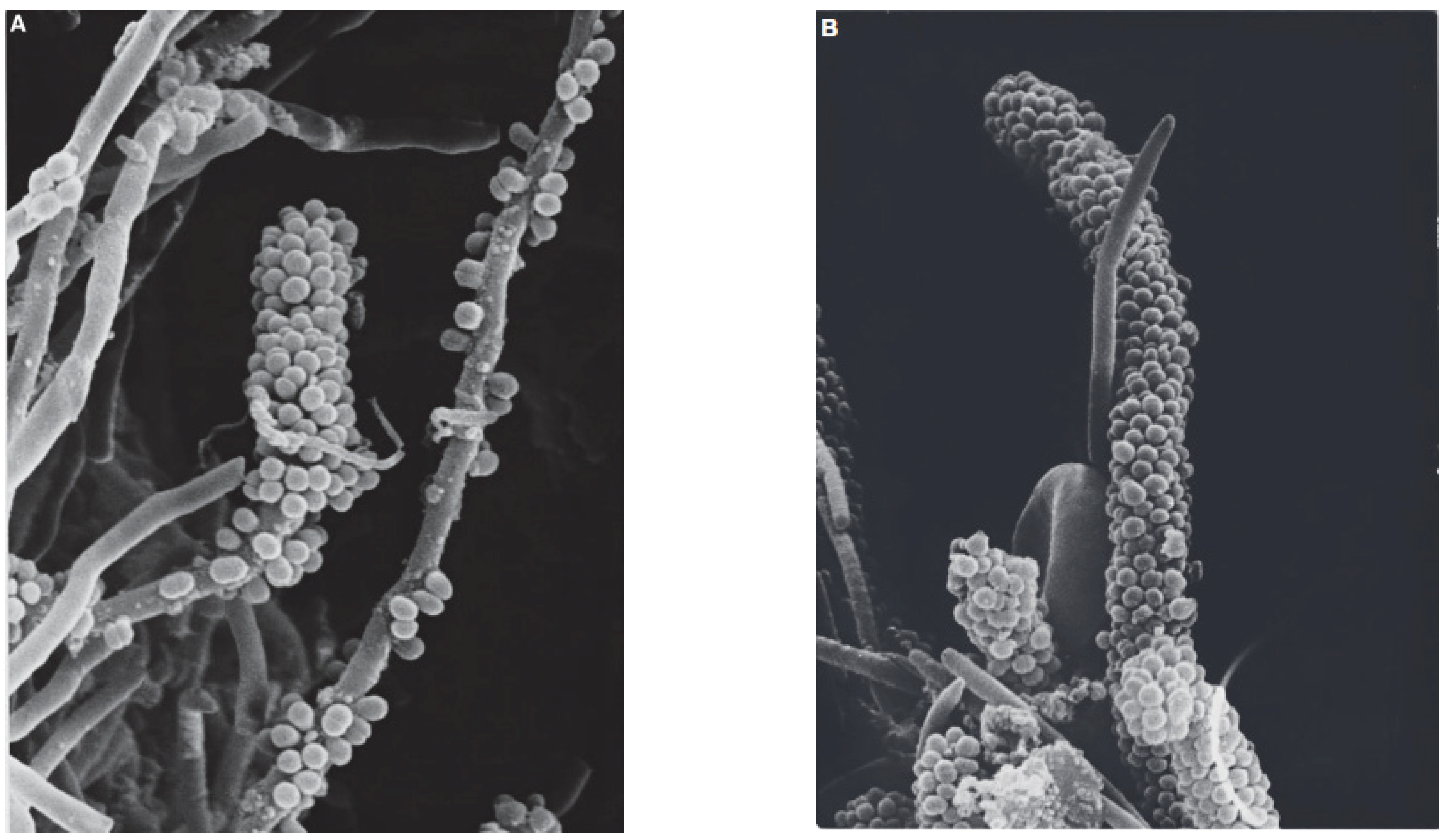
2.1.1. Streptococcus mutans
2.1.2. Streptococcus sanguinis
2.2. Gram-Negative Bacteria
Veillonella parvula
3. Antibacterial
3.1. Definition
3.2. Antibacterial Mechanism of Secondary Metabolic Compounds
3.2.1. Phenol
3.2.2. Flavonoids
3.2.3. Saponins
3.2.4. Terpenoids
3.2.5. Alkaloids
3.2.6. Tannins
3.3. Antibacterial Mechanism with MurA Enzyme
3.4. Commonly Used Dental Caries Antibiotics
3.4.1. Fluoride
3.4.2. AIK(SO4)2
3.4.3. Chlorhexidine (CHX)
4. Piper crocatum Ruiz and Pav
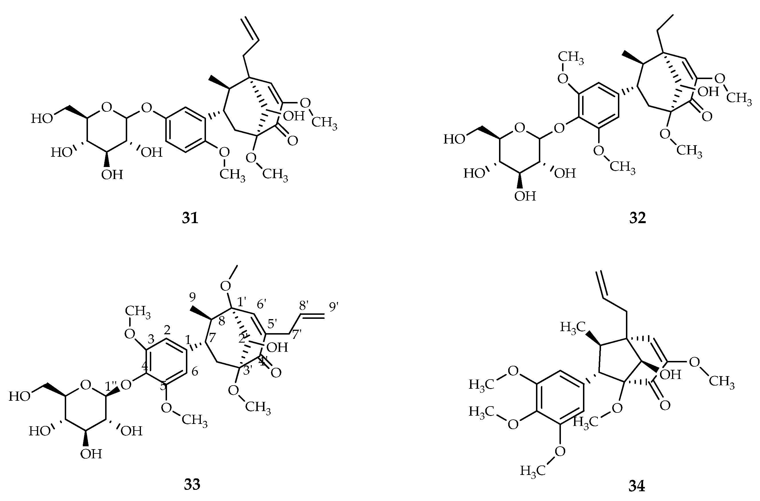
4.1. Isolation of Secondary Metabolites of Piper crocatum Ruiz and Pav
4.2. Bioactivity of Piper crocatum Ruiz and Pav
4.3. Antibacterial Activity of Red Betel Extract
5. Conclusions
Author Contributions
Funding
Institutional Review Board Statement
Informed Consent Statement
Data Availability Statement
Acknowledgments
Conflicts of Interest
References
- Kuang, X.; Chen, V.; Xu, X. Novel Approaches to the Control of Oral Microbial Biofilms. Biomed. Res. Int. 2018, 2018, 6498932. [Google Scholar] [CrossRef] [PubMed] [Green Version]
- Jiao, Y.; Tay, F.R.; Niu, L.-N.; Chen, J. Hua Advancing Antimicrobial Strategies for Managing Oral Biofilm Infections. Int. J. Oral Sci. 2019, 11, 28. [Google Scholar] [CrossRef] [PubMed] [Green Version]
- Verma, D.; Garg, P.K.; Dubey, A.K. Insights into the Human Oral Microbiome. Arch. Microbiol. 2018, 200, 525–540. [Google Scholar] [CrossRef]
- Peres, M.A.; Macpherson, L.M.D.; Weyant, R.J.; Daly, B.; Venturelli, R.; Mathur, M.R.; Listl, S.; Celeste, R.K.; Guarnizo-Herreño, C.C.; Kearns, C.; et al. Oral Diseases: A Global Public Health Challenge. Lancet 2019, 394, 249–260. [Google Scholar] [CrossRef]
- Irani, S. Oral Health and Related Factors: An Update. J. Int. Oral Health 2016, 8, 1140–1144. [Google Scholar] [CrossRef]
- Huang, R.; Li, M.; Gregory, R.L. Bacterial Interactions in Dental Biofilm. Virulence 2011, 2, 435–444. [Google Scholar] [CrossRef]
- Kolenbrander, P.E. Multispecies Communities: Interspecies Interactions Influence Growth on Saliva as Sole Nutritional Source. Int. J. Oral Sci. 2011, 3, 49–54. [Google Scholar] [CrossRef]
- Huang, R.; Li, M.; Gregory, R.L. Effect of Nicotine on Growth and Metabolism of Streptococcus mutans. Eur. J. Oral Sci. 2012, 120, 319–325. [Google Scholar] [CrossRef]
- Kassebaum, N.J.; Bernabé, E.; Dahiya, M.; Bhandari, B.; Murray, C.J.L.; Marcenes, W. Global Burden of Untreated Caries: A Systematic Review and Metaregression. J. Dent. Res. 2015, 94, 650–658. [Google Scholar] [CrossRef]
- Saquib, S.A.; Alqahtani, N.A.; Ahmad, I.; Kader, M.A.; Al Shahrani, S.S.; Asiri, E.A. Evaluation and Comparison of Antibacterial Efficacy of Herbal Extracts in Combination with Antibiotics on Periodontal Pathobionts: An In Vitro Microbiological Study. Antibiotics 2019, 8, 89. [Google Scholar] [CrossRef] [Green Version]
- Suri, M.A.; Azizah, Z.; Asra, R. A Review: Traditional Use, Phytochemical and Pharmacological Review of Red Betel Leaves (Piper crocatum Ruiz & Amp; Pav). Asian J. Pharm. Res. Dev. 2021, 9, 159–163. [Google Scholar] [CrossRef]
- Gurning, K.; Lumbangaol, S.; Situmorang, R.F.R.; Silaban, S. Determination of Phenolic Contents and Antioxidant Activity Test of Ethanol Extract of Sirih Merah (Piper crocatum Ruiz & Pav.) Leaves Using the DPPH Method. J. Pendidik. Kim. 2021, 13, 137–142. [Google Scholar] [CrossRef]
- Rizkita, A.D.; Cahyono, E.; Mursiti, S. Isolasi dan Uji Antibakteri Minyak Daun Sirih Hijau dan Merah Terhadap Streptococcus mutans. J. Chem. Sci. 2017, 6, 279–286. [Google Scholar]
- Larsen, T.; Fiehn, N. Dental Biofilm Infections—An Update. APMIS 2017, 125, 376–384. [Google Scholar] [CrossRef]
- Amin, M.; Ain, N.; Mohd, A.; Sharaf, A.; Al-Hammadi, S. Materials Science & Engineering C Organic and Inorganic Antibacterial Approaches In Combating Bacterial Infection for Biomedical Application. Mater. Sci. Eng. C 2021, 118, 111382. [Google Scholar] [CrossRef]
- Cui, T.; Luo, W.; Xu, L.; Yang, B.; Zhao, W.; Cang, H. Progress of Antimicrobial Discovery Against The Major Cariogenic Pathogen Streptococcus mutans. Curr. Issues Mol. Biol. 2019, 32, 601–644. [Google Scholar] [CrossRef]
- Erviana, R. Active Compounds Isolated from Red Betel (Piper crocatum Ruiz & Pav) Leaves Active Against Streptococcus mutans through Its Inhibition Effect on Glucosyltransferase Activity. J. Med. Sci. 2011, 43, 71–78. [Google Scholar]
- Vaillancourt, K.; Lebel, G.; Pellerin, G.; Lagha, A.B.; Grenier, D. Effects of the Licorice Isoflavans Licoricidin and Glabridin on the Growth, Adherence Properties, and Acid Production of Streptococcus mutans, and Assessment of Their Biocompatibility. Antibiotics 2021, 10, 163. [Google Scholar] [CrossRef]
- Avilés-Reyes, A.; Miller, J.H.; Simpson-Haidaris, P.J.; Lemos, J.A.; Abranches, J. Cnm Is A Major Virulence Factor of Invasive Streptococcus mutans and Part of A Conserved Three-Gene Locus. Mol. Oral Microbiol. 2014, 29, 11–23. [Google Scholar] [CrossRef]
- Takahashi, N.; Nyvad, B. The Role of Bacteria in the Caries Process: Ecological Perspectives. J. Dent. Res. 2011, 90, 294–303. [Google Scholar] [CrossRef]
- Xiao, J.; Klein, M.I.; Falsetta, M.L.; Lu, B.; Delahunty, C.M.; Yates, J.R.; Heydorn, A.; Koo, H. The Exopolysaccharide Matrix Modulates The Interaction between 3D Architecture and Virulence of A Mixed-Species Oral Biofilm. PLoS Pathog. 2012, 8, 7–9. [Google Scholar] [CrossRef] [PubMed] [Green Version]
- Palareti, G.; Legnani, C.; Cosmi, B.; Antonucci, E.; Erba, N.; Poli, D.; Testa, S.; Tosetto, A.; De Micheli, V.; Ghirarduzzi, A.; et al. Comparison between Different Imer Cutoff Values to Assess the Individual Risk of Recurrent Venous Thromboembolism: Analysis of Results Obtained in the DULCIS Study. Int. J. Lab. Hematol. 2016, 38, 42–49. [Google Scholar] [CrossRef] [PubMed]
- Wang, C.; Van Der Mei, H.C.; Busscher, H.J.; Ren, Y. Streptococcus mutans Adhesion Force Sensing in Multi-Species Oral Biofilms. npj Biofilms Microbiomes 2020, 6, 25. [Google Scholar] [CrossRef] [PubMed]
- Berger, D.; Rakhamimova, A.; Pollack, A.; Loewy, Z. Oral Biofilms: Development, Control, and Analysis. High-Throughput 2018, 7, 24. [Google Scholar] [CrossRef]
- De Oliveira, R.V.D.; Bonafé, F.S.S.; Spolidorio, D.M.P.; Koga-Ito, C.Y.; De Farias, A.L.; Kirker, K.R.; James, G.A.; Brighenti, F.L. Streptococcus mutans and Actinomyces Naeslundii Interaction in Dual-Species Biofilm. Microorganisms 2020, 8, 194. [Google Scholar] [CrossRef] [Green Version]
- Marin, L.M.; Xiao, Y.; Cury, J.A.; Siqueira, W.L. Modulation of Streptococcus mutans Adherence To Hydroxyapatite By Engineered Salivary Peptides. Microorganisms 2022, 10, 223. [Google Scholar] [CrossRef]
- Paqué, P.N.; Herz, C.; Wiedemeier, D.B.; Mitsakakis, K.; Attin, T.; Bao, K.; Belibasakis, G.N.; Hays, J.P.; Jenzer, J.S.; Kaman, W.E.; et al. Salivary Biomarkers For Dental Caries Detection and Personalized Monitoring. J. Pers. Med. 2021, 11, 235. [Google Scholar] [CrossRef]
- Supiandi, M.I.; Ege, B.; Julung, H.; Zubaidah, S.; Mahanal, S. Ethnobotany of Traditional Medicine In Dayak Jangkang Tribe, Sanggau District, West Kalimantan, Indonesia. Biodiversitas J. Biol. Divers. 2021, 22, 5417–5424. [Google Scholar] [CrossRef]
- Vyas, T.; Bhatt, G.; Gaur, A.; Sharma, C.; Sharma, A.; Nagi, R. Chemical Plaque Control—A Brief Review. J. Fam. Med. Prim. Care 2021, 10, 1562. [Google Scholar] [CrossRef]
- Thi, M.T.T.; Wibowo, D.; Rehm, B.H.A. Pseudomonas aeruginosa Biofilms. Int. J. Mol. Sci. 2020, 21, 8671. [Google Scholar] [CrossRef]
- Mosterd, C.; Moineau, S. Characterization of A Type II-A CRISPR-Cas System in Streptococcus mutans. Msphere. Asm. Org. 2020, 9, e00235-20. [Google Scholar] [CrossRef] [PubMed]
- Agung, I.G.; Ambarawati, D.; Sukrama, I.D.M.; Putu, I.W.; Yasa, S. Deteksi Gen Gtf-B Streptococcus mutans Dalam Plak Dengan Gigi Karies Pada Siswa Di SD N 29 Dangin Puri. Discoversys 2020, 11, 1049–1055. [Google Scholar] [CrossRef]
- Castillo Pedraza, M.C.; Novais, T.F.; Faustoferri, R.C.; Quivey, R.G.; Terekhov, A.; Hamaker, B.R.; Klein, M.I. Extracellular DNA and Lipoteichoic Acids Interact With Exopolysaccharides in the Extracellular Matrix of Streptococcus mutans Biofilms. Biofouling 2017, 33, 722–740. [Google Scholar] [CrossRef] [Green Version]
- Castillo Pedraza, M.C.; Rosalen, P.L.; De Castilho, A.R.F.; Freires, I.D.A.; De Sales Leite, L.; Faustoferri, R.C.; Quivey, R.G.; Klein, M.I. Inactivation of Streptococcus mutans Genes Lytst and Dltad Impairs Its Pathogenicity In Vivo. J. Oral Microbiol. 2019, 11, 1607505. [Google Scholar] [CrossRef] [PubMed] [Green Version]
- Juntarachot, N.; Sirilun, S.; Kantachote, D.; Sittiprapaporn, P.; Tongpong, P.; Peerajan, S.; Chaiyasut, C. Anti- Streptococcus mutans and Anti-Biofilm Activities of Dextranase and Its Encapsulation in Alginate Beads for Application In Toothpaste. PeerJ 2020, 8, E10165. [Google Scholar] [CrossRef] [PubMed]
- Iwabuchi, Y.; Nakamura, T.; Kusumoto, Y.; Nakao, R.; Iwamoto, T.; Shinozuka, O.; Senpuku, H. Effects Of Ph on the Properties of Membrane Vesicles Including Glucosyltransferase in Streptococcus mutans. Microorganisms 2021, 9, 2308. [Google Scholar] [CrossRef] [PubMed]
- Zhang, Q.; Ma, Q.; Wang, Y.; Wu, H.; Zou, J. Molecular Mechanisms of Inhibiting Glucosyltransferases for Biofilm Formation In Streptococcus mutans. Int. J. Oral Sci. 2021, 13, 30. [Google Scholar] [CrossRef]
- Gong, T.; He, X.; Chen, J.; Tang, B.; Zheng, T.; Jing, M.; Lin, Y.; Pan, Y.; Ma, Q.; Li, Y.; et al. Transcriptional Profiling Reveals the Importance of Rcrr in the Regulation of Multiple Sugar Transportation and Biofilm Formation in Streptococcus mutans. Msystems 2021, 6, e00788-21. [Google Scholar] [CrossRef]
- Bin, Z.; Lorna, C.M.; Todd, K.; Ping, X. Streptococcus sanguinis Biofilm Formation & Interaction with Oral Pathogens. Future Microbiol. 2018, 13, 915–932. [Google Scholar]
- Diaz, P.I.; Chalmers, N.I.; Rickard, A.H.; Kong, C.; Milburn, C.L.; Palmer, R.J.; Kolenbrander, P.E. Molecular Characterization of Subject-Specific Oral Microflora During Initial Colonization of Enamel. Appl. Environ. Microbiol. 2006, 72, 2837–2848. [Google Scholar] [CrossRef] [Green Version]
- Giacaman, R.A.; Torres, S.; Gómez, Y.; Muñoz-Sandoval, C.; Kreth, J. Correlation of Streptococcus mutans and Streptococcus sanguinis Colonization and Ex Vivo Hydrogen Peroxide Production in Carious Lesion-Free and High Caries Adults. Arch. Oral Biol. 2015, 60, 154–159. [Google Scholar] [CrossRef] [PubMed]
- Valdebenito, B.; Tullume-Vergara, P.O.; González, W.; Kreth, J.; Giacaman, R.A. In Silico Analysis of The Competition Between Streptococcus sanguinis and Streptococcus mutans in The Dental Biofilm. Mol. Oral Microbiol. 2017, 33, 168–181. [Google Scholar] [CrossRef] [PubMed]
- Inagaki, S.; Fujita, K.; Takashima, Y.; Nagayama, K.; Ardin, A.C.; Matsumi, Y.; Matsumoto-Nakano, M. Regulation of Recombination between Gtfb/Gtfc Genes in Streptococcus mutans By Recombinase A. Sci. World J. 2013, 2013, 405075. [Google Scholar] [CrossRef] [PubMed] [Green Version]
- Kozmos, M.; Virant, P.; Rojko, F.; Abram, A.; Rudolf, R.; Raspor, P.; Zore, A.; Bohinc, K. Bacterial Adhesion of Streptococcus Mutans to Dental Material Surfaces. Molecules 2021, 26, 1152. [Google Scholar] [CrossRef]
- Matsumi, Y.; Fujita, K.; Takashima, Y.; Yanagida, K.; Morikawa, Y.; Matsumoto-Nakano, M. Contribution of Glucan-Binding Protein A to Firm and Stable Biofilm Formation by Streptococcus mutans. Mol. Oral Microbiol. 2015, 30, 217–226. [Google Scholar] [CrossRef]
- Rinaudo, C.D.; Rosini, R.; Galeotti, C.L.; Berti, F.; Necchi, F.; Reguzzi, V.; Ghezzo, C.; Telford, J.L.; Grandi, G.; Maione, D. Specific Involvement of Pilus Type 2a in Biofilm Formation in Group B Streptococcus. PLoS ONE 2010, 5, e9216. [Google Scholar] [CrossRef]
- Redanz, S.; Cheng, X.; Giacaman, R.A.; Pfeifer, C.S.; Merritt, J.; Kreth, J. Live and Let Die: Hydrogen Peroxide Production by the Commensal Flora and Its Role in Maintaining A Symbiotic Microbiome. Mol. Oral Microbiol. 2018, 33, 337–352. [Google Scholar] [CrossRef]
- Redanz, S.; Masilamani, R.; Cullin, N.; Giacaman, R.A.; Merritt, J.; Kreth, J. Distinct Regulatory Role of Carbon Catabolite Protein A (Ccpa) in Oral Streptococcal Spxb Expression. J. Bacteriol. 2018, 200, e00619-17. [Google Scholar] [CrossRef] [Green Version]
- Cheng, X.; Redanz, S.; Cullin, N.; Zhou, X.; Xu, X.; Joshi, V.; Koley, D.; Merritt, J.; Kreth, J. Plasticity of The Pyruvate Node Modulates Hydrogen Peroxide Production and Acid Tolerance in Multiple Oral Streptococci. Appl. Environ. Microbiol. 2018, 84, e01697-17. [Google Scholar] [CrossRef] [Green Version]
- Liao, S.; Klein, M.I.; Heim, K.P.; Fan, Y.; Bitoun, J.P.; Ahn, S.J.; Burne, R.A.; Koo, H.; Brady, L.J.; Wen, Z.T. Streptococcus mutans Extracellular DNA Is Upregulated During Growth in Biofilms, Actively Released via Membrane Vesicles, And Influenced By Components of The Protein Secretion Machinery. J. Bacteriol. 2014, 196, 2355–2366. [Google Scholar] [CrossRef] [Green Version]
- Klein, M.I.; Hwang, G.; Santos, P.H.S.; Campanella, O.H.; Koo, H. Streptococcus mutans-Derived Extracellular Matrix in Cariogenic Oral Biofilms. Front. Cell. Infect. Microbiol. 2015, 5, 10. [Google Scholar] [CrossRef] [PubMed] [Green Version]
- Matera, G.; Muto, V.; Vinci, M.; Zicca, E.; Abdollahi-Roodsaz, S.; Van De Veerdonk, F.L.; Kullberg, B.J.; Liberto, M.C.; Van Der Meer, J.W.M.; Focà, A.; et al. Receptor Recognition of and Immune Intracellular Pathways for Veillonella parvula Lipopolysaccharide. Clin. Vaccine Immunol. 2009, 16, 1804–1809. [Google Scholar] [CrossRef] [PubMed] [Green Version]
- Mashima, I.; Liao, Y.; Miyakawa, H.; Theodorea, C.F.; Thawboon, B.; Thaweboon, S.; Scannapieco, F.A.; Nakazawa, F. Veillonella infantium sp. Nov., An Anaerobic, Gram-Stain-Negative Coccus Isolated from Tongue Biofilm of A Thai Child. Int. J. Syst. Evol. Microbiol. 2018, 68, 1101–1106. [Google Scholar] [CrossRef] [PubMed]
- Mashima, I.; Theodorea, C.F.; Djais, A.A.; Kunihiro, T.; Kawamura, Y.; Otomo, M. Veillonella nakazawae sp. Nov., An Anaerobic Gram-negative Coccus Isolated from the Oral Cavity of Japanese Children. Int. J. Syst. Evol. Microbiol. 2021, 71, 004583. [Google Scholar] [CrossRef] [PubMed]
- Hamidou, A.; Des, C.; Bonnet, M.; Fournier, P.; Raoult, D.; Million, M. Veillonella Massiliensis, A New Anaerobic Species Isolated from Human Colostrum. Hum. Microbiome J. 2017, 4, 20–21. [Google Scholar] [CrossRef]
- Kreth, J.; Herzberg, M.C. Molecular Principles of Adhesion and Biofilm Formation. In The Root Canal Biofilm; Springer: Berlin/Heidelberg, Germany, 2015; Volume 9, pp. 23–53. ISBN 9783662474150. [Google Scholar] [CrossRef]
- Lahiri, D.; Nag, M.; Banerjee, R.; Mukherjee, D.; Garai, S.; Sarkar, T.; Dey, A.; Sheikh, H.I.; Pathak, S.K.; Edinur, H.A.; et al. Amylases: Biofilm Inducer or Biofilm Inhibitor? Front. Cell. Infect. Microbiol. 2021, 11, 1–13. [Google Scholar] [CrossRef] [PubMed]
- Denger, K.; Schink, B. Energy Conservation by Succinate Decarboxylation in Veillonella parvula. J. Gen. Microbiol. 1992, 138, 967–971. [Google Scholar] [CrossRef] [Green Version]
- Ng, S.K.C.; Hamilton, I.R. Lactate Metabolism by Veillonella parvula. J. Bacteriol. 1971, 105, 999–1005. [Google Scholar] [CrossRef] [Green Version]
- Aas, J.A.; Paster, B.J.; Stokes, L.N.; Olsen, I.; Dewhirst, F.E. Defining The Normal Bacterial Flora of The Oral Cavity. J. Clin. Microbiol. 2005, 43, 5721–5732. [Google Scholar] [CrossRef] [Green Version]
- Becker, M.R.; Paster, B.J.; Leys, E.J.; Moeschberger, M.L.; Kenyon, S.G.; Galvin, J.L.; Boches, S.K.; Dewhirst, F.E.; Griffen, A.L. Molecular Analysis of Bacterial Species Associated with Childhood Caries. J. Clin. Microbiol. 2002, 40, 1001–1009. [Google Scholar] [CrossRef] [Green Version]
- Mashima, I.; Theodorea, C.F.; Thaweboon, B.; Thaweboon, S.; Scannapieco, F.A.; Nakazawa, F. Exploring the Salivary Microbiome of Children Stratified by the Oral Hygiene Index. PLoS ONE 2017, 12, E0185274. [Google Scholar] [CrossRef] [PubMed]
- Djais, A.A.; Theodorea, C.F.; Mashima, I.; Otomo, M.; Saitoh, M.; Nakazawa, F. Identification and Phylogenetic Analysis of Oral Veillonella Species Isolated from the Saliva of Japanese Children. F1000Research 2019, 8, 616. [Google Scholar] [CrossRef] [PubMed]
- Mashima, I.; Theodorea, C.F.; Thaweboon, B.; Thaweboon, S.; Nakazawa, F. Identification of Veillonella Species in the Tongue Biofilm by Using A Novel One-Step Polymerase Chain Reaction Method. PLoS ONE 2016, 11, E0157516. [Google Scholar] [CrossRef] [PubMed]
- Distler, W.; Kröncke, A. The Lactate Metabolism of the Oral Bacterium Veillonella from Human Saliva. Arch. Oral Biol. 1981, 26, 657–661. [Google Scholar] [CrossRef]
- Preza, D.; Olsen, I.; Aas, J.A.; Willumsen, T.; Grinde, B.; Paster, B.J. Bacterial Profiles of Root Caries in Elderly Patients. J. Clin. Microbiol. 2008, 46, 2015–2021. [Google Scholar] [CrossRef] [Green Version]
- Aas, J.A.; Griffen, A.L.; Dardis, S.R.; Lee, A.M.; Olsen, I.; Dewhirst, F.E.; Leys, E.J.; Paster, B.J. Bacteria of Dental Caries in Primary and Permanent Teeth in Children And Young Adults. J. Clin. Microbiol. 2008, 46, 1407–1417. [Google Scholar] [CrossRef] [Green Version]
- Belstrøm, D.; Holmstrup, P.; Nielsen, C.H.; Kirkby, N.; Twetman, S.; Heitmann, B.L.; Klepac-Ceraj, V.; Paster, B.J.; Fiehn, N.E. Bacterial Profiles of Saliva in Relation to Diet, Lifestyle Factors, and Socioeconomic Status. J. Oral Microbiol. 2014, 6, 23609. [Google Scholar] [CrossRef] [Green Version]
- Gross, E.L.; Beall, C.J.; Kutsch, S.R.; Firestone, N.D.; Leys, E.J.; Griffen, A.L. Beyond Streptococcus mutans: Dental Caries Onset Linked to Multiple Species by 16S Rrna Community Analysis. PLoS ONE 2012, 7, e47722. [Google Scholar] [CrossRef]
- Paju, N.; Yamlean, P.V.Y.; Kojong, N. Uji Efektivitas Salep Ekstrak Daun Binahong (Anredera cordifolia (Ten.) Steenis) Pada Kelinci (Oryctolagus cuniculus) Yang Terinfeksi Bakteri Staphylococcus aureus. Pharmacon 2013, 2, 51–62. [Google Scholar] [CrossRef]
- Safitri, S.; Rofiza, Y.; Eti, M. Studi Etnobotani Tumbuhan Obat di Kecamatan Rambah Kabupaten Rokan Hulu. Ejournal 2015, 2, 2–3. [Google Scholar] [CrossRef] [Green Version]
- Liu, Y.; Breukink, E. The Membrane Steps of Bacterial Cell Wall Synthesis as Antibiotic Targets. Antibiotics 2016, 5, 28. [Google Scholar] [CrossRef] [PubMed]
- Stokes, J.M.; Lopatkin, A.J.; Lobritz, M.A.; Collins, J.J. Bacterial Metabolism and Antibiotic Efficacy. Cell Metab. 2019, 30, 251–259. [Google Scholar] [CrossRef] [PubMed]
- Qiu, H.; Si, Z.; Luo, Y.; Feng, P.; Wu, X.; Hou, W.; Zhu, Y.; Chan-Park, M.B.; Xu, L.; Huang, D. The Mechanisms and the Applications of Antibacterial Polymers in Surface Modification on Medical Devices. Front. Bioeng. Biotechnol. 2020, 8, 910. [Google Scholar] [CrossRef] [PubMed]
- Tenda, P.E.; Lenggu, M.Y.; Ngale, M.S.; Farmasi, J.; Kupang, P.K.; Activity, A. Antibacterial Activity Test of Ethanol Extract of Faloak Tree Skin (Sterculia sp.) on Staphylococcus aureus Bacteria. J. Info. Kesehat. 2017, 15, 227–239. [Google Scholar] [CrossRef]
- Dohude, G.A.; Ginting, F.C. The Effectivity of Binahong (Anredera Cordifolia (Ten.) Steenis) Leaves Extract for Growth Inhibition of Streptococcus mutans in Oral Cavity. J. Dentomaxillofacial Sci. 2021, 6, 151. [Google Scholar] [CrossRef]
- Kurek-Górecka, A.; Walczyńska-Dragon, K.; Felitti, R.; Baron, S.; Olczyk, P. Propolis and Diet Rich in Polyphenols as Cariostatic Agents Reducing Accumulation of Dental Plaque. Molecules 2022, 27, 271. [Google Scholar] [CrossRef]
- Kanasi, E.; Dewhirst, F.E.; Chalmers, N.I.; Kent, R., Jr.; Moore, A.; Hughes, C.V.; Pradhan, N.; Loo, C.Y.; Tanner, A.C.R. Clonal Analysis of the Microbiota of Severe Early Childhood Caries. Caries Res. 2010, 44, 485–497. [Google Scholar] [CrossRef] [Green Version]
- Kusuma, S.A.F.; Manan, W.S.; Budiman, F. Inhibitory Effect of Red Piper betel Leaf Ethanol Extract (Piper crocatum Ruiz and Pav.) against Trichomonas Vaginalis Trophozoites In Vitro. Asian J. Pharm. Clin. Res. 2017, 10, 311–314. [Google Scholar] [CrossRef] [Green Version]
- Farhadi, F.; Khameneh, B.; Iranshahi, M.; Iranshahy, M. Antibacterial Activity of Flavonoids and Their Structure-Activity Relationship: An Update Review. Phyther. Res. 2019, 33, 13–40. [Google Scholar] [CrossRef] [Green Version]
- Adamczak, A.; Ożarowski, M.; Karpiński, T.M. Antibacterial Activity of Some Flavonoids and Organic Acids Widely Distributed in Plants. J. Clin. Med. 2019, 9, 109. [Google Scholar] [CrossRef] [Green Version]
- Divaris, K.; Shungin, D.; Rodríguez-Cortés, A.; Basta, P.V.; Roach, J.; Cho, H.; Wu, D.; Ferreira Zandoná, A.G.; Ginnis, J.; Ramamoorthy, S.; et al. The Supragingival Biofilm in Early Childhood Caries: Clinical and Laboratory Protocols and Bioinformatics Pipelines Supporting Metagenomics, Metatranscriptomics, and Metabolomics Studies of the Oral Microbiome. Methods Mol. Biol. 2019, 1922, 525–548. [Google Scholar] [PubMed]
- Zuhrotun, R.K.B.; Subarkah, A.R.; Dhana Rizkita, A.; Cahyono, E.; Sri Mursiti, D.F.; Rana, J.; Campos, J.L.S.; Poggianella, M.; Bestagno, M. Antioxidant Activity, Phenolic, Flavonoid and Tannin Content of Piper betle and Leucosyke Capitella Murni. Int. J. Infect. Dis. 2019, 8, 60. [Google Scholar] [CrossRef] [Green Version]
- Dong, S.; Yang, X.; Zhao, L.; Zhang, F.; Hou, Z.; Xue, P. Industrial Crops & Products Antibacterial Activity and Mechanism of Action Saponins from Chenopodium Quinoa Willd. Husks Against Foodborne Pathogenic Bacteria. Ind. Crop. Prod. 2020, 149, 112350. [Google Scholar] [CrossRef]
- Zhao, Y.; Su, R.; Zhang, W.; Yao, G.; Chen, J. Industrial Crops & Products Antibacterial Activity of Tea Saponin from Camellia Oleifera Shell by Novel Extraction Method. Ind. Crop. Prod. 2020, 153, 112604. [Google Scholar] [CrossRef]
- Yang, W.; Chen, X.; Li, Y.; Guo, S.; Wang, Z.; Yu, X. Advances in Pharmacological Activities of Terpenoids. Nat. Prod. Commun. 2020, 15, 1934578X2090355. [Google Scholar] [CrossRef] [Green Version]
- Banerjee, M.; Parai, D.; Chattopadhyay, S. Andrographolide: Antibacterial Activity Against Common Bacteria of Human Health Concern and Possible Mechanism of Action. Folia Microbiol. (Praha). 2017, 62, 237–244. [Google Scholar] [CrossRef] [PubMed]
- He, N.; Wang, P.; Wang, P.; Ma, C.; Kang, W. Antibacterial Mechanism of Chelerythrine Isolated from Root Of Toddalia Asiatica (Linn) Lam. BMC Complement. Altern. Med. 2018, 18, 261. [Google Scholar] [CrossRef] [PubMed] [Green Version]
- Khin, M.; Jones, A.M.; Cech, N.B.; Caesar, L.K. Phytochemical Analysis and Antimicrobial Efficacy of Macleaya Cordata Against Extensively Drug-Resistant Staphylococcus aureus. Nat. Prod. Commun. 2018, 13, 1934578X1801301. [Google Scholar] [CrossRef] [Green Version]
- Gutiérrez-Del-Río, I.; Fernández, J.; Lombó, F. Plant Nutraceuticals As Antimicrobial Agents in Food Preservation: Terpenoids, Polyphenols and Thiols. Int. J. Antimicrob. Agents 2018, 52, 309–315. [Google Scholar] [CrossRef]
- Guimarães, A.C.; Meireles, L.M.; Lemos, M.F.; Guimarães, M.C.C.; Endringer, D.C.; Fronza, M.; Scherer, R. Antibacterial Activity of Terpenes and Terpenoids Present in Essential Oils. Molecules 2019, 24, 2471. [Google Scholar] [CrossRef] [Green Version]
- Mahizan, N.A.; Yang, S.-K.; Moo, C.-L.; Song, A.A.-L.; Chong, C.-M.; Chong, C.-W.; Abushelaibi, A.; Lim, S.-H.E.; Lai, K.-S. Terpene Derivatives As A Potential Agent Against Antimicrobial Resistance (AMR) Pathogens. Molecules 2019, 24, 2631. [Google Scholar] [CrossRef] [PubMed] [Green Version]
- Cox-Georgian, D.; Ramadoss, N.; Dona, C.; Basu, C. Therapeutic and Medicinal Uses of Terpenes. Med. Plants Farm Pharm. 2019, 15, 333–359. [Google Scholar] [CrossRef]
- Cappiello, F.; Loffredo, M.R.; Del Plato, C.; Cammarone, S.; Casciaro, B.; Quaglio, D.; Mangoni, M.L.; Botta, B.; Ghirga, F. The Revaluation of Plant-Derived Terpenes to Fight Antibiotic-Resistant Infections. Antibiotics 2020, 9, 325. [Google Scholar] [CrossRef] [PubMed]
- Ortega-Ramirez, L.A.; Gutiérrez-Pacheco, M.M.; Vargas-Arispuro, I.; González-Aguilar, G.A.; Martínez-Téllez, M.A.; Fernando Ayala-Zavala, J. Inhibition of Glucosyltransferase Activity and Glucan Production as An Antibiofilm Mechanism of Lemongrass Essential Oil Against Escherichia Coli O157:H7. Antibiotics 2020, 9, 102. [Google Scholar] [CrossRef] [Green Version]
- Presentato, A.; Piacenza, E.; Scurria, A.; Albanese, L.; Zabini, F.; Meneguzzo, F.; Nuzzo, D.; Pagliaro, M.; Martino, D.C.; Alduina, R.; et al. A New Water-Soluble Bactericidal Agent for the Treatment of Infections Caused by Gram-Positive and Gram-Negative Bacterial Strains. Antibiotics 2020, 9, 586. [Google Scholar] [CrossRef]
- Alibi, S.; Crespo, D.; Navas, J. Plant-Derivatives Small Molecules with Antibacterial Activity. Antibiotics 2021, 10, 231. [Google Scholar] [CrossRef]
- Casciaro, B.; Mangiardi, L.; Cappiello, F.; Romeo, I.; Loffredo, M.R.; Iazzetti, A.; Calcaterra, A.; Goggiamani, A.; Ghirga, F.; Mangoni, M.L.; et al. Naturally-Occurring Alkaloids of Plant Origin as Potential Antimicrobials Against Antibiotic-Resistant Infections. Molecules 2020, 25, 3619. [Google Scholar] [CrossRef]
- Jain, A.; Parihar, D.K. Antibacterial, Biofilm Dispersal and Antibiofilm Potential of Alkaloids and Flavonoids of Curcuma. Biocatal. Agric. Biotechnol. 2018, 16, 677–682. [Google Scholar] [CrossRef]
- Yan, Y.; Li, X.; Zhang, C.; Lv, L.; Gao, B.; Li, M. Research Progress on Antibacterial Activities and Mechanisms of Natural Alkaloids: A Review. Antibiotics 2021, 10, 318. [Google Scholar] [CrossRef]
- Othman, T.A.M.; Hanafiah, R.M.; Nam, N.A.; Mohd-Said, S.; Adnan, S.N.A. Chemical Composition and In Vitro Antimicrobial Properties of Phyllanthus columnaris Stem Bark Tannins Against Oral Pathogens. J. Int. Dent. Med. Res. 2018, 12, 848–853. [Google Scholar]
- Kabeer, A.; Yang, Q.; Kim, G.; Li, H.; Zhu, F.; Liu, H.; Gan, R.; Corke, H. Food Bioscience Tannins as an Alternative to Antibiotics. Food Biosci. 2020, 38, 100751. [Google Scholar] [CrossRef]
- Juniarti, D.E.; Kusumaningsih, T.; Juliastuti, W.S.; Soetojo, A.; Wungsu, N.D. Phytochemical Analysis and Antibacterial Activity of Purple Leaf Extract [Graptophyllum pictum (L.) Griff] Against Streptococcus mutans. Acta Med. Philipp. 2021, 55, 802–806. [Google Scholar] [CrossRef]
- Khan, M.F.; Karim, M.A. Alum: Role In Dentistry As A Potent Anti-Plaque and Anti-Caries Agent. Int. J. 2019, 3, 6–8. [Google Scholar]
- El Zoeiby, A.; Sanschagrin, F.; Levesque, R.C. Structure and Function of The Mur Enzymes: Development of Novel Inhibitors. Mol. Microbiol. 2003, 47, 1–12. [Google Scholar] [CrossRef]
- Mihalovits, L.M.; Ferenczy, G.G.; Keserű, G.M. Catalytic Mechanism and Covalent Inhibition of UDP-N-Acetylglucosamine Enolpyruvyl Transferase (MurA): Implications to the Design of Novel Antibacterials. J. Chem. Inf. Model. 2019, 59, 5161–5173. [Google Scholar] [CrossRef]
- Autio-Gold, J. The Role of Chlorhexidine in Caries Prevention. Oper. Dent. 2008, 33, 710–716. [Google Scholar] [CrossRef]
- Mahajan, R.; Khinda, P.K.; Gill, A.S.; Kaur, J.; Saravanan, S.P.; Shewale, A.; Taneja, M.; Joshi, V. Comparison of Efficacy of 0.2% Chlorhexidine Gluconate and Herbal Mouthrinses on Dental Plaque: An In Vitro Comparative Study. Eur. J. Med. Plants 2016, 13, 1–11. [Google Scholar] [CrossRef]
- Vinod, K.S.; Sunil, K.S.; Sethi, P.; Bandla, R.C.; Singh, S.; Patel, D. A Novel Herbal Formulation Versus Chlorhexidine Mouthwash in Efficacy Against Oral Microflora. Orig. Artic. 2018, 8, 184. [Google Scholar] [CrossRef]
- Cheng, L.; Li, J.; He, L.; Zhou, X. Natural Products and Caries Prevention. Caries Res. 2015, 49, 38–45. [Google Scholar] [CrossRef]
- Lee, H.J.; Song, J.; Kim, J.N. Genetic Mutations That Confer Fluoride Resistance Modify Gene Expression and Virulence Traits of Streptococcus mutans. Microorganisms 2021, 9, 849. [Google Scholar] [CrossRef]
- Astuti, I.P.; Munawaroh, E. Karakteristik Morfologi Daun Sirih Merah: Piper crocatum Ruitz & Pav dan Piper porphyrophyllum N.E.Br. Koleksi Kebun Raya Bogor. Berk. Penel. Hayati Ed. Khusus 2011, 7A, 83–85. [Google Scholar]
- Gong, Y.; Li, H.X.; Guo, R.H.; Widowati, W.; Kim, Y.H.; Yang, S.Y.; Kim, Y.R. Anti-Allergic Inflammatory Components from the Leaves of Piper crocatum Ruiz & Pav. Biol. Pharm. Bull. 2021, 44, 245–250. [Google Scholar] [CrossRef] [PubMed]
- Wardhana, Y.W.; Warya, S.; Trisnawaty, A. Formulation of Toothpaste Gel Containing Mixture of Aloe Vera (Aloe Barbadensis Mill.) and Red Betel (Piper crocatum) Extract in Prevention of Dental Caries. J. Pharm. Sci. Res. 2017, 9, 2172–2174. [Google Scholar]
- Al-Shamahy, H.A. Efficacy of Some Antibiotics Against Streptococcus mutans Associated with Tooth Decay in Children and Their Mothers. Online J. Dent. Oral Health 2019, 2, 1–4. [Google Scholar] [CrossRef]
- Safithri, M.; Fahma, F. Potency of Piper crocatum Decoction as an Antihiperglycemia in Rat Strain Sprague Dawley. HAYATI J. Biosci. 2008, 15, 45. [Google Scholar] [CrossRef] [Green Version]
- Li, H.X.; Widowati, W.; Azis, R.; Yang, S.Y.; Kim, Y.H.; Li, W. Chemical Constituents of the Piper crocatum Leaves and Their Chemotaxonomic Significance. Biochem. Syst. Ecol. 2019, 86, 103905. [Google Scholar] [CrossRef]
- Emrizal; Fernando, A.; Yuliandari, R.; Rullah, K.; Indrayani, N.R.; Susanty, A.; Yerti, R.; Ahmad, F.; Sirat, H.M.; Arbain, D. Cytotoxic Activities of Fractions and Two Isolated Compounds from Sirih Merah (Indonesian Red Betel), Piper crocatum Ruiz & Pav. Procedia Chem. 2014, 13, 79–84. [Google Scholar] [CrossRef] [Green Version]
- Arbain, D.; Nofrizal; Syafni, N.; Ismed, F.; Yousuf, S.; Choudhary, M.I. Bicyclo[3.2.1]Octanoid Neolignans from Indonesian Red Betle Leaves (Piper Crocatum Ruiz & Pav.). Phytochem. Lett. 2018, 24, 163–166. [Google Scholar] [CrossRef]
- Chai, Y.J.; Go, Y.; Zhou, H.Q.; Li, H.X.; Lee, S.J.; Park, Y.J.; Widowatib, W.; Rizal, R.; Kim, Y.H.; Yang, S.Y.; et al. Unusual Bicyclo[3.2.1]Octanoid Neolignans from Leaves of Piper crocatum and Their Effect on Pyruvate Dehydrogenase Activity. Plants 2021, 10, 1855. [Google Scholar] [CrossRef]
- Atiya, A.; Sinha, B.N.; Ranjan Lal, U. New Chemical Constituents from the Piper betle Linn. (Piperaceae). Nat. Prod. Res. 2018, 32, 1080–1087. [Google Scholar] [CrossRef]
- Liu, T.; Liang, Q.; Zhang, X.M.; Huang, S.Y.; Xu, W.H. A New Furofuran Lignan from Piper terminaliflorum tseng. Nat. Prod. Res. 2018, 32, 335–340. [Google Scholar] [CrossRef] [PubMed]
- Lago, J.H.G.; Ito, A.T.; Fernandes, C.M.; Young, M.C.M.; Kato, M.J. Secondary Metabolites Isolated from Piper Chimonantifolium and Their Antifungal Activity. Nat. Prod. Res. 2012, 26, 770–773. [Google Scholar] [CrossRef] [PubMed]
- Alves, H.D.S.; De Souza, M.D.F.V.; Chaves, M.C.D.O. Three New Compounds form Piper montealegreanum Yuncker (Piperaceae). J. Braz. Chem. Soc. 2011, 22, 1610–1615. [Google Scholar] [CrossRef] [Green Version]
- Ruiz, C.; Haddad, M.; Alban, J.; Bourdy, G.; Reategui, R.; Castillo, D.; Sauvain, M.; Deharo, E.; Estevez, Y.; Arevalo, J.; et al. Activity-Guided Isolation of Antielishmanial Compounds from Piper hispidum. Phytochem. Lett. 2011, 4, 363–366. [Google Scholar] [CrossRef]
- Hashim, N.A.; Ahmad, F.; Salleh, W.M.N.H.W.; Khamis, S. A New Amide from Piper maingayi Hk.F. (Piperaceae). Nat. Prod. Commun. 2019, 14, 3–7. [Google Scholar] [CrossRef] [Green Version]
- Salleh, W.M.N.H.W.; Ahmad, F.; Yen, K.H. Antioxidant and Anti-Tyrosinase Activities from Piper officinarum C.DC (Piperaceae). J. Appl. Pharm. Sci. 2014, 4, 87–91. [Google Scholar] [CrossRef] [Green Version]
- Chen, J.J.; Wang, S.W.; Chen, C.L.; Kuo, Y.H.; Cheng, M.J.; Chang, T.H.; Sung, P.J.; Kuo, W.L.; Lim, Y.P. A New Amide and Antioxidant Constituents of Piper taiwanense. Chem. Nat. Compd. 2017, 53, 1117–1121. [Google Scholar] [CrossRef]
- Ugusman, A.; Zakaria, Z.; Hui, C.K.; Nordin, N.A.M.M.; Mahdy, Z.A. Flavonoids of Piper sarmentosum and Its Cyto- Protective Effects Against Oxidative Stress. EXCLI J. 2011, 4, 10–19. [Google Scholar]
- De Campos, M.P.; Cechinel Filho, V.; Da Silva, R.Z.; Yunes, R.A.; Zacchino, S.; Juarez, S.; Bella Cruz, R.C.; Bella Cruz, A. Evaluation of Antifungal Activity of Piper solmsianum C. DC. Var. Solmsianum (Piperaceae). Biol. Pharm. Bull. 2005, 28, 1527–1530. [Google Scholar] [CrossRef] [Green Version]
- Batubara, I.; Rahminiwati, M.; Darusman, L.K.; Mitsunaga, T. Tyrosinase Activity of Piper Betle and Piper crocatum Essential Oil. Oral 2011, 50–53. [Google Scholar]
- Kurnia, D.; Hutabarat, G.S.; Windaryanti, D.; Herlina, T.; Herdiyati, Y.; Satari, M.H. Potential Allylpyrocatechol Derivatives as Antibacterial Agent Against Oral Pathogen of S. sanguinis ATCC 10,556 and As Inhibitor of Mura Enzymes: In Vitro and In Silico Study. Drug Des. Devel. Ther. 2020, 14, 2977–2985. [Google Scholar] [CrossRef] [PubMed]
- Reshmi, S.K.; Sathya, E.; Devi, P.S. Isolation of Piperdine from Piper Nigrum and Its Antiproliferative Activity. African J. Pharm. Pharmacol. 2010, 4, 562–573. [Google Scholar] [CrossRef]
- Li, H.X.; Yang, S.Y.; Kim, Y.H.; Li, W. Isolation of Two New Compounds and Other Constituents from Leaves of Piper crocatum and Study of Their Soluble Epoxide Hydrolase Activities. Molecules 2019, 24, 489. [Google Scholar] [CrossRef] [Green Version]
- Dyah Astuti, S.; Dysan Tirtana, R.; Fitriana Mahmud, A.; Mawaddah, A.; Abdurachman; Yasin, M. Ultraviolet (UV) Activation Effect on Antibacterial Agents of Red Betel (Piper crocatum) Extract To Streptococcus Mutans. J. Phys. Conf. Ser. 2020, 1445, 012004. [Google Scholar] [CrossRef]
- Puspita, P.J.; Safithri, M.; Sugiharti, N.P. Antibacterial Activitieso of Sirih Merah (Piper crocatum) Leaf Extracts. Curr. Biochem. 2019, 5, 1–10. [Google Scholar] [CrossRef]
- Januarti, I.B.; Wijayanti, R.; Wahyuningsih, S.; Nisa, Z. Potensi Ekstrak Terpurifikasi Daun Sirih Merah (Piper crocatum Ruiz & Pav) Sebagai Antioksidan dan Antibakteri. JPSCR J. Pharm. Sci. Clin. Res. 2019, 4, 60. [Google Scholar] [CrossRef]
- Soleha, F. Pengaruh Metode Ekstraksi Maserasi Terhadap Aktivitas Antibakteri Daun Sirih Merah (Piper crocatum Ruiz & Pav) pada Bakteri Staphylococcus Aureus Menggunakan Metode Sumur Difusi. J. Anal. Farm. 2018, 3, 62–70. [Google Scholar] [CrossRef]
- Pujiastuti, P.; Lestari, S.; Fakultas, P.; Gigi, K.; Jember, U.; Konservasi, B.; Kedokteran, F.; Universitas, G. Perbedaan Efektifitas Antibakteri Ekstrak Daun Sirih Merah (Piper Crocatum) pada Porphyromonas Gingivalis dan Streptococcus Viridans. JKG Unej 2015, 12, 1–4. [Google Scholar]


| No. | Species | Secondary Metabolites | Plant Parts | Bioactivity | References |
|---|---|---|---|---|---|
| 1 | P. betle | Phenylpropanoid | Leaf | Antioxidant | Atiya et al., 2018 [121] |
| 2 | P. terminaliflorum tseng | Furfuran Lignan | All parts of plant | Anticancer | T. Liu et al., 2018 [122] |
| 3 | P. chimonantifolium | Flavonoids Steroids | Leaf | Antifungal | Lago et al., 2012 [123] |
| 4 | P. montealegreanum | Monoterpens Seskuiterpens | Twig | Da S. Alves et al., 2011 [124] | |
| 5 | P. hispidum | Chalcones, Flavanone | Leaf | Antileishmanial | Ruiz et al., 2011 [125] |
| 6 | P. maingayi | Amida | Twig | Antibacterial | Hashim et al., 2019 [126] |
| 7 | P. officinarum | Phenylpropanoid Alkaloids Triterpene | Twig | Antioxidant | Salleh et al., 2014 [127] |
| 8 | P. taiwanense | Amida | Aerial | Antioxidant | Chen et al., 2017 [128] |
| 9 | P. sarmentosum | Flavonoids | Leaf | Antioxidant | Ugusman et al., 2011 [129] |
| 10 | P. solmsianum C. | Flavonoids | Twig | Antifungal | De Campos et al., 2005 [130] |
| 11 | P. betle L. | Terpenoid | Leaf | Antibacterial | Batubara et al., 2011 [131] |
| 12 | P. betle L. | Phenolic | Leaf | Antibacterial | Kurnia et al., 2020 [132] |
| 13 | P. ningrum | Alkaloid-piperidine | Fruit | Anticancer | Reshmi et al., 2010 [133] |
| No. | Secondary Metabolites | Plant Parts | Bioactivity | References |
|---|---|---|---|---|
| 1 | Flavonoids Terpenoids Steroids | Leaf | Antitumor | Emrizal et al., 2014 [118] |
| 2 | 2 flavonoids 2 monoterpenes 3 seskuiterpenes 17 Glucoside | Leaf | Anti-inflammatory | Li et al., 2019 [117] |
| 3 | 12 Phenolic | Leaf | Hypoallergenic | Li et al., 2019 [134] |
| 4 | Bicyclo[3,2,1]Octanoid Neolignane | Leaf | Pyruvate dehydrogenase inhibitors | Chai et al., 2021 [120] |
| 5 | Essential Oil | Leaf | Antibacterial | Rizkita et al., 2017 [13] |
| No. | Compounds | Types of Bacteria | Methods | References |
|---|---|---|---|---|
| 1 | Flavonol Chalcone Anthocyanins | S. mutans | The Kirby–Bauer method of the disc diffusion test combined with UV irradiating treatment was used. The results showed the diameter of the inhibition zone (15.00 ± 0.05) mm for 10 watt and (15.96 ± 0.05) mm for 15 watt. | Dyah Astuti et al., 2020 [135] |
| 2 | Alkaloids Steroids Tannins | B. subtilis P. aeuruginosa | Antibacterial activity was tested using the well method. Inhibited the growth of B. substilis and P. aeruginosa bacteria but the activity was weak, the inhibition zone was < 5 mm. | Puspita et al., 2019 [136] |
| 3 | Flavonoid Saponin Tannins Phenolic | Staphylococcus epidermidis | Bacterial test was carried out using the well method, extract concentrations of 50 and 100% could inhibit the growth of S. epidermidis. | Januarti et al., 2019 [137] |
| 4 | Tannins | Staphylococcus aureus | Tests using the well method can inhibit S. aureus bacteria. Maceration extraction technique to get the average inhibition zone of 12.30 mm. | Soleha, 2018 [138] |
| 5 | Flavonoids Alkaloids Tannins Essential oil | Porphyromonas gingivalis S. viridians | The antibacterial test was carried out using the well method, the inhibition zone on P. gingivalis was 10.34 mm while S. viridians was 8.42 mm. | Pujiastuti et al., 2015 [139] |
Publisher’s Note: MDPI stays neutral with regard to jurisdictional claims in published maps and institutional affiliations. |
© 2022 by the authors. Licensee MDPI, Basel, Switzerland. This article is an open access article distributed under the terms and conditions of the Creative Commons Attribution (CC BY) license (https://creativecommons.org/licenses/by/4.0/).
Share and Cite
Heliawati, L.; Lestari, S.; Hasanah, U.; Ajiati, D.; Kurnia, D. Phytochemical Profile of Antibacterial Agents from Red Betel Leaf (Piper crocatum Ruiz and Pav) against Bacteria in Dental Caries. Molecules 2022, 27, 2861. https://doi.org/10.3390/molecules27092861
Heliawati L, Lestari S, Hasanah U, Ajiati D, Kurnia D. Phytochemical Profile of Antibacterial Agents from Red Betel Leaf (Piper crocatum Ruiz and Pav) against Bacteria in Dental Caries. Molecules. 2022; 27(9):2861. https://doi.org/10.3390/molecules27092861
Chicago/Turabian StyleHeliawati, Leny, Seftiana Lestari, Uswatun Hasanah, Dwipa Ajiati, and Dikdik Kurnia. 2022. "Phytochemical Profile of Antibacterial Agents from Red Betel Leaf (Piper crocatum Ruiz and Pav) against Bacteria in Dental Caries" Molecules 27, no. 9: 2861. https://doi.org/10.3390/molecules27092861