
Chemical Composition of Leaves, Stem, and Roots of Peperomia pellucida (L.) Kunth

 ,
,  , ,
, ,
Abstract
:1. Introduction
2. Experimental Section
2.1. Plant Material
2.2. Extraction, Sample Preparation, and Isolation Process
2.3. UHPLC-MS/MS Analysis
2.4. Compound Characterization
3. Results
4. Discussion
5. Conclusions
Author Contributions
Funding
Institutional Review Board Statement
Informed Consent Statement
Data Availability Statement
Acknowledgments
Conflicts of Interest
Sample Availability
References
- Samain, M.-S.; Vanderschaeve, L.; Chaerle, P.; Goetghebeur, P.; Neinhuis, C.; Wanke, S. Is Morphology Telling the Truth about the Evolution of the Species Rich Genus Peperomia (Piperaceae)? Osterr. Bot. Z. 2009, 280, 251–254. [Google Scholar] [CrossRef]
- Wanke, S.; Samain, M.-S.; Vanderschaeve, L.; Mathieu, G.; Goetghebeur, P.; Neinhuis, C. Phylogeny of the Genus Peperomia (Piperaceae) Inferred from the TrnK/MatK Region (CpDNA). Plant Biol. Stuttg. 2006, 8, 93–102. [Google Scholar] [CrossRef] [PubMed]
- Mathieu, G.; Vergara-Rodriguez, D.; Krömer, T.; Karger, D.N. Peperomia (Piperaceae) Novelties from Veracruz State, Mexico. Phytotaxa 2015, 205, 268. [Google Scholar] [CrossRef]
- Machado-Silva, T.; Carvalho-Silva, M.; Temponi, L.G. Peperomia (Piperaceae) No Parque Estadual de Vila Velha, Paraná. Rodriguésia 2020, 71, 1–15. [Google Scholar] [CrossRef]
- Filardi, F.L.R.; de Barros, F.; Baumgratz, J.F.A.; Bicudo, C.E.M.; Cavalcanti, T.B.; Coelho, M.A.N.; Costa, A.F.; Costa, D.P.; Goldenberg, R.; Labiak, P.H.; et al. Brazilian Flora 2020: Innovation and Collaboration to Meet Target 1 of the Global Strategy for Plant Conservation (GSPC). Rodriguésia 2018, 69, 1513–1527. [Google Scholar]
- Wang, Q.-W.; Yu, D.-H.; Lin, M.-G.; Zhao, M.; Zhu, W.-J.; Lu, Q.; Li, G.-X.; Wang, C.; Yang, Y.-F.; Qin, X.-M.; et al. Antiangiogenic Polyketides from Peperomia Dindygulensis Miq. Molecules 2012, 17, 4474–4483. [Google Scholar] [CrossRef] [Green Version]
- Malquichagua Salazar, K.J.; Delgado Paredes, G.E.; Lluncor, L.R.; Young, M.C.M.; Kato, M.J. Chromenes of Polyketide Origin from Peperomia Villipetiola. Phytochemistry 2005, 66, 573–579. [Google Scholar] [CrossRef] [PubMed]
- Govindachari, T.R.; Krishna Kumari, G.N.; Partho, P.D. Two Secolignans from Peperomia Dindigulensis. Phytochemistry 1998, 49, 2129–2131. [Google Scholar] [CrossRef]
- Zhang, G.-L.; Li, N.; Wang, Y.-H.; Zheng, Y.-T.; Zhang, Z.; Wang, M.-W. Bioactive Lignans from Peperomia Heyneana. J. Nat. Prod. 2007, 70, 662–664. [Google Scholar] [CrossRef]
- Li, Y.-Z.; Huang, J.; Gong, Z.; Tian, X.-Q. A Novel Norlignan and a Novel Phenylpropanoid FromPeperomia Tetraphylla. Helv. Chim. Acta 2007, 90, 2222–2226. [Google Scholar] [CrossRef]
- Chouna, H.S.D.; Bankeu, J.J.K.; Fongang, Y.S.F.; Dize, D.; Ponou, B.K.; Bitchagno, G.T.M.; Awantu, A.F.; Lenta, B.N.; Fekam, F.B.; Ngouela, S.A.; et al. Constituents of Peperomia Vulcanica Baker & C. H. Wright (Piperaceae) with Antiparasitic Activity. Phytochem. Lett. 2021, 41, 14–20. [Google Scholar]
- Ngemenya, M.; Metuge, H.; Mbah, J.; Zofou, D.; Babiaka, S.; Titanji, V. Isolation of Natural Product Hits from Peperomia Species with Synergistic Activity against Resistant Plasmodium Falciparum Strains. European J. Med. Plants 2015, 5, 77–87. [Google Scholar] [CrossRef]
- Gutierrez, Y.V.; Yamaguchi, L.F.; de Moraes, M.M.; Jeffrey, C.S.; Kato, M.J. Natural Products from Peperomia: Occurrence, Biogenesis and Bioactivity. Phytochem. Rev. 2016, 15, 1009–1033. [Google Scholar] [CrossRef]
- De Fátima Arrigoni-Blank, M.; Dmitrieva, E.G.; Franzotti, E.M.; Antoniolli, A.R.; Andrade, M.R.; Marchioro, M. Anti-Inflammatory and Analgesic Activity of Peperomia Pellucida (L.) HBK (Piperaceae). J. Ethnopharmacol. 2004, 91, 215–218. [Google Scholar] [CrossRef]
- Loc, N.H.; Bach, N.H.; Kim, T.-G.; Yang, M.-S. Tissue Culture and Expression of Escherichia Coli Heat-Labile Enterotoxin B Subunit in Transgenic Peperomia Pellucida. Protein Expr. Purif. 2010, 72, 82–86. [Google Scholar] [CrossRef]
- Da Silva, R.M.F.; Gomes, T.C.B.L.; Campos, A.F.; Vilela, W.T.; Silva, P.A.; de Albuquerque Wanderley Sales, V.; de Medeiros Schver, G.C.R.; da Silva, K.E.R.; Costa, S.P.M.; de Souza, F.S.; et al. Obtainment of the Spray-Dried Extracts of Peperomia Pellucida, L. (H.B.K.) Using Different Atomization Temperatures: Physicochemical Characterization and Technological Development for Pharmaceutical Applications. Daru 2021, 29, 147–158. [Google Scholar]
- De Moraes, M.M.; Kato, M.J. Biosynthesis of Pellucidin A in Peperomia Pellucida (L.) HBK. Front. Plant Sci. 2021, 12, 641717. [Google Scholar] [CrossRef]
- Alves, N.S.F.; Setzer, W.N.; da Silva, J.K.R. The Chemistry and Biological Activities of Peperomia Pellucida (Piperaceae): A Critical Review. J. Ethnopharmacol. 2019, 232, 90–102. [Google Scholar] [CrossRef]
- Ng, Z.X.; Than, M.J.Y.; Yong, P.H. Peperomia Pellucida (L.) Kunth Herbal Tea: Effect of Fermentation and Drying Methods on the Consumer Acceptance, Antioxidant and Anti-Inflammatory Activities. Food Chem. 2021, 344, 128738. [Google Scholar] [CrossRef]
- Ooi, D.-J.; Iqbal, S.; Ismail, M. Proximate Composition, Nutritional Attributes and Mineral Composition of Peperomia Pellucida, L. (Ketumpangan Air) Grown in Malaysia. Molecules 2012, 17, 11139–11145. [Google Scholar] [CrossRef] [Green Version]
- Mutee, A.F.; Salhimi, S.M.; Yam, M.F.; Lim, C.P.; Abdullah, G.Z.; Ameer, O.Z.; Abdulkarim, M.F.; Asmawi, M.Z. In Vivo Anti-Inflammatory and in Vitro Antioxidant Activities of Peperomia Pellucida. Int. J. Pharmacol. 2010, 6, 686–690. [Google Scholar] [CrossRef] [Green Version]
- Amirah, S.; Zain, H.H.M.; Husni, I.; Kassim, N.K.; Amin, I. In Vitro Antioxidant Capacity of Peperomia Pellucida (L.) Kunth Plant from Two Different Locations in Malaysia Using Different Solvents Extraction. Res. J. Pharm. Technol. 2020, 13, 1767. [Google Scholar] [CrossRef]
- Gomes, P.; Quirós-Guerrero, L.; Muribeca, A.; Reis, J.; Pamplona, S.; Lima, A.H.; Trindade, M.; Silva, C.; Souza, J.N.S.; Boutin, J.; et al. Constituents of Chamaecrista Diphylla (L.) Greene Leaves with Potent Antioxidant Capacity: A Feature-Based Molecular Network Dereplication Approach. Pharmaceutics 2021, 13, 681. [Google Scholar] [CrossRef] [PubMed]
- Gomes, P.; Quirós-Guerrero, L.; Silva, C.; Pamplona, S.; Boutin, J.A.; Eberlin, M.; Wolfender, J.-L.; Silva, M. Feature-Based Molecular Network-Guided Dereplication of Natural Bioactive Products from Leaves of Stryphnodendron Pulcherrimum (Willd.) Hochr. Metabolites 2021, 11, 281. [Google Scholar] [CrossRef] [PubMed]
- Allwood, J.W.; Goodacre, R. An Introduction to Liquid Chromatography—mass Spectrometry Instrumentation Applied in Plant Metabolomic Analyses. Phytochem. Anal. 2010, 21, 33–47. [Google Scholar] [CrossRef] [PubMed]
- Mao, X.; Xia, L.; Yang, L.; You, Y.; Luo, P.; Li, Y.; Wu, Y.; Jiang, G. Data Mining of Natural Hazard Biomarkers and Metabolites with Integrated Metabolomic Tools. J. Hazard. Mater. 2021, 127912, 1–7. [Google Scholar] [CrossRef]
- Pilon, A.; Vieira, N.; Amaral, J.; Monteiro, A.; Silva, R.; Spíndola, L.; Castro-Gamboa, I.; Lopes, N. Redes moleculares: Uma análise sobre anotações e descoberta de novos ativos. Quim. Nova 2021, 44, 1168–1179. [Google Scholar] [CrossRef]
- Weggler, B.A.; Gruber, B.; Teehan, P.; Jaramillo, R.; Dorman, F.L. Inlets and sampling. In Separation Science and Technology; Elsevier: Amsterdam, The Netherlands, 2020; pp. 141–203. ISBN 9780128137451. [Google Scholar]
- Kumar, K.; Srivastav, S.; Sharanagat, V.S. Ultrasound Assisted Extraction (UAE) of Bioactive Compounds from Fruit and Vegetable Processing by-Products: A Review. Ultrason. Sonochem. 2021, 70, 105325. [Google Scholar] [CrossRef]
- Holman, J.D.; Tabb, D.L.; Mallick, P. Employing ProteoWizard to Convert Raw Mass Spectrometry Data. Curr. Protoc. Bioinform. 2014, 46, 1–9. [Google Scholar] [CrossRef]
- Pluskal, T.; Castillo, S.; Villar-Briones, A.; Oresic, M. MZmine 2: Modular Framework for Processing, Visualizing, and Analyzing Mass Spectrometry-Based Molecular Profile Data. BMC Bioinform. 2010, 11, 395. [Google Scholar] [CrossRef] [Green Version]
- Wang, M.; Carver, J.J.; Phelan, V.V.; Sanchez, L.M.; Garg, N.; Peng, Y.; Nguyen, D.D.; Watrous, J.; Kapono, C.A.; Luzzatto-Knaan, T.; et al. Sharing and Community Curation of Mass Spectrometry Data with Global Natural Products Social Molecular Networking. Nat. Biotechnol. 2016, 34, 828–837. [Google Scholar] [CrossRef] [PubMed] [Green Version]
- Shannon, P.; Markiel, A.; Ozier, O.; Baliga, N.S.; Wang, J.T.; Ramage, D.; Amin, N.; Schwikowski, B.; Ideker, T. Cytoscape: A Software Environment for Integrated Models of Biomolecular Interaction Networks. Genome Res. 2003, 13, 2498–2504. [Google Scholar] [CrossRef] [PubMed]
- Ernst, M.; Kang, K.B.; Caraballo-Rodríguez, A.M.; Nothias, L.-F.; Wandy, J.; Chen, C.; Wang, M.; Rogers, S.; Medema, M.H.; Dorrestein, P.C.; et al. MolNetEnhancer: Enhanced Molecular Networks by Integrating Metabolome Mining and Annotation Tools. Metabolites 2019, 9, 144. [Google Scholar] [CrossRef] [PubMed] [Green Version]
- Dührkop, K.; Fleischauer, M.; Ludwig, M.; Aksenov, A.A.; Melnik, A.V.; Meusel, M.; Dorrestein, P.C.; Rousu, J.; Böcker, S. SIRIUS 4: A Rapid Tool for Turning Tandem Mass Spectra into Metabolite Structure Information. Nat. Methods 2019, 16, 299–302. [Google Scholar] [CrossRef] [PubMed] [Green Version]
- Dührkop, K.; Nothias, L.-F.; Fleischauer, M.; Reher, R.; Ludwig, M.; Hoffmann, M.A.; Petras, D.; Gerwick, W.H.; Rousu, J.; Dorrestein, P.C.; et al. Systematic Classification of Unknown Metabolites Using High-Resolution Fragmentation Mass Spectra. Nat. Biotechnol. 2021, 39, 462–471. [Google Scholar] [CrossRef]
- Luca, S.V.; Minceva, M.; Gertsch, J.; Skalicka-Woźniak, K. LC-HRMS/MS-Based Phytochemical Profiling of Piper Spices: Global Association of Piperamides with Endocannabinoid System Modulation. Food Res. Int. 2021, 141, 110123. [Google Scholar] [CrossRef]
- Xu, R.; Chen, X.; Wang, X.; Yu, L.; Zhao, W.; Ba, Y.; Wu, X. Development and Validation of an Ultra-High Performance Supercritical Fluid Chromatography-Photodiode Array Detection-Mass Spectrometry Method for the Simultaneous Determination of 12 Compounds in Piper Longum L. Food Chem. 2019, 298, 125067. [Google Scholar] [CrossRef]
- Pring, B.G. Isolation and Identification of Amides from Piper Callosum. Synthesis of Pipercallosine and Pipercallosidine. J. Chem. Soc. Perkin Trans. 1 1982, 1493–1498. [Google Scholar] [CrossRef]
- Ngo, Q.M.T.; Cao, T.Q.; Hoang, L.S.; Ha, M.T.; Woo, M.H.; Min, B.S. Cytotoxic Activity of Alkaloids from the Fruits of Piper Nigrum. Nat. Prod. Commun. 2018, 13, 1934578X1801301. [Google Scholar] [CrossRef] [Green Version]
- Rho, M.-C.; Lee, S.W.; Park, H.R.; Choi, J.-H.; Kang, J.Y.; Kim, K.; Lee, H.S.; Kim, Y.K. ACAT Inhibition of Alkamides Identified in the Fruits of Piper Nigrum. Phytochemistry 2007, 68, 899–903. [Google Scholar] [CrossRef]
- Joseph, R.C.; Silva Da Fonseca Diniz, M.; Magno do Nascimento, V.; Barbosa Muribeca, A.D.J.; Costa Santiago, J.C.; da Cunha Borges, L.; da Costa Sá, P.R.; Portal Gomes, P.W.; da Silva Cardoso, J.C.; Rocha De Castro, M.N.; et al. Secure and Sustainable Sourcing of Plant Tissues for the Exhaustive Exploration of Their Chemodiversity. Molecules 2020, 25, 5992. [Google Scholar] [CrossRef] [PubMed]
- Bayma, J.D.; Arruda, M.S.; Müller, A.H.; Arruda, A.C.; Canto, W.C. A Dimeric ArC2 Compound from Peperomia Pellucida. Phytochemistry 2000, 55, 779–782. [Google Scholar] [CrossRef]
- Da Silva Mota, J.; do Ó Pessoa, C.; Bergamo, D.C.B.; Silva, G.H.; Kato, M.J.; Young, M.C.M.; da Silva Bolzani, V.; Furlan, M. Estudo Fitoquímico Das Folhas de Peperomia Obtusifolia (Piperaceae): Avaliação Da Atividade Antitumoral e Antifúngica. 29 Reunião Anual SBQ 2006. [Google Scholar]
- Soares, M.G.; de Felippe, A.P.V.; Guimarães, E.F.; Kato, M.J.; Ellena, J.; Doriguetto, A.C. 2-Hydroxy-4,6-Dimethoxyacetophenone from Leaves of Peperomia Glabella. J. Braz. Chem. Soc. 2006, 17, 1–8. [Google Scholar] [CrossRef] [Green Version]
- Batista, J.M., Jr.; Batista, A.N.L.; Kato, M.J.; Bolzani, V.S.; López, S.N.; Nafie, L.A.; Furlan, M. Further Monoterpene Chromane Esters from Peperomia Obtusifolia: VCD Determination of the Absolute Configuration of a New Diastereomeric Mixture. Tetrahedron Lett. 2012, 53, 6051–6054. [Google Scholar] [CrossRef]
- Felippe, L.G.; Baldoqui, D.C.; Kato, M.J.; da Silva Bolzani, V.; Guimarães, E.F.; Cicarelli, R.M.B.; Furlan, M. Trypanocidal Tetrahydrofuran Lignans from Peperomia Blanda. Phytochemistry 2008, 69, 445–450. [Google Scholar] [CrossRef]
- Saga Kitamura, R.O.; Romoff, P.; Young, M.C.M.; Kato, M.J.; Lago, J.H.G. Chromenes from Peperomia Serpens (SW.) Loudon (Piperaceae). Phytochemistry 2006, 67, 2398–2402. [Google Scholar] [CrossRef] [PubMed]
- Li, N.; Wu, J.-L.; Hasegawa, T.; Sakai, J.-I.; Wang, L.-Y.; Kakuta, S.; Furuya, Y.; Tomida, A.; Tsuruo, T.; Ando, M. Bioactive Dibenzylbutyrolactone and Dibenzylbutanediol Lignans from Peperomia Duclouxii. J. Nat. Prod. 2006, 69, 234–239. [Google Scholar] [CrossRef]
- Mahiou, V.; Roblot, F.; Hocquemiller, R.; Cavé, A.; Rojas De Arias, A.; Inchausti, A.; Yaluff, G.; Fournet, A. New Prenylated Quinones from Peperomia Galioides. J. Nat. Prod. 1996, 59, 694–697. [Google Scholar] [CrossRef]
- Da Silva Mota, J.; Leite, A.C.; Batista Junior, J.M.; Noelí López, S.; Luz Ambrósio, D.; Duó Passerini, G.; Kato, M.J.; da Silva Bolzani, V.; Barretto Cicarelli, R.M.; Furlan, M. In Vitro Trypanocidal Activity of Phenolic Derivatives from Peperomia Obtusifolia. Planta Med. 2009, 75, 620–623. [Google Scholar] [CrossRef]
- Velozo, L.S.M.; Ferreira, M.J.P.; Santos, M.I.S.; Moreira, D.L.; Guimarães, E.F.; Emerenciano, V.P.; Kaplan, M.A.C. C-Glycosyl Flavones from Peperomia Blanda. Fitoterapia 2009, 80, 119–122. [Google Scholar] [CrossRef] [PubMed]
- Wu, J.-L.; Li, N.; Hasegawa, T.; Sakai, J.-I.; Kakuta, S.; Tang, W.; Oka, S.; Kiuchi, M.; Ogura, H.; Kataoka, T.; et al. Bioactive Tetrahydrofuran Lignans from Peperomia Dindygulensis. J. Nat. Prod. 2005, 68, 1656–1660. [Google Scholar] [CrossRef] [PubMed]
- Wu, J.-L.; Li, N.; Hasegawa, T.; Sakai, J.-I.; Mitsui, T.; Ogura, H.; Kataoka, T.; Oka, S.; Kiuchi, M.; Tomida, A.; et al. Bioactive Secolignans from Peperomia Dindygulensis. J. Nat. Prod. 2006, 69, 790–794. [Google Scholar] [CrossRef] [PubMed]
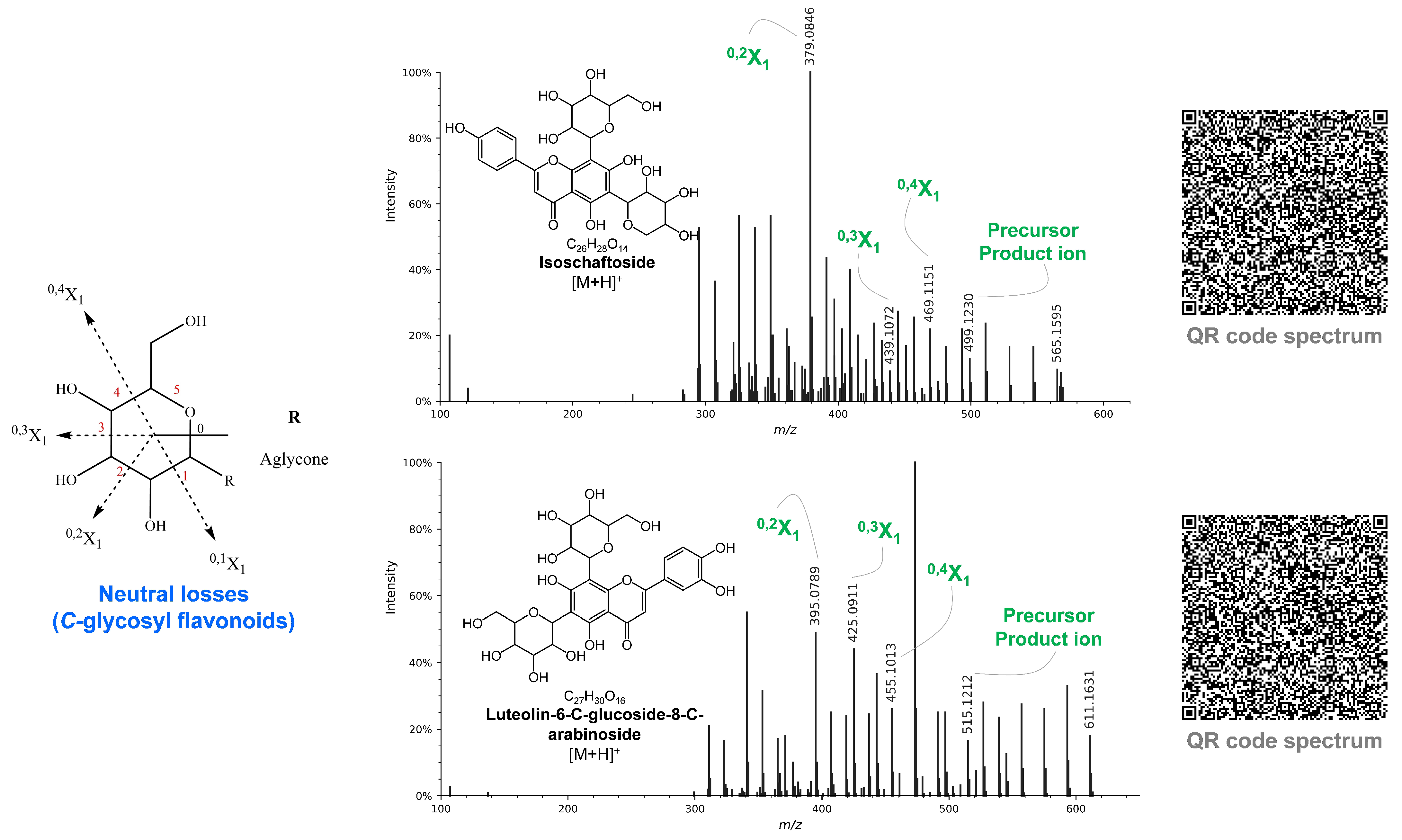
- Hooper, A.M.; Tsanuo, M.K.; Chamberlain, K.; Tittcomb, K.; Scholes, J.; Hassanali, A.; Khan, Z.R.; Pickett, J.A. Isoschaftoside, a C-Glycosylflavonoid from Desmodium Uncinatum Root Exudate, Is an Allelochemical against the Development of Striga. Phytochemistry 2010, 71, 904–908. [Google Scholar] [CrossRef] [PubMed]
- Gomes, A.C.C.; da Silva Sampaio, L.; da Silva, P.A.; Lamas, M.E.; Sakuragui, C.M.; Barreto Junior, C.B.; Simas, N.K.; Kuster, R.M. In Vitro effect of isoschaftoside isolated from Syngonium Podophyllum on pig kidney Na+, K+-ATPASE. Quim. Nova 2014, 37, 1606–1609. [Google Scholar] [CrossRef]
- Bendini, A.; Cerretani, L.; Pizzolante, L.; Toschi, T.G.; Guzzo, F.; Ceoldo, S.; Marconi, A.M.; Andreetta, F.; Levi, M. Phenol Content Related to Antioxidant and Antimicrobial Activities of Passiflora Spp. Extracts. Eur. Food Res. Technol. 2006, 223, 102–109. [Google Scholar] [CrossRef]
- Carocho, M.; Ferreira, I.C.F.R. A Review on Antioxidants, Prooxidants and Related Controversy: Natural and Synthetic Compounds, Screening and Analysis Methodologies and Future Perspectives. Food Chem. Toxicol. 2013, 51, 15–25. [Google Scholar] [CrossRef]
- Zeb, A. Phenolic Antioxidants in Foods: Chemistry, Biochemistry and Analysis; Springer International Publishing: Cham, Switzerland, 2021; ISBN 9783030747671. [Google Scholar]
- Rajudin, E.; Ahmad, F.; Sirat, H.M.; Arbain, D.; Aboul-Enein, H.Y. Chemical Constituents from Tiger’s Betel, Piper Porphyrophyllum N.E.Br. (Fam. Piperaceae). Nat. Prod. Res. 2010, 24, 387–390. [Google Scholar] [CrossRef]
- Baldoqui, D.C.; da Silva Bolzani, V.; Furlan, M.; Kato, M.J.; Marques, M.O.M. Flavonas, Lignanas e Terpeno de Piper Umbellata (Piperaceae). Quim. Nova 2009, 32, 1107–1109. [Google Scholar] [CrossRef]
- Jensen, S.; Olsen, C.E.; Dutt Tyagi, O.; Boll, P.M.; Hussaini, F.A.; Gupta, S.; Bisht, K.S.; Parmar, V.S. Neolignans and an Isoprenylated Phenol from Piper Clarkii. Phytochemistry 1994, 36, 789–792. [Google Scholar] [CrossRef]
- Santoso, B.B.; Hernandez, H.P.; Rodriguez, E.B.; Dalmacio, I.F. Two Antibacterial Compounds: Velutin and 4- (Hydroxy (Oxiran-2-Yl)Methyl)-2-Methoxyphenol from the Stem Bark of Drimys Arfakensis Gibbs. KnE Life Sci. 2017, 3, 51. [Google Scholar] [CrossRef] [Green Version]
- Jung, S.-H.; Heo, H.-Y.; Choe, J.-W.; Kim, J.; Lee, K. Anti-Melanogenic Properties of Velutin and Its Analogs. Molecules 2021, 26, 3033. [Google Scholar] [CrossRef] [PubMed]
- Hassan, S.; Hamed, S.; Almuhayawi, M.; Hozzein, W.; Selim, S.; AbdElgawad, H. Bioactivity of Ellagic Acid and Velutin: Two Phenolic Compounds Isolated from Marine Algae. Egypt. J. Bot. 2020, 61, 219–231. [Google Scholar] [CrossRef]
- López, K.S.E.; Marques, A.M.; Moreira, D.D.E.L.; Velozo, L.S.; Sudo, R.T.; Zapata-Sudo, G.; Guimarães, E.F.; Kaplan, M.A.C. Local Anesthetic Activity from Extracts, Fractions and Pure Compounds from the Roots of Ottonia Anisum Spreng. (Piperaceae). An. Acad. Bras. Cienc. 2016, 88, 2229–2237. [Google Scholar] [CrossRef] [Green Version]
- Morikawa, T.; Matsuda, H.; Yamaguchi, I.; Pongpiriyadacha, Y.; Yoshikawa, M. New Amides and Gastroprotective Constituents from the Fruit of Piper Chaba. Planta Med. 2004, 70, 152–159. [Google Scholar]
- Nicolussi, S.; Viveros-Paredes, J.M.; Gachet, M.S.; Rau, M.; Flores-Soto, M.E.; Blunder, M.; Gertsch, J. Guineensine Is a Novel Inhibitor of Endocannabinoid Uptake Showing Cannabimimetic Behavioral Effects in BALB/c Mice. Pharmacol. Res. 2014, 80, 52–65. [Google Scholar] [CrossRef]
- Kumar, V.; Marković, T.; Emerald, M.; Dey, A. Herbs: Composition and dietary importance. In Encyclopedia of Food and Health; Elsevier: Amsterdam, The Netherlands, 2016; pp. 332–337. ISBN 9780123849533. [Google Scholar]
- Chicca, A.; Nicolussi, S.; Bartholomäus, R.; Blunder, M.; Aparisi Rey, A.; Petrucci, V.; del Carmen Reynoso-Moreno, I.; Viveros-Paredes, J.M.; Dalghi Gens, M.; Lutz, B.; et al. Chemical Probes to Potently and Selectively Inhibit Endocannabinoid Cellular Reuptake. Proc. Natl. Acad. Sci. USA 2017, 114, E5006–E5015. [Google Scholar] [CrossRef] [Green Version]


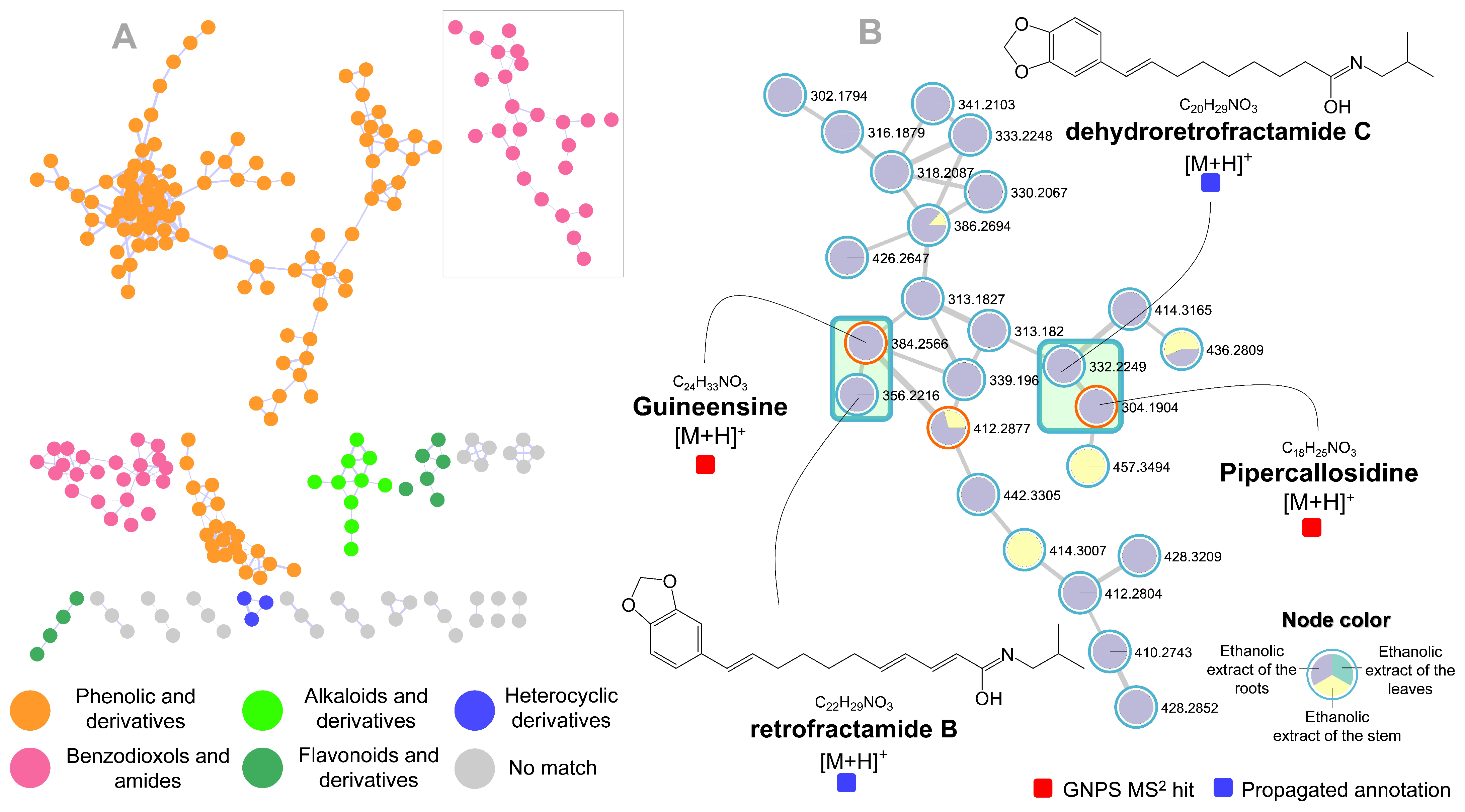
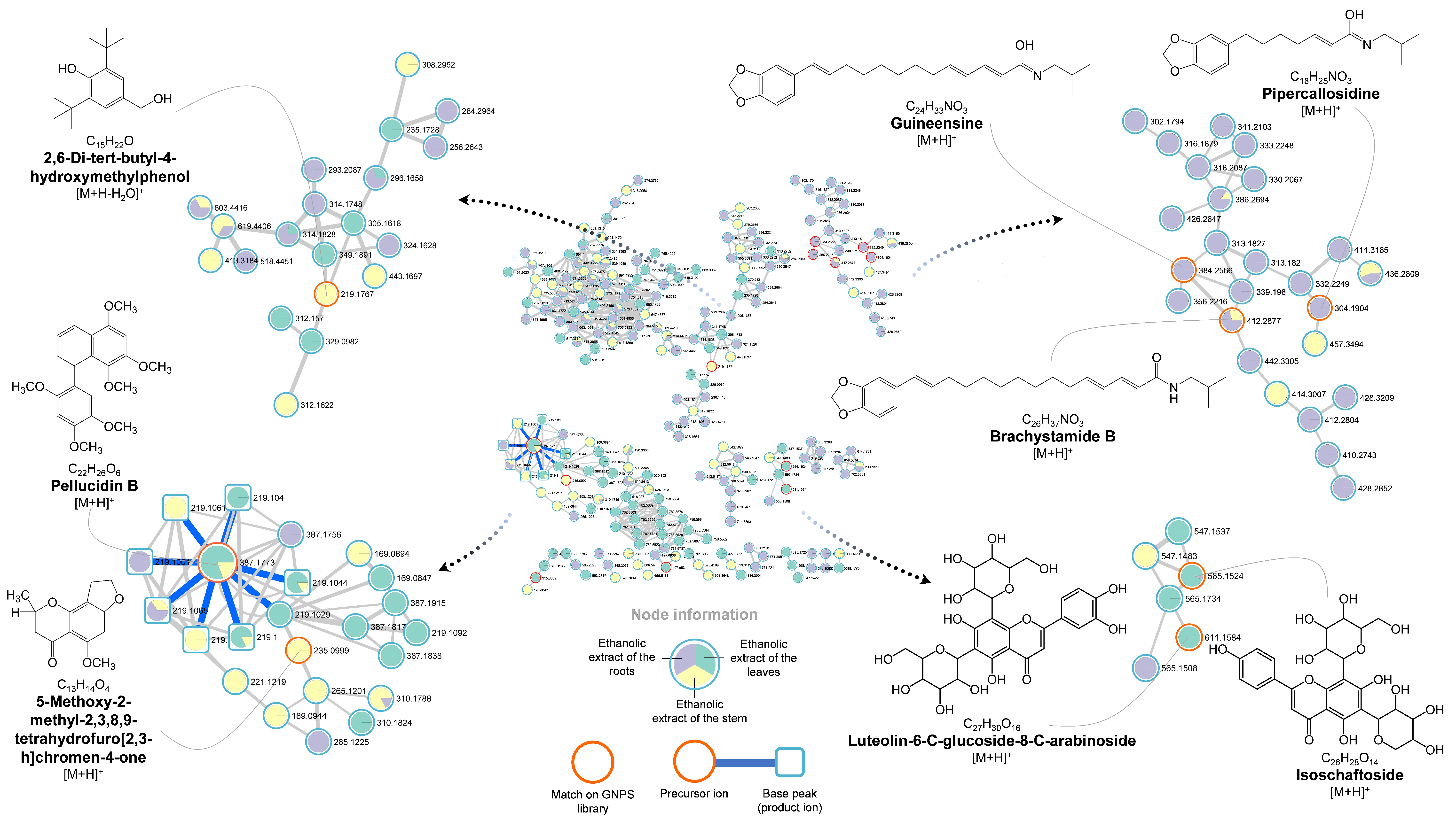
| Peak | Rt (min) | Molecular | [M + H]+ (m/z) | Main Product Ions (MS/MS) | Putative Compound | ||
|---|---|---|---|---|---|---|---|
| Formula | Calculated | Accurate | mDa | ||||
| 1 | 0.55 | C10H13N5O4 | 268.1046 | 268.102 | 2.6 | 136, 119 | vidarabine |
| 2 | 2 | C27H30O16 | 611.1612 | 611.1584 | 2.8 | 593, 575, 557, 539, 527, 515, 491, 473, 455, 443, 425, 395, 371, 341, 311 | luteolin-6-C-glucoside-8-C-arabinoside |
| 3 | 2.34 | C26H28O14 | 565.1557 | 565.1524 | 3.3 | 547, 529, 511, 499, 481, 469, 457, 439 427, 409, 397, 379, 349, 337, 325, 307, 295 | isoschaftoside |
| 4 | 3.41 | C11H16O3 | 197.1178 | 197.1189 | 1.1 | 179, 161, 133 | liolide |
| 5 | 3.78 | C12H15NO3 | 222.113 | 222.1116 | 1.4 | 204, 165, 150, 133, 105 | N-methylcorydaldine |
| 6 | 4.04 | C10H12O4 | 197.0814 | 197.083 | 1.6 | 182, 169, 154, 138, 123, 111 | 2′,4′,5′-trihydroxybutyrophenone |
| 7 | 5.26 | C15H22O | 219.1749 | 219.1767 | 1.8 | 203, 187, 173, 161, 145, 133, 119, 105 | 2,6-di-tert-butyl-4-hydroxymethylphenol |
| 8 | 6.18 | C22H26O6 | 387.1808 | 387.1773 | 3.5 | 219, 169, 145 | pellucidin B |
| 9 | 6.27 | C17H14O6 | 315.0869 | 315.0869 | 0 | 300, 272, 257, 243, 201, 187, 167, 149 | velutin |
| 10 | 7 | C13H14O4 | 235.097 | 235.0999 | 2.9 | 205, 189, 177, 161, 146, 133, 118, 105 | 5-methoxy-2-methyl-2,3,8,9-tetrahydrofuro [2,3-h]chromen-4-one |
| 11 | 7.18 | C18H25NO3 | 304.1913 | 304.1904 | 0.9 | 231, 213, 187, 173, 159, 135, 123, 111, 102 | pipercallosidine |
| 12 | 7.77 | C22H28O6 | 389.1964 | 389.2002 | 3.8 | 315, 221, 190, 181, 174, 169, 147, 129, 114, 105 | pellucidin A |
| 13 | 8.2 | C20H29NO3 | 332.2226 | 332.2249 | 2.3 | 259, 161, 149, 135, 123, 102 | dehydroretrofractamide C |
| 14 | 8.52 | C22H29NO3 | 356.2226 | 356.2216 | 1 | 283, 269, 215, 187, 175, 167, 161, 149, 135, 123, 102 | retrofractamide B |
| 15 | 9.39 | C24H33NO3 | 384.2539 | 384.2566 | 2.7 | 311, 283, 175, 161, 135, 123, 102 | guineensine |
| 16 | 10.3 | C26H37NO3 | 412.2852 | 412.2877 | 2.5 | 339, 311, 290, 203, 185, 175, 161, 149, 135, 123, 102 | brachystamide B |
Publisher’s Note: MDPI stays neutral with regard to jurisdictional claims in published maps and institutional affiliations. |
© 2022 by the authors. Licensee MDPI, Basel, Switzerland. This article is an open access article distributed under the terms and conditions of the Creative Commons Attribution (CC BY) license (https://creativecommons.org/licenses/by/4.0/).
Share and Cite
Gomes, P.W.P.; Barretto, H.; Reis, J.D.E.; Muribeca, A.; Veloso, A.; Albuquerque, C.; Teixeira, A.; Braamcamp, W.; Pamplona, S.; Silva, C.; et al. Chemical Composition of Leaves, Stem, and Roots of Peperomia pellucida (L.) Kunth. Molecules 2022, 27, 1847. https://doi.org/10.3390/molecules27061847
Gomes PWP, Barretto H, Reis JDE, Muribeca A, Veloso A, Albuquerque C, Teixeira A, Braamcamp W, Pamplona S, Silva C, et al. Chemical Composition of Leaves, Stem, and Roots of Peperomia pellucida (L.) Kunth. Molecules. 2022; 27(6):1847. https://doi.org/10.3390/molecules27061847
Chicago/Turabian StyleGomes, Paulo Wender P., Hugo Barretto, José Diogo E. Reis, Abraão Muribeca, Alice Veloso, Carlos Albuquerque, Andrew Teixeira, Wandson Braamcamp, Sônia Pamplona, Consuelo Silva, and et al. 2022. "Chemical Composition of Leaves, Stem, and Roots of Peperomia pellucida (L.) Kunth" Molecules 27, no. 6: 1847. https://doi.org/10.3390/molecules27061847