Review

π–π Stacking Interaction of Metal Phenoxyl Radical Complexes

1
Center for Integrative Quantum Beam Science (CIQuS), Institute of Materials Structure Science (IMSS), High Energy Accelerator Research Organization (KEK), 1-1 Oho, Tsukuba 305-0801, Ibaraki, Japan
2
Graduate School of Science and Engineering, Ibaraki University, Bunkyo, Mito 310-8512, Ibaraki, Japan
*
Author to whom correspondence should be addressed.
Molecules 2022, 27(3), 1135; https://doi.org/10.3390/molecules27031135
Submission received: 27 December 2021 / Revised: 29 January 2022 / Accepted: 2 February 2022 / Published: 8 February 2022

Abstract

:
π–π stacking interaction is well-known to be one of the weak interactions. Its importance in the stabilization of protein structures and functionalization has been reported for various systems. We have focused on a single copper oxidase, galactose oxidase, which has the π–π stacking interaction of the alkylthio-substituted phenoxyl radical with the indole ring of the proximal tryptophan residue and catalyzes primary alcohol oxidation to give the corresponding aldehyde. This stacking interaction has been considered to stabilize the alkylthio-phenoxyl radical, but further details of the interaction are still unclear. In this review, we discuss the effect of the π–π stacking interaction of the alkylthio-substituted phenoxyl radical with an indole ring.

1. Introduction

Weak interactions in biological system are important in terms of the structural and functional aspects [1,2,3,4,5,6,7,8,9,10,11,12,13,14,15,16,17,18,19,20,21,22,23,24,25,26,27,28]. Of particular significance are hydrogen bonds [19,20], π–π stacking interactions [12,13,14] and other non-covalent interactions, including cation-π, NH-π and CH-π interactions [21,22,23,24,25,26,27,28]. These interactions greatly contribute to the construction and stabilization of the highly ordered structures of proteins and other biological molecules, molecular recognition of the substrate and catalytic activity of enzymes [1,2,3,4,5,6,7,8,9,10,11,12,13,14,15,16,17,18,19,20,21,22,23,24,25,26,27,28]. From the importance of weak interactions, the functionalization of the metal complexes has been investigated. In particular, the hydrogen bond was found to stabilize the active dioxygen species [29,30,31]. Thus, the metal complexes with some groups capable of hydrogen bond formation exhibited novel properties and reactivities. On the other hand, the other weak interactions, such as π–π stacking interaction, have remained yet to be employed for the functionalization of the artificial metal complexes.
In order to find the way to the functionalization of metal complexes by π–π stacking interaction, we focused on model studies of the single copper enzyme, galactose oxidase (GO), which catalyzes primary alcohol oxidation to give the corresponding aldehyde [32,33,34,35,36,37]. The catalytic alcohol oxidation mechanism involves the hydrogen atom abstraction from the π-position of primary alcohol to the phenoxyl radical bound to the copper(II) ion in the active form of GO [35]. The active site structure of GO has been revealed to have a square pyramidal structure with two imidazole moieties of the histidine residues and two phenol moieties of the tyrosine residues coordinated to the copper(II) ion (Figure 1) [36]. One of the two phenol moieties is deprotonated and bound at an equatorial position of the copper center as a phenolate ligand, to which the sulfur atom derived from the cysteine residue is bound at the ortho-position to form an ortho-alkylthiophenolate moiety. The active form of GO is known to be the copper(II)-alkylthiophenoxyl radical, and this phenoxyl radical has been proposed to be stabilized by π–π stacking interaction with the indole ring of the proximal tryptophan residue (Trp 290) [37].
A number of model studies of GO have been reported with emphasis on the characteristic electronic structure of copper(II)-phenoxyl radical and its reactivity [37,38,39,40,41,42,43,44,45,46,47]. Copper(II)-phenoxyl radical complexes have quite different geometric and electronic structures compared with the copper(III)-phenolate species, which is the same one-electron oxidized form of the copper(II)-phenolate species [48,49,50,51,52,53]. The difference is reflected to the reactivity on the primary alcohol oxidation; the copper(III)-phenolate is less reactive for the primary alcohol oxidation in comparison with the copper(II)-phenoxyl radical, suggesting that the hydrogen abstraction by the phenoxyl radical is significant in the course of the oxidation [52,53,54]. However, the effect of the π–π stacking interaction of the phenoxyl radical with the indole ring is still unclear.
With these points in mind, we focus on the effects of the π–π stacking interaction of the phenoxyl radical bound to a copper ion. The geometric and electronic structural changes of the phenoxyl radical by the π–π stacking interaction and its effects on reactivity are discussed on the basis of recent results. This review discusses π–π stacking interactions of the phenoxyl radical species with the indole ring around the metal center.

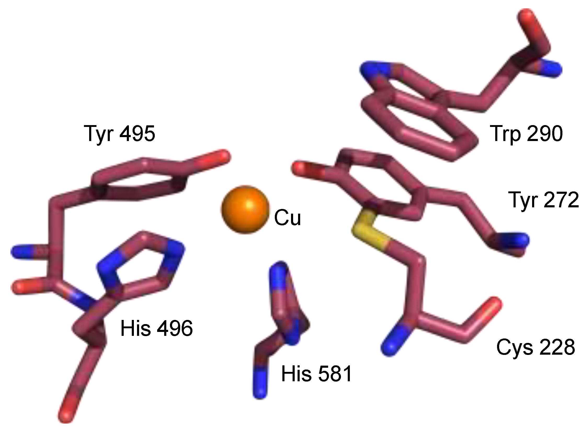
2. The π–π Stacking Interaction in the Active Site of GO

One of the aromatic amino acids tryptophan (Trp) has a side chain indole ring, which is a fused-ring aromatic molecule composed of a pyrrole and a benzene ring. The indole ring has been considered to stabilize the active species or to be involved in electron transfer pathways by π–π stacking interaction with the other aromatic ring. Therefore, it is frequently located at the proximal position of the active or important site of enzymes [41,55]. GO catalyzes oxidation of the primary alcohol at the 6-position of galactose to generate the corresponding aldehyde [35,56], and the active species of the catalytic oxidation has been well known to be the copper(II)-phenoxyl radical derived from the one-electron oxidation of the phenolate moiety of tyrosine 272 (Tyr 272) bound to the copper(II) ion [32,33,34,35,36,37]. The phenoxyl radical moiety of Tyr 272 is modified by the carbon-sulfur covalent bond at the ortho-position, derived from the cross linking with the cysteine 228 (Cys 228), and the active form of GO is described as a copper(II)-alkylthiophenoxyl radical species [51]. The indole ring of Trp 290 is located in the second coordination sphere of the active site of GO, most probably for stabilization of the copper(II)-alkylthiophenoxyl radical species by the π–π stacking interaction with the alkylthiophenoxyl radical of Tyr 272 [57,58,59,60,61]. Previous studies using the GO mutants with Trp 290 replaced with various other amino acids, such as glycine (gly), phenylalanine (Phe) and histidine (His), revealed a shorter lifetime of the Cu(II)-alkylthiophenoxyl radical, indicating that the π–π stacking interaction plays an important role in the stabilization of the radical in the active form of GO [58,59,60].
Apo-GO, which has no Cu(II) ion in the active site, shows a significantly different structure, especially in the active site, where the phenoxyl radical could not be detected and no C-S bond formation in the phenol moiety of Tyr 272 was found (Scheme 1) [60,61]. In addition, the π–π stacking interaction of the indole moiety of Trp 290 with the phenol moiety of Tyr 272 was not observed, and the indole ring was exposed to the outer sphere of protein [60]. However, both the stacking interaction and the C-S cross link were generated upon the addition of copper(II) ion to the apo-GO by the soaking experiments [60]. These results suggests that the indole ring of Trp 290 selectively interacts with the alkylthio-phenoxyl radical moiety.
On the other hand, the effect of the C-S cross link of the alkylthiophenoxyl radical have been also reported [62]. The mutation study replacing Cys with glycine (Gly) and serine (Ser) of the GO homologue, GlxA, revealed that the unpaired electron of the phenoxyl radical was transferred to the indole moiety of Trp to form the indolyl or indole–π–cation radical [62]. The result suggests that the alkylthiophenoxyl radical is also stabilized by the C-S cross link. It was proposed that in the absence of the C-S cross link, the indole ring of Trp may be also located in the proximal position of the phenoxyl radical, but that the conformation of the indole moiety is different from that of the native GO. The indole ring was found to be in a position not suitable for the π–π stacking interaction, and thus it was rotated to form the pseudo-physical (van der Waals) interaction mainly of the indole C–H bonds with the phenolate moiety (Scheme 2). The experimental and calculation studies of the mutants proposed that the spin density in these mutants is distributed mainly on the indole moiety coupled with the Tyr phenolate moiety [62]. Thus, the methylthio group is important for the stabilization of the phenoxyl radical by the face-to-face π–π stacking interaction, and, for easy transfer of the unpaired electron of the phenoxyl radical, may be easily transferred to the indole ring by rotation.

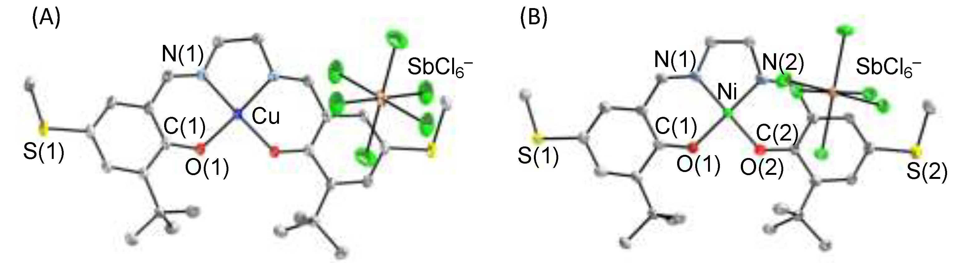
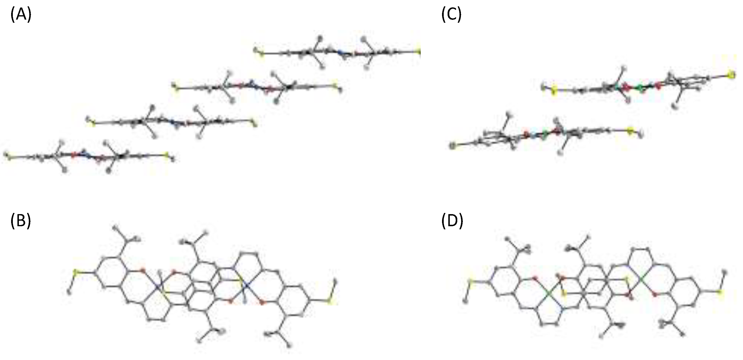
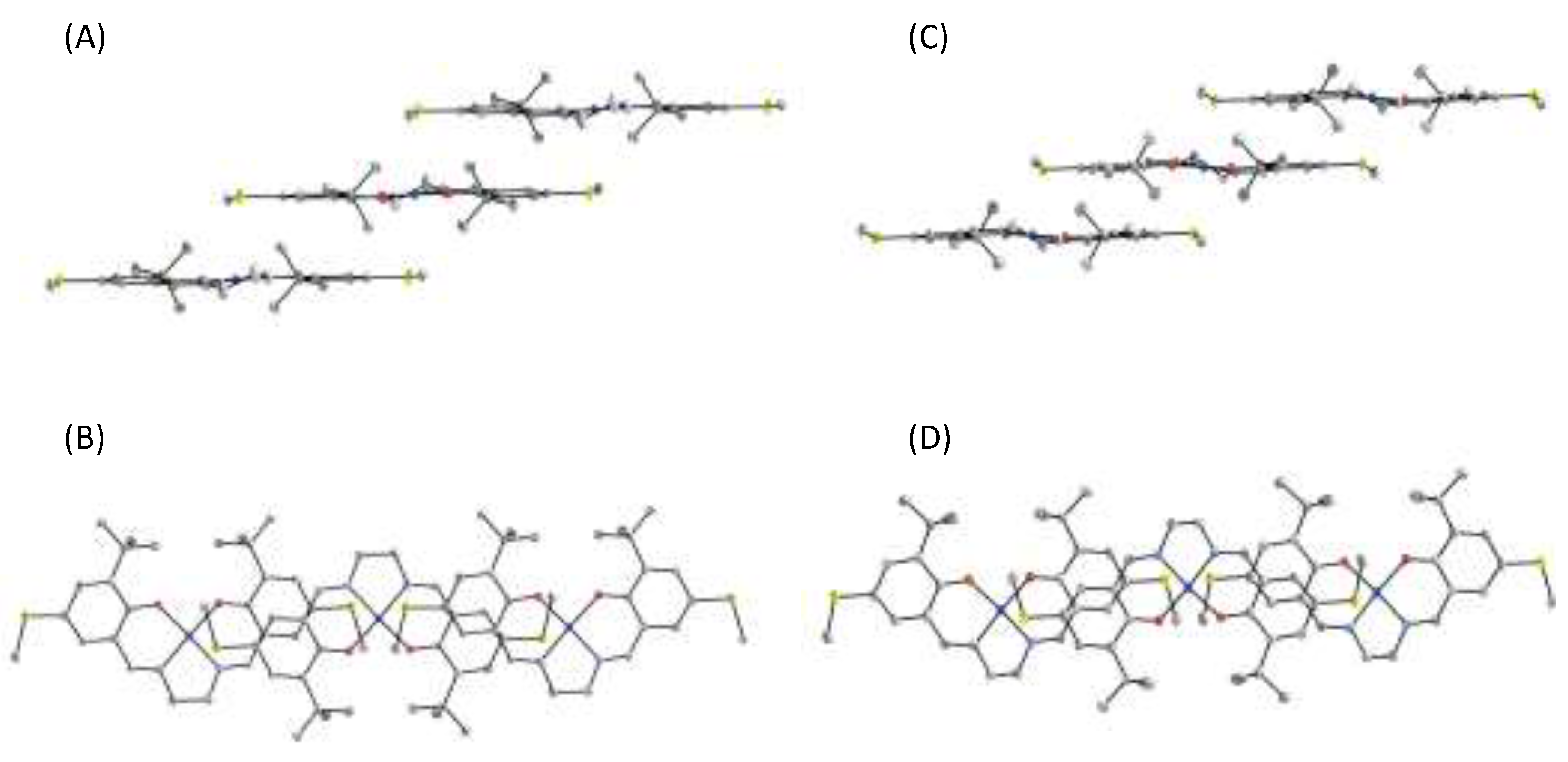
3. π–π Stacking Interaction of Methylthio-Phenoxyl Radical Metal Complexes

The π–π stacking interaction between two phenoxyl radicals is slightly different from that involving the neutral phenol and phenolate anion, due to the existence of one unpaired electron on the π conjugated system. The π orbital is assigned to the SOMO on the phenoxyl radical as whole; therefore, a SOMO–SOMO interaction can be considered in general [63]. Such a SOMO–SOMO interaction could not be observed in the crystals of metal phenoxyl radical complexes except alkylthio-phenoxyl radical species [39,40,64,65,66]. In many cases, the phenoxyl radical moiety interacted only with the counter anion weakly [39,40,64]. On the other hand, the alkylthio-phenoxyl radical, which is similar to the phenoxyl radical in the active form of GO, showed a significant interaction with the other phenoxyl radical by π–π stacking interaction in the solid state [65]. Furthermore, the stacking interaction between alkylthio-phenoxyl radicals exhibited a different dependence on the central metal ion [65,66].
Recently, the X-ray crystal structures of oxidized copper(II)- and nickel(II)-diphenolate complexes have been successfully determined. One-electron oxidized copper(II) di(methylthiophenolate) complex with a salen-type ligand, [Cu(MeS-salen)]SbCl6 ([1]SbCl6), was prepared by reaction of Cu(MeS-salen) (1) with one equivalent of thiantrenyl radical hexachloroantimante salt (Th+SbCl6) (Scheme 3) [65]. Similar oxidation by addition of one equivalent of Th+SbCl6 to the solution of Ni(MeS-salen) (2) afforded the one-electron oxidized complex, [Ni(MeS-salen)]SbCl6 ([2]SbCl6) [66]. The Cu and Ni K-edge X-ray absorption near edge structures (XANES) of these one-electron oxidized complexes showed no significant difference from that of the complexes before oxidation, whereas the sulfur K-edge XANES exhibited a noticeable increase in the pre-edge intensity of [1]SbCl6 [65,66,67,68]. These results indicate that the one-electron oxidized complexes [1]SbCl6 and [2]SbCl6 are ligand centered oxidation species and thus can be assigned to the metal(II)-phenoxyl radical complexes.
The X-ray crystal structure of [1]SbCl6 revealed that it has half of the molecule as a crystallographically independent unit, indicating that [1]SbCl6 has a perfect C2 axis through the center of the ethylenediamine and the copper ion (Figure 2A) [65]. The results indicate that the structural features of the two phenolate moieties are identical. The bond length of Cu–O in [1]SbCl6 (1.920 (1) Å) was longer than that of 1 (1.902(1) Å), and that of C–O of the phenolate moiety in [1]SbCl6 (1.295(2) Å) was shortened compared with 1 (1.307(2) Å). On the other hand, the X-ray crystal structure of [2]SbCl6 showed no symmetry axis, indicating that the two phenolate moieties in [2]SbCl6 are structurally not identical (Figure 2B) [66]. The differences of the two Ni–O and two C–O bond lengths in [2]SbCl6 (Ni–O(1) 1.875(2) Å, Ni–O(2) 1.843(2) Å; C(1)–O(1) 1.279(4) Å, C(2)–O(2) 1.315(4) Å) were larger than the differences observed between 1 and [1]SbCl6. The bond lengths of Ni–O(2) and C(2)–O(2) in [2]SbCl6 were similar to those of complex 2 (Ni–O 1.8586(9) Å, C–O 1.312(1) Å), supporting that one of the phenolate moieties in [2]SbCl6 maintains the phenolate electronic structure, so that [2]SbCl6 can be described as the nickel(II) localized phenoxyl radical complex, [NiII(phenolate)(phenoxyl radical)]+ [66]. In the case of copper complex [1]SbCl6, the difference of the Cu–O and C–O bond lengths was rather small in comparison with the nickel complexes. From the results together with the symmetric features in crystals, [1]SbCl6 can be assigned to the copper(II)-delocalized radical species described as [CuII(0.5-phenoxyl radical)2]+ with the electron equally distributed on the two phenolate moieties [65].
The crystal packing of these one-electron oxidized complexes revealed the existence of the intermolecular interaction. The crystal packing of [1]SbCl6 showed the intermolecular π–π stacking interaction between the two half-phenoxyl radical moieties of the neighboring molecules with the distance of 3.18 Å, resulting in the one-dimensional chain formation. Furthermore, the sulfur atom of the 0.5-alkylthiophenoxyl radical was in close contact with the central copper ion with the distance of 3.04 Å (Figure 3A,B) [65]. On the other hand, the crystal packing of the localized phenoxyl radical complex [2]SbCl6 showed a different view of the crystal packing (Figure 3C,D). The intermolecular π–π stacking interaction was observed between the localized phenoxyl radical moieties of the neighboring molecules with the distance of 3.1 Å to give a dimerization species, but no one-dimensional chain formation could be detected [66].
Such a difference in the intermolecular π–π stacking interaction of copper and nickel complexes was considered to arise from the difference in the population of the unpaired electron spin on the phenoxyl radical moiety. In general, the SOMO–SOMO interaction shows some characteristics [63]; π–π stacking geometry involving the SOMO–SOMO interaction exhibits the atom-over-atom configurations with a distance shorter than the sum of the van der Waals radii (less than 3.19 Å in the case of the C–C distance), as opposed to the atom-over-bond or atom-over-ring configurations, which are typical of van der Waals π–π stacking [14,63,69]. Furthermore, possible minor deviations from the planarity of the constituent molecules indicate the primary role of the SOMO–SOMO interaction [63]. In the case of [2]SbCl6, the intermolecular π–π stacking interaction between two localized phenoxyl radical moieties can be assigned to the characteristic SOMO–SOMO interaction, the atom-over-atom configurations being observed with the shortest C–C distance of 3.1 Å and small deviation of the stacked phenoxyl radical moieties from planarity (Figure 3C,D) [66]. On the other hand, [1]SbCl6 having two half-phenoxyl radical moieties showed the intermolecular interaction typical of van der Waals π–π stacking, which is very similar to that before oxidation (Figure 4) [65]. These results suggest that the alkylthio-phenoxyl radical moiety prefers the π–π stacking interaction and that the mode of the stacking depends on the electronic structure of the phenoxyl radical moiety.
The π–π stacking interaction can give rise to a different electronic structure of the metal phenoxyl radical species. The phenoxyl radical localization and delocalization can be assigned by the NIR band intensity and band width, which is similar to the mixed valence dinuclear complexes [70,71,72]. In the case of the completely localized phenoxyl radical unpaired electron on one of the two phenolate moieties, no characteristic band is observed in the NIR region. However, an increase in the degree of the delocalization gradually causes the appearance of the NIR band, and the full delocalization of the radical unpaired electron on the two phenolate moieties described as (0.5-phenoxyl radical)2 gives a sharp intense band with a narrow bandwidth in the NIR region [73,74,75]. The UV-vis-NIR absorption spectrum of the CH2Cl2 solution of [1]SbCl6 showed a broad NIR band with a small intensity at 9100 cm−1, which is a feature similar to the one-electron oxidized methoxy-substituted diphenolate copper(II) complex, [Cu(MeO-salen)]+ (Figure 5A) [65,73]. The band was assigned to the phenolate to phenoxyl radical charge transfer, which was also supported by TD-DFT calculation. On the other hand, the solid sample of [1]SbCl6 showed a large shift of the NIR band to 5000 cm−1, which is different from that of the solid sample of [Cu(MeO-salen)]+ (7800cm−1 in CH2Cl2 vs. 7200 cm−1 in the solid state) [65,73]. Furthermore, the bandwidth of the NIR band of [1]SbCl6 in the solid state was narrow in comparison with the solid sample of [Cu(MeO-salen)]+ and the CH2Cl2 solution of [1]SbCl6 (Figure 5B) [65,73]. These results indicate that [1]SbCl6 has different electronic structures, the localized phenoxyl radical in CH2Cl2 and the delocalized phenoxyl radical on two phenolate moieties in the solid state due to the stacking interaction, which was supported by DFT calculations [65].
The difference arising from the stacking interaction was also observed in EPR measurement. In general, copper(II)-phenoxyl radical complexes are EPR inactive or show the characteristic EPR signal at ca. g = 4, due to the magnetic coupling of two unpaired electron spins between the copper d-electron and the phenoxyl radical electron, resulting in S = 0 or 1 in total [39,76,77]. However, the solid sample and the frozen sample of the CH2Cl2 solution of [1]SbCl6 showed a significant isotropic EPR signal at ca. g = 2.0, which is of similar intensity to the signal of the di(phenoxyl radical) copper(II) complex of the same ligand, [1](SbCl6)2 (Figure 6) [65,67,68]. These results can be considered to show that all unpaired electron spins in both the copper(II) ion and phenoxl radical line up ferromagnetically in the one-dimensional chain of [1]SbCl6 by the π–π stacking interaction, which is similar to the other ferromagnetic one-dimensional chain compounds. In fact, the solid sample of [1]SbCl6 exhibited the ferromagnetic interaction intra- and inter-molecularly [65]. Thus, the π–π stacking interaction of the methylthio-phenoxyl radical moiety in [1]SbCl6 influences the electronic structure.

4. The π–π Stacking Interaction of Phenoxyl Radical with an Indole Ring

In order to understand π-π stacking properties of the indole ring with the phenoxyl radical, copper(II) and nickel(II) diphenolate salen-type complexes with two para-methoxy- or para-methylthio-phenolate moieties and a side chain indole ring on the ethylenediamine backbone were synthesized and characterized (Figure 7) [78,79,80]. Their one-electron oxidized complexes were characterized, particularly focusing on the different behavior of the indole ring due to the oxidation state.

4.1. The π–π Stacking Interaction of Methoxyphenoxyl Radical with an Indole Ring

The X-ray crystal structure of copper(II) salen-type complex 3 having a pendent indole ring and methoxy substitution at para-position of two phenolate moieties revealed no significant intra- and inter-molecular interaction (Figure 8A), and thus the coordination sphere of complex 3 was very similar to that of the MeO-salen complex without the pendent indole ring Cu(MeO-salen) [65,73]. This result well supports the previous reports that the indole ring does not interact with an electron-rich aromatic ring, such as the phenolate moiety [79,80,81,82,83,84,85]. Complex 3 was oxidized by the addition of one equivalent of AgSbF6 as a one-electron oxidant in CH2Cl2 to form the one-electron oxidized complex [3]SbF6, whose X-ray crystal structure revealed that [3]SbF6 consists of two crystallographically independent species in the unit cell (Figure 8B,C) [78]. They showed similar structural characteristics in the first coordination sphere, and one of the two Cu-O bonds in each species was ca. 0.1 Å longer than the other, indicating that the radical electron was fully localized on one of the phenolate moieties. Therefore, the two complexes in the unit cell could be assigned to the localized phenoxyl radical complexes described as [CuII(phenolate)(phenoxyl radical)]+ [64]. The position of the indole ring in [3]SbF6 was completely different from that in the neutral complex 3; the indole ring was found to lie on the phenoxyl radical moiety of salen ligand by the π–π stacking interaction in both complexes. The distance between indole ring and the phenoxyl radical moiety was determined to be ca. 3.4 Å, which is in the acceptable range of the π–π stacking interaction [14,86]. Furthermore, this stacking conformation of the indole ring and phenoxyl radical moiety showed the atom-over-bond or atom-over-ring configuration, which is different from the π–π stacking interaction by SOMO–SOMO overlapping [63]. These results indicate that the indole ring recognizes the phenoxyl radical moiety and forms a π–π stacking structure with it selectively.
The oxidized complex [3]+ also showed a different behavior in CH2Cl2 in comparison with the complex having a side chain methyl group, [4]+ [78]. The CH2Cl2 solutions of [3]+ and [4]+ exhibited the specific band in the NIR region, which was assigned to the phenolate to phenoxyl radical intervalence charge transfer indicating the relatively localized Cu(II)-methoxyphenoxyl radical on one of the phenolate moieties (Figure 9A) [46,53,66,67,68]. Notably, the NIR band of [3]+ (8700 cm−1) shifted to the higher energy region by 1100 cm−1 compared with that of [4]+ (7600 cm−1), showing that the phenoxyl radical moiety was affected by the pendent indole ring. The energy difference could be estimated from this NIR band difference to be 13 kJ/mol, which is in line with the energy of the π–π stacking interaction (4–20 kJ/mol) [14,78,86]. Therefore, the indole-phenoxyl radical stacking interaction was also maintained in the CH2Cl2 solution. It has been reported that the π–π stacking interaction of the indole ring depends on the solvent properties [12,87]. In CH3CN (dielectronic parameter [1/η2 − 1/ε] of CH3CN: 0.528; CH2Cl2: 0.382) a similar NIR band shift to the higher energy region was also observed for [3]+ (9500 cm−1) in comparison with [4]+ (8700 cm−1) [78]. In toluene (dielectric parameter [1/η2 − 1/ε]: 0.026), however, the NIR band peak difference between [3]+ (8060 cm−1) and [Cu(MeO-salen)]+ (7880 cm−1) [65,73,88] was rather small (Figure 9B), and the energy was estimated to be 2.4 kJ/mol. These results are consistent with the fact that the π–π stacking interaction is more favored in the polar solvent [78]. Furthermore, toluene, being an aromatic solvent, may be also effective for inhibition of the intramolecular π–π stacking interaction by solvation.
DFT calculations supported the intramolecular π–π stacking interaction of the indole ring with the phenoxyl radical. The calculation results suggested that the π–π stacking form is more stable than the open structure having no significant stacking interaction [78]. Furthermore, TD-DFT calculation of [3]+ showed that the NIR band of [3]+ consists of the two characteristic components at 7175 cm−1 and 9291 cm−1 in CH2Cl2. The 7175-cm−1 band could be assigned to the intervalence charge transfer (IVCT) band from the phenolate to the phenoxyl radical moiety as the transition from HOMO to LUMO [78]. The assignment is in good agreement with the NIR peak of the complexes without a pendent indole moiety, [4]+ and [Cu(MeO-salen)]+ [65,73]. On the other hand, the 9291-cm−1 band was assigned to the charge transition from the indole moiety to the phenoxyl radical moiety. Such a charge transfer band was also suggested at 28428 cm−1 (352 nm) by TD-DFT calculation (Figure 10), which agrees well with the previous report on the appearance of the charge transfer band in the near UV region due to aromatic ring stacking [12,79,87]. In fact, the different UV-vis spectrum between [3]+ and [4]+ exhibited a band at 352 nm in CH2Cl2, and the CD spectrum of [3]+ showed a significant CD peak at ca. 350 nm, which is considered to be due to the proximal effect on the charge transfer band. The difference UV-vis spectrum and the CD peak were solvent-dependent, and the UV band shift and the CD peak could not be detected in toluene [78]. From the experimental and calculation results, the indole ring was concluded to stabilize the phenoxyl radical by the π–π stacking interaction, where it serves as an electron-donor to the phenoxyl radical moiety [78].

4.2. The π–π Stacking Interaction of Methylthiophenoxyl Radical with an Indole Ring

The methylthiophenoxyl radical complex [1]SbCl6 favors the π–π stacking interaction in the solid state. As discussed in the previous section, its electronic structure is changed from the localized phenoxyl radical species to the delocalized species by the stacking interaction [65]. The effect of the proximal indole ring in the methylthiophenoxyl radical complex could be clarified by characterization of complex 5 having the same coordination structure as 1 but with a pendent indole ring.
One-electron oxidized complex [5]+ could be generated by the addition of 1 equivalent of thianthrenyl cation radical salt (Th+SbCl6) to the CH2Cl2 solution of 5. The UV-vis-NIR spectrum of 5 showed a spectral feature similar to that of complex [1]SbCl6 in CH2Cl2, suggesting that the phenoxyl radical was localized on one of the phenolate moieties in CH2Cl2 [65]. In comparison with the other phenoxyl radical complexes having a side chain, such as a methyl ([6]+) and a phenyl ([7]+) group, complex [5]+ exhibited the NIR band assigned to the phenolate to phenoxyl radical IVCT at a quite different position [79]. The NIR band of complex [5]+ shifted to the higher energy region than the bands of [6]+ and [7]+ by ca. 1000 cm−1 (9.6 kJ/mol). This energy difference is in line with the energy of π–π stacking interaction (4–20 kJ/mol) and the stabilization energy of the methoxy-substituted complex [3]+, indicating that the pendent indole ring of [5]+ contacts with the phenoxyl radical moiety by the π–π stacking interaction in CH2Cl2 solution [14,78,79,86]. Considering the difficulty of the stacking interaction between the two methylthiophenoxyl radical complexes in CH2Cl2 solution of [1]SbCl6, the interaction of the indole ring is more effective for perturbation of the methylthiophenoxyl radical [65,79]. On the other hand, it is noticed that the phenyl group in [7]+ may not be effective for the perturbation by the stacking interaction [79].
The methylthiophenoxyl radical complex [1]+ showed a significantly intense EPR signal at ca. g = 2 in frozen solution, though copper(II)-phenoxyl radical complexes such as [1]+ having a two-spin system with the magnetic interaction are generally EPR-silent [65,67,68]. This can be considered to be due to the formation of a polymerization structure in the frozen solution by intermolecular π–π stacking interaction, and thus the intense EPR signal of the frozen sample of [1]+ may arise from various magnetic interactions in the polymerization structure [65]. Although complex [6]+ showed a similar intensity of the EPR signal at g = ca. 2, complexes [5]+ and [7]+ exhibited the EPR intensity decrease estimated to be more than 70% in the case of [5]+ [79]. The result indicates that the stacked indole ring and phenyl group inhibited the formation of the polymerization structure by the intermolecular stacking between the methylthiophenoxyl radical moieties, and as a result, the electronic structure change of the methylthiophenoxyl radical to form the localized phenoxyl radical species on one of the phenolate moieties was also inhibited [79]. DFT calculation using gradient isosurfaces methods[89] suggested that complexes [5]+ and [7]+ could form the π–π stacking structure of the methylthiophenoxyl radical with the pendent aromatic ring, with the indole ring showing a larger degree of the interaction than the phenyl ring (Figure 11) [79]. Thus, the π–π stacking interaction of the pendent indole moiety with the methylthiophenoxyl radical is more effective than the intermolecular stacking between two methylthiophenoxyl radical moieties.

4.3. The Effect of the Electronic Structure of the Phenoxyl Radical on π–π Stacking Interaction with an Indole Ring

The above-mentioned discussions in this review focused mainly on the π–π stacking interaction of the localized phenoxyl radical copper(II) complexes [78,79]. The ligand centered oxidation metal complexes with the salen-type ligand sometime show that the radical unpaired electron spin is delocalized on the two phenolate moieties [46,64,66,85,90,91,92,93]. The phenoxyl radical localization and delocalization depend on the central metal ion. Copper(II) complexes generally favor the localized phenoxyl radical described as [CuII(phenolate)(phenoxyl radical)]+ [46,53,66,67,68], whereas some nickel complexes show the delocalization of the radical to form [NiII(0.5-phenoxyl radical)2]+ configuration [46,64,66,75,81,93,94]. In this section, the effect of the π–π stacking interaction of the indole ring with the 0.5-phenoxyl radical moiety is discussed for the Ni(II) complex of the ligand of complex 3 having a pendent indole ring as an example [80].
The neutral and the methoxyphenoxyl radical nickel(II) complexes, 8 and [8]SbF6, were prepared by the procedures similar to those employed for the isolation of copper(II) complexes 3 and [3]+, respectively, in 1,1,2,2-tetrachloroethane(C2H2Cl4)/n-hexane. The X-ray crystal structures of complexes 8 and [8]+ revealed that the structures are very similar to those of complexes 3 and [3]SbF6, respectively (Figure 12) [78,80]. Although the neutral complex 8 showed no significant interaction of the pendent indole ring intra- and intermolecularly, the crystal structure of [8]SbF6 consisted of two different stacking structures in the unit cell as revealed for [3]SbF6, the position of the phenolate ring stacked with the indole moiety being different. In addition, details of the coordination structures of the two molecules in the unit cell are different. The two Ni–O lengths of [8]SbF6 (a) were the same (1.841(9) Å), whereas they were slightly different in the other molecule [8]SbF6 (b) (1.833(7) Å and 1.860(7) Å). The bond length between copper ion and the phenoxyl radical oxygen atom is ca. 0.1 Å longer than that between copper ion and the phenolate oxygen atom in complex [3]+, which was assigned to the phenoxyl radical localized on one of the phenolate moieties [80]. Form the results, complex [8]SbF6 was mainly assigned to the radical with the unpaired electron spin delocalized on two phenolate moieties, described as [Ni(0.5-phenoxyl radical)2]. On the other hand, complex [8]+, which has the structure showing no significant interaction of the pendent indole ring with the phenoxyl radical moiety, could be isolated by changing the solvent from 1,1,2,2-tetrachloroethane to chloroform. (Figure 12B). This result suggests that the π–π interaction in this complex may be less favored than that in the localized phenoxyl radical complex [3]+[80].
Delocalization of the unpaired electron on the two phenolate moieties in [8]SbF6 was maintained in CH2Cl2 solution, where [8]SbF6 showed the intense NIR band at 4700 cm−1 (ε = 16800 M−1cm−1) [78,79]. The intensity of this band was much stronger than that of the localized phenoxyl radical copper(II) complexes (Figure 13) [80]. The NIR band feature with a strong intensity and a narrow bandwidth could be assigned to the fully delocalized system based on the mixed valence systems by Robin and Day classification [46,53,66,67,68,69,70,71,72]. Therefore, the radical unpaired electron fully delocalized on two phenolate moieties and described as [Ni(0.5-phenoxyl radical)2]+ is maintained in the CH2Cl2 solution, which is in good agreement with the other NiII-phenoxyl radical species of the salen-type ligands.
On the other hand, the indole ring in the phenoxyl radical complex [8]+ may be considered to be in close contact with the 0.5-phenoxyl radical moiety in the CH2Cl2 solution, whereas the experimental difference between complexes with and without a pendent indole ring was rather small. The NIR band in the UV-vis-NIR spectrum of NiII-phenoxyl radical of [8]+exhibited a slight decrease in the intensity at 4700 cm−1 compared with that of [9]+ but a slight intensity increase at 10200 cm−1 (Figure 13B) [80]. The results indicated that the effect of the π–π stacking interaction of the indole ring with the phenoxyl radical in complex [8]+ was rather small as compared with that in the copper complex [3]+ [78,80]. In fact, the complex [8]+with an open structure could be isolated by using a different solvent. Thus, the π–π stacking interaction of the indole ring with the 0.5-phenoxyl radical moiety is less effective than that with the localized phenoxyl radical in the symmetric two-phenolate ligand system [80].
TD-DFT calculation of complex [8]+ suggested that the NIR band characteristics of [8]+ are different from those of the copper(II)-phenoxyl radical complexes and the oxidized nickel(II)-salen complexes without the pendent indole moiety. Especially, the NIR band of [8]+ at 10686.0 cm−1 was predicted as a characteristic transition from βHOMO-4 to LUMO. The contribution of the Ni ion orbital in βHOMO-4 was estimated to be ca. 47%, whereas the Ni ion contribution in LUMO was only 13% (Figure 14) [80]. Therefore, this band could be described as a MLCT band from the Ni(II) ion to the indole ring. It should be mentioned in this connection that the electronic structure of the one-electron oxidized Ni-salen complexes could change from the Ni(II)-phenoxyl radical to the Ni(III)-phenolate state by addition of exogenous ligands [94,95]. In addition, the transition at 9693.1 cm−1 was predicted to be the LLCT band, which could be described as the charge transfer from the indole to the delocalized phenoxyl radical (Figure 14) [80]. These CT-bands characteristics at ca. 10000 cm−1 support the close contact of the indole ring with the coordination plane in [8]+. Thus, theoretical calculations showed that the indole moiety selectively interacts with the phenoxyl radical moiety and stabilizes the nickel(II)-phenoxyl radical complex by the π–π stacking interaction.

5. Summary and Conclusions

The roles of the π–π stacking interaction in the metal-phenoxyl radical complexes have been discussed in this review, especially focusing on the interaction of the alkylthiophenoxyl radical with the indole ring as seen in the single copper enzyme GO. GO has an alkyltiophenoxyl radical bound to copper ions in the active site, and the indole moiety of Trp 290 located at the proximal position of the phenoxyl radical is involved in the π–π stacking interaction. The alkylthio group is important for the stabilization of the phenoxyl radical state. In the absence of the alkylthio group, the phenoxyl radical is less stable and is reduced by the electron transfer from indole, where the indole ring showed a different mode of interaction from the π–π stacking interaction in the native form of GO.
The alkylthio-phenoxyl radical can be stabilized by the π–π stacking interaction, which gives rise to a slightly different electronic structure. However, the electronic structure and the effect of the π–π stacking interaction depend on the central metal ion. In contrast with the van der Waals π–π stacking interaction in the copper complexes, the nickel complex exhibits the SOMO–SOMO interaction. The π–π stacking interaction of the phenoxyl radical with the indole ring significantly stabilizes the phenoxyl radical state, and the indole to phenoxyl radical charge transfer can be detected in the NIR region in the absorption spectrum. This assignment may be taken to show the close energy gap between indole and phenoxyl radical. Therefore, a small perturbation of the phenoxyl radical can lead to the electron transfer. These observations indicate that the π–π stacking interaction of phenoxyl radical with the indole ring is more effective than that between two phenoxyl radicals, and that the electronic structures of the phenoxyl radical can be controlled by the π–π stacking interaction with the indole moiety.
Taken together, the π–π stacking interaction of the phenoxyl radical with the indole ring is important in the active site of GO, and we believe that unique properties of the π–π stacking interaction involving phenoxyl, indole and various other aromatic rings may lead to novel functionalization of the metal complexes.

Author Contributions

Conceptualization, H.O. and Y.S.; writing—original draft preparation, H.O.and Y.S.; writing—review and editing, Y.S.; funding acquisition, Y.S. All authors have read and agreed to the published version of the manuscript.

Funding

This work was supported in part by Grants-in-Aid for Scientific Research (No. 16K05716 to Y. S.) from the Ministry of Education, Culture, Sports, Science, and Technology of Japan, Cooperative Research Program of “Network Joint Research Center for Materials and Devices” (Institute for Materials Chemistry and Engineering, Kyushu University).

Institutional Review Board Statement

Not applicable.

Informed Consent Statement

Not applicable.

Data Availability Statement

Data is contained within the article.

Acknowledgments

The authors would like to thank Takashi Suzuki, Graduate School of Science and Engineering, Ibaraki University and Tomoyuki Takeyama, School of Materials and Chemical Technology, Tokyo Institute of Technology for reading our manuscript and providing useful comments and suggestions.

Conflicts of Interest

The authors declare no conflict of interest.

References

  1. Yamauchi, O. Non-covalent interactions in biocomplexes. In New-Generation Bioinorganic Complexes; Jastrzab, R., Tylkowski, B., Eds.; De Gruyter: Berlin, Germany, 2016; pp. 1–40. [Google Scholar]
  2. Alberts, B.; Bray, D.; Lewis, J.; Raff, M.; Roberts, K.; Watson, J.D. Molecular Biology of the Cell; Garland Publishing: New York, NY, USA, 1994; pp. 89–138. [Google Scholar]
  3. Hobza, P.; Müller-Dethlefs, K. Non-covalent Interactions: Theory and Experiment; Royal Society of Chemistry: Cambridge, UK, 2009. [Google Scholar]
  4. Scheiner, S.; Biswal, H.S.; Takahashi, O.; Li, Q.Z.; Li, H.B.; Kozelka, J.; Grabowski, S.J.; Del Bene, J.E.; Alkorta, I.; Elguero, J.; et al. Noncovalent Forces.; Scheiner, S., Ed.; Springer: Dordrecht, The Netherlands, 2015. [Google Scholar]
  5. Karshikoff, A. Noncovalent Interactions in Proteins; Imperial College Press: London, UK, 2006. [Google Scholar]
  6. Maharramov, A.M.; Mahmudov, K.T.; Kopylovich, M.N.; da Silva, M.F.C.G.; Pombeiro, A.J.L.; Antonio, J.P.M.; Farias, G.D.V.; Santos, F.M.F.; Oliveira, R.; Cal, P.M.S.D.; et al. Noncovalent Interactions in the Synthesis and Design of New Compounds; Maharramov, A.M., Mahmudov, K.T., Kopylovich, M.N., Pombeiro, A.J.L., Eds.; John Wiley & Sons, Inc.: Hoboken, NJ, USA, 2016. [Google Scholar]
  7. Burley, S.K.; Petsko, G.A. Stability of protein pharmaceuticals. Adv. Protein Chem. 1988, 39, 125–189. [Google Scholar] [PubMed]
  8. Guin, D.; Gruebele, M. Weak Chemical Interactions That Drive Protein Evolution: Crowding, Sticking, and Quinary Structure in Folding and Function. Chem. Rev. 2019, 119, 10691–10717. [Google Scholar] [CrossRef] [PubMed]
  9. Hopza, P.; Zahradnik, R. Weak Intermolecular Interactions in Chemistry and Biology; Elsevier Scientific Publishing: Amsterdam, The Netherlands, 1980. [Google Scholar]
  10. Dykstra, C.E. Electrostatic interaction potentials in molecular force fields. Chem. Rev. 1993, 93, 2339–2353. [Google Scholar] [CrossRef]
  11. Mahmudov, K.T.; Kopylovich, M.N.; Guedes da Silva, M.F.C.; Pombeiro, A.J.L. Noncovalent interactions in the synthesis of coordination compounds: Recent advances. Coord. Chem. Rev. 2017, 345, 54–72. [Google Scholar] [CrossRef]
  12. Yamauchi, O.; Odani, A.; Takani, M. Metal-amino acid chemistry. Weak interaction and related functions of side chain groups. Dalton Trans. 2002, 3411–3421. [Google Scholar] [CrossRef]
  13. Burley, S.K.; Petsko, G.A. Aromatic-aromatic interaction: A mechanism of protein structure stabilization. Science 1985, 229, 23–28. [Google Scholar] [CrossRef]
  14. Mayer, E.A.; Castellano, R.K.; Diederich, F. Interactions with aromatic rings in chemical and biological recognition. Angew. Chem. Int. Ed. 2003, 42, 1210–1250. [Google Scholar] [CrossRef]
  15. Raynal, M.; Ballester, P.; Vidal-Ferran, A.; van Leeuwen, P.W.N.M. Supramolecular catalysis. Part 1: Non-covalent interactions as a tool for building and modifying homogeneous catalysts. Chem. Soc. Rev. 2014, 43, 1660–1733. [Google Scholar] [CrossRef]
  16. Raynal, M.; Ballester, P.; Vidal-Ferran, A.; van Leeuwen, P.W.N.M. Supramolecular catalysis. Part 2: Artificial enzyme mimics. Chem. Soc. Rev. 2014, 43, 1734–1787. [Google Scholar] [CrossRef] [PubMed]
  17. Mahmudov, K.T.; Gurbanov, A.V.; Guseinov, F.I.; Guedes da Silva, M.F.C. Noncovalent interactions in metal complex catalysis. Coord. Chem. Rev. 2019, 387, 32–46. [Google Scholar] [CrossRef]
  18. Brown, C.J.; Toste, F.D.; Bergman, R.G.; Raymond, K.N. Supramolecular Catalysis in Metal−Ligand Cluster Hosts. Chem. Rev. 2015, 115, 3012–3035. [Google Scholar] [CrossRef]
  19. Desiraju, G.R.; Steiner, T. The Weak Hydrogen Bond: In Structural Chemistry and Biology; Oxford University Press: Oxford, UK, 1999. [Google Scholar]
  20. Jeffrey, G.A. An Introduction to Hydrogen Bonding; Oxford University Press: Oxford, UK, 1997. [Google Scholar]
  21. Dougherty, D.A. Cation–π interactions in chemistry and biology: A new view of benzene, Phe, Tyr, and Trp. Science 1996, 271, 163–168. [Google Scholar] [CrossRef]
  22. Mahmudov, K.T.; Gurbanov, A.V.; da Silva, M.F.C.G.; Pombeiro, A.J.L.; Arimitsu, S.; Higashi, M.; Lillo, J.M.S.V.J.; Mansilla, J.; Sarazin, Y.; Carpentier, J.-F.; et al. Noncovalent Interactions in Catalysis; Mahmudov, K.T., Kopylovich, M.N., Guedes da Silva, M.F.C., Pombeiro, A.J.L., Eds.; Royal Society of Chemistry: Cambridge, UK, 2019. [Google Scholar]
  23. Dougherty, D.A. Cation−π Interactions Involving Aromatic Amino Acids. J. Nutrition. 2007, 137, 1504S–1508S. [Google Scholar] [CrossRef] [Green Version]
  24. Dougherty, D.A. Cation–π interactions in aromatics of biological and medicinal interest: Electrostatic potential surfaces as a useful qualitative quide. Proc. Natl. Acad. Sci. USA 1996, 93, 10566–10571. [Google Scholar]
  25. Dougherty, D.A. Cation–π interactions in structural biology. Proc. Natl. Acad. Sci. USA 1999, 96, 9459–9464. [Google Scholar]
  26. Zarić, S.D. Metal ligand aromatic cation–π interactions. Eur. J. Inorg. Chem. 2003, 2197–2209. [Google Scholar] [CrossRef]
  27. Dougherty, D.A. The cation–π interaction. Acc. Chem. Res. 2013, 46, 885–893. [Google Scholar] [CrossRef] [Green Version]
  28. Yamada, S. Cation−π Interactions in Organic Synthesis. Chem. Rev. 2018, 118, 11353–11432. [Google Scholar] [CrossRef]
  29. Collman, J.P.; Gagne, R.R.; Reed, C.A.; Robinson, W.T.; Rodley, G.A. Structure of an Iron(II) Dioxygen Complex; A Model for Oxygen Carrying Hemeproteins. Proc. Natl. Acad. Sci. USA 1974, 71, 1326–1329. [Google Scholar] [CrossRef] [Green Version]
  30. Borovic, A.S. Bioinspired Hydrogen Bond Motifs in Ligand Design: The Role of Noncovalent Interactions in Metal Ion Mediated Activation of Dioxygen. Acc. Chem. Res. 2005, 38, 54–61. [Google Scholar] [CrossRef]
  31. Shook, R.L.; Borovic, A.S. Role of the Secondary Coordination Sphere in Metal-Mediated Dioxygen Activation. Inorg. Chem. 2010, 49, 3646–3660. [Google Scholar] [CrossRef] [PubMed] [Green Version]
  32. Whittaker, J.W. Free radical catalysis by galactose oxidase. Chem. Rev. 2003, 103, 2347–2364. [Google Scholar] [CrossRef] [PubMed]
  33. Rokhsana, D.; Shepard, E.M.; Brown, D.E.; Dooley, D.M. Amine Oxidase and Galactose Oxidase. In Copper Oxygen Chemistry; Karlin, K.D., Itoh, S., Eds.; John Wiley & Sons: Hoboken, NJ, USA, 2011; pp. 53–106. [Google Scholar]
  34. Stubbe, J.; van der Donk, W.A. Protein Radicals in Enzyme Catalysis. Chem. Rev. 1998, 98, 705–762. [Google Scholar] [CrossRef]
  35. Whittaker, M.M.; Whittaker, J.W. Catalytic Reaction Profile for Alcohol Oxidation by Galactose Oxidase. Biochemistry 2001, 40, 7140–7148. [Google Scholar] [CrossRef] [PubMed]
  36. Ito, N.; Phillips, S.E.V.; Stevens, C.; Ogel, Z.B.; McPherson, M.J.; Keen, J.N.; Yadav, K.D.; Knowles, P.F. Novel thioether bond revealed by a 1.7 Å crystal structure of galactose oxidase. Nature 1991, 350, 87–90. [Google Scholar] [CrossRef] [PubMed]
  37. Oshita, H.; Shimazaki, Y. Recent Advances in One-electron Oxidized CuII-Diphenoxide Complexes as Models of Galactose Oxidase: Importance of the Structural Flexibility in the Active Site. Chem. Eur. J. 2020, 26, 8324–8340. [Google Scholar] [CrossRef]
  38. Thomas, F. Metal Coordinated Phenoxyl Radicals. In Stable Radicals: Fundamentals and Applied Aspects of Odd Electron Compounds; Hicks, R.G., Ed.; John Wiley & Sons: Chichester, UK, 2010; pp. 281–316. [Google Scholar]
  39. Shimazaki, Y. Phenoxyl radical-metal complexes. In The Chemistry of Metal Phenolates; Zabicky, J., Ed.; John Wiley & Sons: Chichester, UK, 2014; Volume 1, pp. 593–668. [Google Scholar]
  40. Shimazaki, Y. Recent advances in the field of phenoxyl radical-metal complexes. In The Chemistry of Metal Phenolates Volume II; Zabicky, J., Ed.; John Wiley & Sons: Chichester, UK, 2016; pp. 269–294. [Google Scholar]
  41. Shimazaki, Y.; Takani, M.; Yamauchi, O. Metal complexes of amino acids and amino acid side chain groups. Structures and properties. Dalton Trans. 2009, 7854–7869. [Google Scholar] [CrossRef] [PubMed]
  42. Pierre, J.-L. One electron at a time oxidations and enzymatic paradigms: From metallic to non-metallic redox centers. Chem. Soc. Rev. 2000, 29, 251–257. [Google Scholar] [CrossRef]
  43. Jazdzewski, B.A.; Tolman, W.B. Understanding the Copper-Phenoxyl Radical Array in Galactose Oxidase: Contributions From Synthetic Modeling Studies. Coord. Chem. Rev. 2000, 200–202, 633–685. [Google Scholar] [CrossRef]
  44. Itoh, S.; Taki, M.; Fukuzumi, S. Active Site Models for Galactose Oxidase and Related Enzymes. Coord. Chem. Rev. 2000, 198, 3–20. [Google Scholar] [CrossRef]
  45. Thomas, F. Ten Years of a Biomimetic Approach to the Copper(II) Radical Site of Galactose Oxidase. Eur. J. Inorg. Chem. 2007, 17, 2379–2404. [Google Scholar] [CrossRef]
  46. Lyons, C.T.; Stack, T.D.P. Recent advances in phenoxyl radical complexes of salen-type ligands as mixed-valent galactose oxidase models. Coord. Chem. Rev. 2013, 257, 528–540. [Google Scholar] [CrossRef] [Green Version]
  47. Chaudhuri, P.; Wieghardt, K. Phenoxyl radical complexes. Prog. Inorg. Chem. 2001, 50, 151–216. [Google Scholar]
  48. Dyrkacz, G.R.; Libby, R.D.; Hamilton, G.A. Trivalent copper as a probable intermediate in the reaction catalyzed by galactose oxidase. J. Am. Chem. Soc. 1976, 98, 626–628. [Google Scholar] [CrossRef]
  49. Hamilton, G.A.; Adolf, P.K.; de Jersey, J.; DuBois, G.C.; Dyrkacz, G.R.; Libby, R.D. Trivalent copper, superoxide, and galactose oxidase. J. Am. Chem. Soc. 1978, 100, 1899–1912. [Google Scholar] [CrossRef]
  50. Clark, K.; Penner-Hahn, J.E.; Whittaker, M.M.; Whittaker, J.W. Oxidation-state assignments for galactose oxidase complexes from x-ray absorption spectroscopy. Evidence for copper(II) in the active enzyme. J. Am. Chem. Soc. 1990, 112, 6433–6434. [Google Scholar] [CrossRef]
  51. McGlashen, M.L.; Eads, D.D.; Spiro, T.G.; Whittaker, J.W. Resonance Raman Spectroscopy of Galactose Oxidase: A New Interpretation Based on Model Compound Free Radical Spectra. J. Phys. Chem. 1995, 99, 4918–4922. [Google Scholar] [CrossRef]
  52. Storr, T.; Verma, P.; Pratt, R.C.; Wasinger, E.C.; Shimazaki, Y.; Stack, T.D.P. Defining the Electronic and Geometric Structure of One-Electron Oxidized Copper−Bis-phenoxide Complexes. J. Am. Chem. Soc. 2008, 130, 15448–15459. [Google Scholar] [CrossRef] [Green Version]
  53. Asami, K.; Tsukidate, K.; Iwatsuki, S.; Tani, F.; Karasawa, S.; Chiang, L.; Storr, T.; Thomas, F.; Shimazaki, Y. New Insights into the Electronic Structure and Reactivity of One-Electron Oxidized Copper(II)-(Disalicylidene)diamine Complexes. Inorg. Chem. 2012, 51, 12450–12461. [Google Scholar] [CrossRef] [PubMed]
  54. Asami, K.; Takashina, A.; Kobayashi, M.; Iwatsuki, S.; Yajima, T.; Kochem, A.; van Gastel, M.; Tani, F.; Kohzuma, T.; Thomas, F.; et al. Characterization of one-electron oxidized copper(ii)-salophen-type complexes; effects of electronic and geometrical structures on reactivities. Dalton Trans. 2014, 43, 2283–2293. [Google Scholar] [CrossRef] [PubMed]
  55. Shimazaki, Y.; Yajima, T.; Yamauchi, O. Properties of the indole ring in metal complexes. A comparison with the phenol ring. J. Inorg. Biochem. 2015, 148, 105–115. [Google Scholar] [CrossRef]
  56. Avigad, G.; Amaral, D.; Asensio, C.; Horecker, B.L. The D-galactose oxidase of Polyporus circinatus. J. Biol. Chem. 1962, 237, 2736–2743. [Google Scholar] [CrossRef]
  57. Whittaker, M.M.; Whittaker, J.W. Cu(I)-dependent Biogenesis of the Galactose Oxidase Redox Cofactor. J. Biol. Chem. 2003, 278, 22090–22101. [Google Scholar] [CrossRef] [PubMed] [Green Version]
  58. Baron, A.J.; Stevens, C.; Wilmot, C.M.; Knowles, P.F.; Phillips, S.E.V.; McPherson, M.J. Preliminary studies of two active site mutants of galactose oxidase. Biochem. Soc. Trans. 1993, 21, 319S. [Google Scholar] [CrossRef] [PubMed]
  59. Rogers, M.S.; Tyler, E.M.; Akyumani, N.; Kurtis, C.R.; Spooner, R.K.; Deacon, S.E.; Tamber, S.; Firbank, S.J.; Mahmoud, K.; Knowles, P.F.; et al. The stacking tryptophan of galactose oxidase: A second coordination sphere residue that has profound effects on tyrosyl radical behavior and enzyme catalysis. Biochemistry 2007, 46, 4606–4618. [Google Scholar] [CrossRef] [PubMed] [Green Version]
  60. Firbank, S.J.; Rogers, M.S.; Wilmot, C.M.; Dooley, D.M.; Halcrow, M.A.; Knowles, P.F.; McPherson, M.J.; Phillips, S.E.V. Crystal structure of the precursor of galactose oxidase: An unusual self-processing enzyme. Proc. Natl. Acad. Sci. USA 2001, 98, 12932–12937. [Google Scholar] [CrossRef] [PubMed] [Green Version]
  61. Rogers, M.S.; Hurtado-Guerrero, R.; Firbank, S.J.; Halcrow, M.A.; Dooley, D.M.; Phillips, S.E.V.; Knowles, P.F.; McPherson, M.J. Cross-Link Formation of the Cysteine 228−Tyrosine 272 Catalytic Cofactor of Galactose Oxidase Does Not Require Dioxygen. Biochemistry 2008, 47, 10428–10439. [Google Scholar] [CrossRef] [Green Version]
  62. Chaplin, A.K.; Bernini, C.; Sinicropi, A.; Basosi, R.; Worrall, J.A.R.; Svistunenko, D.A. Tyrosine or Tryptophan? Modifying a Metalloradical Catalytic Site by Removal of the Cys–Tyr Cross-Link in the Galactose 6-Oxidase Homologue GlxA. Angew. Chem. Int. Ed. 2017, 56, 6502–6506. [Google Scholar] [CrossRef]
  63. Kertesz, M. Pancake Bonding: An Unusual Pi-Stacking Interaction. Chem. Eur. J. 2019, 25, 400–416. [Google Scholar] [CrossRef] [PubMed]
  64. Shimazaki, Y. Recent Advances in X-Ray Structures of Metal-Phenoxyl Radical Complexes. Adv. Mater. Phys. Chem. 2013, 3, 60–71. [Google Scholar] [CrossRef] [Green Version]
  65. Takeyama, T.; Suzuki, T.; Kikuchi, M.; Kobayashi, M.; Oshita, H.; Kawashima, K.; Mori, S.; Abe, H.; Hoshino, N.; Iwatsuki, S.; et al. Solid State Characterization of One- and Two-Electron Oxidized CuII-salen Complexes with para-Substituents: Geometric Structure-Magnetic Property Relationship. Eur. J. Inorg. Chem. 2021, 2021, 4133–4145. [Google Scholar] [CrossRef]
  66. Oshita, H.; Kikuchi, M.; Mieda, K.; Ogura, T.; Yoshimura, T.; Tani, F.; Yajima, T.; Abe, H.; Mori, S.; Shimazaki, Y. Characterization of Group 10-Metal-p-Substituted Phenoxyl Radical Complexes with Schiff Base Ligands. Chemistry Select 2017, 2, 10221–10231. [Google Scholar] [CrossRef]
  67. Verma, P.; Pratt, R.C.; Storr, T.; Wasinger, E.C.; Stack, T.D.P. Sulfanyl stabilization of copper-bonded phenoxyls in model complexes and galactose oxidase. Proc. Natl. Acad. Sci. USA 2011, 108, 18600–18605. [Google Scholar] [CrossRef] [PubMed] [Green Version]
  68. Pratt, R.C.; Lyons, C.T.; Wasinger, E.C.; Stack, T.D.P. Electrochemical and Spectroscopic Effects of Mixed Substituents in Bis(phenolate)–Copper(II) Galactose Oxidase Model Complexes. J. Am. Chem. Soc. 2012, 134, 7367–7377. [Google Scholar] [CrossRef] [Green Version]
  69. Neel, A.J.; Hilton, M.J.; Sigman, M.S.; Toste, F.D. Exploiting non-covalent π interactions for catalyst design. Nature 2017, 543, 637–646. [Google Scholar] [CrossRef] [PubMed]
  70. Robin, M.B.; Day, P. Mixed Valence Chemistry-A Survey and Classification. Adv. Inorg. Chem. Radiochem. 1968, 10, 247–422. [Google Scholar]
  71. Creutz, C.; Taube, H. Direct approach to measuring the Franck-Condon barrier to electron transfer between metal ions. J. Am. Chem. Soc. 1969, 91, 3988–3989. [Google Scholar] [CrossRef]
  72. Hush, N.S. Distance Dependence of Electron Transfer Rates. Coord. Chem. Rev. 1985, 64, 135–157. [Google Scholar] [CrossRef]
  73. Kanso, H.; Clarke, R.M.; Kochem, A.; Arora, H.; Philouze, C.; Jarjayes, O.; Storr, T.; Thomas, F. Effect of Distortions on the Geometric and Electronic Structures of One-Electron Oxidized Vanadium(IV), Copper(II), and Cobalt(II)/(III) Salen Complexes. Inorg. Chem. 2020, 59, 5133–5148. [Google Scholar] [CrossRef]
  74. Hua, C.; Doheny, P.W.; Ding, B.; Chan, B.; Yu, M.; Kepert, C.J.; D’Alessandro, D.M. Through-Space Intervalence Charge Transfer as a Mechanism for Charge Delocalization in Metal–Organic Frameworks. J. Am. Chem. Soc. 2018, 140, 6622–6630. [Google Scholar] [CrossRef]
  75. Shimazaki, Y.; Stack, T.D.P.; Storr, T. Detailed Evaluation of the Geometric and Electronic Structures of One-electron Oxidized Group 10 (Ni, Pd, and Pt) Metal(II)-(Disalicylidene)diamine Complexes. Inorg. Chem. 2009, 48, 8383–8392. [Google Scholar] [CrossRef] [Green Version]
  76. Müller, J.; Weyhermüller, T.; Bill, E.; Hildebrandt, P.; Moussa, L.O.; Glaser, T.; Wieghardt, K. Why Does the Active Form of Galactose Oxidase Possess a Diamagnetic Ground State? Angew. Chem. Int. Ed. 1998, 37, 616–619. [Google Scholar] [CrossRef]
  77. Bill, E.; Müller, J.; Weyhermüller, T.; Wieghardt, K. Intramolecular Spin Interactions in Bis(phenoxyl)metal Complexes of Zinc(II) and Copper(II). Inorg. Chem. 1999, 38, 5795–5802. [Google Scholar] [CrossRef]
  78. Oshita, H.; Suzuki, T.; Kawashima, K.; Abe, H.; Tani, F.; Mori, S.; Yajima, T.; Shimazaki, Y. π–π Stacking Interaction in an Oxidized CuII–Salen Complex with a Side-Chain Indole Ring: An Approach to the Function of the Tryptophan in the Active Site of Galactose Oxidase. Chem. Eur. J. 2019, 25, 7649–7658. [Google Scholar] [CrossRef] [PubMed]
  79. Oshita, H.; Yoshimura, T.; Mori, S.; Tani, F.; Shimazaki, Y.; Yamauchi, O. Characterization of the one-electron oxidized Cu(II)-salen complexes with a side chain aromatic ring: The effect of the indole ring on the Cu(II)-phenoxyl radical species. J. Biol. Inorg. Chem. 2018, 23, 51–59. [Google Scholar] [CrossRef] [PubMed]
  80. Oshita, H.; Suzuki, T.; Kawashima, K.; Abe, H.; Tani, F.; Mori, S.; Yajima, T.; Shimazaki, Y. The effect of π–π stacking interaction of the indole ring with the coordinated phenoxyl radical in a nickel(II)-salen type complex. Comparison with the corresponding Cu(II) complex. Dalton Trans. 2019, 48, 12060–12069. [Google Scholar] [CrossRef] [PubMed]
  81. Shimazaki, Y.; Yajima, T.; Takani, M.; Yamauchi, O. Metal complexes involving indole rings: Structures and effects of metal–indole interactions. Coord. Chem. Rev. 2009, 253, 479–492. [Google Scholar] [CrossRef]
  82. Sugimori, T.; Masuda, H.; Ohata, N.; Koiwai, K.; Odani, A.; Yamauchi, O. Structural Dependence of Aromatic Ring Stacking and Related Weak Interactions in Ternary Amino Acid−Copper(II) Complexes and Its Biological Implication. Inorg. Chem. 1997, 36, 576–583. [Google Scholar] [CrossRef]
  83. Yajima, T.; Shimazaki, Y.; Ishigami, N.; Odani, A.; Yamauchi, O. Conformational preference of the side chain aromatic ring in Cu(II) and Pd(II) complexes of 2N1O-donor ligands. Inorg. Chem. Acta. 2002, 337, 193–202. [Google Scholar] [CrossRef]
  84. Yajima, T.; Takamido, R.; Shimazaki, Y.; Odani, A.; Nakabayashi, Y.; Yamauchi, O. π–π Stacking assisted binding of aromatic amino acids by copper(II)–aromatic diimine complexes. Effects of ring substituents on ternary complex stability. Dalton Trans. 2007, 3, 299–307. [Google Scholar] [CrossRef] [Green Version]
  85. Taborosi, A.; Yamaguchi, T.; Odani, A.; Yamauchi, O.; Kohzuma, T. The Role for the Weak Interaction on the Stabilization of Copper-containing Complex: DFT Investigation of Noncovalent Interactions in ternary-Cu(II) (DA)(AA) Complexes (DA=diamine and AA=amino acids) as a Model of Metalloprotein. Bull. Chem. Soc. Jpn. 2019, 92, 1874–1882. [Google Scholar] [CrossRef]
  86. Hunter, C.A.; Sanders, J.K.M. The Nature of π–π Interaction. J. Am. Chem. Soc. 1990, 112, 5525–5534. [Google Scholar] [CrossRef]
  87. Yamauchi, O.; Odani, A.; Hirota, S. Metal Ion-Assisted Weak Interactions Involving Biological Molecules. From Small Complexes to Metalloproteins. Bull. Chem. Soc. Jpn. 2001, 74, 1525–1545. [Google Scholar] [CrossRef]
  88. Johnson, E.R.; Keinan, S.; Mori-Sánchez, P.; Contreras-Grarcía, J.; Cohen, A.J.; Yang, W. Revealing Non-Covalent Interactions. J. Am. Chem. Soc. 2010, 132, 6498–6506. [Google Scholar] [CrossRef] [PubMed] [Green Version]
  89. Contreras-Grarcía, J.; Johnson, E.R.; Keinan, S.; Chaudret, R.; Piquemal, J.-P.; Beratan, D.N.; Yang, W. NCIPLOT: A program for plotting non-covalent interaction regions. J. Chem. Theory. Comput. 2011, 7, 625–632. [Google Scholar] [CrossRef]
  90. Shimazaki, Y.; Yajima, T.; Tani, F.; Karasawa, S.; Fukui, K.; Naruta, Y.; Yamauchi, O. Syntheses and Electronic Structures of One-Electron-Oxidized Group 10 Metal(II)-(Disalicylidene)diamine Complexes (Metal = Ni, Pd, Pt). J. Am. Chem. Soc. 2007, 129, 2559–2568. [Google Scholar] [CrossRef]
  91. Thomas, F. Ligand-centred oxidative chemistry in sterically hindered salen complexes: An interesting case with nickel. Dalton Trans. 2016, 45, 10866–10877. [Google Scholar] [CrossRef]
  92. Storr, T.; Wasinger, E.C.; Pratt, R.C.; Stack, T.D.P. The Geometric and Electronic Structure of a One-Electron-Oxidized Nickel(II) Bis(salicylidene)diamine Complex. Angew. Chem., Int. Ed. 2007, 46, 5198–5201. [Google Scholar] [CrossRef] [PubMed]
  93. Shimazaki, Y.; Arai, N.; Dunn, T.J.; Yajima, T.; Tani, F.; Ramogida, C.F.; Storr, T. Influence of the chelate effect on the electronic structure of one-electron oxidized group 10 metal(ii)-(disalicylidene)diamine complexes. Dalton Trans. 2011, 40, 2469–2479. [Google Scholar] [CrossRef]
  94. Shimazaki, Y.; Tani, F.; Fukui, K.; Naruta, Y.; Yamauchi, O. One-Electron Oxidized Nickel(II)-(Disalicylidene)diamine Complex: Temperature-Dependent Tautomerism between Ni(III)-Phenolate and Ni(II)-Phenoxyl Radical States. J. Am. Chem. Soc. 2003, 125, 10512–10513. [Google Scholar] [CrossRef] [PubMed]
  95. Kawai, M.; Yamaguchi, T.; Masaoka, S.; Tani, F.; Kohzuma, T.; Chiang, L.; Mieda, K.; Ogura, T.; Szilagyi, R.K.; Shimazaki, Y. Influence of Ligand Flexibility on the Electronic Structure of Oxidized NiIII-Phenoxide Complexes. Inorg. Chem. 2014, 53, 10195–10202. [Google Scholar] [CrossRef] [PubMed]
Figure 1. The active site structure of galactose oxidase.
Molecules 27 01135 g001
Scheme 1. Reaction of the apo-GO with copper(II) ion to form the mature-GO.
Molecules 27 01135 sch001
Scheme 2. (A) Stacking interaction of the indole ring in native mature GO homologue. (B) The electron transfer from the phenoxyl radical to the indole moiety in the absence of the C-S cross link.
Molecules 27 01135 sch002
Scheme 3. (A) Structure of alkylthio-phenolate complexes 1 and 2; (B) Formation of one-electron oxidized alkylthio-phenolate complexes, [1]+ and [2]+.
Molecules 27 01135 sch003
Figure 2. Crystal structures of one-electron oxidized complexes; (A) [1]SbCl6 and (B) [2]SbCl6.
Molecules 27 01135 g002
Figure 3. Crystal packing of the phenoxyl radical complexes; (A) side view of [1]SbCl6, (B) top view of [1]SbCl6, (C) side view of [2]SbCl6, (D) top view of [2]SbCl6.
Molecules 27 01135 g003
Figure 4. Comparison of crystal packing of copper complexes; (A) side view of 1, (B) top view of 1, (C) side view of [1]SbCl6, (D) top view of [1]SbCl6.
Molecules 27 01135 g004
Figure 5. UV-vis-NIR and reflectance spectra of copper complexes. (A) UV-vis-NIR spectra in CH2Cl2, (B) Reflectance spectra in the solid state. Blackline: [Cu(MeO-salen)]SbF6; red line: [1]SbCl6.
Molecules 27 01135 g005
Figure 6. ESR spectra of [1]SbCl6 (black line) and [1](SbCl6)2 (red line) in the solid state at 123 K.
Molecules 27 01135 g006
Figure 7. Structures of copper(II) and nickel(II) diphenolate salen-type complexes with two para-methoxy- or para-methylthio-phenolate moieties and a side chain group.
Molecules 27 01135 g007
Figure 8. X-ray crystal structures of Cu complexes having a pendent indole ring. (A) neutral complex 3. (B) one of the two crystallographically independent molecules in the unit cell of oxidized complex [3]SbF6. (C) the other molecule of oxidized complex [3]SbF6 in the same unit cell.
Molecules 27 01135 g008
Figure 9. (A) Solvent-dependent UV-vis-NIR spectral difference between complexes [3]+ (red line) and [4]+ (blue line) in CH2Cl2. (B) Solvent-dependent NIR band difference between complexes [3]+ (red line) and [CuMeO-salen]+ (blue line) in toluene. Insets in both (A) and (B); expanded views of the NIR spectra.
Molecules 27 01135 g009
Figure 10. (A) UV-vis-NIR spectrum of [3]+ in CH2Cl2 and band positions and intensities due to the indole ring predicted by the TD-DFT calculations and drawn as vertical black lines. (B) TD-DFT assignments of these transitions.
Molecules 27 01135 g010
Figure 11. Gradient isosurfaces for [5]+ (A) and [7]+ (B); side view (left) and top view (right).
Molecules 27 01135 g011
Figure 12. X-ray crystal structures of Ni complexes having a pendent indole ring. (A) neutral complex of 8. (B) one-electron oxidized complex [8]SbF6 with an open form crystallized from CHCl3/hexane. (C) one of the two crystallographically independent molecules of oxidized complex [8]SbF6 with a closed form crystallized from C2H2Cl4/hexane. (D) the other crystallographically independent molecule of oxidized complex [8]SbF6.
Molecules 27 01135 g012
Figure 13. (A) UV-vis-NIR spectra of [8]+ (red line) and [9]+ (blue line) in CH2Cl2. (B) expanded view of the spectra of [8]+ (red line) and [9]+ (blue line) at NIR region.
Molecules 27 01135 g013
Figure 14. (A) UV-vis-NIR spectrum of [8]+ in CH2Cl2 and band positions and intensities relevant to the indole ring drawn as vertical black lines concerning the indole ring predicted by the TD-DFT calculations. (B) TD-DFT assignments of these transitions.
Molecules 27 01135 g014
Publisher’s Note: MDPI stays neutral with regard to jurisdictional claims in published maps and institutional affiliations.

