
Inhibitory Effects of Selected Medicinal Plants on Bacterial Growth of Methicillin-Resistant Staphylococcus aureus


Abstract
:1. Introduction
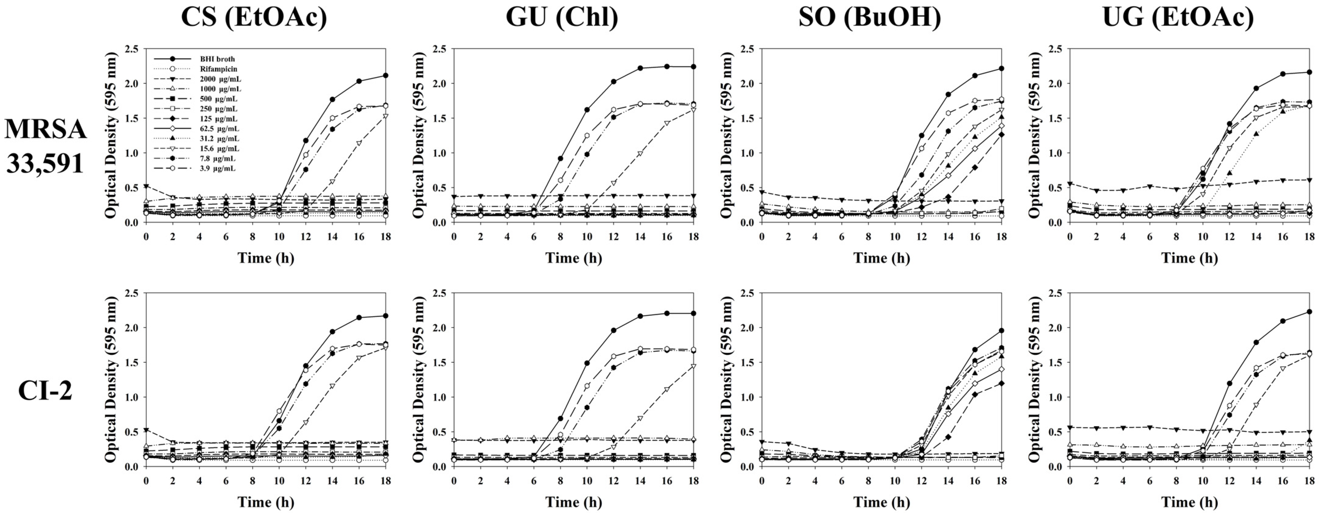
2. Results and Discussion
3. Materials and Methods
3.1. Plant materials
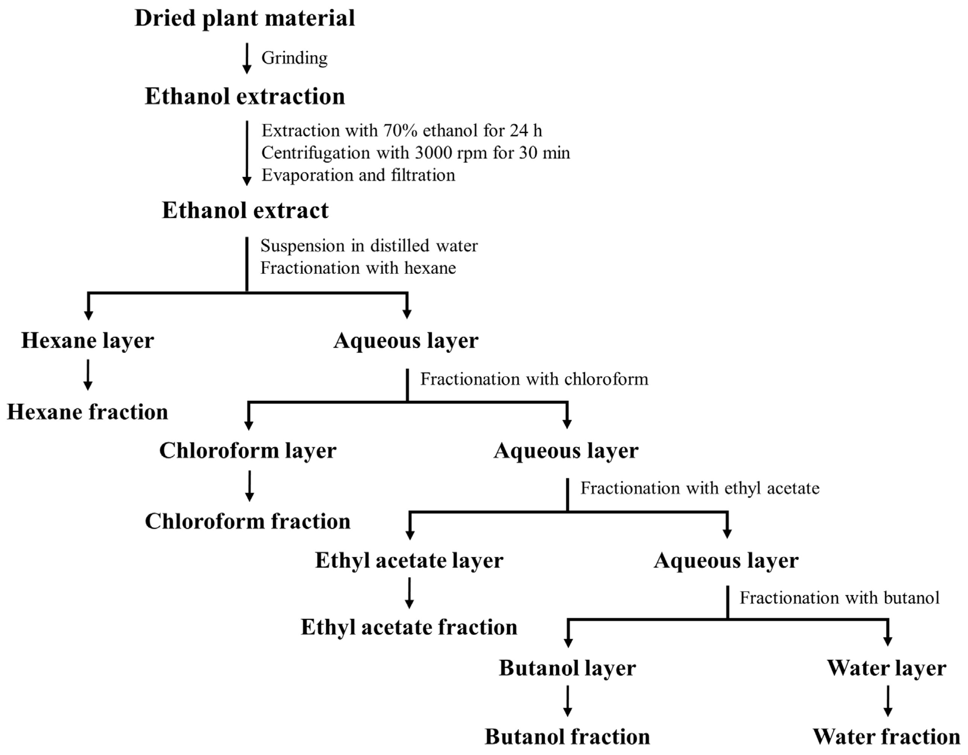
3.2. Preparation and Fractionation of Plant Extracts
3.3. Bacteria Culture
3.4. Antibiotic Susceptibility Testing
3.5. Disc Diffusion Assay
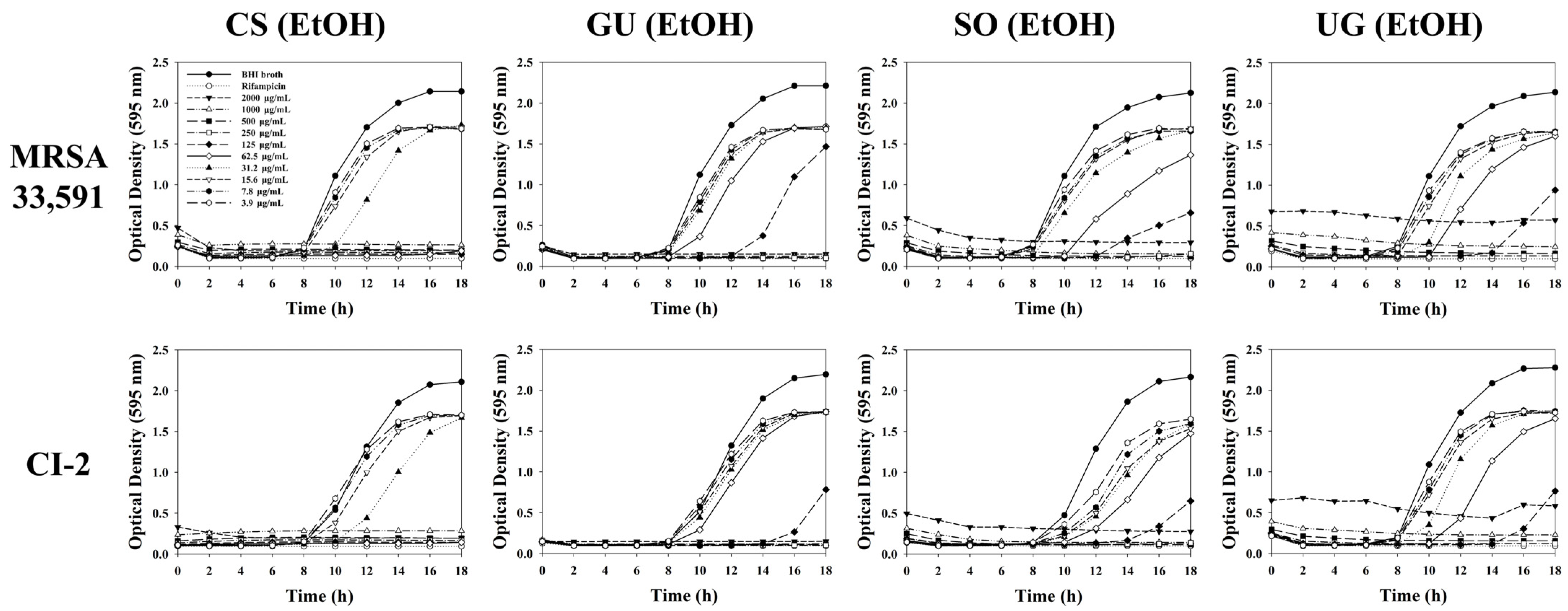
3.6. Determination of Minimum Inhibitory Concentration (MIC)
3.7. Determination of Minimum Bactericidal Concentration (MBC)
3.8. Cell Culture
3.9. Cell Viability Assay
3.10. Statistical Analysis
4. Conclusions
Supplementary Materials
Author Contributions
Funding
Institutional Review Board Statement
Informed Consent Statement
Data Availability Statement
Conflicts of Interest
References
- Mun, Y.S.; Hwang, Y.J. Novel spa and Multi-Locus Sequence Types (MLST) of Staphylococcus aureus Samples Isolated from Clinical Specimens in Korean. Antibiotics 2019, 8, 202. [Google Scholar] [CrossRef] [Green Version]
- Garoy, E.Y.; Gebreab, Y.B.; Achila, O.O.; Tekeste, D.G.; Kesete, R.; Ghirmay, R.; Kiflay, R.; Tesfu, T. Methicillin-Resistant Staphylococcus aureus (MRSA): Prevalence and Antimicrobial Sensitivity Pattern among Patients—A Multicenter Study in Asmara, Eritrea. Can. J. Infect. Dis. Med. Microbiol. 2019, 2019, 8321834. [Google Scholar] [CrossRef] [PubMed] [Green Version]
- Li, Y.; Lee, Y.; Seo, Y.; Hwang, Y. Relationship of multidrug-resistant gene and extended-spectrum carbapenem-resistance in Staphylococcus aureus. BIOCELL 2019, 43, 263–269. [Google Scholar] [CrossRef] [Green Version]
- Kim, H.J.; Choi, Q.; Kwon, G.C.; Koo, S.H. Molecular epidemiology and virulence factors of methicillin-resistant Staphylococcus aureus isolated from patients with bacteremia. J. Clin. Lab. Anal. 2020, 34, e23077. [Google Scholar] [CrossRef] [PubMed] [Green Version]
- Mendes, R.E.; Mendoza, M.; Singh, K.K.B.; Castanheira, M.; Bell, J.M.; Turnidge, J.D.; Lin, S.S.F.; Jones, R.N. Regional Resistance Surveillance Program Results for 12 Asia-Pacific Nations (2011). Antimicrob. Agents Chemother. 2013, 57, 5721–5726. [Google Scholar] [CrossRef] [PubMed] [Green Version]
- Masimen, M.A.A.; Harun, N.A.; Maulidiani, M.; Ismail, W.I.W. Overcoming Methicillin-Resistance Staphylococcus aureus (MRSA) Using Antimicrobial Peptides-Silver Nanoparticles. Antibiotics 2022, 11, 951. [Google Scholar] [CrossRef]
- Uc-Cachón, A.H.; Dzul-Beh, A.D.J.; Palma-Pech, G.A.; Jiménez-Delgadillo, B.; Flores-Guido, J.S.; Gracida-Osorno, C.; Molina-Salinas, G.M. Antibacterial and antibiofilm activities of Mayan medicinal plants against Methicillin-susceptible and -resistant strains of Staphylococcus aureus. J. Ethnopharmacol. 2021, 279, 114369. [Google Scholar] [CrossRef]
- World Health Organization. Global Priority List of Antibiotic-Resistant Bacteria to Guide Research, Discovery, and Development of New Antibiotics. 2017. Available online: https://www.who.int/news/item/27-02-2017-who-publishes-list-of-bacteria-for-which-new-antibiotics-are-urgently-needed (accessed on 12 October 2022).
- Alibi, S.; Crespo, D.; Navas, J. Plant-Derivatives Small Molecules with Antibacterial Activity. Antibiotics 2021, 10, 231. [Google Scholar] [CrossRef]
- Archana, H.; Bose, V.G. Evaluation of phytoconstituents from selected medicinal plants and its synergistic antimicrobial activity. Chemosphere 2022, 287, 132276. [Google Scholar] [CrossRef]
- Panda, S.K.; Das, R.; Lavigne, R.; Luyten, W. Indian medicinal plant extracts to control multidrug-resistant S. aureus, including in biofilms. S. Afr. J. Bot. 2020, 128, 283–291. [Google Scholar] [CrossRef]
- George, B.; Abrahamse, H.; Hemmaragala, N.M. Caspase dependent apoptotic inhibition of melanoma and lung cancer cells by tropical Rubus extracts. Biomed. Pharmacother. 2016, 80, 193–199. [Google Scholar] [CrossRef]
- Duarte, A.E.; De Menezes, I.R.A.; Bezerra Morais Braga, M.F.; Leite, N.F.; Barros, L.M.; Waczuk, E.P.; Pessoa da Silva, M.A.; Boligon, A.; Teixeira Rocha, J.B.; Souza, D.O.; et al. Antimicrobial Activity and Modulatory Effect of Essential Oil from the Leaf of Rhaphiodon echinus (Nees & Mart) Schauer on Some Antimicrobial Drugs. Molecules 2016, 21, 743. [Google Scholar] [CrossRef] [Green Version]
- Guadie, A.; Dakone, D.; Unbushe, D.; Wang, A.; Xia, S. Antibacterial activity of selected medicinal plants used by traditional healers in Genta Meyche (Southern Ethiopia) for the treatment of gastrointestinal disorders. J. Herb. Med. 2020, 22, 100338. [Google Scholar] [CrossRef]
- Panda, S.K.; Mohanta, Y.K.; Padhi, L.; Park, Y.-H.; Mohanta, T.K.; Bae, H. Large Scale Screening of Ethnomedicinal Plants for Identification of Potential Antibacterial Compounds. Molecules 2016, 21, 293. [Google Scholar] [CrossRef] [PubMed]
- Famuyide, I.M.; Aro, A.O.; Fasina, F.O.; Eloff, J.N.; McGaw, L.J. Antibacterial activity and mode of action of acetone crude leaf extracts of under-investigated Syzygium and Eugenia (Myrtaceae) species on multidrug resistant porcine diarrhoeagenic Escherichia coli. BMC Vet. Res. 2019, 15, 162. [Google Scholar] [CrossRef] [Green Version]
- Obakiro, S.B.; Kiprop, A.; K’Owino, I.; Andima, M.; Owor, R.O.; Chacha, R.; Kigondu, E. Phytochemical, Cytotoxicity, and Antimycobacterial Activity Evaluation of Extracts and Compounds from the Stem Bark of Albizia coriaria Welw ex. Oliver. Evid.-Based Complement. Altern. Med. 2022, 2022, 7148511. [Google Scholar] [CrossRef]
- Foti, M.; Grasso, R.; Fisichella, V.; Mascetti, A.; Zafarana, M.A.; Colnaghi, M.; Grasso, M.; Spena, M.T. Analysis of Eurasian Stone curlew (Burhinus oedicnemus) microbial flora reveals the presence of multi-drug resistant pathogens in agro-pastoral areas of Sicily (Italy). Heliyon 2020, 6, e05401. [Google Scholar] [CrossRef]
- Bag, A.; Bhattacharyya, S.K.; Pal, N.K.; Chattopadhyay, R.R. In vitro antibacterial potential of Eugenia jambolana seed extracts against multidrug-resistant human bacterial pathogens. Microbiol. Res. 2012, 167, 352–357. [Google Scholar] [CrossRef]
- Álvarez-Martínez, F.; Barrajón-Catalán, E.; Herranz-López, M.; Micol, V. Antibacterial plant compounds, extracts and essential oils: An updated review on their effects and putative mechanisms of action. Phytomedicine 2021, 90, 153626. [Google Scholar] [CrossRef]
- De Zoysa, M.H.N.; Rathnayake, H.; Hewawasam, R.P.; Wijayaratne, W. Determination of In Vitro Antimicrobial Activity of Five Sri Lankan Medicinal Plants against Selected Human Pathogenic Bacteria. Int. J. Microbiol. 2019, 2019, 7431439. [Google Scholar] [CrossRef]
- Dirar, A.; Alsaadi, D.; Wada, M.; Mohamed, M.; Watanabe, T.; Devkota, H. Effects of extraction solvents on total phenolic and flavonoid contents and biological activities of extracts from Sudanese medicinal plants. S. Afr. J. Bot. 2019, 120, 261–267. [Google Scholar] [CrossRef]
- Arokiyaraj, S.; Bharanidharan, R.; Agastian, P.; Shin, H. Chemical composition, antioxidant activity and antibacterial mechanism of action from Marsilea minuta leaf hexane: Methanol extract. Chem. Cent. J. 2018, 12, 105–111. [Google Scholar] [CrossRef] [PubMed] [Green Version]
- Ren, L.; Hemar, Y.; Perera, C.O.; Lewis, G.; Krissansen, G.W.; Buchanan, P.K. Antibacterial and antioxidant activities of aqueous extracts of eight edible mushrooms. Bioact. Carbohydr. Diet. Fibre 2014, 3, 41–51. [Google Scholar] [CrossRef]
- Elshikh, M.; Ahmed, S.; Funston, S.; Dunlop, P.; McGaw, M.; Marchant, R.; Banat, I.M. Resazurin-based 96-well plate microdilution method for the determination of minimum inhibitory concentration of biosurfactants. Biotechnol. Lett. 2016, 38, 1015–1019. [Google Scholar] [CrossRef] [PubMed] [Green Version]
- Lee, J.W.; Ji, Y.J.; Yu, M.H.; Bo, M.H.; Seo, H.J.; Lee, S.P.; Lee, I.S. Antimicrobial effect and resistant regulation of Glycyrrhiza uralensis on methicillin-resistant Staphylococcus aureus. Nat. Prod. Res. 2009, 23, 101–111. [Google Scholar] [CrossRef]
- Pattananandecha, T.; Apichai, S.; Julsrigival, J.; Ogata, F.; Kawasaki, N.; Saenjum, C. Antibacterial Activity against Foodborne Pathogens and Inhibitory Effect on Anti-Inflammatory Mediators’ Production of Brazilin-Enriched Extract from Caesalpinia sappan Linn. Plants 2022, 11, 1698. [Google Scholar] [CrossRef]
- Mogana, R.; Adhikari, A.; Tzar, M.N.; Ramliza, R.; Wiart, C. Antibacterial activities of the extracts, fractions and isolated compounds from Canarium patentinervium Miq. against bacterial clinical isolates. BMC Complement. Med. Ther. 2020, 20, 55. [Google Scholar] [CrossRef] [Green Version]
- Rivas-Cáceres, R.R.; Stephano-Hornedo, J.L.; Lugo, J.; Vaca, R.; Del Aguila, P.; Yañez-Ocampo, G.; Mora-Herrera, M.E.; Díaz, L.M.C.; Cipriano-Salazar, M.; Alaba, P.A. Bactericidal effect of silver nanoparticles against propagation of Clavibacter michiganensis infection in Lycopersicon esculentum Mill. Microb. Pathog. 2018, 115, 358–362. [Google Scholar] [CrossRef]
- Nazir, S.; Rabbani, A.; Mehmood, K.; Maqbool, F.; Shah, G.M.; Khan, M.F.; Sajid, M. Antileishmanial activity and cytotoxicity of ZnO-based nano-formulations. Int. J. Nanomed. 2019, 14, 7809–7822. [Google Scholar] [CrossRef] [Green Version]
- Ramamoorthy, R.; Muthalagu, M.; Andra, S.; Ravichandran, B.; Narayanasamy, M. Investigation on antimicrobial, antioxidant and cytotoxicity properties of triple bark extract formulated using traditional medicinal plants. SN Appl. Sci. 2019, 1, 772. [Google Scholar] [CrossRef]
- Asong, J.A.; Amoo, S.O.; McGaw, L.J.; Nkadimeng, S.M.; Aremu, A.O.; Otang-Mbeng, W. Antimicrobial Activity, Antioxidant Potential, Cytotoxicity and Phytochemical Profiling of Four Plants Locally Used against Skin Diseases. Plants 2019, 8, 350. [Google Scholar] [CrossRef] [PubMed] [Green Version]
- Bauer, A.W.; Kirby, W.M.M.; Sherris, J.C.; Turck, M. Antibiotic susceptibility testing by a standardized single disk method. Am. J. Clin. Pathol. 1966, 36, 493–496. [Google Scholar] [CrossRef]
- Lee, Y.H.; Park, S.Y.; Hwang, Y.J.; Park, J.K. Molecular Weight Determination of Chitosan with Antibacterial Activity Using Matrix-Assisted Laser Desorption/Ionization-Time of Flight Mass Spectrometry Analysis. Macromol. Res. 2022, 30, 90–98. [Google Scholar] [CrossRef]
- Bostanci, M.T.; Bulbul, A.S.; Celik, I.S.; Kocabas, Y.Z.; Burhan, H.; Bayat, R.; Sen, F.; Zakariae, N.; Esmaeili, R.; Jafari, H.; et al. Investigation of antibacterial, antifungal, antibiofilm, antioxidant and anticancer properties of methanol extracts of Salvia marashica Ilcim, Celep & Dogan and Salvia caespitosa Montbret & Aucher ex Benth plants with medicinal importance. Chemosphere 2022, 288 Pt 2, 132602. [Google Scholar] [PubMed]
- Ndlovu, B.; De Kock, M.; Klaasen, J.; Rahiman, F. In Vitro Comparison of the Anti-Proliferative Effects of Galenia africana on Human Skin Cell Lines. Sci. Pharm. 2021, 89, 12. [Google Scholar] [CrossRef]


| Scientific Name | Common Name | Family | Parts Used | Origin | Extraction Yield (%) |
|---|---|---|---|---|---|
| Areca catechu L. | Areca nut | Arecaceae | Seeds | Indonesia | 17.62 |
| Caesalpinia sappan L. | Sappan wood | Leguminosae | Heartwoods | Indonesia | 10.47 |
| Curcuma aromatica Salisb. | Wild turmeric | Zingiberaceae | Roots | Indonesia | 19.65 |
| Cinnamomum loureiroi Nees. | Saigon cinnamon | Lauraceae | Barks | Vietnam | 11.48 |
| Euphorbia humifusa Wild. | Creeping euphorbia | Euphorbiaceae | Leaves and stems | Korea | 31.22 |
| Glechoma grandis (A. Gray) Kuprian. | Ground ivy | Lamiaceae | Leaves and stems | Korea | 35.16 |
| Glycyrrhiza uralensis Fisch. | Chinese liquorice | Fabaceae | Roots | China | 20.95 |
| Lonicera japonica Thunb. | Japanese honeysucke | Caprifoliaceae | Floral buds | China | 38.63 |
| Morus alba L. | White mulberry | Moraceae | Leaves | Korea | 20.25 |
| Phellinus linteus | Sanghuang | Hymenochaetaceae | Fruit bodies | China | 3.80 |
| Polygonum tinctorium Ait. | Chinese indigo | Polygonaceae | Leaves | China | 9.05 |
| Quercus salicina Blume | Japanese willow leaf oak | Fagaceae | Leaves | Korea | 31.92 |
| Sanguisorba officinalis L. | Greater burnet | Rosaceae | Roots | China | 23.84 |
| Scutellaria baicalensis Georgi. | Baikal skullcap | Lamiaceae | Roots | Korea | 62.37 |
| Sophora flavescens Ait. | shrubby sophora | Leguminosae | Roots | Korea | 29.70 |
| Uncaria gambir Roxb. | Gambir | Rubiaceae | Leaves and twigs | Indonesia | 61.84 |
| Strains | Phenotype | Antibiotic Resistance Pattern |
|---|---|---|
| S. aureus ATCC 29,213 | MSSA | - |
| MRSA ATCC 33,591 | MRSA | Amp, Pen, Kan, Eryth, Strep, Tet, Gen, Chlo, Meth |
| CI-2 * | MDRSA | Amp, Pen, Kan, Eryth, Strep, Tet, Gen, Meth |
| CI-21 * | MRSA | Amp, Pen, Kan, Strep, Gen, Meth |
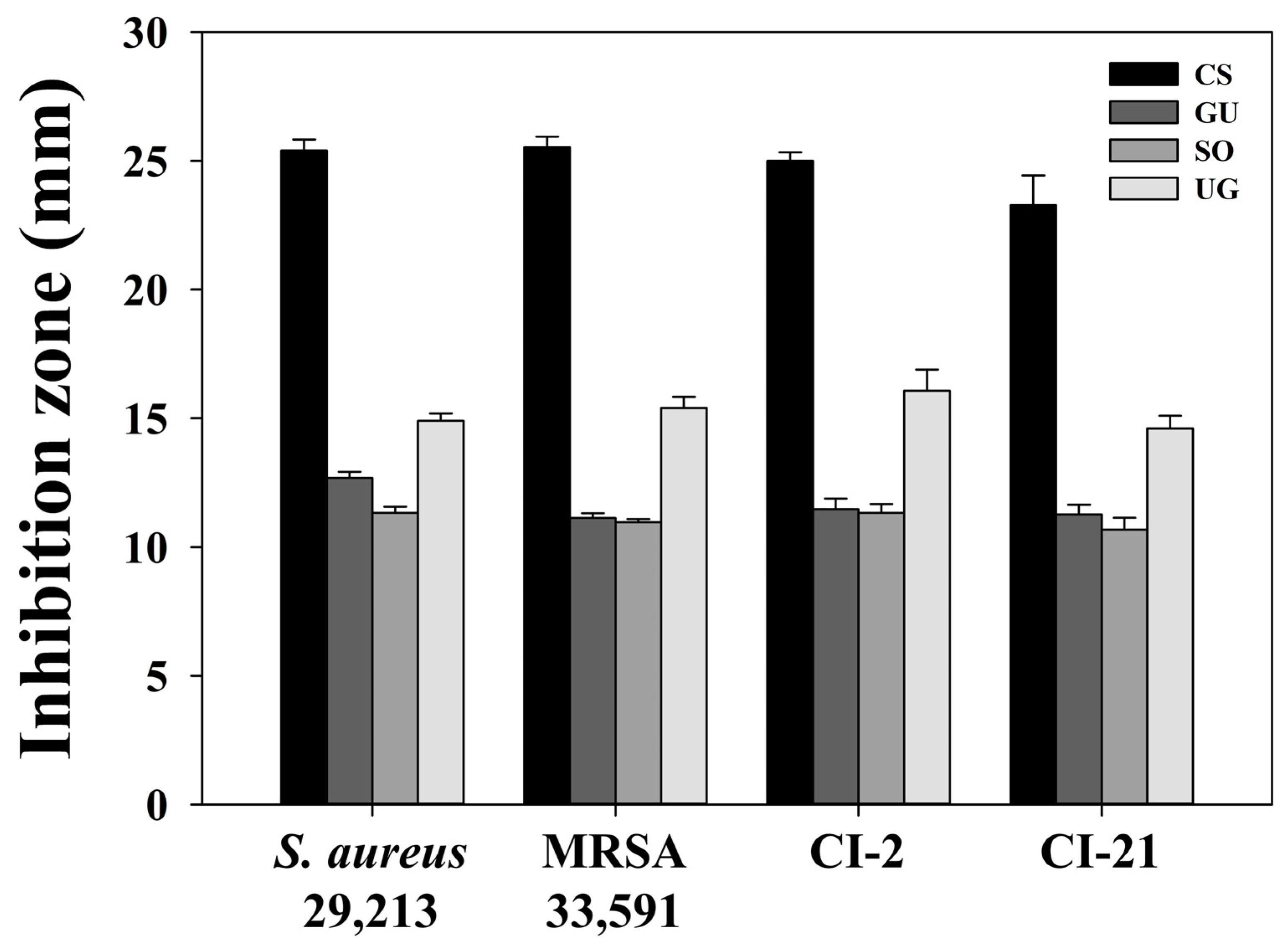
| Strains | Medicinal Plants | Diameter of the Clear Zone (mm) | ||||||
|---|---|---|---|---|---|---|---|---|
| Distilled Water | Rifampicin (30 μg) | Extract Concentration (mg/mL) | ||||||
| 40 | 20 | 10 | 5 | 2.5 | ||||
| S. aureus 29,213 | CS | - | 29.87 ± 0.09 | 25.40 ± 0.43 | 20.67 ± 0.24 | 17.27 ± 0.52 | 12.43 ± 0.33 | 9.77 ± 0.21 |
| GU | - | 29.87 ± 0.19 | 12.67 ± 0.24 | 10.00 ± 0.41 | 8.10 ± 0.08 | - | - | |
| SO | - | 30.00 ± 0.00 | 11.33 ± 0.24 | 8.80 ± 0.28 | - | - | - | |
| UG | - | 30.30 ± 0.51 | 14.90 ± 0.29 | 11.80 ± 0.24 | 9.17 ± 0.37 | - | - | |
| MRSA 33,591 | CS | - | 30.73 ± 0.57 | 25.53 ± 0.41 | 21.60 ± 0.43 | 18.07 ± 0.09 | 13.90 ± 0.65 | 10.73 ± 0.52 |
| GU | - | 30.67 ± 0.47 | 11.13 ± 0.19 | 9.73 ± 0.57 | 8.17 ± 0.09 | - | - | |
| SO | - | 30.67 ± 0.47 | 10.97 ± 0.12 | 10.20 ± 0.16 | - | - | - | |
| UG | - | 30.67 ± 0.62 | 15.40 ± 0.43 | 12.33 ± 0.47 | 9.13 ± 0.12 | - | - | |
| CI-2 | CS | - | 30.00 ± 0.00 | 25.00 ± 0.33 | 21.93 ± 0.74 | 18.07 ± 0.09 | 13.87 ± 0.41 | 11.13 ± 0.34 |
| GU | - | 29.67 ± 0.47 | 11.47 ± 0.41 | 9.60 ± 0.85 | 8.17 ± 0.08 | - | - | |
| SO | - | 29.67 ± 0.47 | 11.33 ± 0.34 | 9.87 ± 0.19 | - | - | - | |
| UG | - | 29.27 ± 0.90 | 16.07 ± 0.82 | 14.93 ± 0.82 | 10.00 ± 0.82 | - | - | |
| CI-21 | CS | - | 29.33 ± 0.47 | 23.27 ± 1.16 | 20.47 ± 0.34 | 17.50 ± 0.41 | 13.17 ± 0.24 | 9.87 ± 0.19 |
| GU | - | 29.83 ± 0.62 | 11.27 ± 0.38 | 9.60 ± 0.85 | 8.20 ± 0.16 | - | - | |
| SO | - | 29.33 ± 0.47 | 10.67 ± 0.47 | 9.67 ± 0.47 | - | - | - | |
| UG | - | 29.27 ± 0.90 | 14.60 ± 0.49 | 12.27 ± 0.38 | 9.93 ± 0.09 | - | - | |
| Strains | Medicinal Plants | Diameter of the Clear Zone (mm) | ||||||
|---|---|---|---|---|---|---|---|---|
| Distilled Water | Rifampicin (30 μg) | Fraction Concentration (10 mg/mL) | ||||||
| HEX | Chl | EtOAc | BuOH | Water | ||||
| S. aureus 29,213 | CS | - | 30.83 ± 0.24 | - | - | 23.23 ± 0.61 | 16.77 ± 0.95 | - |
| GU | - | 30.60 ± 0.43 | 8.67 ± 0.47 | 11.93 ± 0.33 | 10.70 ± 0.22 | - | - | |
| SO | - | 30.67 ± 0.47 | - | - | - | - | - | |
| UG | - | 30.43 ± 0.42 | - | - | 14.67 ± 1.09 | 10.00 ± 0.00 | - | |
| MRSA 33,591 | CS | - | 31.50 ± 0.41 | - | - | 23.10 ± 1.22 | 16.77 ± 0.21 | - |
| GU | - | 31.00 ± 0.00 | 8.33 ± 0.24 | 11.70 ± 0.79 | 10.93 ± 0.42 | - | - | |
| SO | - | 30.67 ± 0.47 | - | - | - | - | - | |
| UG | - | 30.83 ± 0.24 | - | - | 16.33 ± 1.09 | 9.93 ± 0.09 | - | |
| CI-2 | CS | - | 31.67 ± 0.24 | - | - | 23.83 ± 1.10 | 17.77 ± 0.92 | - |
| GU | - | 30.83 ± 0.24 | 8.67 ± 0.47 | 11.37 ± 0.49 | 10.57 ± 0.49 | - | - | |
| SO | - | 30.93 ± 0.09 | - | - | - | - | - | |
| UG | - | 31.43 ± 0.33 | - | - | 14.43 ± 0.66 | 10.27 ± 0.21 | - | |
| CI-21 | CS | - | 31.83 ± 0.24 | - | - | 23.33 ± 0.50 | 16.40 ± 0.83 | - |
| GU | - | 30.90 ± 0.29 | 8.77 ± 0.21 | 10.93 ± 0.09 | 10.23 ± 0.21 | - | - | |
| SO | - | 31.00 ± 0.00 | - | - | - | - | - | |
| UG | - | 31.33 ± 0.24 | - | - | 14.83 ± 1.68 | 9.83 ± 0.29 | - | |
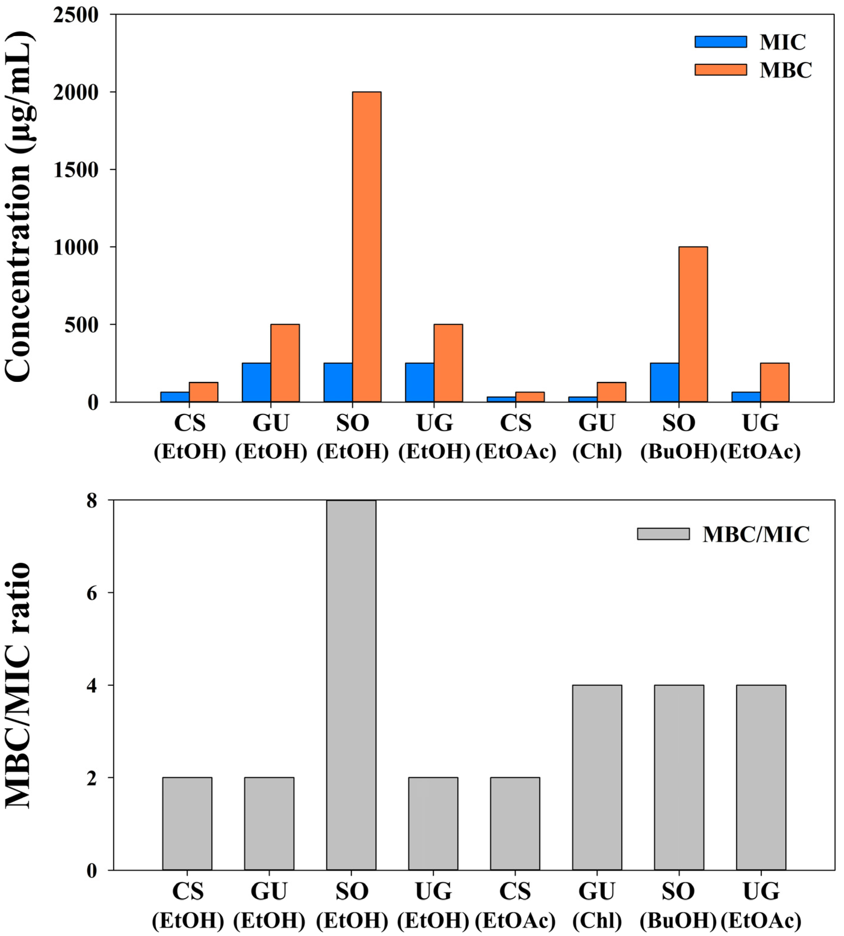
| Medicinal Plants | Extracts | Fractions | S. aureus 29,213 | MRSA 33,591 | CI-2 | CI-21 | ||||||||
|---|---|---|---|---|---|---|---|---|---|---|---|---|---|---|
| MIC | MBC | MBC/MIC | MIC | MBC | MBC/MIC | MIC | MBC | MBC/MIC | MIC | MBC | MBC/MIC | |||
| CS | EtOH | 62.5 | 125 | 2 | 62.5 | 125 | 2 | 62.5 | 125 | 2 | 62.5 | 125 | 2 | |
| HEX | 2000 | >2000 | ND | 2000 | >2000 | ND | 2000 | >2000 | ND | 2000 | >2000 | ND | ||
| Chl | 1000 | 2000 | 2 | 1000 | 2000 | 2 | 1000 | 2000 | 2 | 1000 | 1000 | 1 | ||
| EtOAc | 31.2 | 62.5 | 2 | 31.2 | 62.5 | 2 | 31.2 | 62.5 | 2 | 31.2 | 62.5 | 2 | ||
| BuOH | 62.5 | 125 | 2 | 125 | 125 | 1 | 125 | 125 | 1 | 125 | 125 | 1 | ||
| Water | 1000 | 2000 | 2 | 1000 | 2000 | 2 | 1000 | 2000 | 2 | 1000 | 2000 | 2 | ||
| GU | EtOH | 250 | 500 | 2 | 250 | 500 | 2 | 250 | 500 | 2 | 250 | 500 | 2 | |
| HEX | 62.5 | 250 | 4 | 62.5 | 250 | 4 | 62.5 | 250 | 4 | 62.5 | 250 | 4 | ||
| Chl | 31.2 | 125 | 4 | 31.2 | 125 | 4 | 31.2 | 125 | 4 | 31.2 | 125 | 4 | ||
| EtOAc | 125 | 500 | 4 | 125 | 500 | 4 | 125 | 500 | 4 | 125 | 500 | 4 | ||
| BuOH | >2000 | ND | ND | >2000 | ND | ND | >2000 | ND | ND | >2000 | ND | ND | ||
| Water | >2000 | ND | ND | >2000 | ND | ND | >2000 | ND | ND | >2000 | ND | ND | ||
| SO | EtOH | 250 | 2000 | >4 | 250 | 2000 | >4 | 250 | 2000 | >4 | 250 | 2000 | >4 | |
| HEX | 1000 | >2000 | ND | 1000 | >2000 | ND | 1000 | >2000 | ND | 1000 | >2000 | ND | ||
| Chl | 1000 | >2000 | ND | 1000 | >2000 | ND | 1000 | >2000 | ND | 1000 | >2000 | ND | ||
| EtOAc | 500 | 1000 | 2 | 500 | 2000 | 4 | 500 | 2000 | 4 | 500 | 2000 | 4 | ||
| BuOH | 250 | 1000 | 4 | 250 | 1000 | 4 | 250 | 1000 | 4 | 250 | 1000 | 4 | ||
| Water | 250 | 2000 | >4 | 250 | 2000 | >4 | 250 | 2000 | >4 | 250 | 2000 | >4 | ||
| UG | EtOH | 250 | 500 | 2 | 250 | 500 | 2 | 250 | 500 | 2 | 250 | 500 | 2 | |
| HEX | >2000 | ND | ND | >2000 | ND | ND | >2000 | ND | ND | >2000 | ND | ND | ||
| Chl | 1000 | 1000 | 1 | 1000 | 2000 | 2 | 1000 | 2000 | 2 | 1000 | 2000 | 2 | ||
| EtOAc | 62.5 | 125 | 2 | 62.5 | 250 | 4 | 62.5 | 250 | 4 | 62.5 | 250 | 4 | ||
| BuOH | 125 | 250 | 2 | 125 | 250 | 2 | 125 | 250 | 2 | 125 | 250 | 2 | ||
| Water | 250 | 1000 | 4 | 250 | 1000 | 4 | 250 | 1000 | 4 | 250 | 1000 | 4 | ||
| Medicinal Plants | Extracts/Fractions | Cell Viability (%) | |
|---|---|---|---|
| HepG2 | A549 | ||
| CS | EtOH | 90.15 ± 0.48 | 91.94 ± 1.24 |
| EtOAc | 90.75 ± 0.43 | 90.99 ± 0.50 | |
| GU | EtOH | 81.24 ± 0.58 | 83.05 ± 0.28 |
| Chl | 85.38 ± 2.62 | 87.94 ± 0.44 | |
| SO | EtOH | 81.30 ± 0.52 | 81.92 ± 1.07 |
| BuOH | 79.71 ± 0.30 | 80.64 ± 0.21 | |
| UG | EtOH | 82.05 ± 0.56 | 83.43 ± 0.57 |
| EtOAc | 85.45 ± 0.26 | 85.14 ± 0.29 | |
Publisher’s Note: MDPI stays neutral with regard to jurisdictional claims in published maps and institutional affiliations. |
© 2022 by the authors. Licensee MDPI, Basel, Switzerland. This article is an open access article distributed under the terms and conditions of the Creative Commons Attribution (CC BY) license (https://creativecommons.org/licenses/by/4.0/).
Share and Cite
Jung, I.-G.; Jeong, J.-Y.; Yum, S.-H.; Hwang, Y.-J. Inhibitory Effects of Selected Medicinal Plants on Bacterial Growth of Methicillin-Resistant Staphylococcus aureus. Molecules 2022, 27, 7780. https://doi.org/10.3390/molecules27227780
Jung I-G, Jeong J-Y, Yum S-H, Hwang Y-J. Inhibitory Effects of Selected Medicinal Plants on Bacterial Growth of Methicillin-Resistant Staphylococcus aureus. Molecules. 2022; 27(22):7780. https://doi.org/10.3390/molecules27227780
Chicago/Turabian StyleJung, In-Geun, Jae-Young Jeong, Seung-Hoon Yum, and You-Jin Hwang. 2022. "Inhibitory Effects of Selected Medicinal Plants on Bacterial Growth of Methicillin-Resistant Staphylococcus aureus" Molecules 27, no. 22: 7780. https://doi.org/10.3390/molecules27227780