
Unexpected Room Temperature Ferromagnetism of a Ball-Milled Graphene Oxide—Melamine Mixture
 ,
,  , , , and
Abstract
:1. Introduction
2. Experimental
2.1. Synthesis of Nitrogen-Doped Carbon Material
2.2. Characterization
3. Results
3.1. Elemental Analysis and SEM
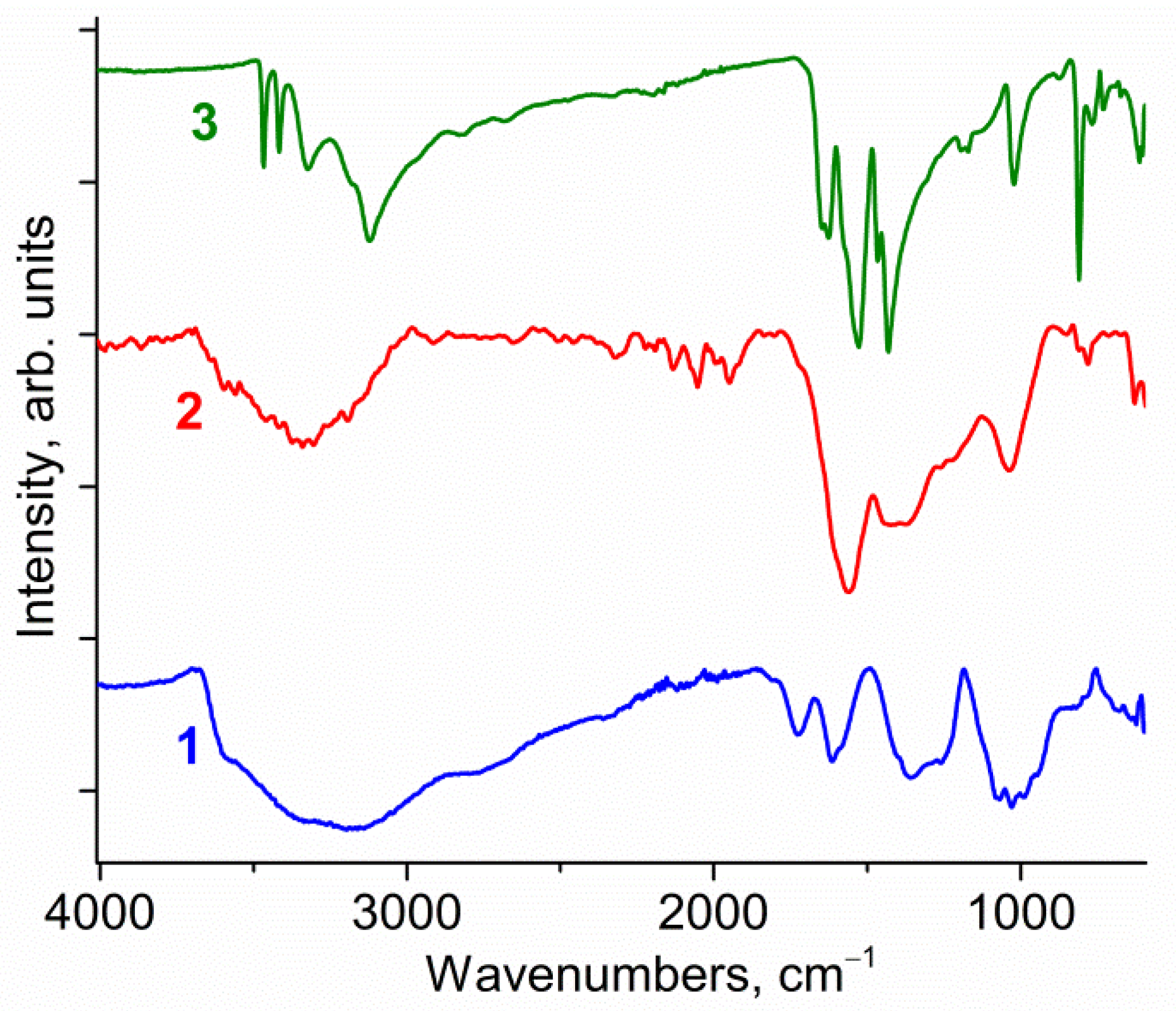
3.2. IR Spectra
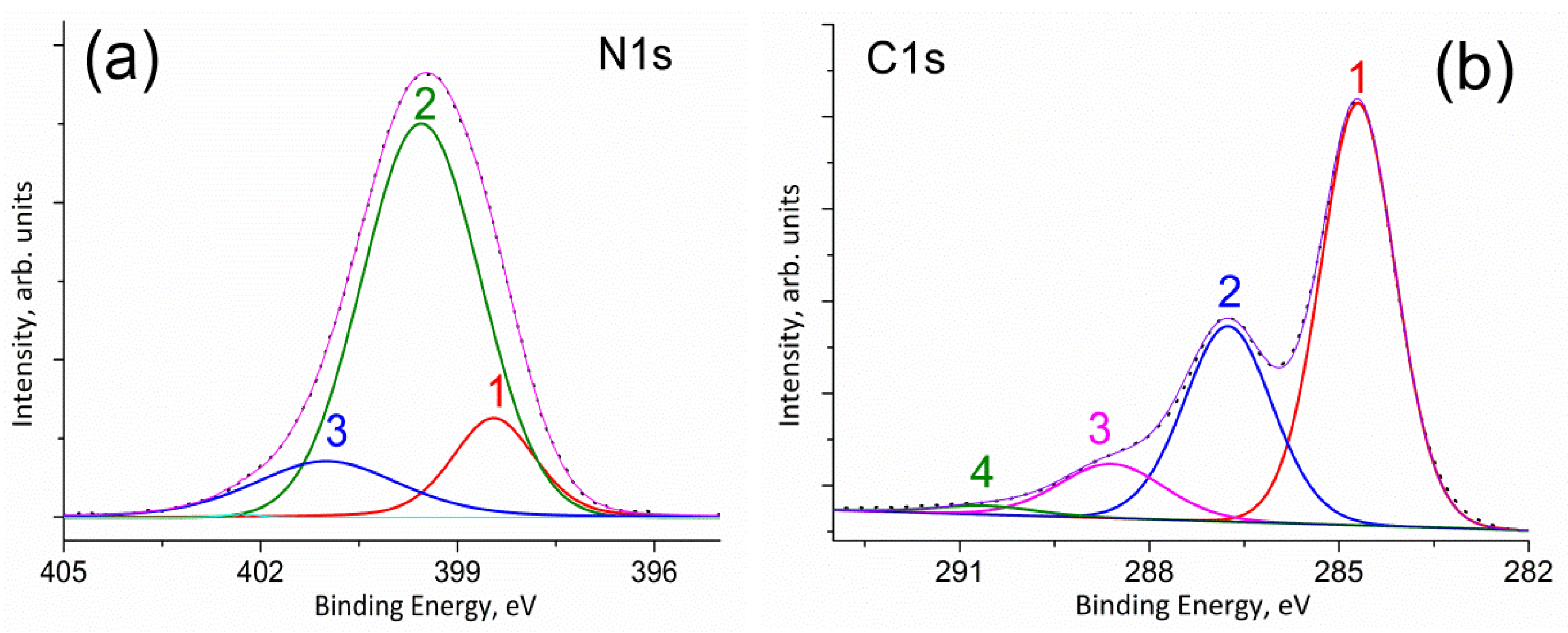
3.3. XPS Spectra
3.4. ESR Spectra
3.5. Magnetic Measurements
4. Discussion
5. Conclusions
Author Contributions
Funding
Institutional Review Board Statement
Informed Consent Statement
Data Availability Statement
Acknowledgments
Conflicts of Interest
Sample Availability
References
- Korshak, Y.V.; Medvedeva, T.V.; Ovchinnikov, A.A.; Spector, V.N. Organic polymer ferromagnet. Nature 1987, 326, 370–372. [Google Scholar] [CrossRef]
- Ovchinnikov, A.A. Multiplicity of the ground state of large alternant organic molecules with conjugated bonds. Theor. Chim. Acta 1978, 47, 297–304. [Google Scholar] [CrossRef]
- Makarova, T.L.; Sundqvist, B.; Höhne, R.; Esquinazi, P.; Kopelevich, Y.; Scharff, P.; Davydov, V.A.; Kashevarova, L.S.; Rakhmanina, A.V. Magnetic carbon. Nature 2001, 413, 716–718. [Google Scholar] [CrossRef] [PubMed]
- Makarova, T.L.; Sundqvist, B.; Höhne, R.; Esquinazi, P.; Kopelevich, Y.; Scharff, P.; Davydov, V.; Kashevarova, L.S.; Rakhmanina, A.V. Retraction Note: Magnetic carbon. Nature 2006, 440, 707. [Google Scholar] [CrossRef] [Green Version]
- Coey, J.M.D. d0 ferromagnetism. Solid State Sci. 2005, 7, 660–667. [Google Scholar] [CrossRef]
- Coey, J.M.D.; Stamenov, P.; Gunning, R.D.; Venkatesan, M.; Paul, K. Ferromagnetism in defect-ridden oxides and related materials. New J. Phys. 2010, 12, 053025. [Google Scholar] [CrossRef]
- Ghosh, S.; Khan, G.G.; Mandal, K. d0 ferromagnetism in oxide nanowires: Role of intrinsic defects. EPJ Web Conf. 2013, 40, 03001. [Google Scholar] [CrossRef] [Green Version]
- Qi, B.; Ólafsson, S.; Gíslason, H.P. Vacancy defect-induced d0 ferromagnetism in undoped ZnO nanostructures: Controversial origin and challenges. Prog. Mater. Sci. 2017, 90, 45–74. [Google Scholar] [CrossRef]
- Garg, S.; Gautam, S.; Singh, J.P.; Kandasami, A.; Goya, N. Characterizing the defects and ferromagnetism in metal oxides: The case of magnesium oxide. Mater. Charact. 2021, 179, 111366. [Google Scholar] [CrossRef]
- Chouhan, L.; Bouzerar, G.; Srivastava, S.K. d0 ferromagnetism in Li-doped ZnO compounds. J. Mater. Sci. Mater. Electron. 2021, 32, 6389–6397. [Google Scholar] [CrossRef]
- Chouhan, L.; Srivastava, S.K. A comprehensive review on recent advancements in d0 ferromagnetic oxide materials. Mater. Sci. Semicond. Process. 2022, 147, 106768. [Google Scholar] [CrossRef]
- Chouhan, L.; Bouzerar, G.; Srivastava, S.K. Effect of Mg-doping in tailoring d0 ferromagnetism of rutile TiO2 compounds for spintronics application. J. Mater. Sci. Mater. Electron. 2021, 32, 11193–11201. [Google Scholar] [CrossRef]
- Rani, N.; Chahal, S.; Kumar, P.; Kumar, A.; Shukla, R.; Singh, S.K. MgO nanostructures at different annealing temperatures for d0 ferromagnetism. Vacuum 2020, 179, 109539. [Google Scholar] [CrossRef]
- Chouhan, L.; Narzary, R.; Dey, B.; Panda, S.K.; Manglam, M.K.; Roy, L.; Brahma, R.; Mondal, A.; Kar, M.; Ravi, S.; et al. Tailoring room temperature d0 ferromagnetism, dielectric, optical, and transport properties in Ag-doped rutile TiO2 compounds for spintronics applications. J. Mater. Sci. Mater. Electron. 2021, 32, 28163–28175. [Google Scholar] [CrossRef]
- Chouhan, L.; Srivastava, S.K. Observation of room temperature d0 ferromagnetism, band-gap widening, zero dielectric loss and conductivity enhancement in Mg doped TiO2 (rutile + anatase) compounds for spintronics applications. J. Solid State Chem. 2022, 307, 122828. [Google Scholar] [CrossRef]
- Chouhan, L.; Panda, S.K.; Bhattacharjee, S.; Das, B.; Mondal, A.; Parida, B.N.; Brahma, R.; Manglam, M.K.; Kar, M.; Bouzerar, G.; et al. Room temperature d0 ferromagnetism, zero dielectric loss and ac-conductivity enhancement in p-type Ag-doped SnO2 compounds. J. Alloys Compd. 2021, 870, 159515. [Google Scholar] [CrossRef]
- Narzary, R.; Dey, B.; Chouhan, L.; Kumar, S.; Ravi, S.; Srivastava, S.K. Optical band gap tuning, zero dielectric loss and room temperature ferromagnetism in (Ag/Mg) co-doped SnO2 compounds for spintronics applications. Mater. Sci. Semicond. Process. 2022, 147, 106477. [Google Scholar] [CrossRef]
- Beltrán, J.I.; Monty, C.; Balcells, L.; Martínez-Boubeta, C. Possible d0 ferromagnetism in MgO. Solid State Commun. 2009, 149, 1654–1657. [Google Scholar] [CrossRef]
- Graf, T.; Goennenwein, S.T.B.; Brandt, M.S. Prospects for carrier-mediated ferromagnetism in GaN. Phys. Status Solidi (B) 2003, 239, 277–290. [Google Scholar] [CrossRef]
- Morozov, I.G.; Belousova, O.V.; Belyakov, O.A.; Parkin, I.P.; Sathasivam, S.; Kuznetcov, M.V. Titanium nitride room-temperature ferromagnetic nanoparticles. J. Alloys Compd. 2016, 276, 266–276. [Google Scholar] [CrossRef]
- Xu, M.; Liang, T.; Shi, M.; Chen, H. Graphene-like two-dimensional materials. Chem. Rev. 2013, 113, 3766–3798. [Google Scholar] [CrossRef] [PubMed]
- Si, C.; Zhou, J.; Sun, Z. Half-metallic ferromagnetism and surface functionalization-induced metal–insulator transition in graphene-like two-dimensional Cr2C crystals. ACS Appl. Mater. Interfaces 2015, 7, 17510–17515. [Google Scholar] [CrossRef] [PubMed]
- Feng, P.; Zhang, S.; Liu, D.; Gao, M.; Ma, F.; Yan, X.-W.; Xie, Z.Y. Achieving high-temperature ferromagnetism by means of magnetic ion dimerization in the graphene-like Mn2N6C6 monolayer. J. Phys. Chem. C 2022, 126, 10139–10144. [Google Scholar] [CrossRef]
- Zhang, H.; Liao, Z.; Xia, B.; Herng, T.S.; Ding, J.; Gao, D. Ferromagnetism of Mn-N4 architecture embedded graphene. J. Phys. D Appl. Phys. 2022, 55, 225001. [Google Scholar] [CrossRef]
- Wen, J.Q.; Tong, X.; Lei, Y.T.; Tian, P.H.; Wu, H.; He, W.L. Ferromagnetism induced by C doped graphene-like ZnO films with different concentrations. Solid State Commun. 2019, 299, 11366. [Google Scholar] [CrossRef]
- Wen, J.; Lin, P.; Han, Y.; Li, N.; Chen, G.; Bai, L.; Guo, S.; Wu, H.; He, W.; Zhang, J. Insights into enhanced ferromagnetic activity of P doping graphene-ZnO monolayer with point defects. Mater. Chem. Phys. 2021, 270, 124855. [Google Scholar] [CrossRef]
- Bafekry, A. Graphene-like BC6N single-layer: Tunable electronic and magnetic properties via thickness, gating, topological defects, and adatom/molecule. Phys. E Low-Dimens. Syst. Nanostruct. 2020, 118, 113850. [Google Scholar] [CrossRef]
- Han, K.H.; Spemann, D.; Esquinazi, P.; Hohne, R.; Riede, V.; Butz, T. Ferromagnetic spots in graphite produced by proton irradiation. Adv. Mater. 2003, 15, 1719–1722. [Google Scholar] [CrossRef]
- Esquinazi, P.; Spemann, D.; Hohne, R.; Setzer, A.; Han, K.H.; Butz, T. Induced magnetic ordering by proton irradiation in graphite. Phys. Rev. Lett. 2003, 91, 227201. [Google Scholar] [CrossRef] [Green Version]
- Rode, A.V.; Gamaly, A.G.; Christy, J.G.; Fitz Gerald, S.T.; Hyde, R.G.; Elliman, B.; Luther-Davies, A.I.; Veinger, E.G.; Androulakis, J.; Giapintzakis, J. Unconventional magnetism in all-carbon nanoform. Phys. Rev. B Condens. Matter Mater. Phys. 2004, 70, 054407. [Google Scholar] [CrossRef]
- Castro, E.V.; Peres, N.M.R.; Stauber, T.; Silva, N.A.P. Low-density ferromagnetism in biased bilayer graphene. Phys. Rev. Lett. 2008, 100, 186803. [Google Scholar] [CrossRef] [PubMed] [Green Version]
- Antonov, V.E.; Bashkin, I.O.; Khasanov, S.S.; Moravsky, A.P.; Morozov, Y.G.; Shulga, Y.M.; Ossipiyan, Y.A.; Ponyatovsky, E.G. Magnetic ordering in hydrofullerene C60H24. J. Alloys Compd. 2002, 330–332, 365–368. [Google Scholar] [CrossRef]
- Zhou, J.; Wang, Q.; Sun, Q.; Chen, X.S.; Kawazoe, Y.; Jena, P. Ferromagnetism in semihydrogenated graphene sheet. Nano Lett. 2009, 9, 3867–3870. [Google Scholar] [CrossRef] [PubMed]
- Wang, Y.; Huang, Y.; Song, Y.; Zhang, X.; Ma, Y.; Liang, J. Room-temperature ferromagnetism of graphene. Nano Lett. 2009, 9, 220–224. [Google Scholar] [CrossRef] [PubMed]
- Qin, S.; Guo, X.; Cao, Y.; Ni, Z.; Xu, Q. Strong ferromagnetism of reduced graphene oxide. Carbon 2014, 78, 559–565. [Google Scholar] [CrossRef]
- Sinha, A.; Ali, A.; Thakur, A.D. Ferromagnetism in graphene oxide. Mater. Today Proc. 2021, 46, 6230–6233. [Google Scholar] [CrossRef]
- Błoński, P.; Tucek, J.; Sofer, Z.; Mazanek, V.; Petr, M.; Pumera, M.; Otyepka, M.; Zbořil, R. Doping with graphitic nitrogen triggers ferromagnetism in graphene. J. Am. Chem. Soc. 2017, 139, 3171–3180. [Google Scholar] [CrossRef] [Green Version]
- Ning, G.; Xu, C.; Hao, L.; Kazakova, O.; Fan, Z.; Wang, H. Ferromagnetism in nanomesh graphene. Carbon 2013, 51, 390–396. [Google Scholar] [CrossRef]
- Sarkar, S.K.; Raul, K.K.; Pradhan, S.S.; Basu, S.; Nayak, A. Magnetic properties of graphite oxide and reduced graphene oxide. Phys. E Low-Dimens. Syst. Nanostruct. 2014, 64, 78–82. [Google Scholar] [CrossRef]
- Liu, Y.; Tang, N.; Wan, X.; Feng, Q.; Li, M.; Xu, Q.; Liu, F.; Du, Y. Realization of ferromagnetic graphene oxide with high magnetization by doping graphene oxide with nitrogen. Sci. Rep. 2013, 3, 2566. [Google Scholar] [CrossRef]
- Qin, S.; Xu, Q. Room temperature ferromagnetism in N2 plasma treated graphene oxide. J. Alloys Compd. 2017, 692, 332–338. [Google Scholar] [CrossRef]
- Khurana, G.; Kumar, N.; Kotnala, R.K.; Nautiyal, T.; Katiyar, R.S. Temperature tuned defect induced magnetism in reduced graphene oxide. Nanoscale 2013, 5, 3346–3351. [Google Scholar] [CrossRef] [PubMed]
- Raj, K.G.; Joy, P.A. Ferromagnetism at room temperature in activated graphene oxide. Chem. Phys. Lett. 2014, 605–606, 89–92. [Google Scholar] [CrossRef]
- Rao, C.N.R.; Ramakrishna Matte, H.S.S.; Subrahmanyam, K.S. Synthesis and selected properties of graphene and graphene mimics. Acc. Chem. Res. 2013, 46, 149–159. [Google Scholar] [CrossRef] [PubMed]
- Bagani, K.; Ray, M.K.; Satpati, B.; Ray, N.R.; Sardar, M.; Banerjee, S. Contrasting magnetic properties of thermally and chemically reduced graphene oxide. J. Phys. Chem. C 2014, 118, 13254–13259. [Google Scholar] [CrossRef]
- Lee, D.; Seo, J.; Zhu, X.; Cole, J.M.; Su, H. Magnetism in graphene oxide induced by epoxy groups. Appl. Phys. Lett. 2015, 106, 172402. [Google Scholar] [CrossRef]
- Kim, S.W.; Kim, H.K.; Lee, K.; Roh, K.C.; Han, J.T.; Kim, K.B.; Lee, S.; Jung, M.-H. Studying reduction of graphene oxide with magnetic measurements. Carbon 2019, 142, 373–378. [Google Scholar] [CrossRef]
- Fu, L.; Wang, Y.; Zhang, K.; Zhang, W.; Chen, J.; Deng, Y.; Du, Y.; Tang, N. Realization of ambient-stable room-temperature ferromagnetism by low-temperature annealing of graphene oxide nanoribbons. ACS Nano 2019, 13, 6341–6347. [Google Scholar] [CrossRef]
- Nair, R.R.; Sepioni, M.; Tsai, I.-L.; Lehtinen, O.; Keinonen, J.; Krasheninnikov, A.V.; Thomson, T.; Geim, A.K.; Grigorieva, I.V. Spin-half paramagnetism in graphene induced by point defects. Nat. Phys. 2012, 8, 199–202. [Google Scholar] [CrossRef] [Green Version]
- Zhang, X.; Li, G.; Li, Q.; Shaikh, M.S.; Li, Z. The pure paramagnetism in graphene oxide. Results Phys. 2021, 26, 104407. [Google Scholar] [CrossRef]
- Vasiliev, V.P.; Manzhos, R.A.; Krivenko, A.G.; Kabachkov, E.N.; Shulga, Y.M. Nitrogen-enriched carbon powder prepared by ball-milling of graphene oxide with melamine: An efficient electrocatalyst for oxygen reduction reaction. Mendeleev Commun. 2021, 31, 529–531. [Google Scholar] [CrossRef]
- Vasiliev, V.P.; Manzhos, R.A.; Kochergin, V.K.; Krivenko, A.G.; Kabachkova, E.N.; Kulikov, A.V.; Shulga, Y.M.; Gutsev, G.L. A Facile synthesis of noble-metal-free catalyst based on nitrogen doped graphene oxide for oxygen reduction reaction. Materials 2022, 15, 821. [Google Scholar] [CrossRef] [PubMed]
- Shulga, Y.M.; Baskakov, S.A.; Smirnov, V.A.; Shulga, N.Y.; Belay, K.G.; Gutsev, G.L. Graphene oxide films as separators of polyaniline-based supercapacitors. J. Power Sources 2014, 245, 33–36. [Google Scholar] [CrossRef]
- Stylianakis, M.M.; Spyropoulos, G.D.; Stratakis, E.; Kymakis, E. Solution-processable graphene linked to 3,5-dinitrobenzoyl as an electron acceptor in organic bulk heterojunction photovoltaic devices. Carbon 2012, 50, 5554–5561. [Google Scholar] [CrossRef]
- Shahriary, L.; Athawale, A. Graphene oxide synthesized by using modified hummers approach. Int. J. Renew. Energy Environ. Eng. 2014, 2, 58–63. [Google Scholar]
- Saleem, H.; Haneef, M.; Abbasi, H.Y. Synthesis route of reduced graphene oxide via thermal reduction of chemically exfoliated graphene oxide. Mater. Chem. Phys. 2018, 204, 1–7. [Google Scholar] [CrossRef]
- Ikram, R.; Jan, B.M.; Ahmad, W. An overview of industrial scalable production of graphene oxide and analytical approaches for synthesis and characterization. J. Mater. Res. Technol. 2020, 9, 11587–11610. [Google Scholar] [CrossRef]
- Meier, R.J.; Maple, J.R.; Hwang, M.-J.; Hagler, A.T. Molecular modeling urea- and melamine-formaldehyde resins. 1. A force field for urea and melamine. J. Phys. Chem. 1995, 99, 5445–5456. [Google Scholar] [CrossRef]
- Kitson, R.E.; Griffith, N.E. Infrared absorption band due to nitrile stretching vibration. Anal. Chem. 1952, 24, 334–337. [Google Scholar] [CrossRef]
- Dows, D.A.; Haim, A.; Wilmarth, W.K. Infra-red spectroscopic detection of bridging cyanide groups. J. Inorg. Nucl. Chem. 1961, 21, 33–37. [Google Scholar] [CrossRef]
- Kumar, M.; Chung, J.S.; Kong, B.-S.; Kim, E.J.; Hur, S.H. Synthesis of graphene–polyurethane nanocomposite using highly functionalized graphene oxide as pseudo-crosslinker. Mater. Lett. 2013, 106, 319–321. [Google Scholar] [CrossRef]
- Pimentel, G.C.; McClellan, A.L. Hydrogen bonding. Annu. Rev. Phys. Chem. 1971, 22, 347–385. [Google Scholar] [CrossRef]
- Zhang, C.; Dabbs, D.M.; Liu, L.-M.; Aksay, I.A.; Car, R.; Selloni, A. Combined effects of functional groups, lattice defects, and edges in the infrared spectra of graphene oxide. J. Phys. Chem. C 2015, 119, 18167–18176. [Google Scholar] [CrossRef]
- Lazar, P.; Mach, R.; Otyepka, M. Spectroscopic fingerprints of graphitic, pyrrolic, pyridinic, and chemisorbed nitrogen in N-doped graphene. J. Phys. Chem. C 2019, 123, 10695–10702. [Google Scholar] [CrossRef]
- Lesiak, B.; Kövér, L.; Tóth, J.; Zemek, J.; Jiricek, P.; Kromka, A.; Rangam, N. C sp2/sp3 hybridisations in carbon nanomaterials—XPS and (X) AES study. Appl. Surf. Sci. 2018, 452, 223–231. [Google Scholar] [CrossRef]
- Shulga, Y.M.; Kabachkov, E.N.; Korepanov, V.I.; Khodos, I.I.; Kovalev, D.Y.; Melezhik, A.V.; Tkachev, A.G.; Gutsev, G.L. Concentration of C sp3 atoms and other properties of an activated carbon with over 3000 m2/g BET surface area. Nanomaterials 2021, 11, 1324. [Google Scholar] [CrossRef]
- Wang, B.; Fielding, A.J.; Dryfe, R.A.W. Electron paramagnetic resonance as a structural tool to study graphene oxide: Potential dependence of the EPR response. J. Phys. Chem. C 2019, 123, 22556–22563. [Google Scholar] [CrossRef]
- Zener, C. Interaction between the d-shells in the transition metals. II. Ferromagnetic compounds of manganese with perovskite structure. Phys. Rev. 1951, 82, 403–405. [Google Scholar] [CrossRef]


| Sample | C | H | N | S |
|---|---|---|---|---|
| Melamine * | 28.57 | 4.76 | 66.67 | 0.000 |
| melamine (grinding) | 28.71 | 4.544 | 65.24 | 0.042 |
| graphene oxide | 52.13 | 2.113 | 1.07 | 0.070 |
| GO (grinding) | 53.28 | 2.277 | 1.13 | 0.065 |
| NDCNM | 52.47 | 2.473 | 6.58 | 0.033 |
| Sample | Element | |||
|---|---|---|---|---|
| C | N | O | S | |
| graphene oxide | 74.3 | 0.3 | 23.5 | 1.9 |
| GO (grinding) | 76.8 | 0.0 | 23.0 | 0.2 |
| NDCNM | 76.7 | 5.5 | 17.4 | 0.4 |
Publisher’s Note: MDPI stays neutral with regard to jurisdictional claims in published maps and institutional affiliations. |
© 2022 by the authors. Licensee MDPI, Basel, Switzerland. This article is an open access article distributed under the terms and conditions of the Creative Commons Attribution (CC BY) license (https://creativecommons.org/licenses/by/4.0/).
Share and Cite
Vasiliev, V.P.; Kabachkov, E.N.; Kulikov, A.V.; Manzhos, R.A.; Morozov, I.G.; Shulga, Y.M. Unexpected Room Temperature Ferromagnetism of a Ball-Milled Graphene Oxide—Melamine Mixture. Molecules 2022, 27, 7698. https://doi.org/10.3390/molecules27227698
Vasiliev VP, Kabachkov EN, Kulikov AV, Manzhos RA, Morozov IG, Shulga YM. Unexpected Room Temperature Ferromagnetism of a Ball-Milled Graphene Oxide—Melamine Mixture. Molecules. 2022; 27(22):7698. https://doi.org/10.3390/molecules27227698
Chicago/Turabian StyleVasiliev, Vladimir P., Eugene N. Kabachkov, Alexander V. Kulikov, Roman A. Manzhos, Iurii G. Morozov, and Yury M. Shulga. 2022. "Unexpected Room Temperature Ferromagnetism of a Ball-Milled Graphene Oxide—Melamine Mixture" Molecules 27, no. 22: 7698. https://doi.org/10.3390/molecules27227698