Article

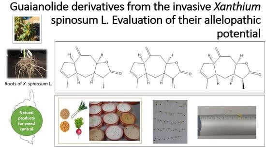
Guaianolide Derivatives from the Invasive Xanthium spinosum L.: Evaluation of Their Allelopathic Potential

Université de Corse, UMR CNRS 6134 Sciences Pour l’Environnement, Laboratoire Chimie des Produits Naturels, BP 52, 20250 Corte, France
*
Author to whom correspondence should be addressed.
Molecules 2022, 27(21), 7297; https://doi.org/10.3390/molecules27217297
Submission received: 31 August 2022 / Revised: 12 October 2022 / Accepted: 15 October 2022 / Published: 27 October 2022
(This article belongs to the Special Issue Natural Products That Might Change Society)

Abstract

:
Ziniolide, xantholide B (11α-dihydroziniolide), and 11β-dihydroziniolide, three sesquiterpene lactones with 12,8-guaianolide skeletons, were identified as volatile metabolites from the roots of Xanthium spinosum L., an invasive plant harvested in Corsica. Essential oil, as well as hydrosol and hexane extracts, showed the presence of guaianolide analogues. The study highlights an analytical strategy involving column chromatography, GC-FID, GC-MS, NMR (1D and 2D), and the hemi-synthesis approach, to identify compounds with incomplete or even missing spectral data from the literature. Among them, we reported the 1H- and 13C-NMR data of 11β-dihydroziniolide, which was observed as a natural product for the first time. As secondary metabolites were frequently involved in the dynamic of the dispersion of weed species, the allelopathic effects of X. spinosum root’s volatile metabolites were assessed on seed germination and seedling growth (leek and radish). Essential oil, as well as hydrosol- and microwave-assisted extracts inhibited germination and seedling growth; root metabolite phytotoxicity was demonstrated. Nevertheless, the phytotoxicity of root metabolites was demonstrated with a more marked selectivity to the benefit of the monocotyledonous species compared to the dicotyledonous species. Ziniolide derivatives seem to be strongly involved in allelopathic interactions and could be the key to understanding the invasive mechanisms of weed.

Graphical Abstract

1. Introduction

Invasive alien species represent one of the current main environmental issues; they are recognized by the Convention on Biological Diversity as the fourth cause of global biodiversity loss, after the disappearance of natural habitats, overexploitation of resources, and pollution [1]. Invasive species generally have rapid growth, high fertility, high dispersal power, and resistance to pathogens. Their numerous nuisance mechanisms (hybridization, modification of natural habitats, pathogenic organisms, etc.) allow them to take advantage of native species with the consequence of their disappearance and radical landscape modifications [2]. However, the chemistry of invasive species must vary, and with enormous biological activity potential that remains to be explored [3]. Invasive plants may serve as inexpensive and renewable sources of bioactive compounds.
The European Plant Protection Organization (E.P.P.O.) lists 6658 exotic species, of which 168 are considered invasive (78 in France) [4]. In Corsica, among the 2978 plant species listed, 454 introduced species have been recorded [5], of which, 52 are considered as worrying and 17 as invasive [6]. Among them, Xanthium spinosum (Spiny cocklebur) is a highly invasive plant originating from South America and is now widespread throughout the world [6,7,8,9,10,11,12]. X. spinosum is one of the worrying species because of its good adaptation to the Mediterranean climate as well as its affinity for nitrogenous soils. The plant is frequently found in farmlands where it causes a sanitary risk for cattle [8]. X. spinosum is provided with hooked spines, which can attach to animal coats and clothing, contributing to the dispersal over large areas.
X. spinosum is known by traditional medicine in many countries, such as Romania, Serbia, Egypt, South Africa, and Argentina [9]. The plant is used in the treatment of rabies, chronic fevers, and diabetes, and also to stimulate saliva production and for diuretic effects [10]. In Romania, plants are used to treat urinary problems and various prostate pathologies [11]. In Oltenia (southern Romania), X. spinosum and X. italicum seeds are used in infusion to help cardiac disorders [12]. In Bolivia, root decoctions are used to treat arteriosclerosis and hypertension, leaving decoction to inflammatory pathologies, such as oophoritis, hepatitis, toothache, cystitis, nephritis, and gastritis [13]. In North America, the Cherokee uses the plant to treat lung problems and snakebites [14]. Nowadays, the plant is used around the world in a wide range of medical applications. In Spain, the leaves are used as contraceptive drugs [15], and fruits are used to treat kidney malfunctions and hyperglycemia [16]. In Italy, a seed decoction is used to treat diarrhea [17].
Many studies have focused on the composition of polar solvent extracts and the chemical compounds identified are sorted into three main groups: phenolics, sesquiterpenes, and diterpenes [11,18,19,20,21,22,23,24,25,26,27]. Furthermore, X. spinosum is recognized for its biological and pharmaceutical activities, imputed to the presence of sesquiterpene lactones called xanthanolides [20,21,22,23,24,25,26,27,28,29,30,31,32,33,34,35,36,37,38,39,40,41,42,43,44]. These lactones, provided with a non-cyclic carbon chain and a seven-membered ring, appear to be responsible for cytotoxic [29,30,31], antitumor [20,31,32,33], antibacterial [34,35,36,37], anti-fungal [38], anti-leishmaniosis [38], anti-malarial [39], anti-inflammatory [30], and anti-ulcer activities [40,41]. In particular, xanthatin, isolated from the aerial parts of X. spinosum, is recognized for antibacterial and antifungal properties [37,42], as well as anti-angiogenesis [43] and phytotoxic properties [21,44].
Few studies relate to the chemical characterization of essential oils of the genus Xanthium, eleven describe the essential oil obtained from aerial parts of eight different species with various geographical origins: X. cavanelesii from Argentina [45], X. canadense from Japan [46], X. sibiricum from China [47], X. brasilicum from Iran [48], X. pennsylvanicum from Russia [49], X. strumarium from Brazil [50], Iran [51], India [52], and Egypt [53], and X. italicum [54] and X. spinosum [8] from Corsica.
To our knowledge, X. spinosum volatile root metabolites have never been investigated, and it might be interesting to study their involvement in the plant invasion mechanism. Indeed, through the exudation of a wide variety of compounds, roots have a critical ecological impact on soil and can inhibit the growth of competition plant species [55,56].
In this context, our project was interested in studying the ecological role of a Corsican invasive Xanthium spinosum, through the analysis of its volatile root metabolites and the evaluation of their allelopathic potential with the ambition to preserve biodiversity and provide a lasting response to the economic and ecological problems raised by invasive plant species.

2. Results and Discussion

2.1. Chemical Composition of X. spinosum Root Extracts

2.1.1. Essential Oil and Hydrosol

Essential oil and hydrosol were prepared by hydrodistillation from X. spinosum roots. The integrated analysis of essential oil (EO) identified 49 components, which accounted for 92.8% of the total amount (Table 1). Chromatogram of the EO is available in Supplementary Material (Figure S3). Essential oil showed the same proportion of oxygenated and hydrocarbon compounds (46.4%). Hydrocarbons were mainly represented by sesquiterpenes (36.1%) while monoterpenes were weak (10.3%). Oxygenated sesquiterpenes amounted to 39.1%, and among them, sesquiterpene lactones were higher (22.3%). The main components were α-isocomene 32 (6.1%), β-elemene 33 (8.5%), neryl 2-methylbutyrate 53 (6.2%), carotol 55 (9.4%), and ziniolide 66 (19.3%). The structures of the main EO components were reported in Figure 1.
Hydrosol was treated by liquid–liquid extraction and the integrated analysis of hydrosol extract (HYD) identified 40 components that accounted for 83.5% of the total amount (Table 1). Chromatogram of the HYD is available in Supplementary Material (Figure S4). Relative to EO, hydrosol extract was exclusively composed of oxygenated compounds; monoterpenoids amounted to 13.3% and the number of sesquiterpene lactones reached 3 times higher than EO (61.6% vs. 22.3%, respectively). The main components of HYD were terpinen-4-ol 21 (4.9%), xantholide B 65 (15.0%), and ziniolide 66 (42.6%).

2.1.2. Hexane Extracts

The integrated analysis of the cold maceration extract (MAC) and the assisted microwave extract (MAE) identified, respectively, 30 and 32 components, which accounted for 74.1 and 84.0% of the total amount (Table 1). Chromatograms of MAC and MAE extracts are available in Supplementary Materials (Figures S5 and S6). Both hexane extracts showed relatively similar compositions, which do not greatly differ from EO concerning hydrocarbon sesquiterpenes. However, we should note that hydrocarbon monoterpenes and most polar compounds were missing, to the benefit of sesquiterpene lactones. The main components were germacrene D 41 (9.2 and 6.9%), xantholide B 65 (11.7 and 15.0%), and ziniolide 66 (25.2 and 30.4%) for MAC and MAE extracts, respectively.

2.2. Analytical Strategy Applied to Identify X. spinosum Sesquiterpenic Lactones

We should note that 76 components were identified by comparing their EI–MS and RI with those compiled in the laboratory–MS library and 13 components were identified by the perfect match against RIn from the literature and commercial MS libraries [57,58]. However, compounds 65, 66, and 67 were not indexed and their univocal identification required the development of an analytical strategy involving column chromatography (CC), NMR experiments, and a hemi-synthesis procedure.
MAE extract was selected for its high proportion of compounds that remained unidentified after preliminary analysis, combined with a high extraction yield (0.23% against 0.04, 0.06, and 0.12% for EO, HYD, and MAC, respectively). Thus, two consecutive CC were carried out from the MAE extract: The first was to separate non-polar from polar components, and the second was performed on the polar fraction using a silica gel column impregnated with AgNO3. The 14 fractions obtained were analyzed by GC and GC-MS and GC chromatograms demonstrated that fractions 11 and 10 contained 65 (63.5%) and 66 (69.8%), respectively. Column chromatography resolution was not sufficient to obtain 67 with a convenient purity (7.9% in Fraction 12).

2.3. Contribution of MS and NMR to the Identification of Lactones from X. spinosum Roots
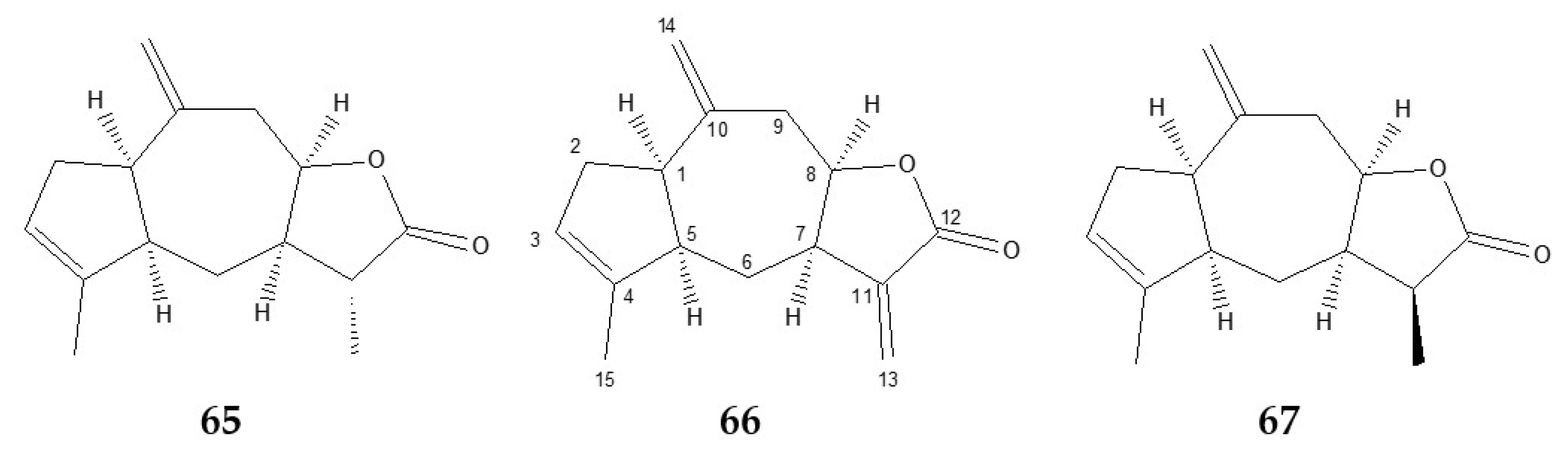
2.3.1. Ziniolide 66

Compound 66 gave a molecular peak at m/z 230, suggesting a C15H18O2 formula. EI-MS of 66 (Figure S1) comes with a base peak at m/z 91 and a peak at m/z 119, such as oxygenated sesquiterpenes. The MS spectrum of 66 showed a satisfactory concordance with sesquiterpene lactones recorded in our MS library, such as dehydrocostuslactone (score matching 50%).
The unequivocal identification of 66 as 3,10(14),11(13)-guaiatrien-12,8-olide, a guaiane-type sesquiterpene lactone commonly known as ziniolide was carried by alignment of 13C-NMR and 1H chemical shifts reported in the literature [28,59]. For completeness, the whole set of recorded NMR data (Figures S7–S12) confirm the above structure. Experimental 13C-NMR chemical shifts were given in Table 2. Relative to the literature data [28], our 13C-NMR assignment differed for C-2, C-4, C-5, C-10, and C-11 (Figure 2). Concerning the C-2 and C-5 chemical shifts, the values given in the literature appear to be inverted. Correct assignments were carried out using APT experiments and the analysis of the long-rang correlations in the HMBC spectrum. The multiplicity of C-2 (δC 35.96) and C-5 (δC 51.83) as CH2 and CH, respectively, as well as the condensed five-membered ring system C1-C5, were clearly established. The chemical shift values of the three ethylenic quaternary carbons C-4, C-10, and C-11 were very close and a source of confusion. Their assignments were aided by HMBC: C-4 (δC 142.83) was correlated to H3-15, C-10 (δC 143.71) with H2-9 and H2-14, as well as C-11 (δC 141.40) with H2-13. In addition, the cis-stereochemistry of the bicyclo[5.3.0]decane junction and the γ-lactone arrangement of 66 were ensured by distinct differences with the 13C-NMR data of centaurolide-B [60], an analogous derivative of trans-trans-guaianolide.
Ziniolide was isolated for the first time from Zinnia peruviana (L.) L. (Asteraceae) (synonym Zinnia multiflora) [59], then also from X. canadense [46,61] and X. catharticum [28].

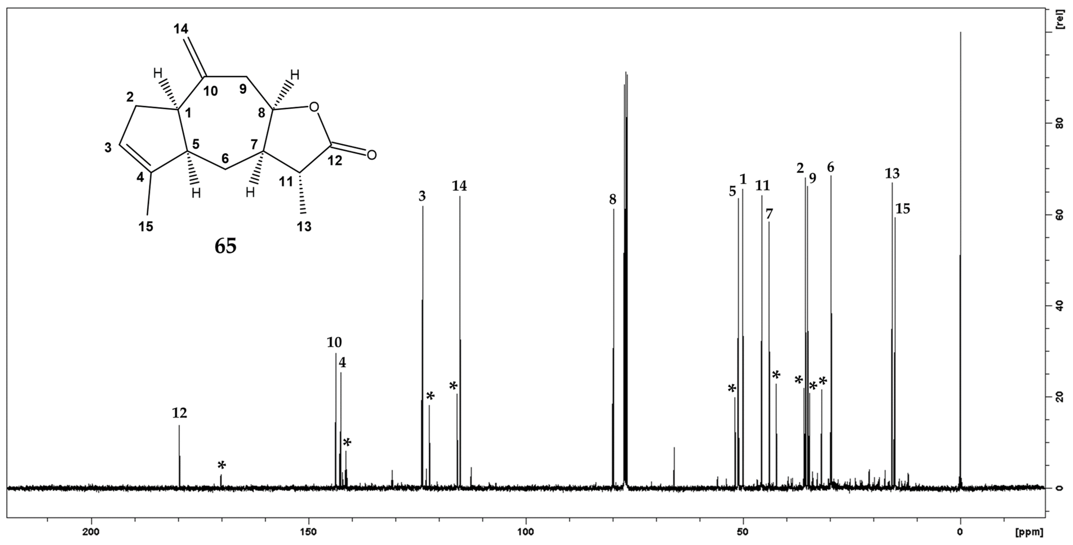
2.3.2. Xantholide B (11α-dihydroziniolide) 65

The EI-MS spectra of 65 and 66 were nearly the same except for heaviest fragments, such as molecular ions at m/z 230 and 232, respectively, which suggested that 65 was a dihydrogenated derivative of 66. Moreover, 65 MS spectrum has a good concordance score with sesquiterpene lactones present in our MS libraries.
Despite the occurrence of ziniolide 66 (21.4%), 13C-NMR chemical shifts of 65 were easily extracted from the fraction 11 spectrum (Figure 3) according to their relative intensities (1:3, respectively) and fifteen resonances were isolated (Table 2). The 13C-NMR spectrum of 65 showed strong similarities with those of ziniolide 66 and indicated that this compound exhibited the same sesquiterpene skeleton with a vinyl methyl, only one exomethylene group, and a lactone functionality. The hypothesis of the occurrence of a dihydro lactone was supported by the comparison of both sets of chemical shifts between 65 and 66, for which C-11, C-12, and C-13 showed the main differences. More precisely, the exomethylene group including C-11 (δC 141.40) and C-13 (δC 122.15) in 66 was replaced by a methine (C-11, δC 45.70) and a methyl (C-13, δC 15.74) groups, respectively, according to the hydrogenation of α-methylene-γ-lactone function. Relative to 66, the carbonylic C-12 (δC 179.70) undergoes a strong shielding (9 ppm) due to the non-conjugated carbonyl system. This chemical shift value was in agreement with those of dihydro guaianolide [62]. In addition, chemical shifts of the methyl group (δC 15.74, δH 1.31) indicated an α orientation as reported in compounds with similar configurations [29]. Consequently, 65 was identified as the 11α-dihydroziniolide 66. Finally, the agreement between 1H chemical shifts of 65 and those reported in the literature [46] supported the identification of 11α-dihydroziniolide, commonly known as xantholide B. It is to be noticed that xantholide B 65 has previously been isolated from X. canadense (L.) L. (Asteraceae) [46,61], and to our knowledge, the 13C-NMR data of 65 are described here for the first time. Recorded NMR data is given in Supplementary Materials (Figures S13 and S14).

2.3.3. 11β-dihydroziniolide 67

We should note that component 67 has demonstrated peak overlapping with ziniolide 66 on our lab non-polar GC column (Rtx-1); the polar GC column (Rtx-wax) was required to obtain sufficient resolution (Figure S2). Compounds 67 and 65 exhibited identical EI-mass spectra, which suggest a diastereoisomeric relationship between both molecules (Figure 4). Therefore, 67 is also a dihydrogenated derivative of ziniolide 66.
The reduction of a rich-ziniolide fraction (MAE-F10, 66, 69.8%) using sodium borohydride (NaBH4) confirms our hypothesis. GC chromatogram and EI-MS of the NaBH4 reduction product have informed about the presence of a mixture of 65 (17.6%) and 67 (73.3%). The ultra-dominant abundance of 67 allowed for extracting the fifteen chemical shifts from the 13C-NMR spectra.
13C-NMR chemical shifts of 67 (Table 2) and more precisely the presence of a quaternary carbon C-12 (δC 179.03) confirmed the good efficiency of the reduction of α-methylene-γ-lactone function. In the same way, the substitution of the exocyclic methylene of 66 by a methyl (H3-13, δH 1.19, d 7.3 Hz) coupled to a methine (H-11, δH 2.88, q 7.3 Hz) confirmed the hydrogenation of the C11–C13 double bond.
The many similarities between the 1H and 13C-NMR data of 65 and 67 supported the hypothesis of a diastereoisomeric relationship emitted from the mass spectra. Most chemical shift variations between 65 and 67 were observed on C-6 (δC 29.74 and 22.67, respectively) and C-13 (δC 15,74 and 10.0, respectively), with reciprocal shielding γ effects resulting from the small dihedral angle between C-13 and C-6, which supposed β orientation of C-13 methyl group. Finally, the complete assignment was supported by HSQC and HMBC experiments (Table 2). Consequently, the structure of compound 67 was established as 11β-dihydroziniolide. The whole set of recorded NMR data is available in Supplementary Materials (Figures S15–S20).
The 11β-dihydroziniolide was previously reported as a reduction product of ziniolide and also as the reaction product of the treatment in basic conditions of xantholide B [46]. However, to our knowledge, the present work report for the first time the occurrence of 11β-dihydroziniolide as a natural product as well as the 13C-NMR data (Table 2) and 1H-NMR data (see experimental) was never published before.

2.4. Allelopathic Effect of X. spinosum Root Extracts

Allelopathic activity of X. spinosum root extracts was assessed on the seed germination and seedling growth of two plants chosen for their respective botanical characteristics: (i) Allium porrum (Alliaceae) is an old, rustic, and perennial vegetable that we have selected as monocotyledon model and (ii) Raphanus sativus (Brassicaceae) is an annual or biennial plant, mainly cultivated for its fleshy and more popular hypocotyl, which was selected as dicotyledon model. The allelopathic potential was evaluated via the phytotoxicity of three root extracts: EO, HYD, and MAE-F10; a rich-lactone fraction obtained by CC from MAE. All extracts were assessed at concentration ranges from 0 to 1000 µg/mL. The phytotoxicity was appreciated through biological indices: root and shoot lengths, wet and dry weights, germination rate, germination percentage, vigor index, length and weight ratios, and allelopathic effect (see the experimental section for further details). The results were reported in Table 3 for A. porrum and R. sativus, respectively.
Our experiments highlighted the inhibitory effect of X. spinosum root extracts on the growth of both species. As seen in Figure 5a, the growth of the monocotyledon A. porrum seeds was strongly disturbed in contact with X. spinosum extracts. Relative to the control, the inhibition growth was clearly correlated to the concentration increase of the three natural extracts. Concerning R. sativus seeds (Figure 5b), the growth inhibition was disturbed to a lesser extent. The dicot seeds of R. sativus appear less sensitive than the monocot seeds of A. porrum to the toxic effects of X. spinosum extracts. Nevertheless, used at a high concentration (1000 μg/ml), the hydrosol extract was the most efficient to inhibit the seedling lengths of R. sativus.
Analysis of variance on the mean of seedling lengths (L) (Table 3) indicated that monocotyledon L was significantly different from control for all extracts (no treatment showed a common letter with control). While for the dicot plant, L was significantly different from the control only with hydrosol extract and MAE-F10 at high concentrations (from 250 to 1000 µg/mL).
The effect of X. spinosum natural mixtures on the seedling length could be analyzed using the length ratio (LR). While the root and shoot growths were little differentiated for the monocot seeds of A. porrum without treatment, a higher concentration of lactone-rich extract (MAE-F10) appeared to improve the specific growth of aerial shoots to the detriment of the roots. Concerning the seeds of dicot R. sativum, the root growth was twice higher than aerial shoot growth without treatment. Our experiment highlighted an increase of length ratio (LR) when the treatment concentration increased, whatever the X. spinosum extract used. This indicated that dicot radicles were more sensitive to extracts than the hypocotyl.
Relative to control, the treatment of the monocot seeds of A. porrum by an increasing concentration of X. spinosum extracts clearly affected the germination rate (GR). In addition, most seeds did not germinate. For completeness, a germination percentage (GP) followed the same tendency, the increase of extract concentrations caused a lack of germination success. The MAE-F10 extract at 1000 µg/mL was the more efficient, only a third of A. porrum seeds reached the germination state at the end of the experiment. Concerning dicot seeds of R. sativus, whatever the concentration, GR was less affected by X. spinosum extracts, and GP was not affected by treatments.
Comparison of vigor index (VI) and the allelopathic effect (AE) of both species confirmed that monocot seedling growth was more affected by the root extracts than the dicot seedlings; VI of the dicot plant was slightly affected whereas the VI of the monocot plant was from 8 to 45 times smaller at higher extract concentrations.
Treated monocot seeds that have successfully grown appeared to be significantly smaller than the control and a negative allelopathic effect (AE) was observed for all treatments. A greater effect was observed with MAE-F10 extract with an AE of −95.7% at 1000 µg/mL. Dry weight and wet weight were both uniformly affected, as shown by the constant weight ratio (WR), suggesting that the inhibition of seedling growth could be attributed to the inhibition of mitosis (increase in biomass). We should note that HYD extract treatment stimulated seedling growth at 100 µg/mL, as shown by the positive AE, a phenomenon known as “low dose simulation–high dose stimulation” or “hormesis” [63].
Sesquiterpene lactones are recognized for their allelopathic potential thanks to their α,β-unsaturated carbonyl group, which can undergo 1,4-conjugated additions with nucleophiles, such as the sulfhydryl group very abundant in proteins and nucleic acid [64]. Moreover, the biological effect of ziniolide 66 has already been the object of various studies and demonstrated larval growth inhibition of Drosophila melanogaster [46] as well as antibacterial activity [28], anti-inflammatory, cytotoxic, and antitumor potential [42].
Our biological assessments underlined the lactones responsibility in the phytotoxicity of X. spinosum extracts and the α-methylene-γ-lactone group present in ziniolide 66 could be the main actor. Thus, guaianolide in X. spinosum roots and their recent introduction into the island territory might be one of the factors encouraging the invasion of the species; as supposed by the “novel weapon hypothesis”, natives species may not yet have the time to develop metabolic pathways to eliminate and counteract these new phytotoxins [65].
However, the phytotoxic effect was not proportional to lactone content and lactones do not seem to be the only ones involved. Indeed, monoterpenes, such as pinene isomers [66], possess allelopathic properties, hydrocarbon monoterpenes contained in X. spinosum essential oil could explain the difference in the inhibition mechanism and the overall effect probably results from the positive or negative synergy between several metabolites.

3. Materials and Methods

3.1. Plant Material

X. spinosum roots were harvested in central Corsica (Corte, France, 42°17’56.5”N, 9°10’13.0”E) during a dormant state in January 2019. The botanical determination of the plants was performed according to the botanical keys summarized in Flora Corsica [5].

3.2. Isolation of Volatile Metabolites

Four sample preparation techniques including hydrodistillation, hydrosol extraction, cold maceration in hexane, and microwave-assisted extraction in hexane were used in order to produce exhaustive volatile extracts.
Air-dried roots (200 g) were subjected to hydrodistillation (5 h) using a Clevenger-type apparatus, according to the method recommended in the European pharmacopoeia [67]. Hydrodistillation produced a yellow essential oil (EO) with a yield of 0.04% (w/dw, based on the weight of the dried plant material) and aromatic water, called hydrosol.
Hydrosol was recovered by removing co-coating (the first 300 mL) of the Clevenger apparatus during the hydrodistillation. Then, hydrosol was submitted to liquid–liquid extraction (LLE). A total of 300 mL was extracted successively with 3 × 50 mL of diethyl ether; the organic phase was then washed with 50 mL of water saturated with NaCl, dried over Na2SO4, and filtered before being concentrated to produce hydrosol extract (HYD). Hydrosol extraction produced a colorless extract with a yield of 0.06% (w/dw).
Ground air-dried roots (20 g) were subjected to maceration in hexane (200 mL) at room temperature (48 h). The solvent was then filtered and concentrated. The resulting extract was next taken up in absolute ethanol and centrifuged (20 min at 6000 rpm), and the supernatant was collected and concentrated to finally obtain the macerate extract (MAC). Maceration in hexane produced an orange–yellow extract with yields of 0.12% (w/dw).
Air-dried roots were extracted using Multiwave 3000 (Anton Paar, Gratz, Austria) apparatus provided with 16 ceramic vessels. For each vessel, ground roots (5 g) were introduced with hexane (40 mL) and extraction was realized at 180 °C (150 W per vessel) for 20 min followed by 40 min of cooling. The solvent was then filtered and concentrated. The resulting extract was next taken up in absolute ethanol and centrifuged (20 min at 6000 rpm), and the supernatant was collected and concentrated to finally obtain the microwave extract (MAE). The microwave-assisted extraction produced an orange–yellow extract with a yield of 0.23% (w/dw).

3.3. Fractions

The MAE extract (1.44 g) was first submitted to column chromatography on a silica gel column (40 × 2 cm, 63–200 µm, 50 g) with two elution gradients (100/0 and 0/100 of Hex/DIPE) giving two fractions. The polar fraction (1.15 g) was next submitted to column chromatography on a silica gel column impregnated with 10% AgNO3 (40 × 2 cm, 40–63 µm, 50 g) using gradients of Hex/DIPE. TLC fingerprint grouping gave 14 fractions, among them, F10 (200 mg, 65: 10.1%, 66: 69.8%, 67: 2.8%), F11 (75 mg, 65: 63.5%, 66: 21.4%, 67: 4.3%) and F12 (9 mg, 65: 16.4%, 66: 6.8%, 67: 7.9%).

3.4. NaBH4 Reduction

β-dihydroziniolide 67 was obtained by treating the MAE fraction 10 (20 mg, 69.8% of ziniolide 66) with the NaBH4 solution (1.23.10−4 mol) in ethanol (10 mL). The resulting mixture was stirred at room temperature and then refluxed for 60 min. After treatment, 10 mL of water saturated with NaCl and 2 drops of glacial acetic acid were added. The mixture was then extracted by 3 × 15 mL of hexane and dried over sodium sulfate before being concentrated under a vacuum. The resulting mixture (12 mg) contained xantholide B 65 (17.6%) and β-dihydroziniolide 67 (73.3%).

3.5. GC-FID Analysis

Analyses were carried out using a Perkin–Elmer Clarus 600 gas chromatography (GC) apparatus (Waltham, MA, USA) equipped with a single injector and two flame ionization detectors (FIDs) for simultaneous sampling to two fused–silica capillary columns (60 m × 0,22 mm i.d., film thickness 0.25 μm; Restek, Bellefonte, PA, USA) with stationary phases of different polarity, i.e., a nonpolar Rtx–1 (polydimethylsiloxane) and a polar Rtx–Wax (polyethylene glycol). The oven temperature was programmed to increase from 60 to 230 °C at 2 °C min−1 and was held isothermal at 230 °C for 30 min. The injector temperature was maintained at 280 °C and the detector temperature at 280 °C, the carrier gas was H2 (0,7 mL.min−1) and the samples were injected (0.2 μL of pure oil) in the split mode (1:80). Retention indices (RIs) of the mixture components were determined relative to the retention times (tR) of a series of n-alkanes (C5–C30; commercial solution, obtained from Restek, Bellefonte, PA, USA) using the Van den Dool and Kratz equation [68].

3.6. GC-MS Analysis

The plant extracts and the fractions obtained by CC were investigated using a Perkin Elmer Turbo Mass quadrupole detector directly coupled to a Perkin Elmer SQ8 (Walton, MA, USA), equipped with the two same fused-silica capillary columns as described above. Both columns were used with the same quadrupole MS detector. The analyses were consecutively carried out on the nonpolar and the polar column. Hence, for each sample, two reconstructed ion chromatograms (RIC) were provided, which were investigated consecutively. The GC conditions were the same as described above and the MS parameters were as follows: ion–source temperature, 150 °C, ionization energy, 70 eV; electron ionization mass spectra acquired over a mass range of 35–350 amu during a scan time 1 s. The injection volumes were 0.1 μL.

3.7. NMR Analysis

Nuclear magnetic resonance (NMR) spectra were recorded on MAE fractions 10 and 11, and the NaBH4 reduction product of MAE fraction 10.
NMR experiments were acquired in CDCl3 (EuroIsotop, Saint Aubin, France), at 300 K using a Bruker Avance DRX 500 NMR spectrometer (Karlsruhe, Germany) operating at 500.13 MHz for 1H and 125.77 MHz for 13C Larmor frequency with a double resonance broadband fluorine observe (BBFO) 5 mm probe head. 13C-NMR experiments were recorded using a one–pulse excitation pulse sequence (90° excitation pulse) with 1H decoupling during signal acquisition (performed with WALTZ–16); the relaxation delay has been set at 2 s. For each analyzed sample, depending on the compound concentration, 3 up to 5 k free induction decay (FID) 64 k complex data points were collected using a spectral width of 30,000 Hz (240 ppm). Chemical shifts (δ in ppm) were reported relative to the residual signal of CDCl3C 77.04 ppm). Complete 1H and 13C assignments of the new compound were obtained using 2D gradient–selected NMR experiments, 1H–1H COSY (correlation spectroscopy), 1H–13C HSQC (heteronuclear single quantum correlation), 1H–13C HMBC (heteronuclear multiple bond coherence) and 1H–1H NOESY (nuclear Overhauser effect spectroscopy), for which conventional acquisition parameters were used, as described in the literature [69].

3.8. Compound Identification and Quantification

Identification of individual components in plant extracts or CC fractions was based on a methodology involving integrated techniques, such as GC retention indices, GC-MS (EI), and NMR. The identification of individual components was based (i) on the comparison of the retention indices (RIs) determined on the polar and nonpolar columns with those of authentic compounds or literature data [57,58]; (ii) on computer matching of the mass spectra with commercial MS libraries and the mass spectra with those listed in our homemade MS library of authentic compounds or literature data [57,58,70,71]; (iii) comparing the 13C-NMR chemical shifts of CC fraction components with those of reference spectra reported in the literature; (iv) NMR assignments using 1D and 2D experiments. The relative quantification percentage was obtained by internal normalization of the GC-FID peak area without response factors.

11β-dihydroziniolide (67)

Colorless oil; Rf 0.14 (Hex/DIPE 6:4), 0.67 (Hex/Ethyl acetate 6:4); MS m/z 232 see Figure 4a; 1H NMR data (CDCl3, 600 MHz): δ 5.37 (1H, br s, H-3), δ 5.01 (1H, s, H-14a), δ 4.97 (1H, s, H-14b), δ 4.56 (1H, dt, J = 4.6, 11.2 Hz, H-8), δ 3.19 (1H, m, H-1), δ 2.88 (1H, q, J = 7.3 Hz, H-11), δ 2.82 (1H, dd, J = 6.8, 13.7 Hz, H-9b), δ 2.58 (1H, dd, J = 4.2, 13.7 Hz, H-9a), δ 2.41 (1H, br m, H-7), δ 2.40 (1H, br m, H-2a), δ 2.38 (1H, br m, H-5), δ 2.31 (1H, br m, H-2b), δ 1.74 (1H, s, H-15), δ 1.42 (1H, d, J = 13.2 Hz, H-6b), δ 1.19 (3H, d, J = 7.3 Hz, H-13), δ 1.17 (1H, s, H-6a); 13C (CDCl3, 125 MHz) see Table 2.

3.9. Allelopathic Effect Evaluation

The allelopathic activity was assessed on three X. spinosum root extracts (EO, HYD, and the MAE fraction 10) selected according to their chemical compositions. Allelopathic tests were performed using the methodology reported in the literature [72] and implemented in our laboratory.
Commercial seeds of Allium porrum (monocotyledon) and Raphanus sativus (dicotyledon) were used to assess the phytotoxicity of X. spinosum extracts. Stock solutions of essential oil were prepared in dimethylsulfoxide (DMSO) as the initial solvent followed by dilution with distilled water to a final concentration of 1000 μg·mL−1. The concentration of DMSO in the stock solution was 1% v/v. Other test solutions (100, 250, and 500 μg·mL−1) were prepared by dilution of the stock solution with distilled water. Control treatment (0) was an aqueous solution of DMSO (1% v/v). Three replicates, each of 10 seeds, were prepared for each treatment using glass Petri dishes (9 cm) lined with Whatman no. 4 filter paper. A total of 3 mL of test solution was added to each Petri dish. The Petri dishes were hermetically closed with stretch film and placed in an incubator at 20 °C in the dark. Germinated seeds were counted each day over a period of 7 days and root length, shoot length and wet seedling weight were determined after 7 days. Finally, germinated seeds were kept in a laboratory oven at 60 °C for one week to determine dry seedling weight.
At the end of the experiment germination rate (GR) [66,73], germination percentage (GP) [74], vigor index (VI) [75], lengths ratio (LR), weights ratio (WR) [66], and allelopathic effect (AE) [76] were determined from the following equations:
GR = i = 1 n n i d i  
GP   = n f N × 100
VI   = GP   ×   L 100
LR   = S R
WR   = d w ww
A E = ( L C 1 ) × 100
ni: number of germinated seeds at each counting; di: number of days until x counting; x: counting number; nf: number of germinated seeds at the end of the experiment; N: total number of seeds; L: mean of seedling lengths (mm); dw: mean of seedling dry weights (mg); ww: mean of seedling wet weights (mg); R: mean of root lengths (mm); S: mean of shoot lengths (mm); C: mean of control seedling lengths (mm).

3.10. Statistical Analysis

Seedling length data from allelopathic bioassays were subjected to one-way analysis of variance (ANOVA) using Minitab statistical software. Means of multiple treatments were compared using Tukey HSD (honestly significant difference) test at a 5% level of significance. All results are expressed as mean ± SD. Means not sharing a common letter are significantly different.

4. Conclusions

The volatiles of X. spinosum roots were characterized by an analytical strategy involving column chromatography, GC-FID, GC/MS, NMR, as well as hemi-synthesis to identify guaianolide sesquiterpenes with incomplete or even missing spectral data from the literature. Instead of the xanthanolides usually found in the species, ziniolide, xantholide B (11α-dihydroziniolide), and 11β-dihydroziniolide, three sesquiterpene lactones with 12,8-guaianolide skeleton were identified from the essential oil, hydrosol extract, and hexane extracts from Xanthium spinosum L. Among them, 1H- and 13C-NMR data of 11β-dihydroziniolide, as well as its occurrence as a natural product were described for the first time. Our study aimed to highlight the involvement of volatile compounds of X. spinosum roots in the allelopathic interactions between these invasive weeds and plants. Treatments inflicted on leek and radish seeds involved essential oil, as well as hydrosol and lactone-rich extracts of X. spinosum roots, which have shown phytotoxicity. Allium porrum, chosen as a monocot model, appears more sensitive than the dicot Raphanus sativus concerning seed germination. Nevertheless, used at a high concentration (1000 μg/ml), the hydrosol extract was the most efficient to inhibit the seedling length of R. sativus and selective inhibition of radicle seedlings was observed according to the concentration treatment. The present study demonstrated great seedling growth inhibition and the anti-germination potential of X. spinosum. The involvement of the ziniolide analogs appears to be effective; nevertheless, the aid of other natural products in the extracts can contribute to synergetic effects. In order to develop valorization opportunities for X. spinosum, further investigations were required to determine its possible use as a natural herbicide.

Supplementary Materials

The following supporting information can be downloaded at: https://www.mdpi.com/article/10.3390/molecules27217297/s1, Figure S1: EI MS spectrum of 66; Figure S2: GC DIF Spectrum centered on compounds 6567: (a) non-polar column. (b) polar column; Figure S3: GC DIF Chromatogram of the EO; Figure S4: GC DIF Chromatogram of the HYD; Figure S5: GC DIF Chromatogram of the MAC; Figure S6: GC DIF Chromatogram of the MAE; Figure S7: 13C NMR Spectrum of 66; Figure S8: 1H NMR Spectrum of 66; Figure S9: HMBC Spectrum of 66; Figure S10: HSQC Spectrum of 66; Figure S11: COSY Spectrum of 66; Figure S12: NOESY Spectrum of 66; Figure S13: 13C NMR Spectrum of 65; Figure S14: 1H NMR Spectrum of 65; Figure S15: 13C NMR Spectrum of 67; Figure S16: 1H NMR Spectrum of 67; Figure S17: HMBC Spectrum of 67; Figure S18: HSQC Spectrum of 67; Figure S19: COSY Spectrum of 67; Figure S20: NOESY Spectrum of 67.

Author Contributions

Conceptualization. A.M. and P.B.; methodology. A.M and P.B.; validation. A.M and P.B.; investigation. S.B and P.B.; sourcing. S.B.; writing—original draft preparation. S.B.; writing—review and editing. A.M and P.B.; supervision. A.M and P.B.; project administration. A.M. All authors have read and agreed to the published version of the manuscript.

Funding

This research was funded by Collectivité de Corse, S.B. research grant (CONV-18-DESR-03).

Institutional Review Board Statement

Not applicable.

Informed Consent Statement

Not applicable.

Data Availability Statement

The study did not report any data.

Acknowledgments

A. Tintaru (Aix Marseille Univ, CNRS, UMR 7325, CINaM, 13288 Marseille, France) for the acquisition of NRM data.

Conflicts of Interest

The authors declare no conflict of interest.

Sample Availability

The samples are available from the authors, Université de Corse, UMR CNRS SPE, Lab. Chimie des Produits Naturels, 20250 Corte, Corsica, France.

References

  1. Jeanmonod, D.; Schlüssel, A.; Gamisans, J. Status and Trends in the Alien Flora of Corsica: Status and Trends in the Alien Flora of Corsica. EPPO Bull. 2011, 41, 85–99. [Google Scholar] [CrossRef] [Green Version]
  2. Lockwood, J.L.; Hoopes, M.F.; Marchetti, M.P. Invasion Ecology; Blackwell Publishing: Malden, MA, USA, 2007; ISBN 978-1-4051-1418-9. [Google Scholar]
  3. Máximo, P.; Ferreira, L.M.; Branco, P.S.; Lourenço, A. Invasive Plants: Turning Enemies into Value. Molecules 2020, 25, 3529. [Google Scholar] [CrossRef] [PubMed]
  4. EPPO Global Database. Available online: https://gd.eppo.int/ (accessed on 5 October 2022).
  5. Jeanmonod, D.; Gamisans, J. Flora Corsica; Edisud: Aix en Provence, France, 2007; ISBN 978-2-7449-0662-6. [Google Scholar]
  6. Paradis, G.; Hugot, L.; Spinosi, P. Les Plantes Envahissantes: Une Menace Pour La Biodiversité. Stantari 2008, 13, 18–26. [Google Scholar]
  7. Weber, E. Invasive Plant Species of the World: A Reference Guide to Environmental Weeds; CABI Publishing: Wallingford, UK, 2003; ISBN 978-1-78064-386-1. [Google Scholar]
  8. Andreani, S.; Paolini, J.; Costa, J.; Muselli, A. Chemical Composition of Essential Oils of Xanthium spinosum L., an Invasive Species of Corsica. Chem. Biodivers. 2017, 14, e1600148. [Google Scholar] [CrossRef]
  9. Amorin, J.L. Xanthium spinosum L. (Compositae), Weed Used in Argentine Folk Medecine. Rev. Fac. Agron. Univ. Nac. Plata. 1972, 48, 155–169. [Google Scholar]
  10. Dragendorff, G. Die Heilpflanzen der Verschiedenen Völker und Zeiten: Ihre Anwendung, Wesentlichen Bestandteile und Geschichte; Verlag von Ferdinand Enke: Stuttgart, Germany, 1898. [Google Scholar]
  11. Varga, E.; Domokos, E.; Kelemen, H.; Fulop, I.; Kursinszki, L. HPLC-ESI-MS/MS Profiling of Phenolic Acids, Flavonoids And Sesquiterpene Lactones from Xanthium spinosum. Rev. Chim. 2020, 71, 558–564. [Google Scholar] [CrossRef]
  12. Ciocirlan, V. Flora Ilustrata a Romanici; Ceres: Bucuresti, Romania, 2000. [Google Scholar]
  13. Fernandez, E.; Sandi, Y.; Kokosta, L. Ethnobotanical Inventory of Medicinal Plants Used in the Bustillo Province of the Potosi Department, Bolivia. Fitoterapia 2003, 74, 407–416. [Google Scholar] [CrossRef]
  14. Hamel, P.B.; Chiltoskey, M.U. Cherokee Plants and Their Uses: A 400 Years History; Herald Publishing: Kings Mountain, NC, USA, 1975; ISBN 978-0-935741-25-4. [Google Scholar]
  15. Kumar, D.; Kumar, A.; Prakash, O. Potential Antifertility Agents from Plants: A Comprehensive Review. J. Ethnopharmacol. 2012, 140, 1–32. [Google Scholar] [CrossRef]
  16. Benitez, G.; Gonzalez-Tejero, M.R.; Molero-Mesa, J. Pharmaceuticial Ethnobotany in the Western Part of Granada Province (Southern Spain): Ethnopharmacological Synthesis. J. Ethnopharmacol. 2010, 129, 87–105. [Google Scholar] [CrossRef]
  17. Palmese, M.T.; Manganelli, R.E.U.; Tomei, P.E. An Ethno-Pharmacobotanical Survey in the Sarrabus District (South-East Sardinia). Fitoterapia 2001, 72, 619–643. [Google Scholar] [CrossRef]
  18. Erzsébet, D.; László, K.; Hajnal, K.; Erzsébet, V. A szúrós szerbtövis (Xanthium spinosum L.) növényfaj fitofarmakológiai áttekintése. Acta Pharm. Hung. 2016, 86, 35–40. [Google Scholar]
  19. Aldibekova, D.A.; Kizaibek, M.; Aisijiang, M.; Dyuskaliyeva, G.; Taldau, A.; Erkinbek, M. Morphology, Anatomy, Chlorogenic Acid Content and Antioxidant Capacity of Xanthium strumarium L. and Xanthium spinosum L. OnLine J. Biol. Sci. 2018, 18, 237–246. [Google Scholar] [CrossRef] [Green Version]
  20. Ansari, A.H.; Dubey, K.S. 2-Desacetyl-8-Epi-Xanthumanol-4-O-b-D-Galactopyranoside: The Potential Antitumor Sesquiterpenoidal Lactone from Xanthium spinosum Bark. Asian J. Chem. 2000, 12, 521–526. [Google Scholar]
  21. Yuan, Z.; Zheng, X.; Zhao, Y.; Liu, Y.; Zhou, S.; Wei, C.; Hu, Y.; Shao, H. Phytotoxic Compounds Isolated from Leaves of the Invasive Weed Xanthium spinosum. Molecules 2018, 23, 2840. [Google Scholar] [CrossRef] [PubMed]
  22. Sanz, M.J.; Terencio, M.C.; Manez, S.; Peris, J.B.; Rios, J.L. Phenolic Compounds from Two Xanthium Species. Planta Med. 1991, 57, A131. [Google Scholar] [CrossRef]
  23. Piacente, S.; Pizza, C.; De Tommasi, N.; De Simone, F. Sesquiterpene and Diterpene Glycosides from Xanthium spinosum. Phytochemistry 1996, 41, 1357–1360. [Google Scholar] [CrossRef]
  24. Abdei-Mogib, A.; Dawidar, A.M.; Metwally, M.A.; Abou-Elzahab, M. Xanthanolides from Xanthium spinosum. Phytochemistry 1991, 30, 3461–3462. [Google Scholar] [CrossRef]
  25. Omar, A.A.; Elrashidy, E.M.; Ghazy, N.A.; Metwally, M.A.; Ziesche, J.; Bohlmann, F. Xanthanolides from Xanthium spinosum. Phytochemistry 1984, 23, 915–916. [Google Scholar] [CrossRef]
  26. Marco, J.A.; Sanz-Cervera, J.F.; Corral, J.; Carda, M.; Jakupovic, J. Xanthanolides from Xanthium: Absolute Configuration of Xanthanol, Isoxanthanol and Their C-4 Epimers. Phytochemistry 1993, 34, 1569–1576. [Google Scholar] [CrossRef]
  27. Ciulei, I.; Grigorescu, E.; Stanescu, U. Plante Medicinale, Fitochimie, Fitoterapie; Medicala: Bucuresti, Romania, 1993. [Google Scholar]
  28. Cumanda, J.; Marinoni, G.; Bernardi, M.; Vidari, G.; Finzi, P.V. New Sesquiterpenes from Xanthium Catharticum. J. Nat. Prod. 1991, 54, 460–465. [Google Scholar] [CrossRef]
  29. Bui, V.-B.; Liu, S.-T.; Zhu, J.-J.; Xiong, J.; Zhao, Y.; Yang, G.-X.; Xia, G.; Hu, J.-F. Sesquiterpene Lactones from the Aerial Parts of Xanthium sibiricum and Their Cytotoxic Effects on Human Cancer Cell Lines. Phytochem. Lett. 2012, 5, 685–689. [Google Scholar] [CrossRef]
  30. Kim, Y.S.; Kim, J.S.; Park, S.H.; Choi, S.U.; Lee, C.O.; Kim, S.K.; Kim, Y.K.; Kim, S.H.; Ryu, S.Y. Two Cytotoxic Sesquiterpene Lactone from the Leave of Xanthium strumarium and Their in Vitro Inhibitory Activity of Farnesyltransferase. Planta Med. 2003, 69, 375–377. [Google Scholar] [CrossRef] [PubMed]
  31. Kovacs, A.; Vasas, A.; Forgo, P.; Rethy, B.; Zupko, I.; Hohmann, J. Xanthanolides with Antitumour Activity from Xanthium Italicum. Z. Nat. C 2009, 64, 343–349. [Google Scholar] [CrossRef] [PubMed]
  32. Ramirez-Erosa, I.; Huang, Y.; Hickie, R.A.; Sutherland, R.G.; Branka, B. Xanthatin and Xanthinosin from the Burs of Xanthium strumarium L. as Potential Anticancer Agents. Can. J. Physiol. Pharmacol. 2007, 85, 1160–1172. [Google Scholar] [CrossRef]
  33. Réthy, B.; Csupor-Löffler, B.; Zupko, I.; Hajdu, Z.; Mathé, I.; Hohmann, J.; Rédei, T.; Falkay, G. Antiproliferative Activity of Hungarian Asteraceae Species against Human Cancer Cell Lines. Part I. Phytother. Res. 2007, 21, 1200–1208. [Google Scholar] [CrossRef]
  34. Little, J.E.; Foote, M.W.; Johnstone, D.B. Xanthatin: An Antimicrobial Agent from Xanthium pennsylvanicum. Arch. Biochem. 1950, 21, 247–254. [Google Scholar]
  35. Pinel, B.; Audo, G.; Mallet, S.; Lavault, M.; Poype, F.; Seraphin, D.; Richomme, P. Multi Grams Scale Purification of Xanthanolides from Xanthium macrocarpum: Centrifugal Partition Chromatography versus Silica Gel Chromatography. J. Chromatogr. A 2007, 1151, 14–19. [Google Scholar] [CrossRef]
  36. Tsankova, E.T.; Trendafilova, A.B.; Kujumgiev, A.I.; Galabov, A.S.; Robeva, P.R. Xanthanolides from Xanthium Italicum Moretti and Their Biological Activity. Z. Nat. C 1994, 49, 154–155. [Google Scholar] [CrossRef]
  37. Ginesta-Peris, E.; Garcia-Breijo, F.J.; Primo-Yúfera, E. Antimicrobial Activity of Xanthatin from Xanthium spinosum L. Lett. Appl. Microbiol. 1994, 18, 206–208. [Google Scholar] [CrossRef]
  38. Lavault, M.; Landreau, A.; Larcher, G.; Bouchara, J.P.; Pagniez, F.; Le Pape, P.; Richomme, P. Antileschmanial and antifungal activities of xanthanolides isolated from Xanthium macrocarpum. Fitoterapia 2005, 76, 363–366. [Google Scholar] [CrossRef]
  39. Chandel, S.; Bagai, U.; Vashishat, V. Antiplasmodial Activity of Xanthium strumarium against Plasmodium Berghei-Infected BALB/C Mice. Parasitol. Res. 2012, 110, 1179–1183. [Google Scholar] [CrossRef] [PubMed]
  40. Favier, L.S.; Agosta, G.; Gomez, M.R.; Maria, A.G.; Tonn, C.E. Determination and assay validation of the bioactive sesquiterpene lactone from Xanthium cavanillesii usig capillary electrophoresis. Pharmazie 2006, 61, 981–984. [Google Scholar] [PubMed]
  41. Favier, L.S.; Maria, A.O.M.; Wendel, G.H.; Borkowski, E.J.; Giordano, O.S.; Petzer, L.; Tonn, C.E. Anti-ulcerogenic activity of xanthanolide sesquiterpenes from Xanthium cavanillesii in rats. J. Ethnopharmacol. 2005, 100, 260–267. [Google Scholar] [CrossRef] [PubMed]
  42. Bader, A.; Giner, R.M.; Martini, F.; Schinella, G.R.; Rios, J.L.; Braca, A.; Prieto, J.M. Modulation of COX, LOX and NFκB Activities by Xanthium spinosum L. Root Extract and Ziniolide. Fitoterapia 2013, 91, 284–289. [Google Scholar] [CrossRef]
  43. Romero, M.; Zanuy, M.; Rosell, E.; Cascante, M.; Piulats, J.; Font-Bardia, M.; Balzarini, J.; De Clerq, E.; Pujol, M.D. Optimization of Xanthatin Extraction from Xanthium spinosum L. and Its Cytotoxic, Anti-Angiogenesis and Antiviral Properties. Eur. J. Med. Chem. 2015, 90, 491–496. [Google Scholar] [CrossRef]
  44. Linh, N.T.T.; Son, N.T.T.; Ha, N.T.T.; Tra, N.T.; Anh, L.T.T.; Chen, S.; Van Tuyen, N. Biologically Active Constituents from Plants of the Genus Xanthium. Prog. Chem. Org. Nat. Prod. 2021, 116, 135–209. [Google Scholar] [CrossRef]
  45. Taher, H.A.; Ubiergo, G.O.; Talenti, E.C.J. Constituents of the essential oil of Xanthium cavanillesii. J. Nat. Prod. 1985, 48, 857. [Google Scholar] [CrossRef]
  46. Sakuda, Y.; Tahara, T. The constituents of essential oil from Xanthium canadense Mill. J. Jpn. Oil Chem. Soc. 1982, 31, 151–153. [Google Scholar] [CrossRef] [Green Version]
  47. Liu, X.Q.; Li, L.L.; Zeng, L.S.; Zou, F.H.; Feng, S. Study of Volatiles Oils Constituents from 20 Kinds of Compositate in Hunan with GC-MS. J. Univ. Hunan 2010, 30, 28–31. [Google Scholar]
  48. Habibi, Z.; Laleh, A.; Masoudi, S.; Rustaiyan, A. Composition of the Essential Oil of Xanthium brasilicum Vellozo from Iran. J. Essent. Oil Res. 2004, 16, 31–32. [Google Scholar] [CrossRef]
  49. Kozhin, S.A.; Ikonnikov, V.V. Composition of essential oil of Xanthium pennsylvanicum Wallroth. Rastit. Resur. 1980, 16, 204–208. [Google Scholar]
  50. Scherer, R.; Wagner, R.; Meireles, M.A.A.; Godoy, H.T.; Duarte, M.C.T.; Filho, J.T. Biological Activity and Chemical Composition of Hydrodistilled and Supercritical Extracts of Xanthium strumarium L. Leaves. J. Essent. Oil Res. 2010, 22, 424–429. [Google Scholar] [CrossRef]
  51. Esmaeili, A.; Rustaiyan, A.; Akbari, M.T.; Moazami, N.; Masoudi, S.; Amiri, H. Composition of the Essential Oils of Xanthium strumarium L. and Cetaurea solstitialis L. from Iran. J. Essent. Oil Res. 2006, 18, 427–429. [Google Scholar] [CrossRef]
  52. Ahuja, M.M.; Nigam, S.S. Chemical examination of the essential oil from the leaves of Xanthium strumarium. Flavour Ind. 1970, 1, 627–630. [Google Scholar]
  53. El-Gawad, A.; Elshamy, A.; El Gendy, A.; Gaara, A.; Assaeed, A. Volatiles Profiling, Allelopathic Activity, and Antioxidant Potentiality of Xanthium strumarium Leaves Essential Oil from Egypt: Evidence from Chemometrics Analysis. Molecules 2019, 24, 584. [Google Scholar] [CrossRef] [Green Version]
  54. Andreani, S.; Barboni, T.; Desjobert, J.M.; Paolini, J.; Costa, J.; Muselli, A. Essential Oil Composition and Chemical Variability of Xanthium Italicum Moretti from Corsica. Flavour Fragr. J. 2012, 27, 227–236. [Google Scholar] [CrossRef]
  55. Bertin, C.; Yang, X.; Weston, L.A. The Role of Root Exudates and Allelochemicals in the Rhizosphere. Plant Soil 2003, 256, 67–83. [Google Scholar] [CrossRef]
  56. Bais, H.P.; Weir, T.L.; Perry, L.G.; Gilroy, S.; Vivanco, J.M. The Role of Root Exudates in Rhizosphere Interactions with Plants and Other Organisms. Annu. Rev. Plant Biol. 2006, 57, 233–266. [Google Scholar] [CrossRef]
  57. Konig, W.A.; Hochmuth, D.H.; Joulain, D. Terpenoids and Related Constituents of Essential Oils, Library of Mass Finder 2.1; Institute of Organic Chemistry, University of Hamburg: Hamburg, Germany, 2001. [Google Scholar]
  58. Adams, R.P. Identification of Essential Oils by Capillary Gas Chromatography/Mass Spectroscopy, 4th ed.; Allured Publishing Corporation: Carol Stream, IL, USA, 2007. [Google Scholar]
  59. Bohlmann, F.; Zdero, C.; King, R.M.; Robinson, H. Neue Elemanolide Und Guajanolide Aus Zinnia-Arten. Phytochemistry 1979, 18, 1343–1348. [Google Scholar] [CrossRef]
  60. Kebbi, S.; Ciavatta, M.L.; Mahmoud, A.M.; Carbone, M.; Ligresti, A.; Seghiri, R.; Gavagnin, M. Sesquiterpene Lactones with the 12,8-Guaianolide Skeleton from Algerian Centaurea Omphalotricha. Biomolecules 2021, 11, 1053. [Google Scholar] [CrossRef]
  61. Tahara, T.; Sakuda, Y. Structure of Xanthanolides A et B, Tow New Guaianolides from Xanthium Canadense Mill. Tetrahedron Lett. 1980, 21, 1861–1862. [Google Scholar] [CrossRef]
  62. Buděšínský, M.; Šaman, D. Carbon-13 NMR Spectra of Sesquiterpene Lactones. In Annual Reports on NMR Spectroscopy; Elsevier: Amsterdam, The Netherlands, 1995; Volume 30, pp. 231–475. ISBN 978-0-12-505330-3. [Google Scholar]
  63. Mattson, M.P. Hormesis defined. Ageing Res Rev. 2008, 7, 1–7. [Google Scholar] [CrossRef] [PubMed]
  64. Macias, F.A.; Galindo, J.C.; Massanet, G.M. Potential Allelopathic Activity of Several Sesquiterpene Lactone Models. Phytochemistry 1992, 31, 1969–1977. [Google Scholar] [CrossRef]
  65. Callaway, R.M.; Aschehoug, E.T. Invasive Plants Versus Their New and Old Neighbors: A Mechanism for Exotic Invasion. Science 2000, 290, 521–523. [Google Scholar] [CrossRef] [PubMed]
  66. Areco, V.A.; Figueroa, S.; Cosa, M.T.; Dambolena, J.S.; Zygadlo, J.A.; Zunino, M.P. Effect of Pinene Isomers on Germination and Growth of Maize. Biochem. Syst. Ecol. 2014, 55, 27–33. [Google Scholar] [CrossRef]
  67. Council of Europe. European Pharmacopoeia; Council of Europe: Strasbourg, France, 1997; ISBN 978-92-871-2991-8. [Google Scholar]
  68. van Der Dool, H.; Kratz, P. A Generalization of the Retention Index System Including Linear Temperature Programmed Gas—Liquid Partition Chromatography. J. Chromatogr. A 1963, 11, 463–471. [Google Scholar] [CrossRef]
  69. Braun, S.; Kalinowski, H.-O.; Berger, S. 150 and More Basic NMR Experiments. A Pratical Course. J. Chem. Educ. 1998, 7, 831. [Google Scholar] [CrossRef] [Green Version]
  70. NIST. Spectral Database for Organic Compounds.; NIST: Gaithersburg, MD, USA, 2008. [Google Scholar]
  71. NIST. PC Version of the NIST/EPA/NIH Mass Spectral Library; NIST: Gaithersburg, MD, USA, 2008. [Google Scholar]
  72. Abdelgaleil, S.; Hashinaga, F. Allelopathic Potential of Two Sesquiterpene Lactones from Magnolia grandiflora L. Biochem. Syst. Ecol. 2007, 35, 737–742. [Google Scholar] [CrossRef]
  73. Maguire, J.D. Speed of Germination—Aid In Selection And Evaluation for Seedling Emergence And Vigor. Crop. Sci. 1962, 2, 176–177. [Google Scholar] [CrossRef]
  74. Hartmann, H.T.; Kester, D.E. Plant Propagation: Principles and Practices; Prentice Hall: Hoboken, NJ, USA, 1983. [Google Scholar]
  75. van Staden, J.; Sparg, S.G.; Kulkarni, M.G.; Light, M.E. Post-Germination Effects of the Smoke-Derived Compound 3-Methyl-2H-Furo[2,3-c]Pyran-2-One, and Its Potential as a Preconditioning Agent. Field Crop. Res. 2006, 98, 98–105. [Google Scholar] [CrossRef]
  76. Pannequin, A.; Tintaru, A.; Desjobert, J.-M.; Costa, J.; Muselli, A. New Advances in the Volatile Metabolites of Frullania tamarisci. Flavour Fragr. J. 2017, 32, 409–418. [Google Scholar] [CrossRef]
Figure 1. Structures of main components of X. spinosum root essential oil from Corsica; α-isocomene 32, β-elemene 33, neryl 2-methylbutyrate 53, carotol 55, ziniolide 66.
Molecules 27 07297 g001
Figure 2. Structures of xantholide B 65, ziniolide 66, and 11β-dihydroziniolide 67.
Molecules 27 07297 g002
Figure 3. NMR 13C spectrum of the MAE fraction 11 in CDCl3 (125.77 MHz, at 300 K) in which two compounds are evidenced: xantholide B 65 (63.5%)—signal assignments refer to numbering in the inserted structure; and ziniolide 66 (21.4%)—signals marked with (*).
Molecules 27 07297 g003
Figure 4. EI-MS spectrum of (a) compound 67; (b) xantholide B 65.
Molecules 27 07297 g004
Figure 5. Effect of X. spinosum extracts on seed growth. Data displayed is the mean of three replicates of 10 seeds each, individual standard deviations were used to calculate the intervals. Mean of seedling lengths (L) were measured after an incubation time of 7 days at 20 °C in the dark using seeds of (a) Allium porrum (monocot plant) and (b) Raphanus sativus (dicot plant). Seeds were treated using five extract concentrations from 0 (control) to 1000 μg.mL−1. EO: essential oil, HYD: hydrosol extract, MAE-F10: rich-lactone fraction obtained by CC from MAE.
Molecules 27 07297 g005aMolecules 27 07297 g005b
Table 1. Chemical compositions of X. spinosum roots.
aConstituents RInlit bRInexp cRIpexp d Content (%) e Identification f
EOHYDMACMAE
1Hexanal8017701055 -0.1-- RI, MS
2Benzaldehyde9419291525 -0.2-- RI, MS
3Tricyclene9279201020 0.1--- RI, MS
4α-Thujene9329281023 0.2--- RI, MS
5α-Pinene9369311022 1.5--- RI, MS
6Camphene9509431066 1.9--- RI, MS
7β-Pinene9789701110 1.9--- RI, MS
8α-Terpinene101310081178 2.0--- RI, MS
9p-Cymene101510111268 1.1--- RI, MS
10Limonene102510201199 0.8--- RI, MS
11γ-Terpinene105110471243 0.6--- RI, MS
12Terpinolene108210781280 0.2--- RI, MS
13cis-Sabinene hydrate104810511605 -0.5-- RI, MS
14trans-Sabinene hydrate108210831541 -0.6-- RI, MS
15cis-p-Menth-2-ene-1-ol110811061621 -0.3-- RI, MS
16Camphor112311211522 -0.1-- RI, MS
17trans-p-Menth-2-ene-1-ol112311221606 -0.2-- RI, MS
18trans-Verbenol114011291676 -0.1-- RI, MS
19Mentha-1,5-dien-8-ol116611451698 -0.1-- RI, MS
20Borneol115011481698 -1.6-- RI, MS
21Terpinen-4-ol116411611600 1,04.9-- RI, MS
22α-Terpineol117611791700 tr0.6-- RI, MS
23Cosmen-2-ol118711981824 -3.9-- RI, MS
24Nerol121012111799 -0.3-- RI, MS
25Geraniol123512441731 tr0.1-- RI, MS
267α-Silphiperfol-5-ene132913281429 --0.1- RI, MS
27Silphin-1-ene135013481474 1.2-0.50.4 RI, MS
28Cyclosativene137813761483 0.2--0.1 RI, MS
29Daucene138013821502 tr-0.10.6 RI, MS
30α-Copaene137913791488 1.3-0.30.5 RI, MS
31Modhephene138313821522 1.4-0.80.6 RI, MS
32α-Isocomene138913881533 6.1-4.23.7 RI, MS
33β-Elemene138913881589 8.5tr2.44.2 RI, MS
34β-Isocomene141114061571 2.0tr1.10.9 RI, MS
35(E)-Caryophyllene142114241591 0.5-0.50.5 RI, MS
36γ-Elemene142914291638 0.1-0.2- RI, MS
37trans-α-Bergamotene143414321580 0.5-0.40.4 RI, MS
38α-Humulene145514561665 3.6tr1.61.2 RI, MS
394,5-di-epi-Aristocholene147114671665 0.1--- RI, MS
40γ-Muurolene147414711681 --0.1- RI, MS
41Germacrene-D147914801704 1.1tr9.26.9 RI, MS
42β-Selinene148614831712 3.4tr0.40.4 RI, MS
43α-Selinene149414951720 3.1-0.40.5 RI, MS
44α-Bulnesene150315021711 ---0.3 RI, MS
45β-Bisabolene150315091744 0.4-0.40.3 RI, MS
46γ-Cadinene150715071752 0.3--0.6 RI, MS
47δ-Cadinene152015161752 1.6-0.3tr RI, MS
48trans-Cadina-1,4-diene152315231763 0.2--- RI, MS
49α-Calacorene152715311895 0.3--- RI, MS
50β-Calacorene154115481939 0.2--- RI, MS
51Spathulenol157215682125 0.50.2-- RI, MS
524-Formyl-5-nor-β-caryophyllene156815641994 -0.1-- RI, MS
53Neryl 2-methylbutyrate157015651865 6.2tr2.31.4 RI, MS
54Caryophyllene oxyde157815761980 0.20.10.1- RI, MS
55Carotol159415942018 9.41.55.02.6 RI, MS
56Humulene epoxyde II160216012044 1,00.50.50.6 RI, MS
57epi-Cubenol162316402059 -0.3-- RI, MS
58α-Cadinol164316452231 1.3-0.2- RI, MS
59t-Muurolol163316342143 0.2tr0.8- RI, MS
60Selin-11-en-4-α-ol165816592231 3.12.30.73.7 RI, MS
61Bulnesol166516592204 tr0.2-1.0 RI, MS
6214-Hydroxy-9-epi-(E)-caryophyllene166816572316 -0.7-- RI, MS
63Eudesma-4(15),7-dien-1-β-ol167116722347 -1.2-- RI, MS
6414-hydroxy-α-Muurolene177917582531 1.10.90.3- RI, MS
65Xantholide B (11-α-dihydroziniolide)-18962785 3.015.011.715.0 RI, MS, NMR
66Ziniolide (Xantholide A)-19212853 19.342.625.230.4 RI, MS, NMR
6711-β-dihydroziniolide-19252838 tr2.11.8- RI, MS, NMR
68Hexadecenoic acid195119512870 0.1tr2.41.2 RI, MS
69Dihydrocollumellarin19001956 -0.9-- RI, MS
70Collumellarin195219582891 -1.0-6.0 RI, MS
Total identified 92.883.574.184.0
Hydrocarbon compounds 46.4tr23.0522.1
Oxygenated compounds 46.483.551.061.9
Hydrocarbon monoterpenes 10.3---
Hydrocarbon sesquiterpenes 36.1tr23.0522.1
Oxygenated monoterpenes 7.213.32.31.4
Oxygenated sesquiterpenes 39.169.946.359.3
Other oxygenated compounds 0.10.32.41.2
Sesquiterpenic lactones 22.361.638.751.4
a Compounds are listed in order of their elution from non-polar Rtx-1 column. b RInlit: retention indices for a non-polar column taken from Konïg et al. [57], and from Adams et al. [58]. c RInexp: Retention indices determined experimentally on the non-polar Rtx-1 column. d RIpexp: retention indices determined experimentally on the polar Rtx-wax column. e The contents (normalized abundances) were determined on the non-polar column Rtx-1 column; tr, trace (< 0.1%); EO: essential oil, HYD: hydrosol extract, MAC: cold-macerate in hexane, MAE: microwave-assisted extraction in hexane. f Identification methods: RI, comparison with retention indices; MS, comparison of mass spectra with those listed in mass-spectral libraries; NMR, structural elucidation via chemical shifts assignment. For details, see Experimental Section.
Table 2. 13C-NMR data a,b in CDCl3 for xantholide B 65, ziniolide 66, and 11β-dihydroziniolide 67.
C *65
δC, type
66
δC, type
HMBC67
δC, type
HMBC
150.10, CH51.01, CH3, 1450.68, CH2, 3, 14
235.67, CH235.96, CH21, 335.83, CH21, 3
3123.75, CH123.98, CH1, 2, 15123.84, CH2, 15
4142.55, C142.83, C2, 15142.70, C2, 15
551.13, CH51.83, CH1, 6, 1550.69, CH1, 6, 15
629.74, CH231.96, CH21, 7, 822.67, CH21, 11
744.00, CH42.37, CH8, 9, 1343.75, CH6, 9, 11
879.77, CH80.02, CH979.61, CH6, 9
935.15, CH234.76, CH28, 1434.96, CH214
10143.68, C143.71, C1, 9, 14142.56, C1, 9, 14
1145.70, CH141.40, C7, 1340.57, CH6
12179.70, C170.13, C13179.03, C11, 13
1315.74, CH3122.15, CH2710.00, CH36
14115.14, CH2115.78, CH21, 9115.68, CH21, 9
1515.04, CH315.07, CH3 15.12, CH3
* Atom number referred to Figure 2; a Spectra recorded on a 125.77 MHz instrument; b assignments aided by APT and 2D NMR experiments (see the experimental section for further details).
Table 3. Allelopathic effect of X. spinosum root extracts on (a) Allium porrum (monocot plant) and (b) Raphanus sativus (dicot plant).
(a) A. porrum
Treatment[C] (µg/mL)L (mm) *GRGPVILRWRAE
EO0 (Control)34.2 ± 18.8 a2.186.73.01.20.2-
10014.1 ± 10.0 b1.883.31.21.10.2−58.8
2505.6 ± 6.5 c1.273.30.41.30.2−83.5
5004.9 ± 4.4 c1.176.70.41.30.2−85.8
10003.7 ± 3.2 c0.966.70.21.20.4−89.2
HYD0 (Control)29.0 ± 20.7 a2.280.02.31.2 0.2-
10015.3 ± 10.7 b1.883.31.31.20.2−47.2
2507.5 ± 7.4 b,c1.266.70.51.10.1−73.4
5006.9 ± 6.1 bc1.783.30.61.00.1−76.1
10005.3 ± 7.6 c0.960.00.31.60.2−81.6
MAE-F100 (Control)48.6 ± 20.6 a2.693.34.51.20.2-
10030.9 ± 21.8 b2.283.32.61.30.2−36.4
25025.2 ± 14.8 b2.083.32.11.40.1−48.2
5004.2 ± 7.8 c0.940.00.21.90.3−91.3
10002.1 ± 4.5 c0.733.30.11.90.3−95.7
(b) R. sativus
Treatment[C] (µg/mL)L (mm)*GRGPVILRWRAE
EO0 (Control)147.3 ± 56.2 a6.296.714.20.50.1-
100137.6 ± 56.5 a6.596.713.30.50.1−6.6
250135.5 ± 53.3 a6.096.713.10.70.1−8.0
500126.4 ± 36.8 a5.9100.012.60.60.1−14.2
1000125.1 ± 37.9 a5.5100.012.50.60.1−15.1
HYD0 (Control)147.3 ± 56.2 a6.296.714.20.50.1-
100154.4 ± 45.7 a6.0100.015.40.50.14.9
250134.4 ± 51.9 a,b5.4100.013.40.50.1−8.8
500121.7 ± 47.3 b4.996.711.80.80.1−17.4
100080.6 ± 37.7 c5.3100.08.11.00.1−45.3
MAE-F100 (Control)158.3 ± 50.6 a8.5100.015.80.40.1-
100152.0 ± 48.7 a,b7.9100.015.20.40.1−4.0
250127.2 ± 57.8 b,c8.0100.012.70.60.1−19.7
50096.1 ± 53.5 c7.3100.09.60.60.1−39.3
1000114.7 ± 39.2 c7.3100.011.50.60.1−27.6
C: treatment concentration (µg/mL); L: mean of the total length (mm), *: means within treatment row followed by the same letter (a, b or c) are not significantly different at p = 0.05 level according to Tukey test; GR: germination rate; GP: germination percentage (%); VI: vigor index; LR: lengths ratio; WR: weights ratio; AE: allelopathic effect (%). See Experimental section for further details.
Publisher’s Note: MDPI stays neutral with regard to jurisdictional claims in published maps and institutional affiliations.

