Article

Anticholinesterase Activity of Selected Medicinal Plants from Navarra Region of Spain and a Detailed Phytochemical Investigation of Origanum vulgare L. ssp. vulgare

by
María Pilar de Torre
1,
Rita Yolanda Cavero
2,3 and
María Isabel Calvo
1,3,*
1
Department of Pharmaceutical Technology and Chemical, Faculty of Pharmacy and Nutrition, University of Navarra, Irunlarrea s/n, 31008 Pamplona, Spain
2
Department of Environmental Biology, Faculty of Sciences, University of Navarra, Irunlarrea s/n, 31008 Pamplona, Spain
3
Instituto de Investigación Sanitaria de Navarra (IDISNA), 31008 Pamplona, Spain
*
Author to whom correspondence should be addressed.
Molecules 2022, 27(20), 7100; https://doi.org/10.3390/molecules27207100
Submission received: 26 August 2022 / Revised: 10 October 2022 / Accepted: 19 October 2022 / Published: 20 October 2022

Abstract

:
Alzheimer’s disease is a neurodegenerative disease characterized by progressive memory loss and cognitive impairment due to a severe loss of cholinergic neurons in specific brain areas. It is the most common type of dementia in the aging population. Although many anti-acetylcholinesterase (AChE) drugs are already available on the market, their performance sometimes yields unexpected results. For this reason, research works are ongoing to find potential anti-AChE agents both from natural and synthetic sources. In this study, 90 extracts from 30 native and naturalized medicinal plants are tested by TLC and Ellman’s colorimetric assay at 250, 125 and 62.5 μg/mL in order to determine the inhibitory effect on AChE. In total, 21 out of 90 extracts show high anti-AChE activity (75–100% inhibition) in a dose-dependent manner. Among them, ethanolic extract from aerial parts of O. vulgare ssp. vulgare shows an IC50 value 7.7 times lower than galantamine. This research also establishes the chemical profile of oregano extract by TLC, HPLC-DAD and LC-MS, and twenty-three compounds are identified and quantified. Dihydroxycinnamic acids and flavonoids are the most abundant ones (56.90 and 25.94%, respectively). Finally, total phenolic compounds and antioxidant properties are quantified by colorimetric methods. The total phenolic content is 207.64 ± 0.69 µg/mg of extract. The antioxidant activity is measured against two radicals, DPPH and ABTS. In both assays, the oregano extract shows high activity. The Pearson correlation matrix shows the relationship between syringic acids, a type of dihydroxybenzoic acid, and anti-AChE (r2 = −0.9864) and antioxidant activity (r2 = 0.9409 and 0.9976). In conclusion, the results of this study demonstrate promising potential new uses of these medicinal herbs for the treatment of Alzheimer’s. Origanum vulgare ssp. vulgare and syringic acids, which have anti-AChE activity and beneficial antioxidant capacity, can be highlighted as potential candidates for the development of drugs for the treatment of Alzheimer’s disease and other diseases characterized by a cholinergic deficit.

1. Introduction

Alzheimer’s disease (AD) is the most common cause of dementia. This chronic neurodegenerative disease develops slowly and progressively by causing deterioration of intellectual capacity in the following Wernicke areas: learning and memory, language abilities, reading and writing, praxis, interaction with the environment and personality changes. Early detection of the disease is important because, as of now, medicine cannot reverse degeneration but can only delay the neurodegenerative progression. Risk factors to develop AD include both genetic (gene ApoE4) and environmental factors (age, depression, metabolic syndrome: HTA, diabetes and hyperlipidaemia) [1].
Since the 1970–1980s, science has focused on “The Cholinergic hypothesis of AD” because of the highly consistent findings on the alteration of some selective neurotransmitter systems in patients with AD. A presynaptic reduction of acetylcholine (ATCh) was found in patients with AD and amongst other Alzheimer’s treatments, inhibitors of acetylcholinesterase, that increase this neurotransmitter in the neocortical synaptic space, are the most common ones. The most important ones are donepezile, galantamine and rivastigmine, especially used in the early stages of the disease [2], having hepatotoxicity and gastrointestinal disorders as side effects [3].
As aging and oxidative stress (production of reactive oxygen species—ROS) are involved in AD [4], antioxidants might also be potentially helpful in Alzheimer’s treatment.
The ethnopharmacological study is one of the best ways for drug discovery and development. This research is mainly carried out by academic institutions rather than by the pharmaceutical industry. In the first steps of investigations, traditional use and preparation techniques of medicinal plants can be used as a guide for the extraction methods and in vitro pharmacological screening. Later on, the industry can conduct in vivo targeted screenings and clinical trials [5].
In northern Spain, where the province of Navarra is located, there is a great diversity of native and naturalized medicinal plants. In 2003, our research group started an ethnopharmacological investigation that continues to the present day. The high number of plants collected to date has allowed the publication of several manuscripts for various affections, neurological and mental disorders among them [6]. The aim of the current study is the analysis of 90 extracts obtained from 38 plant species used in Navarra for the nervous system, followed by the selection of the most active ones for chemical characterization.

2. Results and Discussion

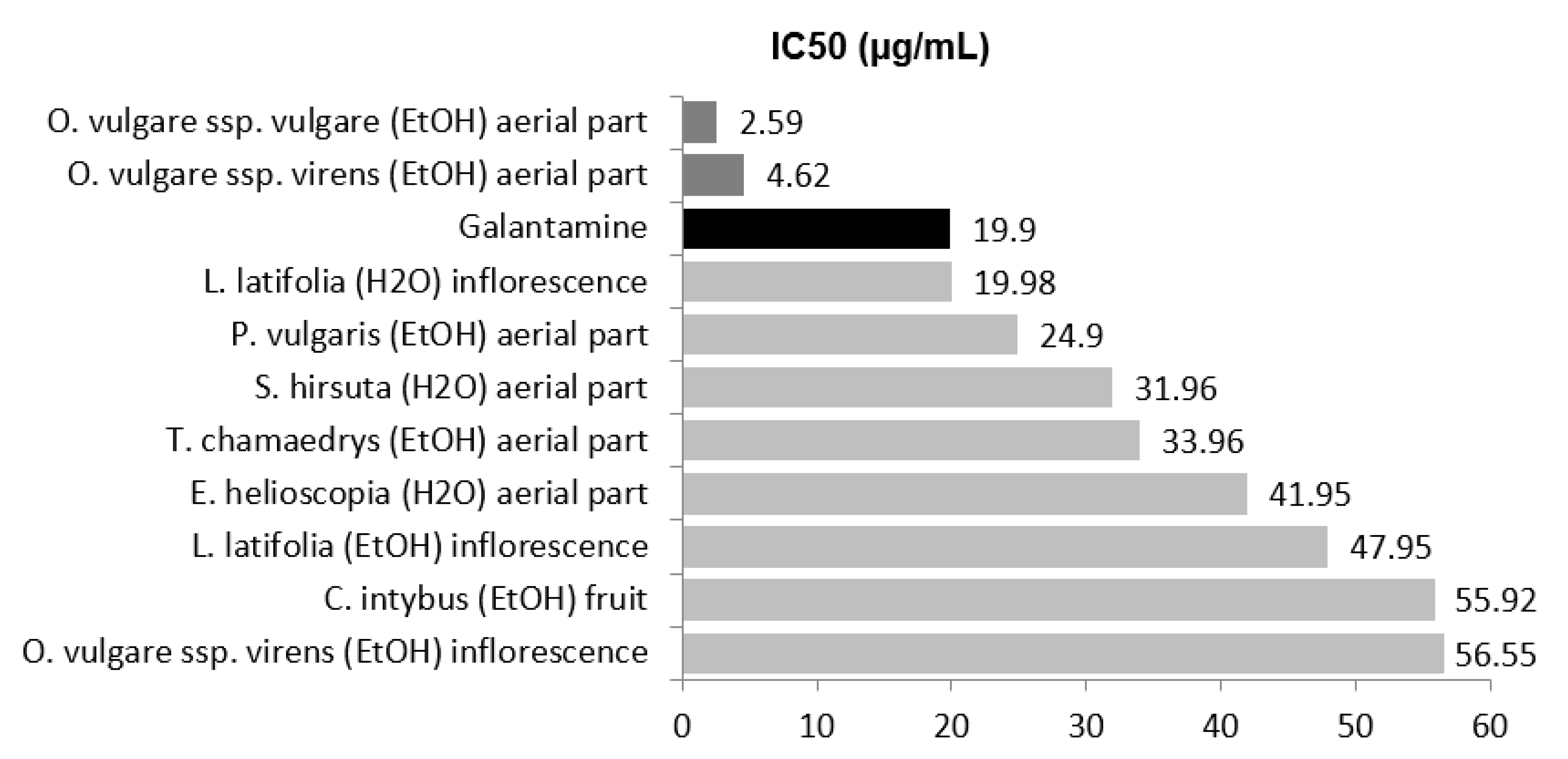
2.1. Antiacethylcholinesterase Activity

In the last two decades, the mechanism of inhibition of AChE has acquired high importance in treating AD symptoms from a clinical point of view. Some extracts and phytochemicals have shown this activity [7]. Several methods have been described for the determination of AChE inhibitory activity, such as colorimetric methods using Ellman’s reagent or Fast Blue B salt reagent, fluorometric methods or HPLC online detection. Ellman´s method, which is based on the determination of the amount of thiocholine released when acetylthiocholine is hydrolyzed by AChE, is the most widely employed method because it is simple and gives quick access to information in plant extracts [8].
In order to select plant extracts with high AChE inhibitory activity, in this study, 90 ethanolic and aqueous extracts of 38 medicinal plants collected in Navarra (Spain) were analyzed. These medicinal plants, belonging to nine botanical families (Asteraceae, Lamiaceae, Crassulaceae, Equisetaceae, Euphorbiaceae, Lythraceae, Papaveraceae, Primulaceae and Verbenaceae), showed high antioxidant activity in previous researches of our group [9,10].
Qualitative screening by TLC showed that 20 out of the 90 extracts were inactive at doses of 0.20 mg. Since the crude extracts may sometimes yield false positive or negative results in the TLC assay, a quantitative microplate assay was performed. In total, 20 extracts showed an inhibition rate below 10% at a dose of 250 μg/mL (Table 1). A summary of screening studies of these extracts is provided in Table 1, alphabetically ordered by family, showing scientific name, botanical part, extraction solvent, yield of extraction, percentage of inhibition and concentration at which 50% of the enzyme is inhibited. Seventy extracts showed inhibitory activity towards acetylcholinesterase at a concentration of 250 μg/mL, 21 of them with high activity (75–100% inhibition), 34 with moderate activity (50–75% inhibition) and 15 with low activity (10–50% inhibition) (Table 1).
The screening of 30 extracts from 11 medicinal plants of the Asteraceae family revealed that two of them exhibited very strong activity with inhibition percentages higher than 75%: the aqueous extract from the leaves of Tussilago farfara (80.25 ± 13.78%) and ethanolic extracts from the inflorescence of Santolina chamaecyparissus (75.82 ± 6.69%) at 250 μg/mL. Ethyl acetate extracts of T. dubius and T. farfara have been described as potent inhibitors of acetylcholinesterase and butyrylcholinesterase [11].
In relation to S. chamaecyparissus, only the essential oil obtained by steam distillation has been described as a control agent against termites due to this activity [12]. It has also been described this activity in the essential oil of other species from Santolina, such as S. impresa [13] and S. semidentata [14]. However, it is important to highlight that the ethanolic extract of S. chamaecyparissus has a different chemical composition compared to the essential oil and contains non-volatile compounds, which could be a source of new bioactive compounds.
Lamiaceae species have been reported to possess a wide range of biological activity and a diversity of phytochemicals. This botanical family is rich in essential oils, hydroxycinnamic acids and flavonoids as active constituents, which significantly contribute to its neuroprotective properties. For this reason, the anti-AChE activity of this family has been widely studied [15]. As can be seen in Table 1, Lamiaceae family extracts were generally stronger than the ones of the Asteraceae family. Out of the 42 ethanolic and aqueous extracts of 18 medicinal plants that were tested, 13 extracts showed high inhibitory activity, achieving values above 75%, whereas another 14 achieved moderate inhibition of the AChE (values between 50 and 75%) at a concentration of 250 μg/mL.
The ethanolic extract of the inflorescence, stem and leaf from Lavandula latifolia showed values higher than 90% at 125 μg/mL. The aqueous extract also showed similar values at 250 μg/mL. There is bibliographic information about the AChE inhibitory activity of the essential oils from L. angustifolia and L. intermedia [16], L. luisieri [17], L. pedunculata [18], L. stoechas [19] and L. viridis [20]. However, no investigations about L. latifolia have been found.
In this study, differences were detected between four Mentha species, and the best inhibitory results were obtained with ethanolic and aqueous extracts from M. longifolia, at 77.98% and 90.45%, respectively. M. aquatica, M. pullegium and M. suaveolens showed lower activity. It is worth mentioning that there are similar data in the literature for the essential oil of the following aforementioned Mentha species: M. longifolia [21], M. aquatic [22], M. arvensis [23], M. gentilis [24], M. piperita and M. spicata [25], M. pulegium [26] and M. suaveolens [27].
The ethanolic extracts of aerial parts from two Origanum vulgare subspecies, virens and vulgare, showed similar results, with inhibition percentages of 91.75 ± 1.38 and 95.61 ± 2.02%, respectively. Both extracts also showed the lowest IC50 values, 4.62 ± 0.01 and 2.59 ± 0.01 μg/mL, which are even lower than values for galantamine (19.90 ± 4.80 μg/mL). These results are similar to those published by other authors for O. vulgare subspecies [28] and other closely related species such as O. majorana [29], O. compactum [30], O. syriacum [31] and O. ehrenbergii [32]. The difference between these works and our study relies on the polarity of the extracts. Most of the previous analyses were determined in essential oil or hydrophobic extracts (dichloromethane or ethyl acetate solvent).
A clear difference between the two extracts of Prunella vulgaris was found; whilst the ethanolic extract provided an inhibition percentage of 78.86 ± 7.39% at 250 μg/mL, the aqueous extract could be considered inactive (< 10%). To the best of our knowledge, there are only two literature data concerning the AChE inhibitory properties of the genus Prunella. Park et al. [33] studied the effects of the ethanolic extract of the flower of P. vulgaris var. lilacina on drug-induced memory impairment, concluding that this plant would be useful for treating cognitive impairments induced by cholinergic dysfunction and that it exerts its effects via NMDA receptor signaling. Qu et al. [34] determined that ethyl acetate extracts of P. vulgaris attenuated scopolamine-induced memory impairment in rats by improving behavioural performance and decreasing brain cell damage, which was associated with a notable reduction in AChE activity and MDA level, as well as an increase in SOD and GPx activities.
The following two different species of Sideritis have been analyzed: S. hirsuta and S. hyssopifolia. Only the aqueous extract of S. hirsuta showed high AChE inhibitory activity (93.27 ± 3.85%). No results about these species have been published; however, bibliographic information about the anti-AChE activity of this genus has been found for S. arborescens [35], S. congesta [36], S. arguta, S. libanotica, S. perfoliata and S. pisidica [37].
Ethanolic and aqueous extracts of Teucrium chamaedrys showed higher activity (98.09 ± 1.10% and 98.96 ± 9.85%) than galantamine at 250 μg/mL. These results are corroborated by the investigation of different species of Teucrium genus against AD, T. arduini, T. chamaedrys, T. montanum and T. polium [15], T. hyrcanicum [38] and T. royleanum [39]. The methanolic extract of T. royleanum and its fractions were also examined as inhibitors of acetylcholinesterase and a significant enzyme inhibition activity (52–83%) was found [39].
The genus Thymus contains about 350 species of aromatic perennial herbaceous plants and subshrubs. Many studies focused on the in vitro inhibitory activity of essential oil from the plants of this genus on acetylcholinesterase [40,41]. In this sense, our results are in accordance with them, ethanolic extract of T. praecox and T. vulgaris showed high AChE inhibition, 97.81 ± 10.90 and 82.48 ± 9.05%, respectively.
Eighteen extracts from seven different families (Crassulaceae, Equisetaceae, Euphorbiaceae, Lytraceae, Papaveraceae, Primulaceae and Verbenaceae) were studied. Six of them showed high AChE inhibition (>75%). The ethanolic extracts of E. arvense and E. telmateia demonstrated a similar effect and were more effective than the aqueous ones, with inhibitory values higher than 84% at 250 μg/mL. Since both species are close botanically and chemically, similar pharmacological results were to be expected.
Euphorbiaceae is a large family of flowering plants with around 300 genera and 7500 species. Euphorbia species contain glucosinolates and cyanogenic glycosides, such as linamarin, in different proportions. The quantitative difference in the chemical composition could justify the variability of results found between the aqueous extracts of the two species, E. characias (44.25 ± 5.01 mg/mL) and E. helioscopia (78.74 ± 4.15 mg/mL). Finally, the aqueous extracts of Lytrum salicaria (Lytraceae) and Anagallis arvensis (Primulaceae) and the ethanolic extract of Papaver rhoeas (Papaveraceae) also showed high AChE inhibitory activity (98.50 ± 13.50, 80.02 ± 1.25 and 99.78 ± 7.57 mg/mL, respectively). It is important to highlight the different chemical compositions of these species, L. salicaria is rich in tannins; A. arvensis in saponins and P. rhoeas in alkaloids. To the best of our knowledge, there is no anti-AChE activity reported in any of them, except for Euphorbia species, E. antisyphlitica [42], E. characias [43], E. hirta [44], E. royleana [45], E. splendens [46], E. tirucalli [47], E. fischeriana [48] and Papaveraceae [49].
Half-maximal inhibitory concentration (IC50) is the most widely used measure of a drug’s efficacy in pharmacological research. It indicates how much drug is needed to inhibit a biological process down to half, thus providing a measure of the potency of an antagonist drug. The potential anti-AChE can be classified into the following categories based on the IC50 values: high potency, IC50 < 15 µM; moderate potency, 15 < IC50 < 50 µM; low potency, 50 < IC50 < 1000 µM [7]. Figure 1 shows the TOP 10 extracts in relation to their IC50 value and the comparison with galantamine.
Two alcoholic extracts presented higher anti-AChE potency than galantamine (19.9 ± 4.80 µg/mL), aerial parts of O. vulgare ssp. virens (4.62 ± 0.01 µg/mL) and O. vulgare ssp. vulgare (2.59 ± 0.01 µg/mL). The aqueous extract of inflorescence from L. latifolia showed an IC50 value (19.98 ± 0.49 µg/mL) equal to galantamine.
Based on the results of the screening, the ethanolic extract from aerial parts of O. vulgare ssp. vulgare showed the best anti-AChE activity, 7,7 times higher than galantamine. For this reason, the investigation continued with the chemical characterization of this extract. Antioxidant activity and total phenolic compounds were also determined. Finally, in order to establish structure-activity relationships, the results were analyzed by a correlation matrix.

2.2. Chemical Characterization of Origanum vulgare ssp. vulgare Aerial Parts

For chemical characterization, thin-layer chromatography, high-performance liquid chromatography with diode array detection (HPLC-DAD) and liquid chromatography-mass spectrometry (LC-MS) were used. Besides, the chemical characterization was complemented with the determination of total phenolic compounds.

2.2.1. Total Phenolic Compounds Determination

Total phenolic compounds (TPC) were spectrophotometrically quantified following the Folin–Ciocalteu colorimetric method [50]. In this assay, phenolic compounds are oxidized in an alkaline medium by the Folin–Ciocalteu reagent (composed of a mixture of phosphowolframic acid and phosphomolybdenic acid) producing a reduced mixture of blue oxides of tungsten and molybdenum that can be quantified at 765 nm. The TPC of ethanolic extract was 207.64 ± 0.69 µg/mg of lyophilized extract. Previous studies with oregano also determined the TPC of the extracts [51], and sometimes they showed different results to the ones obtained in this work. To explain these differences, it is important to highlight that the chemical composition of an extract varies depending on the plant material, the growing conditions and the preparation method (solvent, time, temperature).

2.2.2. Identification and Quantification of Main Groups of Phenolic Compounds by TLC and HPLC-DAD

The chemical composition of the ethanolic extract of O. vulgare was firstly qualitatively analyzed by TLC with two different mobile phases (Figure 2a,b). Both TLC plates, after exposition to natural products reagent (NP), allowed the identification by the colour of the main compounds.
At the top of the TLC plate (Rf = 0.90) developed with ethyl acetate:methanol:water (65:15:5, v/v/v), a pink colored spot was detected. These spots could be chlorophylls since the aerial parts of O. vulgare were used as starting material. Wagner and Bladt [52] found similar fluorescent spots with high Rf values (> 0.70) on the TLC plate and identified them as chlorophylls. Chlorophylls are green pigments involved in photosynthesis and located in the leaves of plants. Blue spots, a characteristic colour of phenolic acids, with Rf = 0.70, 0.55, 0.40 and 0.25 were also detected. Phenolic acids have been described for O. vulgare [53,54,55], being the most importants 3,4-dihydroxybenzoic acid [56,57,58], rosmarinic acid [50,53,59] and caffeic acid [53,60,61]. TLC showed a yellow spot at the bottom of the plate (Rf = 0). This colour indicates the presence of flavonoids [52], potentially bioactive compounds already described in O. vulgare [50,55,62,63,64].
To confirm the chemical profile of the extracts, complementary TLC plates were prepared by modifying the mobile phase. According to Wagner and Bladt [52], ethyl acetate:glacial acetic acid:formic acid:water (100:11:11:26, v/v/v/v) is one of the best mobile phases to detect flavonoids and phenolic acids after NP treatment. At first sight, the separation of compounds was better than with the first mobile phase. The yellow spots at the baseline on the previous TLC were here separated into several spots. The presence of phenolic acids (in blue), flavonoids (in yellow), and chlorophylls (in pink) was also confirmed. The same profile was described previously in oregano hydroalcoholic extract [65].
TLC is a qualitative chromatographic technique in which neither the intensity of the bands should be used as a formal quantification nor the color given under certain conditions (reagent and observation wavelength) can be used for the identification of compounds beyond their chemical group (chlorophylls, flavonoids, phenolic acids...). To obtain quantitative results, techniques such as high-performance liquid chromatography (HPLC) should be used. The HPLC-DAD provides separation and the UV spectrum of compounds, allowing their assignment to a specific chemical group [66]. In this way, the peaks were grouped into the following six groups based on their UV spectrum: dihydroxycinnamic acids (λmax 325–329 nm), dihydroxybenzoic acids (λmax 220, 259.4, 293.7 nm), syringic acids (λmax 220 sh, 260–280 nm), essential oils with an aromatic ring (λmax 254 nm), salvianolic acids λmax 289, 323 sh nm) and flavonoids (λmax 254.6–267, 338–348.5 nm) (Figure 2c). The main compounds were luteolin derivative (31.4 min), 3,4-dihydroxybenzoic acid (31.8 min) and rosmarinic acid (37.9 min), previously described [65]. Dihydroxybenzoic acids, syringic acids, dihydroxycinnamic acids and salvianolic acids are phenolic acids obtained through the shikimic acid pathway in plants, but they were considered as different groups in the quantification and discussion of our results.
The area under curve (AUC) of each peak was transformed into concentration by linear regression analysis [65]. Ethanolic extract showed 15.56 ± 0.14 mg/100 mg of flavonoids expressed in terms of luteolin (#L9283, Sigma-Aldrich Co., St. Louis, MO, USA); 35.35 ± 1.13 mg/100 mg of dihydroxycinnamic acids (24,21 ± 1.08 mg was rosmarinic acid) and 11.14 ± 0.15 mg/100 mg of salvianolic acids expressed in terms of caffeic acid (#C0625, Sigma-Aldrich Co., St. Louis, MO, USA); 5.23 ± 0.04 mg/100 mg of dihydroxybenzoic acids and 2.89 ± 0.24 mg/100 mg of syringic acids expressed in terms of 3,4-dihydroxibenzoic acid (#D109800, Sigma-Aldrich Co., St. Louis, MO, USA). Essential oils typical of oregano showed low-intensity peaks in HPLC-DAD, so they were not considered in the chemical quantification. Figure 2d shows the distribution of the main group expressed in percentage. The most abundant compounds are dihydroxycinnamic acids (56.90%), with rosmarinic acid being the highest percentage (38.97%) and flavonoids (25.94%).

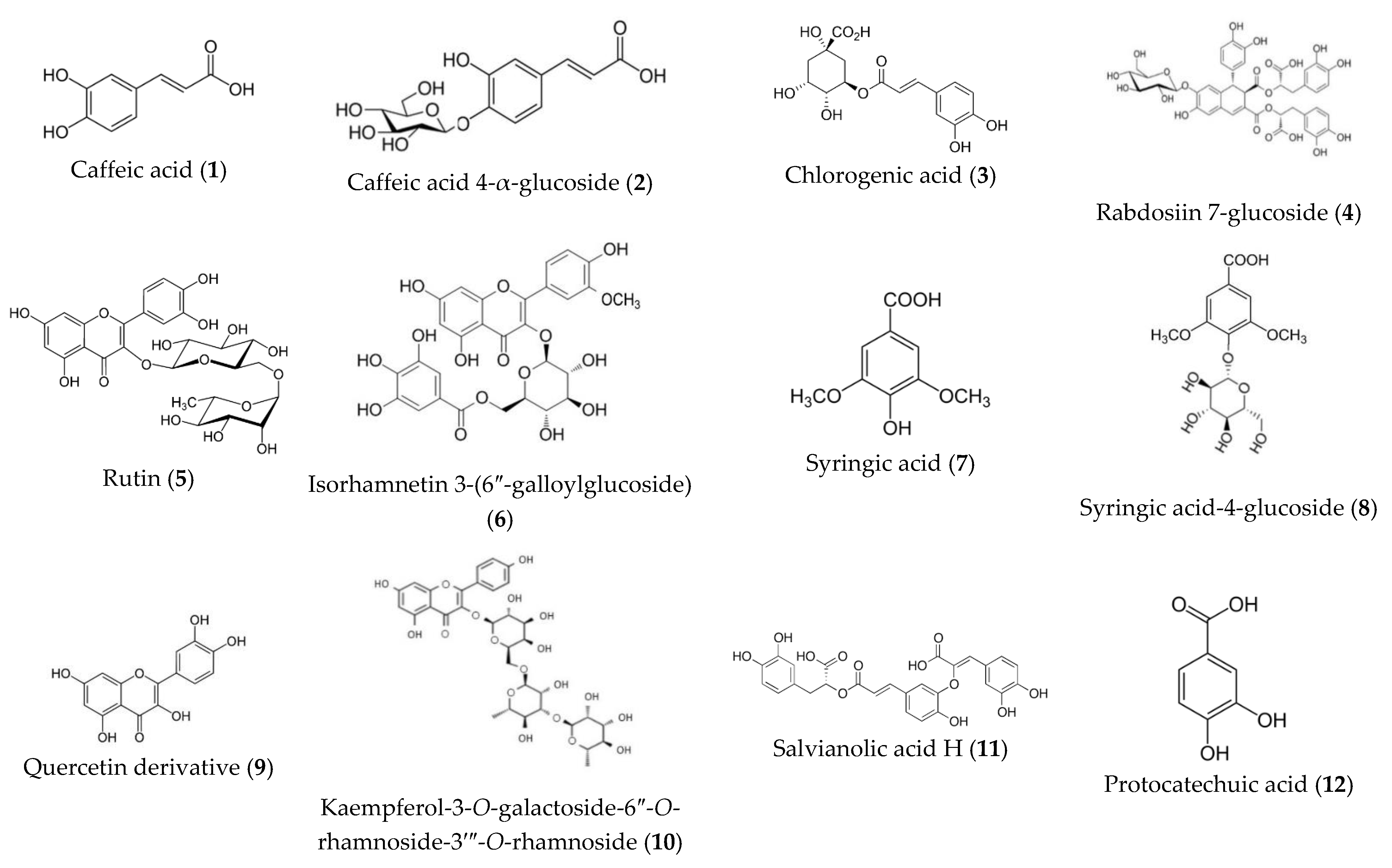
2.2.3. Identification of Main Compounds by LC-ESI-QTOF-MS

After separating compounds from a sample by liquid chromatography (HPLC-DAD), highly sensitive instrumental analytical techniques, such as mass spectrometry (LC-ESI-QTOF-MS), can be applied for the identification of individual compounds. This technology is based on the ionization of the separated compounds to obtain structural information [67]. A large number of secondary metabolites are glycosylated compounds and the fragmentation by LC-MS allows the revealing of the main structure and the attached sugars, making it a useful technique for the phytochemical identification of compounds extracted from plants.
The peaks were preliminarily assigned to a family of phenolic compounds based on their UV-vis spectra. The structure of each compound was proposed based on fragmentation patterns using ESI-MS-MS experiments as well as by co-elution with several standards. In total, 23 compounds were thus detected and identified or tentatively identified. They are listed in Table 2, with UV-visible and MS data.
Figure 3 shows the structures of identified compounds. The identification of tentatively characterized compounds present in the oregano aerial part’s extract is explained below.
Dihydroxycinnamic acids (λmax 316–331 nm) were detected first in LC-MS analysis. They were also previously described in Lamiaceae species [53,58,68]. These compounds could be related to the blue spots on TLC (mobile phase: ethyl acetate:glacial acetic acid:formic acid:water (100:11:11:26, v/v/v/v)).
One monomer, caffeic acid (compound 1), was identified at 0.9 min (m/z 179.05, λmax 296sh, 324 nm). This compound yielded an ion at m/z 179.05 [M-H] and a prominent fragment at m/z 135.04 [M-H-44] through the loss of a CO2 group. The extract also showed four more complex forms of caffeic acid (compounds 2, 3, 4 and 17). Fragment m/z 179.05 of caffeic acid appears in the mass spectra of all of them. Compound 2 at 1.1 min showing [M-H] at m/z 341.07 and [M-H-162 (glucose residue)] at m/z 179.03 was tentatively characterized as caffeic acid 4-α-D-glucoside. The loss of 162 amu is likely due to the cleavage of a glucose moiety. Chlorogenic acid (compound 3), a combination of caffeic acid and quinic acid (m/z 354.31), was also detected at 1.3 min. The highly intense characteristic ion at m/z 191.01 [M-H-162 (caffeoyl residue)] corresponding to quinic acid confirmed the structure. Compound 17 was assigned to rosmarinic acid, an ester of caffeic acid and 3,4-dihydroxyphenyl lactic acid and it is widely described in Lamiaceae and Boraginaceae families. The MS fragmentation of rosmarinic acid pseudomolecular ion (m/z 358.97) lead to three peaks at m/z 197.01 [M-H-162 (caffeoyl)], 179.05 [M-H-180] and 161.02 [M-H-198], corresponding to the deprotonated form of 3-(3,4-dihydroxyphenyl) lactic and caffeic acids and their dehydrated forms. These results agree with the fragmentation scheme proposed by Lecomte et al. [69]. Rosmarinic acid, whose name derives from Rosmarinus officinalis L., has been identified as one of the most active compounds in several plants from the Lamiaceae family, such as rosemary and oregano [70]. Its identification by HPLC-DAD and LC-MS is widely reported in the literature [20,50,58,71]. Definitive elucidation of these structures was also confirmed by co-injection with reference standards. Compound 4 was identified as rabdosiin 7-O-β-D-glucoside (m/z 879.05 [M-H], λmax 287 sh, 329 nm). This compound is a caffeic acid tetramer connected to a lignan skeleton (m/z 718.6) and a glucose unit [M-H-162]. Originally, rabdosiin has been isolated and identified from the stem of Rabdosia japonica Hara, Labiatae [72]. According to published data, it has been suggested as a potential anti-HIV, antiallergic, and antiproliferative agent [73]. This is the first report about the presence of rabdosiin 7-O-β-D-glucoside in Origanum species.
The second group of compounds, dihydroxybenzoic acids from the shikimic acid pathway, was identified. These acids, showing a blue spot on the TLC plate, can be the result of the transformation of caffeic acid and have been described in previous studies with oregano [57,74]. According to the retention time (6.5 min), UV-spectra (λmax 217.0, 261.7, 294.9 nm), m/z 153.01 [M-H] and a fragment ion [M-H-44 (CO2)] at m/z 109.02, compound 12, the major component, could be identified as the 3,4-dihydroxibenzoic acid or protocatechuic acid, also previously described in O. vulgare [66,75,76]. Final identification was carried out by co-injection of 3,4-DHBA standard (#D109800, Sigma-Aldrich Co., St. Louis, MO, USA).
The third type of UV peak (λmax 220 sh, 280 nm) corresponds to the group of syringic acids, already described in O. vulgare [54,63,77]. Syringic acids are phenolic compounds strictly named 4-hydroxy-3,5-dimethoxybenzoic acids, synthesized from ferulic acid and caffeic acid by a series of enzymatic reactions in the shikimic acid pathway [70,78]. Despite being derivates of dihydroxybenzoic acid, they present a different UV spectrum. For this reason, these compounds are treated separately. Two syringic acids were detected (compounds 7 and 8), at 3.4 and 3.7 min. Compound 7 was identified as syringic acid (m/z 197.03 [M-H]) and compound 8 as a glycosylated variation, syringic acid-4-β-D-glucoside (at m/z 359.08 [M-H] and m/z 197.04 [M-H-162].
Most of the published studies on oregano use essential oils as plant material due to the important bioactivity of these compounds [74,79,80]. As they are volatile, only one essential oil (compound 19) was detected in this study. Compound 19, (λmax 254.4 nm, m/z 149.1 [M-H]-) is identified as thymol, the most important essential oil in oregano. Final identification was carried out by co-injection of the thymol standard.
The next group was one of the salvianolic acids, which was also previously reported in oregano [74,81,82]. From a chemical point of view, they are considered a large group of acids whose names are attributed with letters as follows: salvianolic acid A, B, E,… These compounds have a complex chemical structure derived from rosmarinic acid, and they were distinguished from dihydroxycinnamic acids because they showed a different UV spectrum (λmax 289, 323 sh nm). In LC-MS, four salvianolic acids were identified (compounds 11, 14, 15, 16 and 18). Fragments m/z 179.04 of caffeic acid appear in the mass spectra of all detected salvianolic acids. Compound 11 (6.3 min), caffeic acid trimer with deprotonated ion [M-H] at m/z 537.09 was assigned as salvianolic acid H or salvianolic acid I (pair of isomers). Compounds 14 (6.8 min), 15 (7.1 min) and 18 (7.5 min), caffeic acid tetramers, generated the same pseudomolecular ion [M-H] at m/z 717.12 and were identified as salvianolic acid E, salvianolic acid B and salvianolic acid L, respectively. Finally, a caffeic acid hexamer, compound 16, at 7.6 min and with a pseudomolecular ion m/z of 987.22 was detected. The final identification of these compounds was determined by comparison with retention times and MS fragmentation data [83], except for compound 16, whose structure was not completely elucidated.
The last chemical group present in oregano and Lamiaceae is flavonoids [71,84]. These secondary metabolites are generally present in glycosylated forms, with the main molecule attached to one or more sugars (glucose, galactose) [85]. In UV-spectra, two separated and characteristic shoulders easily identify these compounds. Nine flavonoids with three different types of spectra were detected. Compounds 5, 6, 9, 10 and 21 showed typical UV spectra of the flavonol. Compounds 5 (2.4 min) showed molecular ions at m/z 609.17 [M-H] and was identified as rutin (quercetin-3-O-rutinose, λmax 254.6, 348.5 nm), producing an MS ion at m/z 463.3 [M-H-146], by loss of rhamnose moiety and a quercetin ion at m/z 301.8 [M-H-146-162]. The loss of 308 amu is characteristic of compounds having rutinose. In a similar way, compound 21 (8 min) was identified as syringetin 3-O-rutinoside, m/z 653.14 [M-H], 507.4 [M-H-146] and 345.07 [M-H-146-162]. Syringetin is a dimethoxyflavone, myricetin, in which the hydroxy groups at positions 3′ and 5′ have been replaced by methoxy groups. The ion m/z 330.1 [M-H-308-15] and 315.2 [M-H-308-30] confirmed the presence of two -OCH3 groups. Compound 10 (5.8 min) with λmax 266, 346 nm in the DAD spectrum and showing a molecular anion at m/z 739.05 in the negative ESI spectra, and ions at m/z 659.07 [M-H-146], m/z 447.01, [M-H-146-146] was identified as a kaempferol-3-galactoside-6″-rhamnoside-3′″-rhamnoside. Cleavage of this glycoside gave the aglycone at m/z 285.03 [M-H], kaempferol. Compound 6 (2.5 min) was an isorhamnetin derivate with m/z 315.02 [M-H-314] corresponding to the aglycone that has lost galloylhexoside fragment. The mass spectrum of compound 6 showed the fragment m/z 447.03 [M-H-152] corroborating the galloyl substitution. Moreover, the additional mass loss of 162 amu confirmed the presence of a hexoside (glucoside or galactoside). Compound 6 was thus identified as isorhamnetin 3-(6″-galloylglucoside). Finally, a quercetin oxalate was detected at 5.6 min (compound 9) with m/z 388.20 [M-H] and m/z 301.80 [M-H-87].
Compound 20 (7.9 min) is a 5-hydroxy-3,3′,4′,7-tetramethoxyflavone, namely, retusin with the ion m/z 357.09 [M-H] and λmax 350, 268 nm. The ions at m/z 342.12 [M-H-15], 327.07 [M-H-15], 312.02 [M-H-15] and 297.02 [M-H-15] confirmed the four methoxyl groups.
In the MS identification of C-glycosides, the key fragmentations used were [M-60], [M-90], [M-120] and [M-240]. Compounds 22 (orientin) and 23 (homoorientin) were identified as C-glycosyl derivatives of luteolin (λmax 254, 267 nm). Ion fragmentation of both were m/z 447.05 [M-H], 357.78 [M-H-90] and 327.22 [M-H-120] and 285.5 [M-H-162].
Finally, one flavanone glycoside (compound 13) at 6.6 min and λmax 285, 325 nm was identified as eriodictyol-7-O-glucoside. Ions at m/z 449.19 [M-H] and m/z 287.05 [M-H-162] confirmed the structure.
Rosmarinic acid, apigenin, luteolin and quercetin are the most recurrent compounds in this Lamiaceae species [50,59,86]. With increasing evidence of the biological activity of flavonoids and phenolic acids from oregano species, quantification of these compounds is important. Reports from different oregano species have shown that flavones are the most abundant flavonoid subgroup, followed by flavonols, flavanones and flavanols [87]. The most common phenolic acids in oregano are hydroxycinnamic acid and hydroxybenzoic acid derivatives [87]. However, their content and distribution can vary depending on geographical, environmental growing factors and the vegetative stage of the plant [88,89], showing a different chemical profile within the same species [54]. For these reasons, it is very important for the chemical characterization and the establishment of quimiotaxonomic markers for each species and subspecies. In aerial parts of O. vulgare spp vulgare, rosmarinic acid and 3,4-dihydroxybenzoic acid could be two optimal candidates for markers.

2.3. Antioxidant Activity

Phenolic compounds are secondary metabolites present in a wide range of medicinal plants with a chemical structure that can act as an H donor, making them potentially antioxidant compounds. Molecular oxygen (O2) is involved in metabolic functions. However, it can also be present as short-lived highly reactive derivatives (reactive oxygen species—ROS) as the result of these enzymatic reactions. Superoxide (O2•−), hydrogen peroxide (H2O2) and the hydroxyl radical (OH) are some of these derivatives that can cause cell damage [90]. They can affect DNA and polyunsaturated fatty acids in the membrane [91]. Organisms are prepared to counteract these damages through antioxidant defense systems. However, according to the aging and free radical theory, the effectiveness of these protective systems tends to decrease with age, and the accumulation of these harmful molecules can create pathologies in the body, developing diseases such as Alzheimer’s and diabetes [90,92]. Most of the current research with natural products is focused on finding external co-adjuvants to counteract this oxidative damage, either as prevention or treatment [93,94]. Compounds that are able to counteract this oxidative damage are called antioxidants. As an exogenous aid to prevent damage to the body, these antioxidant compounds can reduce the formation of these free radicals or neutralize them [90].

2.3.1. Antioxidant Activity In Vitro against DPPH Radical

Among in vitro assays, the DPPH-based method is probably the most popular one due to its simplicity, speed and low cost. DPPH (1,1-diphenyl-2-picrylhydrazyl) is a stable free radical that can be reduced by transferring hydrogen from other compounds. Since 1995, when Brand-Williams first published and discussed in depth the methodology [95], some variants have been developed. Depending on the equipment and the interest of the study, the reaction can be quantified at a pre-defined time (30 min. mainly) or kinetic studies can be performed. Nonetheless, the principle of the reaction is always the same as follows: the reduction of DPPH is followed by monitoring the decrease in its absorbance at a characteristic wavelength during the reaction. In its radical form, DPPH absorbs at 517 nm, but upon reduction by an antioxidant (AH) or a radical species (R), the absorption disappears.
In fact, as Brand-Williams recommends, the reaction was monitored over time to establish a kinetic scale depending on the stabilization time-point of the reaction. A sample is considered a fast antioxidant if the stabilization point of the reaction is reached before 30 min, an intermediate antioxidant if it stabilizes between 30 and 60 min, and a slow antioxidant if it needs more than 60 min to stabilize. The reaction is stable when no statistical differences (p > 0.05) are observed between two consecutive values [96]. Ethanolic extract was an intermediate antioxidant (stabilization points between 30 and 60 min) with an IC50 = 3.22 ± 0.19 µg/mL at 60 min (Table 3).
IC50 values of antioxidant activity depend on the concentration of DPPH, and this makes difficult the comparison with other published studies. Nevertheless, the antioxidant activity index (AAI), which is independent of the concentration of DPPH, can be calculated by dividing the concentration of DPPH in the final solution (20 µg/mL) by the IC50 value [97]. This index determines the strength of the antioxidant activity regardless of the concentration of DPPH. According to the current classification, plant extracts are considered poor antioxidants when AAI < 0.5, moderate when AAI is between 0.5 and 1.0, strong if AAI is between 1.0 and 2.0 and very strong antioxidants when AAI > 2.0. In this sense, the antioxidant activity index (AAI) was also calculated to determine the strength of the antioxidant activity of the extracts regardless of the concentration of DPPH. The results showed that the extract was a very strong antioxidant, with an AAI = 6.21 ± 0.10 (Table 3).

2.3.2. Antioxidant Activity In Vitro against ABTS Radical

As a complement to DPPH antioxidant determination, the Trolox equivalent antioxidant capacity (TEAC) method, also known as the ABTS radical cation decolorization assay, was performed in vitro. This assay determines, through a simple and inexpensive protocol, the ability of an antioxidant compound to counteract the free radical ABTS. Unlike other common in vitro antioxidant tests to determine this activity, this method does not require enzymes or special conditions [94]. In addition, the method could be applicable to the study of hydrophobic and hydrophilic antioxidants. The ABTS in vitro assay was carried out according to García-Herreros et al. [98]. It is based on the formation of an ABTS cation radical that exhibits a colour change that is measurable by spectrophotometry at 741 nm. The assay was performed with the extract at a 1 mg/mL concentration and the results were expressed as the amount of Trolox (TE) per mg of lyophilized extract, after substituting the data in the Trolox calibration curve. The antioxidant activity was 34.24 ± 0.20 mg/100 mg of extract.

2.4. Chemical Composition—Biological Activity Relationship

Correlation is a type of association between two countable variables that evaluates the trend in the data (positive or negative). In a correlation, a positive value indicates a positive direct relationship, while a negative value indicates a negative indirect relation between the variables. The magnitude indicated the strength of the link, being values between −1 and 1. The closer to the unit, the stronger the relationship, which on a graph is generally observed as a smaller dispersion of the values. One of the most widely used coefficients for calculating lineal correlation is Pearson’s, which assumes that the trend must be linear, there are no outliers and the variables must be numeric with a reasonable number of values.
The Pearson correlation coefficients, which show the relationship between the biological activity of the ethanolic extract of O. vulgare ssp. vulgare and its chemical composition, are presented in Table 4.
The AChE-IC50 activity was strongly correlated (R2 > 0.85) in a linear, negative manner to antioxidant activity (DPPH-AAI (R2 = −0.8649) and ABTS (R2 = −0.9487)), syringic acids (R2 = −0.9864), flavonoid (R2 = −0.9563) and dihydroxybenzoic acids’ (R2 = −0.9247) content. A moderate and negative correlation (R2 = 0.76) between AChE-IC50 activity and dihydroxycinnamic and rosmarinic acid content was also observed. Free DPPH radical scavenging activity, expressed as antioxidant activity index (AAI), had a strong correlation to ABTS activity (R2 = 0.9378) and all the main compounds analyzed (R2 > 0.9). These results are according to many studies of the activity of polyphenols as AChE inhibitors, which, in addition to inhibiting AChE activity, also have an antioxidant effect, including scavenging free radical forms of oxygen and the ability to chelate transition metals, which reduces the formation of inflammation that can cause the destruction of neuronal structures [99].
The neuroprotective effect of flavonoids and dihydroxycinnamic acids has been widely studied by many authors [2,15,100]. The inhibitory effect on AChE activity was also reported for individual phenolic acids, in the following order: rosmarinic acid > caffeic acid > gallic acid = chlorogenic acid > homovanillic acid > sinapic acid. Flavonoids, such as quercetin, kaempferol and, to a lesser extent, luteolin were also reported as efficient AChE inhibitors [101].
However, it is important to highlight the AchE activity of syringic acids. To our knowledge, there are currently a few works focused on them [102]. Syringic acids show a wide range of therapeutic applications in the prevention of diabetes, CVDs, cancer, cerebral ischemia; antioxidant, antimicrobial, anti-inflammatory, antiendotoxic, neuro- and hepatoprotective activities have been described [103]. Recently, a study analyzed 16 hydroxybenzoic acids using calorimetry and docking simulation as AchE inhibitors. All tested compounds were shown to inhibit the hydrolysis of ACh, and the best properties were shown by methyl syringinate; syringic acid also showed a high inhibition percentage [104]. Considering that AChE inhibitory potential has been mainly investigated for essential oils in the Lamiaceae family, these findings suggest the great influence of other chemical constituents such as syringic, which may have great relevance in pharmacological fields and open a new research line.

3. Materials and Methods

3.1. Plant materials and extraction

Plants were collected in Santacara, Navarra, Spain, (Longitude: O1°32′38.33″ and Latitude: N42°22′47.71″) and identified by the botanist, Dr. Rita Yolanda Cavero. Voucher specimens have been deposited in the PAMP Herbarium of the University of Navarra. Plants were air-dried in the dark at room temperature. All species are listed in Table 1.
Plant materials (10 g) were ground into fine powder (180 mesh) and extracted by maceration with 250 mL of ethanol (EtOH) and water (H2O) at room temperature in a closed container (3 times each 24 h). The extracts were dried under reduced pressure at 30 °C in a rotary evaporator (Buchi R-300) and then were lyophilized (Virtis BT3-SL, NY, EEUU). Finally, the dry extracts were stored in glass vial at −80 °C.

3.2. Antiacetylcholinesterase Activity

A qualitative antiacetylcholinesterase activity was studied by TLC according to the method described by Uriarte-Pueyo and Calvo [105]. Extracts and galantamine (#Y0001191, Sigma-Aldrich Co., St. Louis, MO, USA) were spotted at 0.20 mg onto the TLC plate and developed with ethyl acetate:methanol:water (65:15:5, v/v/v) as mobile phase. Then, the plates were sprayed with DTNB or 5,5′-dithiobis(2-nitrobenzoic acid) (#D218200, Sigma-Aldrich Co., St. Louis, MO, USA)/ATCI or acetylthiocholine iodide (#01480, Sigma-Aldrich Co., St. Louis, MO, USA) 1:1. It was allowed to dry for 3–5 min and 3 U/mL of acetylcholinesterase (AchE) (#C2888, Sigma-Aldrich Co., St. Louis, MO, USA) solution was sprayed. After AChE application, a yellow background appeared, with white spots for AChE inhibiting extracts or compounds.
Quantitative AChE inhibitory activity was measured by spectrophotometric method developed by Rhee et al. [106] and modified by Carpinella et al. [107]. The lyophilized extracts were diluted in their corresponding solvent (ethanol or water) to give a stock solution of 20 mg/mL and three serial solutions were prepared (10–2.5 mg/mL). Twenty-five μL of each solution was added to 25 μL of 15 mM ATCI, 125 μL of 3 mM DTNB, 25 μL of acetylcholinesterase and 5.0 μL of 0.1 mM sodium phosphate buffer (pH 8.0) into a 96-well microplate and incubated for 15 min at 25 °C. The hydrolysis of acetylthiocholine iodide was monitored by the formation of the yellow 5-thio-2-nitrobenzoate anion as a result of the reaction of DTNB with thiocholine, catalyzed by enzymes. Absorbance was read at a wavelength of 405 nm using a PowerWave™ Microplate Spectrophotometer (BioTek, Winooski, VT, EEUU) and results were processed with KC Junior BioTek data analysis software. Inhibition (%) of AChE was calculated by using the following equation: Inhibition (%) = [1-(Asamp/Acon)/Astd] × 100, where Asamp, Acon and Astd are the absorbances measured with a sample, with sample but without enzyme and without a sample, respectively. The inhibitory concentration (IC50) was calculated by GraphPad Prism v 4.00 analysis. Galantamine, dissolved in methanol, was used as a positive control. Each measurement was made at least in triplicate.

3.3. Chemical Characterizationof Origanum vulgare ssp. vulgare Aerial Parts

3.3.1. Total Phenolic Compounds Determination

Total phenolic compounds (TPC) were spectrophotometrically quantified following the Folin–Ciocalteu colorimetric method [50]. The ethanolic extract of O. vulgare ssp. vulgare was dissolved in ethanol at 1 mg/mL. For the reaction, 15 µL of sample were mixed with 75 µL of Folin–Ciocalteu reagent (#47641, Sigma-Aldrich Co., St. Louis, MO, USA) allowing to react for 2 min. Ethanol was used as a blank sample. Then, 225 µL of Na2CO3 and 1,185 µL of distilled water were added and, after shaking, the mixture was incubated at room temperature for 2 h.
In a 96-well plate, 300 µL of the solution was disposed per well and the absorbance at 765 nm was monitored. The absorbance was transformed into µg of gallic acid per mg of lyophilized extract by extrapolation from a previously obtained calibration curve (y = 0.001x + 0.0038, R2 = 0.999, where y corresponds to absorbance and x to gallic acid concentration).

3.3.2. Identification and Quantification of Main Groups of Phenolic Compounds by TLC and HPLC-DAD

This activity was firstly confirmed by using thin-layer chromatography (TLC) as a qualitative assay disposing 10 μL of hydroalcoholic extract (10 mg/mL) in a Silicagel 60 F254nm with plastic base (#105554, Merck KGaA, Darmstadt, Germany) that were eluted with ethyl acetate:methanol:water (65:15:5, v/v/v) and ethyl acetate:acetic acid:formic acid:water (100:11:11:26, v/v/v) in a chromatography chamber. Spots were observed at 366 nm after treatment with NP reagent (#126705, Sigma-Aldrich Co., St. Louis, MO, USA).
Then, the main groups of compounds of the extract were qualitative and quantitatively identified by high-performance liquid chromatography with diode array detector (Waters HPLC 600E multi-solvent delivery system, a Waters U6K sampler and a Waters 991 photodiode-array detector, Waters Corp., Milford, MA, USA). Samples were injected in a C18 reversed-phase column (Nova-Pak 15 0 mm × 3.9 mm, 4 μm, Waters Corp., Milford, MA, USA) at 25 °C with a flow rate of 0.8 mL/min and were eluted with acetonitrile (solution A) and acidified water type I adjusted to pH 2 with formic acid (solution B), in different proportions (%) of solution B: 0–10 min, 95%; 10–20 min, 95–90%; 20–35 min, 90–80%; 35–45 min, 80–60%; 45–50 min, 80–20% and then 95% in 5 min. The range of detection was established between 190 and 600 nm. Quantification of the main groups of compounds was carried out according to the previously published method by our group [65]. The areas under the curve (AUC) of the main peaks were expressed in terms of mg of the standard compound per 100 mg of extract by linear regression analysis.

3.3.3. Identification of Main Compounds by LC-ESI-QTOF-MS

The individual compounds were identified by LC-ESI-QTOF-MS (Ultimate 3000 RSLCnano system (Thermo Fischer Scientific, Idstein, Germany) interfaced with a quadrupole time-of-flight (QqToF) Impact II mass spectrometer equipped with an electrospray source (Bruker Daltonics, Bremen, Germany) [108]. Conditions of the method applied were the following: column Nova-Pack® C18 (150 × 2.1 mm, 1.7 µm) as Stationary phase, at 25 °C with a flow rate of 0.8 mL/min and were eluted with distilled water (0.1% formic acid) (solution A) and acetonitrile (0.1% formic acid) (solution B) as mobile phase, in different proportions (%) of solution B: 0–1.5 min, 5%; 1.5–13 min, 5–75%; 13–18 min, 75–100%; 18–21 min, 100%; 21–23 min, 100–50% and then 5% in 7 min. Optimized parameters were set as ion spray voltage, +4.5/−2.5 kV; end plate offset, 500 V, nebulizer gas (N2), 2.8 bars; dry gas (N2), 8 L/min; dry heater, 200 °C. Internal calibration was performed in High-Precision Calibration (HPC) mode with a solution of sodium formate 10 mM introduced into the ion source via a 20 µL loop at the beginning of each analysis using a six-port valve. Acquisition was performed in full-scan mode in the m/z 50–1300 range, and in a data-depending MS/MS mode with 3 Hz acquisition using a dynamic method with a fixed cycle time of 3 s. The duration of dynamic exclusion was 0.4 min. The acquired data were processed by Data Analysis 4.1 software (Bruker Daltoniks, Bremen, Germany). The peaks were automatically numbered and the mass of the fragmentation was compared with the data obtained from the PubChem online database.

3.4. Antioxidant Activity

3.4.1. Antioxidant Activity In Vitro against DPPH Radical

Antioxidant activity can be monitored using the scavenging effect of radicals on DPPH (#D9132, Sigma-Aldrich Co., St. Louis, MO, USA), which changes from purple to yellow in the presence of an antioxidant compound. This change can be quantified by spectrophotometry at 517 nm (spectrophotometer UV PowerWave XS, BioTek Instruments, Inc., Winooski, VT, USA) according to the method previously described [95]. The results were expressed as scavenging activity (percentage of inhibition, %) and IC50, the concentration in which the 50% of the free radical DPPH is reduced. Furthermore, by using IC50 values the index of antioxidant activity (AAI) was calculated with the following formula: AAI = final DPPH concentration (µg/mL)/IC50 (µg/mL).

3.4.2. Antioxidant Activity In Vitro against ABTS Radical

The ABTS (#10102946001, Sigma-Aldrich Co., St. Louis, MO, USA) in vitro assay was carried out according to García-Herreros et al. [98]. The absorbance at 741 nm was measured with an FLUO Star Omega spectrofluorometric analyser (BMG Labtechnologies, Offenburg, Germany). The results were expressed in terms of Trolox (6-hydroxy-2,5,7,8-tetramethylchroman-2-carboxylic acid, TE). Data transformation was obtained by extrapolation from the Trolox calibration curve whose equation was y = 0.2802x + 0.8694, R2 = 0.9952, where y is the inhibition percentage (% I) and x corresponds to Trolox concentration (mM).

3.5. Statistical Analysis

Means, standard deviations and graphs were obtained with Microsoft Excel 2013 (Microsoft Corp., Redmond, WA, USA). The experiments were performed in triplicate. Statistical analysis was performed using Stata v.12 (StataCorp LLC, College Station, TX, USA) and differences were calculated on each pair of interest by two-tailed, equal variance Student t-test. They were considered significant at p < 0.05. The relationship between TPC, individual groups of compounds and the antioxidant and AChE inhibition activity was analyzed by Pearson correlation coefficients.

4. Conclusions

The alcoholic and aqueous extracts of plants used in the traditional medicine of Navarra for neurological diseases were screened for AChE inhibition. The inhibitory activities of these extracts support the traditional use of these species. In total, 21 out of 90 extracts showed a high AChE activity (75–100 % inhibition). Among them, the ethanolic extract from aerial parts of Origanum vulgare ssp. vulgare was selected as a promising candidate for a source of potent AChE inhibitor as well as an antioxidant agent. A phytochemical investigation of the extract resulted in 23 phenolic compounds. Among these, syringic acids could be interesting due to their neuroprotective and antioxidant effects. Further evaluation is required to assess their safety and bioavailability in vivo animal models.
Considering that O. vulgare L. comprises several subspecies such as hirtum (Link) Ietsw., vulgare L., viridulum (Martrin-Donos) Nyman, glandulosum (Desfontaines) Ietswaart, gracile (Koch) Ietsw., virens (Hoffmanns. & Link) Ietsw., and viride L., further studies of these subspecies should be carried out in order to look for leads for the treatment of Alzheimer and other neurological diseases.

Author Contributions

R.Y.C. collected the plant material. M.P.d.T., R.Y.C. and M.I.C. conceived the experimental design. M.I.C. and M.P.d.T. performed in vitro experiments and statistical analyzes. M.P.d.T., R.Y.C. and M.I.C. critically reviewed the data contributing to the draft of the manuscript. All authors have read and agreed to the published version of the manuscript.

Funding

The authors are grateful to the PIUNA (Plan Investigador de la Universidad de Navarra, Ref. PIUNA/2018) for the financial support of this work.

Institutional Review Board Statement

Not applicable.

Informed Consent Statement

Not applicable.

Data Availability Statement

The data presented in this study are available on request from the corresponding author.

Acknowledgments

The authors thank the Center of Chemistry and Biochemistry (University of Lisbon) poor technical support in LC-MS analysis. The authors wish to thank Aizpurua for his help and technical assistance.

Conflicts of Interest

The authors declare no conflict of interest.

Sample Availability

Samples of the extracts are available available from the authors.

References

  1. Breijyeh, Z.; Karaman, R. Comprehensive Review on Alzheimer’s Disease: Causes and Treatment. Molecules 2020, 25, 5789. [Google Scholar] [CrossRef] [PubMed]
  2. Uriarte-Pueyo, I.; Calvo, M.I. Flavonoids as Acetylcholinesterase Inhibitors. Curr. Med. Chem. 2011, 18, 5289–5302. [Google Scholar] [CrossRef] [PubMed]
  3. Yiannopoulou, K.G.; Papageorgiou, S.G. Current and future treatments for Alzheimer’s disease. Ther. Adv. Neurol. Disord. 2013, 6, 19–33. [Google Scholar] [CrossRef] [Green Version]
  4. Gnanaraj, C.; Sekar, M.; Fuloria, S.; Swain, S.S.; Gan, S.H.; Chidambaram, K.; Rani, N.N.I.M.; Balan, T.; Stephenie, S.; Lum, P.T.; et al. In Silico Molecular Docking Analysis of Karanjin against Alzheimer’s and Parkinson’s Diseases as a Potential Natural Lead Molecule for New Drug Design, Development and Therapy. Molecules 2022, 27, 2834. [Google Scholar] [CrossRef] [PubMed]
  5. Süntar, I. Importance of ethnopharmacological studies in drug discovery: Role of medicinal plants. Phytochem. Rev. 2020, 19, 1199–1209. [Google Scholar] [CrossRef]
  6. Calvo, M.I.; Cavero, R.Y. Medicinal plants used for neurological and mental disorders in Navarra and their validation from official sources. J. Ethnopharmacol. 2015, 169, 263–268. [Google Scholar] [CrossRef]
  7. Santos, T.C.; Gomes, T.M.; Pinto, B.A.S.; Camara, A.L.; Paes, A.M.A. Naturally Occurring Acetylcholinesterase Inhibitors and Their Potential Use for Alzheimer’s Disease Therapy. Front. Pharmacol. 2018, 9, 1192. [Google Scholar] [CrossRef] [Green Version]
  8. Marston, A.; Kissling, J.; Hostettmann, K. A rapid TLC bioautographic method for the detection of acetylcholinesterase and butyrylcholinesterase inhibitors in plants. Phytochem. Anal. 2002, 13, 51–54. [Google Scholar] [CrossRef]
  9. López, V.; Akerreta, S.; Casanova, E.; García-Mina, J.M.; Cavero, R.Y.; Calvo, M.I. In vitro antioxidant and anti-rhizopus activities of Lamiaceae herbal extracts. Plant Food. Hum. Nutr. 2007, 62, 151–155. [Google Scholar] [CrossRef]
  10. López, V.; Akerreta, S.; Casasnova, E.; García-Mina, J.M.; Cavero, R.Y.; Calvo, M.I. Screening of Spanish medicinal plants for antioxidant and antifungal activities. Pharm. Biol. 2008, 46, 602–609. [Google Scholar] [CrossRef]
  11. Uysal, S.; Senkardes, I.; Mollica, A.; Zengin, G.; Bulut, G.; Dogan, A.; Glamočlija, J.; Soković, M.; Lobine, D.; Mahomoodally, F.M. Biologically active compounds from two members of the Asteraceae family: Tragopogon dubius Scop. and Tussilago farfara L. J. Biomol. Struct. Dyn. 2019, 37, 3269–3281. [Google Scholar] [CrossRef] [PubMed]
  12. Seo, S.M.; Kim, J.; Kang, J.; Koh, S.H.; Ahn, Y.J.; Kang, K.S.; Park, I.K. Fumigant toxicity and acetylcholinesterase inhibitory activity of 4 Asteraceae plant essential oils and their constituents against Japanese termite (Reticulitermes speratus Kolbe). Pestic. Biochem. Phys. 2014, 13, 55–61. [Google Scholar] [CrossRef] [PubMed]
  13. Rodrigues, A.M.; Fale, P.L.; Ascensao, L.; Serralheiro, M.L. Santolinaimpressa, a Portuguese endemic species: Inhibition of acetylcholinesterase, antioxidant activity and cell toxicity. Planta Med. 2014, 80, 1491. [Google Scholar] [CrossRef]
  14. Gomes, A.; Pimpão, R.C.; Fortalezas, S.; Figueira, I.; Miguel, C.; Ferreira, R.B.; Santos, C.N.; Aguiar, C.; Salgueiro, L.; Cavaleiro, C.; et al. Chemical characterization and bioactivity of phytochemicals from Iberian endemic Santolina semidentata and strategies for ex situ propagation. Ind. Crop. Prod. 2015, 74, 505–513. [Google Scholar] [CrossRef] [Green Version]
  15. Vladimir-Knežević, S.; Blažeković, B.; Kindl, M.; Vladić, J.; Lower-Nedza, A.D.; Brantner, A.H. Acetylcholinesterase inhibitory, antioxidant and phytochemical properties of selected medicinal plants of the Lamiaceae family. Molecules 2014, 19, 767–782. [Google Scholar] [CrossRef] [Green Version]
  16. Truzzi, E.; Chaouch, M.A.; Rossi, G.; Tagliazucchi, L.; Bertelli, D.; Benvenuti, S. Characterization and Valorization of the Agricultural Waste Obtained from Lavandula Steam Distillation for Its Reuse in the Food and Pharmaceutical Fields. Molecules 2022, 27, 1613. [Google Scholar] [CrossRef]
  17. Videira, R.; Castanheira, P.; Graos, M.; Salgueiro, L.; Faro, C.; Cavaleiro, C.A. Necrodane monoterpenoid from Lavandula luisieri essential oil as a cell-permeable inhibitor of BACE-1, the beta-secretase in Alzheimer’s disease. Flavour Frag. J. 2013, 28, 380–388. [Google Scholar] [CrossRef]
  18. Costa, P.; Gonalves, S.; Valentào, P.; Andrade, P.B.; Almeida, C.; Nogueira, J.M.F.; Romano, A. Metabolic profile and biological activities of Lavandula pedunculata subsp. lusitanica (Chaytor) Franco: Studies on the essential oil and polar extracts. Food Chem. 2013, 141, 2501–2506. [Google Scholar]
  19. Sebai, H.; Selmi, S.; Rtibi, K.; Gharbi, N.; Sakly, M. Protective Effect of Lavandula stoechas and Rosmarinus officinalis Essential Oils Against Reproductive Damage and Oxidative Stress in Alloxan-Induced Diabetic Rats. J. Med. Food 2015, 18, 241–249. [Google Scholar] [CrossRef]
  20. Costa, P.; Grevenstuk, T.; Rosa da Costa, A.M.; Gonçalves, S.; Romano, A. Antioxidant and anti-cholinesterase activities of Lavandula viridis L’Hér extracts after in vitro gastrointestinal digestion. Ind. Crop. Prod. 2014, 55, 83–89. [Google Scholar] [CrossRef]
  21. Al-Sarar, A.S.; Hussein, H.I.; Abobakr, Y.; Bayoumi, A.E.; Al-Otaibi, M.T. Fumigant toxicity and antiacetylcholinesterase activity of Saudi Mentha longifolia and Lavandula dentata species against Callosobruchus maculatus (F.) (Coleoptera: Bruchidae). Turk. Entomol. Derg. 2014, 38, 11–18. [Google Scholar]
  22. Stafford, G.I.; Pedersen, M.E.; van Staden, J.; Jäger, A.K. Review on plants with CNS-effects used in traditional South African medicine against mental diseases. J. Ethnopharmacol. 2008, 119, 513–537. [Google Scholar] [CrossRef] [PubMed]
  23. Lou, H.Y.; Fan, P.H.; Perez, R.G.; Lou, H.X. Neuroprotective effects of linarin through activation of the PI3K/Akt pathway in amyloid-beta-induced neuronal cell death. Bioorgan. Med. Chem. 2011, 19, 4021–4027. [Google Scholar] [CrossRef] [PubMed]
  24. Miyazawa, M.; Watanabe, H.; Kameoka, H.; Umemoto, K. Inhibition of Acetylcholinesterase Activity by Essential Oils of Mentha Species. J. Agr. Food Chem. 1998, 46, 3431–3434. [Google Scholar] [CrossRef]
  25. de Sousa Barros, A.; de Morais, S.M.; Ferreira, P.A.T.; Vieira, Í.G.P.; Craveiro, A.A.; dos Santos Fontenelle, R.O.; de Menezes, J.E.S.A.; da Silva, F.W.F.; de Sousa, H.A. Chemical composition and functional properties of essential oils from Mentha species. Ind. Crop. Prod. 2015, 76, 557–564. [Google Scholar] [CrossRef]
  26. Politeo, O.; Bektašević, M.; Carev, I.; Jurin, M.; Roje, M. Phytochemical Composition, Antioxidant Potential and Cholinesterase Inhibition Potential of Extracts from Mentha pulegium L. Chem. Biodivers. 2018, 15, e1800374. [Google Scholar] [CrossRef]
  27. Ferreira, A.; Proença, C.; Serralheiro, M.L.M.; Araújo, M.E.M. The in vitro screening for acetylcholinesterase inhibition and antioxidant activity of medicinal plants from Portugal. J. Ethnopharmacol. 2006, 108, 31–37. [Google Scholar] [CrossRef]
  28. Sarikurkcu, C.; Zengin, G.; Oskay, M.; Uysal, S.; Ceylan, R.; Aktumsek, A. Composition, antioxidant, antimicrobial and enzyme inhibition activities of two Origanum vulgare subspecies (subsp. vulgare and subsp. hirtum) essential oils. Ind. Crop. Prod. 2015, 70, 178–184. [Google Scholar] [CrossRef]
  29. Yoo, K.Y.; Park, S.Y. Terpenoids as Potential Anti-Alzheimer’s Disease Therapeutics. Molecules 2012, 17, 3524–3538. [Google Scholar] [CrossRef] [Green Version]
  30. López, V.; Cascella, M.; Benelli, G.; Maggi, F.; Gómez-Rincón, C. Green drugs in the fight against Anisakis simplex-larvicidal activity and acetylcholinesterase inhibition of Origanum compactum essential oil. Parasitol. Res. 2018, 117, 861–867. [Google Scholar] [CrossRef] [Green Version]
  31. López, V.; Pavela, R.; Gómez-Rincón, C.; Les, F.; Bartolucci, F.; Galiffa, V.; Petrelli, R.; Cappellacci, L.; Maggi, F.; Canale, A.; et al. Efficacy of Origanum syriacum Essential Oil against the Mosquito Vector Culex quinquefasciatus and the Gastrointestinal Parasite Anisakis simplex, with Insights on Acetylcholinesterase Inhibition. Molecules 2019, 24, 2563. [Google Scholar] [CrossRef] [Green Version]
  32. Loizzo, M.R.; Menichini, F.; Conforti, F.; Tundis, R.; Bonesi, M.; Saab, A.M.; Statti, G.A.; Cindio, B.; Houghton, P.J.; Menichini, F.; et al. Chemical analysis, antioxidant, antiinflammatory and anticholinesterase activities of Origanum ehrenbergii Boiss and Origanum syriacum L. essential oils. Food Chem. 2009, 117, 174–180. [Google Scholar] [CrossRef]
  33. Park, S.J.; Kim, D.H.; Lee, I.K.; Jung, W.Y.; Park, D.H.; Kim, J.M.; Lee, K.R.; Lee, K.T.; Shin, C.Y.; Cheong, J.H.; et al. The ameliorating effect of the extract of the flower of Prunella vulgaris var. lilacina on drug-induced memory impairments in mice. Food Chem. Toxicol. 2010, 48, 1671–1676. [Google Scholar] [CrossRef]
  34. Qu, Z.; Zhang, J.; Yang, H.; Gao, J.; Chen, H.; Liu, C.; Gao, W. Prunella vulgaris L., an Edible and Medicinal Plant, Attenuates Scopolamine-Induced Memory Impairment in Rats. J. Agric. Food Chem. 2017, 65, 291–300. [Google Scholar] [CrossRef] [PubMed]
  35. Miguel, M.; Bouchmaaa, N.; Aazza, S.; Gaamoussi, F.; Lyoussi, B. Antioxidant, anti-inflammatory and anti-acetylcholinesterase activities of eleven extracts of Moroccan plants. Fresen. Environ. Bull. 2014, 23, 1–14. [Google Scholar]
  36. Topcu, G.; Ertas, A.; Ozturk, M.; Dincel, D.; Kilic, T.; Halfon, B. Ent-kaurane diterpenoids isolated from Sideritis congesta. Phytochem. Lett. 2011, 4, 436–439. [Google Scholar] [CrossRef]
  37. Erdogan-Orhan, I.; Baki, E.; Şenol, S.; Yilmaz, G. Sage-called plant species sold in Turkey and their antioxidant activities. J. Serb. Chem. Soc. 2010, 75, 1491–1501. [Google Scholar] [CrossRef]
  38. Golfakhrabadi, F.; Yousefbeyk, F.; Mirnezami, T.; Khanavi, M.; Laghaei, P.; Hajimahmoodi, M. Antioxidant and Antiacetylcholinesterase Activity of Teucriumhyrcanicum. Pharmacog. Res. 2015, 7, S15–S19. [Google Scholar]
  39. Ahmad, B.; Mukarram Shah, S.M.; Khan, H.; Hassan Shah, S.M. Enzyme inhibition activities of Teucrium royleanum. J. Enzyme Inhib. Med. Chem. 2007, 22, 730–732. [Google Scholar] [CrossRef] [Green Version]
  40. Rabiei, Z.; Mokhtari, S.; Asgharzade, S.; Rahnama, S.; Rafieian-kopaei, M.; Gholami, M. Inhibitory effect of Thymus vulgaris extract on memory impairment induced by scopolamine in rat. Asian Pac. J. Trop. Biomed. 2015, 5, 845–851. [Google Scholar] [CrossRef] [Green Version]
  41. Sezer Senol, F.; Orhan, I.E.; Ozgen, U.; Renda, G.; Bulut, G.; Guven, L.; Karaoglan, E.S.; Sevindik, H.G.; Skalicka-Wozniak, K.; Koca Caliskan, U.; et al. Memory-vitalizing effect of twenty-five medicinal and edible plants and their isolated compounds. S. Afr. J. Bot. 2016, 102, 102–109. [Google Scholar] [CrossRef]
  42. Singh, D.K.; Agarwal, R.A. Latex of Euphorbia antisyphlitica, a new potent molluscicide having antiacetylcholinesterase activity against the snail Lymnaea acuminata. Sci. Total Environ. 1987, 61, 211–215. [Google Scholar] [CrossRef]
  43. Pisano, M.B.; Cosentino, S.; Viale, S.; Spanò, D.; Corona, A.; Esposito, F.; Tramontano, E.; Montoro, P.; Tuberoso, C.I.; Medda, R.; et al. Biological Activities of Aerial Parts Extracts of Euphorbia characias. Biomed. Res. Int. 2016, 2016, 1538703. [Google Scholar] [CrossRef] [PubMed] [Green Version]
  44. Anuradha, H.; Srikumar, B.N.; Deepti, N.; Shankaranarayana Rao, B.S.; Lakshmana, M. Restoration of acetylcholinesterase activity by Euphorbia hirta in discrete brain regions of chronically stressed rats. Pharm. Biol. 2010, 48, 499–503. [Google Scholar] [CrossRef]
  45. Tiwari, S.; Singh, A. Piscicidal and anti-acetylcholinesterase activity of Euphorbia royleana stem bark extracts against freshwater common predatory fish Channa punctatus. Environ. Toxicol. Phar. 2004, 18, 47–53. [Google Scholar] [CrossRef]
  46. Bakry, F.A. Use of some plant extracts to control Biomphalaria alexandrina snails with emphasis on some biological effects. Pestic. Biochem. Phys. 2009, 95, 159–165. [Google Scholar] [CrossRef]
  47. Tiwari, S.; Singh, A. Biochemical stress response in freshwater fish Channa punctatus induced by aqueous extracts of Euphorbia tirucalli plant. Chemosphere 2006, 64, 36–42. [Google Scholar] [CrossRef]
  48. Wei, J.C.; Zhang, X.Y.; Gao, Y.N.; Wang, D.D.; He, X.L.; Gao, X.X.; Hu, G.S.; Wang, A.H.; JJia, J.M. Euphorfinoids E-L: Diterpenoids from the roots of Euphorbia fischeriana with acetylcholinesterase inhibitory activity. Phytochemistry 2021, 190, 112867. [Google Scholar] [CrossRef]
  49. Kim, D. Inhibitory effect of corynoline isolated from the aerial parts of Corydalis incisa on the acetylcholinesterase. Arch. Pharm. Res. 2002, 25, 817–819. [Google Scholar] [CrossRef]
  50. Gayoso, L.; Roxo, M.; Cavero, R.Y.; Calvo, M.I.; Ansorena, D.; Astiasarán, I.; Wink, M. Bioaccessibility and biological activity of Melissa officinalis, Lavandula latifolia and Origanum vulgare extracts: Influence of an in vitro gastrointestinal digestion. J. Funct. Foods 2018, 44, 146–154. [Google Scholar] [CrossRef] [Green Version]
  51. Mahomoodally, M.F.; Zengin, G.; Aladag, M.O.; Ozparlak, H.; Diuzheva, A.; Jekő, J.; Cziáky, Z.; Aumeeruddy, M.Z. HPLC-MS/MS chemical characterization and biological properties of Origanum onites extracts: A recent insight. Int. J. Environ. Health Res. 2019, 29, 607–621. [Google Scholar] [CrossRef] [PubMed]
  52. Wagner, H.; Bladt, S. Plant Drug Analysis, 2nd ed.; Springer: Berlin/Heidelberg, Germany, 1996. [Google Scholar]
  53. Gonçalves, S.; Moreira, E.; Grosso, C.; Andrade, P.B.; Valentão, P.; Romano, A. Phenolic profile, antioxidant activity and enzyme inhibitory activities of extracts from aromatic plants used in Mediterranean diet. J. Food Sci. Technol. 2017, 54, 219–227. [Google Scholar] [CrossRef] [PubMed] [Green Version]
  54. Oniga, I.; Pușcaș, C.; Silaghi-Dumitrescu, R.; Olah, N.K.; Sevastre, B.; Marica, R.; Marcus, I.; Sevastre-Berghian, A.C.; Benedec, D.; Pop, C.E. Origanum vulgare ssp. vulgare: Chemical composition and biological studies. Molecules 2018, 23, 2077. [Google Scholar] [CrossRef] [PubMed] [Green Version]
  55. Martins, N.; Barros, L.; Santos-Buelga, C.; Henriques, M.; Silva, S.; Ferreira, I.C.F.R. Decoction, infusion and hydroalcoholic extract of Origanum vulgare L.: Different performances regarding bioactivity and phenolic compounds. Food Chem. 2014, 158, 73–80. [Google Scholar] [CrossRef] [PubMed]
  56. Lin, Y.L.; Wang, C.N.; Shiao, Y.J.; Liu, T.Y.; Wang, W.Y. Benzolignanoid and polyphenols from Origanum vulgare. J. Chin. Chem. Soc. 2003, 50, 1079–1083. [Google Scholar] [CrossRef]
  57. Agiomyrgianaki, A.; Dais, P. Simultaneous determination of phenolic compounds and triterpenic acids in oregano growing wild in Greece by 31P NMR spectroscopy. Magn. Reson. Chem. 2012, 50, 739–748. [Google Scholar] [CrossRef]
  58. González, M.D.; Luis, C.M.; Lanzelotti, P.L. Polyphenolic profile of Origanum vulgare L. ssp. viridulum from Argentina. Fyt. Issn. 2014, 9457, 179–184. [Google Scholar]
  59. Koldaş, S.; Demirtas, I.; Ozen, T.; Demirci, M.A.; Behçet, L. Phytochemical screening, anticancer and antioxidant activities of Origanum vulgare L. ssp. viride (Boiss.) Hayek, a plant of traditional usage. J. Sci. Food Agric. 2015, 95, 786–798. [Google Scholar] [CrossRef]
  60. Skotti, E.; Anastasaki, E.; Kanellou, G.; Polissiou, M.; Tarantilis, P.A. Total phenolic content, antioxidant activity and toxicity of aqueous extracts from selected Greek medicinal and aromatic plants. Ind. Crops Prod. 2014, 53, 46–54. [Google Scholar] [CrossRef] [Green Version]
  61. Roby, M.H.H.; Sarhan, M.A.; Selim, K.A.H.; Khalel, K.I. Evaluation of antioxidant activity, total phenols and phenolic compounds in thyme (Thymus vulgaris L.), sage (Salvia officinalis L.), and majoram (Origanum majorana L.) extracts. Ind. Crops Prod. 2013, 43, 827–831. [Google Scholar] [CrossRef]
  62. Lin, L.Z.; Mukhopadhyay, S.; Robbins, R.J.; Harnly, J.M. Identification and quantification of flavonoids of Mexican oregano (Lippia graveolens) by LC-DAD-ESI/MS analysis. J. Food Compost. Anal. 2007, 20, 361–369. [Google Scholar] [CrossRef] [PubMed] [Green Version]
  63. Ličina, B.Z.; Stefanović, O.D.; Vasić, S.M.; Radojević, I.D.; Dekić, M.S.; Čomić, L.R. Biological activities of the extracts from wild growing Origanum vulgare L. Food Control 2013, 33, 498–504. [Google Scholar] [CrossRef]
  64. Hossain, M.B.; Rai, D.K.; Brunton, N.P.; Martin-Diana, A.B.; Barry-Ryan, A.C. Characterization of phenolic composition in Lamiaceae spices by LC-ESI-MS/MS. J. Agric. Food Chem. 2010, 58, 10576–10581. [Google Scholar] [CrossRef]
  65. de Torre, M.P.; Vizmanos, J.L.; Cavero, R.Y.; Calvo, M.I. Improvement of antioxidant activity of oregano (Origanum vulgare L.) with an oral pharmaceutical form. Biomed. Pharmacother. 2020, 129, 110424. [Google Scholar] [CrossRef] [PubMed]
  66. Liu, B.; Hu, T.; Yan, W. Authentication of the Bilberry Extracts by an HPLC Fingerprint Method Combining Reference Standard Extracts. Molecules 2020, 25, 2514. [Google Scholar] [CrossRef]
  67. Jorge, T.F.; Mata, A.T.; António, C. Mass spectrometry as a quantitative tool in plant metabolomics. Philos. Trans. A. Math. Phys. Eng. Sci. 2016, 374, 20150370. [Google Scholar] [CrossRef] [PubMed] [Green Version]
  68. Hamasaki, N.; Ishii, E.; Tominaga, K.; Tezuka, Y.; Nagaoka, T.; Kadota, S.; Kuroki, T.; Yano, I. Highly selective antibacterial activity of novel alkyl quinolone alkaloids from a Chinese herbal medicine, Gosyuyu (Wu-Chu-Yu), against Helicobacter pylori in vitro. Microbiol. Immunol. 2000, 44, 9–15. [Google Scholar] [CrossRef]
  69. Lecomte, J.; López-Giraldo, L.J.; Laguerre, M.; Baréa, B.; Villeneuve, P. Synthesis, Characterization and Free Radical Scavenging Properties of Rosmarinic Acid Fatty Esters. J. Am. Oil Chem. Soc. 2010, 87, 615–620. [Google Scholar] [CrossRef]
  70. Petersen, M.; Abdullah, Y.; Benner, J.; Eberle, D.; Gehlen, K.; Hücherig, S.; Janiak, V.; Kim, K.H.; Sander, M.; Weitzel, C.; et al. Evolution of rosmarinic acid biosynthesis. Phytochemistry 2009, 70, 1663–1679. [Google Scholar] [CrossRef]
  71. Gayoso, L.; Claerbout, A.S.; Calvo, M.I.; Cavero, R.Y.; Astiasarán, I.; Ansorena, D. Bioaccessibility of rutin, caffeic acid and rosmarinic acid: Influence of the in vitro gastrointestinal digestion models. J. Funct. Foods 2016, 26, 428–438. [Google Scholar] [CrossRef]
  72. Agata, I.; Hatanp, T.; Okudaq, T.A. Tetrameric derivative of caffeic acid from Rabdosia japonica. Phytochemistry 1989, 28, 2447–2450. [Google Scholar] [CrossRef]
  73. Flegkas, A.; Milosević Ifantis, T.; Barda, C.; Samara, P.; Tsitsilonis, O.; Skaltsa, H. Antiproliferative Activity of (-)-Rabdosiin Isolated from Ocimum sanctum L. Medicines 2019, 6, 37. [Google Scholar] [CrossRef] [PubMed] [Green Version]
  74. Teixeira, B.; Marques, A.; Ramos, C.; Serrano, C.; Matos, O.; Neng, N.R.; Nogueira, J.M.F.; Saraiva, J.A.; Nunes, M.L. Chemical composition and bioactivity of different oregano (Origanum vulgare) extracts and essential oil. J. Sci. Food Agric. 2013, 93, 2707–2714. [Google Scholar] [CrossRef] [PubMed]
  75. Liang, C.H.; Chan, L.P.; Ding, H.Y.; So, E.C.; Lin, R.J.; Wang, H.M.; Chen, Y.G.; Chou, T.H. Free radical scavenging activity of 4-(3,4-Dihydroxybenzoyloxymethyl) phenyl-O-β-d-glucopyranoside from Origanum vulgare and its protection against oxidative damage. J. Agric. Food Chem. 2012, 60, 7690–7696. [Google Scholar] [CrossRef]
  76. Zengin, G.; Ferrante, C.; Orlando, G.; Zheleva-Dimitrova, D.; Gevrenova, R.; Recinella, L.; Chiavaroli, A.; Leone, S.; Brunetti, L.; Aumeeruddy, M.Z.; et al. Chemical profiling and pharmaco-toxicological activity of Origanum sipyleum extracts: Exploring for novel sources for potential therapeutic agents. J. Food Biochem. 2019, 43, 13003. [Google Scholar] [CrossRef]
  77. Li, F.; Tsona, N.T.; Li, J.; Du, L. Aqueous-phase oxidation of syringic acid emitted from biomass burning: Formation of light-absorbing compounds. Sci. Total Environ. 2021, 765, 144239. [Google Scholar] [CrossRef]
  78. Li, Y.; Zhang, L.; Wang, X.; Wu, W.; Qin, R. Effect of Syringic acid on antioxidant biomarkers and associated inflammatory markers in mice model of asthma. Drug Dev. Res. 2019, 80, 253–261. [Google Scholar] [CrossRef]
  79. Taleb, M.H.; Abdeltawab, N.F.; Shamma, R.N.; Abdelgayed, S.S.; Mohamed, S.S.; Farag, M.A.; Ramadan, M.A. Origanum vulgare L. essential oil as a potential anti-acne topical nanoemulsion-in vitro and in vivo study. Mol. A J. Synth. Chem. Nat. Prod. Chem. 2018, 23, 2164. [Google Scholar] [CrossRef] [Green Version]
  80. Llana-Ruiz-Cabello, M.; Gutiérrez-Praena, D.; Puerto, M.; Pichardo, S.; Jos, Á.; Cameán, A.M. In vitro pro-oxidant/antioxidant role of carvacrol, thymol and their mixture in the intestinal Caco-2 cell line. Toxicol. Vitr. 2015, 29, 647–656. [Google Scholar] [CrossRef]
  81. Gutiérrez-Grijalva, E.P.; Angulo-Escalante, M.A.; León-Félix, J.; Heredia, J.B. Effect of in vitro digestion on the Total Antioxidant Capacity and Phenolic Content of 3 species of oregano (Hedeoma patens, Lippia graveolens, Lippia palmeri). J. Food Sci. 2017, 82, 2832–2839. [Google Scholar] [CrossRef]
  82. Laothaweerungsawat, N.; Sirithunyalug, J.; Chaiyana, W. Chemical compositions and anti-skin-ageing activities of Origanum vulgare L. essential oil from tropical and mediterranean region. Molecules 2020, 25, 1101. [Google Scholar] [CrossRef] [PubMed] [Green Version]
  83. Zhang, Q.; Wang, S.; Yu, Y.; Sun, S.; Zhang, Y.; Zhang, Y.; Yang, W.; Li, S.; Qiao, Y. Salvianolic Acid A, as a Novel ETA Receptor Antagonist, Shows Inhibitory Effects on Tumor in Vitro. Int. J. Mol. Sci. 2016, 17, 1244. [Google Scholar] [CrossRef] [PubMed] [Green Version]
  84. Chishti, S.; Kaloo, Z.A.; Sultan, P. Medicinal importance of genus Origanum: A review. J. Pharmacogn. Phytotherapy 2013, 5, 170–177. [Google Scholar]
  85. Gulcin, I. Antioxidants and antioxidant methods: An updated overview. Arch. Toxicol. 2020, 94, 651–715. [Google Scholar] [CrossRef] [PubMed] [Green Version]
  86. Zhang, X.L.; Guo, Y.S.; Wang, C.H.; Li, G.Q.; Xu, J.J.; Chung, H.Y.; Ye, W.C.; Li, Y.L.; Wang, G.C. Phenolic compounds from Origanum vulgare and their antioxidant and antiviral activities. Food Chem. 2014, 152, 300–306. [Google Scholar] [CrossRef] [PubMed]
  87. Gutiérrez-Grijalva, E.P.; Picos-Salas, M.A.; Leyva-López, N.; Criollo-Mendoza, M.S.; Vazquez-Olivo, G.; Heredia, J.B. Flavonoids and phenolic acids from oregano: Occurrence, biological activity and health benefits. Plants 2018, 7, 2. [Google Scholar] [CrossRef]
  88. Baâtour, O.; Kaddour, R.; Mahmoudi, H.; Tarchoun, I.; Bettaieb, I.; Nasri, N.; Mrah, S.; Hamdaoui, G.; Lachaâl, M.; Marzouk, B. Culture conditions and salt effects on essential oil composition of sweet marjoram (Origanum majorana) from Tunisia. Acta Pharm. 2012, 62, 251–261. [Google Scholar] [CrossRef] [Green Version]
  89. De Falco, E.; Roscigno, G.; Landolfi, S.; Scandolera, E.; Senatore, F. Growth, essential oil characterization, and antimicrobial activity of three wild biotypes of oregano under cultivation condition in Southern Italy. Ind. Crops Prod. 2014, 62, 242–249. [Google Scholar] [CrossRef]
  90. Valko, M.; Leibfritz, D.; Moncol, J.; Cronin, M.T.D.; Mazur, M.; Telser, J. Free radicals and antioxidants in normal physiological functions and human disease. Int. J. Biochem. Cell Biol. 2007, 39, 44–84. [Google Scholar] [CrossRef]
  91. Viña, J.; Borras, C.; Abdelaziz, K.M.; Garcia–Valles, R.; Gomez-Cabrera, M.C. The Free Radical Theory of Aging Revisited The Cell Signaling Disruption Theory of Aging. Antioxid. Redox Signal. 2013, 19, 779–787. [Google Scholar] [CrossRef] [Green Version]
  92. Bajaj, S.; Khan, A. Antioxidants and diabetes. Indian J. Endocrinol. Metab. 2012, 16, 267–271. [Google Scholar] [CrossRef] [PubMed]
  93. Botsoglou, N.A.; Taitzoglou, I.A.; Botsoglou, E.; Lavrentiadou, S.N.; Kokoli, A.N.; Roubies, N. Effect of Long-Term Dietary Administration of Oregano on the Alleviation of Carbon Tetrachloride-Induced Oxidative Stress in Rats. J. Agric. Food Chem. 2008, 56, 6287–6293. [Google Scholar] [CrossRef] [PubMed]
  94. Nur Alam, M.; Jahan Bristi, N.; Rafiquzzaman, M. Review on in vivo and in vitro methods evaluation of antioxidant activity. Saudi Pharm. J. 2013, 21, 143–152. [Google Scholar] [CrossRef] [PubMed] [Green Version]
  95. Brand-Williams, W.; Cuvelier, M.E.; Berset, C. Use of a free radical method to evaluate antioxidant activity. LWT–Food Sci. Technol. 1995, 28, 25–30. [Google Scholar] [CrossRef]
  96. de Torre, M.P.; Cavero, R.Y.; Calvo, M.I.; Vizmanos, J.L. A Simple and a Reliable Method to Quantify Antioxidant Activity In Vivo. Antioxidants 2019, 8, 142. [Google Scholar] [CrossRef] [Green Version]
  97. Scherer, R.; Godoy, H.T. Antioxidant activity index (AAI) by the 2,2-diphenyl-1-picrylhydrazyl method. Food Chem. 2009, 112, 654–658. [Google Scholar] [CrossRef]
  98. García-Herreros, C.; García-Iñiguez, M.; Astiasarán, I.; Ansorena, D. Antioxidant activity and phenolic content of water extracts of Borago officinalis L.: Influence of plant part and cooking procedure. Ital. J. Food Sci. 2010, 22, 156–164. [Google Scholar]
  99. Orhan, I.E.; Kucukboyaci, N.; Calis, I.; Cerón-Carrasco, H.P.; den Haan Alonso, H.; Peña-García, J.; Pérez-Sánchez, H. Acetylcholinesterase inhibitory assessment of isolated constituents from Salsola grandis Freitag, Vural & Adıgüzel and molecular modeling studies on N-acetyltryptophan. Phytochem. Lett. 2017, 20, 373–378. [Google Scholar]
  100. Khan, H.; Marya; Amin, S.; Kamal, M.A.; Patel, S. Flavonoids as acetylcholinesterase inhibitors: Current therapeutic standing and future prospects. Biomed. Pharmacother. 2018, 101, 860–870. [Google Scholar] [CrossRef]
  101. Szwajgier, D. Anticholinesterase activity of selected phenolic acids and flavonoids—Interaction testing in model solutions. Ann. Agric. Environ. Med. 2015, 22, 690–694. [Google Scholar] [CrossRef]
  102. Szwajgier, D.; Baranowska-Wójcik, E.; Borowiec, K. Phenolic acids exert anticholinesterase and cognition-improving effects. Curr. Alzheimer Res. 2018, 15, 531–543. [Google Scholar] [CrossRef] [PubMed]
  103. Srinivasulu, C.; Ramgopal, M.; Ramanjaneyulu, G.; Anuradha, C.M.; Suresh Kumar, C. Syringic acid (SA)—A Review of Its Occurrence, Biosynthesis, Pharmacological and Industrial Importance. Biomed. Pharmacother. 2018, 108, 547–557. [Google Scholar] [CrossRef] [PubMed]
  104. Budryn, G.; Majak, I.; Grzelczyk, J.; Szwajgier, D.; Rodríguez-Martínez, A.; Pérez-Sánchez, H. Hydroxybenzoic Acids as Acetylcholinesterase Inhibitors: Calorimetric and Docking Simulation Studies. Nutrients 2022, 14, 2476. [Google Scholar] [CrossRef] [PubMed]
  105. Uriarte-Pueyo, I.; Calvo, M.I. Structure-activity relationships of acetylated flavone glycosides from Galeopsis ladanum L. (Lamiaceae). Food Chem. 2010, 120, 679–683. [Google Scholar] [CrossRef]
  106. Rhee, I.K.; Meent, M.V.; Ingkaninan, K.; Verpoorte, R. Screening for acetylcholinesterase inhibitors from Amaryllidaceae using silica gel thin-layer chromatography in combination with bioactivity staining. J. Chromatogr. A 2001, 915, 217–223. [Google Scholar] [CrossRef]
  107. Carpinella, M.C.; Andrione, D.G.; Ruiz, G.; Palacios, S.M. Screening for acetylcholinesterase inhibitory activity in plant extracts from Argentina. Phytother. Res. 2010, 24, 259–263. [Google Scholar] [CrossRef]
  108. Guedes, L.; Reis, P.B.P.S.; Machuqueiro, M.; Ressaissi, A.; Pacheco, R.; Serralheiro, M.L. Bioactivities of Centaurium erythraea (Gentianaceae) Decoctions: Antioxidant activity, enzyme inhibition and docking studies. Molecules 2019, 24, 3795. [Google Scholar] [CrossRef]
Figure 1. TOP 10 ranking of antioxidant extracts expressed as IC50 (µg/mL) values.
Molecules 27 07100 g001
Figure 2. Chemical characterization of ethanolic extract from Origanum vulgare ssp. vulgare. (a) TLC plate with ethyl acetate:methanol:water (65:15:5, v/v/v) as mobile phase, and NP reagent; (b) TLC plate with ethyl acetate:glacial acetic acid:formic acid:water (100:11:11:26, v/v/v/v) as mobile phase, and NP reagent; (c) HPLC profile at 325 nm; (d) Quantification main groups of compounds expressed as percentage (%).
Molecules 27 07100 g002
Figure 3. Main compounds of ethanolic extract from Origanum vulgare ssp. vulgare. Dihydroxycinnamic acids (1–4, 17); dihydroxybenzoic acids (12); syringic acids (7, 8); essential oil (19); salvianolic acids (11, 14, 15, 18); flavonoids (5, 6, 9, 10, 13, 2123).
Molecules 27 07100 g003aMolecules 27 07100 g003b
Table 1. Yields (w/w %) and acetylcholinesterase inhibitory activity, expressed as percentage (%) and IC50 (μg/mL) of the ethanolic and aqueous extracts.
Ethanolic ExtractAqueous Extract
Yield (w/w %)Inhibitory Activity (%)IC50 (μg/mL)Yield (w/w %)Inhibitory Activity (%)IC50
(μg/mL)
250 μg/mL125 μg/mL62.5 μg/mL250 μg/mL125 μg/mL62.5 μg/mL
Asteraceae
Achillea millefolium L. ssp. millefoliumLeaf8.3956.96 ± 4.9345.17 ± 6.1038.70 ± 6.43190.80 ± 8.0210.7<10n.q.n.q.n.d.
Stem8.47<10n.qn.q.n.d.4.6646.89 ± 4.2932.57 ± 5.0821.99 ± 5.83>250
Inflorescence11.9960.78 ± 5.2149.40 ± 6.1439.00 ± 2.51128.87 ± 2.579.7338.95 ± 5.9335.40 ± 6.0332.16 ± 3.96>250
Anthemis arvensis L. ssp. arvensisAerial part20.0953.80 ± 5.0133.82 ± 6.8029.24 ± 1.93234.76 ± 5.5410.2667.17 ± 15.3046.53 ± 4.7936.73 ± 2.12158.84 ± 1.58
Anthemis cotula L.Aerial part13.8356.50 ± 5.9649.62 ± 5.0438.43 ± 5.61156.84 ± 3.7610.87<10n.q.n.q.n.d.
Cichorium intybus L.Fruit9.6866.57 ± 9.3163.49 ± 1.0661.24 ± 1.1955.92 ± 4.7514.8260.08 ± 3.0635.22 ± 4.7930.82 ± 2.51189.81 ± 2.57
Helichrysum stoechasAerial part4.42<10n.q.n.q.n.d.8.049.57 ± 7.6429.48 ± 4.4825.52 ± 4.92>250
Jasonia glutinosaInflorescence5.6857.81 ± 6.9948.60 ± 8.3033.02 ± 7.15239.76 ± 13.1713.5356.82 ± 1.9056.10 ± 7.6952.93 ± 4.5560.83 ± 9.80
Jasonia tuberosaAerial part17.17<10n.q.n.q.n.d.15.7955.94 ± 4.2530.35 ± 1.0128.67 ± 5.63219.10 ± 5.73
Santolina chamaecyparesus L. ssp. squarrosa NymanInflorescence13.1575.82 ± 6.6952.18 ± 6.0024.20 ± 6.57100.90 ± 5.748.43<10n.q.n.q.n.d.
Sylibum marianum (L.) GaertnerInflorescence5.3949.79 ± 2.4422.43 ± 8.3516.45 ± 0.42>2506.87<10n.q.n.q.n.d.
Tanacetum parthenium (L.) SchultzStem10.2540.83 ± 6.7739.69 ± 5.0214.71 ± 2.14>3008.2153.46 ± 5.2238.86 ± 4.0240.96 ± 8.15193.80 ± 4.25
Leaf15.6167.57 ± 1.2559.66 ± 19.7348.53 ± 2.2492.80 ± 4.2910.9549.80 ± 1.3245.60 ± 1.1928.61 ± 1.35>250
Inflorescence8.0769.74 ± 1.0259.46 ± 1.1844.77 ± 8.61116.95 ± 3.568.825.78 ± 2.06n.q.n.q.>300
Tussilago farfara L.Leaf5.6870.76 ± 8.8744.61 ± 6.9831.65 ± 3.06160.83 ± 4.251080.25 ± 13.7837.47 ± 7.1334.95 ± 1.18178.92 ± 8.71
Crassulaceae
Hylotelephium maximumAerial part20.94<10n.q.n.q.n.d.4.3544.85 ± 4.70 238.76 ± 1.68
Equisetaceae
Equisetum arvense L.Sterile stem20.6284.72 ± 9.2060.95 ± 6.6750.53 ± 3.8062.16 ± 2.3517.6562.59 ± 9.0259.85 ± 1.9942.73 ± 1.56155.61 ± 7.52
Equisetum telmateia LSterile stem13.0684.79 ± 1.9774.61 ± 6.1448.21 ± 1.3363.93 ± 1.8815.5242.90 ± 6.4125.18 ± 6.6219.49 ± 5.71>300
Euphorbiaceae
Euphorbia characiasAerial part17.29<10n.q.n.q.n.d.8.9344.25 ± 5.0132.03 ± 7.0927.14 ± 6.72>300
Euphorbia helioscopiaAerial part16.26<10n.q.n.q.n.d.6.0678.74 ± 4.1568.74 ± 4.1556.24 ± 5.0141.95 ± 0.69
Lamiaceae
Calamintha sylavatica Bromf ssp. ascendens (Jordan) P.W.BallAerial part12.2764.58 ± 3.3951.835.1727.13 ± 2.2067.90 ± 2.579.0368.64 ± 2.0628.58 ± 3.2627.15 ± 3.53157.84 ± 3.06
Lavandula latifolia MedicusInflorescence13.3898.73 ± 5.0091.73 ± 9.6766.13 ± 6.5247.95 ± 0.598.7691.25 ± 13.1459.13 ± 1.9072.18 ± 8.2719.98 ± 0.49
Stem and leaf12.6599.79 ± 4.0098.27 ± 1.9723.32 ± 2.5970.92 ± 0.203.5596.32 ± 2.7163.07 ± 5.5333.75 ± 1.7071.92 ± 2.47
Melissa officinalis L. ssp. officinalisAerial part6.7961.81 ± 1.5258.55 ± 16.8148.99 ± 3.3284.91 ± 2.1611.8259.40 ± 1.1245.52 ± 3.5823.81 ± 3.77171.82 ± 4.25
Mentha aquatica L.Aerial part14.40<10n.q.n.q.n.d.9.8961.81 ± 1.2555.00 ± 0.7029.47 ± 3.56118.87 ± 7.32
Mentha longifolia (L.) HudsonAerial part9.4577.98 ± 2.1844.47 ± 2.4030.18 ± 1.78130.86 ± 7.226.4490.45 ± 5.3564.74 ± 1.0044.08 ± 5.2762.93 ± 1.68
Mentha pullegium L.Aerial part10.84<10n.q.n.q.n.d.5.8846.29 ± 1.4038.91 ± 4.9530.79 ± 1.34226.7 ±10.98
Mentha suaveolens Ehrh.Aerial part10.3462.29 ± 8.9147.37 ± 5.8134.01 ± 8.04217.78 ± 11.488.6157.74 ± 5.6055.73 ± 7.0847.91 ± 2.85113.88 ± 3.96
Origanum vulgare L. spp. virens Bonnier and LayensInflorescence8.5061.89 ± 4.1159.38 ± 2.1056.25 ± 3.9256.55 ± 0.626.5462.91 ± 8.2250.76 ± 2.1543.54 ± 1.37120.85 ± 1.88
Aerial part14.091.75 ± 1.3880.21 ± 2.8864.77 ± 5.354.62 ± 0.018.3832.50 ± 3.3018.07 ± 7.3416.38 ± 1.09>250
Origanum vulgare L. spp. vulgareAerial part8.1395.61 ± 2.0291.50 ± 3.0175.45 ± 2.922.59 ± 0.0113.9152.55 ± 7.8644.35 ± 9.5132.65 ± 1.03175.82 ± 5.84
Phlomis herba-venti L.Aerial part13.5472.47 ± 6.3447.82 ± 5.1448.57 ± 6.52189.1 ± 2.676.1662.84 ± 6.2047.66 ± 4.5242.84 ± 7.91190.90 ± 4.15
Phlomis lychnitis L.Inflorescence14.5253.63 ± 13.7530.21 ± 2.9039.33 ± 9.94248.73 ± 5.646.22<10n.q.n.q.n.d.
Stem and leaf7.9055.26 ± 5.0428.27 ± 4.6321.65 ± 4.53247.75 ± 6.134.79<10n.q.n.q.n.d.
Prunella vulgaris L.Aerial part6.1578.86 ± 7.3968.78 ± 1.8663.05 ± 3.1124.97 ± 1.4815.7<10n.q.n.q.n.d.
Salvia pratensis L.Aerial part9.9941.17 ± 6.5037.20 ± 5.1421.86 ± 2.26>30013.95<10n.q.n.q.n.d.
Sideritis hirsutaAerial part13.44<10n.q.n.q.n.d.11.3293.27 ± 3.8587.40 ± 9.2874.68 ± 3.6731.96 ± 0.39
Sideritis hyssopifolia ssp. guilloniiAerial part3.44<10n.q.n.q.n.d.4.8347.45 ± 6.6430.94 ± 1.0628.33 ± 6.44>250
Teucrium chamaedrysAerial part11.0198.09 ± 1.1094.58 ± 11.8469.98 ± 1.4933.96 ± 0.197.4598.96 ± 9.8559.00 ± 8.8041.05 ± 1.0197.90 ± 1.88
Thymus praecox Opiz ssp. polytrichusAerial part10.1797.81 ± 10.960.16 ± 4.2660.79 ± 5.3971.92 ± 3.0711.0851.03 ± 1.1633.05 ± 7.9525.79 ± 3.04>250
Thymus vulgaris L. ssp. vulgarisAerial part5.0982.48 ± 9.0568.62 ± 8.9145.47 ± 2.4379.92 ± 3.369.64<10n.q.n.q.n.d.
Lytraceae
Lythrum salicaria L.Aerial part20.04<10n.q.n.q.n.d.4.0798.50 ± 13.5074.28 ± 6.0448.62 ± 9.3169.93 ± 1.48
Papaveraceae
Papaver rhoeas L.Capsule/petal29.6599.78 ± 7.5756.02 ± 9.8814.80 ± 1.5776.92 ± 8.5115.1352.64 ± 5.3142.64 ± 8.7736.42 ± 1.40150.84 ± 9.70
Primulaceae
Anagallis arvensis L.Aerial part14.0663.10 ± 1.0857.92 ± 4.5751.42 ± 5.5657.94 ± 3.3613.0480.02 ± 1.2558.98 ± 9.4138.08 ± 8.33102.89 ± 2.97
Verbenaceae
Verbena officinalis L.Aerial part10.9458.04 ± 7.6343.20 ± 3.1728.40 ± 5.13166.83 ± 1.788.8058.05 ± 1.6139.96 ± 1.2433.54 ± 1.85140.85 ± 6.73
Galantamine 91.33 ± 1.3188.38 ± 2.2374.26 ± 6.2019.9 ± 4.80
n.d.–not determined; n.q.–not quantified.
Table 2. Spectrometric data, identification and molecular formula of phenolic constituents of ethanolic extract from Origanum vulgare ssp. vulgare aerial part.
CompoundRt (min)λmax (nm)[M–H] (m/z)Fragment Ions (m/z)Tentative IdentificationMolecular
Formula
11.1296 sh, 324179.05135.04, 89.03Caffeic acid
(3,4-Dihydroxycinnamic acid)
C9H8O4
20.9287 sh, 331341.07179.03, 149.04, 96.95Caffeic acid 4-α-glucosideC15H18O9
31.3287 sh, 329353.10191.01, 179.03Chlorogenic acidC16H18O9
41.4287 sh, 329879.05717, 1-, 179.05, 96.95Caffeic acid tetramer glucoside
(Rabdosiin 7-O-β-glucoside)
C42H40O21
52.4254.6 348.5609.17463.3, 301.80RutinC27H30O16
62.5253, 290 sh, 370629.13477,03, 315.06, 96.95Isorhamnetin 3-(6″-galloylglucoside)C29H26O16
73.4220.5 sh, 278.3197.03Syringic acidC9H10O5
83.7213.4 sh, 280.7359.08197.04Syringic acid-4-β-glucosideC15H20O10
95.6269, 290 sh, 355387.15301.80Quercetin oxalateC17H8O11
105.8266, 346739.05659.07, 593.3, 447.01, 285.03Kaempferol-3-O-galactoside-6″-O-rhamnoside-3′″-O-rhamnosideC33H40O19
116.3289.0, 323.1 sh537.09493.11, 358.06, 295.06, 253.04, 185.02, 179.04, 135.04Salvianolic acid H or Salvianolic IC27H22O12
126.5217.0, 261.7, 294.9153.01109.023,4-Dihydroxybenzoic acid
(Protocatechuic acid)
C7H6O4
136.6285, 325449.19377.04, 287.05 153.01Eriodictyol-7-O-glucosideC21H22O11
146.8289.0, 323.1 sh717.12553.08, 519.09, 419.21, 358.06, 339.05, 321.04, 295.06, 179.04Salvianolic acid DC36H30O16
157.1289.0, 323.1 sh717.12519.09, 421.1, 358.06, 339.05, 321.04, 179.04Salvianolic acid BC36H30O16
167.3289.0, 323.1 sh987.22451.11, 179.04Caffeic acid hexamerC52H44O20
177.4329.1359.06197,1, 179.05, 161.3, 135.04, 133.03, 123.04Rosmarinic acidC18H16O8
187.6289.0, 323.1 sh717.12553.08, 519.09, 419.21, 358.06, 339.05, 321.04, 185.02, 179.04Salvianolic acid LC36H30O16
197.8254,4149.1--ThymolC10H14O
207.9350, 268357.06357.09, 342.12, 327.07, 312.02, 297.02RetusinC19H18O7
218.0254.6 348.5653.14507.4, 345.07, 330.1, 315.2, 96.95Syringetin 3-O-rutinosideC29H34O17
229.2254, 267447.05357.78, 327.21, 285.4Orientin (Luteolin 8-C-glucoside)C21H20O11
239.4254, 267447.09357.78, 327.22, 285.4Homoorientin (Luteolin 6-C-glucoside)C21H20O11
Table 3. Antioxidant activity against DPPH radical of ethanolic extract of Origanum vulgare ssp. vulgare. Results are expressed as percentage of inhibition (%), IC50 (µg/mL) values and activity index (AAI).
Time (min)
[Extract]
(µg/mL)
153045607590
125105.29 ± 0.65105.21 ± 0.64105.35 ± 0.59105.51 ± 0.71105.49 ± 0.64105.71 ± 0.67
62.5104.89 ± 0.77105.14 ± 0.59105.16 ± 0.45105.41 ± 0.71105.28 ± 0.65105.43 ± 0.67
31.25102.98 ± 1.83104.36 ± 0.85104.71 ± 0.73105.21 ± 0.89105.28 ± 0.68105.50 ± 0.64
15.6290.40 ± 8.90100.97 ± 6.86103.29 ± 7.71105.23 ± 6.09106.00 ± 6.18106.37 ± 5.29
7.8171.11 ± 1.3867.81 ± 6.3772.48 ± 7.3677.24± 6.1078.11 ± 8.2080.10 ± 8.31
3.9150.46 ± 1.8151.00 ± 2.2751.02 ± 1.3150.90 ± 1.2649.53 ± 1.1850.19 ± 1.41
1.9536.66 ± 0.9436.27 ± 0.7638.89± 1.2338.91 ± 1.4541.78 ± 2.1735.37 ± 1.68
0.9819.45 ± 0.8722.52 ± 2.4621.71± 1.1829.01 ± 0.8029.26± 0.8828.00 ± 1.32
IC50 (µg/mL)4.05 ± 0.22 b3.82 ± 0.27 b3.58 ± 0.38 b3.22 ± 0.19 a3.15 ± 0.34 a3.28 ± 0.29 a
AAI4.94 ± 0.09 b5.23 ± 0.07 b5.59 ± 0.05 b6.21 ± 0.10 a6.35 ± 0.06 a6.10 ± 0.07 a
Data expressed as means ± SD of triplicate analysis. Values with different letter present significant differences (p < 0.05) and same letter indicates no significant differences (p > 0.05). Value in bold means IC50max (stabilization point).
Table 4. Pearson correlation coefficients between the AChE inhibition and antioxidant activity and the main compound content values.
DPPH-AAIABTSTPCFLDHBADHCASRASALVARA
AChE-IC50−0.8649−0.9487−0.5984−0.9563−0.9247−0.7667−0.9864−0.8806−0.7693
DPPH-AAI 0.93780.81450.91410.93240.90110.94090.92080.9022
ABTS 0.82100.98780.97620.93040.99760.99230.9318
TPC 0.72200.92530.97320.85840.74390.9722
FL 0.93040.86180.97470.99950.8639
DHBA 0.98770.98880.94170.9884
DHCA 0.95340.87771.0000
SRA 0.98140.9546
SALVA 0.8796
AChE-IC50–acetylcholinesterase inhibition (IC50 µg/mL); DPPH-AAI–antioxidant activity index; ABTS (mg TE/100 mg extract); TPC–total phenolic compounds (mg/100 mg); FL–flavonoids (mg /100 mg); DHBA–dihydroxybenzoic acids (mg/100 mg); DHCA–dihydroxycinnamic acids (mg/100 mg); SRA–syringic acids (mg/100 mg); SALVA–salvianolic acids (mg/100 mg); RA–rosmarinic acid (mg RA/100 mg).
Publisher’s Note: MDPI stays neutral with regard to jurisdictional claims in published maps and institutional affiliations.

