Article

Comprehensive Lichenometabolomic Exploration of Ramalina conduplicans Vain Using UPLC-Q-ToF-MS/MS: An Identification of Free Radical Scavenging and Anti-Hyperglycemic Constituents

by
Tatapudi Kiran Kumar
1,2,†,
Bandi Siva
1,†,
Ajay Anand
1,
Komati Anusha
1,
Satish Mohabe
3,4,
Araveeti Madhusudana Reddy
3,
Françoise Le Devehat
5,
Ashok Kumar Tiwari
1,2,
Joël Boustie
5,* and
Katragadda Suresh Babu
1,2,*
1
Centre for Natural Products & Traditional Knowledge, CSIR-Indian Institute of Chemical Technology, Uppal Road, Tarnaka, Hyderabad 500007, India
2
Academy of Scientific and Innovative Research (AcSIR), Ghaziabad 201002, India
3
Department of Botany, Yogi Vemana University, Vemanapuram, Kadapa 516003, India
4
Faculty of Sciences & IT, Madhyanchal Professional University, Ratibad, Bhopal 462044, India
5
Institut des Sciences Chimiques de Rennes, Université Rennes, CNRS, ISCR-UMR6226, 35000 Rennes, France
*
Authors to whom correspondence should be addressed.
These authors contributed equally to this work.
Molecules 2022, 27(19), 6720; https://doi.org/10.3390/molecules27196720
Submission received: 24 August 2022 / Revised: 28 September 2022 / Accepted: 30 September 2022 / Published: 9 October 2022
(This article belongs to the Special Issue Natural Polyphenols in Human Health)

Abstract

:
In this study, we propose ultra-performance liquid chromatography coupled with quadrupole/time-of-flight mass spectrometry (UPLC-QToF-MS/MS)-guided metabolite isolation as a choice analytical approach to the ongoing structure–activity investigations of chemical isolates from the edible lichen, Ramalina conduplicans Vain. This strategy led to the isolation and identification of a new depside (5) along with 13 known compounds (14, 614), most of which being newly described in this lichen species. The structures of the isolates were established by detailed analysis of their spectral data (IR, NMR, and Mass). The acetone extract was further analyzed by UPLC-Q-ToF-MS/MS in a negative ionization mode, which facilitated the identification and confirmation of 18 compounds based on their fragmentation patterns. The antioxidant capacities of the lichen acetone extract (AE) and isolates were measured by tracking DPPH and ABTS free radical scavenging activities. Most isolates displayed marked radical scavenging activities against ABTS while moderate activities were observed against DPPH radical scavenging. Except for atranol (14), oxidative DNA damage was limited by all the tested compounds, with a marked protection for the novel isolated compound (5), as previously noted for the acetone extract (p < 0.001). Furthermore, compound (4) and acetone extract (AE) have inhibited intestinal α-glucosidase enzyme significantly (p < 0.01). Although some phytochemical studies were already performed on this lichen, this study provided new insights into the isolation and identification of bioactive compounds, illustrating interest in future novel analytical techniques.

1. Introduction

Lichens are structurally complex and self-sustaining unique consortia comprised of a fungus host (mycobiont) living with algae or cyanobacteria (photobiont partner) in the framework of a unique symbiotic type of relationship. In recent years, much attention has been paid to the biological roles of lichen metabolites because of their potential applications in perfumery, cosmetics, creative crafts, the dye industry, and the pharmaceutical sector [1,2]. Moreover, many lichens and their extracts and metabolites have been utilized as ingredients in ethnic food preparations and specialties, along with ethnomedicinal applications [3]. For example, a mixture of lichens called Yangben in the Rai and Limbu communities of East Nepal is mainly composed of Ramalina species [4]. Among these fruticose epiphytic species, Ramalina conduplicans Vain is common and one of the most widely-used edible lichen of the Ramalinaceae family, this is distributed in Central and Southeastern Asian countries [5].
In Southwestern China, people used to prepare their traditional cold dishes with this lichen at their marriage banquets [6], and it also has a long history of consumption as a spice in many places in India and as a traditional food by selected communities in East Nepal [7,8]. In addition to its useful edible properties, crude extracts from this lichen are used as ethnomedicine to counteract inflammation, anthelminthic [9], and act as an anti-diabetic [10], along with antibiotic activities [3,11,12]. Many studies on this lichen have focused on their nutritional value along with the important trace elements [13] and antioxidant properties of R. conduplicans [9,14] concerning sekikaic acid and homosekikaic acid [15]. However, systematic investigations of its constituents for their bioactive potentials have not been carried out to date.
Therefore, the antioxidant and alpha-glucosidase inhibiting properties of metabolites from Ramalina conduplicans were investigated here as part of our ongoing exploration of natural flora for the isolation of bioactive secondary metabolites [16,17]. Accordingly, we have designed a strategy and workflow based on the Total Ion Current Chromatography (TIC) of the acetone extract (AE) to recognize and to isolate compounds from R. conduplicans by UPLC-Q-ToF-MS/MS. AE and all the isolates were assayed for their antioxidant free-radical scavenging properties, including DNA damage protection and anti-hyperglycemic potential, through α-glucosidase inhibition.

2. Results and Discussion

The R. conduplicans sample was identified by morphological characteristics and thallus reactions: K+ pale yellow, KC−, P−, and also negative reaction of the medulla to calcium hypochlorite solution (C−) (Figure S2, Supplementary Materials). These usual spot tests are based on the presence of lichen metabolites, but have to be supplemented by accurate analytical studies to reveal the metabolite content.

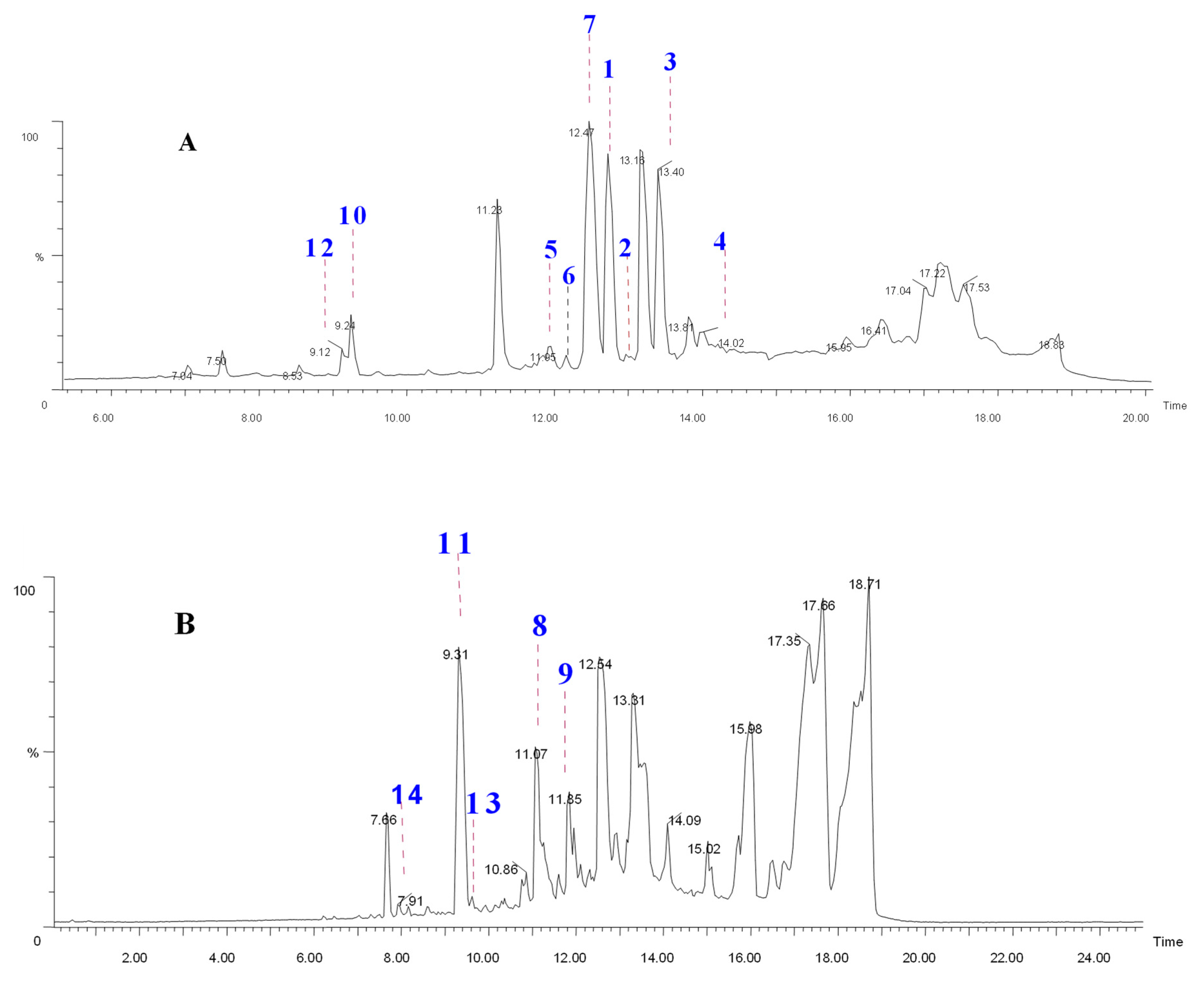
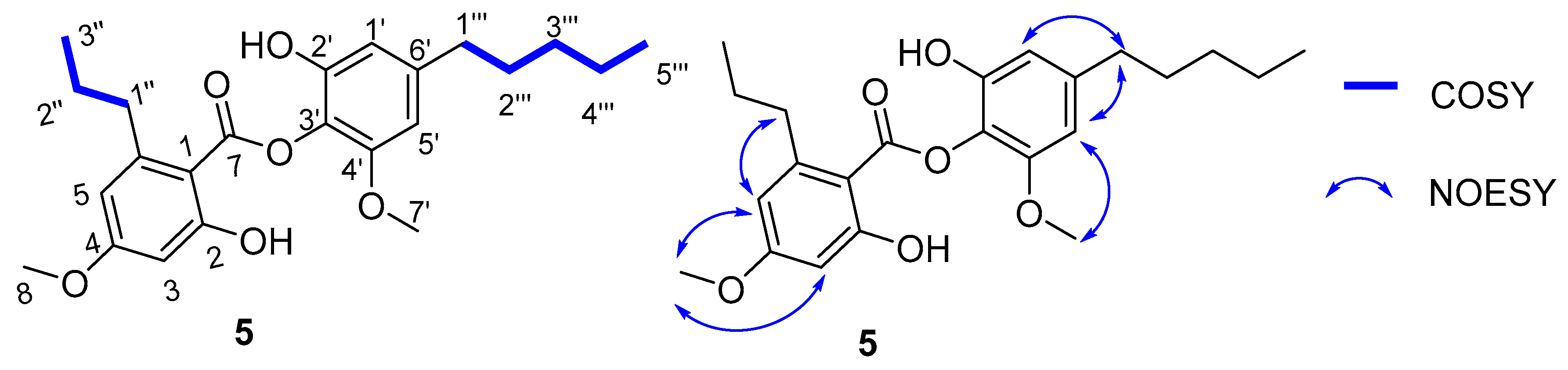
2.1. Chemical Profiling, Isolation, and Structure Elucidation

A HPTLC (Figure S3, Supplementary Materials) co-migration with standards and the UPLC-PDA profile (Figure S4, Supplementary Materials) of the acetone extract of R. conduplicans suggested the presence of a dozen of visible compounds, among which salazinic acid, usnic acid, sekikaic acid, homosekikaic acid, and divaricatic acid were identified against standards and appeared to represent the most abundant compounds. Initial LC-QToF-MSE analyses of the acetone extract of R. conduplicans indicated the presence of depsides, depsidones, and monophenolic acids based on High-Resolution Mass Spectroscopy (HRMS). Molecular formulae for C10–35H10–50O2–15 were generated from mass ranges m/z 150–750 coupled with the fragment ions and their MS spectral data (accurate mass and fragmentation pattern) and compared to online databases (DNP, Reaxys, SciFinder).
Mass spectrometry (MS) and, particularly, quadruple time-of-flight coupled to Liquid Chromatography (UPLC-Q-ToF-MS) has been widely utilized for profiling metabolites due to its superiority in high-resolution mass, precision, and sensitivity [18], and was helpful to clearly discriminate between the depsides, depsidones, simple phenol acids, dibenzofurans, and hydroxyl fatty acids based on the fragmentation of lichen molecules [16]. Therefore, the acquired TIC of the R. conduplicans extracts, obtained within 16 min, were analyzed from spectra obtained in negative mode and, thus, are effective for characterizing trace components (Figure 1). Metabolite assignments were made based on their polarity related to their retention time (Rt) and molecular formulae from accurate molecular weight measurement, along with adducts [M − H]/ fragment ions and Ring Double Bond Equivalence (RDBE). In the present study, a total of 18 compounds were clearly characterized from the crude extract of R. conduplicans by molecular formulae generated by ToF-MS/MS and MS/MS including their fragmentation profiles, as reported in the literature and presented in Table S1 (Supplementary Materials).
Based on the fragmentation of isolates, we have identified compounds (15) and (79) along with atranorin belonging to depsides. The literature clearly indicates that sekikaic acid is an abundant molecule in Ramalina species [19]. Sekikaic acid (1) is a m-depside corresponding to the esterification of two divaricatinic acid units and is found with Rt at 11.98 min and m/z 417.1547 (C22H25O8) with fragments m/z 209 and m/z 225 corresponding to the A ring and B ring, respectively [20]. Compounds 1, 3, 5, 7, 8, and 9, having a common fragment m/z 209 (Figures S47 and S48, Supplementary Materials), clearly indicate the difference in locating the other Bring. These depsides can be considered as ester derivatives of divaricatinic acid (11) while compound 2 is a divaric acid derivative (recognized at Rt 7.50 min, m/z 195.0657). The other identified monoaromatic compounds correspond to 2,4-di-O-methyldivaric acid (6), 4-O-methyldivaricatic acid (10), divaricatinic acid (11), olivetolic acid (12), divarinolmonomethylether (13), and atranol (14). In this run, three additional compounds were ionized and fragmented (Rt = 8.53 min, Rt = 11.88 min, and Rt = 13.17 min) and not determined. The fragmentation feature of Compound 5 (m/z 401.1954 [M − H] (calcd. for [C23H28O6] 401,1964)) suggested the coupling of a divaricatinic acid moiety to an olivetol monomethylether moiety (Figure S17, Supplementary Materials). Based on these fragmentation studies, we assigned compounds as shown in the Supporting Information section and in Table S1, including the monoaromatic divaric acid, along with the common and already-described atranorin (depside), usnic acid (related to dibenzofurans), and salazinic acid (a depsidone). The structures were concluded through MS/MS fragmentation patterns and compared with in-house standards.
Subsequently, the acetone extract was subjected to column chromatography to give eight fractions (I to VIII). An LC–MSE analysis of all fractions revealed the presence of depsides in III–VI fractions (Figures S4–S6, Supplementary Materials). Thus, the targeted isolation and purification of III–VI fractions yielded the isolation of one new depside (5), along with other known depsides (14 and79) and monoaromatic compounds (6 and 1014). The spectra and fragmentation patterns of these molecules were shown in the Supporting Information section (Figures S7–S46, Supplementary Materials).
The structures of the isolated compounds (Figure 2) were determined by a combination of spectroscopic data (HRESIMS, 1H and 13C NMR) and in comparison with the reported literature data. They were identified as sekikaic acid (1) [21], 4′-O-methylnorhomosekikaic acid (2) [22], homosekikaic acid (3) [22], hyperhomosekikaic acid (4) [23], 2,4-dimethyldivaric acid (6) [24], divaricatic acid (7) [25], decarboxydivaricatic acid (8) [26], decarboxystenosporic acid (9) [26], methyldivaricatinate (10) [24], divaricatinic acid (11) [21], olivetolic acid (12) [27], divarinolmonomethylether (13) [21], and atranol (14) [28].
Compound 5 was isolated as white amorphous powder and identified as a new compound. Its molecular formula was established as C23H29O6 based on a HRESIMS ion at m/z 401.1954 [M − H] (calcd. for [C23H28O6], 401.1964). The 1H and 13C NMR data of 5 (Table 1) showed the presence of four aromatic protons, (δH 6.53 (d, J = 1.8 Hz, 1H), 6.51 (d, J = 1.8 Hz, 1H), 6.46 (d, J = 2.6 Hz), and 6.40 (d, J = 2.6 Hz); δC 120.0, 110.7, 105.4, and 100.8) and one ester carbonyl (δC 170.3). In addition, two methoxyl groups (δH 3.86 (3H, s), 3.81 (3H, s)) and two n-alkane side chains of two methylene groups that were adjacent to a benzene ring (δH 3.02–2.93 (m, 2H), 2.62–2.51 (m, 2H)) were also distinguished from the NMR spectra, respectively (Table S2, Supplementary Materials). These spectral features, together with the characteristic ester carbonyl group at C-7 (δC 170.3) in the 13C NMR spectrum, strongly imply that 5 is a depside-type derivative [16,29].
A comparison of 1H NMR and 13C NMR data from5 with those of 4′-O-methylnorhomosekikaic acid, which were isolated from the same species, indicated an overall similarity, except for the absence of a COOH group and the presence of two additional methylenes. This reasoning was further supported by its 13C NMR spectrum, which showed the absence of a carbonyl COOH group, and its 1H NMR spectrum indicated the presence of an additional aromatic proton at 6.53 (d, J = 1.8 Hz, 1H). A comprehensive analysis of 2D NMR (COSY, and HSQC) data, especially the 1H–1H COSY spectrum, revealed two discrete spin systems, including -CH-CH2-CH3- (from H-1″, H-2″ and H-3″) and -CH-CH2-CH2-CH2-CH3 (from H-1′″ to 5′″), as drawn with bold lines in Figure 3. The position of the n-pentyl group at C-6′ and n-propyl chain at C-6 was confirmed on the basis of the NOESY correlations (H-1′″/H-5′, H-1′″/H-1′ and H-1″/H-5) (Figure 3) and in comparison with the sekikaic acid data. In addition, the MS/MS spectrum of 5 showed (Figure 4) product ions m/z 209, thereby indicating the breakage of the C–O bond between two aromatic rings supported by the fragments at m/z 165 and 137. Based on these spectral characteristics, the structure of 5 was established and trivially named as decarboxyhomosekikaic acid.

3. Biological Activity

3.1. Assessment of Compounds and Extract for Free Radicals Scavenging and Antioxidant Activity

The amphiphilic nature of the ABTS•+ cation was used to identify both hydrophilic and hydrophobic antioxidants in dietary materials, whereas the DPPH- radical was used to measure an antioxidant’s reducing power [30]. These fundamental chemical experiments reveal the radical scavenging and reduction characteristics of the potential antioxidant candidates.
Acetone extract (AE) and all isolated compounds (114) scavenged ABTS•+ and DPPH- radicals and the results are presented in Table 2. The results have demonstrated that AE and all the other compounds potently neutralized ABTS•+ radicals (more than 70%) and have shown activity equal to the ascorbic acid standard, except for compound 13, which only scavenged radicals by 50%.The pattern and potentials in decreasing order of ABTS•+ scavenging potentials were observed as follows:olivetolic acid (12) > compound 5 > divarinolmonomethylether (13) > decarboxydivaricatic acid (8) > 4-O-methylnorhomo sekikaic acid (2) > atranol (14) > divaricatic acid (7) > decarboxystenosporic acid (9) > sekikaic acid (1) > 2,4-dimethyldivaric acid (6) > homo sekikaic acid (3) > methyldivaricatinate (10). In the case of the DPPH-radical scavenging assay, acetone extract (AE) and compound 14 scavenged DPPH-radicals potently by more than 50%, whereas 1, 2, 3, 4, 5, 6, 12, and 13 countervailed DPPH-radicals (20–40%) moderately. It is important to mention that potent ABTS•+ scavenging activities were observed in all compounds, but DPPH scavenging activity was detected to be moderate in all compounds except compound 14 (Table 2). As ABTS•+ is a planar radical, it can be used to identify antioxidants even with low redox potentials. However, due to the steric barrier of the N radical, they may react slowly or not at all when tested on DPPH radicals [31]. This might be the reason why extracts and compounds scavenge ABTS•+ more potently than DPPH-radicals. To check whether this radical scavenging activity is related to antioxidant properties, we challenged genomic DNA with hydrogen peroxide (H2O2)-induced oxidative damage.

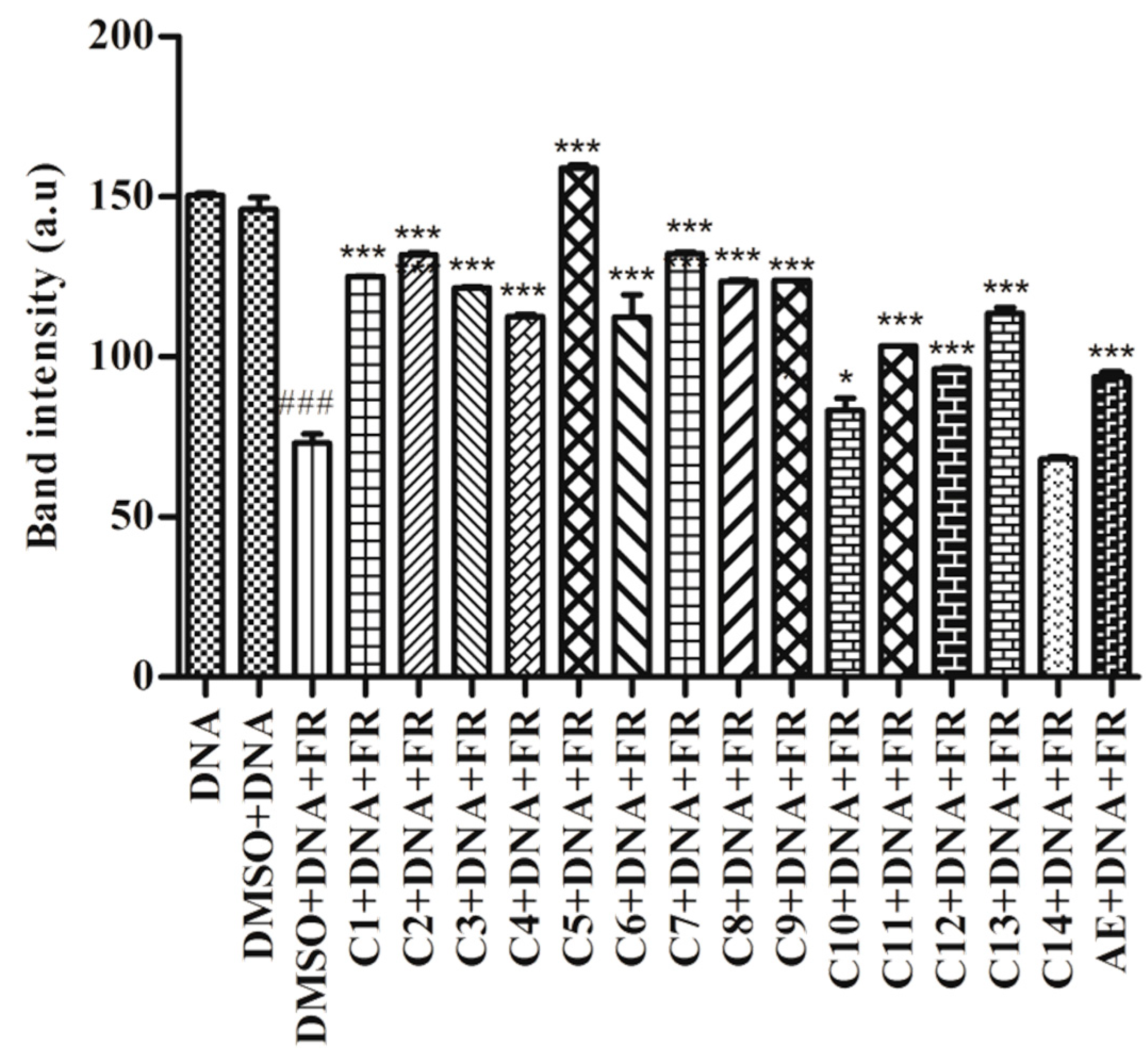
3.2. Protective Effect of R. conduplicans AE and Isolated Compounds on Oxidative DNA Damage

The Fenton’s reaction produces the hydroxyl radical, which is a ROS that is detrimental to the human body. Hydroxyl radicals react with different nucleobases, thereby inducing the formation of mutated bases that eventually lead to DNA damage [32]. Figure 5 demonstrated that FR damaged DNA significantly (p < 0.001) compared to the control (DMSO + DNA). Though all compounds showed significant protection against hydroxyl radical-induced DNA damage (p < 0.001, cpd 10: p < 0.05), compound 14 could not prevent the oxidative damage to DNA (Figure 5 and Figure S49). The genoprotective activity of these compounds and the AE may be attributed to the presence of free radical scavenging potential.

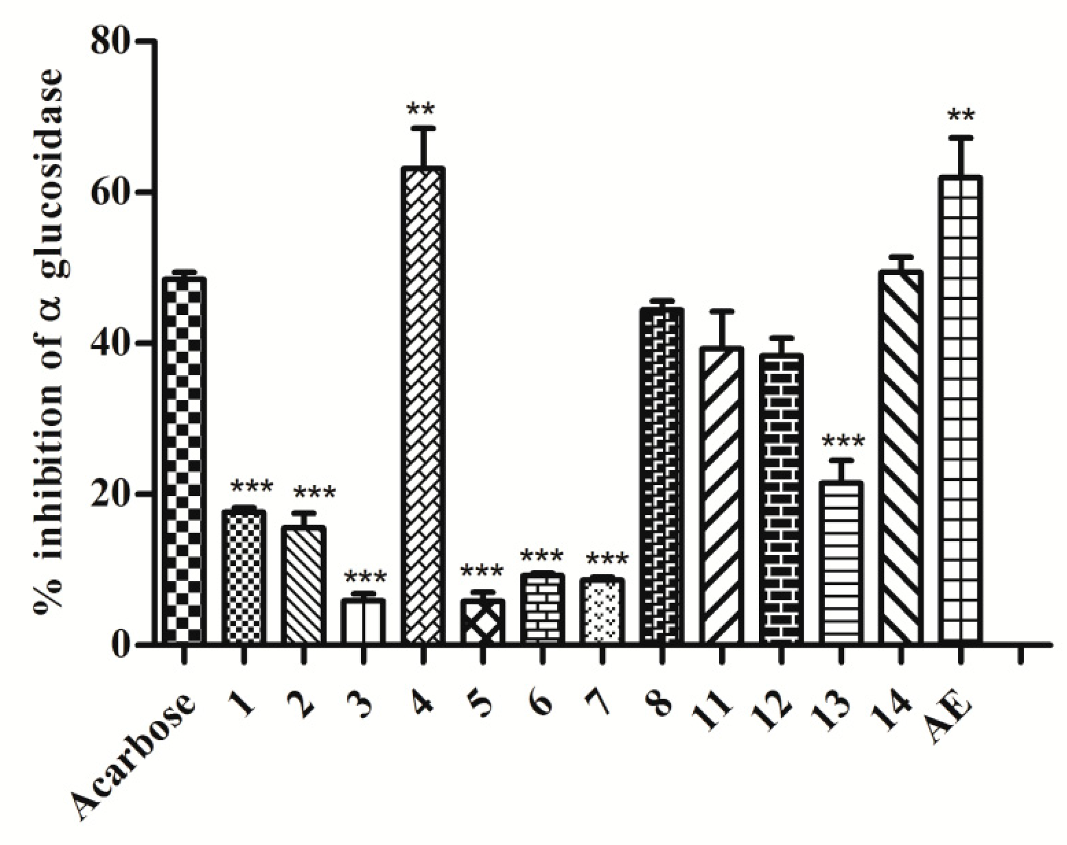
3.3. Assessment of In Vitro Antihyperglycemic Activity of Compounds and Extract as Intestinal α-Glucosidase Enzyme Inhibition

The α-glucosidase enzyme is a key enzyme that catalyses disaccharide digestion. The inhibition of α-glucosidase in the intestine slows digestion and the overall rate of glucose absorption into the blood. This has proven to be one of the most effective ways for lowering post-prandial blood glucose levels and, as a result, preventing the onset of late diabetes complications [33]. Sekikaic acid (1) was already recognized to inhibit α-glucosidase along with usnic acid and salazinic acid from other Ramalina species, but it is not the most effective compound [34]. As per Figure 6, it was stated that acetone extract (AE) and compound 4 have displayed better α-glucosidase inhibition (p < 0.01) than Acarbose.
On the other hand, compounds 8, 11, 12, and 14 demonstrated inhibitory effects comparable to those of the standard Acarbose (Figure 6). As contrasted activities can be observed between structurally-related compounds, structure–activity relationships can be considered. This is the case between depsides 3 and 4, suggesting a positive influence of the C1-pentyl chain with regard to substitution by a C1-propyl chain. When this length modification of the alkyl chain occurs on the B ring of decarboxylated derivatives (active compound 8 versus inactive compound 9) the opposite influence can be observed. The presence of a C6′-carboxylic group lowers the α-glucosidase inhibitory activity as compound 7 is less active than compound 8. Methylation of the carboxylic function of the monoaromatic divaricatinic acid 11 resulted in a complete loss of activity. Nevertheless, most of the tested compounds were found with some activity, such as compounds 1, 2, 3, 5, 6, 7, and 13, which displayed mild to moderate enzyme inhibition (p < 0.001). These results are to be pooled with the growing number of reports on the antidiabetic potential of lichen extracts or molecules [34,35,36]. The combination of activities with different mechanisms of action is of particular interest to develop potent antihyperglycemic effects. Lowering glucose absorption and limiting oxidative damages due to hyperglycemia, as expected from the lichen extract, could be promising. The challenge is to use standardized extracts that were previously checked to be safe for acute and chronic intake.

4. Materials and Methods

4.1. General

The NMR spectra were recorded on a Bruker FT-400 MHz NMR spectrometer and samples were dissolved in deuterated acetone-d6. Mass data were acquired on aXevoTM G2 XS-ESI-QTof mass spectrometer (Waters Corp., Manchester, UK). For thin layer chromatography (TLC) analysis, precoated Merck plates (silica gel 60 F254) were utilized. Silica gel (100–200 mesh) (Qing-dao Marine Chemical, Inc., Qingdao, China) was chosen for column chromatographic separation. Semi-preparative chromatography was performed on a Gilson HPLC (Middleton, WI, USA) instrument equipped with a 321 binary pump, GX-281 liquid handler, and UV-155 detector with X Select HSS T3 (250 mm × 100 mm, 5 µm) (Waters Corp., Drinagh, Ireland) as a stationary phase using a Trilution LC v2.1 platform. Formic acid (OptimaTM Mass spec grade) (Thermo Fisher Scientific, Geel, Belgium), HPLC-grade acetonitrile, LiChrosolv (Merck, Darmstadt, Germany), and ultra-pure water (Millipore System, Randolph, MA, USA) were used.

4.2. Instrumental UPLC Conditions

The instrumental conditions were set-up as per our recent report (Reddy et al., 2019) with slight modifications. Chromatographic separation was performed on an Acquity H Class UPLC system (Waters, Milford, MA, USA) with a conditioned auto sampler using an ACQUITY UPLC CSH Phenyl-Hexyl column (100 mm × 2.1 mm id., 1.7 μm particle size) (Waters, Milford, MA, USA). Column temperature was maintained at 40 °C. High-resolution masses of secondary metabolites were measured after UPLC separation. A mobile phase consisting of water with 0.1% formic acid in water (solvent A) and acetonitrile with 0.1% formic acid (solvent B) was pumped at a flow rate of 0.4 mL/min. The gradient elution program was as follows: 0 min, 5% B; 3.00 min, 20% B; 5.00min, 35% B; 7.50 min, 50% B; 10.00 min, 70% B; 12.50 min, 95% B; 17.00 min 95% B; and 21.00 min 5% B. The equilibration time was 4.0 min and the injection volume was 2 μL. The LC-QTof-MSE mode was applied to analyze the samples in both TIC as well as the MS/MS mode, where the collision energy was ramped at 15–45 eV. Eluted compounds were detected from m/z 50 to 1200 using a Xevo G2-XS Q-Tof mass spectrometer (Waters, Manchester, UK), which was connected to Electro-spray ionization (ESI) interface with a negative ion mode using the following instrument settings: capillary voltage, 2.0 KV; sample cone, 40 V; source temperature, 120 °C; desolvation temperature 350 °C; cone gas flow rate 50 L/h; desolvation gas (N2) flow rate 850 L/h, argon as CID gas for MS/MS experiments. All analyses were performed using lock spray, which ensured accuracy and reproducibility. Leucine–Enkephalin (5 ng/mL) was used as a lock mass, generating a reference ion in the negative mode at m/z 554.2615, and was introduced by a lock spray at 10 μL/min for accurate mass acquisition. Data acquisition was achieved using MassLynx ver. 4.1. Acquiring data in this manner provided information on intact precursor ions as well as fragment ions.

4.3. Lichen Sample Collection and Identification

The lichen, Ramalina conduplicans, was collected from tree bark in Bichpuri Range, Bijrani Zone of Corbett National Park, alt. N 29°26′40″ E79°04′06 (1283 m) in the month of May 2019. The morphological features of lichen thallus and ascomata were observed under Magnüs MS 24/13, and spot tests for color reaction were carried out by 10% aqueous solution of potassium hydroxide (K), Steiner’s stable p-phenylene diamine solution (PD), and calcium hypochlorite solution (C). For the anatomical investigation of fruiting bodies, a light microscope from ZEISS Axiostar was used. The lichen substances were identified with thin layer chromatography in solvent system ‘A’ following White and James’s methods (1985). The standard literature [37] was referred to for identification of lichen samples. The voucher specimens (Satish Mohabe & A. Madhusudhana Reddy 7658YVUH) of species were deposited at the Herbarium, Department of Botany, Yogi Vemana University, Kadapa, Andhra Pradesh. The corresponding data are shown in Figures S1–S3, Supporting Materials.

4.4. Extraction and Isolation

The sorted-out lichen Ramalina conduplicans (300 g) was shade-dried, powdered, and extracted with acetone (6L) at room temperature for 48h. The result was that acetone extract was evaporated to dryness under reduced pressure, thereby affording a syrupy residue (20 g). This crude extract was subjected to gradient column chromatography (SiO2, 60–120 mesh) and eluted with a hexane/EtOAc mixture of increasing polarity with 10% intervals that yielded 8 fractions. These eight fractions were reconstituted in acetonitrile and subjected to UPLC Q-ToF MSE. Based on the TIC profile, we selected fractions 3–6 for purification (mass profile shown in supporting information, discussion in results section). All these fractions were subjected to semi-preparative HPLC (X Select HSST3 OBD Prep Column,5 μm, 10 mm × 250 mm), 0.1 % formic acid with water (solvent A) and acetonitrile (solvent B) as mobile phase at flow rate 4 mL/min, detected at 254 nm. Semi-preparative HPLC were conducted by gradient elution programs to obtain compounds as follows: Fraction 3 (quantity 70 mg, loop volume 250 μL was eluted by 0 min, 5% B; 5 min, 5% B; 10.00 min, 35% B; 16.00 min, 60% B; 25 min, 95% B; 30 min, 95% B; 5% B; 30.50 min, 5% B; 35.00 min. to yield 3 (homosekikaic acid, 7 mg), 7 (divaricatic acid, 4 mg), 11 (divaricatinic acid, 3 mg), 12 (olivetolic acid, 5 mg), and 14 (atranol, 2 mg). Fraction 4 (quantity 50 mg, loop volume 250 μL was eluted by 0 min, 30% B; 5 min, 30% B; 10.00 min, 50% B; 23.00 min, 95% B; 27.00 min, 95% B; 27.50 min, 30% B; 30% B; 30.00 min at flow rate 4 mL/min, detected at 254 nm to yield 10 (methyldivaricatinate, 3 mg), 8 (decarboxydivaricatic acid, 5 mg), 9 (decarboxystenosporic acid, 2 mg), and 4 (hyperhomosekikaic acid, 1mg). Fraction 5 (quantity 25 mg, loop volume 250 μL was eluted by 0 min, 5% B; 8.50 min, 30% B; 15.00 min, 50% B; 22.00 min, 95% B; 28.00 min, 95% B; 29.0 min, 5% B; 5% B; 35.00 min at flow rate 4 mL/min, detected at 254 nm) to yield 13 (divarinolmonomethylether, 3 mg) and compound 5 (2 mg). Fraction 6 (quantity 40 mg, loop volume 250 μL was eluted by 0 min, 10% B; 8.50 min, 40% B; 18.00 min, 55% B; 25.00 min, 75% B; 32.00 min, 95% B; 36.0 min, 95% B; 10% B; 37.00 min, 10% B; 42.00 min at flow rate 4 mL/min, detected at 254 nm) to yield 1 (sekikaic acid, 5 mg), 2 (4-O-methylnorhomosekikaic acid, 7 mg) and 2,4-di-O-methyldivaric acid 6 (2 mg). Physicochemical data are shown in the Supporting Materials.

4.5. In Vitro Antihyperglycemic and Antioxidant Assay

4.5.1. DPPH Radical Scavenging Activity

A DPPH radical scavenging assay was carried out as previously reported [38]. Scavenging of 2,2-diphenyl-1-picryhydrazyl (DPPH) radicals by the acetone extract (AE) (50 µg of 2 mg/mL solution dissolved in DMSO) and compounds (114) (50 µg of 2 mg/mL solution dissolved in DMSO) was measured in 100 mM Tris-HCl buffer (pH 7.4) by recording the absorbance at 517 nm spectrophotometrically. Ascorbic acid (50 µg of 2 mg/mL solution dissolved in DMSO) served as the standard. The results were expressed as %-scavenging and calculated by using the following formula: (Ac−At)/100 × Ac, where Ac was the absorbance of control and At was the absorbance of the test sample. Different concentrations of compounds were evaluated to obtain 50% scavenging activity (SC50). The SC50 was calculated based on the equation obtained from regression analysis.

4.5.2. ABTS Radical Scavenging Activity

Scavenging of the 2,2′-azino-bis(3-ethylbenzothiazoline-6-sulphonic acid) radical cation (ABTS•+) was performed as per the earlier method [39]. Acetone extract (AE) (20 µg of 2 mg/mL solution dissolved in DMSO) and compounds (114) (20 µg of 2 mg/mL solution dissolved in DMSO) were incubated with ABTS•+ solution in 6.8 mM phosphate buffer (pH 8.0) as described earlier. The discoloration of the ABTS•+ solution was determined by measuring the absorbance at 734 nm spectrophotometrically. Ascorbic acid (20 µg of 2 mg/mL solution dissolved in DMSO) served as the standard. The activity was expressed as %-scavenging and calculated as follows: (Ac − At)/(100 × Ac), where Ac was the absorbance of control and At was the absorbance of the test sample. The SC50 of compounds was calculated as per the above formula.

4.5.3. Free Radical Induced DNA Damage

The protective effect of acetone extract (AE) and compounds (114) on oxidative DNA damage was evaluated as per the previous method [40]. A total of 2 µL calf-thymus DNA mixed with 5 µL of 39 mM Tris buffer (pH 7.4) and 5 µL (10 µg) acetone extract and compounds (114) (10 µg of 2 mg/mL solution dissolved in DMSO) mixture was incubated at room temperature for 20 min. The reaction was initiated by adding 5 µL FeCl3 (500 µM) and 10 µL H2O2 (0.8 M) and incubated for 10 min at 37 °C. The reaction was stopped by adding 3 µL DNA loading dye. Finally, the mixture was subjected to 0.8% agarose gel electrophoresis in TAE (40 mM Tris, 20 mM acetic acid and 0.5 M EDTA) buffer (pH 7.2). A total of 3 µL of Ethidium bromide was added to agarose solution to stain DNA bands. The image was viewed under transilluminating UV light and photographed (Bio-Rad, ChemiDocTM XRS, Hercules, CA, USA with Image LabTM software (ver. 6.0.1, build34, standard edition, 2017). The band intensity of the DNA was measured by using ImageJ software (ver. 1.4.3.67, Broken Symmetry Software, Scottsdale, AZ, USA).

4.5.4. Intestinal α-Glucosidase Inhibition

An intestinal α-glucosidase enzyme inhibition assay was performed as per the previous method [36]. A total of 20 μL (40 µg) of acetone extract and compounds (114) (40 µg of 2 mg/mL solution dissolved in DMSO) were incubated with 50 μL of rat intestinal α-glucosidase enzyme (89.93 mM, prepared in 0.9% NaCl) in 100 mM phosphate buffer (pH 6.8) for 10 min. After the incubation period, 50 μL of substrate (4-nitroplenyl α-d-glucopyranoside) solution was added. The release of p-nitrophenol from substrate was measured by recording the absorbance at 405 nm spectrophotometrically. Acarbose (40 µg of 2 mg/mL solution dissolved in DMSO) was taken as the standard. The activity was expressed and calculated as follows: (Ac − At)/100 × Ac, where Ac was the absorbance of control and At was the absorbance of the test sample.

4.5.5. Statistical Analysis

Comparisons within the groups were done by applying one-way ANOVA followed by a post-test Tukey’s Multiple comparison test. Statistical significance was set at p < 0.05. Data analysis was performed by using GraphPad Prism ver. 5.01 (GraphPad Software Inc., San Diego, CA, USA).

5. Conclusions

A novel UPLC-QToF-MS/MS-guided strategy was proposed here for the isolation and characterization of one new depside, decarboxyhomosekikaic acid, along with 13 known metabolites from Ramalinaconduplicans—most of them being minor metabolites that were reported on for the first time from this species. In the whole experimental design, UPLC-QToF-MS/MS was selected for multiple purposes, including targeting, finding, profiling, and isolating active constituents. Three hitherto unreferenced compounds were detected in this lichen, with their molecular formulae being deduced from HR-QToF-MS. Although in minute amounts, one isolate could be identified as an additional homosekikaic derivative. The expected major compounds atranorin, usnic acid, salazinic acid, and sekikaic acid were also obtained. However, efforts for isolating, identifying, and testing mainly targeted alkyldepsides- and monoaromatic-related compounds.
These compounds were tested for their antioxidant and α-glucosidase inhibition potential. Most of them, and the crude acetone extract (AE), have displayed antioxidant potential by scavenging ABTS and DPPH radicals and protected DNA from oxidative damage. Five compounds, and particularly hyperhomosekicaic acid, exhibited a comparable or better α-glucosidase inhibition to that of the acarbose standard. On the basis of these results, it is suggested that these lichen substances have a great potential to be used as bioresources or as structural models for novel bioactive candidate compounds. Docking experiments are necessary to document the structure–activities observed in this study along with pharmacomodulation studies to evaluate the antidiabetic properties. Acetone extract unexpectedly showed a comparable effect to that of the Acarbose standard, though it was not sufficient to consider its hypoglycemic activity in the context of the traditional use made of this edible lichen [10].
It should be kept in mind that activities obtained from the crude extract or from any of the active metabolites cannot be claimed to support a preventive or a therapeutic activity as no clinical assay has been carried out to validate an effect with a standardized dosage. Unexpected side effects can occur when preparations differ from the real traditional use, and toxicity trials have to be carried out at once.

Supplementary Materials

The following supporting information can be downloaded at: https://www.mdpi.com/article/10.3390/molecules27196720/s1. Detailed Materials and Method. Table S1: LC-QToF-MSE data of compounds from acetone extract of R. conduplicans. Physicochemical data of isolated compounds. Figure S1: Authenticate Ramalina conduplicans. Figure S2: Shows the spot tests for identification of Ramalina conduplicans. Figure S3: Depicts the HPTLC profiles for R. conduplicans extract. Figure S4: UPLC-PDA chromatogram of acetone extract of R. conduplicans. Figure S5: Total Ion Chromatogram (TIC) of fractions and pure compounds. Figure S6–S46: Spectral data (FTIR spectra, HRESIMS, MS/MS spectra and fragmentation pattern, 1H & 13C NMR, DEPT, HSQC, DQF-COSY and NOESY spectra) of compounds 114, salazinic acid, usnic acid and atronarin. Figure S47: TIC of R.conduplicans. Figure S48: Common fragmentation of depsides. Figure S49. Invitro DNA damage assay.

Author Contributions

K.S.B., the biologist A.K.T. and J.B. conceived and designed the experiments; T.K.K., B.S. and K.S.B. performed the experiments; K.S.B., F.L.D. and J.B. analyzed the data; A.M.R. and S.M. contributed to the material identification reagents/materials and tools; A.A. and K.A. performed in vitro biological experiments and analyzed data; B.S., A.K.T., K.S.B. and J.B. wrote/revised the paper. All authors have read and agreed to the published version of the manuscript.

Funding

This research received no external funding.

Institutional Review Board Statement

Not applicable.

Informed Consent Statement

Not applicable.

Data Availability Statement

The data presented in this study are available on request from the corresponding authors.

Acknowledgments

This research was performed as a part of the Indo-French IRP ‘Natural Products and Synthesis for Affordable Health’ and acknowledgments are due to CSIR (India) and CNRS (France). Authors thank to Director, IICT for his constant encouragement and support and to S. Labarre for English revision. B.S. thanks CSIR for fellowship program and financial support. IICT communication no. IICT/Pubs./2022/082.

Conflicts of Interest

The authors declare no conflict of interest.

Sample Availability

Samples of the compounds 114 are available from the authors, and the samples were deposited in the institutional depository.

References

  1. Shukla, V.; Joshi, G.P.; Rawat, M.S.M. Lichens as a potential natural source of bioactive compounds: A review. Phytochem. Rev. 2010, 9, 303–314. [Google Scholar] [CrossRef]
  2. Ingelfinger, R.; Henke, M.; Roser, L.; Ulshöfer, T.; Calchera, A.; Singh, G.; Parnham, M.J.; Geisslinger, G.; Fürst, R.; Schmitt, I.; et al. Unraveling the pharmacological potential of lichen extracts in the context of cancer and inflammation with a broad screening approach. Front. Pharmacol. 2020, 11, 1322. [Google Scholar] [CrossRef]
  3. Zhao, Y.; Wang, M.; Xu, B.A. Comprehensive review on secondary metabolites and health-promoting effects of edible lichen. J. Funct. Foods 2021, 80, 104283. [Google Scholar] [CrossRef]
  4. Bhattarai, T.; Subba, D.; Subba, R. Nutritional value of some edible lichens of east Nepal. Angew. Bot. 1999, 73, 11–14. [Google Scholar]
  5. Awasthi, D.D. A Compendium of the Macrolichens from India, Nepal and Sri Lanka; Bishen Singh Mahendra Pal Singh: Dehradun, India, 2000. [Google Scholar]
  6. Wang, L.S.; Narui, T.; Harada, H.; Culberson, C.F.; Culberson, W.L. Ethnic uses of lichens in Yunnan, China. Bryologist 2001, 104, 345–349. [Google Scholar] [CrossRef]
  7. Hanus, L.O.; Temina, M.; Dembitsky, V. Biodiversity of the chemical constituents in the epiphytic lichenized ascomycete Ramalinalacera grown on different substrates Crataegussinaicus, Pinus halepensis, and Quercus calliprinos. Biomed. Pap. Med. Fac. Univ. Palacky Olomouc. Czech Repub. 2008, 152, 203–208. [Google Scholar] [CrossRef] [Green Version]
  8. Upreti, D.K.; Divakar, P.K.; Nayaka, S. Commercial and ethnic use of lichens in India. Econ. Bot. 2005, 59, 269–273. [Google Scholar] [CrossRef]
  9. Kumar, S.P.; Kekuda, T.P.; Vinayaka, K.S.; Sudharshan, S.J. Anthelmintic and antioxidant efficacy of two macrolichens of Ramalinaceae. Pharmacogn. J. 2009, 1, 238–242. [Google Scholar]
  10. Vinitha, M.T.; Veranja, K. Potential of lichen compounds as antidiabetic agents with antioxidative properties: A review. Oxid. Med. Cell. Longev. 2017, 2017, 2079697. [Google Scholar]
  11. Devi, A.B.; Mohabe, S.; Nayaka, S.; Reddy, M. In-vitro antimicrobial activity of lichen Ramalina conduplicans Vain collected from Eastern Ghats, India. Sci. Res. Rep. 2016, 6, 99–108. [Google Scholar]
  12. Ankith, G.N.; Rajesh, M.R.; Karthik, K.N.; Avinash, H.C.; Kekuda, P.T.; Vinayaka, K.S. Antibacterial and antifungal activity of three Ramalina species. J. Drug Deliv. Ther. 2017, 7, 27–32. [Google Scholar] [CrossRef]
  13. Liu, X.Y. Determination of trace element of 4 lichens in Yunnan. Stud. Trans. Elem. Health 2003, 20, 30–31. [Google Scholar]
  14. Ramya, K.; Thirunalasundari, T. Lichens: A myriad hue of bioresources with medicinal properties. Int. J. Life Sci. 2017, 5, 387–393. [Google Scholar]
  15. Luo, H.; Wei, X.; Yamamoto, Y.; Liu, Y.; Wang, L.; Jung, J.S.; Hur, J.S. Antioxidant activities of edible lichen Ramalina conduplicans and its free radical-scavenging constituents. Mycoscience 2010, 51, 391–395. [Google Scholar] [CrossRef]
  16. Kumar, K.; Siva, B.; Sarma, V.; Mohabe, S.; Reddy, A.M.; Boustie, J.; Tiwari, A.K.; Rao, N.R.; Babu, K.S. UPLC-MS/MS quantitative analysis and structural fragmentation study of five Parmotrema lichens from the Eastern Ghats. J. Pharm. Biomed. Anal. 2018, 156, 45–57. [Google Scholar] [CrossRef]
  17. Reddy, S.D.; Siva, B.; Kumar, K.; Babu, V.S.P.; Sravanthi, V.; Boustie, J.; Nayak, V.L.; Tiwari, A.K.; Rao, C.; Sridhar, B.; et al. Comprehensive analysis of secondary metabolites in Usnealongissima (Lichenized Ascomycetes, Parmeliaceae) using UPLC-ESI-QTOF-MS/MS and Pro-apoptotic activity of barbatic acid. Molecules 2019, 24, 2270. [Google Scholar] [CrossRef]
  18. Olivier-Jimenez, D.; Chollet-Krugler, M.; Rondeau, D.; Beniddir, M.A.; Ferron, S.; Delhaye, T.; Allard, P.-M.; Wolfender, J.L.; Sipman, H.; Lücking, R.; et al. A database of high-resolution MS/MS spectra for lichen metabolites. Sci. Data 2019, 6, 294. [Google Scholar] [CrossRef] [Green Version]
  19. Moreira, A.S.; Braz-Filho, R.; Mussi-Dias, V.; Vieira, I.J. Chemistry and biological activity of Ramalina lichenized fungi. Molecules 2015, 20, 8952–8987. [Google Scholar] [CrossRef] [Green Version]
  20. Musharraf, S.G.; Kanwal, N.; Thadhani, V.M.; Choudhary, M.I. Rapid identification of lichen compounds based on the structure–fragmentation relationship using ESI-MS/MS analysis. Anal. Methods 2015, 7, 6066–6076. [Google Scholar] [CrossRef]
  21. Linh, N.T.T.; Danova, A.; Truong, T.L.; Chavasiri, W.; Phung, N.K.P.; Chi, H.B.L. Chemical constituents of chloroform extract from the lichen Ramalina peruviana Arch (Ramalinaceae). Vietnam J. Chem. 2020, 58, 231–236. [Google Scholar] [CrossRef] [Green Version]
  22. Culberson, C.F.; Culberson, W.L.; Johnson, A. Orcinol-type depsides and depsidones in the lichens of the Cladoniachlorophaea group (Ascomycotina, Cladoniaceae). Bryologist 1985, 88, 380. [Google Scholar] [CrossRef]
  23. Gunasekaran, S.; Rajan, V.P.; Ramanathan, S.; Murugaiyah, V.; Samsudin, M.W.; Din, L.B. Antibacterial and antioxidant activity of lichens Usnearubrotincta, Ramalinadumeticola, Cladoniaverticillata and their chemical constituents. Malays. J. Anal. Sci. 2016, 20, 1–13. [Google Scholar] [CrossRef]
  24. Sun, W.; Zhuang, C.; Li, X.; Zhang, B.; Lu, X.; Zheng, Z.; Dong, Y. Varic acid analogues from fungus as PTP1B inhibitors: Biological evaluation and structure–activity relationships. Bioorg. Med. Chem. Lett. 2017, 27, 3382–3385. [Google Scholar] [CrossRef] [PubMed]
  25. Brandão, L.F.; Alcantara, G.B.; Matos, M.; Bogo, D.; Freitas, D.; Oyama, N.M.; Honda, N.K. Cytotoxic evaluation of phenolic compounds from lichens against melanoma cells. Chem. Pharm. Bull. 2013, 61, 176–183. [Google Scholar]
  26. Elix, J.A.; Wardlaw, J.H. New depsides from the lichen Neofusceliadepsidella. Aust. J. Chem. 1997, 50, 1145–1150. [Google Scholar] [CrossRef]
  27. Ismed, F.; Farhan, A.; Bakhtiar, A.; Zaini, E.; Nugraha, Y.P.; Putra, O.D.; Uekusa, H. Crystal structure of olivetolic acid: A natural product from Cetreliasanguinea (Schaer.). Acta Crystallogr. E Crystallogr. Comm. 2016, 72, 1587–1589. [Google Scholar] [CrossRef] [Green Version]
  28. Bouges, H.; Monchot, A.; Antoniotti, S. Enzyme-catalysed conversion of atranol and derivatives into dimeric hydrosoluble materials: Application to the preparation of a low-atranol oakmoss absolute. Cosmetics 2018, 5, 69. [Google Scholar] [CrossRef]
  29. Bauer, J.; Waltenberger, B.; Noha, S.M.; Schuster, D.; Rollinger, J.M.; Boustie, J.; Chollet, M.; Stuppner, H.; Werz, O. Discovery of depsides and depsidones from lichen as potent inhibitors of microsomal prostaglandin E2 synthase-1 using pharmacophore models. ChemMedChem 2012, 7, 2077–2081. [Google Scholar] [CrossRef]
  30. Munteanu, I.G.; Apetrei, C. Analytical methods used in determining antioxidant activity: A review. Int. J. Mol. Sci. 2021, 22, 3380. [Google Scholar] [CrossRef]
  31. Siddeeg, A.; Al-Kehayez, N.M.; Abu-Hiamed, H.A.; Al-Sanea, E.A.; Al-Farga, A.M. Mode of action and determination of antioxidant activity in the dietary sources: An overview. Saudi J. Biol. Sci. 2021, 28, 1633–1644. [Google Scholar] [CrossRef]
  32. Lloyd, D.R.; Phillips, D.H. Oxidative DNA damage mediated by copper (II), iron (II) and nickel (II) Fenton reactions: Evidence for site-specific mechanisms in the formation of double-strand breaks, 8-hydroxydeoxyguanosine and putative intrastrand cross-links. Mutat. Res. Fundam. Mol. Mech. Mutagen. 1999, 424, 23–36. [Google Scholar] [CrossRef]
  33. Komati, A.; Anand, A.; Nagendla, N.K.; Madhusudana, K.; Mudiam, M.K.; Babu, K.S.; Tiwari, A.K. Bombax ceiba calyx displays antihyperglycemic activity via improving insulin secretion and sensitivity: Identification of bioactive phytometabolomes by UPLC-QTof-MS/MS. J. Food Sci. 2022, 87, 1865–1881. [Google Scholar] [CrossRef] [PubMed]
  34. Verma, N.; Behera, B.C.; Sharma, B.O. Glucosidase inhibitory and radical scavenging properties of lichen metabolites salazinic acid, sekikaic acid and usnic acid. Hacet. J. Biol. Chem. 2012, 40, 7–21. [Google Scholar]
  35. Schinkovitz, A.; Le Pogam, P.; Derbre, S.; Roy-Vessieres, E.; Blanchard, P.; Thirumaran, S.L.; Breard, D.; Aumond, M.C.; Zehl, M.; Urban, E.; et al. Secondary metabolites from lichen as potent inhibitors of advanced glycation end products and vasodilative agents. Fitoterapia 2018, 131, 182–188. [Google Scholar] [CrossRef] [PubMed]
  36. Duong, T.H.; Hang, T.X.H.; Le Pogam, P.; Tran, T.N.; Mac, D.D.; Dinh, M.H.; Sichaem, J. α-Glucosidase inhibitory depsidones from the lichen Parmotrematsavoense. Planta Med. 2020, 86, 776–781. [Google Scholar] [PubMed]
  37. Orange, A.; James, P.W.; White, F.J. Microchemical methods for the identification of lichens; British Lichen Society: London, UK, 2001. [Google Scholar]
  38. Tiwari, A.K.; Manasa, K.; Kumar, D.A.; Zehra, A. Raw horse gram seeds possess more in vitro antihyperglycaemic activities and antioxidant properties than their sprouts. Nutrafoods 2013, 12, 47–54. [Google Scholar] [CrossRef]
  39. Deepthi, S.; Anusha, K.; Anand, A.; Manasa, A.; Babu, K.S.; Mudiam, M.K.R.; Tiwari, A.K. Micronutrients and phytochemicals content in various rice (Oryzasativa Linn.) samples control carbohydrate digestion variedly and present differential antioxidant activities: An in vitro appraisal. Indian J. Tradit. Knowl. 2020, 19, 821–831. [Google Scholar]
  40. Chang, M.; Bellaoui, M.; Boone, C.; Brown, G.W. A genome-wide screen for methyl methanesulfonate-sensitive mutants reveals genes required for S phase progression in the presence of DNA damage. Proc. Natl. Acad. Sci. USA 2002, 99, 16934–16939. [Google Scholar] [CrossRef]
Figure 1. TIC of (A) R. conduplicans acetone extract and (B) enriched fraction-4.
Molecules 27 06720 g001
Figure 2. Isolated compounds (114) from Ramalina conduplicans Vain.
Molecules 27 06720 g002
Figure 3. Key COSY and NOESY correlations of compound 5.
Molecules 27 06720 g003
Figure 4. MS/MS spectrum and proposed fragmentation of compound 5.
Molecules 27 06720 g004
Figure 5. In vitro DNA damage assay. Compounds (114) and R. conduplicans. AE were incubated with DNA and Fenton’s Reagent and DNA damage was recorded with Agarose Gel electrophoresis. Respective graphical representation. ### p < 0.001; vs. control (DMSO + DNA). *** p < 0.001, * p < 0.05; vs.DMSO + DNA + FR, One-way ANOVA followed by Tukey’s multiple comparison test was used to calculate values. Values are represented as mean ± SD, n = 3. AE = Acetone Extract, FR = Fenton’s Reagent.
Molecules 27 06720 g005
Figure 6. Intestinal α-glucosidase inhibitory assay. Compounds (18), (1114), and acetone extract (AE) were incubated with α-glucosidase enzyme and the release of p-nitrophenol was determined. *** p < 0.001, ** p < 0.01; vs. Acarbose. One-way ANOVA followed by Tukey’s multiple comparison test was applied to compare differences. Values are represented as mean ± SD, n = 3. AE = Acetone Extract. Activity was not detected for compounds 9 and 10.
Molecules 27 06720 g006
Table 1. NMR data of compound 5 (400 & 100 MHz, acetone-d6) *.
S no1H NMR of 513C NMR of 5
1--107.35
2--166.10
36.46 (d, J = 2.6 Hz, 1H)112.07
4--166.10
56.40 (d, J = 2.6 Hz, 1H)100.65
6--149.91
7--170.47
1′6.53 (d, J = 1.8 Hz, 1H)110.80
2′--151.36
3′--154.12
4′--143.54
5′6.51 (d, J = 1.8 Hz, 1H)105.49
6′--150.38
1″3.0–2.93 (m, 2H)40.06
2″1.82–1.68 (m, 2H)26.58
3″0.93 (t, J = 7.6, 3H)15.60
1‴2.62–2.51 (m, 2H)37.69
2‴1.67–1.59 (m, 2H)32.89
3‴1.41–1.30 (m, 2H)33.25
4‴1.41–1.30 (m, 2H)24.18
5‴0.93 (t, J = 7.6 Hz, 3H)15.30
OMe-7′3.81 (s, 3H)57.31
OMe-83.86 (s, 3H)56.83
* = values are assigned with the comparison of sekikaic acid data and COSY/NOESY correlations.
Table 2. Free radical scavenging activities of AE and compounds (114) of Ramalina conduplicans.
Compound Name (Code)DPPH Assay
% Scavenging
(SC50, µg/mL)
ABTS Assay
% Scavenging,
(SC50, µg/mL)
Sekikaic acid (1)37.75 ± 0.6599.05 ± 0.00 (2.45)
4-O-methylnorhomosekikaic acid (2)36.78 ± 1.5798.57 ± 0.00 (1.40)
Homosekikaic acid (3)38.28 ± 1.2298.10 ± 0.00 (2.81)
Hyperhomosekikaic acid (4)27.32 ± 0.3479.52 ± 1.02 (17.44)
Compound 546.29 ± 3.70100.48 ± 0.68 (0.44)
2,4-dimethyldivaric acid (6)29.67 ± 0.89100.20 ± 0.34 (2.46)
Divaricatic acid (7)8.57 ± 1.6599.28 ± 1.02 (2.09)
Decarboxydivaricatic acid (8)17.17 ± 1.74100.28 ± 0.00 (0.75)
Decarboxystenosporic acid (9)13.04 ± 0.7396.7 ± 0.5 (2.41)
Methyl divaricatinate (10)6.50 ± 0.76100.0 ± 1.0 (2.90)
Divaricatinic acid (11)ND100.0 ± 0.5 (2.63)
Olivetolic acid (12)41.94 ± 1.1192.7 ± 0.0 (0.13)
Divarinolmonomethylether (13)21.12 ± 1.2151.7 ± 2.5 (0.57)
Atranol (14)74.66 ± 2.59 (18.65)88.9 ± 7.3 (2.05)
Acetone Extract (AE)50.64 97.3
Ascorbic Acid93.25 ± 1.23 (3.96)99.02 ± 0.03 (0.47)
ND = Not determined. The activity is expressed as % scavenging with regard to ascorbic acid scavenging activity. The SC50 is indicated for the most active compounds.
Publisher’s Note: MDPI stays neutral with regard to jurisdictional claims in published maps and institutional affiliations.

