Article

An Evaluation of Antimicrobial, Anticancer, Anti-Inflammatory and Antioxidant Activities of Silver Nanoparticles Synthesized from Leaf Extract of Madhuca longifolia Utilizing Quantitative and Qualitative Methods

1
Department of Biophysics, Government Institute of Science, Aurangabad 431004, India
2
Department of Botany, The Institute of Science, 15 Madame Cama Road, Mumbai 400032, India
3
Department of Zoology, Dr. Babasaheb Ambedkar Marathwada University, Aurangabad 431004, India
4
Amity Institute of Biotechnology, Amity University Maharashtra, Mumbai-Pune Expressway, Mumbai 410206, India
5
Department of Biotechnology, School of Engineering & Technology, Sharda University, Greater Noida 201310, India
6
Department of Pharmacy, State University of Bangladesh, 77 Satmasjid Road, Dhanmondi, Dhaka 1205, Bangladesh
7
Laboratory Medicine Department, Faculty of Applied Medical Sciences, Umm Al-Qura University, Makkah 24382, Saudi Arabia
8
Department of Clinical Laboratory Sciences, College of Applied Medical Sciences, Taif University, Taif 21944, Saudi Arabia
9
Department of Public Health, Health Sciences College at Al-Leith, Umm Al-Qura University, Makkah 24382, Saudi Arabia
10
Department of Pharmacology, Faculty of Medicine, University Kebangsaan Malaysia, Jalan Yaacob Latif, Kuala Lumpur 56000, Malaysia
*
Authors to whom correspondence should be addressed.
Molecules 2022, 27(19), 6404; https://doi.org/10.3390/molecules27196404
Submission received: 25 August 2022 / Revised: 19 September 2022 / Accepted: 21 September 2022 / Published: 28 September 2022

Abstract

:
In the current decade, nanoparticles are synthesized using solvents that are environmentally friendly. A number of nanoparticles have been synthesized at room temperature using water as a solvent, such as gold (Au) and silver (Ag) nanoparticles. As part of nanotechnology, nanoparticles are synthesized through biological processes. Biological methods are the preferred method for the synthesis of inorganic nanoparticles (AgNPs) as a result of their simple and non-hazardous nature. Nanoparticles of silver are used in a variety of applications, including catalysts, spectrally selective coatings for solar absorption, optical objectives, pharmaceutical constituents, and chemical and biological sensing. Antimicrobial agents are among the top uses of silver nanoparticles. In the current study, silver nanoparticles were biologically manufactured through Madhuca longifolia, and their antibacterial activity against pathogenic microorganisms, anticancer, anti-inflammatory, and antioxidant activities were assessed. UV-Vis spectroscopy, XRD (X-ray diffraction), transmission electron microscopy, Zeta Potential, and FTIR were used to characterize silver nanoparticles. The current work describes a cheap and environmentally friendly method to synthesize silver nanoparticles from silver nitrate solution by using plant crude extract as a reducing agent.

1. Introduction

Modern material science is experiencing a boom in nanotechnology, and the synthesis of nanoparticles has become increasingly important because of its applications in a wide range of fields. The science of nanotechnology, dealing with materials at the nanometer scale, can be applied to all fields of science, including chemistry, biology, physics, materials science, and engineering [1].
Nanoparticles are being synthesized using plants due to their simplicity and eco-friendliness. Madhuca longifolia, also known as Mahua, is a member of the Sapotaceae family. Mahua is a deciduous tree that grows in shady areas. Various parts of the plant are used in the ancient Indian medicine system, including the seeds, flowers, roots, bark, and whole young plants [2]. Parts of the plants are also used for treating diseases including cholera, paralysis, snake bites, debility, tonsillitis, influenza, piles, arthritic pain, helminthiasis, headaches, low semen count, flatulence, rheumatoid arthritis, tuberculosis, and infection [3]. The bioactive molecules present in Madhuca longifolia plant parts are carbohydrates, flavonoids, glycosides, triterpenoids, phenolic compounds and tannins, which are responsible for its ability to protect against pathogens and have been a rich source of medicinal compounds [4,5]. Quercetin is a flavone that occurs in the leaves of Madhuca longifolia; it has beneficial antibacterial, antiviral, anti-carcinogenic, and anti-inflammatory effects as well as being a potential therapeutic drug for treating psoriasis [6]. There are important secondary metabolites in Madhuca longifolia seeds that can be used to prepare drugs to treat skin diseases [7]. Madhuca seed oil and flower oil are rich in medicinal values and can be used to treat ailments like pain relief, oleation, nausea, and urinary ailments. Ayurvedic medicine holds Madhuca longifolia in high regard as a universal panacea. It is used as an antidote for snakebites [8]. The extract of Madhuca longifolia is regarded as a potential antimicrobial agent [9]. Phytochemical study of absolute ethanol extract of the whole plant has shown that it contains flavonoids, glycosides, carbohydrates, and tannins responsible for pharmacological activity having anti-inflammatory, antioxidant, chemoprotective, anti-diabetic, antianxiety, and antidepressant effects [10].
Nanomaterials have very unique properties compared to their bulk counterparts. In addition, they have a very large surface area which typically results in their high reactivity [11]. The synthesis of silver nanoparticles has received considerable attention due to their potential application in reduced oxidative stress, diabetes, liver diseases, inflammatory, cancer and pharmacological application, dairy products [12], and other industrial uses [13], In the present study, we have used Madhuca longifolia (vernacular name—mahua or mohe) leaves for the synthesis of silver nanoparticles. The synthesized nanoparticles were characterized for their size, shape, crystallinity, and surface morphology and evaluated for antimicrobial activity against human pathogenic microorganisms, antioxidation, anti-inflammatory activity, and anticancer activity.

2. Materials and Methods

2.1. Materials

Silver nitrate (Sigma Aldrich, Waltham, MA, USA) was used as received. Other chemicals such as 2,2,diphenyl-1-picrylhydrazyl (DPPH) (Sigma Aldrich), ascorbic acid, and ready-to-use bacterial media (Muller Hilton agar, Hi-Media) were used without any further processing. Double distilled water was used in all experiments. Whatman filter paper number 1 (11 µm) was used to filter the plant extract.

2.2. Methods

2.2.1. Preparation of Leaf Extract

The mature, undamaged, and disease-free leaves of Madhuca longifolia were collected from Chauka ghat, the north region of district Aurangabad, Maharashtra, India. Taxonomic identification was made with the help of the herbarium obtained from the Department of Botany at Dr. Babasaheb Ambedkar Marathwada University, Aurangabad, India. Accession No: 0709. The plant leaves were washed thoroughly with double distilled water wrapped in newspaper and dried under shade. The dried leaves were segregated, pulverized by a mechanical grinder and sieved using 1.5 mm mesh, and further stored in a plastic box at room temperature. The crude extract of the plant material was prepared as described by [14] with some modifications. Half a gram of plant powder was mixed with 100 mL of double-distilled water followed by mild sonication (50 Hz frequencies for 1 min. with the pulse rate of 10:2 using bath sonicator Ultrasonic cleaner, K615HTDP). The crude extract was then filtered using Whatman filter paper number 1 to get a clear solution, which was used as the stock solution.

2.2.2. Synthesis of Silver Nanoparticles

Under room temperature, 10 mL of a leaf extract stock solution was added to 90 mL of silver nitrate solution at 1 mM. After mixing thoroughly, the solution was exposed to sunlight for 5 min. The solution changed from yellow to brown. The colored solution was centrifuged at 10,000 rpm for 15 min to obtain pellets [15]. To produce nanoparticles free of any biological material present in synthesized silver nanoparticles, the pellets were washed with absolute ethanol and dispersed in sterile distilled water [16].

2.2.3. Characterization of Silver Nanoparticles

Spectroscopic studies (UV-visible spectrophotometer Systronic 2203) confirmed the formation of silver nanoparticles. An FTIR analysis was performed of the reaction mixture and plant extract after exposure to sunlight using FTIR (BRUKER Alpha) to identify possible biomolecules involved in the reduction of silver ions. This was described by using FTIR (BRUKER Alpha) in the spectral range of 400–4000 cm−1. With an X-ray diffractometer BRUKER D8 Advance operating at 40 kv and 40 mA, Cukα radian at a 2θangle was used to determine the crystalline structures of the silver nanoparticles.
A TEM (PHILIPS CM 200) was used to determine the morphology and the mean particle size of the silver nanoparticles. A TEM analysis was performed according to [17].

2.2.4. Antibacterial Studies

Using a paper disk diffusion method, Madhuca longifolia leaf extract and silver nanoparticles synthesized from the leaves extract of Madhuca longifolia were tested against E. coli gram-negative bacteria [18]. As the growth medium for bacterial cultures, Mueller Hinton Agar media was prepared, sterilized, and poured into sterilized Petri plates. The sterilized media was covered and allowed to solidify. A sterile cotton swab was used to seed the plates; meanwhile, the sterilized 6 mm paper disks were soaked in the silver nanoparticle solution, varying its concentration, and allowed to dry at room temperature. Once the paper disk was dried, it was placed on plates and incubated for 24 h at 37 °C to measure inhibition zones. As an antibacterial standard, Ciprofloxacin 5 μg/disc was used.

2.2.5. Anti-Inflammatory Studies

Silver nanoparticles synthesized were tested for their anti-inflammatory properties by inhibiting protein denaturation and using the HRBC method. For inhibition of protein denaturation, 1% aqueous solutions of BSA (bovine serum albumin) were diluted with different concentrations of silver nanoparticles synthesized from Madhuca longifolia. The reaction mixture was carried out at the pH of 7. After incubating at 37 °C for 20 min, the mixtures were heated again to 51 °C for 20 min. Once the samples were cooled, spectrophotometric measurements were performed at 660 nm. A standard drug was used, diclofenac sodium. Three replicates of each experiment were performed [19].
Percent inhibition of protein denaturation was calculated as follows:
Percent Inhibition = 100 − (O. D. of test sample − O. D. of control sample) × 100/O.D. of control.
The HRBC method has been used to study heat-induced hemolysis and hypotonicity-induced hemolysis. Before conducting the experiments, the membranes were stabilized. Blood was collected from a healthy human volunteer who had not taken NSAIDs (Non-Steroidal Anti-Inflammatory Drugs) for 2 weeks before the experiment to stabilize the membrane. Centrifuged for 10 min at 3000 rpm and washed three times with normal saline. The blood volume was measured and reconstituted as 10% v/v suspension with normal saline. The method was developed by [20] and modified by [21].
During heat-induced hemolysis, the reaction mixture (2 mL) included a 1 mL test sample, control and a standard drug (100–500 g/mL AgNPs, saline, and aspirin, respectively), and a 1 mL suspension of 10% RBCs that were mixed and centrifuged. After centrifugation, the reaction mixture was incubated in a water bath at 56 °C for 30 min. The tubes were cooled under running tap water at the end of the incubation. The reaction mixture was centrifuged again at 2500 rpm for 5 min, and the absorbance of the supernatants was measured at 560 nm. All samples were tested in triplicate [22,23].
The Percentage inhibition of Hemolysis was calculated as follows:
Percentage inhibition = (Abs control − Abs sample) × 100/Abs control)

Hypotonicity-Induced Hemolysis

In this test, Madhuca longifolia AgNps samples were suspended in distilled water (hypotonic solution). Each centrifuge tube was filled with hypotonic solution (5 mL) containing graded doses of the extract (100, 200, 300, 400, and 500 μg/mL). Additionally, duplicate pairs (per dose) of isotonic solution (5 mL) containing graded doses of the extracts (100–500 μg/ mL) were placed in the centrifuge tubes. The control tubes contained 5 mL of the vehicle (distilled water) and 5 mL of sodium Diclofenac 200 mg/mL. The erythrocyte suspension (0.1 mL) was added to each tube and mixed gently. After incubation for 1 h at room temperature (37 °C), the mixtures were centrifuged for 3 min at 1300 g. To determine the hemoglobin concentration in the supernatant, using a spectrophotometer (Chemito UV 2100), the absorbance (OD) was measured at 560 nm. The hemolysis percentage was calculated by considering the hemolysis produced in distilled water as 100%. As a result, the percent inhibition of hemolysis by the extract was calculated.
Percentage protection = 100 − (OD sample/OD control) × 100 [20].

2.2.6. Antioxidation Studies

The DPPH dot blot method was used in the study of antioxidation as described by [24]. To begin, 3μl of synthesized silver nanoparticles were loaded on silica gel on a glass plate. After allowing the spot to dry, it was placed in a coupling jar containing 0.4 mM DPPH for ten seconds. Free radical scavenging was indicated by a white spot on a light purple background.
In addition to spectrophotometric studies, the scavenging activity of free radicals was also studied [25]. 24 mg of DPPH were dissolved in 100 mL of methanol to prepare the stock solution, which was stored at 20 °C until needed. With the spectrophotometer, we measured the absorbance of the DPPH working solution at 517 nm as 0.98 ± 0.02. A 3 mL aliquot of this solution was mixed with 100 μl of the silver nanoparticles at various concentrations (100–500 μg/mL). After shaking well, the reaction mixture was incubated in the dark at room temperature for 15 min. After that, the absorbance was measured at 517 nm. We used ascorbic acid as a reference standard and a reaction without a sample as a control.
Based on the percentage of DPPH radicals scavenged, the following equation can estimate the scavenging activity:
Scavenging effect % = [(control absorbance-sample absorbance)/(control absorbance)] × 100

2.2.7. Anticancer Studies

The anticancer properties of silver nanoparticles prepared using M longifolia extract in different concentrations (10, 20, 40, 80 μg/mL) have been examined using the SiHa cervical cancer cell line as well as the MDA-MB-231 breast cancer cell line. ACTREC (Advanced Centre for Treatment Research and Education in Cancer) in Mumbai, India, conducted anticancer studies. An SRB assay was used to test cell viability. Each concentration of silver nanoparticles was tested in triplicate. Growth curves were plotted against silver nanoparticle concentrations of μg/mL and percent control growth with adriamycin as a positive control.
Experimental procedure or SRB assay
Human breast cancer cell line MDA-MB-231 and human cervical cancer cell line SiHa were grown in RPMI 1640 medium containing 10% Fetal bovine serum and 2 mM L-glutamine. A 96-well microtiter plate was used for screening silver nanoparticles. Before adding silver nanoparticles of varying concentrations to the plates, the microtiter plates were incubated at 37 °C, 5% CO2, 95% air, and 100% relative humidity for 24 h after cell inoculation. In situ fixation of a single plate of each cell line with TCA was performed after 24 h of incubation to give a representation of the cell population before silver nanoparticles were added. To achieve concentrations of 10, 20, 40, and 80 nanoparticles μ/mL silver nanoparticles, 10 μl aliquots of silver nanoparticles were added to microtiter wells already containing 90 μl of the medium. The assay was terminated with the addition of cold TCA after 48 h of incubation after the addition of silver nanoparticles. The cells were fixed by 10% TCA. A 60-min incubation at 40 C was followed by 5 washes with distilled water and air drying, followed by staining with Sulforhodamine B (SRB) solution (50 mL) at 0.4% (w/v) in 1% acetic acid. After 20 min at room temperature, the plates were incubated. After removing the excess stain with 1% acetic acid, the plate was air dried and read on an Elisa plate reader at 540 nm with a reference wavelength of 690 nm [26]. The percent growth was calculated by comparing the average absorbance of the test well with the average absorbance of the control wells × 100. As a result of the six absorbance measurements (time zero (Tz), control growth (C)), and test growth in the presence of silver nanoparticles at four concentration levels (Ti), growth inhibition was 50%.
GI50 = [(Ti-Tz)/(C-Tz)] × 100
GI50 is the concentration of silver nanoparticles that results in a 50% reduction in the increases in protein in control cells (as measured by SRB staining) during the incubation with silver nanoparticles. The silver nanoparticles concentration resulting in total growth inhibition (TGI) was calculated from Ti = Tz. LC50 is the concentration of silver nanoparticles that causes a 50% reduction in protein measured at the end of the treatment compared to the beginning. There is a net loss of 50% cells following this treatment, calculated as [(Ti-Tz)/Tz] × 100 = −50 [27].
Statistical Analysis
These analyses were done at least in triplicate and the results are presented as averages. ANOVA was used to analyze differences between groups, and Tukey’s multiple comparison test was used to determine significance.

3. Result and Discussion

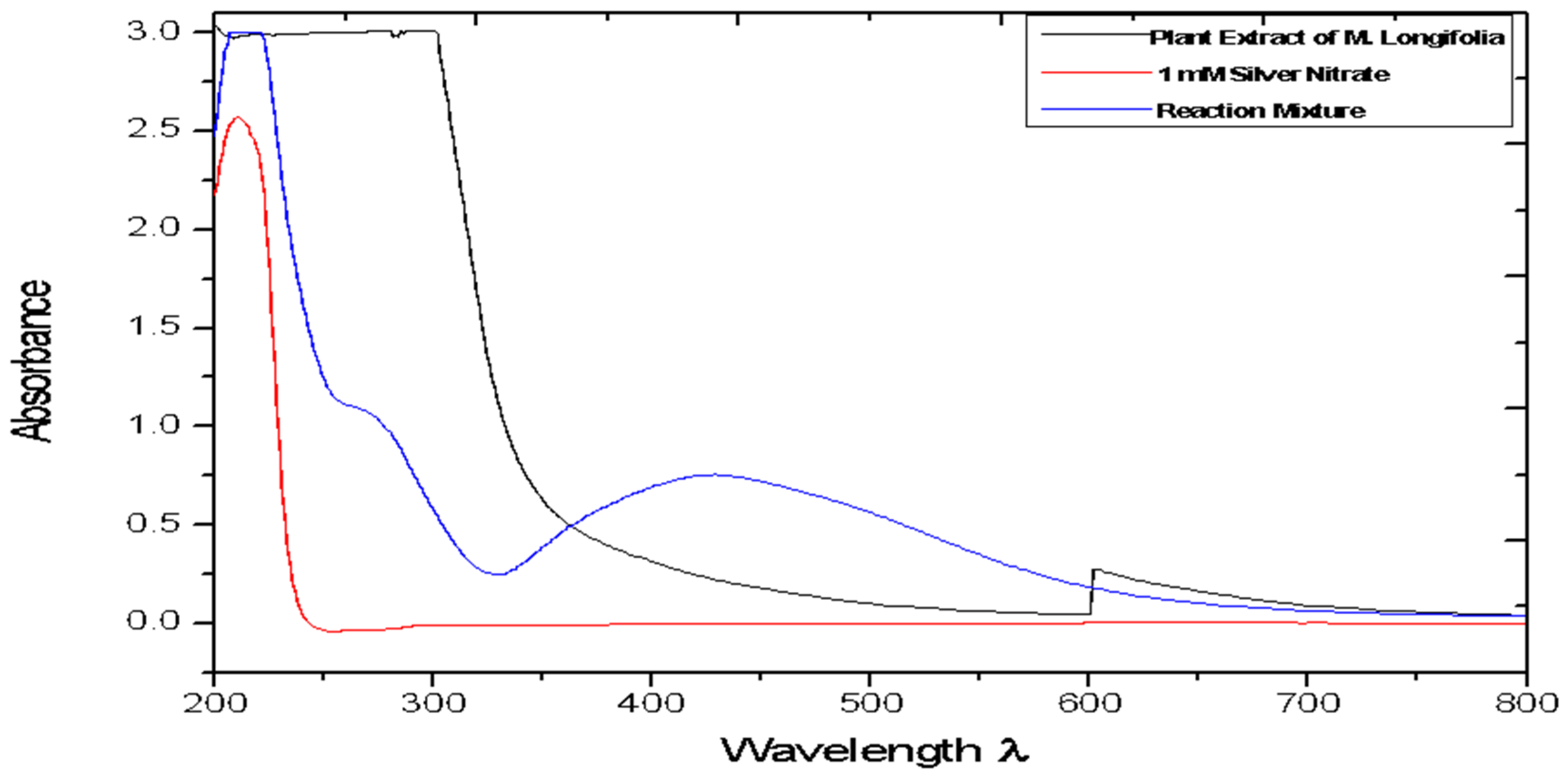
3.1. UV-Vis (Ultraviolet-Visible Spectroscopy Analysis)

The UV visible spectroscopy measurements were used to confirm the formation of silver nanoparticles. The spectra of the reaction mixture after 5 min of exposure to sunlight are shown in Figure 1. It is well known that the silver nanoparticles exhibit absorption at around wavelength 440 nm. This absorption of the spectra occurs as electrons on the metal surface undergo oscillations when excited by light at specific wavelengths. In the present study, the reaction mixture exhibits a unique peak at around 440 nm confirming the formation of silver nanoparticles [14].
A plant is the best option for producing nanoparticles, since the methods are free of toxic chemicals; in addition, plants also produce natural capping agents [28]. A UV-vis spectrophotometer was used to monitor the formation and stability of silver nanoparticles in the colloidal solution. It is generally recognized that UV–Vis spectroscopy could be used to examine the size and shape-controlled nanoparticles in aqueous suspensions [19]. In the present study, it has been found that the leaves of Madhuca longifolia, a traditional medicinal plant, have the potential to reduce silver nitrate ions to silver nanoparticles. The light brown color of the aqueous extract of Madhuca longifolia leaves was changed to reddish brown after 5 min of exposure to sunlight. The intensity of the color increased with the incubation time [24]. Maximum absorbance at 440 nm was observed, which is characteristic of silver nanoparticles [14].

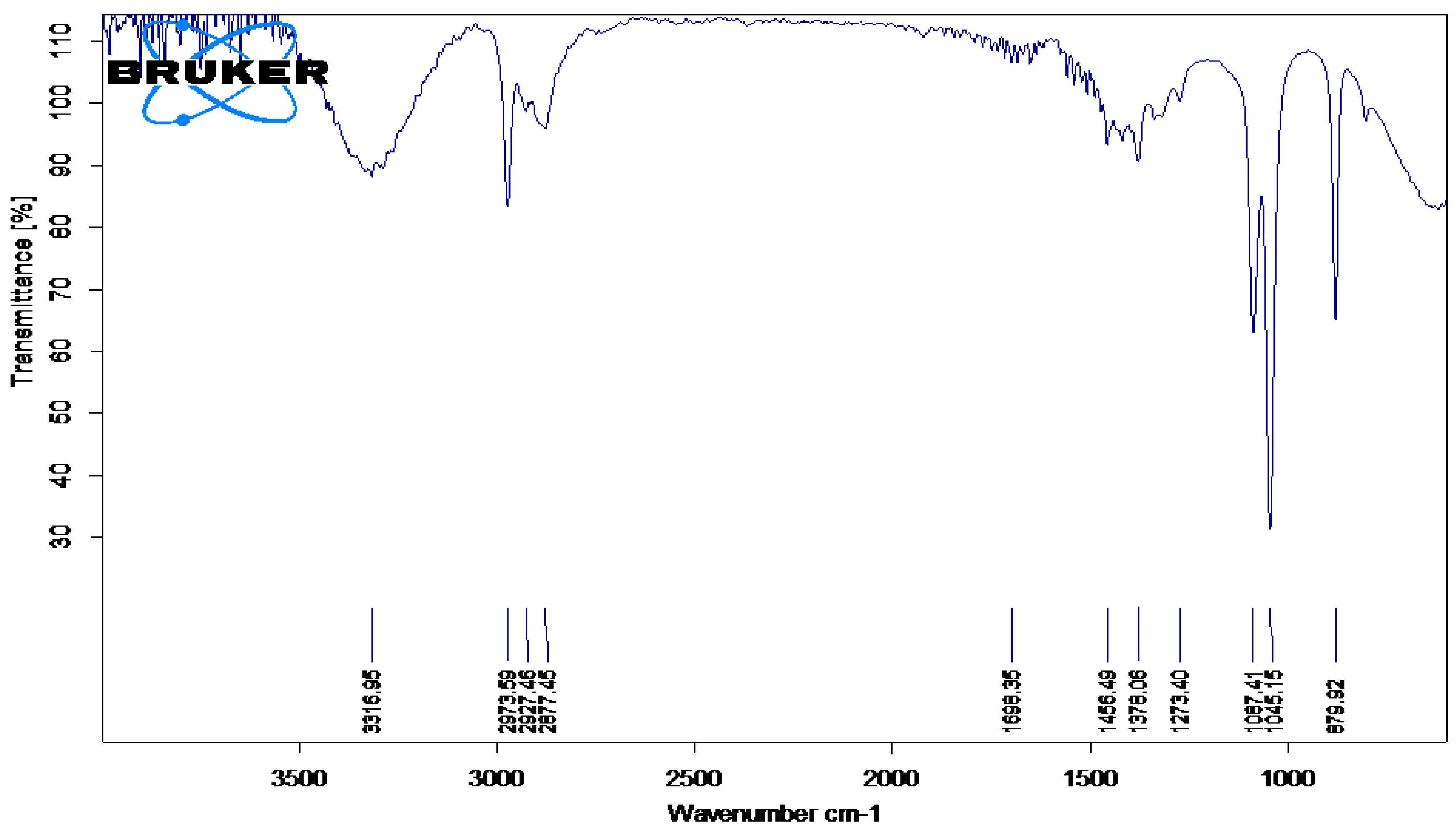
3.2. FTIR-(Fourier Transforms Infrared Spectroscopy)

The IR spectra of plant extract of Madhuca longifolia, and of the reaction mixture exposed to sunlight, are shown in Figure 2 and Figure 3, respectively. Peak assignments were used to identify the functional groups [29].
The IR spectral analysis of the plant extract and reaction mixture reveals the presence of carbohydrates, glycosides and flavonoids. These biomolecules might have played a major role in capping and reducing agents during the formation of silver nanoparticles [30]. The IR spectra for both (plant extract and reaction mixture) showed an intense peak at around 3200 cm−1 that can be assigned to phenolic O-H stretching. The peak at 3000 cm−1 can be assigned to N-H stretching, 1610 cm−1 can be assigned to C=C aromatic stretching, while the peak at 1500 cm−1 may be because of N-O stretching. The intense peak at 1112 cm−1 can be assigned to C=O stretching (s) and 1000 cm−1 assigned to C=C bending of the molecules, probably flavonoids, glycosides, carbohydrates, tannins present in the extract and reaction mixture [22].
FT-IR (Fourier-Transform InfraRed spectroscopy) analysis of an infrared spectrum represents a fingerprint of a sample with absorption peaks which correspond to the frequencies of vibrations between the bonds of the atoms making up the material. Because each different material is a unique combination of atoms, no two compounds produce the same infrared spectrum. Therefore, infrared spectroscopy can result in an identification (qualitative analysis) of every different kind of material [31]. The FTIR spectrophotometer was used to identify the functional groups present in phytoconstituents responsible for reducing and capping AgNPs [32]. FTIR spectrum analysis in this study detected several absorption peaks. It exhibited N–H stretching vibrations, indicating strong hydrogen bonding, and C=O extension vibrations attributed to carboxylic acids, ketones, and aldehydes, which were linked to the silver ions reduction [14]. FTIR study indicates that the carboxyl (-C=O), hydroxyl (-OH), and amine (N-H) groups present in leaf extract are involved in the reduction of silver ions [33].

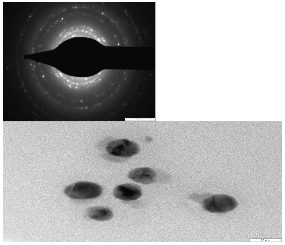
3.3. TEM (Transmission Electron Microscopy)

The shape and size of the resultant particles were elucidated with the help of TEM Figure 4. Aliquots of Ag nanoparticle solution were placed on a carbon-coated copper grid and allowed to dry under ambient conditions and TEM images were recorded. The TEM micrographs suggest that the particles are mostly spherical and are of a uniform size of around 20 nm. The SAED pattern indicates the crystalline nature of silver nanoparticles. Analysis of leaf-mediated synthesis of silver nanoparticles by TEM images proved that the size of AgNPs is in the range of nanoscale, almost spherically shaped, and that they have a mean diameter of 20 nm. Most of the nanoparticles had smooth edges and were circular. In the TEM image, the AgNPs were in physical contact but scattered by an adequately uniform distance between particles [34].

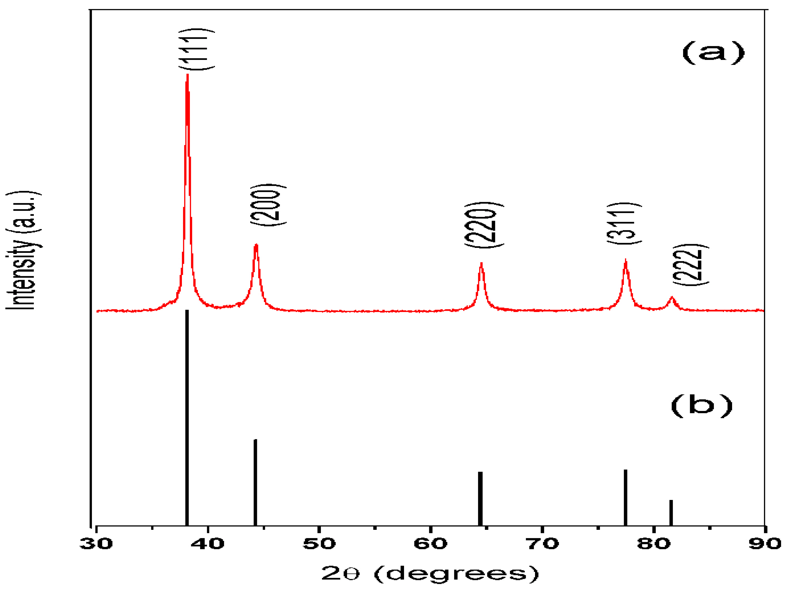
3.4. X-ray Diffraction Analysis (XRD)

The synthesized silver nanoparticles purified by repeated centrifugation and washing were dried and analyzed on an X-ray diffractometer. X-ray powder diffraction is a rapid analytical technique that provides information on unit cell dimensions. Figure 5 shows the XRD pattern of silver nanoparticles with characteristic peaks observed at 2 θ values of 38.11º, 44.27º, 64.42º, 77.47º, and 82.0° that can be assigned to planes of (111), (200), (220), (311) and (222), respectively (JCPDS No. 04–0783) [22,23,24].

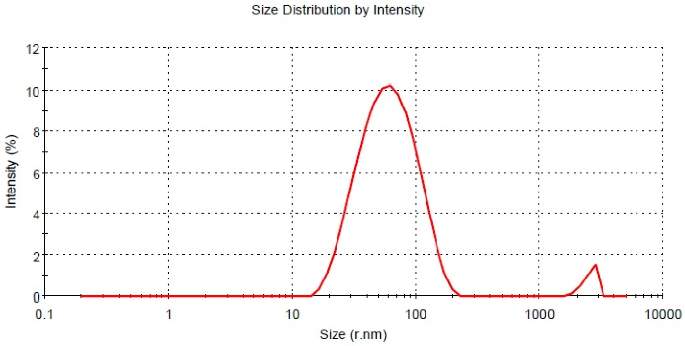
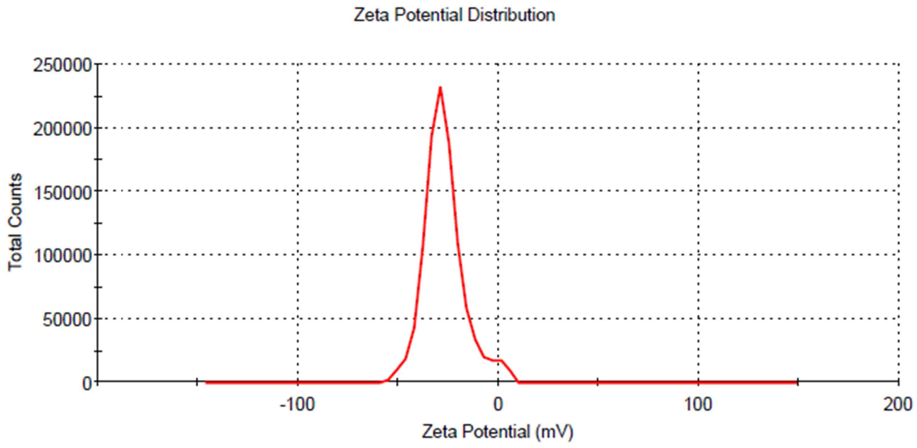
3.5. DLS (Dynamic Light Scattering)

The average hydrodynamic radius of AgNps synthesized using leaf extract of M. longifolia was found to be 45.30 nm Figure 6, with an asymmetric distribution between 13 to 200 nm. However, the TEM micrograph suggests that the particle size is around 20 nm. This disparity may be because all particles were taken into account for the calculation of hydrodynamic radius in DLS. The DLS data suggest that the particles are polydisperse. The Zeta potential of AgNps determined in water as a dispersant was −27.0 mV Figure 7. The high negative charge constitutes a repulsive barrier that physically separates the nanoparticles, avoiding aggregation [35].
A nanoparticle’s zeta potential is a key indicator for its stability in aqueous solution. For M. longifolia silver nanoparticles zeta potential measured was found to be −28 mV with a peak area of 100% intensity. These values indicate that the synthesized silver nanoparticles have excellent stabilization [36].

3.6. Phytochemical Screening of Leaf Extracts (Qualitative Screening)

To get an insight into the chemical constituent present in the leaves of M. longifolia, qualitative phytochemical analysis was carried out using standard procedures as described by Harborne (1973) [37]. The leaf extract of Madhuca longifolia shows positive tests for the bioactive compounds such as alkaloids, saponin, phytosterols, phenol, tannins, flavonoids, terpenoids, and ascorbic acid. Various phytochemical compounds detected are known to have medicinal importance.

3.7. Antibacterial Activity

The silver nanoparticles exhibited activity against the gram-negative bacteria E. coli. All the concentration studies showed a zone of inhibition. The zone of inhibition was concentration dependent (Table 1). The maximum antibacterial activity against E. coli with a zone of inhibition was 16 mm at 200 μg/ mL of silver nanoparticles; furthermore, there was no increase in the zone of inhibition with an increase in the concentration of silver nanoparticles [38].
The antimicrobial efficacy of the nanoparticles depends on the shapes and size of the nanoparticles. The mechanism of action of silver nanoparticles on bacterial cells is well studied. It has been reported that the silver ions, after their entry into the bacterial cell, interact with sulfur and phosphorus present in DNA; the nanoparticles can act on these soft bases and destroy the DNA, which leads to cell death [39]. It also hampers DNA replication and terminates the growth of microbes [40]. In the present study, AgNPs showed maximum activity against E. coli with a zone of inhibition of 20 mm, which is comparable with standard antibiotics.

3.8. In-Vitro Anti-Inflammatory Activity

The in vitro anti-inflammatory activity was studied using protein denaturation and the (human red blood cell) HRBC membrane hemolysis method. Table 2 shows the percent inhibition of protein denaturation for different concentrations of silver nanoparticles and sodium diclofenac. The silver nanoparticles exhibited good anti-inflammatory properties that are comparable with standard sodium diclofenac. Protein denaturation is a process by which proteins lose their tertiary and secondary structures by application of external stress compounds such as strong acid, base, concentrated inorganic-organic salt, or heat. Protein denaturation is a well-documented causative of inflammation. During this process, the proteins lose their quaternary structure, thereby inducing aggregation, which activates deleterious inflammatory signals [41]. Inhibition of albumin denaturation by silver nanoparticles in the present study indicated anti-inflammatory activity [15].
The stabilization of the HRBC membrane by hypotonicity-induced and heat-induced membrane lysis was studied to establish the mechanism of anti-inflammatory action of silver nanoparticles synthesized using Madhuca longifolia. The percent inhibition of hemolysis studied using heat and hypotonicity showed an increasing trend, with an increase in the concentration of silver nanoparticles (Table 3). At 500 μg/mL concentration of AgNps, it was comparable with standard sodium diclofenac 100 μg/mL concentration in both cases. The results indicate a reduction in inflammation that can be due to the presence of active-principle components capped on silver nanoparticles. There was a significant increase in absorbance due to the treatment of Madhuca longifolia AgNPs in comparison to that with sodium diclofenac. For all the data, the values are significant at the P < 0.001 level. Thus, the results reveal that sodium diclofenac has hepatoprotective potential.
The membrane stabilization effect as a measure of anti-inflammatory activity was used in the present study. On exposure of RBCs to the hypotonic solution, excessive accumulation of fluid within the cell resulted in membrane rupture followed by hemolysis [25].
It is well established that protein denaturation inhibitors’ agents function by suppressing different types of inflammatory mediators involved in the inflammation process [42]. Denaturation of proteins is a well-documented cause of inflammation; drugs such as Phenylbutazone, salicylic acid, sodium diclofenac, and flufenamic acid show dose-dependent ability against protein denaturation [43]. It has also been reported that various plant extracts and their isolated compounds showed good anti-inflammatory activity comparable to that of synthetic anti-inflammatory drugs [44].
One of the causes of rheumatoid arthritis is the denaturation of proteins, and inhibition denaturation is one of the in vitro tests used to screen anti-inflammatory drugs [45]. Those drugs that are available are known as nonsteroidal anti-inflammatory drugs (NSAIDs), which act by inhibiting the function of prostaglandin. Prostaglandin is an autocoid that is released extracellularly and triggers pain. Anti-inflammatory agents block this autacoid’s synthesis by either inhibiting the COX enzyme or protecting the lysosomal membrane from breakdown [46]. The majority of the anti-inflammatory drugs stabilize the plasma membrane of mammalian erythrocytes and thereby inhibit the heat-induced and hypotonicity-induced hemolysis [47].
The AgNPs at concentrations of 100 to 500 μg/ mL protected the human RBC membrane against lysis induced by hypotonic solution and heat during inflammation. Membrane stabilization leads to the prevention of leakage of serum protein and fluids into the tissues during a period of increased permeability caused by inflammatory mediators [48]. The AgNPs perhaps stabilized the red blood cell membranes by preventing the release of lytic enzymes and active mediators of inflammation. The RBC membrane was effective against hypotonicity-induced hemolysis, and hence it would be effective in the form of non-steroidal complex anti-inflammatory compounds in the control of inflammation [33].

Antioxidant Activity by TLC DPPH Method TLC Profiling

The dot blot study has shown an intense white spot on a light purple background (Figure 8) indicating the scavenging activity of silver nanoparticles. The spectrophotometric analysis of percent scavenging activity demonstrated by various concentrations of silver nanoparticles is shown in Table 4. All the concentrations have shown scavenging activity; however, it was highest with 500 μg/mL of AgNps.
In the present study, DPPH radical was used as a substrate to identify the free radical scavenging activity of AgNps. Among different concentrations of 100–500 μg/ mL the scavenging activity of AgNPs expressed the highest antioxidant activity causing 23.33, 24.33, and 29.66% inhibition of DPPH free radicals at 300, 400, and 500 μg/mL concentration, respectively. Ascorbic acid was used as positive control and its percent inhibition was 84% at 500 μg/mL concentration. The soluble compounds (flavanoids, glycosides, phenolic compounds etc) present in the methanolic leaf extract serve as a significant indicator of its potential antioxidant activity. Results suggests that the Madhuca longifolia possesses potent antioxidant properties by reducing reactive oxygen species as well as having the capability to donate hydrogen atoms. There was significant variation in DPPH scavenging activity among different concentrations, P = 0.05. In comparison to ascorbic acid, the DPPH scavenging activity significantly decreased under the influence of various concentrations.
DPPH is a stable free radical that accepts an electron or hydrogen radical to become a stable diamagnetic molecule. The decrease in the absorbance of DPPH radical is caused by antioxidants, because the reaction between antioxidant molecules and radicals progresses, which results in the scavenging of the radical by hydrogen donation. It is visually noticeable as a change in color from purple to yellow. Hence, DPPH is usually used as a substrate to evaluate the antioxidative activity of antioxidants. From the results, it can be assumed that the silver nanoparticle synthesized using Madhuca longifolia possesses hydrogen-donating capabilities and acts as an antioxidant [49].

3.9. Anticancer Activity

The silver nanoparticles synthesized using M. longifolia leaf extract showed more significant anticancer activity (Table 5) compared to the positive control group, i.e., Adriamycin GI50 = Growth inhibition of 50% (GI550) calculated from [(Ti-Tz)/(C-Tz)] × 100 = 50, drug concentration resulting in a 50% reduction in the net protein increase. TGI = Drug concentration resulting in total growth inhibition (TGI) will be calculated from Ti = Tz LC50 = the percentage of drug concentration at the end of the drug treatment, indicating a net loss of 50% cells following treatment, which is calculated from [(Ti-Tz)/Tz] × 100 = −50.

4. Conclusions

Synthesis of nanoparticles using biological agents is eco-friendly, low cost, and can be produced at room temperature. The present study has shown that phytochemicals from Madhuca longifolia leaf extracts have acted as both reducing and stabilizing agents. The silver nanoparticles were synthesized rapidly when exposed to sunlight for 5 min. They are spherical and uniform, capped with phytochemicals present in the leaf extract of Madhuca longifolia, as confirmed by various characterization techniques. In this process, the main phenolic compounds, triterpenoids, glycosides present in the leaf extract may be related with the chemical reduction of silver ions and thereby form nanoparticles with an average diameter on 20 nm. The AgNps are potent in inhibiting E. coli bacteria and showed significant antioxidant, anticancer, and anti-inflammatory activity. The outcome of this research confirms that M. longifolia extract is able to form silver nanoparticles and exhibits potent biological activities.

Author Contributions

Conceptualization, P.S., A.V., A.R., R.R., R.C., J.L.; Data curation, P.S., A.V., A.R., R.R., R.C., J.L.; Resources, P.S., A.V., A.R., R.R., R.C., J.L., M.J.H., S.A., M.A. (Mamdouh Allahyani), O.A., M.A. (Mazen Almehmadi), A.S.D., M.M.R.S., M.F.N.A.; Investigation P.S., A.V., A.R., R.R.; writing—original draft preparation, P.S., A.V., A.R., R.R., R.C., J.L.; writing—review and editing, P.S., A.V., A.R., R.R., R.C., J.L., M.J.H., S.A., M.A. (Mazen Almehmadi), O.A., M.A. (Mazen Almehmadi), A.S.D., M.M.R.S., M.F.N.A.; Funding acquisition M.M.R.S., M.F.N.A. All authors have read and agreed to the published version of the manuscript.

Funding

The authors would like to thank the Faculty of Medicine, Universiti Kebangsaan Malaysia for the approval and funding of this research (Approval code: FF-2020-016).

Data Availability Statement

All data used to support the findings of this study are included within the article.

Acknowledgments

Authors are very thankful to Babasaheb Ambedkar Marathwada University Aurangabad Botany department Arvind Dhabe. The authors are also thankful to the dept. of Biophysics, Government Institute of Science, Aurangabad.

Conflicts of Interest

The authors have declared that there are no conflict of interest.

References

  1. Khan, A.; Roy, A.; Bhasin, S.; Bin Emran, T.; Khusro, A.; Eftekhari, A.; Moradi, O.; Rokni, H.; Karimi, F. Nanomaterials: An alternative source for biodegradation of toxic dyes. Food Chem. Toxicol. 2022, 164, 112996. [Google Scholar] [CrossRef] [PubMed]
  2. Singh, P.; Goel, A.; Sehgal, M. Phytochemical analysis and antimicrobial activity of Madhuca longifolia and Clerodendrum infortunatum: Medicinal plants for application as textile finishes. Int. J. Chem. Stud. 2018, 6, 2031–2040. [Google Scholar]
  3. Sunita, T.; Kumar, M.V.; Manjusha, M.P.; Shanmukha, I. Correlation of antioxidant principles with cardioprotective activity of Madhuca longifolia (Koenig) leaves on isoproterenol induced myocardial infarction. Res. J. Pharm. Biol. Chem. Sci. 2016, 7, 971–977. [Google Scholar]
  4. Sarma, D.S.K.; Reddy, A.S.K.; Akhil, M.; Sankar, C.H.S. Phytochemical and Antimicrobial Activity of Whole Plant of Madhuca indica. Int. J. Res. Pharm. Chem. 2013, 3, 15–19. [Google Scholar]
  5. Kulkarni, A.; Autade, A.; Awari, D.; Chitlange, S. Pharmacological Evaluation of Anti-Psoriatic Activity of Madhuca longfolia on Experimental Animals. Int. J. Pharm. Pharm. Res. 2017, 8, 218–235. [Google Scholar]
  6. Umadevi, U.; Kamalam, M. Screening of an Indigenous Medicinal Plant-Madhuca Longifolia for Its Antioxidant and Antimicrobial Properties. Int. J. Pharm. Sci. Res. 2015, 6, 273–276. [Google Scholar]
  7. Akshatha, K.N.; Murthy, S.M.; Lakshmidevi, N. Ethnomedical Uses of Madhuca Longifolia—A Review. Int. J. Life Sci. Pharma Res. 2013, 3, 44–53. [Google Scholar]
  8. Khyade, M.S.; Takate, Y.A.; Divekar, M.V. Plants Used as an Antidote against Snakebite in Akole Taluka of Ahmednagar District (MS), India. J. Nat. Remedies 2011, 11, 182–192. [Google Scholar]
  9. Chakma, S.; Patel, M.P. Antimicrobial Activity of the Fruit-Seeds Madhuca Longifolia (Koenig). Int. Res. J. Pharm. 2011, 2, 192–193. [Google Scholar]
  10. Khare, P.; Kishore, K.; Sharma, D.K. Medicinal uses, Phytochemistry and Pharmacological profile of Madhuca longifolia. Asian J. Pharm. Pharmacol. 2018, 4, 570–581. [Google Scholar] [CrossRef]
  11. Roy, A. Plant derived silver nanoparticles and their therapeutic applications. Curr. Pharm. Biotechnol. 2021, 22, 1834–1847. [Google Scholar] [CrossRef] [PubMed]
  12. Ramadan, M.F.; Sharanabasappa, G.; Parmjyothi, S.; Seshagiri, M.; Moersel, J.T. Profile and levels of fatty acids and bioactive constituents in mahua butter from fruit-seeds of buttercup tree [Madhuca longifolia (Koenig)]. Eur. Food Res. Technol. 2006, 6, 710–718. [Google Scholar] [CrossRef]
  13. Arun, S.; Ajay, S.; Vadivel, V. Effect of extraction conditions on the total phenolic yield of Madhuca longifolia leaves and evaluation of its physico-chemical and antioxidant properties. J. Pharm. Sci. Res. 2017, 9, 1188–1194. [Google Scholar]
  14. Raut, R.W.; Mendhulkar, V.D.; Kashid, S.B. Photosensitized synthesis of silver nanoparticles using Withania somnifera leaf powder and silver nitrate. J. Photochem. Photobiol. B Biol. 2014, 132, 45–55. [Google Scholar] [CrossRef] [PubMed]
  15. Ananthi, P.; Jeyapaul, U.; Balgan, J.; Kala, S.M.J. Green synthesis and characterization of silver nanoparticles using Triumfetta rotundifolia plant extract and its antibacterial activities. J. Nat. Prod. Plant Resour. 2016, 12, 21–27. [Google Scholar]
  16. Zia, F.; Ghafoor, N.; Iqbal, M.; Mehboob, S. Green synthesis and characterization of silver nanoparticles using Cydonia oblong seed extract. Appl. Nanosci. 2016, 6, 1023–1029. [Google Scholar] [CrossRef]
  17. Srirangam, G.M.; Rao, K.P. Synthesis and charcterization of silver nanoparticles from the leaf extract of Malachra capitata (L.). Rasayan J. Chem. 2017, 10, 46–53. [Google Scholar]
  18. Khanra, K.; Roy, A.; Bhattacharyya, N.; Bengal, W. Evaluation of Antibacterial Activity and Cytotoxicity of Green Synthesized Evaluation of Antibacterial Activity and Cytotoxicity of Green Synthesized Silver Nanoparticles Using Hemidesmus Indicus R. Br. Am. J. Nanosci. Nanotechnol. Res. 2013, 1, 1–6. [Google Scholar]
  19. Choudhury, M.; Maiti, S.; Bepari; Pradhan, A.; Maity, P.; Bengal, W. In Vitro and In Vivo anti-inflammatory potential of silver nanoparticles synthesized from Anacardium. World J. Pharm. Pharm. Sci. 2018, 7, 1297–1309. [Google Scholar]
  20. Shinde, U.A.; Phadke, A.S.; Nair, A.M.; Mungantiwar, A.A.; Dikshit, V.J.; Saraf, M.N. Membrane stabilizing activity—A possible mechanism of action for the anti-inflammatory activity of Cedrus deodara wood oil. Fitoterapia 1999, 70, 251–257. [Google Scholar] [CrossRef]
  21. Leelaprakash, G.; Dass, S.M. Invitro anti-inflammatory activity of methanol extract of Enicostemma axillare. Int. J. Drug Dev. Res. 2011, 3, 189–196. [Google Scholar]
  22. Singh, D.; Singh, M.; Yadav, E.; Falls, N.; Komal, U.; Dangi, D.S.; Kumar, V.; Verma, A. Amelioration of diethylnitrosamine (DEN)-induced hepatocellular carcinogenesis in animal models: Via knockdown oxidative stress and proinflammatory markers by Madhuca longifolia embedded silver nanoparticles. RSC Adv. 2018, 8, 6940–6953. [Google Scholar] [CrossRef] [PubMed]
  23. Gaikwad, R.D.; Ahmed, M.L.; Khalid, M.S.; Swamy, P. Anti-inflammatory activity of Madhuca longifolia seed saponin mixture. Pharm. Biol. 2009, 47, 592–597. [Google Scholar] [CrossRef]
  24. Torre, G.L.T.D.; Arollado, E.C.; Atienza, A.A.; Manalo, R.A.M. Evaluation of antioxidant capacity and identification of bioactive compounds of crude methanol extracts of Caesalpinia pulcherrima (L.) Swartz. Indian J. Pharm. Sci. 2017, 79, 113–123. [Google Scholar]
  25. Ferrali, M.; Signorini, C.; Ciccoli, L.; Comporti, M. Iron release and membrane damage in erythrocytes exposed to oxidizing agents, phenylhydrazine, divicine and isouramil. Biochem. J. 1992, 285, 295–301. [Google Scholar] [CrossRef] [Green Version]
  26. Vichai, V.; Kirtikara, K. Sulforhodamine B colorimetric assay for cytotoxicity screening. Nat. Protoc. 2006, 1, 1112–1116. [Google Scholar] [CrossRef]
  27. Pandey, K.; Sharma, P.K.; Dudhe, R. Anticancer Activity of Parthenium hysterophorus Linn and Oldenlandia corymbosa Lam by Srb Method. Open Access Sci. Reports 2012, 1, 1–3. [Google Scholar]
  28. Kanchana, R.; Zantye, P. Plant-mediated synthesis of silver nanoparticles with diverse applications. Asian J. Pharm. Clin. Res. 2016, 9, 124–128. [Google Scholar]
  29. Rautela, A.; Rani, J.; Debnath, M. Green synthesis of silver nanoparticles from Tectona grandis seeds extract: Characterization and mechanism of antimicrobial action on different microorganisms. J. Anal. Sci. Technol. 2019, 10, 5. [Google Scholar] [CrossRef]
  30. Hamouda, R.A.; Hussein, M.H.; Abo-Elmagd, R.A.; Bawazir, S.S. Synthesis and biological characterization of silver nanoparticles derived from the cyanobacterium Oscillatoria limnetica. Sci. Rep. 2019, 9, 13071. [Google Scholar] [CrossRef]
  31. Gnanasundaram, I.; Balakrishnan, K. Synthesis and Evaluation of Anti-Inflammatory Activity of Silver Nanoparticles from Cissus vitiginea Leaf Extract. J. Nanosci. Technol. 2017, 3, 266–269. [Google Scholar]
  32. Jasmine, R.; Rajasulochana, M.; Aude, R. Evaluation of in vitro antioxidant and anti-inflammatory property exhibited by silver nanoparticles stabilized by Adathoda vasica. J. Chem. Pharm. Res. 2016, 8, 128–137. [Google Scholar]
  33. Krithika, S.; Niraimathi, K.L.; Arun, K.P.; Narendran, R.; Balaji, K.; Brindha, P. In Vitro Anti-Inflammatory Studies on Silver Nanoparticles Synthesized from Centratherum punctatum Cass. Int. J. Res. Ayurveda Pharm. 2016, 7, 61–66. [Google Scholar]
  34. Masum, M.M.I.; Siddiqa, M.M.; Ali, K.A.; Zhang, Y.; Abdallah, Y.; Ibrahim, E.; Qiu, W.; Yan, C.; Li, B. Biogenic synthesis of silver nanoparticles using phyllanthus emblicafruit extract and its inhibitory action against the pathogen acidovorax oryzaestrain RS-2 of rice bacterial brown stripe. Front. Microbiol. 2019, 10, 820. [Google Scholar] [CrossRef]
  35. Seeram, H.; Santhosh, K.J.; Sravani, D.; Ravi, K.G.; Madhu, C.; Susheela, B.G. Green synthesis, Characterization and anti microbial activity of silver nano particles—Review Paper. Int. J. Eng. Res. Appl. 2015, 5, 30–34. [Google Scholar]
  36. Sharma, M.; Yadav, S.; Srivastava, M.; Ganesh, N.; Srivastava, S. Promising anti-inflammatory bio-efficacy of saponin loaded silver nanoparticles prepared from the plant Madhuca longifolia. Asian J. Nanosci. Mater. 2018, 1, 244–261. [Google Scholar]
  37. Bagyalakshmi, J.; Haritha, H. Green Synthesis and Characterization of Silver Nanoparticles Using Pterocarpus marsupium and Assessment of its In Vitro Antidiabetic Activity. Am. J. Adv. Drug Deliv. 2017, 5. [Google Scholar] [CrossRef]
  38. Harbone, J.B. Phytochemical Methods A Guide to Modern Techniques of Plant Analysis, 3rd ed.; Chapman and Hall: Madras, India; Springer: Dordrecht, The Netherlands, 1998; pp. 1–317. [Google Scholar]
  39. Srinivas, K.; Celestin, R.V.; Babu, A.M.S.S.; Rajavel, P. Pharmacognostic, Phytochemical and Biological Studies of Leaves of Indigofera barberi. Asian J. Phytomed. Clin. Res. 2013, 1, 1–13. [Google Scholar]
  40. Parvin, M.S.; Das, N.; Jahan, N.; Akhter, M.A.; Nahar, L.; Islam, M.E. Evaluation of in vitro anti-inflammatory and antibacterial potential of Crescentia cujete leaves and stem bark Pharmacology and Toxicology. BMC Res. Notes 2015, 8, 412. [Google Scholar] [CrossRef]
  41. Prabhu, S.; Poulose, E.K. Silver nanoparticles: Mechanism of antimicrobial. Int. Nano Lett. 2012, 2, 32–41. [Google Scholar] [CrossRef]
  42. Varghese, R.E.; Ragavan, D.; Sivaraj, S.; Gayathri, D.; Kannayiram, G. Anti-inflammatory activity of Syzygium aromaticum silver nanoparticles: In vitro and in silico study. Asian J. Pharm. Clin. Res. 2017, 10, 370–373. [Google Scholar] [CrossRef]
  43. Kumari, C.S.; Yasmin, N.; Hussain, M.R.; Babuselvam, M. Invitro anti-inflammatory and anti-artheritic property of Rhizopora mucronata leaves. Int. J. Pharm. Sci. Res. 2015, 6, 482–485. [Google Scholar]
  44. Sakat, S.S.; Juvekar, A.R.; Gambhire, M.N. In vitro antioxidant and anti-inflammatory activity of methanol extract of Oxalis corniculata Linn. Int. J. Pharm. Pharm. Sci. 2010, 2, 146–155. [Google Scholar]
  45. Manivannan, D.; Sukumar, R. The RBC Membrane stabilization in an in-vitro method by the drug isolated from Leucas aspera. Int. J. Appl. Sci. Eng. 2007, 5, 133–138. [Google Scholar]
  46. Pant, K.; Kshitij, A.; Prem, S. To study in vitro anti-inflammatory activity of Anthracephalus cadamba leaves extract. DHR Int. J. Pharm. Sci. 2012, 3, 55–60. [Google Scholar]
  47. Chowdhury, A.; Azam, S.; Jainul, M.A.; Faruq, K.O.; Islam, A. Antibacterial activities and in vitro anti-inflammatory (Membrane Stability) properties of methanolic extracts of Gardenia coronaria leaves. Int. J. Microbiol. 2014, 2014, 410935. [Google Scholar] [CrossRef] [PubMed]
  48. Navale, G.; Patil, D.D.; Patil, A.A.; Patil, K.B.; Patil, N.B. Membrane Stabilization assay for Anti-inflammatory activity yields misleading results for samples containing traces of Methanol. Asian J. Pharm. Res. 2019, 9, 169. [Google Scholar] [CrossRef]
  49. Dahake, A.P.; Chakma, C.; Joshi, D.; Chakma, R.; Tripathi, A. Antioxidant activity of methanolic extract of Madhuca longifolia bark. J. Pharm. Res. 2010, 3, 1709–1711. [Google Scholar]
Figure 1. UV-Vis spectra of reaction mixture and control recorded after 5 min of exposure to sunlight.
Molecules 27 06404 g001
Figure 2. FTIR spectrum of Madhuca longifolia leaf extract.
Molecules 27 06404 g002
Figure 3. FTIR spectrum of AgNps synthesized by Madhuca longifolia leaf extract.
Molecules 27 06404 g003
Figure 4. TEM images of AgNps synthesized using dried leaf extract M. longifolia insight Selected Area Electron Diffraction (SAED) pattern of AgNPs.
Molecules 27 06404 g004
Figure 5. (a) XRD pattern for synthesized silver nanoparticles (b) Pattern from JCPDS file No. 04-078.
Molecules 27 06404 g005
Figure 6. Size distribution of silver nanoparticles.
Molecules 27 06404 g006
Figure 7. Zeta potential for silver nanoparticles.
Molecules 27 06404 g007
Figure 8. TLC-DPPH assay of M. longifolia leaf extract after spraying DPPH.
Molecules 27 06404 g008
Table 1. Antimicrobial activity of M. longifolia Leaf Nanoparticles.
Name of Organism

E. coli
Madhuca longifola AgNPs concentration (µg/mL) wt/vSTANDARD
(ciprofloxacin zone of inhibition in mm) (5 µg/disc)
50100150200250
Zone of inhibition in mm including disk (AgNPs)101113162020
Zone of inhibition in mm including disk (Crude)1013131422
Table 2. Anti-inflammatory activity of silver nanoparticles studied using BSA denaturation method.
Concentration of AgNps (μg/mL)
and sodium diclofenac (μg/mL)
Percent inhibition
Sodium DiclofenacAgNps
10021.48 ± 0.5120.60 ± 0.22
20028.17 ± 0.7823.80 ± 0.41
30033.28 ± 1.1233.17 ± 1.35
40042.16 ± 0.3139.48 ± 0.26
50056.68 ± 0.9253.15 ± 0.87
Table 3. Effect of different concentrations of AgNPs and standard (sodium diclofenac) on HRBC membrane hemolysis of erythrocyte using heat-induced and hypotonicity studies.
Treatment(s)Concentration (μg/mL)Absorbance at 560 nm%Inhibition of hemolysis
For Heat-induced studiesFor Hypotonicity induced studiesFor Heat-induced studiesFor Hypotonicity induced studies
Control0.320.33
M.longifolia AgNps
1000.390.4221.8727.27
2000.240.402521.21
3000.20 *0.24 NS37.527.27
4000.18 *0.21 NS43.7536.36
500016 NS0.14 NS5057.57
Sodium Diclofenac1000.080.137560.60
SE = of six individual observations, * values are significant at P < 0.001, NS = non-significant.
Table 4. Percent scavenging activity showed by AgNPs and ascorbic acid.
Concentration (μg/mL)DPPH% Scavenging activity
AgNpsCrud extract of leaf Ascorbic acid
1007.6 *7.078
20014.33 *10.8980
30023.33 *16.0082
40024.33 *20.4583
50029.6622.1884
SE= of six individual observations, * values are significant at P < 0.005 and P < 0.001.
Table 5. Anticancer activities of silver nanoparticles and control (ADR).
SamplesHuman Cervical Cancer Cell Line SiHa
Drug Concentration (μg/mL)
10204080
Silver nanoparticles96.4105.6107.9133.5
ADR−51.2−27.6−34.5−23.1
Human Breast Cancer CellLine MDA-MB-231
Silver nanoparticles104.599.882.853.7
ADR−48.0−55.6−56.7−49.9
LC 50TGIGI50 studies
Human Cervical Cancer CellLine SiHa
LC 50TGIGI50
Silver nanoparticles>100>100>80
ADR52.5<10<10
Human Breast Cancer CellLine MDA-MB-231
Silver nanoparticles>100>100>80
ADR44.1<10<10
LC50 = Concentration of drug causing 50% cell kill; GI50 = Concentration of drug causing 50% inhibition of cell growth; TGI = Concentration of drug causing total inhibition of cell growth; ADR = Adriamycin, Positive control compound.
Publisher’s Note: MDPI stays neutral with regard to jurisdictional claims in published maps and institutional affiliations.

