Article

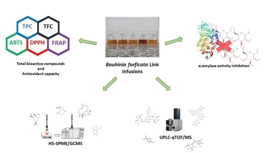
Bauhinia forficata Link Infusions: Chemical and Bioactivity of Volatile and Non-Volatile Fractions

by
Eliane Przytyk Jung
1,
Beatriz Pereira de Freitas
2,
Claudete Norie Kunigami
1,
Davyson de Lima Moreira
3,*,
Natália Guimarães de Figueiredo
4,
Leilson de Oliveira Ribeiro
1,* and
Ricardo Felipe Alves Moreira
5
1
Laboratory of Organic and Inorganic Chemical Analysis, National Institute of Technology, Rio de Janeiro 20081-312, Brazil
2
Faculty of Chemical Engineering, Federal University of Fluminense, Niterói 24210-240, Brazil
3
Laboratory of Natural Products, Rio de Janeiro Botanical Garden Research Institute, Rio de Janeiro 22460-030, Brazil
4
Laboratory of Tobacco and Derivatives, National Institute of Technology, Rio de Janeiro 20081-312, Brazil
5
Food and Nutrition Graduate Program, Federal University of Rio de Janeiro State (UNIRIO), Rio de Janeiro 22290-250, Brazil
*
Authors to whom correspondence should be addressed.
Molecules 2022, 27(17), 5415; https://doi.org/10.3390/molecules27175415
Submission received: 20 July 2022 / Revised: 17 August 2022 / Accepted: 20 August 2022 / Published: 24 August 2022

Abstract

:
This study aimed to evaluate Bauhinia forficata infusions prepared using samples available in Rio de Janeiro, Brazil. As such, infusions at 5% (w/v) of different brands and batches commercialized in the city (CS1, CS2, CS3, and CS4) and samples of plant material botanically identified (BS) were evaluated to determine their total phenolic and flavonoid contents (TPC and TFC), antioxidant capacity (ABTS•+, DPPH, and FRAP assays), phytochemical profile, volatile compounds, and inhibitory effects against the α-amylase enzyme. The results showed that infusions prepared using BS samples had lower TPC, TFC and antioxidant potential than the commercial samples (p < 0.05). The batch averages presented high standard deviations mainly for the commercial samples, corroborating sample heterogeneity. Sample volatile fractions were mainly composed of terpenes (40 compounds identified). In the non-volatile fraction, 20 compounds were identified, with emphasis on the CS3 sample, which comprised most of the compounds, mainly flavonoid derivatives. PCA analysis demonstrated more chemical diversity in non-volatile than volatile compounds. The samples also inhibited the α-amylase enzyme (IC50 value: 0.235–0.801 mg RE/mL). Despite the differences observed in this work, B. forficata is recognized as a source of bioactive compounds that can increase the intake of antioxidant compounds by the population.

Graphical Abstract

1. Introduction

Bauhinia is a genus comprising over 300 species widely distributed in tropical and subtropical forests. In Brazil, 64 species belonging to the Fabaceae family were identified and are commonly known as “pata-de-vaca” due to the shape of their leaves. Most species are of Asian origin; however, Bauhinia longifolia (Bong.) Steud. and Bauhinia forficata Link are native species from Brazil [1,2].
B. forficata is widely used in Brazilian folk medicine due to its beneficial effects on different diseases and human disorders such as rheumatism, local pain, uric acid, and uterine problems [3], but it is primarily used to treat type II diabetes [4]. The beneficial effects are associated with various biocompounds present in B. forficata, such as flavonoids, alkaloids, and terpenes/terpenoids [2,5]. The flavonoid compounds are highlighted since they are the major class in B. forficata extracts. Farag et al. [6] registered the presence of quercetin and kaempferol derivatives in different species of the Bauhinia genus, including B. forficata.
In Brazil, B. forficata is mainly commercialized dried and used to prepare infusions. Thus, under Brazilian law, the Bauhinia tea is associated to food products, so it is not mandatory to indicate the content of bioactive or toxic compounds, as in a limited manner in herbal products [7]. B. forficata infusions were used in different in vivo studies, such as that reported by Salgueiro et al. [8], who evaluated the effects of infusions on oxidative stress, liver damage, and glycemia in mice. Nevertheless, data on the content of bioactive compounds, antioxidant capacity, and volatile compounds, among other parameters of this plant, to compare botanically identified and commercialized samples and their infusions are scarce in the literature. Since it is well known that various factors such as climate, processing, and storage conditions may influence the content of bioactive compounds and the volatile fraction of medicinal plants [9,10], there is a clear need for further studies.
Despite that, to date, there are no data available on the volatile composition of B. forficata infusions. This fraction cannot be underestimated since B. forficata is prepared by infusion or decoction and, therefore, some of the volatile content may disperse in the beverage (hydrolate) and contribute to its beneficial actions besides the aroma. This approach has already been evaluated for other medicinal plants, and the migration of terpenoid and other compounds classes present in the essential oil of the plant for infusion was observed [11,12].
For such an evaluation, headspace solid-phase microextraction coupled to gas chromatography–mass spectrometry (HS-SPME/GC–MS) has been reported as a fast, sensitive, and solvent-free technique for analyzing the extraction and isolation of volatile and semi-volatile compounds, and it has been widely used since its invention in 1989 [13,14]. Furthermore, interference from the infusion matrix may be drastically reduced while the headspace analytes are trapped in the fiber [15]. Thus, this technique has been successfully applied to analyze volatile compounds in infusions and teas [16,17].
In this sense, this work aimed to perform a comprehensive chemical characterization of the volatile and non-volatile fractions of botanically identified and commercial samples of B. forficata used to prepare infusions at 5%. The antioxidant capacity measured by ABTS•+, DPPH and FRAP assays and inhibitory activity of α-amylase of the samples were also determined.

2. Results

2.1. Bioactive Compounds and Antioxidant Capacity of B. forficata Infusions

The TPC, TFC and antioxidant capacity of the B. forficata infusions are summarized in Table 1. It should be pointed out that the results presented in this study for TPC and TFC are expressed as rutin equivalents (RE) since this compound belongs to the flavonoids class, which is the major class in this species [6]. The values of TPC varied from 1923 to 6355 mg RE/100 g. Compared to the literature, the highest value found in this study, which was for the dry basis (7222 mg RE/100 g), is superior to that reported by Port’s et al. [18], who evaluated different infusions of herbs from the Brazilian Amazonian region. Even though these authors did not evaluate B. forficata. However, their approach was the closest to this study, reporting results of the chemical evaluation for a B. ungulata infusion at 2% (g/mL) (2367 mg GAE/100 g dry basis). By calculation, at 5%, 5918 mg GAE/100 g dry basis would be found. Comparisons with data from the literature are difficult since few studies used the same species, and even when the species were the same, the results were expressed using different chemical standards, as in the example above. Additionally, it is easier to find data on B. forficata extracted with organic solvent than with hot water (infusion). Thus, our discussion will be focused on the differences observed among the brands and respective batches evaluated herein.
The values for TPC, TFC, and antioxidant capacity measured by DPPH, ABTS•+, and FRAP assays varied from 1923 to 6355 mg RE/100 g, 482 to 3700 mg RE/100 g, 19 to 206 µmol Trolox/g, 27 to 204 µmol Trolox/g, and 85 to 644 µmol Fe2+/g, respectively. This corroborates that variations among samples and batches were high (Table 1). Among batches of the commercial samples, the highest values for TPC, TFC and antioxidant capacity (CS4B2 and CS3B3) were observed. These were higher than values reported to botanically identified sample (BS), which may be explained by differences in cultivation practices and the way the plants were processed. For example, the drying time may increase the degradation of plant bioactive compounds, whereas soil characteristics and precipitation conditions may affect the biosynthesis of secondary metabolites [9,10].
CS4B2 presented the highest TPC and FRAP values. For the TFC and DPPH and ABTS•+ assays, CS3B3 presented the highest values (Table 1). The literature points to a direct relationship between TPC and antioxidant capacity; however, in this study, the sample that presented the highest TPC did not show the highest values for antioxidant capacity measured by all assays employed. This corroborates that the phytochemical composition of plant extracts may interact differently with radical species, which helps explain the results found.
High standard deviations were observed in CS2 and CS4 samples. The variation coefficient for the TFC reached 75% in CS2, for example, confirming the heterogeneity among the sample batches. The low standard deviation of the BS may be associated mainly with the standardization of the processing, which was followed from the harvest of leaves to drying. In addition, the harvest was from the same tree, although it took place in different seasons. This may also justify the low standard deviation of CS1 and CS3. Furthermore, conditions such as storage time, temperature, and kind of package have influence on the stability of bioactive compounds.
Since the samples showed heterogeneous batches according to the statistical analysis for this set of experiments, two groups were observed from their averages: one composed of the BS, CS1, CS2, and CS4 groups, for which no statistically significant differences were observed for the TPC, TFC, DPPH, ABTS•+, and FRAP assays (p > 0.05), and the other represented by CS3 alone. These data provide important information about the production chain of B. forficata, rendering evident the need to standardize the steps that involve from harvest to distribution to deliver to consumers a product that guarantees its bioactive properties. B. forficata is widely used in Brazilian folk medicine due to its beneficial effects for treating rheumatism, local pain, uric acid, uterine problems [3], and, especially, type II diabetes [4]. This is possible due to the phytochemical profile of B. forficata, which is mainly composed of flavonoids, recognized for their antioxidant capacity [19].

2.2. LC-HRMS Analysis

A total of 20 phenolic compounds (Table 2), among flavonoids, phenolic acids, and other phenolic compounds, were tentatively identified in the samples. The majority are kaempferol and quercetin derivatives. The samples comprised flavonoid O-glycosides, thus in accordance with previously reported results, which prove its pharmacological action [20,21]. Additionally, polar compounds were identified in the samples in accordance with the polarity of the infusions.
Most phenolic compounds identified in this study were free phenolic compounds, esterified with sugars or other compounds with low molecular masses, such as quercetin 3-O-rhamnoside, Kaempferol 3-O-glucoside, and Isorhamnetin.
Rutin, Isoquercetin, Quercetin-O-pentoside, Quercetin 3-O-rhamnoside, Kaempferol 3-O-glucoside, Kaempferol 3-O-rutinoside and Isorhamnetin were the compounds detected in all samples. CS3 is the infusion with the greatest number of compounds that vary according to the batch. In CS1, CS2, and CS4, the same 11 flavonoids were identified with differences in the relative abundance of the ions. Compound 3 showed a precursor ion [M–H]¯ at 331.0670 m/z and a typical loss of a hexose in MS2 resulting in a [M–H]¯ m/z 169 fragment. It was assigned as galloyl hexose. Compound 4 was assigned as hydroxibenzoic acid based on precursor ion [M–H]¯ at 137.0244 m/z and a very low error between experimental and theoretical mass of 0.1 ppm [22]. Compound 6 showed a precursor ion [M–H]¯ at 353.0875 m/z and the quinic acid fragment in MS2 at m/z 191, been identified as 3-Caffeoyl quinic acid. Compound 10 was assigned as Quercetin 3-O-glucopyranoside by comparison with literature records (1 ppm error ([M–H]¯ m/z 463.0878) [23]. Compound 19 was assigned as Isorhamnetin 3-O-rhamnosyl-rutinoside based on precursor ion [M–H]¯ m/z 769.2190 and based in the loss of Isorhamnetin fragment at m/z 315. Kaempferol fragment ion at m/z 284 was used to identify compound 20 as Kaempferol 3-O-dirhamnoside along with the precursor ion [M–H]¯ m/z 577.1595 [6]. Identification of the other listed compounds by fragmentation data and exact mass were previously described by the authors [24,25].
The UPLC-ESI-Q-TOF MS/MS chromatographic technique was an efficient tool to characterize and identify the phenolic compounds in B. forficata infusions. It is important to highlight that the advantage of this technique is that, although it is not quantitative, one may relatively quantify the compounds, even the isomeric forms (e.g., Catechin, Epi-catechin, and Quercetin-O-pentoside), and, in case of a lack of standards, the compound assignments may be made by comparison of UV spectra and MS data (accurate mass and fragmentation) with previous literature reports [6,22,23].
A PCA analysis of the non-volatile chemical composition showed three distinct groups: I—CS3B2, CS3B3 and CS4B2; II—CS1B1, CS1B2, CS2B1, CS2B2, CS4B1, CS4B3 and BSB1; III—CS3B1, BSB2 (Figure 1A). These results demonstrate great chemical variability between the different samples, although flavonoids Rutin, Isoquercetin, Quercetin-O-pentoside, Quercetin 3-O-rhamnoside, Kaempferol 3-O-glucoside, Kaempferol 3-O-rutinoside and Isorhamnetin were detected in all samples.

2.3. HS-SPME/CG–MS

The identification and relative concentrations of the volatile compounds in the five herbal infusions of B. forficata are shown in Table 3, in order of retention time (Rt), and increasing Linear Retention Index (LRI). Forty volatile compounds were tentatively identified, of which only seven were detected in all samples: 2-Propyl-heptanol (7.69–19.42%), Geranyl acetone (4.38–7.31%), Dodecanol (3.11–11.37%), β-Ionone (0.71–5.84%), Spathulenol (11.78–30.87%), Caryophyllene oxide (2.76–17.46%), and Benzoic acid 2-ethylhexyl ester (1.12–20.14%). The volatile compounds included terpenoids, represented by C13-norisoprenoids, sesquiterpenes, and monoterpenes, as well as hydrocarbons, alcohols, esters, aldehydes, ketones, and acids. Among all chemical groups found in the volatiles of the B. forficata infusions, sesquiterpenes (hydrocarbon and oxygenated) were present in a higher number (17) and represented most of the composition of the BS (63%), CS1 (61%), CS3 (50%), and CS4 (53%). Esters (31%) and alcohols (29%) accounted for most of the composition of CS2.
There are no data in the literature on the volatile composition of B. forficata infusions or any species of the Bauhinia genus. However, there are two studies that identified constituents of essential oils of this species and demonstrated that they are essentially composed of sesquiterpenoids. Duarte-Almeida et al. [26] and Sartorilli and Correa [27] evaluated the composition of essential oils in B. forficata and reported that the content of sesquiterpenoids was 87% and 96%, respectively. Our results and those from essential oils [26,27] are a great evidence that a mostly sesquiterpenic volatile fraction composition may be characteristic of this species.
It is well established that many sesquiterpenes and their alcohol, aldehyde, and ketone derivatives are biologically active or precursors of metabolites with biological functions, while others have desirable fragrance and flavoring properties [28]. Spathulenol (8.53–25.86%) and Caryophyllene oxide (2.76–17.46%) were two of the major compounds in all samples. Both compounds are known to possess several biological activities. Nascimento et al. [29] demonstrated antioxidant, anti-inflammatory, antiproliferative, and antimycobacterial activities of spathulenol, and a moldy and herbaceous odor is attributed to this compound [30]. In turn, Caryophyllene oxide has a floral and woody odor [31,32], and biological activities such as anticholinesterase, analgesic, anti-inflammatory, and antifungal activities were also reported [33,34]. Regarding the class of norisoprenoids (C13), they were detected in all samples at concentrations ranging from 7.56% to 14.71%, highlighting Geranyl acetone and β-Ionone. It is reported that they present a significant aromatic impact in fruits such as grapes, apples, lychee, and mango [35,36], with a floral odor being attributed to them [37].
Attention is drawn to the identification of Bisphenol A (BPA) and Dibutyl phthalate (DBP) in some samples evaluated here, especially CS2, which showed important concentrations of these contaminants in its volatile fraction (9.24% and 3.82%, respectively). As any agricultural product, these herbs may be subjected to chemical contaminations due to agricultural practices, especially in stages when a plastic material is used as packaging or support or due to soil treatment, cultivation in contaminated soil, and other factors [38,39]. Furthermore, the migration of these plasticizers that constitute the packaging cannot be ruled out since it is known that this is the main source of exposure to this type of contaminant [39]. Di Bella et al. [39] and Lo Turco et al. [40] evaluated the BPA contamination of spices and herbs from different origins and found it to be present in several samples. Despite concluding that the ingestion of these contaminants does not imply a risk to human health, one cannot disregard their existence, and mechanisms to mitigate them must be evaluated, such as proposing other packaging materials free from them.
In general, the observed differences among the volatile fraction patterns of the infusions were lower than those observed for non-volatile (Figure 1B). Only CS2B2 formed another group by PCA analysis (Figure 1B). Indeed, different origins of the samples with their unique ecological settings as well as features intrinsic to the medicinal herbs may explain this difference [12]. Moreover, Arsenijević et al. [12] stressed that compounds present in the volatile fraction of infusions play an important role in the antioxidant capacity of these products, thus rendering this evaluation relevant, although it was still not possible to measure it in this work. Once again, we highlight that the results obtained herein are the first step towards revealing the beneficial health effects of B. forficata infusions through chemical diversity after evaluating their non-volatile and volatile fractions.

2.4. Assay for α-Amylase Inhibition

In this set of experiments the effect of B. forficata infusions that presented better results for TPC, TFC and antioxidant capacity was investigated. The results revealed that all infusions inhibited the α-amylase activity. Based on the IC50 values, which represent the concentration required to inhibit 50% of the enzyme activity, the CS2B1 sample was the one that showed the greatest potential for enzyme inhibition, as it showed the lowest IC50 value (0.235 mg RE/mL). The IC50 values were 0.235 mg RE/mL, 0.245 mg RE/mL, 0.287 mg RE/mL, 0.489 mg RE/mL, and 0.801 mg RE/mL for CS2B1, CS4B2, CS1B2, BSB3, and CS3B2, respectively. Even though CS4B2 presented the highest TPC, this sample exhibited a higher IC50 value. It is suggested that the inhibition of α-amylase activity may be due to other phytochemicals also present in the infusions such as terpenoids, which were detected in the samples by HS-SPME/CG–MS. However, it is well known that phenolic compounds, mainly flavonoids, are excellent inhibitors of digestive enzymes. Flavonoids and their derivatives have the ability to reduce the potency of α-amylase and α-glucosidase by either interacting with or inhibiting specific positions of the enzyme [41]. However, other classes of compounds should not be neglected as published by Papoutsis et al. [42], which reported in their review the positive effects of terpenoids, carotenoids, among others compounds on inhibition of α-amylase activity. It is important to note that these compounds should be bioavailable after digestion to act on digestive enzymes. Thus, future studies on this subject should be addressed.
Acarbose is widely used in medicine as an inhibitor of digestive enzymes related to the breakout of polysaccharides. As these enzymes are inhibited, there is a reduction in glucose absorption and, consequently, a decrease in the postprandial blood glucose level elevation, which helps reduce the risk of Diabetes mellitus, for example [42]. Its IC50 value was found to be 0.034 mg/mL. Thus, a lower concentration of this substance is required to inhibit 50% of the α-amylase activity when compared to B. forficata infusions. However, it should be noted that this medicinal plant is widely used in folk medicine as an adjuvant in treating hyperglycemia by the population, especially those in vulnerable conditions [43].
It is important to demonstrate that infusions prepared from commercially available herbs showed an important inhibitory action on the enzyme despite being less potent than acarbose. Furthermore, cytotoxicity was not observed when different fractions from B. forficata were evaluated by Franco et al. [44]. These facts reinforce the biological and pharmacological potential of B. forficata as hypoglycemiant agent, which has an important role in Brazilian folk medicine, primarily because it is abundant and easily accessible.

3. Material and Methods

3.1. Plant Material

B. forficata leaves were collected in Petropolis, Rio de Janeiro, Brazil (22°30′04.63″ S, 43°07″58.20″ W, altitude: 958 m) in different seasons (winter, spring, and summer-2018/2019). Voucher specimens were deposited at the Herbarium of the Department of Botany of the Federal University of Rio de Janeiro, under registration number RFA 40.615. The samples were dried in an oven with forced air circulation at 45 °C, then disintegrated in a domestic blender to obtain a powered material, which was used to prepare the infusions. These samples were named BSB1 (winter), BSB2 (spring), and BSB3 (summer).
Four commercial samples purchased from local markets in the city of Rio de Janeiro were also evaluated. Two batches of commercial sample 1 (CS1) and three batches of the other samples (CS2, CS3, and CS4) were acquired, resulting in samples CS1B1, CS1B2, CS2B1, CS2B2, CS2B3, CS3B1, CS3B2, CS3B3, CS4B1, CS4B2, and CS4B3, which were used to prepare the infusions.

3.2. Preparing the Infusions

The infusions were prepared by adding 50 mL of boiling water to 2.5 g of the samples (5% w/v). After that, they were allowed rest at room temperature for 20 min. The extracts were filtered and transferred to a volumetric flask, in which the volume was quenched with distilled water until reaching 50 mL [45].

3.3. Analysis

3.3.1. Total Phenolic Content (TPC)

The TPC analysis was performed using the Folin-Ciocalteu reagent (Imbralab, Ribeirão Preto, Brazil), following the method described by Singleton and Rossi [46]. For the reactions, 250 μL of the filtered and appropriately diluted extract was mixed with 1250 μL of 10% Folin-Ciocalteu reagent and 1000 μL of a 7.5% (w/v) sodium carbonate solution. Thereafter, the samples were heated at 50 °C for 15 min and cooled at room temperature. The absorbance was measured at 760 nm. A calibration curve was constructed using the rutin (Sigma-Aldrich, St Louis, MO, USA) standard with concentrations ranging from 16 mg/L to 166 mg/ L (linear regression: y = 0.0034x−0.0128; R2 = 0.9988). The TPC is expressed as milligrams of rutin equivalent per 100 g (mg RE/100 g).

3.3.2. Total Flavonoid Content (TFC)

The TFC was determined based on the method described by Zhishen et al. [47] with minor modifications. Here, 0.5 mL of extract was mixed with 3.2 mL of ultrapure water and 150 μL of NaNO2 (5%, w/v). After homogenization, the mixture was left to rest for 5 min. Thereafter, 150 μL of AlCl3 (10%, w/v) was added to the mixture, and 1 mL of NaOH (1 M) was added after 1 min. The absorbance was recorded at 510 nm with a spectrophotometer (Metash, Shanghai, China) using ultrapure water as a blank. The TFC was calculated using the calibration curve of rutin (Sigma-Aldrich, St. Louis, MO, USA) standard, with the concentration ranging from 99 mg/L to 595 mg/L (linear regression: y = 0.001x + 0.013; R2 = 0.9974). The results are expressed as mg RE/100 g.

3.3.3. ABTS•+ Assay

The antioxidant capacity was determined by the reduction of radical monocation, 2,2′-azinobis-(3-ethylbenzothiazoline-6-sulfonic acid) (ABTS•+), according to the procedure described by Gião et al. [48]. The radical was obtained after the addition of 7 mmol/L of ABTS (2,2′-azinobis-(3-ethylbenzothiazoline-6-sulfonic acid) diammonium salt (Sigma-Aldrich, Saint Louis, MO, USA) to 2.45 mmol/L of a potassium persulfate solution (1:1 (v/v)). The mixture was left to react in the dark for 16 h. To obtain an absorbance of 0.700 ± 0.020 at 734 nm, the ABTS•+ solution was diluted using ultrapure water. For the reactions, 30 μL of each filtered and diluted extract was mixed with 3000 μL of the ABTS•+ solution. After 6 min, the absorbance was measured at 734 nm with a spectrophotometer (Metash, Shanghai, China) using ultrapure water as a blank. The ABTS•+ antiradical activity was calculated using Trolox solutions (Sigma-Aldrich, Buchs, Switzerland) with different concentrations ranging from 240 to 2000 μmol (linear regression: y = 0.0003x + 0.0094; R2 = 0.9989). The results are expressed as μmol of Trolox equivalents per gram (μmol TE/g).

3.3.4. DPPH Assay

The 2,2′-diphenyl-β-picrylhydrazyl radical (DPPH) (Sigma-Aldrich, Steinheim, Germany) scavenging activity of the extracts was determined according to the method described by Hidalgo et al. [49]. For the reactions, 100 μL of each diluted extract was added to 2900 μL of a DPPH solution (6 × 10⁻⁵ M in methanol and diluted to an absorbance of 0.700 at 517 nm). The resulting solutions were allowed to stand for 30 min in the dark at room temperature. Then, the absorbance was measured at 517 nm with a spectrophotometer (Metash, Shanghai, China) using methanol as a blank. The DPPH scavenging activity was calculated using Trolox solutions (Sigma-Aldrich, Buchs, Switzerland) with different concentrations ranging from 80 to 680 μmol (linear regression: y = 0.0008x + 0.017; R2 = 0.9962). The results are expressed as μmol TE/g.

3.3.5. FRAP Assay

The ferric reducing/ antioxidant power (FRAP) assay was performed according to the procedure reported by Benzie and Strain [50] with minor modifications. The stock solutions included 300 mM of an acetate buffer (pH 3.6), 10 mM of 2,4,6-tri(2-pyridyl)-s-triazine (Sigma-Aldrich, Buchs, Switzerland) in 40 mM of HCl, and 20 mM of FeCl3⋅6H2O. The working solution was prepared by mixing 25 mL of the acetate buffer, 2.5 mL of the TPTZ solution, and 2.5 mL of FeCl3⋅6H2O. Thereafter, 100 μL of each extract was reacted with 3000 μL of the working solution at 37 °C for 30 min, and the absorbance was measured at 593 nm. The FRAP activity was calculated using FeSO4⋅7H2O solutions with different concentrations ranging from 150 to 1200 μmol of Fe2+ (linear regression: y = 0.0008x + 0.0042; R2 = 0.9992). The results are expressed as μmol of Fe2+ per gram (μmol Fe2+/g).

3.3.6. LC-HRMS Analysis

The sample extract was dissolved in an aqueous solution containing formic acid (0.1%, v/v) and subjected to an ultra-performance liquid chromatography-quadrupole/time-of-flight mass spectrometry (UPLCqTOF/MS; maXis Impact, Bruker Daltonics, Billerica, MA, USA) analysis. The separation was performed using a Hypersil C18 column (3 μm particle size, 2.1 mm × 150 mm). The column temperature was maintained at 40 °C. Subsequently, an aliquot of 20 μL was injected into the UPLC-ESI-qTOF system with a flow rate of 0.27 mL/min. The linear gradient elution of A (0.1% formic acid in water) and B (acetonitrile) was applied by employing the following method: 5% of B at the beginning; 5% to 9% of B for 5 min, 9% to 16% of B for 10 min, 16% to 36% of B for 18 min, 36% to 95% of B for 1 min, 95% of B for 12 min, 95% to 5% of B for 1 min, and 5% of B for 13 min. Data Analysis 4.2 software (Bruker Daltonics, Billerica, MA, USA) was used to interpret the data. The MS data were acquired in the negative mode using an electrospray ionization (ESI) source. The data were scanned for each test sample at a mass-to-charge ratio (m/z) from 50 to 1200. Highly pure nitrogen was used as the nebulizing gas and ultrahigh purity helium as the collision gas, and the capillary voltage was set at 5000 V. The ESI parameters included dry gas at 200 °C at a flow rate of 8 L/min and a nebulizer pressure of two bar [25].

3.3.7. HS-SPME/CG–MS

The infusions that presented better results for TPC, TFC and antioxidant capacity were subjected to an analysis of the volatile fraction by Headspace Solid-Phase microextraction followed by gas chromatography–mass spectrometry (HS-SPME/GC–MS).
The headspace volatiles analysis using SPME described by Wang et al. [51] was adopted with minor modifications. Volumes of 10 mL of freshly prepared infusions were placed into 20 mL clear glass vials and immediately capped and placed on a temperature-controlled water bath at 60 °C for 60 min with a SPME fiber coated with 100 µm of PDMS (100% polydimethylsiloxane; Supelco®, Bellefonte, PA, USA) pre-conditioned at 250 °C for 60 min and inserted into the headspace above the liquid surface. A system blank with an empty vial was run as a control assay. SPME fibers were desorbed at 250 °C for 5 min in the injection port of the chromatographic system described below.
The GC–MS analysis of the volatile fractions was carried out using an Agilent 6890N gas chromatograph (Agilent Technologies, Palo Alto, CA, USA) with an HP-5MS 5% phenylmethylsiloxane capillary column (30 m × 0.25 mm, 0.25 µm film thickness; Restek, Bellefonte, PA, USA) equipped with an Agilent 5975 mass selective detector in the electron impact mode (ionization energy: 70 eV) operating according to the following conditions. The oven temperature was initially maintained at 60 °C for one 1 min, then raised at the rate of 8 °C/min to 300 °C, staying at this temperature for 15 min. The injector and detector temperatures were set at 250 °C and 260 °C, respectively. The samples were injected in the splitless mode. A normalization technique was used to obtain quantitative data. Linear retention indices (LRI) were calculated for all components using a homologous series of n-alkanes (C7–C30, Sigma-Aldrich, Laramie, WY, USA) analyzed under the same conditions as the samples. The identification of the volatile fraction components was based on LRI relative to n-alkanes and computer matching with the Wiley275.L and Wiley7n.L libraries and comparisons of the fragmentation patterns of the mass spectra with published data [52].

3.3.8. Assay for α-Amylase Inhibition

The infusions that presented better results for TPC, TFC and antioxidant capacity were subjected to the inhibition assay for α-amylase, performed as reported by Meng et al. [53] with minor modifications. Briefly, 100 μL of extract was mixed with an α-amylase solution (100 μL, 1.0 U/mL) (Sigma-Aldrich, St. Louis, MO, USA) in a phosphate buffer (pH 6.9) and 250 μL of a 1% starch solution. The incubation was carried out for 5 min at 37 °C. The enzyme reaction was stopped by adding dinitrosalicylic acid reagent (250 μL) (Sigma-Aldrich, Steinheim, Germany), and incubation was carried out for 15 min in boiling water. For the dilution, 2 mL of distilled water was added to the final reaction mixture. The absorbance was measured at 540 nm. The inhibitory effect was calculated according to Equation (1), where Abscontrol-1 results from the reaction without adding the enzyme, which was replaced by the buffer solution, while the mixture of the enzyme and starch solution without extract was Abscontrol-2. The results were expressed as IC50 (mg RE/mL). Acarbose (Supelco, Laramie, WY, USA) was used as a positive control to compare the inhibitory effects.
Inhibition percentage (%) = [1 − (Abssample−Abscontrol-1)/Abscontrol-2] × 100

3.4. Statistical Analysis

The data were statistically analyzed using Statistica software version 13 (Dell Inc., Tulsa, OK, USA), performing an analysis of variance (ANOVA) and Tukey’s test to verify the differences among averages, considering the 95% confidence level. Experiments were performed in duplicate/triplicate, and the results are presented as the average ± standard deviation. Additionally, the principal component analysis (PCA) were used to assess the variance in the non-volatile and volatile samples. Results were processed using STATISTICA software version 10 (StartSoft Inc., Tulsa, OK, USA).

4. Conclusions

It is concluded that the samples presented different TPC, TFC and antioxidant potentials. The commercial CS4B2 and CS3B3 samples showed higher values for bioactive compounds and antioxidant capacity than botanically identified samples. However, both were mostly composed of flavonoid derivatives. PCA analysis demonstrated more chemical diversity in non-volatile than volatile compounds. This analysis may justify the differences observed in the results of the performed assays. To the best of our knowledge, this is the first time that volatile fraction obtained from B. forficata infusions has been carried out. It is very clear that it is an important fraction with regard to the aroma besides possible contribution to the biological properties. An inhibitory effect of all B. forficata infusions on the α-amylase enzyme was observed. Despite the differences reported in this work, B. forficata presents itself as a source of bioactive compounds that may increase the intake of antioxidant compounds by the population.

Author Contributions

Conceptualization, E.P.J., L.d.O.R. and R.F.A.M.; methodology, C.N.K., L.d.O.R. and E.P.J.; formal analysis, B.P.d.F., L.d.O.R. and N.G.d.F.; investigation, C.N.K., B.P.d.F., E.P.J., D.d.L.M. and L.d.O.R.; resources, L.d.O.R. and R.F.A.M.; data curation, C.N.K., L.d.O.R., D.d.L.M. and R.F.A.M.; writing—original draft preparation, E.P.J., L.d.O.R., N.G.d.F. and B.P.d.F.; writing—review and editing, D.d.L.M., L.d.O.R., N.G.d.F. and R.F.A.M.; supervision, L.d.O.R. and R.F.A.M. All authors have read and agreed to the published version of the manuscript.

Funding

This research received no external funding.

Institutional Review Board Statement

Not applicable.

Informed Consent Statement

Not applicable.

Data Availability Statement

Data is contained within this article.

Acknowledgments

The authors acknowledge the support from the National Institute of Technology (INT), Federal University of Rio de Janeiro State (UNIRIO), Conselho Nacional de Desenvolvimento Científico e Tecnológico (CNPq), Fundação de Amparo à Pesquisa no Estado do Rio de Janeiro (FAPERJ) and Coordenação de Aperfeiçoamento de Pessoal de Nível Superior (CAPES).

Conflicts of Interest

The authors declare no conflict of interest.

Sample Availability

Samples of the compounds are available from the authors.

References

  1. Vaz, A.M.S.F.; Tozzi, A.M.G.A. Sinopse de Bauhinia sect. Pauletia (Cav.) DC. (Leguminosae: Caesalpinioideae: Cercideae) no Brasil. Braz. J. Bot. 2005, 28, 477–491. [Google Scholar] [CrossRef]
  2. López, R.E.S.; Santos, B.C. Bauhinia forficata Link (Fabaceae). Ver. Fitos. 2015, 9, 217–232. [Google Scholar]
  3. Cechinel-Zanchett, C.C.; de Andrade, S.F.; Cechinel-Filho, V. Ethnopharmacological, Phytochemical, Pharmacological and Toxicological Aspects of Bauhinia Forficata: A Mini-Review Covering the Last Five Years. Nat. Prod. Commun. 2018, 13, 1934578X1801300732. [Google Scholar] [CrossRef]
  4. Tonelli, C.A.; de Oliveira, S.Q.; da Silva Vieira, A.A.; Biavatti, M.W.; Ritter, C.; Reginatto, F.H.; de Campos, A.M.; Dal-Pizzol, F. Clinical Efficacy of Capsules Containing Standardized Extract of Bauhinia Forficata Link (Pata-de-Vaca) as Adjuvant Treatment in Type 2 Diabetes Patients: A Randomized, Double Blind Clinical Trial. J. Ethnopharmacol. 2022, 282, 114616. [Google Scholar] [CrossRef]
  5. Maffioletti, N.S.; Rossato, E.A.; Dal-B’o, S.; Amaral, P.A.; Zanette, V.C. Bauhinia forficata Link (Fabaceae) no combate ao diabetes mellitus: Aspectos taxonômicos, agroecológicos, etnobotânicos e terapêuticos. Ver. Tecnol. Ambiente. 2012, 18, 1–18. [Google Scholar]
  6. Farag, M.A.; Sakna, S.T.; El-fiky, N.M.; Shabana, M.M.; Wessjohann, L.A. Phytochemical, Antioxidant and Antidiabetic Evaluation of Eight Bauhinia L. Species from Egypt Using UHPLC–PDA–qTOF-MS and Chemometrics. Phytochemistry 2015, 119, 41–50. [Google Scholar] [CrossRef]
  7. Ministério da Saúde; Agência Nacional de Vigilância Sanitária. Resolução da Diretoria Colegiada n. 267, de 22 de setembro de 2005. In Approves the Technical Regulation of Plant Species for the Preparation of Teas Diário Oficial; União da República Federativa do Brasil: Brasília, Brasil, 2005. [Google Scholar]
  8. Salgueiro, A.C.F.; Folmer, V.; da Silva, M.P.; Mendez, A.S.L.; Zemolin, A.P.P.; Posser, T.; Franco, J.L.; Puntel, R.L.; Puntel, G.O. Effects of Bauhinia Forficata Tea on Oxidative Stress and Liver Damage in Diabetic Mice. Oxid. Med. Cell. Longev. 2016, 2016, 8902954. [Google Scholar] [CrossRef]
  9. Sotiropoulou, Ν.S.D.; Flampouri, E.; Skotti, E.; Pappas, C.; Kintzios, S.; Tarantilis, P.A. Bioactivity and Toxicity Evaluation of Infusions from Selected Greek Herbs. Food Biosci. 2020, 35, 100598. [Google Scholar] [CrossRef]
  10. Lamien-Meda, A.; Nell, M.; Lohwasser, U.; Börner, A.; Franz, C.; Novak, J. Investigation of Antioxidant and Rosmarinic Acid Variation in the Sage Collection of the Genebank in Gatersleben. J. Agric. Food Chem. 2010, 58, 3813–3819. [Google Scholar] [CrossRef] [PubMed]
  11. Tschiggerl, C.; Bucar, F. Investigation of the Volatile Fraction of Rosemary Infusion Extracts. Sci. Pharm. 2010, 78, 483–492. [Google Scholar] [CrossRef] [PubMed]
  12. Arsenijević, J.; Drobac, M.; Šoštarić, I.; Ražić, S.; Milenković, M.; Couladis, M.; Maksimović, Z. Bioactivity of Herbal Tea of Hungarian Thyme Based on the Composition of Volatiles and Polyphenolics. Ind. Crops Prod. 2016, 89, 14–20. [Google Scholar] [CrossRef]
  13. Ma, C.; Li, J.; Chen, W.; Wang, W.; Qi, D.; Pang, S.; Miao, A. Study of the Aroma Formation and Transformation during the Manufacturing Process of Oolong Tea by Solid-Phase Micro-Extraction and Gas Chromatography–mass Spectrometry Combined with Chemometrics. Food Res. Int. 2018, 108, 413–422. [Google Scholar] [CrossRef] [PubMed]
  14. Lin, S.-Y.; Lo, L.-C.; Chen, I.-Z.; Chen, P.-A. Effect of Shaking Process on Correlations between Catechins and Volatiles in Oolong Tea. J. Food Drug Anal. 2016, 24, 500–507. [Google Scholar] [CrossRef] [PubMed]
  15. Du, L.; Li, J.; Li, W.; Li, Y.; Li, T.; Xiao, D. Characterization of Volatile Compounds of Pu-Erh Tea Using Solid-Phase Microextraction and Simultaneous Distillation–extraction Coupled with Gas Chromatography–mass Spectrometry. Food Res. Int. 2014, 57, 61–70. [Google Scholar] [CrossRef]
  16. Lv, H.-P.; Zhong, Q.-S.; Lin, Z.; Wang, L.; Tan, J.-F.; Guo, L. Aroma Characterisation of Pu-Erh Tea Using Headspace-Solid Phase Microextraction Combined with GC/MS and GC–olfactometry. Food Chem. 2012, 130, 1074–1081. [Google Scholar] [CrossRef]
  17. Augusto, F.; Luiz Pires Valente, A. Applications of Solid-Phase Microextraction to Chemical Analysis of Live Biological Samples. TrAC Trends Anal. Chem. 2002, 21, 428–438. [Google Scholar] [CrossRef]
  18. Port’s, P.S.; Chisté, R.C.; Godoy, H.T.; Prado, M.A. The Phenolic Compounds and the Antioxidant Potential of Infusion of Herbs from the Brazilian Amazonian Region. Food Res. Int. 2013, 53, 875–881. [Google Scholar] [CrossRef]
  19. Ferreres, F.; Gil-Izquierdo, A.; Vinholes, J.; Silva, S.T.; Valentão, P.; Andrade, P.B. Bauhinia Forficata Link Authenticity Using Flavonoids Profile: Relation with Their Biological Properties. Food Chem. 2012, 134, 894–904. [Google Scholar] [CrossRef]
  20. Hwang, D.; Kang, M.; Kang, C.; Kim, G. Kaempferol-3-O-β-rutinoside Suppresses the Inflammatory Responses in Lipopolysaccharide-stimulated RAW264.7 Cells via the NF-κB and MAPK Pathways. Int. J. Mol. Med. 2019, 44, 2321–2328. [Google Scholar] [CrossRef]
  21. Jiang, H.; Yamashita, Y.; Nakamura, A.; Croft, K.; Ashida, H. Quercetin and Its Metabolite Isorhamnetin Promote Glucose Uptake through Different Signalling Pathways in Myotubes. Sci. Rep. 2019, 9, 2690. [Google Scholar] [CrossRef]
  22. Aquino, A.J.; Alves, T.d.C.; Oliveira, R.V.; Ferreira, A.G.; Cass, Q.B. Chemical Secondary Metabolite Profiling of Bauhinia longifolia Ethanolic Leaves Extracts. Ind. Crops Prod. 2019, 132, 59–68. [Google Scholar] [CrossRef]
  23. Engels, C.; Gräter, D.; Esquivel, P.; Jiménez, V.M.; Gänzle, M.G.; Schieber, A. Characterization of Phenolic Compounds in Jocote (Spondias Purpurea L.) Peels by Ultra High-Performance Liquid Chromatography/electrospray Ionization Mass Spectrometry. Food Res. Int. 2012, 46, 557–562. [Google Scholar] [CrossRef]
  24. de Oliveira Ribeiro, L.; Conrado Thomaz, G.F.; de Brito, M.; de Figueiredo, N.; Przytyk Jung, E.; Norie Kunigami, C. Siriguela Peels Provide Antioxidant Compounds-Rich Extract by Solid–liquid Extraction. J. Food Process. Preserv. 2020, 44, e14719. [Google Scholar] [CrossRef]
  25. Jung, E.P.; Conrado Thomaz, G.F.; de Brito, M.O.; de Figueiredo, N.G.; Kunigami, C.N.; de Oliveira Ribeiro, L.; Alves Moreira, R.F. Thermal-Assisted Recovery of Antioxidant Compounds from Bauhinia Forficata Leaves: Effect of Operational Conditions. J. Appl. Res. Med. Aromat. Plants 2021, 22, 100303. [Google Scholar] [CrossRef]
  26. Duarte-Almeida, J.M.; Negri, G.; Salatino, A. Volatile Oils in Leaves of Bauhinia (Fabaceae Caesalpinioideae). Biochem. Syst. Ecol. 2004, 32, 747–753. [Google Scholar] [CrossRef]
  27. Sartorilli, P.; Correa, D.S. Constituents of Essential Oil from Bauhinia Forficata Link. J. Essent. Oil Res. 2007, 19, 468–469. [Google Scholar] [CrossRef]
  28. Butnariu, M. Plants as Source of Essential Oils and Perfumery Applications. In Bioprospecting of Plant Biodiversity for Industrial Molecules; John Wiley & Sons Ltd.: Hoboken, NJ, USA, 2021; pp. 261–292. [Google Scholar]
  29. Nascimento, K.F.; Moreira, F.M.F.; Alencar Santos, J.; Kassuya, C.A.L.; Croda, J.H.R.; Cardoso, C.A.L.; do Carmo Vieira, M.; Góis Ruiz, A.L.T.; Ann Foglio, M.; de Carvalho, J.E.; et al. Antioxidant, Anti-Inflammatory, Antiproliferative and Antimycobacterial Activities of the Essential Oil of Psidium Guineense Sw. and Spathulenol. J. Ethnopharmacol. 2018, 210, 351–358. [Google Scholar] [CrossRef]
  30. Zellner, B.D.; Amorim, A.C.L.; Miranda, A.L.P.; Alves, R.J.V.; Barbosa, J.P.; Costa, G.L.; Rezende, C.M. Screening of the odour-activity and bioactivity of the essential oils of leaves and flowers of Hyptis Passerina Mart. from the Brazilian Cerrado. J. Braz. Chem. Soc. 2009, 20, 322–332. [Google Scholar] [CrossRef]
  31. Eyres, G.; Dufour, J.-P. 22-Hop Essential Oil: Analysis, Chemical Composition and Odor Characteristics. In Beer in Health and Disease Prevention; Preedy, V.R., Ed.; Academic Press: San Diego, CA, USA, 2009; pp. 239–254. ISBN 978-0-12-373891-2. [Google Scholar]
  32. He, Z.; Fan, W.; Xu, Y.; He, S.; Liu, X. Aroma Profile of Folium Isatidis Leaf as a Raw Material of Making Bingqu Chixiang Aroma- and Flavor-Type Baijiu. In Sex, Smoke, and Spirits: The Role of Chemistry; ACS Symposium Series; American Chemical Society: Washington, DC, USA, 2019; Volume 1321, pp. 16–263. ISBN 9780841234673. [Google Scholar]
  33. Chavan, M.J.; Wakte, P.S.; Shinde, D.B. Analgesic and Anti-Inflammatory Activity of Caryophyllene Oxide from Annona Squamosa L. Bark. Phytomedicine Int. J. Phyther. Phytopharm. 2010, 17, 149–151. [Google Scholar] [CrossRef]
  34. Yang, D.; Michel, L.; Chaumont, J.-P.; Millet-Clerc, J. Use of Caryophyllene Oxide as an Antifungal Agent in an in Vitro Experimental Model of Onychomycosis. Mycopathologia 2000, 148, 79–82. [Google Scholar] [CrossRef]
  35. Kotseridis, Y.; Baumes, R. Identification of Impact Odorants in Bordeaux Red Grape Juice, in the Commercial Yeast Used for Its Fermentation, and in the Produced Wine. J. Agric. Food Chem. 2000, 48, 400–406. [Google Scholar] [CrossRef] [PubMed]
  36. Ong, P.K.C.; Acree, T.E. Gas Chromatography/Olfactory Analysis of Lychee (Litchi Chinesis Sonn.). J. Agric. Food Chem. 1998, 46, 2282–2286. [Google Scholar] [CrossRef]
  37. Mahattanatawee, K.; Rouseff, R.; Valim, M.F.; Naim, M. Identification and Aroma Impact of Norisoprenoids in Orange Juice. J. Agric. Food Chem. 2005, 53, 393–397. [Google Scholar] [CrossRef]
  38. Rozentale, I.; Yan Lun, A.; Zacs, D.; Bartkevics, V. The Occurrence of Polycyclic Aromatic Hydrocarbons in Dried Herbs and Spices. Food Control 2018, 83, 45–53. [Google Scholar] [CrossRef]
  39. Di Bella, G.; Ben Mansour, H.; Ben Tekaya, A.; Beltifa, A.; Potortì, A.G.; Saiya, E.; Bartolomeo, G.; Dugo, G.; Lo Turco, V. Plasticizers and BPA Residues in Tunisian and Italian Culinary Herbs and Spices. J. Food Sci. 2018, 83, 1769–1774. [Google Scholar] [CrossRef]
  40. Lo Turco, V.; Potortì, A.G.; Ben Mansour, H.; Dugo, G.; Di Bella, G. Plasticizers and BPA in Spices and Aromatic Herbs of Mediterranean Areas. Nat. Prod. Res. 2020, 34, 87–92. [Google Scholar] [CrossRef]
  41. Rohn, S.; Rawel, H.M.; Kroll, J. Inhibitory Effects of Plant Phenols on the Activity of Selected Enzymes. J. Agric. Food Chem. 2002, 50, 3566–3571. [Google Scholar] [CrossRef] [PubMed]
  42. Papoutsis, K.; Zhang, J.; Bowyer, M.C.; Brunton, N.; Gibney, E.R.; Lyng, J. Fruit, Vegetables, and Mushrooms for the Preparation of Extracts with α-Amylase and α-Glucosidase Inhibition Properties: A Review. Food Chem. 2021, 338, 128119. [Google Scholar] [CrossRef] [PubMed]
  43. Marmitt, D.J.; Bitencourt, S.; Silva, A.d.C.e.; Rempel, C.; Goettert, M.I. The Healing Properties of Medicinal Plants Used in the Brazilian Public Health System: A Systematic Review. J. Wound Care 2018, 27, S4–S13. [Google Scholar] [CrossRef]
  44. Franco, R.R.; Mota Alves, V.H.; Ribeiro Zabisky, L.F.; Justino, A.B.; Martins, M.M.; Saraiva, A.L.; Goulart, L.R.; Espindola, F.S. Antidiabetic Potential of Bauhinia Forficata Link Leaves: A Non-Cytotoxic Source of Lipase and Glycoside Hydrolases Inhibitors and Molecules with Antioxidant and Antiglycation Properties. Biomed. Pharmacother. 2020, 123, 109798. [Google Scholar] [CrossRef]
  45. Gastaldi, B.; Marino, G.; Assef, Y.; Silva Sofrás, F.M.; Catalán, C.A.N.; González, S.B. Nutraceutical Properties of Herbal Infusions from Six Native Plants of Argentine Patagonia. Plant Foods Hum. Nutr. 2018, 73, 180–188. [Google Scholar] [CrossRef] [PubMed]
  46. Singleton, V.L.; Rossi, J.A. Colorimetry of Total Phenolics with Phosphomolybdic-Phosphotungstic Acid Reagents. Am. J. Enol. Vitic. 1965, 16, 144. [Google Scholar]
  47. Zhishen, J.; Mengcheng, T.; Jianming, W. The Determination of Flavonoid Contents in Mulberry and Their Scavenging Effects on Superoxide Radicals. Food Chem. 1999, 64, 555–559. [Google Scholar] [CrossRef]
  48. Gião, M.S.; González-Sanjosé, M.L.; Rivero-Pérez, M.D.; Pereira, C.I.; Pintado, M.E.; Malcata, F.X. Infusions of Portuguese Medicinal Plants: Dependence of Final Antioxidant Capacity and Phenol Content on Extraction Features. J. Sci. Food Agric. 2007, 87, 2638–2647. [Google Scholar] [CrossRef] [PubMed]
  49. Hidalgo, M.; Sánchez-moreno, C.; Pascual-teresa, S. De Flavonoid–Flavonoid Interaction and Its Effect on Their Antioxidant Activity. Food Chem. 2010, 121, 691–696. [Google Scholar] [CrossRef]
  50. Benzie, I.F.F.; Strain, J.J. The Ferric Reducing Ability of Plasma (FRAP) as a Measure of Antioxidant Power: The FRAP Assay. Anal. Biochem. 1996, 76, 70–76. [Google Scholar] [CrossRef] [PubMed]
  51. Wang, C.; Zhang, W.; Li, H.; Mao, J.; Guo, C.; Ding, R.; Wang, Y.; Fang, L.; Chen, Z.; Yang, G. Analysis of Volatile Compounds in Pears by HS-SPME-GC×GC-TOFMS. Molecules 2019, 24, 1795. [Google Scholar] [CrossRef] [PubMed]
  52. Adams, R.P. Identification of Essential Oil Components by Gas Chrochromatography/Quadrupole Mass Spectroscopy; Allured Publishing Corporation: Carol Stream, IL, USA, 2001. [Google Scholar]
  53. Meng, Y.; Su, A.; Yuan, S.; Zhao, H.; Tan, S.; Hu, C.; Deng, H.; Guo, Y. Evaluation of Total Flavonoids, Myricetin, and Quercetin from Hovenia Dulcis Thunb. As Inhibitors of α-Amylase and α-Glucosidase. Plant Foods Hum. Nutr. 2016, 71, 444–449. [Google Scholar] [CrossRef]
Figure 1. Principal component analysis of (A) non-volatile compounds and (B) volatile compounds. BS: botanic sample; CS1: commercial sample 1; CS2: commercial sample 2; CS3: commercial sample 3; CS4: commercial sample 4. B is relative to the batch. *I—BSB3 (rich in Caryophyllene oxide), CS1B2, CS3B2 and CS4B2 (rich in Spathulenol); *II—CS2B1 (rich in 2-Propyl-1-heptanol).
Molecules 27 05415 g001
Table 1. Total phenolic content (TPC), total flavonoid content (TFC) and antioxidant capacity of B. forficata infusions.
SamplesAssays
TPC ¹TFC ¹DPPH ²ABTS•+ ²FRAP ³
BSB12126 ± 15 g,h648 ± 19 e20 ± 2 e,f27 ± 4 f89 ± 3 h
BSB22126 ± 29 g,h630 ± 9 e19 ± 0 f30 ± 2 e,f85 ± 6 h
BSB32772 ± 49 e832 ±11 d,e21 ± 1 e,f30 ± 2 e,f136 ± 3 g
Overall average2342 ± 324 B703 ± 97 B20 ± 1 C29 ± 3 B103 ± 25 B
CS1B12364 ± 164 f,g1026 ± 4 d34 ± 1 d,e,f41 ± 0 d,e,f127 ± 2 g
CS1B22733 ± 55 e,f1042 ± 24 d39 ± 2 d,e46 ± 2 d133 ± 4 g
Overall average2549 ± 230 B1034 ± 18 B36 ± 3 B,C43 ± 3 B130 ± 4 B
CS2B14740 ± 69 c3122 ± 114 b108 ± 1 c99 ± 1 c242 ± 5 e
CS2B22245 ± 79 g,h944 ± 40 d37 ± 3 d,e,f45 ± 2 d,e120 ± 2 g
CS2B33203 ± 215 d626 ± 30 e45 ± 2 d39 ± 1 d,e,f176 ± 1 f
OverallAverage3396 ± 1097 B1564 ± 1178 B63 ± 34 B,C61 ± 29 B179 ± 53 B
CS3B14681 ± 251 c2006 ± 64 d114 ± 2 c109 ± 10 c330 ± 11 d
CS3B25448 ± 144 b2422 ± 147 c173 ± 3 b135 ± 12 b385 ±11 c
CS3B34833 ± 166 c3700 ± 161 a206 ± 2 a204 ± 7 a571 ± 4 b
Overall average4987 ± 389 A2710 ± 773 A164 ± 42 A149 ± 44 A429 ± 109 A
CS4B12169 ± 89 g,h1026 ± 18 d45 ± 2 d47 ± 3 d129 ± 1 g
CS4B26355 ± 137 a2628 ± 90 c185 ± 8 b149 ± 6 b644 ± 19 a
CS4B31923 ± 4 h482 ± 15 f25 ± 1 e,f27 ± 1 f86 ± 5 h
Overallaverage3483 ± 2158 A,B1378 ± 967 B85 ± 76 B74 ± 57 B286 ± 269 A,B
Abbreviations in the “Samples” column represent the different batches of each one of the brands evaluated. Different lowercase letters in the same column indicate that the results are statistically different (p < 0.05). Different uppercase letters in the same column indicate a statistically significant difference among groups (BS, CS1, CS2, CS3 and CS4) (p < 0.05). ¹ Results expressed as mg RE/ 100 g. ² Results expressed as µmol Trolox/g. ³ Results expressed as µmol Fe2+/g. BSB1 = botanical sample batch 1; BSB2 = botanical sample batch 2; BSB3 = botanical sample batch 3; CS1B1 = commercial sample 1 batch 1; CS1B2=commercial sample 1 batch 2; CS2B1 = commercial sample 2 batch 1; CS2B2 = commercial sample 2 batch 2; CS2B3 = commercial sample 2 batch 3; CS3B1 = commercial sample 3 batch 1; CS3B2 = commercial sample 3 batch 2; CS3B3 = commercial sample 3 batch 3; CS4B1 = commercial sample 4 batch 1; CS4B2 = commercial sample 4 batch 2; CS4B3 = commercial sample 4 batch 3. Results as the mean ± standard deviation (triplicate).
Table 2. Tentatively identified compounds of B. forficata infusions.
Compoundsm/z
[M–H] exp.
MS2Molecular
Formula [M–H]
Samples
BSCS1CS2CS3CS4
1Caffeoyl tartarate311.0401179; 135C13H11O9+
2Epi-Catechin289.0718245; 203C15H13O6 +
3Galloyl hexose331.0670169; 125C13H15O10+++ +
4Hydroxibenzoic acid137.0244-C7H5O3+++ +
5Dihydroxibenzoic acid hexoside315.0719108; 152C13H15O9 ++
63-Caffeoyl quinic acid353.0875191C16H17O9++ ++
7Kaempferol 3-O-rhamnosyl-rutinoside739.2136284C33H39O19 +
8Rutin609.1468300C27H29O16+++++
9Myricitrin463.0880316C21H29O12 +
10Quercetin 3-O-glucopyranoside (Isoquercetin)463.0917301; 300C21H29O12+++++
11Quercetin-O-pentoside (Quercetin-O-arabinoside)433.0780300; 301C20H17O11+++++
12Quercetin 3-O-rhamnoside447.0933284; 285C21H29O11+++++
13Kaempferol 3-O-glucoside447.0975-C21H29O11+++++
14Kaempferol 3-O-rutinoside593.1533327; 284; 285C27H29O15++ ++
15Isorhamnetin315.0502300C16H11O7+++++
16Isorhamnetin 3-O-rutinoside623.1638300; 315C28H31O16+
17Quercetin 3-O-rhamnosyl-rutinoside755.2087300; 489C33H39O20+ +
18Isorhamnetin 3-O-rhamnosyl-rutinoside769.2201605; 315C34H41O20+ +
19Kaempferol 3-O-dirhamnoside577.1595431, 285, 284C27H29O14 +
20Kaempferol-O-pentoside417.0833285, 284, 255, 227C20H17O10 +
BS: botanic sample; CS1: commercial sample 1; CS2: commercial sample 2; CS3: commercial sample 3; CS4: commercial sample 4. m/z—mass to charge ratio; MS2—fragments of the second stage of mass spectrometry.
Table 3. Tentatively identified compounds of B. forficata infusions with their respective relative percentage (%).
Rt
(min)
LRI (a)CompoundChemical
Class
BSB3CS1B2CS2B1CS3B2CS4B2
14.0011851-DecanalA0.10 ± 0.04-0.57 ± 0.22--
14.3011932-Propyl-1-heptanolAL3.35 ± 0.357.69 ± 0.6219.42 ± 2.529.66 ± 4.938.96 ± 3.07
16.401195EstragolePP-0.30 ± 0.00--0.44 ± 0.21
18.441357EugenolPP0.24 ± 0.00----
20.101428β-CaryophylleneS0.85 ± 0.10----
20.301429α-IononeN3.59 ± 0.471.55 ± 0.081.64 ± 0.04--
20.901448Geranyl acetoneN6.88 ± 1.087.31 ± 0.005.18 ± 0.765.02 ± 1.084.38 ± 1.25
20.921452α-HumuleneS1.22 ± 0.45----
21.001461AlloaromadendreneS0.70 ± 0.03----
21.201472p-BenzoquinoneK-0.66 ± 0.041.50 ± 0.220.99 ± 0.03-
21.401480DodecanolAL4.00 ± 3.753.11 ± 0.517.14 ± 1.393.94 ± 1.278.37 ± 0.01
21.701485Deydro-β-iononeN--5.30 ± 0.50-1.17 ± 0.36
21.801486β-IononeN4.24 ± 0.053.08 ± 0.110.71 ± 0.232.54 ± 0.385.84 ± 1.19
21.991499Germacrene DS-0.99 ± 0.02---
22.701530δ-CadineneS2.03 ± 0.242.72 ± 0.22-2.25 ± 0.28-
22.801538DihydroactinidiolideOM0.70 ± 0.10----
22.901545Eudesma-3,7(11-diene)S0.38 ± 0.07----
23.201554Nerolidol oxygenatedS---3.55 ± 0.76-
24.001582SpathulenolOS11.78 ± 1.0230.87 ± 0.158.53 ± 2.3413.98 ± 1.3925.86 ± 1.76
24.101585Caryophyllene oxideOS15.80 ± 0.4214.32 ± 0.662.76 ± 2.5817.46 ± 1.4814.11 ± 0.28
24.401598LedolOS4.05 ± 0.21----
24.501603GlobulolOS1.47 ± 0.04----
24.701607Humulene epoxide IIOS14.15 ± 0.78-1.58 ± 0.145.71 ± 0.147.08 ± 006
25.2016311,7,7-Trimethyl-2-vinylbicyclo [2.2.1]hept-2-ene (Vinylbornene)-5.21 ± 0.27----
25.401634LongipinocarveolOS1.68 ± 0.03--2.57 ± 0.26-
25.501647τ-MuurololOS1.75 ± 0.50----
25.701659α-CadinolOS5.04 ± 0.5011.99 ± 0.39-4.36 ± 0.016.32 ± 0.44
27.701745Octanal 2-phenylmethyleneA---0.85 ± 0.270.31 ± 0.16
27.901768Tetradecanoic acidCA-0.36 ± 0.251.87 ± 0.931.58 ± 1.170.54 ± 0.63
28.301785AnthraceneH----0.68 ± 0.05
28.701800OctadecaneH--1.04 ± 0.38--
29.6018504,8,12-Tetradecatrienal-5,9,13-trimethylA--1.91 ± 0.59-1.08 ± 0.05
30.4018801-HexadecanolAL-0.63 ± 0.013.25 ± 1.851.49 ± 1.571.87 ± 1.02
34.201881CyclohexadecaneH-1.39 ± 0.00-0.99 ± 0.54-
34.501900NonadecaneH--0.81 ± 0.24--
34.801909Methyl hexadecanoateE--1.89 ± 0.47--
35.001922Dibutyl phtalateE--9.24 ± 3.89--
35.902108Bisphenol APH-0.10 ± 0.063.82 ± 0.482.16 ± 0.30-
39.6023602-MethyltricosaneH--1.23 ± 1.15--
(a) Linear Retention Index (LRI) calculated for all components using a homologous series of n-alkanes analyzed under the same conditions as the samples; (-) not detected. A—aldehyde, AL—alcohol, PP—phenylpropanoid, S—sesquiterpene, N—norisoprenoid, K—ketone, OM—oxygenated monoterpene, OS—oxygenated sesquiterpene, CA—carboxylic acid, HC—hydrocarbon, E—ester, and PH—phenol. BSB3 = botanically identified sample, batch 3; CS1B2 = commercial sample brand 1, batch 2; CS2B1 = commercial sample brand 2, batch 1; CS3B2 = commercial sample brand 3, batch 2; CS4B2 = commercial sample brand 4, batch 2. Relative percentage as the mean ± standard deviation (duplicate).
Publisher’s Note: MDPI stays neutral with regard to jurisdictional claims in published maps and institutional affiliations.

