Article

Anticancer Cytotoxic Activity of Bispidine Derivatives Associated with the Increasing Catabolism of Polyamines

by
Ekaterina V. Neborak
1,*,
Altynay B. Kaldybayeva
2,3,
Lylia Bey
1,
Aigul Y. Malmakova
2,
Anna S. Tveritinova
1,
Abdullah Hilal
4,
Valentina K. Yu
2,
Maria V. Ploskonos
5,
Marina V. Komarova
6,
Enzo Agostinelli
7,8 and
Dmitry D. Zhdanov
1,4,*
1
Department of Biochemistry, Peoples’ Friendship University of Russia (RUDN University), 8 Miklukho-Maklaya St., Moscow 117198, Russia
2
Laboratory of Chemistry of Synthetic and Natural Medicinal Substances, A.B. Bekturov Institute of Chemical Sciences, 59 Tole bi St., Almaty 050000, Kazakhstan
3
Department of Chemistry and Technology of Organic Substances, Natural Compounds and Polymers, Al-Farabi Kazakh National University, 71 al-Farabi Ave, Almaty 050040, Kazakhstan
4
Laboratory of Medical Biotechnology, Institute of Biomedical Chemistry, 10/8 Pogodinskaya St., Moscow 119121, Russia
5
Department of Chemistry, Astrakhan State Medical University, Astrakhan 414000, Russia
6
Department of Laser and Biotechnical Systems, Samara University, 34 Moskovskoye Shosse, Samara 443086, Russia
7
Department of Sensory Organs, Faculty of Medicine and Dentistry, Sapienza University of Rome, University Hospital Policlinico Umberto I, I-00161 Rome, Italy
8
International Polyamines Foundation, ETS-ONLUS, I-00159 Rome, Italy
*
Authors to whom correspondence should be addressed.
Molecules 2022, 27(12), 3872; https://doi.org/10.3390/molecules27123872
Submission received: 30 May 2022 / Revised: 10 June 2022 / Accepted: 14 June 2022 / Published: 16 June 2022
(This article belongs to the Special Issue Recent Advances in Anticancer Drugs III)

Abstract

:
Polyamine (PA) catabolism is often reduced in cancer cells. The activation of this metabolic pathway produces cytotoxic substances that might cause apoptosis in cancer cells. Chemical compounds able to restore the level of PA catabolism in tumors could become potential antineoplastic agents. The search for activators of PA catabolism among bicyclononan-9-ones is a promising strategy for drug development. The aim of the study was to evaluate the biological activity of new 3,7-diazabicyclo[3.3.1]nonan-9-one derivatives that have antiproliferative properties by accelerating PA catabolism. Eight bispidine derivatives were synthetized and demonstrated the ability to activate PA catabolism in regenerating rat liver homogenates. However, only three of them demonstrated a potent ability to decrease the viability of cancer cells in the MTT assay. Compounds 4c and 4e could induce apoptosis more effectively in cancer HepG2 cells rather than in normal WI-38 fibroblasts. The lead compound 4e could significantly enhance cancer cell death, but not the death of normal cells if PAs were added to the cell culture media. Thus, the bispidine derivative 4e 3-(3-methoxypropyl)-7-[3-(1H-piperazin-1-yl)ethyl]-3,7-diazabicyclo[3.3.1]nonane could become a potential anticancer drug substance whose mechanism relies on the induction of PA catabolism in cancer cells.

1. Introduction

Polyamines (PAs) are ubiquitous aliphatic polycations (putrescine (Put), spermidine (Spd), and spermine (Spm)) that can interact with biological polyanionic macromolecules [1,2,3,4]. The binding of PA to proteins results in the modulation of the activity of different enzymes [5] and ion channels [6] and supports the functions of cell membranes. Due to their ability to interact with nucleic acids, PAs can influence gene transcription [7,8] and mRNA translation [9,10]. Thus, PAs are essential for different cellular functions, including cell growth and proliferation [11]. In addition to regulating functions in normal cells, they can also play a crucial role in malignant transformation and tumor cell proliferation, as their levels are often elevated in tumor tissues [12]. This elevation can occur due to an imbalance in PA synthesis and degradation that is often observed in cancer tissues in comparison with normal tissues. The expression of enzymes responsible for PA biosynthesis, namely, S-adenosylmethionine decarboxylase and/or ornithine decarboxylase, is increased in gastric cancers [12], breast cancers [13], neuroblastoma [14,15], prostate cancers, leukemias and other types of cancer [16]. The inhibition of these enzymes resulted in tumor sensitization to anticancer therapy [17,18] or in a decrease in tumor aggressiveness [19]. The degradation pathway of PAs includes different di- and polyamine oxidases [2,3] that are often reduced in cancer tissues [20,21], while the catabolic products of PA oxidation are able to induce apoptosis in normal and tumor cells [22,23,24,25]. Thus, activation of the PA catabolic pathway, primarily through a direct interaction with PA oxidases, may be a promising strategy for cancer therapy [26,27,28]. The aim of the current study was to search for a potential anticancer agent among the activators of PA catabolism.
Bispidines (diazabicyclononanes) are well known in medicinal chemistry, providing a wide variety of biological activities. They are intensively studied as anticancer [29] or antiviral [30] agents as well as suitable ligands for radiopharmaceuticals [31] and theranostic drugs [32]. Our previous study showed that bicyclononane derivatives were among the most potent activators of the PA catabolic pathway [33]. In the present study, a set of previously described diazabicyclononan-9-one derivatives [34,35,36] and novel synthesized diazabicyclononan-9-one derivatives has been tested for their ability to activate the PA-degrading pathway and to induce cancer cell death through such activation. We found an enhancement of PA catabolism in regenerating rat liver homogenates and increased PA-dependent cytotoxicity for human cancer cells for some bispidine derivatives.

2. Results

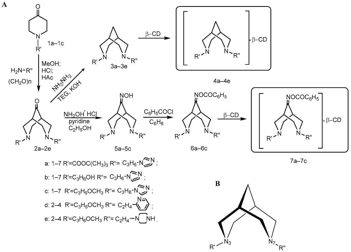
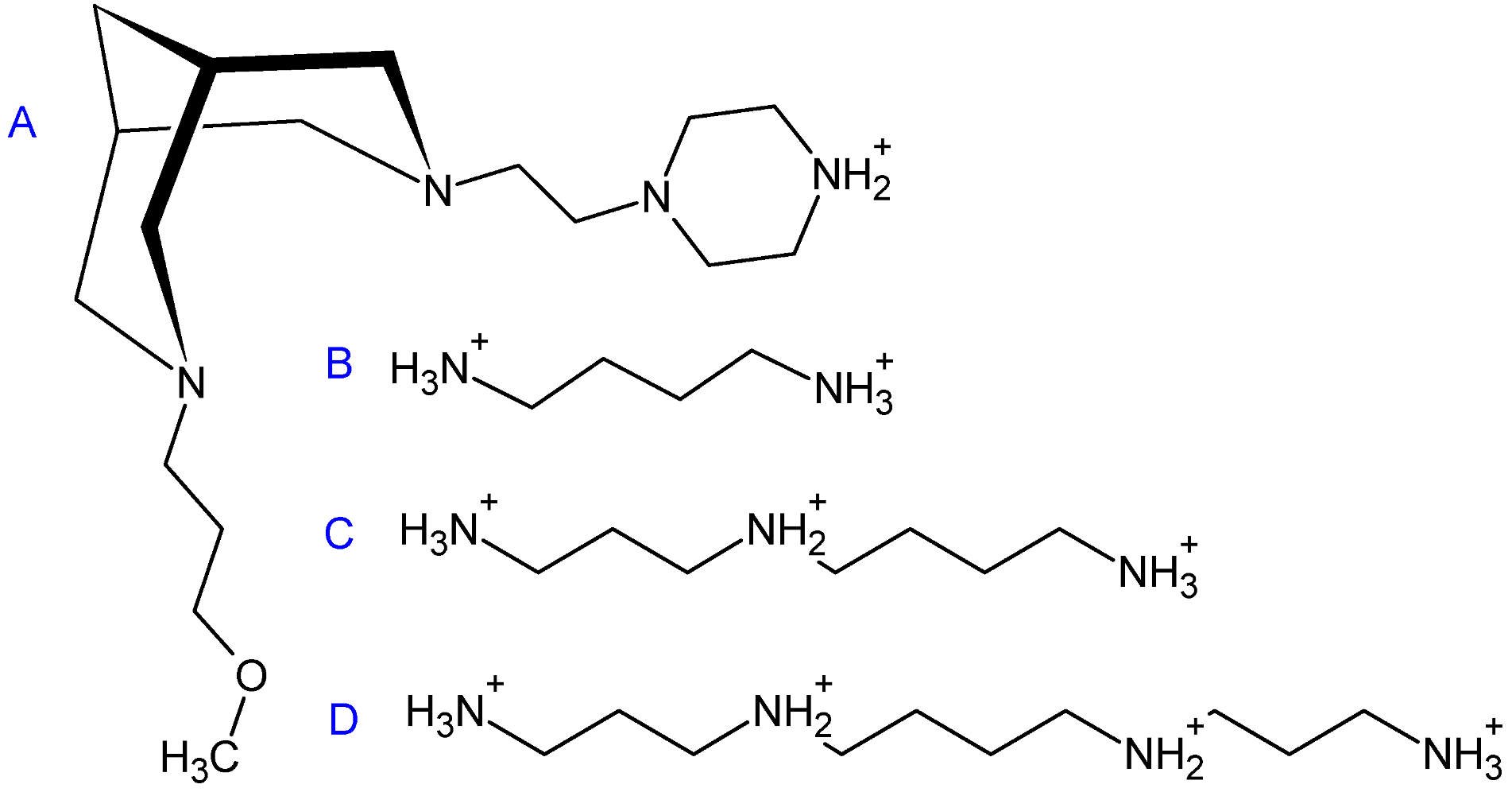
2.1. Synthesis of 3,7-Diazabicyclo[3.3.1]nonane Derivatives

The synthesized 3,7-diazabicyclo[3.3.1]nonan-9-ones (bispidine derivatives) were tested as potential modulators of PA metabolism and anticancer agents. They were prepared in the Laboratory of Chemistry of Synthetic and Natural Medicinal Compounds of A.B. Bekturov Institute of Chemical Sciences. The scheme in Figure 1 illustrates the syntheses and the variety of bicyclo[3.3.1]nonane and O-benzoyloxime complexes prepared with β-cyclodextrin. Compounds 3a3e and 6a6c readily formed complexes 4a4e and 7a7c with β-cyclodextrin, respectively.
3,7-Diazabicyclo[3.3.1]nonan-9-ones (2a2e) were synthesized (Figure 1A) by simultaneous Mannich condensation of 1-Boc-, 1-(3-hydroxypropyl)- and 1-(3-methoxypropyl)-piperidin-4-ones (1a1c) with paraformaldehyde and the primary amines 1-(3-aminopropyl)imidazole, 1-(2-aminoethyl)pyridine and 1-(2-aminoethyl)piperazine in an acetic-methanol medium.
The corresponding 3,7-diazabicyclo[3.3.1]nonanes (3a3e) were obtained by the Kizhner-Wolf reaction, reducing compounds 2a2e under the action of hydrazine hydrate in the presence of KOH in triethylene glycol.
Prolonged reflux of bicyclic ketones 2a2e with hydroxylamine hydrochloride led to the corresponding oximes 5a5c, the subsequent acylation of which by benzoyl chloride synthesized O-benzoyloximes 6a6c.
Column chromatography (on III activity alumina, the eluent is benzene:dioxane 5:1) was used for purification of novel bicyclic ketones, nonanes, bicyclic oximes and O-benzoyl oximes. The completion of the reactions was monitored by TLC. 1H and 13C NMR spectroscopies were used to determine the structures of the synthesized substances.
In the IR spectrum of 3,7-diazabicyclo[3.3.1]nonan-9-ones (2a2e), characteristic bands of stretching vibrations of the carbonyl group appear.
The formation of bicyclic amines was evidenced by the absence of the absorption band of the carbonyl group in the IR spectra of compounds 3a3e.
When comparing the 13C NMR spectra of 3,7-diazabicyclo[3.3.1]nonanes (3a3e) with the spectra of the starting bicyclic ketones (2a2e), it can be seen that they lack the signal of the carbon atom characteristic of the carbonyl group, while in the upfield part of the spectrum, a triplet signal of the carbon atom of the methylene group appears in the 9th position.
The absorption bands of the C=N bond and the OH group are identified in the IR spectra of oximes (5a5c). The most intense band in 6a6c corresponds to the benzoyloxy-carbonyl group. The 13C NMR spectra of oximes (5a5c) and their esters (6a6c) showed resonances for C-9. In the 1H NMR spectra, H1,5 protons were observed as separate signals. The weakest signal was assigned to the proton of the NOH group (5a5c). Signals for the protons of the phenyl ring were observed in the spectrum of the O-benzoyloxime derivatives (6a6c).
To study the biological properties of the novel derivatives of 3,7-diazabicyclo[3.3.1]nonan-9-ones, their water soluble complexes with β-cyclodextrin (4a4e, 7a7c) were synthesized. The complexes melted above 240 °C with decomposition.
3,7-Diazabicyclo[3.3.1]nonan-9-one derivatives (1c, 2a, 2c, 2d, 3a, 3c, 3d, 4a, 4c, 4d) have been studied for myelo-stimulating and plant growth-regulating activity [34,35,36]. The physicochemical properties of novel derivatives 2b, 2e, 3b, 3e, 4b, 4e, and 7a7c are described in the chemical experimental section.
The core bispidine structure of 3,7-diazabicyclo[3.3.1]nonane contains two heterocyclic nitrogens—N3 and N7 (Figure 1B). Different combinations of side N3- and N7-radicals provide a set of five compounds 4a4e, three of which were supplied with benzoyloxim partners 7a7c and were also included in the investigation.

2.2. The Activation of Polyamine Catabolism in Regenerating Rat Liver Homogenates

Eight synthetized bispidine derivatives were tested for their ability to activate oxidation of PAs in the homogenates of regenerating rat liver. Put is normally oxidized by diamine oxidase, which is a copper-containing enzyme that produces ammonia and H2O2 [37]. Spd and Spm are substrates of Spd/Spm-acetyltransferase and acetyl-polyamine oxidase. The results demonstrate that all the compounds could activate the oxidation of all three PAs (Put, Spd, Spm) at varying rates (Figure 2).
Benzoyloxym compounds might be considered prodrugs with partly blocked activity due to the presence of this specific group that might be eliminated by hepatic enzymes. Our set of bispidine derivatives includes three such “drug–prodrug” pairs: 4a/7a, 4b/7b, and 4c/7c. Pairs 4a/7a and 4c/7c contain a hydrophobic fragment at N3, while the 4b/7b pair contains a terminal OH group at N3 that provides some hydrophilicity to this fragment. The general tendency for oxidation of different PAs against the background of the action of the compounds is the following: only pairs 4b/7b demonstrated true drug–prodrug-like activity, as the accelerating effect on PA catabolism was significantly lowered by benzoyloxymation. Compounds 7a and 7c, by contrast, were more active than their benzoyloxym-free partners 4a and 4c, respectively, which demonstrates that the benzoyloxym moiety might enhance the activatory action of these diazabicyclononanes toward PA oxidation. Compounds 4d and 4e are both benzoyloxym-free but differ by the side radical at N3, namely, 4d contains an aryl moiety, while 4e contains an aliphatic heterocycle. The comparison of these two compounds reveals that the aromatic radical provides stronger PA catabolism activatory properties to the molecule as the PA oxidation becomes 3–5-fold higher under the action of 4d than that of 4e. Thus, 4d becomes a leader among all the tested compounds in regard to the activation of Spm oxidation.

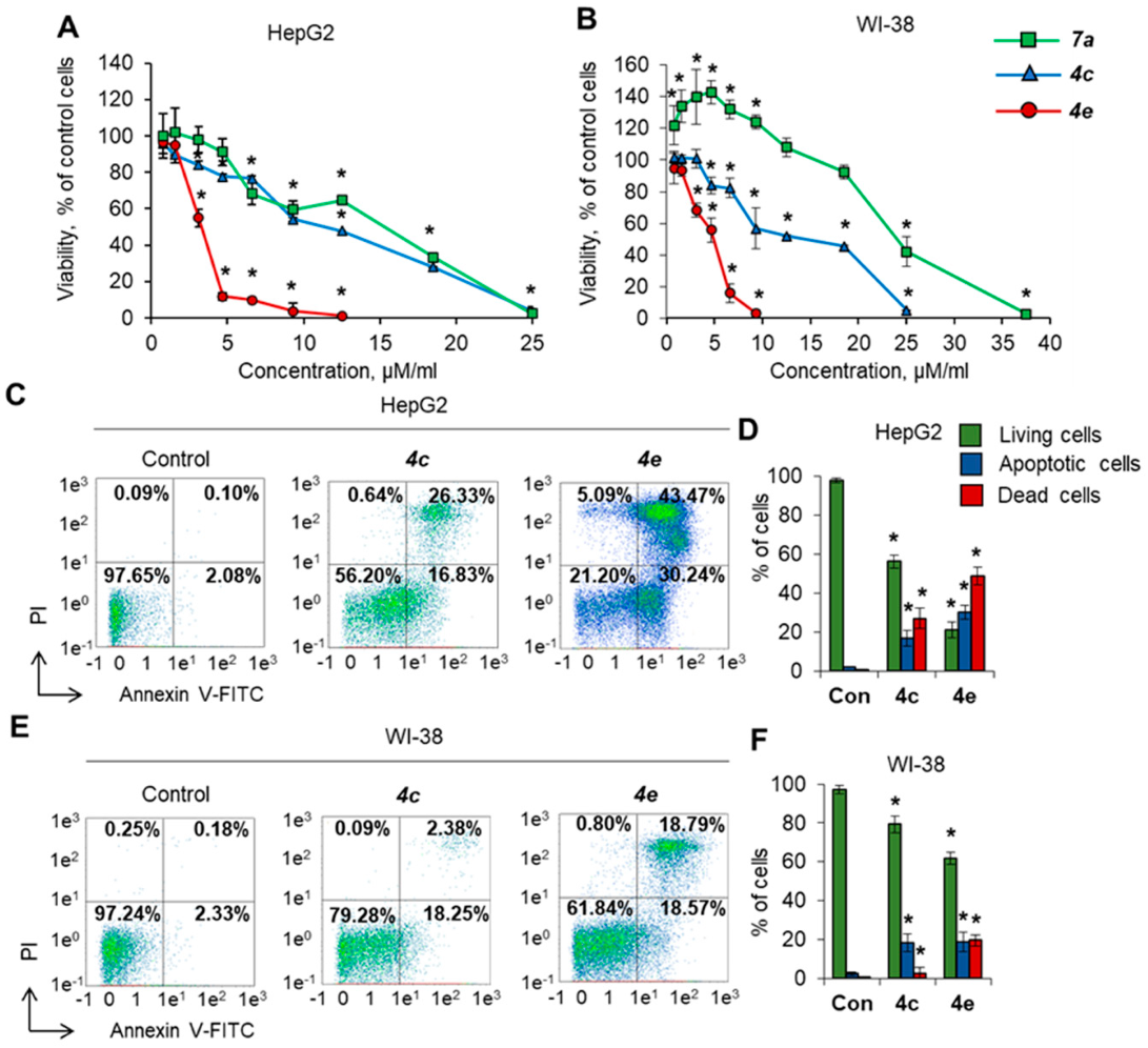
2.3. Toxicity of Bicyclononane Derivatives toward Cancer Cells

To test the cytotoxic activity (loss of cell viability) of the compounds, HepG2 human liver carcinoma cells and WI-38 normal fibroblasts were cultivated in the presence of different concentrations of synthesized compounds, and cell viability and apoptosis induction were measured after 72 h of incubation. Table 1 represents IC50 values toward the chosen cell lines. Only three compounds (4c, 4e, 7a) demonstrated selectivity toward cancer HepG2 cells (Figure 3A) with significantly less cytotoxicity and higher IC50 toward normal cells (Figure 3B). Compound 4e was the most active and could induce a gradual decrease in HepG2 cell viability in the range of concentrations of 3–9 µM. Almost all cells were not viable after incubation at higher concentrations. Compound 7a was less active and could induce a gradual decrease in cell viability at concentrations of 7–25 µM. Compound 4c demonstrated moderate activity and gradually suppressed cell growth at concentrations of 3–25 µM. Compound 7a demonstrated the ability to increase the viability of normal WI-38 fibroblasts at low concentrations of 1–9 µM and was therefore excluded from further studies.
To investigate the type of cell death, cells were incubated with 25 µM 4c or 4e, and the induction of apoptosis was assessed by labeling cell membrane phosphatidylserine with annexin V-FITC and cell DNA with PI combined with flow cytometry. The results of the apoptosis evaluation were in good agreement with the results from the MTT test. Both compounds induced apoptosis more efficiently in cancer HepG2 cells than in normal WI-38 cells: 56% and 20% of cells remained alive after incubation with 4c or 4e, respectively (Figure 3C,D). The tested compound 4c did not induce apoptosis in normal fibroblasts at a concentration of 25 µM, whereas cells were more sensitive to 4e (Figure 3E,F). Thus, 4e demonstrated cytotoxicity against both normal and cancer cells. Taken together, the results demonstrate that the bispidine derivatives 4c and 4e possess strong cytotoxic activity and can induce apoptosis in cancer cells, while normal cells are less sensitive to their action.

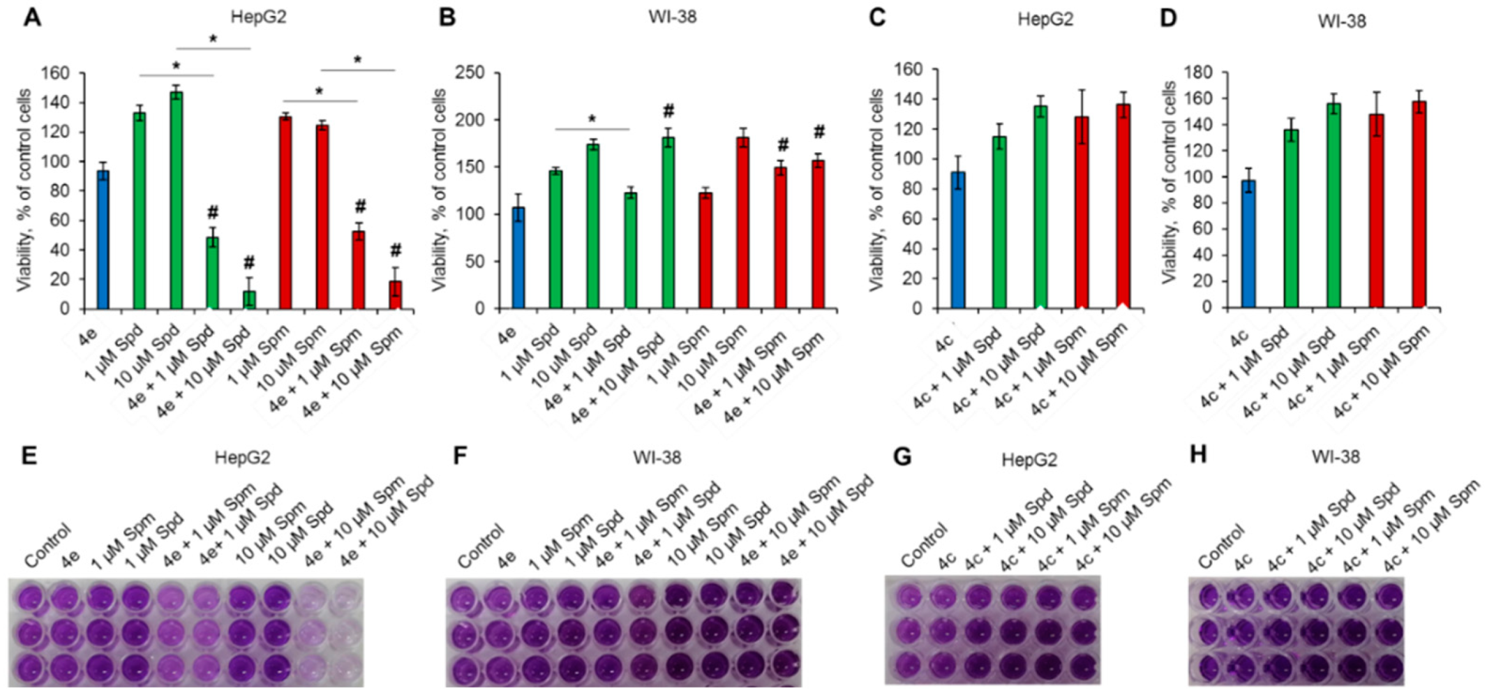
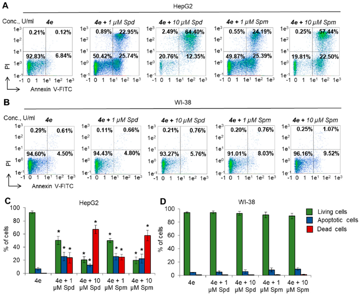
2.4. Polyamines Enhance the Toxicity of Diazabicyclononane Derivative 4e toward Cancer Cells

The study of cell viability and apoptosis induction demonstrated that compound 4e could induce a higher rate of cancer cell death than 4c, although no correlation was revealed between the ability to enhance PA catabolism (Figure 2) and cell viability or apoptosis induction (Figure 3) of the tested compounds. Therefore, subsequent experiments were performed with the most active compounds, 4c and 4e.
The degradation of PAs is commonly reduced and abnormal in cancer cells [20,21], while the products of PA oxidation are known to be able to induce cancer cell death by apoptosis [40,41]. To determine whether the cytotoxic activity of diazabicyclononane derivatives is truly associated with the activation of PA catabolism, we incubated HepG2 or WI-38 cells with a maximum nontoxic concentration (MNTC) of 2 µM for compounds 4c and 4e in the presence of 1 or 10 µM Spm or Spd. The controls were incubated with 4c and 4e and with each of PAs separately.
Both PAs in a single compound treatment promoted the viability of HepG2 and WI-38 cells in the MTT test (Figure 4A,B,E,F). The combination of nontoxic concentrations of 4e and 4c with PAs resulted in different effects on cell viability. Namely, the 4e + 1 µM Spd/Spm combination caused 50% cell death in HepG2 cells (Figure 4A,E). The combination of 4e + 10 µM Spd/Spm demonstrated even greater cytotoxicity, leaving only 10–20% of living cells. This effect suggests a PA-dose-dependent enhancement of the cytotoxicity of 4e. In contrast, Spd/Spm-induced WI-38 proliferation was slightly lowered by coincubation with 4e (Figure 4B,F). Compound 4c was not able to induce any significant reduction in cell viability in the presence of PAs (Figure 4C,D,G,H). Moreover, 4c did not affect the viability of either cancer or normal cells in PA-containing media. These results suggest that the cytotoxic activity of 4c, which is shown in Figure 3, is not associated with the catabolism of PAs, and the addition of PAs to 4c-treated cells did affect cell viability.
To reveal the type of cell death induced by the combination of 4e and PAs, cells were labeled with Annexin-V and FITC and subjected to flow cytometry. The results were in good agreement with the MTT test. Significant induction of HepG2 cell apoptosis was observed after incubation with 4e in the presence of each PA (Figure 5A,C). Higher concentrations of Spd or Spm could induce a greater degree of cell death through apoptosis. However, incubation of normal WI-38 cells with PAs did not have any significant influence on cell death (Figure 5B,D). The results of this experiment demonstrate that diazabicyclononane derivative 4e is able to induce apoptosis at the highest nontoxic concentration in the presence of PAs in HepG2 cancer cells but not in normal WI-38 fibroblasts.

3. Discussion

Our study revealed biological activity among novel bispidine derivatives. The tested compounds can activate PA catabolism, and two of them, 4e and 4c, demonstrate pronounced toxic activity toward cancer cells. Our experiment also revealed a PA-dependent enhancement of the cytotoxicity of the most active bispidine derivative 4e. The results of our study demonstrate that tumor selectivity and potent anticancer activity for this compound could be achieved in combination with PAs.
Our study is in accordance with the recent work by Kanamori et al. [42], who demonstrated that exogenous bovine serum amine oxidase and PA could induce apoptosis in cancer but not normal cells. However, we aimed to enhance the same PA-degrading pathway using small molecule drug substances to activate endogenous enzymes of PAs catabolism.
Previously, it was shown that exogenous PAs can promote tumor and normal cell proliferation [43] and provide a cytoprotective action for normal fibroblasts [44]. In accordance, we demonstrated the promotion of WI-38 and HepG2 viability after cell incubation with PAs (Figure 4A,B). The viability of normal WI-38 cells in the presence of combinations 4e + Spd/Spm and 4c + Spd/Spm was not significantly different from that induced by PAs alone. These results suggest that the tested bispidine derivatives had no effect on the viability of normal cells. In case of cancer HepG2 cells, these two compounds demonstrated opposite effects: 4c, which was not active at MNTC, caused no cell death when added in combination with PAs, while 4e cytotoxic activity was potentiated by exogenous PAs.
The products of PA catabolism (mainly reactive oxygen species (ROS), such as hydrogen peroxide and acrolein) are capable of inducing cell death via apoptosis [25,26,32,33]. Apoptosis was the type of cell death in HepG2 cells when 4e was coincubated with Spd/Spm. The absence of cell death at the MNTC of the compound suggests that the initial cellular content of PAs in tumor HepG2 cells (Table 2) is not enough high to produce sufficient amounts of toxic metabolites of PA decomposition to cause apoptosis. We believe that the addition of exogenous PAs resulted in their oxidation by bispidine-activated PA catabolic enzymes and the generation of cytotoxic products that could trigger cell death. Such an effect was specific only for 4e but not for 4c, despite its more pronounced ability to accelerate PA catabolism shown in regenerating rat liver homogenates.
We believe that the low transmembrane penetration or intracellular metabolism of 4c may be the reason for its low activity in living cells. The tested compounds were converted into complexes with β-cyclodextrins that are widely used in medicinal chemistry to improve water solubility and the transportation of hydrophobic drugs to the target cell membrane barriers. The penetration of such complexes inside the cell may occur via two main pathways [45]. The first is endo- and/or pinocytosis, which is typical for similar complexes regardless of the chemical composition. The second mechanism relies on the dissociation of complexes with the release of active substances that can pass through the cell membrane. This pathway depends on the physical and chemical properties of the active compound. The mechanism of cellular uptake of bispidines has not yet been reported. Compound 4e has some structural features that distinguish it from other compounds and may potentiate its activity. In particular, it contains nonaromatic piperazinyl-ethyl-side radicals at position N7, while all other tested substances include aromatic radicals at that position. This saturated diazacyclic fragment has a secondary amino group (Figure 6), is protonated and positively charged under physiological conditions due to the basic properties of piperazine, which are much stronger than those in imidazoles or pyridines [46]. Moreover, the positive charge is not delocalized in aliphatic piperazine ring unlike aromatic imidazole or pyridine rings. The saturation of the C-C bonds and positively charged nitrogen make 4e structurally more similar to natural aliphatic PAs (Figure 6). This structural similarity may promote the interaction of 4e with natural PA transporters [47] and their penetration through the membrane after the dissociation of the bispidine-β-cyclodextrin complex. Thus, we can assume that the piperazine moiety unique to the lead compound 4e may facilitate its uptake by the cells, thus enhancing its intracellular toxic effect.
Another interesting observation is the selectivity of 4e toward the cancer cell line HepG2, which is obvious and needs to be explained. The concentrations of PAs in HepG2 and WI-38 cells are comparable (Table 2), according to the literature data, and are much lower than the amount of exogenously added PA in our experiments. Thus, they cannot be responsible for the different effects of 4e on cell viability in these two cell lines. We suppose that the differences in PA metabolism enzyme activities in cancer and normal cells are more likely to be the reason for the observed selective cytotoxicity of 4e. The available data on PA metabolism characteristics in the experimental cell lines are summarized in Table 2. Additionally, normal hepatocytes and mesenchymal fibroblasts have been added for a better illustration, as not all the parameters for the experimental cell lines have been found in the literature. The absolutely zero transcription level of one PA catabolic enzyme, acetl-polyamine oxidase (APAO), in HepG2 cells was reported in earlier studies. This feature is one of the key differences between normal and tumor tissue metabolism [2,3,20,21,22,23,24,25]. In contrast, spermine oxidase (SMOX) is more active in cancer HepG2 cells than in normal cells. The overexpression of this PA catabolic enzyme is often associated with cellular malignization [37] due to a double-edged role of ROS in tumor progression. Our compounds are potent activators of the PA catabolic pathway, and a rapid ROS storm may cause apoptosis rather than malignization and tumor promotion. In addition to direct activation, induction of the corresponding gene expression might also take place, although this hypothesis needs to be further verified.
The benzoylated and nonbenzoylated pairs did not demonstrate any pronounced drug/prodrug activity. Instead, adding benzoyloxim to the basic structures provided mostly greater activity toward PA catabolism and antiproliferative action.
As demonstrated in Figure 4, adding each of PAs as a substrate to boost toxicity provided convincing evidence for the potential of bispidines. Minimally toxic bispidines strongly synergized the potency of the two PAs in the tumor cell line but not in fibroblasts. Taken together, the results of the study demonstrate that synthesized bispidine derivatives can induce cancer cell death and the activation of PA catabolism. In the present study, we showed for the first time that the bispidine derivative 4e 3-(3-methoxypropyl)-7-[3-(1H-piperazin-1-yl)ethyl]-3,7-diazabicyclo[3.3.1]nonane can be subjected to PA-based drug development.

4. Materials and Methods

4.1. Chemical Experimental Part

4.1.1. Reagents and Equipment

Primary amines, paraform and 1-Boc-piperidin-4-one (1a) were purchased from Aldrich (Louis Street, MO, USA). IR spectra were recorded on a Nicolet 5700 instrument between KBr plates. 1H and 13C NMR spectra were recorded on a JNM-ECA Jeol 400 spectrometer (frequencies 399.78 and 100.53 MHz, respectively) using DMSO-d6 solvent. The elemental analysis data were consistent with the calculated values. Column chromatography and thin-layer chromatography were carried out on alumina (Al2O3) of the third degree of activity, and Rf compounds were given for this type of plate. The spots were developed in iodine vapors. The IR and NMR spectra for the synthesized compounds are presented in the Supplementary File.

4.1.2. Syntheses of Bispidine-9-Ones (Compounds 2a2e)

1-(3-Hydroxypropyl)piperidin-4-one(1b). A total of 385 mL of absolute toluene and 16.26 g (0.707 mol) of metallic Na were placed in a three-necked flask equipped with a mechanical stirrer, reflux condenser with a calcium chloride tube, and dropping funnel. The reaction mixture was heated to 110 °C in an oil bath until the sodium dissolved (to obtain suspensions of sodium in toluene) for 25–30 min. Then, the reaction mixture was cooled to 75–80 °C, then 128 mL of methanol was added dropwise and it was stirred at 75 °C for 2 h. Afterwards, a straight condenser was attached to the flask, and the reaction mixture was heated. A solution of 174.61 g (0.707 mol) of diester was added using a dropping funnel at 110 °C, while the azeotropic mixture of toluene and methanol was simultaneously distilled off by direct distillation. The rate of dropping of the mixture of diester and methyl alcohol should be equal to the rate of distillation of the solvents. When the temperature reached 110 °C, becoming equal to the boiling point of toluene, the heating was stopped. While cooling (ice was used for cooling) and stirring, a solution of 301 mL of concentrated hydrochloric acid and 301 mL of distilled water was gradually added. The formed organic and aqueous layers were separated. The lower aqueous acidic layer was boiled at 100 °C for 8 h. The progress of the reaction was monitored using a solution of 1% FeCl3. The color changed to maroon. The solution was alkalized with NaOH to pH 12, extracted with chloroform, and dried over anhydrous MgSO4. The solvent was evaporated, and the residue was purified by column chromatography on Al2O3, benzene:dioxane = 5:1. A total of 21.04 g (19%) of 1-(3-hydroxypropyl)piperidin-4-one (1b) was obtained in the form of light yellow oil with Rf = 0.65 (Al2O3, benzene:isopropanol = 7:1).
Found, %: C 61.11, H 9.58, N 8.90. C8H15NO2.
Calculated, %: C 61.14, H 9.55, N 8.92.
IR spectrum, cm−1: 1716 ( ν C=О).
1H NMR spectrum, δ, ppm: bicyclononan-9-one: 1.56 (2H, Н-1eq,5eq), 2.07-2.45 (2H, Н-6ax,8ax), 2.85-3.22 (2H, Н-2ax,4ax), 3.47–3.59 (2H, Н-6eq,8 eq), 3.81–3.89 (2H, Н-2 eq,4 eq). hydroxypropyl: 4.60 (1H, NCH2CH2CH2OH).
13C NMR spectrum ppm: bicyclononan-9-one: 59.2 (C-1,5), 60.2 (C-2,4,6,8), 214.5 (C-9). hydroxypropyl: 27.5 (NCH2CH2CH2OH), 49.7, 63.7 (NCH2CH2CH2OH).
1-Methoxypropyl-piperidin-4-one (1c). The procedure for the synthesis of 1-methoxypropyl-piperidin-4-one (1c) was described earlier [34].
3-Boc-7-[3-(1H-imidazol-1-yl)propyl]-3,7-diazabicyclo[3.3.1]nonan-9-one (2a). The procedure for the synthesis of 3-Boc-7-[3-(1H-imidazol-1-yl)propyl]-3,7-diazabicyclo[3.3.1]nonan-9-one (2a) was described earlier [35].
3-(3-Hydroxypropyl)-7-[3-(1H-imidazol-1-yl)propyl]-3,7-diazabicyclo[3.3.1]nonan-9-one (2b). In a three-necked flask equipped with a stirrer, reflux condenser and dropping funnel, 40 mL of methanol was deoxygenated under a stream of nitrogen. After 30 min, a mixture of 9 mL (0.076 mol) of 1-(3-aminopropyl)imidazole), 9.17 g (0.304 mol) of paraform, 4 mL of concentrated hydrochloric acid, and 6 mL of glacial acetic acid was added and stirred for 15 min in an atmosphere of nitrogen. A solution of 12 g (0.076 mol) of 1-(3-hydroxypropyl)piperidin-4-one (1b) and 6 mL of glacial acetic acid in 43 mL of methanol was added dropwise. After 10 h of heating the reaction mixture at 60–65 °C, a second equivalent of paraform was added and held for another 10 h at the same temperature. The solvent was evaporated, and the residue was dissolved in 121 mL of water. The extraction of neutral products was carried out with diethyl ether. The aqueous layer was alkalinized to pH 12, and the organic part was extracted with chloroform and dried over MgSO4. The solvent was evaporated, and the obtained product was purified by column chromatography on Al2O3:benzene:dioxane 5:1. A total of 13.84 g (59%) of 3-(3-hydroxypropyl)-7-[3-(1H-imidazol-1-yl)propyl]-3,7-diazabicyclo[3.3.1]nonan-9-one (2b) was obtained in the form of a light yellow oil with Rf = 0.41 (Al2O3, benzene:isopropanol = 6:1).
Found, %: C 62.72, H 8.52, N 18.27. C16H26N4O2.
Calculated, %: C 62.75, H 8.50, N 18.30.
IR spectrum, cm−1: 1729 ( ν C=О), 1071 ( ν C-О-C).
1H NMR spectrum, δ, ppm: bicyclononan-9-one: 1.54 (2 H, Н-1eq,5eq), 2.07–2.45 (2 H, Н-6ax,8ax), 2.85–3.22 (2 H, Н-2ax,4ax), 3.47–3.59 (2 H, Н-6eq,8 eq), 3.81–3.89 (2 H, Н-2 eq,4 eq). 1H-imidazol-1-yl)propyl: 6.75–6.78, 6.82, 7.33 (3H, CHimidazol). Side chains: 1.54 (2H, NCH2CH2CH2N); 1.69–1.81 (2H, NCH2CH2CH2OH). hydroxypropyl: 4.60 (1H, NCH2CH2CH2OH).
13C NMR spectrum ppm: bicyclononan-9-one: 59.4 (C-1,5), 61.4 (C-2,4,6,8), 214.5 (C-9). (1H-imidazol-1-yl)propyl: 119.0 (CHimidazol), 128.7, 137.2. Side chains: 29.3 (CH2CH2CH2), 44.9, 47.4 (CH2CH2CH2). hydroxypropyl: 27.5 (NCH2CH2CH2OH), 49.7, 63.7 (NCH2CH2CH2OH).
3-(3-Methoxypropyl)-7-[3-(1H-imidazol-1-yl)propyl]-3,7-diazabicyclo[3.3.1]nonan-9-one (2c). The procedure for the synthesis of 3-(3-methoxypropyl)-7-[3-(1H-imidazol-1-yl)propyl]-3,7-diazabicyclo[3.3.1]nonan-9-one (2c) was described earlier [34].
3-(3-methoxypropyl)-7-[2-(pyridin-2-yl)ethyl]-3,7-diazabicyclo[3.3.1]nonan-9-one (2d). The procedure for the synthesis of 3-(3-methoxypropyl)-7-[2-(pyridin-2-yl)ethyl]-3,7-diazabicyclo[3.3.1]nonan-9-one (2d) was described earlier [36].
3-(3-Methoxypropyl)-7-[2-(piperazin-1-yl)]-3,7-diazabicyclo[3.3.1]nonan-9-one (2e). In a three-necked flask equipped with a stirrer, reflux condenser and dropping funnel, 50 mL of methanol was deoxygenated under a stream of nitrogen. After 30 min, a mixture of 7.55 g (0.058 mol) of 1-(2-aminoethyl)piperazine, 7 g (0.304 mol) of paraform, 3 mL of concentrated hydrochloric acid, and 4 mL of glacial acetic acid was added and stirred for 15 min in an atmosphere of nitrogen. A solution of 10 g (0.058 mol) of 1-(3-methoxypropyl)piperidin-4-one (1c) and 4 mL of glacial acetic acid in 43 mL of methanol was added dropwise. After 10 h of heating the reaction mixture at 60–65 °C, a second equivalent of paraform was added and held for another 10 h at the same temperature. The solvent was evaporated, and the residue was dissolved in 119 mL of water. The extraction of neutral products was carried out with diethyl ether. The aqueous layer was alkalinized to pH 12, and the organic part was extracted with chloroform and dried over MgSO4. The solvent was evaporated, and the obtained product was purified by column chromatography on Al2O3:benzene:dioxane 5:1. A total of 17.15 g (67%) of 3-(3-methoxypropyl)-7-[2-(piperazin-1-yl)ethyl]-3,7-diazabicyclo[3.3.1]nonan-9-one (2e) was obtained in the form of a light yellow oil with Rf = 0.32 (Al2O3, benzene:isopropanol = 6:1).
Found, %: C 62.89, H 9.81, N 19.79. C17H32N4O2.
Calculated, %: C 62.96, H 9.88, N 19.75.
IR spectrum, cm−1: 1732 ( ν C=О), 1089 ( ν C-О-C).
1H NMR spectrum, δ, ppm: bicyclononan-9-one: 1.55 (2H, Н-1eq,5eq), 2.80–2.96 (2H, Н-6ax,8ax), 2.98 (2H, Н-2ax,4ax), 3.47–3.59 (2 H, Н-6eq,8 eq), 3.87 (2H, Н-2 eq,4 eq). 2-(Piperazin-1-yl)ethyl: 4.55–4.58 (4H, CHpiperazin). methoxypropyl: 3.60–3.98 (NCH2CH2CH2OCH3).
13C NMR spectrum, ppm: bicyclononan-9-one: 46.9 (C-1,5), 58.5 (C-2,4,6,8), 214.8 (C-9). 2-(Piperazin-1-yl)ethyl: 46.7 (CH2piperazin). methoxypropyl: 56.0, 27.8, 66.0, 71.4 (NCH2CH2CH2OCH3).

4.1.3. Syntheses of β-Cyclodextrin Complexes of Bispidines (Compounds 4a4e)

3-Boc-7-[3-(1H-imidazol-1-yl)propyl]-3,7-diazabicyclo[3.3.1]nonane (3a). The procedure for the synthesis of 3-Boc-7-[3-(1H-imidazol-1-yl)propyl]-3,7-diazabicyclo[3.3.1]nonane (3a) was described earlier [35].
3-(3-Hydroxypropyl)-7-[3-(1H-imidazol-1-yl)propyl]-3,7-diazabicyclo[3.3.1]nonane (3b). A mixture of 2.0 g (0.006 mol) of 3-(3-hydroxypropyl)-7-[3-(1H-imidazol-1-yl)propyl]-3,7-diazabicyclo[3.3.1]nonan-9-one (2b) and 1 mL (0.031 mol) of hydrazine hydrate (99% solution) in 17.5 mL of triethylene glycol at 60 °C was added to 4.17 g (0.074 mol) of KOH. The reaction mixture was heated to 150 °C and stirred at this temperature for 5 h. At a temperature of 190–200 °C, water and excess hydrazine were removed by evaporation. After cooling, 29 mL of distilled water was added, extracted with diethyl ether, and dried over anhydrous MgSO4. The solvent was evaporated, and the obtained product was purified by column chromatography on Al2O3, benzene:dioxane 5:1. A total of 1.28 g (67%) of 3-(3-hydroxypropyl)-7-[3-(1H-imidazol-1-yl)propyl]-3,7-diazabicyclo[3.3.1]nonane (3b) was obtained as a pale yellow oil with Rf = 0.36 (Al2O3, benzene: isopropanol = 7:1).
Found, %: C 65.77, Н 9.61, N 19.20. C16H28N4O.
Calculated, %: C 65.75, Н 9.59, N 19.18.
IR spectrum, cm−1: 1121 ( ν C-О-C).
1H NMR spectrum, δ, ppm: bicyclononane: 2.03–2.11 (2 H, Н-1eq,5eq), 1.42–1.64 (2Н, H-9), 2.37 (4H, Н-2ax,4ax,6ax,8ax), 2.87–3.08 (4 H, Н-2eq,4eq,6eq,8eq). 1H-imidazol-1-yl)propyl: 1.56 (2H, NCH2CH2CH2N), 3.05–3.08, 3.86 (4H, NCH2CH2CH2N), 7.45 (3H, CHimidazole), hydroxypropyl: 4.12 (1H, NCH2CH2CH2OH).
13C NMR spectrum, δ, ppm: bicyclononane: 30.2 (C-1,5), 36.3 (C-9), 58.4 (C-2,4,6,8). (1H-imidazol-1-yl)propyl: 32.8 (CH2CH2CH2), 51.6 (CH2CH2CH2), 45.6 (CH2CH2CH2), 118.9, 128.7, 137.2 (CHimidazol). hydroxypropyl: 27.8 (NCH2CH2CH2OH), 51.6, 59.8 (NCH2CH2CH2OH).
3-(3-Methoxypropyl)-7-[3-(1H-imidazol-1-yl)propyl]-3,7-diazabicyclo[3.3.1]nonane (3c). The procedure for the synthesis of 3-(3-methoxypropyl)-7-[3-(1H-imidazol-1-yl)propyl]-3,7-diazabicyclo[3.3.1]nonane (3c) was described earlier [34].
3-(3-Methoxypropyl)-7-[2-(pyridin-2-yl)ethyl]-3,7-diazabicyclo[3.3.1]nonane (3d). The procedure for the synthesis of 3-(3-methoxypropyl)-7-[2-(pyridin-2-yl)ethyl]-3,7-diazabicyclo[3.3.1]nonan-9-one (3d) was described earlier [36].
3-(3-Methoxypropyl)-7-[2-(piperazin-1-yl)]-3,7-diazabicyclo[3.3.1]nonane (3e). A mixture of 2.0 g (0.006 mol) of 3-(3-methoxypropyl)-7-[2-(piperazin-1-yl)ethyl]-3,7-diazabicyclo[3.3.1]nonan-9-one (2e) and 1 mL (0.031 mol) of hydrazine hydrate (99% solution) in 17.5 mL of triethylene glycol at 60 °C was added to 4.17 g (0.074 mol) of KOH. The reaction mixture was heated to 150 °C and stirred at this temperature for 5 h. At a temperature of 190–200 °C, water and excess hydrazine were removed by evaporation. After cooling, 29 mL of distilled water was added, extracted with diethyl ether, and dried over anhydrous MgSO4. The solvent was evaporated, and the obtained product was purified by column chromatography on Al2O3 and benzene:dioxane 5:1. A total of 1.21 g (63%) of 3-(3-methoxypropyl)-7-[2-(piperazin-1-yl)ethyl]-3,7-diazabicyclo[3.3.1]nonane (3e) was obtained as a pale yellow oil with Rf = 0.34 (Al2O3, benzene: isopropanol = 7:1).
Found, %: C 65.79, H 10.81, N 18.00. C17H34N4O.
Calculated, %: C 65.81, H 10.97, N 18.06.
IR spectrum, cm−1: 1075 ( ν C-О-C), 3374 ( ν N-H).
1H NMR spectrum, δ, ppm: bicyclononan-9-one: 1.59 (2H, Н-1eq,5eq), 2.42 (2H, Н-6ax,8ax), 2.98 (2H, Н-2ax,4ax), 2.94 (2 H, Н-6eq,8 eq), 3.04 (2H, Н-2 eq,4 eq). 2-(Piperazin-1-yl)ethyl: 4.55–4.58 (4 H, CHpiperazin). methoxypropyl: 1.13 (NCH2CH2CH2OCH3).
13C NMR spectrum, ppm: bicyclononan-9-one: 30.0 (C-1,5), 58.6 (C2,4), 58.4 (C6,8), 32,0 (C9). 2-(Piperazin-1-yl)ethyl: 54,8, 53,4, 54,8, 46,3 (7N-CH2CH2piperazine). methoxypropyl: 56.3, 27.6, 66.0, 72.4 (NCH2CH2CH2OCH3).
Complex3-Boc-7-[3-(1H-imidazol-1-yl)propyl]-3,7-diazabicyclo[3.3.1]nonane (4a). The procedure for the synthesis of the complex of 3-Boc-7-[3-(1H-imidazol-1-yl)propyl]-3,7-diazabicyclo[3.3.1]nonane (4a) was described earlier [35].
Complex of 3-(3-hydroxypropyl)-7-[3-(1H-imidazol-1-yl)propyl]-3,7-diazabicyclo[3.3.1]nonane (4b). Hot solutions of 0.5 g (0.002 mol) of 3-(3-hydroxypropyl)-7-[3-(1H-imidazol-1-yl)propyl]-3,7-diazabicyclo[3.3.1]nonane (3b) in 25 mL of ethyl alcohol and 1.94 g (0.002 mol) of β-cyclodextrin in 30 mL of distilled water were mixed. The mixture was placed in a drying cupboard, and ethanol and water were evaporated at 50–55 °C to produce 2.25 g of compound 4b.
Found, %: C 48.84, Н 6.84, N 3.95. C58H98N4O36.
Calculated, %: C 48.81, Н 6.87, N 3.93.
Complex of 3-(3-methoxypropyl)-7-[3-(1H-imidazol-1-yl)propyl]-3,7-diazabicyclo[3.3.1]nonane (4c). The procedure for the synthesis of the complex of 3-(3-methoxypropyl)-7-[3-(1H-imidazol-1-yl)propyl]-3,7-diazabicyclo[3.3.1]nonane (4c) was described earlier [34].
Complex of 3-(3-methoxypropyl)-7-[2-(pyridin-2-yl)ethyl]-3,7-diazabicyclo[3.3.1]nonane (4d). The procedure for the synthesis of the complex of 3-(3-methoxypropyl)-7-[2-(pyridin-2-yl)ethyl]-3,7-diazabicyclo[3.3.1]nonan-9-one (4d) was described earlier [36].
Complex of 3-(3-methoxypropyl)-7-[2-(piperazin-1-yl)]-3,7-diazabicyclo[3.3.1]nonane (4e). Hot solutions of 1.4 g (0.003 mol) of 3-(3-methoxypropyl)-7-[2-(piperazin-1-yl)]-3,7-diazabicyclo[3.3.1]nonane (3e) in 35 mL of ethyl alcohol and 4.0 g (0.003 mol) of β-cyclodextrin in 60 mL of distilled water were mixed. The mixture was placed in a drying cupboard, and ethanol and water were evaporated at 50–55 °C to produce 4.75 g of compound 4e.
Found, %: C 49.01, H 7.14, N 3.91. C59H104N4O36.
Calculated, %: C 49.03, H 7.20, N 3.88.

4.1.4. Syntheses of β-Cyclodextrin Complexes of o-Benzoyloximes of Bispidines (Compounds 7a7e)

Oxime of 3-Boc-7-[3-(1H-imidazol-1-yl)propyl]-3,7-diazabicyclo[3.3.1]nonane-9-one (5a). Five grams (0.014 mol) of 3-Boc-7-[3-(1H-imidazol-1-yl)propyl]-3,7-diazabicyclo[3.3.1]nonan-9-one (2a) in 85 mL of ethyl alcohol and 1.7 mL (0.021 mol) of pyridine were placed in a three-necked flask equipped with a mechanical stirrer, reflux condenser with a calcium chloride tube, and dropping funnel. While stirring, 2.53 g (0.036 mol) of hydroxylamine hydrochloride was added. The reaction mixture was heated at 110–120 °C for 27 h, the solvent was evaporated, and the residue was dissolved in 40 mL of water, alkalized with NaOH to pH 12, extracted with chloroform, and dried over anhydrous MgSO4. The solvent was evaporated, and the residue was purified by column chromatography on Al2O3, benzene:dioxane = 5:1. A total of 4.5 g (86%) of oxime of 3-Boc-7-[3-(1H-imidazol-1-yl)propyl]-3,7-diazabicyclo[3.3.1]nonan-9-one (5a) was obtained in the form of light yellow oils with Rf = 0.16 (Al2O3, benzene:isopropanol = 20:1).
Found, %: C 59.52; H, 8.00; N, 19.30. C18H29N5O3.
Calculated, %: C 59.50; H, 7.98; N, 19.28.
IR spectrum, cm−1: 1651 ( ν C=N), 3380 ( ν О-H).
1Н NMR spectrum, δ, ppm: bicyclononan-9-ketoxime: 2.69 (2H, Н-1eq,5eq), 2.07–4.10 (4 H, Н-2ax,4ax,6ax,8ax), 2.60–4.72 (4H, Н-2eq,4eq,6eq,8eq), 6.96 (Н, N=OH). 1H-imidazol-1-yl)propyl: 1.56 (2H, NCH2CH2CH2N), 3.13–3.22, 3.85 (4H, NCH2CH2CH2N), 6.96–7.49 (3H, CHimidazol). Boc: 1.48 (9H, CH3).
13C NMR ppm, δ, ppm: bicyclononan-9-ketoxime: 36.9 (C-5), 50.5 (C-6,8), 60.1 (C-2,4), 154.6 (C-9). (1H-imidazol-1-yl)propyl: 30.8 (CH2CH2CH2), 54.1 (CH2CH2CH2), 44.1 (CH2CH2CH2), 120.0 (CHimidazol), 128.7, 137.9. Boc: 28.6 (CH3), 79.1 (O-C), 158.3 (C=O).
Oxime of 3-(3-hydroxypropyl)-7-[3-(1H-imidazol-1-yl)propyl]-3,7-diazabicyclo[3.3.1]nonane (5b). A total of 6.0 g (0.020 mol) of 3-(3-hydroxypropyl)-7-[3-(1H-imidazol-1-yl)propyl]-3,7-diazabicyclo[3.3.1]nonan-9-one (2a) in 122 mL of ethyl alcohol and 2.37 g (0.030 mol) of pyridine were placed in a three-necked flask equipped with a mechanical stirrer, reflux condenser with a calcium chloride tube, and dropping funnel. While stirring, 3.61 g (0.052 mol) of hydroxylamine hydrochloride was added. The reaction mixture was heated at 110–120 °C for 29 h, the solvent was evaporated, and the residue was dissolved in 30 mL of water, alkalized with NaOH to pH 12, extracted with chloroform, and dried over anhydrous MgSO4. The solvent was evaporated, and the residue was purified by column chromatography on Al2O3, benzene:dioxane = 5:1. A total of 4.85 g (77%) of oxime of 3-(3-hydroxypropyl)-7-[3-(1H-imidazol-1-yl)propyl]-3,7-diazabicyclo[3.3.1]nonan-9-one (5b) was obtained in the form of light yellow oils with Rf = 0.07 (Al2O3, benzene:isopropanol = 20:1).
Found, %: C 59.79, H 8.40, N 21.84. C16H27N5O2.
Calculated, %: C 59.81, H 8.41, N 21.81.
IR spectrum, cm−1: 1510 ( ν C=N), 3240 ( ν О-H).
1Н NMR spectrum, δ, ppm: bicyclononan-9-ketoxime: 1.60 (2 H, Н-1eq,5eq), 2.06–2.40 (4 H, Н-2ax,4ax,6ax,8ax), 2.99–3.42 (4 H, Н-2eq,4eq,6eq,8eq), 6.40 (Н, N=OH). 1H-imidazol-1-yl)propyl: 1.60 (2H, NCH2CH2CH2N), 3.42, 3.85–3.87 (4H, NCH2CH2CH2N), 6.92, 7.23, 7.45 (3H, CHimidazol). hydroxypropyl 4.83 (1H, NCH2CH2CH2OH), 1.88–1.90 (2H, NCH2CH2CH2OH), 2.40, 3.68 (4H, NCH2CH2CH2OH).
13C NMR ppm, δ, ppm: bicyclononan-9-ketoxime: 42.2 (C-5), 57.7 (C-6,8), 58.1 (C-2,4), 160.9 (C-9). (1H-imidazol-1-yl)propyl: 27.7 (CH2CH2CH2), 50.0 (CH2CH2CH2), 45.4 (CH2CH2CH2), 119.0 (CHimidazol), 128.8, 137.1. hydroxypropyl: 27.7 (NCH2CH2CH2OH), 51.5, 59.5 (NCH2CH2CH2OH).
Oxime of 3-(3-methoxypropyl)-7-[3-(imidazol-1-yl)propyl]-3,7-diazabicyclo[3.3.1]nonan-9-one (5c). A total of 3.5 g (0.008 mol) of 3-(3-methoxypropyl)-7-[3-(imidazol-1-yl)propyl]-3,7-diazabicyclo[3.3.1]nonan-9-one (2c) in 65 mL of ethyl alcohol and 1.3 mL (0.018 mol) of pyridine were placed in a three-necked flask equipped with a mechanical stirrer, reflux condenser with a calcium chloride tube, and dropping funnel. While stirring, 2.15 g (0.027 mol) of hydroxylamine hydrochloride was added. The reaction mixture was heated at 110–120 °C for 27 h, the solvent was evaporated, and the residue was dissolved in 27 mL of water, alkalized with NaOH to pH 12, extracted with chloroform, and dried over anhydrous MgSO4. The solvent was evaporated, and the residue was purified by column chromatography on Al2O3, benzene:dioxane = 5:1. 3.1 g (76%) of oxime of 3-(3-methoxypropyl)-7-[3-(imidazol-1-yl)propyl]-3,7-diazabicyclo[3.3.1]nonan-9-one (5c) was obtained in the form of light yellow oils with Rf = 0.21 (Al2O3, benzene:isopropanol = 20:1).
Found, %: C 60.86, H 9.72, N 20.95. C17H33N5O2.
Calculated, %: C 60.90, H 9.85, N 20.89.
IR spectrum, cm−1: 1659 ( ν C=N), 3365 ( ν О-H).
1Н NMR spectrum, δ, ppm: bicyclononan-9-ketoxime: 2.54 (2 H, Н-1eq,5eq), 2.12–3.78 (4 H, Н-2ax,4ax,6ax,8ax), 2.30–43.57 (4 H, Н-2eq,4eq,6eq,8eq), 6.96 (Н, N=OH). 1H-imidazol-1-yl)propyl: 1.53 (2H, NCH2CH2CH2N), 3.16–3.20, 3.75 (4H, NCH2CH2CH2N), 6.96–7.49 (3H, CHimidazol). methoxypropyl: 3.83 (3H, NCH2CH2CH2OCH3), 1.75 (2H, NCH2CH2CH2OCH3), 2.53, 3.74 (4H, NCH2CH2CH2OCH3).
13C NMR ppm, δ, ppm: bicyclononan-9-ketoxime: 37.9 (C-5), 53.5 (C-6,8), 65.1 (C-2,4), 161.5 (C-9). (1H-imidazol-1-yl)propyl: 28.8 (CH2CH2CH2), 45.9 (CH2CH2CH2), 44.8 (CH2CH2CH2), 115.4, 119.7, 127.5. (CHimidazol). methoxypropyl: 29.2 (CH3), 73.9 (O-C), 149.9 (C=O).
O-Benzoyloxime of 3-Boc-7-[3-(1H-imidazol-1-yl)propyl]-3,7-diazabicyclo[3.3.1] nonan-9-one (6a). Then, 4.42 g (0.012 mol) of oxime of 3-Boc-7-[3-(1H-imidazol-1-yl)propyl]-3,7-diazabicyclo[3.3.1]nonan-9-one (5a) was mixed with 84 mL of absolute benzene, and a mixture of 14 mL of absolute benzene and 2.8 mL (0.024 mol) of benzoyl chloride was added dropwise. The reaction took place at room temperature. Finally, 10 mL of distilled water was added to the reaction mixture and neutralized with potash to pH 10–11. The product was extracted with chloroform, and the combined extracts were dried over anhydrous MgSO4. The solvent was evaporated, and the residue was distilled in vacuo. A total of 2.66 g (50% of theory) of the O-benzoyloxime of 3-Boc-7-[3-(1H-imidazol-1-yl)propyl]-3,7-diazabicyclo[3.3.1]nonan-9-one (6a) was obtained in the form of a yellow oil (16), Rf = 0.56 (Al2O3, benzene:isopropanol = 7:1).
Found, %: C 64.27; H, 7.04; N, 15.01. C25H33N5O4.
Calculated, %: C 64.24; H, 7.07; N, 14.99.
IR spectrum, cm−1: 1725 ( ν C=О).
1Н NMR spectrum, δ, ppm: ester of bicyclononan-9-ketoxime: 2.77 (2 H, Н-1eq,5eq), 2.12–4.15 (4 H, Н-2ax,4ax,6ax,8ax), 2.65–4.77 (4 H, Н-2eq,4eq,6eq,8eq), 7.67–8.05 (5H, Ph). 1H-imidazol-1-yl)propyl: 1.56 (2H, NCH2CH2CH2N), 3.13–3.22, 3.85 (4H, NCH2CH2CH2N), 6.96, 7.03, 7.49 (3H, CHimidazol). Boc: 1.48 (9H, CH3).
13C NMR spectrum, δ, ppm: ester of bicyclononan-9-ketoxime: 36.9, 39.9 (C-1,5), 45.5 60.0 (C-2,4,6,8), 164.7 (C-9). (1H-imidazol-1-yl)propyl: 29.4 (CH2CH2CH2), 50.1 (CH2CH2CH2), 47.9 (CH2CH2CH2), 120.5 (CHimidazol), 133.9, 136.3. Boc: 28.6 (CH3), 80.2 (O-C), 159.5 (C=O).
O-Benzoyloxime of 3-(3-hydroxypropyl)-7-[3-(1H-imidazol-1-yl)propyl]-3,7-diazabicyclo[3.3.1]nonan-9-one (6b). Oxime (0.014 mol) of 3-(3-hydroxypropyl)-7-[3-(1H-imidazol-1-yl)propyl]-3,7-diazabicyclo[3.3.1]nonan-9-one (5b) was mixed with 100 mL of absolute benzene, and a mixture of 14 mL of absolute benzene and 3.3 mL (0.014 mol) of benzoyl chloride was added dropwise. The reaction took place at room temperature. Finally, 10 mL of distilled water was added to the reaction mixture and neutralized with potash to pH 10–11. The product was extracted with chloroform, and the combined extracts were dried over anhydrous MgSO4. The solvent was evaporated, and the residue was distilled in vacuo. A total of 4.01 g (70% of theory) of O-benzoyloxime of 3-(3-hydroxypropyl)-7-[3-(1H-imidazol-1-yl)propyl]-3,7-diazabicyclo[3.3.1]nonan-9-one (6b) was obtained in the form of a yellow oil, Rf = 0.64 (Al2O3, benzene:isopropanol = 7:1).
Found, %: C 64.90, H 7.32, N 16.45. C23H31N5O3.
Calculated, %: C 64.94, H 7.29, N 16.47.
IR spectrum, cm−1: 1737 ( ν C=О).
1Н NMR spectrum, δ, ppm: ester of bicyclononan-9-ketoxime: 2.74 (2H, Н-1eq,5eq), 2.11–2.24 (4 H, Н-2ax,4ax,6ax,8ax), 2.65–2.77 (4H, Н-2eq,4eq,6eq,8eq), 7.67–8.05 (5H, Ph). 1H-imidazol-1-yl)propyl: 1.56 (2H, NCH2CH2CH2N), 3.13–3.22, 3.85 (4H, NCH2CH2CH2N), 6.96, 7.03, 7.49 (3H, CHimidazol). hydroxypropyl: 4.83 (1H, NCH2CH2CH2OH), 1.75 (2H, NCH2CH2CH2OH), 2.56, 3.76 (4H, NCH2CH2CH2OH).
13C NMR spectrum, δ, ppm: ester of bicyclononan-9-ketoxime: 37.7–40.7 (C-1,5), 59.1–59.4 (C-2,4,6,8), 167.6 (C-9). (1H-imidazol-1-yl)propyl: 29.4 (CH2CH2CH2), 50.1 (CH2CH2CH2), 48.0 (CH2CH2CH2), 120.5 (CHimidazol), 133.9, 136.3. hydroxypropyl: 28.1 (NCH2CH2CH2OH), 52.4, 60.6 (NCH2CH2CH2OH).
O-benzoyloxime of 3-(3-methoxypropyl)-7-[3-(imidazol-1-yl)propyl]-3,7-diazabicyclo[3.3.1]nonan-9-one (6c). Three grams (0.009 mol) of oxime of 3-(3-methoxypropyl)-7-[3-(imidazol-1-yl)propyl]-3,7-diazabicyclo[3.3.1]nonan-9-one (5c) was mixed with 90 mL of absolute benzene, and a mixture of 12 mL of absolute benzene and 2 mL (0.014 mol) of benzoyl chloride was added dropwise. The reaction took place at room temperature. Finally, 8 mL of distilled water was added to the reaction mixture and neutralized with potash to pH 10–11. The product was extracted with chloroform, and the combined extracts were dried over anhydrous MgSO4. The solvent was evaporated, and the residue was distilled in vacuo. A total of 3.05 g (64% of theory) of O-benzoyloxime of 3-(3-methoxypropyl)-7-[3-(imidazol-1-yl)propyl]-3,7-diazabicyclo[3.3.1]nonan-9-one (6c) was obtained in the form of a yellow oil, Rf = 0.61 (Al2O3, benzene:isopropanol = 7:1).
Found, %: C 60.86, H 9.72, N 20.95. C17H33N5O2.
Calculated, %: C 60.90, H 9.85, N 20.89.
IR spectrum, cm−1: 1734 ( ν C=О).
1Н NMR spectrum, δ, ppm: ester of bicyclononan-9-ketoxime: 2.71 (2 H, Н-1eq,5eq), 2.01–2.14 (4 H, Н-2ax,4ax,6ax,8ax), 2.35–2.45 (4 H, Н-2eq,4eq,6eq,8eq), 7.67–8.05 (5H, Ph). 1H-imidazol-1-yl)propyl: 1.56 (2H, NCH2CH2CH2N), 3.13–3.22, 3.85 (4H, NCH2CH2CH2N), 6.96, 7.03, 7.49 (3H, CHimidazol). hydroxypropyl: 4.83 (1H, NCH2CH2CH2OH), 1.75 (2H, NCH2CH2CH2OH), 2.56, 3.76 (4H, NCH2CH2CH2OH).
13C NMR ppm, δ, ppm: bicyclononan-9-ketoxime: 36.0 (C-5), 51.9 (C-6,8), 64.5 (C-2,4), 159.5 (C-9). (1H-imidazol-1-yl)propyl: 27.5 (CH2CH2CH2), 43.9 (CH2CH2CH2), 45.2 (CH2CH2CH2), 116.2, 118.4, 129.2. (CHimidazol). methoxypropyl: 27.4 (CH3), 75.1 (O-C).
Complex of O-benzoyloxime of 3-Boc-7-[3-(1H-imidazol-1-yl)propyl]-3,7-diazabicyclo[3.3.1]nonan-9-one (7a). Hot solutions of 1.0 g (0.002 mol) of O-benzoyloxime of 3-Boc-7-[3-(1H-imidazol-1-yl)propyl]-3,7-diazabicyclo[3.3.1]nonan-9-one (6a) in 25 mL of ethyl alcohol and 3.124 g (0.002 mol) of β-cyclodextrin in 40 mL of distilled water were mixed together. The mixture was placed in a drying cupboard, and ethanol and water were evaporated at 50–55 °C to produce 3.57 g of compound 7a.
Found, %: C 50.25, H 6.46, N 4.35. C67H103N5O39.
Calculated, %: C 50.22, H 6.43, N 4.37.
Complex of O-benzoyloxime of3-(3-hydroxypropyl)-7-[3-(1H-imidazol-1-yl)propyl]-3,7-diazabicyclo[3.3.1]nonan-9-one (7b). Hot solutions of 1.5 g (0.003 mol) of O-benzoyloxime of 3-(3-hydroxypropyl)-7-[3-(1H-imidazol-1-yl)propyl]-3,7-diazabicyclo[3.3.1]nonan-9-one (6b) in 35 mL of ethyl alcohol and 4 g (0.003 mol) of β-cyclodextrin in 60 mL of distilled water were mixed together. The mixture was placed in a drying cupboard, and ethanol and water were evaporated at 50–55 °C to produce 4.98 g of compound 7b.
Found, %: C 50.01, H 6.45, N 4.51. C65H101N5O38.
Calculated, %: C 50.03, H 6.48, N 4.49.
Complex of O-benzoyloxime of3-(3-methoxypropyl)-7-[3-(1H-imidazol-1-yl)propyl]-3,7-diazabicyclo[3.3.1]nonan-9-one (7c). Hot solutions of 2.5 g (0.006 mol) of O-benzoyloxime of 3-(3-methoxypropyl)-7-[3-(1H-imidazol-1-yl)propyl]-3,7-diazabicyclo[3.3.1]nonan-9-one (6c) in 45 mL of ethyl alcohol and 6.8 g (0.006 mol) of β-cyclodextrin in 100 mL of distilled water were mixed together. The mixture was placed in a drying cupboard, and ethanol and water were evaporated at 50–55 °C to produce 8.8 g of compound 7c.
Found, %: C 49.81, H 6.52, N 4.31. C66H103N5O39.
Calculated, %: C 49.84, H 6.48, N 4.40.

4.2. Biological Experimental Part

4.2.1. The Preparation of Regenerating Liver Homogenates

The regenerating rat liver was used as the source of the enzymes of PA catabolism [33]. The study was approved by the local Ethics Committee of RUDN University (protocol no. 17/09-2015). Six-month-old female Sprague–Dawley rats (RUDN vivarium) were subjected to partial hepatectomy according to standard surgical techniques [50]. Eighteen hours after the operation, regenerating livers were homogenized with an Omni MultiMix200 homogenizer (Omni, Kennesaw, GA, USA) in two volumes of ice-cold 50 mM Tris-HCl buffer, pH = 9.0, containing 0.05% SDS (Sigma-Aldrich, St. Louis, MO, USA) and protease inhibitors Complete™ Protease Inhibitor Cocktail (Roche, Basel, Switzerland). The homogenates were centrifuged at 10,000× g for 20 min at +4 °C, and the supernatant was used as a source of polyamine oxidases in their natural environment for the following in vitro evaluation of the action of bispidines on their activities.

4.2.2. Determination of Amine Oxidase Activity in Rat Liver Homogenates

Determination of amine oxidase activity in the presence of the tested compounds was performed in rat liver homogenates, as described previously [33]. A 30% liver homogenate in 50 mM Tris-HCl buffer, pH = 9.0, was incubated in 96-well plates for 1 h at 37 °C with 150 U/mg peroxidase (Sigma-Aldrich, St. Louis, MO, USA) in the presence of 10 μM test compounds. O-dianisidine (Sigma-Aldrich), Spd·3HCI (Sigma-Aldrich, St. Louis, MO, USA) and Spm·4HCI (Sigma-Aldrich, St. Louis, MO, USA) were then added to a final concentration of 10.7 μM. The mixture was incubated again for 30 min, and the optical density was measured at 540 nm (Novaspec III, Amersham Biosciences, Amersham, UK). The activity of polyamine oxidases in samples was assessed based on the calibration curve per unit of protein. The protein assay was performed according to the Bradford method [51].

4.2.3. Cell Culture

Humаn hepаtocellulаr cаrcinomа HepG2 cell line (АTCC, Mаnаssаs, VА, USA) wаs grown in RPMI-1640 medium (Gibco, Thermo Fisher Scientific Inc., Wаlthаm, MА, USA). Normаl humаn fibroblаsts WI-38 (АTCC, Mаnаssаs, VА, USA) were grown in DMЕM medium (Gibco, Thermo Fisher Scientific Inc., Wаlthаm, MА, USA) and were used as control noncancer cells. Both cell lines were passage number 4. Аll mediа were supplemented with 5% fetаl bovine serum (Cаpricorn Scientific, Еbsdorfergrund, Germаny) аnd 1% of sodium pyruvаte (Pаneco, Moscow, Russiа). Cells were grown аt 5% CО2/95% аir in а humidified аtmosphere аt 37 °C to confluency 80–90%. To harvest, the cells were washed up to three times with 5 mL prewarmed phosphate-buffered saline (Paneco, Moscow, Russia) and detached with prewarmed 0.25% trypsin/EDTA (Gibco, Thermo Fisher Scientific Inc., Waltham, MA, USA). Cell lines were tested for mycoplаsmа contаminаtion before the experiment using Mycoplаsmа Detection Kit PlаsmoTest™ (InvivoGen, Sаn Diego, CА, USA).

4.2.4. Cell Viability Testing

To determine the influence of the compounds on cell viability, an MTT assay was performed [52]. Briefly, cells were removed from culture flasks by trypsinization and seeded in 96-well plаtes (TPP, Trаsаdingen, Switzerlаnd) at a concentration of 1 × 10⁴ cells per well. In 24 h, synthesized compounds within the rаnge of concentrаtions 1–50 µM were added and incubated for 72 h. Tetrаzolium sаlt (3-(4,5-dimethyl-thiаzol-2-yl)-2,5-diphenyltetrаzolium bromide, Servа, Heidelberg, Germаny) solution (10 µL) was added to each well to reach a final concentration of 0.45 mg/mL and incubated at 37 °C for 4 hr. Аfter incubаtion, the formаzаn crystаls were dissolved in 100 µL dimethyl sulfoxide (DMSO), аnd the аbsorbаnce wаs meаsured аt 570 nm with а CLARIOstar Plus multiplаte reаder (BMG Labtech, Ortenberg, Germany). The percentage of cell viability was calculated relative to nontreated control cells.
The mаximum nontoxic concentrаtion (MNTC) wаs considered the highest concentrаtion of the compound thаt produced no stаtisticаlly significаnt increаse in cell viability. Compound concentrаtions thаt produce 50% of cell viаbility (IC50) were cаlculаted from curves constructed by plotting cell viаbility (%) versus drug concentrаtion (µM) [53]. To test the elevаtion of toxicity in the presence of PАs, cells were incubаted with MNTC of 4c or 4e in the presence of 1 or 10 µM Spm·4HCI or Spd·3HCI (both from Sigmа–Аldrich, St. Louis, MО, USA).

4.2.5. Detection of Аpoptosis

To evаluаte аpoptosis, the incubаted cells were re-suspended in phosphаte sаline buffer (Pаneco, Moscow, Russiа) аnd incubаted with Аnnexin V-FITC аnd propidium iodide (PI) from а FITC Аnnexin V/Deаd Cell Аpoptosis kit (Life Technologies, Cаrlsbаd, CА, USA), аccording to the mаnufаcturer’s protocol. The counting of 5 × 10⁴ cells аt eаch point wаs performed by flow cytometry with а MАCS Quаnt Аnаlyzer 10 (Miltenyi Biotec, Bergisch Glаdbаch, Germаny) аs it wаs previously described [54].

4.2.6. Stаtistics

SPSS 25 softwаre (IBM SPSS Stаtistics, Аrmonk, NY) wаs used for stаtisticаl аnаlysis. The dаtа on PА cаtаbolism influence belonged to the Gаussiаn distribution model, and pаrаmetricаl methods were аpplied. The results аre presented аs meаn ± stаndаrd error of meаn (SЕM). The dаtа on cytotoxicity аnd аpoptosis evаluаtion did not belong to the Gаussiаn distribution model; non-pаrаmetricаl methods were аpplied. Compаrisons between compounds were performed using Mаnn–Whitney U test with correction for multiple testing аccording to Bonferroni method. Differences described by p ≤ 0.05 were considered significаnt. The results аre presented аs meаn ± stаndаrd error of meаn (SЕM).

5. Conclusions

The enhancement of PA catabolism in cancer cells has become a promising approach for the development of antitumor therapy. Synthetized bispidine derivative 4e 3-(3-methoxypropyl)-7-[3-(1H-piperazin-1-yl)ethyl]-3,7-diazabicyclo[3.3.1]nonane demonstrated the ability to activate PA catabolism in regenerating rat liver homogenates. This compound could significantly decrease the viability of cancer cells in vitro. Adding Spd or Spm as a substrate of oxidation to boost toxicity, provided convincing evidence for the anticancer potential of bispidines. Minimally toxic bispidines strongly synergized the potency of the two PAs in the tumor cell line but not in normal fibroblasts. We can conclude that the lead compound 4e can become a potential anticancer drug substance which mechanism relies on the induction of PA catabolism in tumor cells.

Supplementary Materials

The following supporting information can be downloaded at https://www.mdpi.com/article/10.3390/molecules27123872/s1; Figure S1. IR spectrum of 1-(3-hydroxypropyl)piperidin-4-one (1 b).; Figure S2. IR spectrum of 3-(3-hydroxypropyl)-7-[3-(1H-imidazol-1-yl)propyl]-3,7-diazabicyclo[3.3.1]nonan-9-one (2b).; Figure S3. IR spectrum of 3-(3-hydroxypropyl)-7-[3-(1H-imidazol-1-yl)propyl]-3,7-diazabicyclo[3.3.1]nonane (3b).; Figure S4. IR spectrum of oxime 3-Boc-7-[3-(1H-imidazol-1-yl)propyl]-3,7-diazabicyclo[3.3.1]nonan-9-one (5a).; Figure S5. IR spectrum of oxime of 3-(3-hydroxypropyl)-7-[3-(1H-imidazol-1-yl)propyl]-3,7-diazabicyclo[3.3.1]nonan-9-one (5b).; Figure S6. IR spectrum of O-benzoyloxime 3-(3-Boc)-7-[3-(1H-imidazol-1-yl)propyl]-3,7-diazabicyclo[3.3.1]nonan-9-one (6a).; Figure S7. IR spectrum of O-benzoyl oxime of 3-(3-hydroxypropyl)-7-[3-(1H-imidazol-1-yl)propyl]-3,7-diazabicyclo[3.3.1]nonan-9-one (6b).; Figure S8. 1H NMR spectrum of 1-(3-hydroxypropyl)piperidin-4-one (1b)_in_CHCl3.; Figure S9. 13C NMR spectrum of 1-(3-hydroxypropyl)piperidin-4-one (1b)_in_CHCl3.; Figure S10. 1H NMR spectrum of 3-(3-hydroxypropyl)-7-[3-(1H-imidazol-1-yl)propyl]-3,7-diazabicyclo[3.3.1]nonan-9-one (2b)_in_CHCl3.; Figure S11. 13C NMR spectrum of 3-(3-hydroxypropyl)-7-[3-(1H-imidazol-1-yl)propyl]-3,7-diazabicyclo[3.3.1]nonan-9-one (2b)_in_CHCl3.; Figure S12. 1H NMR spectrum of 3-(3-hydroxypropyl)-7-[3-(1H-imidazol-1-yl)propyl]-3,7-diazabicyclo[3.3.1]nonane(3b)_in_CHCl3.; Figure S13. 13C NMR spectrum of 3-(3-hydroxypropyl)-7-[3-(1H-imidazol-1-yl)propyl]-3,7-diazabicyclo[3.3.1]nonane(3b)_in_CHCl3.; Figure S14. 1H NMR spectrum of oxime of 3-(3-Boc)-7-[3-(1H-imidazol-1-yl)propyl]-3,7-diazabicyclo[3.3.1]nonan-9-one (5a)_in_DMSO.; Figure S15. 13C NMR spectrum of oxime of 3-(3-Boc)-7-[3-(1H-imidazol-1-yl)propyl]-3,7-diazabicyclo[3.3.1]nonan-9-one (5a)_in_DMSO.; Figure S16. 1H NMR spectrum of oxime of 3-(3-hydroxypropyl)-7-[3-(1H-imidazol-1-yl)propyl]-3,7-diazabicyclo[3.3.1]nonan-9-one (5b)_in_CHCl3.; Figure S17. 13C NMR spectrum of oxime of 3-(3-hydroxypropyl)-7-[3-(1H-imidazol-1-yl)propyl]-3,7-diazabicyclo[3.3.1]nonan-9-one (5b)_in_CHCl3.; Figure S18. 1H NMR spectrum of O-benzoyloxime of 3-(3-Boc)-7-[3-(1H-imidazol-1-yl)propyl]-3,7-diazabicyclo[3.3.1]nonan-9-one (6a)_in_DMSO.; Figure S19. 13C NMR spectrum O-benzoyloxime of 3-(3-Boc)-7-[3-(1H-imidazol-1-yl)propyl]-3,7-diazabicyclo[3.3.1]nonan-9-one (6a)_in_DMSO.; Figure S20. 1H NMR spectrum of O-benzoyloxime of 3-(3-hydroxypropyl)-7-[3-(1H-imidazol-1-yl)propyl]-3,7-diazabicyclo[3.3.1]nonan-9-one (6b)_in_CHCl3.; Figure S21. 13C NMR spectrum of O-benzoyloxime of 3-(3-hydroxypropyl)-7-[3-(1H-imidazol-1-yl)propyl]-3,7-diazabicyclo[3.3.1]nonan-9-one (6b)_in_CHCl3.

Author Contributions

Conceptualization, E.V.N.; methodology, D.D.Z., A.Y.M., E.V.N., V.K.Y., M.V.P.; software, M.V.K.; validation, D.D.Z.; formal analysis, M.V.K., A.S.T., A.H., L.B.; data curation, A.B.K., E.V.N., A.S.T., A.H., L.B.; resources, D.D.Z.; data curation, E.V.N. and D.D.Z.; writing—original draft preparation, E.V.N., A.B.K., D.D.Z., A.Y.M., L.B.; writing—review and editing, E.V.N., D.D.Z., E.A.; visualization, E.V.N., A.B.K., A.Y.M., L.B., A.H., D.D.Z.; supervision, A.Y.M., V.K.Y., E.A., E.V.N., D.D.Z.; project administration and funding acquisition, D.D.Z. All authors have read and agreed to the published version of the manuscript.

Funding

The study was performed employing “Avogadro” large-scale research facilities and was financially supported by the Ministry of Education and Science of the Russian Federation, Agreement No. 075-15-2021-933, unique project ID: RF00121X0004.

Institutional Review Board Statement

All applicable international, national, and/or institutional guidelines for the care and use of animals were followed. The experiments with the animals were carried out according to the protocols approved by the Ethics Committee of RUDN University (protocol no. 17/09-2015).

Informed Consent Statement

Not applicable.

Data Availability Statement

The datasets used and/or analyzed during the present study are available from the corresponding author on reasonable request.

Conflicts of Interest

The authors declare no conflict of interest.

Sample Availability

Samples of the compounds are available from the authors upon reasonable request.

References

  1. Sánchez-Jiménez, F.; Medina, M.Á.; Villalobos-Rueda, L.; Urdiales, J.L. Polyamines in mammalian pathophysiology. Cell. Mol. Life Sci. 2019, 76, 3987–4008. [Google Scholar] [CrossRef] [PubMed]
  2. Igarashi, K.; Kashiwagi, K. The functional role of polyamines in eukaryotic cells. Int. J. Biochem. Cell Biol. 2019, 107, 104–115. [Google Scholar] [CrossRef] [PubMed]
  3. Bekebrede, A.F.; Keijer, J.; Gerrits, W.J.J.; de Boer, V.C.J. The Molecular and Physiological Effects of Protein-Derived Polyamines in the Intestine. Nutrients 2020, 12, 197. [Google Scholar] [CrossRef] [PubMed] [Green Version]
  4. Acosta-Andrade, C.; Artetxe, I.; Lete, M.G.; Monasterio, B.G.; Ruiz-Mirazo, K.; Goñi, F.M.; Sánchez-Jiménez, F. Polyamine-RNA-membrane interactions: From the past to the future in biology. Colloids Surf. B. Biointerfaces 2017, 155, 173–181. [Google Scholar] [CrossRef] [PubMed]
  5. Zarza, X.; Van Wijk, R.; Shabala, L.; Hunkeler, A.; Lefebvre, M.; Rodriguez-Villalón, A.; Shabala, S.; Tiburcio, A.F.; Heilmann, I.; Munnik, T. Lipid kinases PIP5K7 and PIP5K9 are required for polyamine-triggered K(+) efflux in Arabidopsis roots. Plant J. 2020, 104, 416–432. [Google Scholar] [CrossRef] [PubMed]
  6. Dhara, M.; Matta, J.A.; Lei, M.; Knowland, D.; Yu, H.; Gu, S.; Bredt, D.S. Polyamine regulation of ion channel assembly and implications for nicotinic acetylcholine receptor pharmacology. Nat. Commun. 2020, 11, 2799. [Google Scholar] [CrossRef]
  7. Prusov, A.N.; Smirnova, T.A.; Kolomijtseva, G.Y. Thermodynamic Study of Interactions of Distamycin A with Chromatin in Rat Liver Nuclei in the Presence of Polyamines. Biochemistry 2018, 83, 1231–1244. [Google Scholar] [CrossRef]
  8. Sakamoto, A.; Terui, Y.; Uemura, T.; Igarashi, K.; Kashiwagi, K. Polyamines regulate gene expression by stimulating translation of histone acetyltransferase mRNAs. J. Biol. Chem. 2020, 295, 8736–8745. [Google Scholar] [CrossRef]
  9. Zhang, H.; Alsaleh, G.; Feltham, J.; Sun, Y.; Napolitano, G.; Riffelmacher, T.; Charles, P.; Frau, L.; Hublitz, P.; Yu, Z.; et al. Polyamines Control eIF5A Hypusination, TFEB Translation, and Autophagy to Reverse B Cell Senescence. Mol. Cell 2019, 76, 110–125.e9. [Google Scholar] [CrossRef] [Green Version]
  10. Dever, T.E.; Ivanov, I.P. Roles of polyamines in translation. J. Biol. Chem. 2018, 293, 18719–18729. [Google Scholar] [CrossRef] [Green Version]
  11. Yoshida, T.; Sakamoto, A.; Terui, Y.; Takao, K.; Sugita, Y.; Yamamoto, K.; Ishihama, A.; Igarashi, K.; Kashiwagi, K. Effect of Spermidine Analogues on Cell Growth of Escherichia coli Polyamine Requiring Mutant MA261. PLoS ONE 2016, 11, e0159494. [Google Scholar] [CrossRef] [Green Version]
  12. Xu, L.; You, X.; Cao, Q.; Huang, M.; Hong, L.-L.; Chen, X.-L.; Lei, L.; Ling, Z.-Q.; Chen, Y. Polyamine synthesis enzyme AMD1 is closely associated with tumorigenesis and prognosis of human gastric cancers. Carcinogenesis 2020, 41, 214–222. [Google Scholar] [CrossRef]
  13. Capellen, C.C.; Ortega-Rodas, J.; Morwitzer, M.J.; Tofilau, H.M.N.; Dunworth, M.; Casero, R.A.J.; Chandra, S. Hyperglycemic conditions proliferate triple negative breast cancer cells: Role of ornithine decarboxylase. Breast Cancer Res. Treat. 2021, 190, 255–264. [Google Scholar] [CrossRef] [PubMed]
  14. Bachmann, A.S.; Geerts, D. Polyamine synthesis as a target of MYC oncogenes. J. Biol. Chem. 2018, 293, 18757–18769. [Google Scholar] [CrossRef] [PubMed] [Green Version]
  15. Guo, T.; Li, B.; Gu, C.; Chen, X.; Han, M.; Liu, X.; Xu, C. PGC-1α inhibits polyamine metabolism in Cyclin E1-driven ovarian cancer. Cancer Med. 2019, 8, 7754–7761. [Google Scholar] [CrossRef] [PubMed] [Green Version]
  16. Novita Sari, I.; Setiawan, T.; Seock Kim, K.; Toni Wijaya, Y.; Won Cho, K.; Young Kwon, H. Metabolism and function of polyamines in cancer progression. Cancer Lett. 2021, 519, 91–104. [Google Scholar] [CrossRef]
  17. Geck, R.C.; Foley, J.R.; Murray Stewart, T.; Asara, J.M.; Casero, R.A.J.; Toker, A. Inhibition of the polyamine synthesis enzyme ornithine decarboxylase sensitizes triple-negative breast cancer cells to cytotoxic chemotherapy. J. Biol. Chem. 2020, 295, 6263–6277. [Google Scholar] [CrossRef] [Green Version]
  18. Sweeney, C. Targeting the polyamine pathway-"a means" to overcome chemoresistance in triple-negative breast cancer. J. Biol. Chem. 2020, 295, 6278–6279. [Google Scholar] [CrossRef]
  19. Kaminski, L.; Torrino, S.; Dufies, M.; Djabari, Z.; Haider, R.; Roustan, F.-R.; Jaune, E.; Laurent, K.; Nottet, N.; Michiels, J.-F.; et al. PGC1α Inhibits Polyamine Synthesis to Suppress Prostate Cancer Aggressiveness. Cancer Res. 2019, 79, 3268–3280. [Google Scholar] [CrossRef] [Green Version]
  20. Wallace, H.M.; Duthie, J.; Evans, D.M.; Lamond, S.; Nicoll, K.M.; Heys, S.D. Alterations in polyamine catabolic enzymes in human breast cancer tissue. Clin. Cancer Res. Off. J. Am. Assoc. Cancer Res. 2000, 6, 3657–3661. [Google Scholar]
  21. Thomas, T.J.; Thomas, T. Cellular and Animal Model Studies on the Growth Inhibitory Effects of Polyamine Analogues on Breast Cancer. Med. Sci. 2018, 6, 24. [Google Scholar] [CrossRef] [PubMed] [Green Version]
  22. Agostinelli, E.; Belli, F.; Molinari, A.; Condello, M.; Palmigiani, P.; Vedova, L.D.; Marra, M.; Seiler, N.; Arancia, G. Toxicity of enzymatic oxidation products of spermine to human melanoma cells (M14): Sensitization by heat and MDL 72527. Biochim. Biophys. Acta 2006, 1763, 1040–1050. [Google Scholar] [CrossRef] [PubMed] [Green Version]
  23. Murray Stewart, T.; Dunston, T.T.; Woster, P.M.; Casero, R.A.J. Polyamine catabolism and oxidative damage. J. Biol. Chem. 2018, 293, 18736–18745. [Google Scholar] [CrossRef] [PubMed] [Green Version]
  24. Wang, L.; Liu, Y.; Qi, C.; Shen, L.; Wang, J.; Liu, X.; Zhang, N.; Bing, T.; Shangguan, D. Oxidative degradation of polyamines by serum supplement causes cytotoxicity on cultured cells. Sci. Rep. 2018, 8, 10384. [Google Scholar] [CrossRef] [PubMed]
  25. PLoSkonos, M.V.; Syatkin, S.P.; Neborak, E.V.; Hilal, A.; Sungrapova, K.Y.; Sokuyev, R.I.; Blagonravov, M.L.; Korshunova, A.Y.; Terentyev, A.A. Polyamine Analogues of Propanediamine Series Inhibit Prostate Tumor Cell Growth and Activate the Polyamine Catabolic Pathway. Anticancer Res. 2020, 40, 1437–1441. [Google Scholar] [CrossRef] [PubMed]
  26. Affronti, H.C.; Rowsam, A.M.; Pellerite, A.J.; Rosario, S.R.; Long, M.D.; Jacobi, J.J.; Bianchi-Smiraglia, A.; Boerlin, C.S.; Gillard, B.M.; Karasik, E.; et al. Pharmacological polyamine catabolism upregulation with methionine salvage pathway inhibition as an effective prostate cancer therapy. Nat. Commun. 2020, 11, 52. [Google Scholar] [CrossRef] [PubMed] [Green Version]
  27. Ohkubo, S.; Mancinelli, R.; Miglietta, S.; Cona, A.; Angelini, R.; Canettieri, G.; Spandidos, D.A.; Gaudio, E.; Agostinelli, E. Maize polyamine oxidase in the presence of spermine/spermidine induces the apoptosis of LoVo human colon adenocarcinoma cells. Int. J. Oncol. 2019, 54, 2080–2094. [Google Scholar] [CrossRef] [Green Version]
  28. Obakan, P.; Arisan, E.D.; Coker-Gurkan, A.; Palavan-Unsal, N. Epibrassinolide-induced apoptosis regardless of p53 expression via activating polyamine catabolic machinery, a common target for androgen sensitive and insensitive prostate cancer cells. Prostate 2014, 74, 1622–1633. [Google Scholar] [CrossRef]
  29. Cui, H.; Goddard, R.; Pörschke, K.-R.; Hamacher, A.; Kassack, M.U. Bispidine analogues of cisplatin, carboplatin, and oxaliplatin. synthesis, structures, and cytotoxicity. Inorg. Chem. 2014, 53, 3371–3384. [Google Scholar] [CrossRef]
  30. Shcherbakov, D.; Baev, D.; Kalinin, M.; Dalinger, A.; Chirkova, V.; Belenkaya, S.; Khvostov, A.; Krut’ko, D.; Medved’ko, A.; Volosnikova, E.; et al. Design and Evaluation of Bispidine-Based SARS-CoV-2 Main Protease Inhibitors. ACS Med. Chem. Lett. 2022, 13, 140–147. [Google Scholar] [CrossRef]
  31. Comba, P.; Kerscher, M.; Rück, K.; Starke, M. Bispidines for radiopharmaceuticals. Dalton Trans. 2018, 47, 9202–9220. [Google Scholar] [CrossRef] [PubMed]
  32. Nonat, A.M.; Roux, A.; Sy, M.; Charbonnière, L.J. 2,4-Substituted bispidines as rigid hosts for versatile applications: From κ-opioid receptor to metal coordination. Dalton Trans. 2019, 48, 16476–16492. [Google Scholar] [CrossRef] [PubMed]
  33. Syatkin, S.P.; Neborak, E.V.; Khlebnikov, A.I.; Komarova, M.V.; Shevkun, N.A.; Kravtsov, E.G.; Blagonravov, M.L.; Agostinelli, E. The investigation of structure-activity relationship of polyamine-targeted synthetic compounds from different chemical groups. Amino Acids 2020, 52, 199–211. [Google Scholar] [CrossRef] [PubMed]
  34. Malmakova, A.Y.; Yu, V.K.; Kan, V.M.; Dauletbai, P.; Li, T.E.; Dulatbaev, A.; Kaldybaeva, A.B.; Praliyev, K.D. 1-(3-Aminopropyl)imidazol as a precursor of plant growth stimulators. Chem. J. Kaz. 2018, 4, 42–51. [Google Scholar]
  35. Iksakova, T.K.; Malmakova, A.Y.; Praliev, K.D.; Seylkhanov, T.M. Synthesis of novel 3,7-diazasubstituted 3,7-diazabicyclo[3.3.1]nonane-9-onoes and some of their derivatives. Chem. Chem. Technol. 2014, 57, 2932. [Google Scholar]
  36. Malmakova, A.E.; Yu, V.K.; Iskakova, T.K.; Dauletbay, P.; Praliev, K.D.; Baktybaeva, L.K. Synthesis and Myelostimulatory Activity of β-Cyclodextrin Complexes of 3,7-Diazabicyclo[3.3.1]Nonan-9-One Derivatives. Pharm. Chem. J. 2020, 54, 582–587. [Google Scholar] [CrossRef]
  37. Handa, A.K.; Fatima, T.; Mattoo, A.K. Polyamines: Bio-Molecules with Diverse Functions in Plant and Human Health and Disease. Front. Chem. 2018, 6, 10. [Google Scholar] [CrossRef] [Green Version]
  38. Pascale, F.; Bedouet, L.; Baylatry, M.; Namur, J.; Laurent, A. Comparative Chemosensitivity of VX2 and HCC Cell Lines to Drugs Used in TACE. Anticancer Res. 2015, 35, 6497–6503. [Google Scholar]
  39. Lamie, P.F.; Philoppes, J.N. 2-Thiopyrimidine/chalcone hybrids: Design, synthesis, ADMET prediction, and anticancer evaluation as STAT3/STAT5a inhibitors. J. Enzym. Inhib. Med. Chem. 2020, 35, 864–879. [Google Scholar] [CrossRef]
  40. Lorenz, H.-M.; Schiller, M.; Gabler, C.; Blank, N.; Kriegel, M.; Winkler, S.; Kalden, J.R. Induction of apoptosis by polyamine metabolites in immunocompetent cells and different tumor cell lines. Arthritis Res. Ther. 2002, 4, 53. [Google Scholar] [CrossRef]
  41. Seiler, N.; Raul, F. Polyamines and apoptosis. J. Cell. Mol. Med. 2005, 9, 623–642. [Google Scholar] [CrossRef] [PubMed]
  42. Kanamori, Y.; Finotti, A.; Di Magno, L.; Canettieri, G.; Tahara, T.; Timeus, F.; Greco, A.; Tirassa, P.; Gasparello, J.; Fino, P.; et al. Enzymatic Spermine Metabolites Induce Apoptosis Associated with Increase of p53, caspase-3 and miR-34a in Both Neuroblastoma Cells, SJNKP and the N-Myc-Amplified Form IMR5. Cells 2021, 10, 1950. [Google Scholar] [CrossRef] [PubMed]
  43. Dai, F.; Yu, W.; Song, J.; Li, Q.; Wang, C.; Xie, S. Extracellular polyamines-induced proliferation and migration of cancer cells by ODC, SSAT, and Akt1-mediated pathway. Anticancer. Drugs 2017, 28, 457–464. [Google Scholar] [CrossRef] [PubMed]
  44. Gladilina, Y.A.; Bey, L.; Hilal, A.; Neborak, E.V.; Blinova, V.G.; Zhdanov, D.D. Cytoprotective Activity of Polyamines Is Associated with the Alternative Splicing of RAD51A Pre-mRNA in Normal Human CD4+ T Lymphocytes. Int. J. Mol. Sci. 2022, 23, 1863. [Google Scholar] [CrossRef] [PubMed]
  45. Conceicao, J.; Adeoye, O.; Cabral-Marques, H.M.; Lobo, J.M.S. Cyclodextrins as Drug Carriers in Pharmaceutical Technology: The State of the Art. Curr. Pharm. Des. 2018, 24, 1405–1433. [Google Scholar] [CrossRef]
  46. Bax, B.; Chung, C.W.; Edge, C. Getting the chemistry right: Protonation, tautomers and the importance of H atoms in biological chemistry. Acta Crystallogr. Sect. D Struct. Biol. 2017, 73, 131–140. [Google Scholar] [CrossRef] [Green Version]
  47. Moriyama, Y.; Hatano, R.; Moriyama, S.; Uehara, S. Vesicular polyamine transporter as a novel player in amine-mediated chemical transmission. Biochim. Biophys. Acta. Biomembr. 2020, 1862, 183208. [Google Scholar] [CrossRef]
  48. Hoshino, K.; Momiyama, E.; Yoshida, K.; Nishimura, K.; Sakai, S.; Toida, T.; Kashiwagi, K.; Igarashi, K. Polyamine transport by mammalian cells and mitochondria: Role of antizyme and glycosaminoglycans. J. Biol. Chem. 2005, 280, 42801–42808. [Google Scholar] [CrossRef] [Green Version]
  49. Hyvönen, M.T.; Khomutov, M.; Vepsäläinen, J.; Khomutov, A.R.; Keinänen, T.A. α-Difluoromethylornithine-Induced Cytostasis is Reversed by Exogenous Polyamines, Not by Thymidine Supplementation. Biomolecules 2021, 11, 707. [Google Scholar] [CrossRef]
  50. Higgins, G.; Anderson, R.E.; Higgins, G.; Anderson, R. Experimental pathology of liver: Restoration of liver in white rat following partial surgical removal. Arch. Pathol. 1931, 12, 186–202. [Google Scholar]
  51. Bradford, M.M. A rapid and sensitive method for the quantitation of microgram quantities of protein utilizing the principle of protein-dye binding. Anal. Biochem. 1976, 72, 248–254. [Google Scholar] [CrossRef]
  52. van Tonder, A.; Joubert, A.M.; Cromarty, A.D. Limitations of the 3-(4,5-dimethylthiazol-2-yl)-2,5-diphenyl-2H-tetrazolium bromide (MTT) assay when compared to three commonly used cell enumeration assays. BMC Res. Notes 2015, 8, 47. [Google Scholar] [CrossRef] [PubMed] [Green Version]
  53. Nevozhay, D. Cheburator software for automatically calculating drug inhibitory concentrations from in vitro screening assays. PLoS ONE 2014, 9, e106186. [Google Scholar] [CrossRef] [Green Version]
  54. Zhdanov, D.D.; Vasina, D.A.; Orlova, V.S.; Gotovtseva, V.Y.; Bibikova, M.V.; Pokrovsky, V.S.; Pokrovskayaa, M.V.; Aleksandrova, S.S.; Sokolov, N.N. Apoptotic endonuclease EndoG induces alternative splicing of telomerase catalytic subunit hTERT and death of tumor cells. Biochem. Suppl. Ser. B Biomed. Chem. 2016, 10, 310–321. [Google Scholar] [CrossRef]
Figure 1. Scheme of the synthesis of 3,7-diazabicyclo[3.3.1]nonane derivatives (A) and the general structure of the synthesized compounds (B).
Molecules 27 03872 g001
Figure 2. The influence of the tested compounds on the rate of PA oxidation. Rat liver homogenates were incubated with 20 µM compounds, and polyamine oxidase activity was determined for (A) Put, (B) Spd and (C) Spm as described in the experimental section. N = 4. * p ≤ 0.05 vs. control. # p ≤ 0.05 between bicyclononane and its benzoyloxym-free partner. Con., control.
Molecules 27 03872 g002
Figure 3. Cell viability in the presence of diazabicyclononane derivatives. Cancer cells and normal fibroblasts were cultivated for 72 h in the presence of different concentrations of 4c, 4e and 7a. Cell viability measured by MTT test for (A) HepG2 and (B) WI-38 cell lines. (C,E) Representative flow cytometry diagrams for cells incubated with 25 µM 4c or 4e and labeled with Annexin V-FITC and PI demonstrating the induction of apoptosis. The ratio of living cells (low left quadrants), apoptotic cells (low right quadrants), dead necrotic cells (upper left quadrant) and dead apoptotic cells (upper right quadrant) are presented. (D,F) Histograms of live, apoptotic and dead cells incubated with 4c or 4e. Con, control untreated cells. PI, propidium iodide. N = 8 for MTT assay. N = 4 for the flow cytometry study. * p ≤ 0.05 vs. control untreated cells.
Molecules 27 03872 g003
Figure 4. Cytotoxic activity toward cancer cells of diazabicyclononane derivative 4e but not depends on PA presence in growth media. Cancer cells and normal fibroblasts were cultivated for 72 h in the presence of MNTC of 4c or 4e. Cell viability measured by MTT test for (A,C) HepG2 and (B,D) WI-38 cell lines in the presence of 1 or 10 µM Spd or Spm. (EH) Representative pictures of the MTT test for cells incubated with diazabicyclononane derivative and PAs. N = 8. # p ≤ 0.05 vs. cells treated with diazabicyclonane derivative 4c or 4e. * p ≤ 0.05 between groups of cells.
Molecules 27 03872 g004
Figure 5. Induction of apoptosis in cancer cells by 4e in the presence of PAs. Cells were incubated with 4e MNTC and 1 or 10 µM Spd or Spm, and the induction of apoptosis was detected by flow cytometry (A,B). Representative flow cytometry diagrams for cells labeled with Annexin V-FITC and PI. The ratio of living cells (low left quadrants), early apoptotic cells (low right quadrants), dead necrotic (upper left quadrant) and dead apoptotic (upper right quadrant) cells are presented. Histograms of live, early apoptotic and dead cells incubated with 4e and each PA vs. cells incubated with 4e (C,D). N = 4. * p ≤ 0.05.
Molecules 27 03872 g005
Figure 6. Comparison of the structure of 4e with the positively charged side of piperazin and the structures of PAs. (A)—the most active compound 4e: 3-(3-methoxypropyl)-7-[3-(1H-piperazin-1-yl)ethyl]-3,7-diazabicyclo[3.3.1]nonane. (B)—Put, (C)—Spd, (D)—Spm.
Molecules 27 03872 g006
Table 1. The determined IC50 values (µM) for cancer and normal cell lines.
Cell LineCompound
4a4b4c4d4e7a7b7cCisplatin
HepG216.012.79.312.53.515.611.16.615.9 *
WI-3813.110.113.84.65.124.35.06.618.5 **
The results are presented as the mean from eight different MTT tests. (N = 8). Errors were in the range of ±5% of the reported values. Literature data: * [38], ** [39].
Table 2. Literature data on PA metabolism in the experimental cell lines and in cell lines of theoretical comparison.
PA Metabolism MarkerExperimental Cell LinesCell Lines of Theoretical Comparison
WI-38HepG2Normal Hepatocytes (Epithelial)Fibroblasts (Mesenchymal Cells)
PA levelsSpd, nmol/mg of protein7 *5.1 **NFNF
Spm, nmol/mg of protein9.1 *8.2 **NFNF
PA SynthesisODC transcription level, nTPMNF429.9 #50.3 #63.2 #
S-AdoMetDC transcription level, nTPMNF96.5 #59.3 #132.1 #
PA DegradationSSAT transcription level, nTPMNF503.5 #994.1 #1211.3 #
APAO transcription level, nTPMNF0.0 #13.4 #3.6 #
SMOX transcription level, nTPMNF50.8 #2.3 #11.3 #
# www.proteinatlas.org (accessed on 12 November 2021), NF—not found, * literature data: [48] ** literature data: [49].
Publisher’s Note: MDPI stays neutral with regard to jurisdictional claims in published maps and institutional affiliations.

