Review

The Skeletal Effects of Tanshinones: A Review

Department of Pharmacology, Faculty of Medicine, Universiti Kebangsaan Malaysia, Level 17, Preclinical Building, Jalan Yaacob Latif, Bandar Tun Razak, Cheras Kuala Lumpur 56000, Malaysia
*
Author to whom correspondence should be addressed.
Molecules 2021, 26(8), 2319; https://doi.org/10.3390/molecules26082319
Submission received: 22 February 2021 / Revised: 30 March 2021 / Accepted: 14 April 2021 / Published: 16 April 2021

Abstract

:
Background: Osteoporosis results from excessive bone resorption and reduced bone formation, triggered by sex hormone deficiency, oxidative stress and inflammation. Tanshinones are a class of lipophilic phenanthrene compounds found in the roots of Salvia miltiorrhiza with antioxidant and anti-inflammatory activities, which contribute to its anti-osteoporosis effects. This systematic review aims to provide an overview of the skeletal beneficial effects of tanshinones. Methods: A systematic literature search was conducted in January 2021 using Pubmed, Scopus and Web of Science from the inception of these databases. Original studies reporting the effects of tanshinones on bone through cell cultures, animal models and human clinical trials were considered. Results: The literature search found 158 unique articles on this topic, but only 20 articles met the inclusion criteria and were included in this review. The available evidence showed that tanshinones promoted osteoblastogenesis and bone formation while reducing osteoclastogenesis and bone resorption. Conclusions: Tanshinones modulates bone remodelling by inhibiting osteoclastogenesis and osteoblast apoptosis and stimulating osteoblastogenesis. Therefore, it might complement existing strategies to prevent bone loss.

1. Introduction

Osteoporosis is a condition that increases the fracture risk of patients due to decreased bone strength, which is a result of bone microstructure and declining bone mass [1,2]. Osteoporosis can occur in both sexes, but it is more common in postmenopausal women than their male counterparts. This sex distinction occurs because of lower peak bone mass in women and the rapid decline of bone mass due to sudden cessation of oestrogen production in the body. Oestrogen deficiency leads to increased bone turnover, subsequently bone loss and osteoporosis [3]. Osteoporosis frequently remains undiagnosed due to its asymptomatic nature until it presents as low-trauma hip, spine, proximal humerus, pelvis and/or wrist fractures [4,5]. In 2010, it was estimated that 158 million individuals globally, aged 50 years or older, were at high risk of osteoporotic fracture and this number is expected to double by 2040 [6]. The cost of fractures in the United States is estimated to reach $25 billion annually by 2025 corresponding to three million projected fractures [7].
Pharmacotherapeutics for osteoporosis could be divided into anti-resorptive (i.e., bisphosphonates, oestrogen receptors modulators, oestrogen replacement and denosumab) or anabolic (i.e., teriparatide) medications. Anti-resorptive osteoporotic drugs reduce the rate of bone resorption by inhibiting osteoclasts [8]. Although they manage osteoporosis effectively, bone resorption inhibition is often associated with reduced bone formation since both processes are coupled. Coupling is modulated by osteogenic variables produced by osteoblasts. This event will prevent the repair of bone micro-fractures and jeopardise skeletal microarchitecture [9,10]. Cathepsin K (CatK) inhibitors allow inhibition of bone resorption without disturbing bone formation [11], but most agents are still in development [12,13,14]. Odonacatib is the only CatK inhibitor that had entered phase III clinical trial but was terminated due to cardio-cerebrovascular adverse effects [15,16]. On the other hand, anabolic agents increase bone formation rate more than bone resorption [8], but they are offered to patients with high fracture risk and have failed other therapies [11]. Other preventive agents like calcium and vitamin D are available, but their effectiveness is inconsistent. Ensuring adequate dietary calcium and vitamin D is compulsory in stopping the progression of osteoporosis [17]. However, vitamin D and calcium supplementation alone might not be sufficient to stop osteoporosis from occurring [18].
Tanshinones are the lipid-soluble diterpenoid quinones isolated from Danshen, the dried roots of a well-known traditional medicine Salva miltiorrhiza. They are the major lipid-soluble pharmacological constituents of Danshen and give the root its reddish-brown colour [19]. The major tanshinones isolated from Danshen included 15,16-dihydrotanshinone (D-T), tanshinone I (T-I), cryptotanshinone (C-T) and tanshinone IIA (T-IIA) [20,21]. A previous study indicated that D-T could selectively block the collagenase activity of CatK without affecting protease activity and osteoclastogenesis [22]. In vitro studies have also reported that T-IIA and C-T possess anti-inflammatory effect by inhibiting the activation of nuclear factor kappa B (NF-κB) pathway [23,24,25,26]. These studies suggest the potential of tanshinones as an anti-osteoporotic agent. Therefore, this article aims to review the effects of tanshinones on bone based on evidence from in vitro and in vivo studies.

2. Results

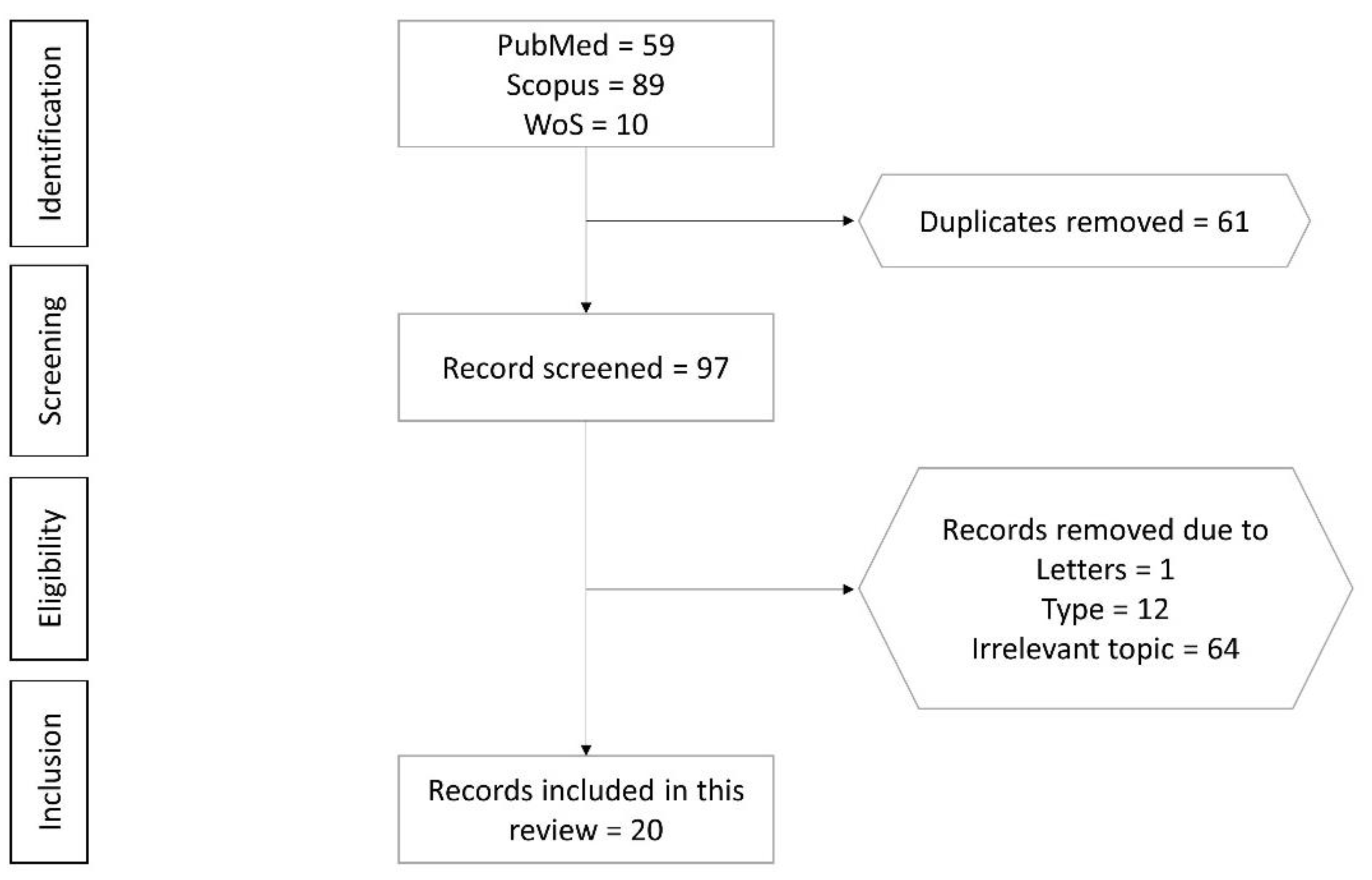
2.1. Selection of Articles

A total of 158 articles were retrieved from the literature search, of which 59 were from PubMed, 89 were from Scopus and 10 were Web of Science. After removing duplicates (n = 61), 97 articles were screened. Seventy-seven articles not meeting the selection criteria were eliminated (12 review articles, 1 editorial letter and 64 articles not relevant to the current review). Finally, 20 articles fulfilling all criteria mentioned were included in the review (Table S2). The selection process from identification, screening, eligibility to inclusion of articles is shown in Figure 1.

2.2. Study Characteristics

The selected studies were published from 2004 to 2020. Thirteen studies were in vitro experiments using primary cells from mouse (bone marrow cells (mBMCs), calvarial osteoblasts), human (bone marrow cells (hBMCs), periodontal ligament stem cells (hPDLSC), bone marrow mononuclear cells (BMMCs), bone marrow mesenchymal stromal cells (BM-MSCs)) and cell lines (RAW264.7, MC3T3-E1, C2C12) [27,28,29,30,31,32,33,34,35,36,37,38,39]. Twelve in vivo studies using Sprague Dawley rats, Wnt1sw/sw mice and C57BL/6J mice were included. Kunming (KM) mice and ICR mice [30,32,36,37,39,40,41,42,43,44,45,46]. No human studies on this topic were reported.
The in vitro studies investigated the effects tanshinone or its derivatives on osteoclast differentiation using macrophage colony-stimulating factor (M-CSF), receptor activator of NF-ĸB (RANK) ligand (RANKL), interleukin 1 alpha (IL-1α), lipopolysaccharide (LPS) or tumour necrosis factor-alpha (TNF-α). They also studied the osteogenic effects and anti-apoptotic effects of tanshinone in the presence of dexamethasone. The doses of tanshinone or its derivatives used ranged between 0.001 and 1000 µM and 1.5 mg/mL (5428.88 µM) [37]. The treatment period was 5–7 days for the differentiation of osteoclasts and 1–7 days for the differentiation of osteoblasts.
For animal studies, the doses of tanshinone used were between 0.36 and 200 mg/kg [30,32,36,37,39,40,41,42,43,44,45,46]. The disease model used in these studies included retinoic acid, ovariectomy (OVX)/oestrogen deficiency, streptozocin/diabetes, lipopolysaccharide/inflammation and collagen/rheumatoid arthritis-induced bone loss, as well as polyethylene (PE) particle-induced calvarial osteolysis mimicking osteolysis in arthroplasty. These studies determined bone microstructure using micro-computed tomography, bone histomorphometry and bone remodelling markers to assess bone health. Table 1 summarises the effects of tanshinones on bone health.

2.3. Results from Cell Culture Studies

Osteoblasts are specialised mesenchymal cells that synthesise bone matrix and coordinate the bone mineralisation [47]. Liu et al. reported that 48 h incubation with tanshinone IIA (T-IIA) (2.5 and 5 μM) increased osteogenic differentiation in human periodontal ligament stem cells (hPDLSC) compared to the control group [27]. Gene expression studies showed that T-IIA upregulated osteoblastogenesis-related genes like osteocalcin (OCN), osteopontin (OPN), runt-related transcription factor 2 (Runx2), alkaline phosphate (ALP). Pre-treatment with T-IIA (1, 5, 10 and 20 μM for 7 days) on BM-MSCs increased osteoblast number at early osteogenesis stages as evidenced by increased ALP expression [28]. Besides this, gene expression studies showed decreased receptor activator of nuclear factor-κΒ ligand (RANKL) expression and increased OPN, osteoprotegerin (OPG), Runx2, bone morphogenetic protein (BMP)-2, ALP and collagen 1 expression [28]. C2C12 cells are muscle cell lines that can differentiate into osteoblasts when cultured in monolayers under the influence of BMP2 [48]. Kim and Kim reported T-IIA (2.5, 5, 10 and 20 μM) for 7 days increased BMP-2-induced trans-differentiation of C2C12 cells into osteoblasts [35]. T-IIA treatment also increased activation of osteogenic genes like ALP, OCN, Runx2, BMP-2, 4, 6, 7 and 9. Similarly, Wang et al. [36] reported that T-IIA (5, 10, 15 and 20 μM for 48 h) increased MC3T3-E1 pre-osteoblast differentiation by increasing Runx2, osterix expression and ALP activity. T-IIA treatment also increased BMP-2 protein expression and activated the c-Jun N-terminal kinases (JNK) signalling pathway [36].
Besides this, T-IIA pre-treatment (1 μM) on MC3T3-E1 pre-osteoblasts for 24 h reduced dexamethasone-induced osteoblast apoptosis by inhibiting NADPH oxidase 4 (Nox4) expression [38]. Nox4 is an enzyme involved in reactive oxygen species (ROS) production [49], and ROS has been reported to promote osteoblast apoptosis [50]. Zhu et al. reported that T-IIA treatment (1.5 mg/mL for 24 h) increased osteoblast viability and decreased osteoblast apoptosis in osteoblast cells by increasing the expression of anti-apoptotic genes such as p53 and B-cell lymphoma 2 and reducing expression of apoptotic genes like apoptotic protease-activating factor 1 and caspase-3 in osteoblasts [37]. T-IIA also exhibited protective effect against oxidative stress in hydrogen peroxide-treated osteoblasts by reducing ROS, thiobarbituric acid reactive substance (TBARS) and reactive nitrogen species (RNS) but did not improve superoxide dismutase level or increase ALP expression [37]. T-IIA could improve osteoblast survival by regulating the NF-κB signalling pathway. In the study, NF-κB phosphorylation in osteoblasts was decreased, the expression levels of NF-κB targets genes, such as TNF-α, inducible nitric oxide synthase (iNOS) and COX-2 and proteins such as p65, an inhibitor of NF-κB alpha (IκBα) and IκB kinase beta (IKK-β) were reduced with T-IIA treatment. However, the expression of tumour necrosis factor receptor-associated factor (TRAF)-1, an inhibitor of apoptosis protein, was increased in the T-IIA-treated osteoblasts [37].
On the other hand, T-IIA treatment (1, 2 and 5 μg/mL) for 7 days reduced the number of tartrate-resistant acid protein (TRAP) positive cells formed from RANKL-treated RAW 264.7 cells in a dose-dependent manner than the untreated cells [39]. Osteoclast function was also reduced dose-dependently as evidenced by decreased resorbing pits and actin rings. Gene expression studies showed that T-IIA reduced osteoclastogenesis by suppressing osteoclastogenesis-related genes, such as nuclear factor of activated T-cells cytoplasmic 1 (NFATc1), TRAP, matrix metalloproteinase 9, cathepsin K, calcitonin receptor, and TRAF 6 [39]. T-IIA also suppressed osteoclastogenesis by inhibiting RANKL-induced activation of NF-κB, as evidenced by inhibition of p65 translocation, mitogen-activated protein kinase (MAPK) and protein kinase B (Akt) pathways [39]. Similarly, Kim et al. reported that pre-treatment with T-IIA (2.5, 5, 10 and 20 μg/mL) for 1 h reduced osteoclast fusion, actin ring formation and resorption area in macrophage colony-stimulating factor (M-CSF) and RANKL-treated calvarial osteoblasts and BMCs coculture [33]. T-IIA inhibited osteoclast differentiation marked by reduced calcitonin receptor, c-Src and integrin β3 expression [33]. Pre-treatment with T-IIA also inhibited the activation of ERK, Akt and NF-κB signal transduction pathways [33]. Kwak et al. [31] reported a reduction in osteoclast differentiation, c-Fos and NFATc1 expression following T-IIA treatment (10 μg/mL) for 4 days in RANKL and M-CSF-treated BMCs and calvarial osteoblast coculture. The anti-osteoclastogenic activity of T-IIA was mainly exerted through its anti-inflammatory properties. Mechanistically, T-IIA reduced the RANKL-induced activation of NF-κB by blocking the p65 subunit nuclear translocation [33,39]. T-IIA reversed the RANKL-induced activation of MAPK and Akt pathways by reducing the levels of ERK, JNK, c-FOS and Akt expression [31,33,39]. Kwak et al. [32] reported that T-IIA treatment (5 μg/mL) for 6 days inhibited osteoclast differentiation in cocultures of calvarial osteoblast cells and BMCs treated with tumour necrosis factor-alpha (TNF-α), interleukin 1 alpha (IL-1α) or LPS. T-IIA regulated the expression of RANKL and OPG in osteoclasts treated with LPS. A mechanistic study revealed that T-IIA reduced osteoclast formation and regulated expression of RANKL and OPG by inhibiting prostaglandin E2 (PGE2) through selective COX-2 inhibition [32].
Tanshinone II A sulfonic sodium (T06) is a water-soluble derivative of T-IIA [51] developed to increase the bioavailability of T-IIA [52]. Panwar et al. reported that treatment with T06 (200 and 500 nM) for 72 h effectively reduced cross-linked C-telopeptide of type I collagen (CTX-1) level in the culture supernatant of M-CSF and RANKL-treated human and mouse BMCs cultured on bone slides [30]. Treatment with 1 μM of T06 for 72 h reduced the total eroded surface of dentin slides created by mouse and human osteoclasts. However, it did not affect the metabolic activity and number of osteoclasts, suggesting that it was not toxic to the cells [30]. The main difference between T-IIA, T-I, D-T and C-T is in their structure and IC50 values. T-IIA and C-T have a dimethyl tetranaphthalene ring with IC50 values of 89.1 and 226.7 µM, respectively, while T-I and D-T contain naphthalene rings A and B with IC50 values of 38.7 and 14.4 µM respectively [53]. Lee et al. reported that T-IIA, T-1, D-T and C-T (0.5, 1 and 2.5 μg/mL for 3 days) decreased TRAP-positive multinuclear cells in M-CSF treated primary osteoblasts and bone marrow cell coculture [29]. Similarly, Kim et al. reported that T-IIA, T-I, D-T and C-T (1 μg/mL for 7 days) reduced osteoclastogenesis, evidenced by a reduced number of TRAP-positive cells but did not affect osteoblastogenesis due to the lack of change in ALP activity in RANKL and M-CSF-treated MC3T3-E1 cells [34].

2.4. Results from Animal Studies

T-IIA is the most widely studied tanshinone type, shown to produce skeletal beneficial effects in both healthy and animals suffering from bone loss. T-IIA intake (22 mg/kg/day for 8 weeks) significantly increased the whole-body and femoral BMD, maximum femoral load and histomorphometric indices of healthy female Wistar rats. Increased serum OCN and lower TRAP levels were also observed in the treated rats [46].
On the other hand, postmenopausal osteoporosis is primarily caused by oestrogen deficiency, which disrupts normal bone remodelling by increasing osteoclastic resorption activity more than bone formation, leading to a net loss of bone [54]. Wang et al. reported that T-IIA treatment (10 mg/kg for 2 weeks) increased bone volume, trabecular number (Tb.N), trabecular thickness (Tb.Th) and reduced trabecular separation (Tb.Sp) in Sprague Dawley rats with OVX-induced osteoporosis [40]. Similarly, OVX osteoporotic C57BL/6 mice treated with T-IIA (10 mg/kg for 6 weeks) showed reduced trabecular bone loss [39]. They also showed increased bone surface area/total volume, BV/TV, BMD, Tb.N and decreased trabecular pattern factor [39]. Cui et al. [43] reported that treatment with 200 mg/kg of total tanshinone (equivalent to 35 mg/kg of T-IIA + 16 mg/kg of C-T) for 30 weeks increased BV/TV, Tb.Th and reduced osteocalcin/bone surface, mineralisation rate and bone formation rate at the proximal tibial metaphysis of OVX osteoporotic rats. Additionally, T-IIA significantly restored the cortical bone thickness and Tb.N with higher serum estradiol levels and lower serum phosphate, ALP and TRAP levels than untreated KM mice with retinoic acid-induced osteoporosis [44]. Panwar at al. [30] reported that treatment with T06 (40 mg/kg for 3 months) could improve the structural properties of OVX C57BL/6 mice by reducing Tb.Sp and increasing BV/TV and Tb.N. Immunohistochemical analysis revealed that T06 treatment increased the number of osteoblasts per bone perimeter, confirmed by increased ALP expression in Western blot analysis and increased plasma procollagen-1 N-terminal peptide concentration [30]. However, T06 treatment did not affect total osteoclast numbers/bone surface and osteoclast numbers/bone perimeter than the OVX mice [30].
A spontaneous mutation in WNT1 (one of the major WNT ligands regulating bone homeostasis) is present in the swaying (Wnt1sw/sw) mouse, wherein a recessive mutation causes osteogenesis imperfecta and heterozygous mutation causes the early onset of osteoporosis [55]. In Wnt1sw/sw osteoporotic mice, T-IIA treatment (10 mg/kg for 6 weeks) increased the stiffness, ultimate strength and elastic modulus compared to mice treated with alendronate [37]. Diabetes has a major impact on bone stability, since both type 1 and type 2 diabetes elevate fracture risk, making skeletal fragility a complication of diabetes [56,57]. In 8-week-old C57BL/J6 mice with streptozotocin-induced diabetic osteoporosis, T-IIA treatment (10 and 30 mg/kg; 3 times a week for 8 weeks) increased the bone mineral density (BMD) of trabecular bone compared to the negative control [42]. Improvement in the bone structural properties was marked by increased volumetric BMD, BV/TV, bone area fraction, connectivity density and a decrease in the structural model index (SMI) [42]. Inflammation also directly affects bone remodelling because proinflammatory cytokines have been implicated in the regulation of osteoblasts and osteoclasts [58]. Kwak et al. [32] reported that ICR mice with LPS-induced bone loss showed decreased osteoclast formation and cancellous bone loss when treated with T-IIA (5 μg/g on days 1, 3, 5 and 7).
Yao et al. reported that T-IIA supplementation (1 and 2 μg /g/day for 21 days) inhibited bone resorption by reducing the number of resorption pits, percentage of porosity in the skull and TRAP-positive cells in 8 weeks old C57BL/J6 mice with PE particle-induced calvarial osteolysis. These events increased BV/TV, BMD and bone strength of the rats [41]. In this study, T-IIA achieved its bone protective potential by decreasing osteoclastogenic markers, such as RANKL/OPG ratio, osteoclast-associated immunoglobulin-like receptor and CTX-1 expression [41]. Zhang et al. also demonstrated that direct gingival mucosa injection of T-IIA significantly reduced the recurrence distance and percentage of first molar tooth movement on Wistar rats by suppressing the osteoclast activity as evidenced by the higher ratio of OPG to osteoclast differentiation factor [45]. Wang et al. also observed that T-IIA treatment (10, 20 and 30 mg/kg for 4 weeks) promoted fracture healing in mice that underwent femur diaphysis osteotomy by enhancing the volume of callus area after damage, callus intensity, low-density bone volume/callus total volume, tissue mineral density and BMD [36].

3. Materials and Methods

3.1. Literature Review

This systematic review was performed in line with Preferred Reporting Items for Systematic Reviews and Meta-Analyses (PRISMA) (Table S1) A systematic literature search was conducted in January 2021 using PubMed, Scopus and Web of Science database to identify studies on the anti-osteoporotic properties of tanshinones. The search string used was (1) tanshinones AND (2) (bone OR osteoporosis OR osteoblasts OR osteoclasts OR osteocytes).

3.2. Article Selection

Articles with the following characteristics were included: (1) original research articles with the primary objective of determining the anti-osteoporotic effects of tanshinones; (2) studies using cell cultures, animal models or human subjects. Articles were excluded if they were: (1) conference abstract, review, letter/commentary, editorial, perspective articles without original data; (2) studies using extracts/formulations containing tanshinones and other bioactive compounds; (3) articles not written in English. Two authors performed the search using both databases and search string mentioned. Inclusion of an article in the review was based on the consensus of both authors. If a consensus could not be reached, the corresponding author would decide on the fate of the article.

3.3. Data Extraction

Data extracted included authors’ names, publication year, study design, dose and treatment period, findings and limitations of the study.

4. Discussion

The current literature revealed that tanshinones exhibited anti-osteoporotic activity by suppressing osteoclastogenesis and improving osteoblastogenesis. Tanshinones exert anti-osteoclastogenic activity by blocking RANKL-induced activation of NF-κB, MAPK, Akt pathways and M-CSF/c-Src signalling. Tanshinones prevent osteoblast apoptosis and facilitate osteoblastogenesis by ameliorating oxidative stress and inflammation. Animal studies revealed that tanshinones prevented skeletal deterioration in osteoporosis models induced by oestrogen deficiency, diabetes and inflammation. They also prevent PE-induced osteolysis and improve fracture healing in animal models.
Osteoblasts are mesenchymal cells that produce and mineralise the bone matrix [47]. Osteoblast differentiation is regulated by Runx2 phosphorylation and transcription, which is mediated by the MAPK cascades. MAPK pathways and its components, JNK, ERK and p38, form the non-canonical BMP2 signal transduction pathways that regulate osteoblastogenesis [59,60,61,62]. T-IIA increases the activation of osteogenic genes which indicate its stimulatory effect on osteogenic differentiation [27,28,35]. Wang et al. pinpointed that the effects of T-IIA were mediated through JNK as inhibition of this pathway negated its effects on osteoblast differentiation and mineralisation [36].
Excessive ROS production could overwhelm intracellular antioxidant defence, causing oxidative stress and osteoporosis [63,64]. Apart from inhibiting osteoblast proliferation [65] and differentiation [66], oxidative stress also induces osteoblast apoptosis [67,68], thereby jeopardising bone formation [69]. Ameliorating oxidative stress could decrease osteoblast apoptosis and increase its differentiation [37]. Hydrogen peroxide, an end-product of Nox4, is one of the major ROS predominately present in mitochondria [70,71]. Blocking Nox4 activation may prevent osteoblast apoptosis. T-IIA was reported to inhibit Nox4 expression, which in turn decreased osteoblast apoptosis [38]. NF-κB activation negatively regulates bone formation by suppressing osteoblast differentiation [72]. T-IIA increased osteoblast differentiation via suppressing NF-κB activation and translation of its target genes (TNF-α, iNOS and COX2). This event is achieved by preventing IKK-β and IκBα degradation and p65 nuclear translocation. These effects translated to improved bone microstructure and biomechanical properties in animals treated with T-IIA [37].
Osteoclasts are derived from hematopoietic lineage cells and are capable of bone resorption [73]. It is necessary for normal bone homeostasis, but excessive resorption can induce pathological bone loss. A variety of hormones and cytokines regulate osteoclast differentiation and activation. Particularly, RANKL and M-CSF are the essential cytokines for osteoclastic differentiation [74]. M-CSF binds to colony-stimulating factor 1 receptor, and RANKL binds to RANK to promote osteoclast differentiation and survival, as well as bone resorptive activity [75,76]. RANK-RANKL binding leads to the recruitment of TRAF factors like TRAF 6 [77], resulting in the activation of NF-kB, Akt and MAPKs (ERK/p38/JNK) pathways. Besides, RANKL signalling activates c-Fos and subsequently NFATc1, a master switch for controlling osteoclast terminal differentiation [78,79,80]. All these pathways act in concert to initiate osteoclast differentiation and bone resorption by inducing transcription and expression of osteoclast-specific genes, such as TRAP, cathepsin K, matrix metalloproteinase 9 (MMP-9) and C-Src [81]. Exogenous factors like LPS can alter RANKL signalling and influence osteoclastogenesis. LPS induces production of proinflammatory cytokines of osteoblasts and precursor cells via COX-2, especially TNF-α, which subsequently augment RANKL signalling in osteoclasts [82,83]. Blocking of COX-2 is reported to inhibit osteoclastogenesis in vitro [84,85].
T-IIA treatment is reported to suppress RANKL and M-CSF-induced osteoclastogenesis from precursor cells and osteoclast function [33,39]. Similar effects were observed with T06, T-1, C-T and D-T treatment, indicating all tanshinones share similar properties [29,30,34]. T-IIA prevents RANKL-induced activation of TRAF 6, which in turn reduces activation of NF-κB, MAPK, Akt and c-Src pathways, and inhibits osteoclast formation and activity, marked by reduced TRAP and MMP9 expression [33,39]. T-IIA also inhibits the expression of RANKL-induced c-Fos and NFATc1, which suppress osteoclast differentiation [31]. Besides, T-IIA is reported to inhibit LPS-mediated COX-2 expression in bone marrow and calvarial osteoblast cells [32]. This action could reduce osteoclast formation and bone loss prevention in mice administered with LPS [32].
RANK-RANKL signalling also regulates CatK expression [86]. CatK represents a potential target of anti-osteoporosis therapy. CatK inhibition does not affect bone formation [87,88], suggesting that bone formation and resorption are uncoupled [16]. Furthermore, osteoclast formation and survival required for osteoblastic bone formation response during remodelling are not affected by CatK inhibition [89]. In contrast, increased bone formation was observed in CatK deficient mice [90]. The different forms of tanshinones (T-IIA and T06) exert distinct effects in CatK-associated bone remodelling. T-IIA suppressed RANKL-induced expression of CatK in RAW264.7 cells and BMMCs, but reduced the function and survival of osteoclasts [39]. This observation suggests that T-IIA may not be a selective inhibitor of CatK. On the other hand, T06 inhibited the activity of CatK but did not alter CatK-positive osteoclast number in mice with OVX-induced osteoporosis [30], which suggests that T06 may be a selective inhibitor of CatK. The selectivity ensures normal osteoblast-osteoclast crosstalk and bone remodelling are not interrupted. In comparison, bisphosphonates, an antiresorptive agent commonly used as the first-line treatment for osteoporosis, also inhibit bone formation [91].
The biological effects of tanshinones suggest their various potential clinical applications. Direct gingival mucosa injection of T-IIA reduced the recurrence distance and percentage of first molar tooth movement in Wistar rats by suppressing the osteoclast activity [45]. This evidence showed that T-IIA could prevent the loosening of teeth in gingival and periodontal diseases. Tanshinones enhanced skeletal health in healthy animals, by increasing BMD, femoral microstructures and strength, as well as lowering osteoclast activity [46]. Therefore, it could serve as an agent for the primary prevention of osteoporosis. Furthermore, various skeletal actions of tanshinones could be harnessed for secondary prevention of osteoporosis. T-IIA restored retinoic acid-induced decrease of cortical bone thickness and Tb.N by increasing serum oestradiol levels and preventing high bone remodelling in Wistar rats [44]. T-IIA also improved bone structural properties in mice with STZ-induced diabetic osteoporosis [42]. Additionally, T-IIA [39,40], T06 [30] and total tanshinones (T-IIA + C-T) [43] prevented OVX-induced bone loss in rats and mice by improving bone microstructures. T06 increased osteoblast number [30] while total tanshinones decreased osteoclast number [43] in vivo. Untreated osteoporosis could lead to fragility fracture. T-IIA could increase fracture healing in mice with femoral osteotomy to mimic fracture [36]. Osteolysis following joint replacement is mainly caused by the abrasive particles introduced by the prosthesis [41]. Recent studies showed that these abrasive particles could induce the release of cytokines associated with osteolysis, such as IL-6, IL-1, TNF-α and PGE2, worsening the inflammatory response [92]. PE particles have been confirmed to induce osteolysis around artificial joints [93]. T-IIA was shown to prevent calvarial bone resorption in PE particle-induced osteolysis [41]. Hence, T-IIA could be embedded with arthroplasty materials to avoid triggering inflammation and bone resorption.
Limited studies examined the safety of tanshinones. T-IIA at high concentration (≥6 μM) caused severe growth inhibition, development malformation and cardiotoxicity in zebrafish [94]. Similarly, T-11A at a high concentration (25 μM) was toxic to human endothelial EAhy926 cells as these cells were killed after a 24 h treatment [95]. There is limited data on the pharmacokinetic properties of tanshinones. Zhang et al. reported that tanshinones have limited bioavailability when administered orally [96]. After oral administration at 100 mg/kg, the systemic bioavailability of C-T was 2.05%, suggesting poor absorption or significant metabolism in the gut or/and liver. After intraperitoneal administration at 100 mg/kg, the systemic bioavailability of C-T was 10.60%, suggesting hepatic metabolism or low solubility of C-T [97]. There is a paucity of pharmacokinetics, pharmacodynamics and safety data of tanshinones in humans. A search through https://clinicaltrials.gov/ (accessed on 30 March 2021) using the term “tashinones” revealed four registered trials on left ventricular remodelling secondary to acute myocardial infarction (identifier: NCT02524964), pulmonary hypertension (identifier: NCT01637675), polycystic ovary syndrome (identifier: NCT01452477) and childhood acute promyeloid leukemia (identifier: NCT02200978). The last trial was recruiting subjects, while the status of the other three trials was unknown. No human clinical trial on the effects of tanshinones on skeletal diseases has been attempted, probably due to poor intestinal absorption and bioavailability [97,98]. Various methods have been developed to address the problem of low bioavailability, including designing water-soluble tanshinone derivatives, loading of tanshinones into discoidal and spherical high-density lipoproteins, liposomes, nanoparticles, microemulsions, cyclodextrin and solid dispersions [99,100,101,102,103]. However, due to the complicated manufacturing process, little progress has been made in the clinical application of these formulations [104]. Therefore, more comprehensive studies in this regard will help to establish tanshinones as one of the clinical therapeutic options for various bone conditions.
The present review did not exclude studies based on quality and assumed that articles published by journals indexed in three main and reputable databases are of sufficient quality to be included. We did not consider articles published in non-indexed journals, unpublished articles, thesis and non-primary research articles, potentially excluding some relevant studies.

5. Conclusions

The preclinical evidence so far supports that tanshinones exert skeletal protective effects by inhibiting osteoclastogenesis/bone resorption and promotes osteoblastogenesis/bone formation. A type of tanshinones, T06, is shown to be a specific CatK inhibitor, which reduces bone degradation without affecting the crosstalk between osteoblasts and osteoclasts. There is a lack of human clinical trials to validate the skeletal effects of tanshinones. Thus, well-planned clinical trials should be conducted to endorse the skeletal properties of tanshinones and prove the safety of these compounds.

Supplementary Materials

Table S1: PRISMA Checklist, Table S2: Literature Search Results.

Author Contributions

Conceptualization, S.O.E. and K.-Y.C.; methodology, S.O.E. and K.-L.P.; validation, K.-Y.C.; writing—original draft preparation, S.O.E.; writing—review and editing, K.-L.P. and K.-Y.C.; visualization, S.O.E.; supervision, K.-Y.C.; funding acquisition, K.-Y.C. All authors have read and agreed to the published version of the manuscript.

Funding

This research was funded by Universiti Kebangsaan Malaysia through Research University Grant (GUP-2020-021). Sophia Ogechi Ekeuku and Kok-Lun Pang are postdoctoral researchers funded by Universiti Kebangsaan Malaysia through FPR-1 and RGA1.

Institutional Review Board Statement

Not applicable.

Informed Consent Statement

Not applicable.

Acknowledgments

The authors thank Universiti Kebangsaan Malaysia for the support.

Conflicts of Interest

The authors declare no conflict of interest.

References

  1. Black, D.M.; Rosen, C.J. Postmenopausal Osteoporosis. N. Engl. J. Med. 2016, 374, 254–262. [Google Scholar] [CrossRef]
  2. Eastell, R.; O’Neill, T.W.; Hofbauer, L.C.; Langdahl, B.; Reid, I.R.; Gold, D.T.; Cummings, S.R. Postmenopausal osteoporosis. Nat. Rev. Dis. Prim. 2016, 2, 16069. [Google Scholar] [CrossRef]
  3. Deeks, E.D. Author Correction to: Denosumab: A Review in Postmenopausal Osteoporosis. Drugs Aging 2018, 35, 261. [Google Scholar] [CrossRef] [PubMed] [Green Version]
  4. Cosman, F.; De Beur, S.J.; LeBoff, M.S.; Lewiecki, E.M.; Tanner, B.; Randall, S.; Lindsay, R. Clinician’s Guide to Prevention and Treatment of Osteoporosis. Osteoporos. Int. 2014, 25, 2359–2381. [Google Scholar] [CrossRef] [Green Version]
  5. Jeremiah, M.P.; Unwin, B.K.; Greenawald, M.H.; Casiano, V.E. Diagnosis and Management of Osteoporosis. Am. Fam. Physician 2015, 92, 261–268. [Google Scholar] [PubMed]
  6. Odén, A.; McCloskey, E.V.; Kanis, J.A.; Harvey, N.C.; Johansson, H. Burden of high fracture probability worldwide: Secular increases 2010–2040. Osteoporos. Int. 2015, 26, 2243–2248. [Google Scholar] [CrossRef] [PubMed]
  7. Burge, R.; Dawson-Hughes, B.; Solomon, D.H.; Wong, J.B.; King, A.; Tosteson, A. Incidence and Economic Burden of Osteoporosis-Related Fractures in the United States, 2005-2025. J. Bone Miner. Res. 2006, 22, 465–475. [Google Scholar] [CrossRef]
  8. Camacho, P.M.; Petak, S.M.; Binkley, N.; Clarke, B.L.; Harris, S.T.; Hurley, D.L.; Kleerekoper, M.; Lewiecki, E.M.; Miller, P.D.; Narula, H.S.; et al. American Association of Clinical Endocrinologists and American College of Endocrinology Clinical Practice Guidelines for the Diagnosis and Treatment of Postmenopausal Osteoporosis—2016. Endocr. Pr. 2016, 22, 1–42. [Google Scholar] [CrossRef] [Green Version]
  9. Chapurlat, R.D.; Arlot, M.; Burt-Pichat, B.; Chavassieux, P.; Roux, J.P.; Portero-Muzy, N.; Delmas, P.D. Microcrack Frequency and Bone Remodeling in Postmenopausal Osteoporotic Women on Long-Term Bisphosphonates: A Bone Biopsy Study. J. Bone Miner. Res. 2007, 22, 1502–1509. [Google Scholar] [CrossRef]
  10. Stepan, J.J.; Burr, D.B.; Pavo, I.; Sipos, A.; Michalska, D.; Li, J.; Fahrleitner-Pammer, A.; Petto, H.; Westmore, M.; Michalsky, D.; et al. Low bone mineral density is associated with bone microdamage accumulation in postmenopausal women with osteoporosis. Bone 2007, 41, 378–385. [Google Scholar] [CrossRef]
  11. Chan, C.K.Y.; Mason, A.; Cooper, C.; Dennison, E. Novel advances in the treatment of osteoporosis. Br. Med Bull. 2016, 119, 129–142. [Google Scholar] [CrossRef] [PubMed] [Green Version]
  12. Peroni, A.; Zini, A.; Braga, V.; Colato, C.; Adami, S.; Girolomoni, G. Drug-induced morphea: Report of a case induced by balicatib and review of the literature. J. Am. Acad. Dermatol. 2008, 59, 125–129. [Google Scholar] [CrossRef] [PubMed]
  13. Rünger, T.M.; Quintanilla-Dieck, M.J.; Bhawan, J. Role of Cathepsin K in the Turnover of the Dermal Extracellular Matrix during Scar Formation. J. Investig. Dermatol. 2007, 127, 293–297. [Google Scholar] [CrossRef] [PubMed] [Green Version]
  14. Tanaka, M.; Yamada, H.; Nishikawa, S.; Mori, H.; Ochi, Y.; Horai, N.; Li, M.; Amizuka, N. Joint Degradation in a Monkey Model of Collagen-Induced Arthritis: Role of Cathepsin K Based on Biochemical Markers and Histological Evaluation. Int. J. Rheumatol. 2016, 2016, 1–10. [Google Scholar] [CrossRef] [Green Version]
  15. McClung, M.R.; O’Donoghue, M.L.; Papapoulos, S.E.; Bone, H.; Langdahl, B.; Saag, K.G.; Reid, I.R.; Kiel, D.P.; Cavallari, I.; Bonaca, M.P.; et al. Odanacatib for the treatment of postmenopausal osteoporosis: Results of the LOFT multicentre, randomised, double-blind, placebo-controlled trial and LOFT Extension study. Lancet Diabetes Endocrinol. 2019, 7, 899–911. [Google Scholar] [CrossRef]
  16. Rongchen, D.; Wu, Z.; Chu, H.Y.; Lu, J.; Lyu, A.; Liu, J.; Zhang, G. Cathepsin K: The Action in and Beyond Bone. Front. Cell Dev. Biol. 2020, 8, 433. [Google Scholar] [CrossRef]
  17. Tang, B.M.P.; Eslick, G.D.; Nowson, C.; Smith, C.; Bensoussan, A. Use of calcium or calcium in combination with vitamin D supplementation to prevent fractures and bone loss in people aged 50 years and older: A meta-analysis. Lancet 2007, 370, 657–666. [Google Scholar] [CrossRef]
  18. The DIPART (Vitamin D Individual Patient Analysis of Randomized Trials) Group. Patient level pooled analysis of 68 500 patients from seven major vitamin D fracture trials in US and Europe. BMJ 2010, 340, b5463. [Google Scholar] [CrossRef] [Green Version]
  19. Dong, Y.; Morris-Natschke, S.L.; Lee, K.-H. Biosynthesis, total syntheses, and antitumor activity of tanshinones and their analogs as potential therapeutic agents. Nat. Prod. Rep. 2011, 28, 529–542. [Google Scholar] [CrossRef]
  20. Chen, X.; Guo, J.; Bao, J.; Lu, J.; Wang, Y. The Anticancer Properties of Salvia Miltiorrhiza Bunge (Danshen): A Systematic Review. Med. Res. Rev. 2013, 34, 768–794. [Google Scholar] [CrossRef]
  21. Xing, L.; Tan, Z.-R.; Cheng, J.-L.; Huang, W.-H.; Zhang, W.; Deng, W.; Yuan, C.-S.; Zhou, H.-H. Bioavailability and pharmacokinetic comparison of tanshinones between two formulations of Salvia miltiorrhiza in healthy volunteers. Sci. Rep. 2017, 7, 4709. [Google Scholar] [CrossRef] [PubMed] [Green Version]
  22. Panwar, P.; Søe, K.; Guido, R.V.C.; Bueno, R.V.C.; Delaisse, J.-M.; Brömme, D. A novel approach to inhibit bone resorption: Exosite inhibitors against cathepsin K. Br. J. Pharmacol. 2015, 173, 396–410. [Google Scholar] [CrossRef] [PubMed] [Green Version]
  23. Ren, Z.H.; Tong, Y.H.; Xu, W.; Ma, J.; Chen, Y. Tanshinone II A attenuates inflammatory responses of rats with myocardial infarction by reducing MCP-1 expression. Phytomedicine 2010, 17, 212–218. [Google Scholar] [CrossRef]
  24. Li, X.; Lian, L.-H.; Bai, T.; Wu, Y.-L.; Wan, Y.; Xie, W.-X.; Jin, X.; Nan, J.-X. Cryptotanshinone inhibits LPS-induced proinflammatory mediators via TLR4 and TAK1 signaling pathway. Int. Immunopharmacol. 2011, 11, 1871–1876. [Google Scholar] [CrossRef] [PubMed]
  25. Li, X.-X.; Zheng, X.; Liu, Z.; Xu, Q.; Tang, H.; Feng, J.; Yang, S.; Vong, C.T.; Gao, H.; Wang, Y. Cryptotanshinone from Salvia miltiorrhiza Bunge (Danshen) inhibited inflammatory responses via TLR4/MyD88 signaling pathway. Chin. Med. 2020, 15, 1–13. [Google Scholar] [CrossRef] [Green Version]
  26. Jang, S.I.; Kim, H.J.; Kim, Y.-J.; Jeong, S.-I.; You, Y.-O. Tanshinone IIA inhibits LPS-induced NF-κB activation in RAW 264.7 cells: Possible involvement of the NIK–IKK, ERK1/2, p38 and JNK pathways. Eur. J. Pharmacol. 2006, 542, 1–7. [Google Scholar] [CrossRef]
  27. Liu, X.; Niu, Y.; Xie, W.; Wei, D.; Du, Q. Tanshinone IIA promotes osteogenic differentiation of human periodontal ligament stem cells via ERK1/2-dependent Runx2 induction. Am. J. Trans. Res. 2019, 11, 340–350. [Google Scholar]
  28. Qian, K.; Xu, H.; Dai, T.; Shi, K. Effects of Tanshinone IIA on osteogenic differentiation of mouse bone marrow mesenchymal stem cells. Naunyn-Schmiedeberg’s Arch. Pharmacol. 2015, 388, 1201–1209. [Google Scholar] [CrossRef]
  29. Lee, S.-Y.; Choi, D.-Y.; Woo, E.-R. Inhibition of osteoclast differentiation by tanshinones from the root ofSalvia miltiorrhiza Bunge. Arch. Pharmacal Res. 2005, 28, 909–913. [Google Scholar] [CrossRef]
  30. Panwar, P.; Xue, L.; Søe, K.; Srivastava, K.; Law, S.; Delaisse, J.-M.; Brömme, D. An Ectosteric Inhibitor of Cathepsin K Inhibits Bone Resorption in Ovariectomized Mice. J. Bone Miner. Res. 2017, 32, 2415–2430. [Google Scholar] [CrossRef] [Green Version]
  31. Kwak, H.B.; Yang, D.; Ha, H.; Lee, J.-H.; Kim, H.-N.; Woo, E.-R.; Lee, S.; Kim, H.-H.; Lee, Z.H. Tanshinone IIA inhibits osteoclast differentiation through down-regulation of c-Fos and NFATc1. Exp. Mol. Med. 2006, 38, 256–264. [Google Scholar] [CrossRef] [PubMed]
  32. Kwak, H.B.; Sun, H.-M.; Ha, H.; Kim, H.-N.; Lee, J.-H.; Kim, H.-H.; Shin, H.-I.; Lee, Z.H. Tanshinone IIA suppresses inflammatory bone loss by inhibiting the synthesis of prostaglandin E2 in osteoblasts. Eur. J. Pharmacol. 2008, 601, 30–37. [Google Scholar] [CrossRef] [PubMed]
  33. Kim, H.-H.; Kim, J.H.; Kwak, H.B.; Huang, H.; Han, S.-H.; Ha, H.; Lee, S.W.; Woo, E.-R.; Lee, Z.H. Inhibition of osteoclast differentiation and bone resorption by tanshinone IIA isolated from Salvia miltiorrhiza Bunge. Biochem. Pharmacol. 2004, 67, 1647–1656. [Google Scholar] [CrossRef] [PubMed]
  34. Kim, H.-K.; Woo, E.-R.; Lee, H.-W.; Park, H.-R.; Kim, H.-N.; Jung, Y.-K.; Choi, J.-Y.; Chae, S.-W.; Kim, H.-R.; Chae, H.-J. The Correlation ofSalvia miltiorrhizaExtract–Induced Regulation of Osteoclastogenesis with the Amount of Components Tanshinone I, Tanshinone IIA, Cryptotanshinone, and Dihydrotanshinone. Immunopharmacol. Immunotoxicol. 2008, 30, 347–364. [Google Scholar] [CrossRef] [PubMed]
  35. Kim, H.J.; Kim, S.H. Tanshinone IIA enhances BMP-2-stimulated commitment of C2C12 cells into osteoblasts via p38 activation. Amino Acids 2010, 39, 1217–1226. [Google Scholar] [CrossRef] [PubMed]
  36. Wang, Y.; Chen, H.; Zhang, H. Tanshinone IIA exerts beneficial effects on fracture healing in vitro and in vivo. Chem. Interactions 2019, 310, 108748. [Google Scholar] [CrossRef]
  37. Zhu, S.; Wei, W.; Liu, Z.; Yang, Y.; Jia, H. Tanshinone-IIA attenuates the deleterious effects of oxidative stress in osteoporosis through the NF-κB signaling pathway. Mol. Med. Rep. 2018, 17, 6969–6976. [Google Scholar] [CrossRef] [Green Version]
  38. Li, J.; He, C.; Tong, W.; Zou, Y.; Li, D.; Zhang, C.; Xu, W. Tanshinone IIA blocks dexamethasone-induced apoptosis in osteoblasts through inhibiting Nox4-derived ROS production. Int. J. Clin. Exp. Pathol. 2015, 8, 13695–13706. [Google Scholar]
  39. Cheng, L.; Zhou, S.; Zhao, Y.; Sun, Y.; Xu, Z.; Yuan, B.; Chen, X. Tanshinone IIA attenuates osteoclastogenesis in ovariectomized mice by inactivating NF-kB and Akt signaling pathways. Am. J. Transl. Res. 2018, 10, 1457–1468. [Google Scholar]
  40. Wang, L.; Cheng, L.; Zhang, B.; Wang, N.; Wang, F. Tanshinone prevents alveolar bone loss in ovariectomized osteoporosis rats by up-regulating phosphoglycerate dehydrogenase. Toxicol. Appl. Pharmacol. 2019, 376, 9–16. [Google Scholar] [CrossRef]
  41. Yao, J.; Ma, S.; Feng, W.; Wei, Y.; Lu, H.; Zhong, G.; Wu, Z.; Wang, H.; Su, W.; Li, J. Tanshinone IIA protects against polyethylene particle-induced osteolysis response in a mouse calvarial model. International Journal of Clinical and Experimental Pathology 2018, 11, 4461–4471. [Google Scholar]
  42. Zhang, J.; Cai, Z.; Yang, M.; Tong, L.; Zhang, Y. Inhibition of tanshinone IIA on renin activity protected against osteoporosis in diabetic mice. Pharm. Biol. 2020, 58, 219–224. [Google Scholar] [CrossRef] [PubMed]
  43. Cui, L.; Wu, T.; Liu, Y.-Y.; Deng, Y.-F.; Ai, C.-M.; Chen, H.-Q. Tanshinone prevents cancellous bone loss induced by ovariectomy in rats. Acta Pharmacol. Sin. 2004, 25, 678–684. [Google Scholar]
  44. Zhou, Y.; Liu, Y.; Gao, Y. Effect of tanshitone on prevention and treatment of retinoic acid induced osteoporosis in mice. China J. Chin. Mater. Medica 2010, 35, 2923–2926. [Google Scholar]
  45. Zhang, S.Y.; Liu, J.G.; Zhao, G. Tanshinone type IIA inhibits osteoprotegerin and osteoclast differentiation factor expression at relapse stage after orthodontic tooth movement. Chinese J. Tissue Engineering Res. 2014, 18, 1730–1736. [Google Scholar] [CrossRef]
  46. Yang, F.-F.; Gao, Y.-H.; Xi, H.-R.; Li, W.-Y.; Ma, H.-P.; Chen, K.-M. Effect of Compound Medicine of Tanshinone 2A and Resveratrol on Peak Bone Mass in Growing Rats. Zhongguo Yi Xue Ke Xue Yuan Xue Bao 2018, 40, 456–462. [Google Scholar]
  47. Dirckx, N.; Moorer, M.C.; Clemens, T.L.; Riddle, R.C. The role of osteoblasts in energy homeostasis. Nat. Rev. Endocrinol. 2019, 15, 651–665. [Google Scholar] [CrossRef]
  48. Katagiri, T.; Yamaguchi, A.; Komaki, M.; Abe, E.; Takahashi, N.; Ikeda, T.; Rosen, V.; Wozney, J.M.; Fujisawa-Sehara, A.; Suda, T. Bone morphogenetic protein-2 converts the differentiation pathway of C2C12 myoblasts into the osteoblast lineage [published erratum appears in J Cell Biol 1995 Feb;128(4):following 713]. J. Cell Biol. 1994, 127, 1755–1766. [Google Scholar] [CrossRef] [PubMed] [Green Version]
  49. Goettsch, C.; Babelova, A.; Trummer, O.; Erben, R.G.; Rauner, M.; Rammelt, S.; Weissmann, N.; Weinberger, V.; Benkhoff, S.; Kampschulte, M.; et al. NADPH oxidase 4 limits bone mass by promoting osteoclastogenesis. J. Clin. Investig. 2013, 123, 4731–4738. [Google Scholar] [CrossRef] [PubMed] [Green Version]
  50. Lee, D.H.; Lim, B.-S.; Lee, Y.-K.; Yang, H.-C. Effects of hydrogen peroxide (H2O2) on alkaline phosphatase activity and matrix mineralization of odontoblast and osteoblast cell lines. Cell Biol. Toxicol. 2006, 22, 39–46. [Google Scholar] [CrossRef] [PubMed]
  51. Zhou, Z.-Y.; Zhao, W.-R.; Zhang, J.; Chen, X.-L.; Tang, J.-Y. Sodium tanshinone IIA sulfonate: A review of pharmacological activity and pharmacokinetics. Biomed. Pharmacother. 2019, 118, 109362. [Google Scholar] [CrossRef] [PubMed]
  52. Shang, Q.; Xu, H.; Huang, L. Tanshinone IIA: A Promising Natural Cardioprotective Agent. Evid. Based Complementary Altern. Med. 2012, 2012, 1–7. [Google Scholar] [CrossRef] [PubMed] [Green Version]
  53. Park, J.-Y.; Kim, J.H.; Kim, Y.M.; Jeong, H.J.; Kim, D.W.; Park, K.H.; Kwon, H.-J.; Park, S.-J.; Lee, W.S.; Ryu, Y.B. Tanshinones as selective and slow-binding inhibitors for SARS-CoV cysteine proteases. Bioorganic Med. Chem. 2012, 20, 5928–5935. [Google Scholar] [CrossRef] [PubMed]
  54. Tella, S.H.; Gallagher, J.C. Prevention and treatment of postmenopausal osteoporosis. J. Steroid Biochem. Mol. Biol. 2014, 142, 155–170. [Google Scholar] [CrossRef] [Green Version]
  55. Joeng, K.S.; Lee, Y.-C.; Jiang, M.-M.; Bertin, T.K.; Chen, Y.; Abraham, A.M.; Ding, H.; Bi, X.; Ambrose, C.G.; Lee, B.H. The swaying mouse as a model of osteogenesis imperfecta caused by WNT1 mutations. Hum. Mol. Genet. 2014, 23, 4035–4042. [Google Scholar] [CrossRef] [PubMed] [Green Version]
  56. Vestergaard, P. Diabetes and Bone. J. Diabetes Metab. 2012, 01, 1–7. [Google Scholar] [CrossRef] [Green Version]
  57. Shivaswamy, V.; Mack, L.R.; Larsen, J. Diabetes, Bone Density, and Fractures. J. Diabetes Met. 2012, 2012, 1–6. [Google Scholar] [CrossRef] [Green Version]
  58. Ginaldi, L.; Di Benedetto, M.C.; De Martinis, M. Osteoporosis, inflammation and ageing. Immun. Ageing I A 2005, 2, 14. [Google Scholar] [CrossRef] [Green Version]
  59. Xiao, G.; Gopalakrishnan, R.; Jiang, D.; Reith, E.; Benson, M.D.; Franceschi, R.T. Bone Morphogenetic Proteins, Extracellular Matrix, and Mitogen-Activated Protein Kinase Signaling Pathways Are Required for Osteoblast-Specific Gene Expression and Differentiation in MC3T3-E1 Cells. J. Bone Miner. Res. 2002, 17, 101–110. [Google Scholar] [CrossRef]
  60. Nohe, A.; Hassel, S.; Ehrlich, M.; Neubauer, F.; Sebald, W.; Henis, Y.I.; Knaus, P. The Mode of Bone Morphogenetic Protein (BMP) Receptor Oligomerization Determines Different BMP-2 Signaling Pathways. J. Biol. Chem. 2002, 277, 5330–5338. [Google Scholar] [CrossRef] [PubMed] [Green Version]
  61. Massagué, J.; Blain, S.W.; Lo, R.S. TGFβ Signaling in Growth Control, Cancer, and Heritable Disorders. Cell 2000, 103, 295–309. [Google Scholar] [CrossRef] [Green Version]
  62. Guo, X.; Wang, X.-F. Signaling cross-talk between TGF-β/BMP and other pathways. Cell Res. 2009, 19, 71–88. [Google Scholar] [CrossRef] [PubMed]
  63. Baek, K.H.; Oh, K.W.; Lee, W.Y.; Lee, S.S.; Kim, M.K.; Kwon, H.S.; Rhee, E.J.; Han, J.H.; Song, K.H.; Cha, B.Y.; et al. Association of Oxidative Stress with Postmenopausal Osteoporosis and the Effects of Hydrogen Peroxide on Osteoclast Formation in Human Bone Marrow Cell Cultures. Calcif. Tissue Int. 2010, 87, 226–235. [Google Scholar] [CrossRef]
  64. Yin, H.; Shi, Z.-G.; Yu, Y.-S.; Hu, J.; Wang, R.; Luan, Z.-P.; Guo, D.-H. Protection against osteoporosis by statins is linked to a reduction of oxidative stress and restoration of nitric oxide formation in aged and ovariectomized rats. Eur. J. Pharmacol. 2012, 674, 200–206. [Google Scholar] [CrossRef] [PubMed]
  65. Li, M.; Zhao, L.; Liu, J.; Liu, A.-L.; Zeng, W.-S.; Luo, S.-Q.; Bai, X.-C. Hydrogen Peroxide Induces G2Cell Cycle Arrest and Inhibits Cell Proliferation in Osteoblasts. Anat. Rec. Adv. Integr. Anat. Evol. Biol. 2009, 292, 1107–1113. [Google Scholar] [CrossRef] [PubMed]
  66. Zhong, Z.-M.; Bai, L.; Chen, J.-T. Advanced Oxidation Protein Products Inhibit Proliferation and Differentiation of Rat Osteoblast-like Cells via NF-κB Pathway. Cell. Physiol. Biochem. 2009, 24, 105–114. [Google Scholar] [CrossRef] [PubMed]
  67. Almeida, M.; Han, L.; Ambrogini, E.; Bartell, S.M.; Manolagas, S.C. Oxidative Stress Stimulates Apoptosis and Activates NF-κB in Osteoblastic Cells via a PKCβ/p66shc Signaling Cascade: Counter Regulation by Estrogens or Androgens. Mol. Endocrinol. 2010, 24, 2030–2037. [Google Scholar] [CrossRef] [PubMed] [Green Version]
  68. Wu, Y.; Wang, D.; Wang, X.; Wang, Y.; Ren, F.; Chang, D.; Chang, Z.; Jia, B. Caspase 3 is Activated through Caspase 8 instead of Caspase 9 during H2O2-induced Apoptosis in HeLa Cells. Cell. Physiol. Biochem. 2011, 27, 539–546. [Google Scholar] [CrossRef] [PubMed]
  69. Moriishi, T.; Kawai, Y.; Komori, H.; Rokutanda, S.; Eguchi, Y.; Tsujimoto, Y.; Asahina, I.; Komori, T. Bcl2 Deficiency Activates FoxO through Akt Inactivation and Accelerates Osteoblast Differentiation. PLoS ONE 2014, 9, e86629. [Google Scholar] [CrossRef]
  70. Schroeder, K.; Zhang, M.; Benkhoff, S.; Mieth, A.; Pliquett, R.; Kosowski, J.; Kruse, C.; Luedike, P.; Michaelis, U.R.; Weissmann, N.; et al. Nox4 Is a Protective Reactive Oxygen Species Generating Vascular NADPH Oxidase. Circ. Res. 2012, 110, 1217–1225. [Google Scholar] [CrossRef] [PubMed] [Green Version]
  71. Cabiscol, E.; Tamarit, J.; Ros, J. Oxidative stress in bacteria and protein damage by reactive oxygen species. Int. Microbiol. 2000, 3, 3–8. [Google Scholar]
  72. Yao, Z.; Li, Y.; Yin, X.; Dong, Y.; Xing, L.; Boyce, B.F. NF-κB RelB Negatively Regulates Osteoblast Differentiation and Bone Formation. J. Bone Miner. Res. 2014, 29, 866–877. [Google Scholar] [CrossRef]
  73. Gallois, A.; Lachuer, J.; Yvert, G.; Wierinckx, A.; Brunet, F.; Rabourdin-Combe, C.; Delprat, C.; Jurdic, P.; Mazzorana, M. Genome-wide expression analyses establish dendritic cells as a new osteoclast precursor able to generate bone-resorbing cells more efficiently than monocytes. J. Bone Miner. Res. 2010, 25, 661–672. [Google Scholar] [CrossRef]
  74. De Vries, T.J.; Schoenmaker, T.; Aerts, D.; Grevers, L.C.; Souza, P.P.; Nazmi, K.; Van De Wiel, M.A.; Ylstra, B.; Van Lent, P.L.V.; Leenen, P.J.M.; et al. M-CSF Priming of Osteoclast Precursors Can Cause Osteoclastogenesis-Insensitivity, Which Can be Prevented and Overcome on Bone. J. Cell. Physiol. 2015, 230, 210–225. [Google Scholar] [CrossRef]
  75. Mun, S.H.; Park, P.S.U.; Park-Min, K.-H. The M-CSF receptor in osteoclasts and beyond. Exp. Mol. Med. 2020, 52, 1239–1254. [Google Scholar] [CrossRef]
  76. Suda, T.; Takahashi, N.; Udagawa, N.; Jimi, E.; Gillespie, M.T.; Martin, T.J. Modulation of Osteoclast Differentiation and Function by the New Members of the Tumor Necrosis Factor Receptor and Ligand Families. Endocr. Rev. 1999, 20, 345–357. [Google Scholar] [CrossRef]
  77. Bharti, A.C.; Takada, Y.; Shishodia, S.; Aggarwal, B.B. Evidence That Receptor Activator of Nuclear Factor (NF)-κB Ligand Can Suppress Cell Proliferation and Induce Apoptosis through Activation of a NF-κB-independent and TRAF6-dependent Mechanism. J. Biol. Chem. 2004, 279, 6065–6076. [Google Scholar] [CrossRef] [Green Version]
  78. Takayanagi, H.; Kim, S.; Koga, T.; Nishina, H.; Isshiki, M.; Yoshida, H.; Saiura, A.; Isobe, M.; Yokochi, T.; Inoue, J.-I.; et al. Induction and Activation of the Transcription Factor NFATc1 (NFAT2) Integrate RANKL Signaling in Terminal Differentiation of Osteoclasts. Dev. Cell 2002, 3, 889–901. [Google Scholar] [CrossRef] [Green Version]
  79. Johnson, R.S.; Spiegelman, B.M.; Papaioannou, V. Pleiotropic effects of a null mutation in the c-fos proto-oncogene. Cell 1992, 71, 577–586. [Google Scholar] [CrossRef]
  80. Ishida, N.; Hayashi, K.; Hoshijima, M.; Ogawa, T.; Koga, S.; Miyatake, Y.; Kumegawa, M.; Kimura, T.; Takeya, T. Large Scale Gene Expression Analysis of Osteoclastogenesisin Vitro and Elucidation of NFAT2 as a Key Regulator. J. Biol. Chem. 2002, 277, 41147–41156. [Google Scholar] [CrossRef] [PubMed] [Green Version]
  81. Nakashima, T.; Hayashi, M.; Fukunaga, T.; Kurata, K.; Oh-Hora, M.; Feng, J.Q.; Bonewald, L.F.; Kodama, T.; Wutz, A.; Wagner, E.F.; et al. Evidence for osteocyte regulation of bone homeostasis through RANKL expression. Nat. Med. 2011, 17, 1231–1234. [Google Scholar] [CrossRef]
  82. Li, L.; Pettit, A.R.; Gregory, L.S.; Forwood, M.R. Regulation of bone biology by prostaglandin endoperoxide H synthases (PGHS): A rose by any other name…. Cytokine Growth Factor Rev. 2006, 17, 203–216. [Google Scholar] [CrossRef]
  83. Ono, K.; Akatsu, T.; Murakami, T.; Kitamura, R.; Yamamoto, M.; Shinomiya, N.; Rokutanda, M.; Sasaki, T.; Amizuka, N.; Ozawa, H.; et al. Involvement of Cyclo-Oxygenase-2 in Osteoclast Formation and Bone Destruction in Bone Metastasis of Mammary Carcinoma Cell Lines. J. Bone Miner. Res. 2002, 17, 774–781. [Google Scholar] [CrossRef]
  84. Ono, K.; Akatsu, T.; Kugai, N.; Pilbeam, C.C.; Raisz, L.G. The effect of deletion of cyclooxygenase-2, prostaglandin receptor EP2, or EP4 in bone marrow cells on osteoclasts induced by mouse mammary cancer cell lines. Bone 2003, 33, 798–804. [Google Scholar] [CrossRef]
  85. Kellinsalmi, M.; Parikka, V.; Risteli, J.; Hentunen, T.; Leskelä, H.-V.; Lehtonen, S.; Selander, K.; Väänänen, K.; Lehenkari, P. Inhibition of cyclooxygenase-2 down-regulates osteoclast and osteoblast differentiation and favours adipocyte formation in vitro. Eur. J. Pharmacol. 2007, 572, 102–110. [Google Scholar] [CrossRef]
  86. Troen, B.R. The Regulation of Cathepsin K Gene Expression. Ann. N. Y. Acad. Sci. 2006, 1068, 165–172. [Google Scholar] [CrossRef] [Green Version]
  87. Brömme, D.; Panwar, P.; Turan, S. Cathepsin K osteoporosis trials, pycnodysostosis and mouse deficiency models: Commonalities and differences. Expert Opin. Drug Discov. 2016, 11, 457–472. [Google Scholar] [CrossRef]
  88. Drake, M.T.; Clarke, B.L.; Oursler, M.J.; Khosla, S. Cathepsin K Inhibitors for Osteoporosis: Biology, Potential Clinical Utility, and Lessons Learned. Endocr. Rev. 2017, 38, 325–350. [Google Scholar] [CrossRef] [PubMed] [Green Version]
  89. Martin, T.J.; Sims, N.A. Osteoclast-derived activity in the coupling of bone formation to resorption. Trends Mol. Med. 2005, 11, 76–81. [Google Scholar] [CrossRef] [PubMed]
  90. Li, C.Y.; Jepsen, K.J.; Majeska, R.J.; Zhang, J.; Ni, R.; Gelb, B.D.; Schaffler, M.B. Mice Lacking Cathepsin K Maintain Bone Remodeling but Develop Bone Fragility Despite High Bone Mass. J. Bone Miner. Res. 2006, 21, 865–875. [Google Scholar] [CrossRef]
  91. George, E.L.; Lin, Y.-L.; Saunders, M.M. Bisphosphonate-related osteonecrosis of the jaw: A mechanobiology perspective. Bone Rep. 2018, 8, 104–109. [Google Scholar] [CrossRef]
  92. Noordin, S.; Masri, B. Periprosthetic osteolysis: Genetics, mechanisms and potential therapeutic interventions. Can. J. Surg. 2012, 55, 408–417. [Google Scholar] [CrossRef] [Green Version]
  93. Neuerburg, C.; Loer, T.; Mittlmeier, L.; Polan, C.; Farkas, Z.; Holdt, L.M.; Utzschneider, S.; Schwiesau, J.; Grupp, T.M.; Böcker, W.; et al. Impact of vitamin E-blended UHMWPE wear particles on the osseous microenvironment in polyethylene particle-induced osteolysis. Int. J. Mol. Med. 2016, 38, 1652–1660. [Google Scholar] [CrossRef] [PubMed] [Green Version]
  94. Wang, T.; Wang, C.; Wu, Q.; Zheng, K.; Chen, J.; Lan, Y.; Qin, Y.; Mei, W.; Wang, B. Evaluation of Tanshinone IIA Developmental Toxicity in Zebrafish Embryos. Molecules 2017, 22, 660. [Google Scholar] [CrossRef] [Green Version]
  95. Yang, L.-J.; Jeng, C.-J.; Kung, H.-N.; Chang, C.-C.; Wang, A.-G.; Chau, G.-Y.; Don, M.-J.; Chau, Y.-P. Tanshinone IIA isolated from Salvia miltiorrhiza elicits the cell death of human endothelial cells. J. Biomed. Sci. 2005, 12, 347–361. [Google Scholar] [CrossRef]
  96. Zhang, Y.; Jiang, P.; Ye, M.; Kim, S.-H.; Jiang, C.; Lü, J. Tanshinones: Sources, Pharmacokinetics and Anti-Cancer Activities. Int. J. Mol. Sci. 2012, 13, 13621–13666. [Google Scholar] [CrossRef] [Green Version]
  97. Zhang, J.; Huang, M.; Guan, S.; Bi, H.-C.; Pan, Y.; Duan, W.; Chan, S.Y.; Chen, X.; Hong, Y.-H.; Bian, J.-S.; et al. A Mechanistic Study of the Intestinal Absorption of Cryptotanshinone, the Major Active Constituent ofSalvia miltiorrhiza. J. Pharmacol. Exp. Ther. 2006, 317, 1285–1294. [Google Scholar] [CrossRef] [Green Version]
  98. Yu, X.-Y.; Lin, S.-G.; Zhou, Z.-W.; Chen, X.; Liang, J.; Liu, P.-Q.; Duan, W.; Chowbay, B.; Wen, J.-Y.; Li, C.-G.; et al. Role of P-Glycoprotein in the Intestinal Absorption of Tanshinone IIA, a Major Active Ingredient in the Root of Salvia miltiorrhiza Bunge. Curr. Drug Metab. 2007, 8, 325–340. [Google Scholar] [CrossRef]
  99. Xu, S.; Liu, P. Tanshinone II-A: New perspectives for old remedies. Expert Opin. Ther. Pat. 2012, 23, 149–153. [Google Scholar] [CrossRef]
  100. Wang, L.; Lai, Y.; Li, C.; Jiang, X. Study on the intestinal absorption profiles of tanshinone IIA and its inclusion complex with cyclodextrin in rats. PDA J. Pharm. Sci. Technol. 2010, 63, 390–400. [Google Scholar]
  101. Liu, X.; An, C.; Jin, P.; Liu, X.; Wang, L. Protective effects of cationic bovine serum albumin-conjugated PEGylated tanshinone IIA nanoparticles on cerebral ischemia. Biomater. 2013, 34, 817–830. [Google Scholar] [CrossRef]
  102. Ma, H.; Fan, Q.; Yu, J.; Xin, J.; Zhang, C. Anticancer activities of tanshinone microemulsion against hepatocellular carcinoma in vitro and in vivo. Mol. Med. Rep. 2012, 7, 59–64. [Google Scholar] [CrossRef] [PubMed] [Green Version]
  103. Jia, X.-B.; Yan, H.-M.; Zhang, Z.-H.; Jiang, Y.-R.; Ding, D.-M.; Sun, E. An attempt to stabilize tanshinone IIA solid dispersion by the use of ternary systems with nano-CaCO3and poloxamer 188. Pharmacogn. Mag. 2014, 10, 311–S317. [Google Scholar] [CrossRef] [PubMed] [Green Version]
  104. Chen, M.; Cai, Y.; Zhang, W.; Chen, Z.; Shi, Z.; He, C. Recent insights into the biological activities and drug delivery systems of tanshinones. Int. J. Nanomed. 2016, 11, 121–130. [Google Scholar] [CrossRef] [PubMed] [Green Version]
Figure 1. Flowchart of the article selection process.
Molecules 26 02319 g001
Table 1. Effects of tanshinone on bone health.
StudiesStudy DesignChanges with Tanshinone Treatment
Cell Culture Studies
Liu et al. [27]Cell: hPDLSC from the premolars of 20 donors without oral or systematic diseases (10 men and 10 women aged 12–25 years old)
Model: osteogenic differentiation
Treatment: 2.5 and 5 μM of T-IIA for 48 h
Negative control: Untreated cells
Positive control: N.A.
  • ↑ osteogenic differentiation
  • ↑ OCN, OPN, Runx2, ALP gene expression vs. negative control
Qian et al. [28]Cell: BM-MSCs isolated from the tibia and femur of BALB/cJ mice (4–6 weeks old)
Model: dexamethasone-induced osteogenic differentiation
Treatment: 1, 5, 10 and 20 μM of T-IIA for 7 days
Negative control: Untreated cells
Positive control: N.A.
  • ↑ ALP expression vs. negative control
  • ↑ OPN, OPG, collagen 1 and ↓ RANKL vs. negative control
  • ↑ Runx2 and BMP 4 vs. negative control
Kim and Kim [35]Cell: C2C12 cells
Model: BMP-2-induced osteoblast differentiation
Treatment: 2.5, 5, 10 and 20 μM of T-IIA for 7 days
Negative control: Untreated cells
Positive control: N.A.
  • ↑ BMP-2-induced osteoblast differentiation and ALP production
  • ↑ activation of osteogenic genes (ALP, OCN, Runx2, BMP-2, 4, 6, 7, 9)
Wang et al. [36]Cell: MC3T3-E1 cells
Model: osteoblast differentiation
Treatment: 5, 10, 15 and 20 μM of T-IIA for 48 h
Negative control: Untreated cells
Positive control: N.A.
  • ↑ Runx2, Osx expression and ALP activity vs. negative control
  • ↑ BMP-2 protein expression vs. negative control
  • ↑ activation of JNK pathway vs. negative control
Li et al. [38]Cell: MC3T3-E1 cells
Model: dexamethasone-induced osteoblast apoptosis
Treatment: 1 μM of T-IIA for 24 h
Negative control: Untreated cells
Positive control: N.A.
  • ↓ dexamethasone-induced osteoblast apoptosis by inhibiting Nox4 expression
Zhu et al. [37]Cell: Osteoblast cells from 10 weeks old female Wnt1sw/sw mice
Model: H2O2-induced osteoblast apoptosis
Treatment: 1.5 mg/mL of T-IIA for 24 h
Negative control: PBS-treated cells
Positive control: 1.5 mg/kg alendronate
  • ↑ osteoblasts viability and ↓ osteoblast apoptosis
  • ↓ ALP, H2O2, ROS, SOD, TBARS and RNS levels vs. osteoblasts from mice treated with alendronate
  • ↓ caspace-3, and Apaf-1 expression and ↑, Bcl-2, TRAF 1, IAP and p53 expression vs. osteoblasts from mice treated with alendronate
  • ↓ activation of NF-κB phosphorylation, NF-κB activity, TNF-α, iNOS and COX2 expression vs. osteoblasts from mice treated with alendronate
  • ↓ p65, IKK-β and IκBα vs. osteoblasts from mice treated with alendronate
Cheng et al. [39]Cell: RAW264.7 cells and BMMCs isolated from the femoral bone marrow of 8-week-old C57BL/6 mice.
Model: RANKL-induced osteoclastogenesis
Treatment: 1, 2 or 5 μg/mL of T-IIA for 7 days
Negative control: Untreated cells
Positive control: N.A.
  • ↓ osteoclastogenesis and osteoclast function vs. negative control
  • ↓ TRAP+ cells (in a dose-dependent manner) vs. negative control
  • ↓ numbers and size of actin ring structures (a characteristic feature of mature osteoclasts during osteoclastogenesis) vs. the negative control
  • ↓ RANKL-induced osteoclast differentiation at early stages vs. negative control
  • ↓ osteoclastogenesis-related genes (TRAP, MMP- 9, cathepsin K, CTR, and TRAF6) vs. negative control
  • ↓ RANKL-induced activation of the NF-κB, MAPK and Akt pathways
  • ↓ ERK, JNK, c-Fos and Akt expression in osteoclasts
Lee et al. [29]Cell: Primary osteoblasts from calvarial cells of ICR newborn mice/bone marrow cells obtained from tibiae of 6- to 7-week-old ICR mice (coculture)
Model: M-CSF-induced osteoclast differentiation
Treatment: 0.5, 1 and 2.5 μg/mL of T-IIA, T-I, C-T, D-T for 3 days (osteoblasts) and 6-7 days (osteoclasts)
Negative control: Untreated cells
Positive control: N.A.
  • ↓ TRAP-positive multinuclear cells vs. negative control
  • ↓ viability of bone marrow cells following treatment with T-I, C-T, D-T
  • NS in the viability of bone marrow cells following treatment with T-IIA
Panwar et al. [30]Cell: mononuclear cells from human bone marrow and bone marrow cells from femur and tibia from 4 months old mice
Model: M-CSF and RANKL-induced osteoclastogenesis
Treatment: 1 and 3 μM of T06 for 72 h
Negative control: DMSO (1%)-treated cells
Positive control: N.A.
  • ↓TRAP-stained osteoclasts and toluidine-stained resorption events in mouse and human osteoclasts vs. negative control
  • ↓ total eroded surface in human and mouse osteoclasts vs. negative control
  • ↓ CTx-1 expression vs. negative control
  • NS for osteoclastogenesis in bone marrow mononuclear cells
Kim et al. [34]Cell: MC3T3-E1 cells and bone marrow cells isolated from the long bone of 7-weeks-old ICR male mice
Model: M-CSF and RANKL-induced osteoclastogenesis
Treatment: 1 μg/mL of T-IIA, T-I, D-T, and C-T for 7 days
Negative control: DMSO-treated cells
Positive control: N.A.
  • ↓ osteoclastogenesis in all tanshinone isoforms vs. negative control
  • NS for osteoblastogenesis in all tanshinone isoforms vs. negative control
Kwak et al. [31]Cell: Bone marrow cells from tibia and femur and mouse calvariae from pericranium of 5 weeks old male ICR mice.
Model: M-CSF and RANKL-induced osteoclastogenesis
Treatment: 10 μg/mL of T-IIA for 4 days
Negative control: M-CSF-treated cells
Positive control: N.A.
  • ↓ RANKL-mediated osteoclast differentiation vs. negative control
  • ↓ c-Fos and NFATc1 expression induced by RANKL
Kim et al. [33]Cell: Calvarial osteoblasts from the new bone of ICR mice and bone marrow cells from tibiae of 6–7 weeks old ICR mice (Mouse bone marrow cells and calvarial osteoblast coculture)
Model: M-CSF and RANKL-induced osteoclastogenesis
Treatment: 2.5–20 μg/mL of T-IIA for 15 min–20 h
Negative control: Untreated cells
Positive control: N.A.
  • ↓ osteoclast differentiation, osteoclast fusion, actin ring formation and resorption area vs. negative control
  • ↓ osteoclast differentiation-related genes (calcitonin receptor, c-Src, and integrin β3)
  • ↓ activation of ERK, Akt and NF-κB signal transduction pathways
Kwak et al. [32]Cell: Calvarial osteoblasts and bone marrow cells isolated from the femur and tibias of 5-weeks-old ICR male mice (Mouse bone marrow cells and calvarial osteoblast coculture)
Model: TNF-α, IL-1α or LPS-induced osteoclast differentiation
Treatment: 5 μg/mL of T-IIA for 7 days
Negative control: Untreated cells
Positive control: N.A.
  • ↓ osteoclast differentiation vs. negative control
  • ↓ LPS-induced RANKL and OPG expression in osteoblasts vs. negative control
  • ↓ LPS-mediated COX-2 expression and LPS-induced PGE2 in osteoblasts
Animal studies
Zhang, et al. [45]Animals: 48 male Wistar rats (2 months old)
Experimental model: Relapse stage after orthodontic mesial movement of maxillary first molar tooth
Treatment: 0.36, 0.72 and 1.44 mg/day of T-IIA for 4 weeks (localised gingival mucosa injection)
Negative control: Normal saline injection
Positive control: N.A.
  • ↓ recurrence distance and percentage of tooth movement by regulating osteoclast activity with ↑ OPG/osteoclast differentiation factor
  • NS in body weight changes
Yang, et al. [46]Animals: 40 female Wistar rats (1 month old, 97 ± 3 g
Disease model: N.A.
Treatment: 22 mg/kg/day of T-IIA for 8 weeks (oral)
Negative control: untreated rats
Positive control: N.A.
Comparative groups: 16.8 mg/kg/day of resveratrol and T-IIA (11 mg/kg/day) + resveratrol (8.4 mg/kg/day) for 8 weeks
  • ↑ serum OCN, whole-body and femoral BMD, maximum femoral load and histomorphometric indices (trabecular width, trabecular separation degree, Tb.N and trabecular area) vs. untreated rats
  • ↓ serum TRAP vs. untreated rats
  • NS in body weight
Wang et al. [36]Animals: 40 male C57BL/J6 mice (3 months old)
Disease model: Open osteotomy at femur diaphysis
Treatment: 10, 20 and 30 mg/kg/day of T-IIA for 4 weeks (oral)
Negative control: Mice with open osteotomy received with methanol
Normal control: No treatment or surgery
Positive control: N.A.
  • ↑ callus area, callus intensity, BV1/TV, TMD and BMD vs. negative control
Yao et al. [41]Animals: 24 male C57BL/J6 mice (2 months old)
Disease model: PE particle-induced calvarial osteolysis
Treatment: 1 and 2 μg/g/day of T-IIA for 21 days (periosteum injection)
Negative control: PE-induced mice treated with PBS
Normal control: Sham treated with PBS
Positive control: N.A.
  • ↓ number of pits and percentage of porosity of the skull vs. negative control
  • ↑ BV/TV and BMD vs. negative control
  • ↓ TRAP (+) osteoclasts, RANKL, OSCAR, CTX-1 and ↑ OPG vs. negative control
Kwak et al. [32]Animals: ICR mice
Disease model: LPS-induced bone loss
Treatment: 5 μg/g of T-IIA (i.p.) on the day before LPS induction, the day 1, 3, 5 and 7 after LPS induction
Negative control: Mice with LPS-induced bone loss
Normal control: Mice with PBS treatment
Positive control: N.A.
  • ↓ LPS-induced bone loss vs. the negative control
  • ↓ LPS-induced osteoclast formation and loss of cancellous bone vs. the negative control
Zhu et al. [37]Animals: 64 female Wnt1sw/sw mice with osteoporosis (2.5 months old, 32–40 g)
Disease model: Spontaneous WNT1 mutation for osteogenesis imperfecta
Treatment: 10 mg/kg of T-IIA (i.p.)/day for 6 weeks (i.p.)
Negative control: Osteoporotic mice with PBS injection
Normal control: N.A.
Positive control: 10 mg/kg alendronate for 6 weeks
  • ↑ stiffness, ultimate strength, elastic modulus, proline/amide 1, phosphate/amide 1 and phosphate/proline vs. negative control and greater potency than positive control
Zhang et al. [42]Animals: 40 male C57BL/J6 mice (2 months old)
Disease model: STZ-induced diabetic osteoporosis
Treatment: 10 and 30 mg/kg of T-IIA 3 times per week for 8 weeks (i.p.)
Negative control: Diabetic mice with corn oil (vehicle)
Normal control: Non-diabetic mice with corn oil
Positive control: 2 mg/kg aliskiren, 3 times a week for 8 weeks
  • ↑ bone mass of trabecular bone vs. negative control
  • ↑ BMD/TV, BV/TV, BA/TA, Conn.D and ↓ SMI vs. negative control
Wang et al. [40]Animals: 32 female Sprague Dawley rats (3 months old)
Disease model: OVX-induced osteoporosis
Treatment: 10 mg/kg/day of T-IIA for 2 weeks (i.v.)
Negative control: untreated OVX rats
Normal control: Sham-operated rats
Positive control: N.A.
  • ↑ bone volume, trabecular number, trabecular thickness and ↓ trabecular separation vs. negative control
Cheng et al. [39]Animals: 18 female C57BL/6 mice (2 months old)
Disease model: OVX-induced osteoporosis
Treatment: 10 mg/kg T-IIA for 6 weeks (i.p.)
Negative control: OVX with normal saline
Normal control: Sham with normal saline
Positive control: N.A.
  • ↓ trabecular bone loss vs. negative control
  • ↑ BS/TV, BV/TV, BMD, Tb.N and ↓ Tb.Pf vs. negative control
Panwar et al. [30]Animals: 29 female C57BL/6 mice
(8 months old, 25g)
Disease model: OVX-induced osteoporosis
Treatment: 40 mg/kg/day of T06 for 3 months (oral)
Negative control: OVX with an unknown vehicle
Normal control: Sham with an unknown vehicle
Positive control: N.A.
  • ↑ N.Ob/B.Pm
  • ↓ Tb.Sp and ↑ BV/TV, Tb.N vs. negative control
  • ↑ ALP-positive osteoblasts vs. negative control.
  • ↑ P1NP concentration vs. negative control
  • NS for CatK-positive osteoclast numbers/bone surface and osteoclast numbers/bone perimeter
Cui et al. [43]Animals: 32 female Sprague-Dawley (4 months old)
Disease model: OVX-induced osteoporosis
Treatment: 200 mg/kg/day of total tanshinone (5 mg/kg/day of T-IIA and 16 mg/kg/day of C-T) for 10 weeks (oral)
Negative control: OVX with oral deionised water
Normal control: Sham with oral deionised water
Positive control: 30 μg/kg/day of 17α-37 for 10 weeks
  • ↑ BV/TV, Tb.Th and ↓ OCS/BS in LV4 vs. negative control
  • ↑ BV/TV, Tb.Th and ↓ OCS/BS, MAR and BFR/ BV vs. negative control
Zhou et al. [44]Animals: 30 male and 30 female KM mice (3 months old, 30 ± 5 g)
Disease model: retinoic acid-induced osteoporosis
Treatment: 40–160 mg/kg/day of tanshinone for 14 days (oral)
Negative control: Untreated osteoporotic mice
Normal control: sham-operated normal mice
Positive control: 3 mg/kg/day of vitamin D3 for 14 days
  • ↑ cortical bone thickness and Tb.N with active epiphysis
  • ↑ serum estradiol levels with ↓ serum phosphate, ALP and TRAP levels vs. negative control
  • NS for serum calcium and OCN
Abbreviations: ↑, increase or upregulate; ↓, decrease or downregulate; % ES/BS, percentage eroded surface; % trench surface/BS, percentage trench surface per bone surface; Akt, protein kinase B; ALP, alkaline phosphatase; Apaf-1, apoptotic protease-activating factor 1; BA/TA, bone area fraction; Bcl-2, B-cell lymphoma 2; BFR/BV, bone formation rate per unit of bone volume; BMD, bone mineral density; BMD/TV, BMD over total volume; BMMCs, bone marrow mononuclear cells; BM-MSCs, bone marrow mesenchymal stem cells; BMP, Bone morphogenetic proteins; BS/TV, Bone surface area/total value; BV/TV, bone volume/total volume; BV1/TV, low-density bone volume/Callus total volume; Conn.D, connectivity density; COX2, cyclooxygenase-2; c-Src, proto-oncogene tyrosine-protein kinase Src, C-T, cryptotanshinone; CTR, calcitonin receptor; CTX-1, cross linked C-telopeptide of type I collagen; D-T, 15,16-dihydrotanshinone; ERK, extracellular signal-related kinase; hPDLSC, human periodontal ligament stem cells; IAP, inhibitor of apoptosis protein, IL-1 α, interleukin 1 alpha; IκBα, inhibitor of NF-κB α; IKK-β, IκB kinase-β; iNOS, inducible nitric oxide synthase; i.p., intraperitoneal injection; i.v., intravenous injection; JNK, c-Jun N-terminal kinase; LPS, lipopolysaccharide; LV4, fourth lumbar vertebrae; MAPK, mitogen-activated protein kinase; MAR, mineral apposition rate; M-CSF, macrophage colony-stimulating factor; MMP-9, matrix metalloproteinase 9; N.A., not available; NFATc1, nuclear factor of activated T-cells cytoplasmic 1; NF-κB, nuclear factor kappa B; Nox4, NADPH oxidase 4; NS, not significant; OCN, osteocalcin; OCS/BS, percent osteoclast surface; OPG, osteoprotegerin; OPN, osteopontin; OSCAR, osteoclast associated receptor; Osx, osterix; OVX, ovariectomy; P1NP, procollagen-1 N-terminal peptide; PBS, phosphate buffer saline; PE, polyethylene; PGE2, prostaglandin E2; pNF-κB, phosphorylated NF-κB; PTM, proximal tibial metaphysis; RANKL, receptor activator of nuclear factor κB ligand; RNS, reactive nitrogen species; ROS, reactive oxygen species; Runx2, runt-related transcription factor 2; SMI, structure model index; SOD, superoxide dismutase; STZ, streptozocin; T-I, tanshinone I; T-IIA, tanshinone II A; T06, tanshinone IIA sulfonic sodium; TBARS, thiobarbituric acid reactive substances; Tb.N, trabecular number; Tb.pf, trabecular pattern factor; Tb.Th., trabecular thickness; TMD, tissue mineral density; TNF-α, tumor necrosis factor alpha; TRAP, Tartrate-resistant acid phosphatase; TRAF-1 and 6, Tumor necrosis factor receptor associated factor 1 and 6.
Publisher’s Note: MDPI stays neutral with regard to jurisdictional claims in published maps and institutional affiliations.

