Article

Chemical Composition of Tomato Seed Flours, and Their Radical Scavenging, Anti-Inflammatory and Gut Microbiota Modulating Properties

1
Department of Nutrition and Food Science, University of Maryland, College Park, MD 20742, USA
2
Food Ingredients and Biopolymers Laboratory, Department of Plant Sciences, North Dakota State University, Fargo, ND 58102, USA
3
Methods and Application of Food Composition Laboratory, Beltsville Human Nutrition Research Center, Agricultural Research Service, United States Department of Agriculture, Beltsville, MD 20705, USA
4
Institute of Food and Nutraceutical Science, School of Agriculture and Biology, Shanghai Jiao Tong University, Shanghai 200240, China
5
Diet, Genomics and Immunology Laboratory, Beltsville Human Nutrition Research Center, Agricultural Research Service, United States Department of Agriculture, Beltsville, MD 20705, USA
*
Author to whom correspondence should be addressed.
Molecules 2021, 26(5), 1478; https://doi.org/10.3390/molecules26051478
Submission received: 1 February 2021 / Revised: 27 February 2021 / Accepted: 3 March 2021 / Published: 9 March 2021

Abstract

:
In the current study, the chemical composition and total phenolic content of tomato seed flours, along with potential health beneficial properties, including free radical scavenging capacities, anti-inflammatory capacities, and gut microbiota profile modulation, were examined using two different batches. Eight compounds were identified in the tomato seed flour, including malic acid, 2-hydroxyadipic acid, salicylic acid, naringin, N-acetyl-tryptophan, quercetin-di-O-hexoside, kaempferol-di-O-hexoside, and azelaic acid. The total phenolic contents of tomato seed flour were 1.97–2.00 mg gallic acid equivalents/g. Oxygen radical absorbing capacities (ORAC), 2,2-diphenyl-1-picrylhydrazyl radical scavenging capacities (DPPH), and 2,2′-azino-bis(3-ethylbenzothiazoline-6-sulfonic acid) cation radical scavenging capacities (ABTS) were 86.32–88.57, 3.57–3.81, and 3.39–3.58 µmoles Trolox equivalents/g, respectively, on a per flour dry weight basis. The mRNA expression of the pro-inflammatory markers, interleukin-1 beta (IL-1β), interleukin-6 (IL-6), and tumor necrosis factor alpha (TNF-α), were dose-dependently suppressed by tomato seed flour extracts. The extracts altered five of the eight bacterial phyla and genera evaluated. The results may provide some scientific support for the use of tomato seed flour as value-added food ingredients.

1. Introduction

Tomato is the world’s second most-consumed vegetable and it accounts for 19% of all vegetable consumption [1]. Tomato is consumed raw, stuffed, grilled, or pickled. However, tomato is more often consumed as processed food products. Among a number of processed tomato products, major products are tomato purée and paste [2]. Tomato purée and paste both serve as the main ingredient for tomato-based food products, such as pasta sauce, pizza sauce, salsas, ketchup, dipping sauce, soups, drinks, and frozen foods. In 2017 and 2018, 35.7 million metric tons of tomatoes were processed by the world’s top 40 companies [2] into mostly tomato purée and paste, since these products are widely used food ingredients. Tomato skins and seeds are removed during tomato purée and paste processing (Figure 1). These wastes are increasing the final disposal costs to the industry and causing environmental contaminations. Developing potential utilization of tomato by-products may reduce the waste and minimize the cost.
In recent years, the use of natural food by-products has been increasing alongside increasing interest in sustainability. This effort can reduce environmental contamination and, at the same time, may add value to the final food products, since natural food by-products are often rich in bioactive compounds. For example, the tomato seed oil is rich in lycopene and tocopherols [3] and tomato skin is rich in essential amino acids and polyphenolic compounds [4]. In addition, tomato seed oil and skin have been evaluated for their potential health beneficial properties. In 2011, Elbadrawy and Sello reported the antioxidant activities of tomato skin extracts [4], whereas Shao and others reported tomato by-products, such as tomato skin, seed oil, and defatted seed for their potentials in lowering plasma and liver cholesterol levels [5]. Because of bioactive content and health beneficial properties, tomato skin and seed oil are currently utilized in salad topping and dressing, respectively. However, other than hypocholesterolemic properties [5], tomato seed flour may possess more health beneficial properties that were worth further study. In addition, there is a little information on the bioactive components that are responsible for the bioactivities in tomato seed flour, warranting additional research.
In this study, two batches of tomato seed flours were extracted and analyzed for their chemical compositions, total phenolic contents, and potential health benefits, particularly free radical scavenging capacities, anti-inflammatory capacities, and gut microbiota profile modulation. The findings could serve as a scientific basis for the development of food products using tomato seed flours to improve human health, as well as further investigation of the biological benefits and molecular mechanisms that are behind it.

2. Results and Discussion

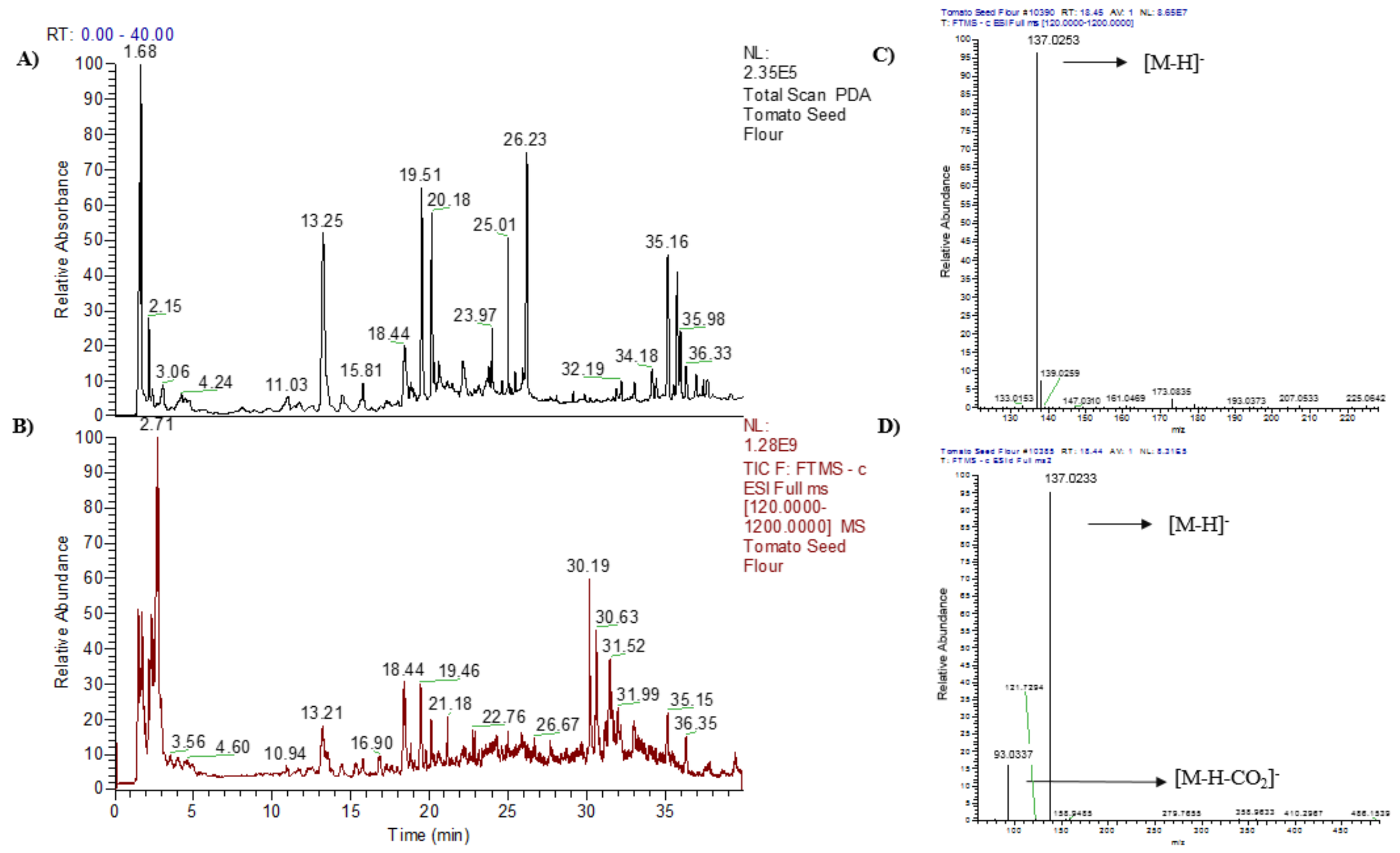
2.1. Chemical Composition of the Tomato Seed Flour Extracts

In both tomato seed flour extracts, named TSF1 and TSF2, a total of eight compounds, namely malic acid, 2-hydroxyadipic acid, salicylic acid, naringin, N-acetyl-tryptophan, quercetin-di-O-hexoside, kaempferol-di-O-hexoside, and azelaic acid, were tentatively identified (Table 1 and Figure 2).
The tentative identification of the eight peaks were based on the theoretical, experimental molecular ions ([M−H]) and the major MS/MS fragment ions, along with the MS data in the literatures. For example, peak 3 had a m/z [M–H] of 137.0233, which refers to the formula, C7H6O3 (mass error, −0.72 mmμ) (Figure 2C). The MS/MS spectrum showed a m/z of 137.0233, which was a parent ion and the fragmental ion had a m/z 93.0337, which was resulted from the elimination of a carboxyl group from the molecular ion (Figure 2D). This fragmentation matched with that previously reported for salicylic acid by Chhonker and Pastor’s groups [6,7]. Taking together, peak 3 was tentatively identified as salicylic acid. Similarly, peak 1 of the tomato seed flour extracts showed a m/z [M − H] of 133.0135, which corresponds to the formula of C4H6O5 (mass error, −0.75 mmμ) (Table 1). The peak 1 had the fragment ion at m/z of 71.0131 (Table 1). These m/z values were in line with the previously reported malic acid m/z values proposed by Kadhi and others [8], and peak 1 was therefore tentatively identified as malic acid. The same approach was applied to identify the other compounds found in tomato seed flour extracts. As a result, 2-hydroxyadipic acid [9], naringin [10,11], N-acetyl-tryptophan [12,13,14], quercetin-di-O-hexoside [15,16], kaempferol-di-O-hexoside [16], and azelaic acid [17,18] were tentatively identified.
Among the eight identified compounds, four compounds, including salicylic acid, naringin, quercetin-di-O-hexoside, and kaempferol-di-O-hexoside, were polyphenolic compounds (Figure S1). Polyphenolic compounds are known to possess potential health beneficial properties, such as reducing the risk of arthritis [19], cancers [20], diabetes [21], obesity [22], and cardiovascular diseases [23].
Salicylic acid is a phenolic acid that is commonly found in plants. It is widely used in medicines treating skin redness. In addition, salicylic acid is also a metabolite of aspirin, acetylsalicylic acid, a pharmaceutical compound. It has been reported that salicylic acid is responsible for the anti-inflammatory acts of aspirin [24]. Naringin, quercetin-di-O-hexoside, and kaempferol-di-O-hexoside are polyphenol glycosides. In many cases, polyphenols that are found in plants exist as glycosides [25]. In polyphenol glycosides, the sugar moiety is often hydrolyzed by the digestive enzymes or gut microflora to release the polyphenol aglycone(s) [26]. Therefore, it is important to understand and evaluate both intact and hydrolyzed forms of polyphenols for potential health beneficial properties. For example, naringin is a flavonoid and polyphenol glycoside found in fruits, vegetables, and herbs, and it may be hydrolyzed to naringenin, the aglycone of naringin in vivo. Previously, Cavia-Saiz and others evaluated the antioxidant properties of both naringin and naringenin [27]. They found that naringenin and naringin both showed antioxidant capacities, but, when compared to naringin, naringenin showed a greater antioxidant capacity. In addition, it has been reported that naringenin possessed cholesterol-lowering capacity [28]. Similarly, quercetin-di-O-hexoside and kaempferol-di-O-hexoside may both be hydrolyzed to their aglycones during digestion. Quercetin is a flavonoid and has a number of health beneficial properties, including free radical scavenging capacities, anti-tumor activities, and reducing the risk of cardiovascular and neurodegenerative diseases [29]. Kaempferol is also a flavonoid that is found in plant food sources, such as kale, spinach, and broccoli. Kaempferol has been a widely investigated polyphenolic compound because of its anti-cancer activity [30]. It has been reported that the anti-cancer activity of kaempferol is mainly by modulating cell signals that are related to the programmed cell death, new blood vessel formation, and the spread of cancer cells. These possible in vivo health beneficial properties of hydrolyzed polyphenols are important. However, it is also important to note that the glycoside forms of polyphenolics may contribute potential health beneficial properties, such as free radical scavenging capacities, anti-inflammatory capacities, and gut microbiota modulation. For instance, a previous study found that glycoside forms of kaempferols, including kaempferol-7-O-glucoside, kaempferol-3-O-rhamnoside, and kaempferol-3-O-rutinoside, showed anti-proliferative capacities, free radical scavenging capacities, and anti-inflammatory capacities [31]. Furthermore, other studies suggest that glycoside forms of kaempferol, quercetin, and naringin are subjects to possible hydrolysis by gut microbiota and they may contribute to gut health [32].
In addition, four non-phenolic compounds were found in the tomato seed flour extracts. These compounds are malic acid, 2-hydroxyadipic acid, N-acetyl-tryptophan, and azelaic acid (Table 1 and Figure S1). Malic acid is an organic acid that is widely found in fruits and vegetables and contributes sour taste. Besides, malic acid can act as a bioavailability enhancer of minerals such as iron [33]. Tryptophan is an essential amino acid and involved in human health conditions including kidney diseases, cardiovascular diseases, diabetes, depression, sleep, and social behavior [34]. In the current study, tryptophan was identified as an acetyl form. However, previous studies suggested that N-acetyl-tryptophan can also act as a tryptophan through the metabolism [35,36]. Azelaic acid is a carboxylic acid that is widely found in grains, such as rye, oat, barley, wheat and sorghum [37]. Azelaic acid has been reported to possess protective effects against oxidative stress in several organs [37]. Together, tomato seed flour has the potential for its utilization as a food additive or a functional food ingredient.

2.2. Total Phenolic Content and Free Radical Scavenging Capacities of Tomato Seed Flour Extracts

Total phenolic content (TPC) is often closely connected to the free radical scavenging capacity and other potential health beneficial properties. In the current study, two different batches of tomato seed flour extracts, TSF1 and TSF2, showed TPC values of 2.00 and 1.97 mg gallic acid equivalents/g of dry seed flour (mg GAE/g), respectively (Table 2). Previously, Toor and Savage evaluated the TPC of tomato seeds while using three different tomato cultivars, including Excell, Tradiro and Flavourine, and found TPC values of 0.35, 0.29, and 0.21 mg GAE/g fresh weight, respectively [38]. When compared to Toor and Savage’s TPC values, TSF1 and TSF2 showed greater TPC values under the experimental conditions. This may be due to the different samples and extraction methods. In the present study, 50% acetone was used to extract the tomato seed flour, where Toor and Savage used whole tomato seeds with 100% hexane and a combination of acetone, water, and acetic acid (70:29.5:0.5, v/v/v) as solvents for lipophilic and hydrophilic compounds, respectively [38]. In 2002, Kaur and Kapoor evaluated the TPC of tomato fruit and found a TPC value of 0.68 mg GAE/g [39]. TSF1 and TSF2 had greater TPC values when compared to tomato fruits (Table 2).
TSF1 and TSF2 showed oxygen radical absorbing capacities (ORAC), 2,2-diphenyl-1-picrylhydrazyl radical scavenging capacities (DPPH) and 2,2′-azino-bis(3-ethylbenzothiazoline-6-sulfonic acid) cation radical scavenging capacities (ABTS) of 88.57–86.32, 3.57–3.81, 3.39–3.58 µmoles Trolox equivalents/g of seed flour (µmoles TE/g), respectively (Table 2). ORAC values were much greater when compared to DPPH or ABTS. These differences may be due to the different structure of the radicals and reactivities between the radical and polyphenolic compounds found in tomato seed flour extracts. During the reaction of free radical scavenging by antioxidants, radicals are quenched by two mechanisms in most cases, namely hydrogen atom transferring (HAT) or single electron transferring (SET) [40]. The mechanisms of ABTS and DPPH assays are based on mixed HAT and SET; however, the ORAC’s mechanism is solely based on HAT. Accordingly, the antioxidant capacity might be different based on different assays; therefore, two or more assays are needed to evaluate a selected antioxidant compound.
Previously, Zhou and Yu evaluated the free radical scavenging capacities of ten tomatoes that were grown in Colorado and found ABTS values in the range of 5.4–20.9 µmoles TE/g dry weight [41]. They also reported TPC values of ten tomatoes. TPC values were in the range of 2.9–5.0 mg GAE/g [41]. When compared to the current study’s TPC and ABTS values, both of the values were greater for tomatoes grown in Colorado. These results suggest a possible correlation between the TPC and free radical scavenging capacities, which was also confirmed by the Pearson correlation analyses in this study. As a result, TPC showed a significantly positive correlation with ORAC (r = 0.975, p ≤ 0.01), DPPH (r = 0.991, p ≤ 0.01), and ABTS (r = 0.987, p ≤ 0.01) values, which suggested that total phenolics in tomato seed may directly contribute to the radical scavenging capacities. In 2018, Perea-Domı’nguez and others tested free radical scavenging capacities of two tomato varieties, Grape and Saladette, and found ORAC and DPPH values of 1631.39–2426.53, 307.8–465.17 µmoles TE/g dry weight, respectively [42]. Overall, tomato seed flours had lower free radical scavenging capacities on a per dry weight basis as compared to tomato fruits.

2.3. Anti-Inflammatory Capacities of Tomato Seed Flour Extracts

TSF1 and TSF2 both demonstrated dose-dependent anti-inflammatory capacities against pro-inflammatory markers, including interleukin-1 beta (IL-1β), interleukin-6 (IL-6), and tumor necrosis factor-alpha (TNF-α) (Figure 3). The treatments with different concentrations of tomato seed flour extracts at 0.2, 0.5, 1.0, and 2.0 mg tomato seed flour (TSF)/mL were capable of inhibiting the lipopolysaccharide (LPS) stimulated IL-1β mRNA expression by 69, 97, 99, 99, 69, 97, 99, and 99%, respectively (Figure 3). Similarly, TSF1 and TSF2 dose dependently inhibited IL-6 and TNF-α mRNA expressions under the experimental conditions (Figure 3). It needs to be pointed out that all of the tomato seed flour extracts with different concentrations used in this test had no adverse effects on THP-1 macrophages viability according to MTT assay (Figure S2).
Navarrete and others previously reported the anti-inflammatory capacities of tomato while using macrophages that were differentiated from THP-1 monocytes and LPS as a stimulant on the expression of proinflammatory cytokines [43]. The tomato extract was able to inhibit the gene expressions of the pro-inflammatory cytokines, including IL-1β and TNF-α. However, the anti-inflammatory capacities of tomato seed flour have not been evaluated in the past. Hence, this is the first report of a potential anti-inflammatory effect for tomato seed flours.
The inflammation process is often closely related to free radicals and chronic diseases. The former, free radicals are produced by a many different sources within the human body. Sources are often divided into two groups, endogenous and exogenous sources. Endogenous sources are mitochondria, peroxisomes, and phagocytes, while exogenous sources are cigarette smoking, air pollution, radiation, some medications, and ozone [44]. A primary cause of chronic diseases is oxidative stress, an imbalance between antioxidants, and free radicals. In the human body, there is a transcription factor, named nuclear factor erythroid-derived 2-like 2 (NRF2), which modulates the production of antioxidants and detoxifying products. These products can protect the damage that is caused by oxidative stress. However, this may not be enough to protect the body from oxidative stress, and the antioxidants from foods are important for overall antioxidative status in the human body.
When oxidative stress is induced, immune cells start to react. Among many immune cells, macrophages play a central role in inflammation and they start to secret pro-inflammatory cytokines and tumor necrosis factors, such as IL-1β, IL-6, and TNF-α. Bioactive compounds in foods can reduce or inhibit the development of these pro-inflammatory cytokines and tumor necrosis factor. For example, kaempferol can act as a nuclear factor kappa-light-chain-enhancer of activated B cells (NF-κB) inhibitor by binding its pathway signaling protein [45]. Similarly, a previous study reported that quercetin inhibited cytokine and inducible nitric oxide synthase expressions through inhibiting the NF-κB pathway [46]. Moreover, naringenin, the aglycone of naringin, has been reported to inhibit enzymes that are responsible for pro-inflammatory responses, such as cyclooxygenase-1 (COX-1) and cyclooxygenase-2 (COX-2) [47]. The result from the present study and the anti-inflammatory capacities of polyphenolic compounds reported by previous studies suggest that tomato seed flour has potential anti-inflammatory capacities, and it may be utilized in food additives or functional foods for improving human health.

2.4. Gut Microbiota Profile Modulation of Tomato Seed Flour Extracts

Research on gut microbiota has gained growing attention in recent years, owing to their notable functions in the human body. The gut microbiota profile has been shown to be correlated with host health conditions, starting from metabolic diseases to digestive diseases and colon cancer [48]. Maintaining a healthy gut microbiota profile is a key to maintaining a good state of health. However, each organism has a different gut microbiota profile and, in addition, along with the aging process, the gut microbiota profile changes. For example, a naturally delivered baby and a cesarean delivered baby may have different gut microbiota profiles. Additionally, each individual has a different metabolism rate. Therefore, the speed of the aging process may be distinct. These intrinsic factors are not changeable. On the other hand, there are extrinsic factors that can shift the gut microbiota profile. These factors include exercise, stress, antibiotics, and diet. Among these extrinsic factors, the diet seems to have the biggest impact, since food is what human beings eat every day.
In the current study, a total of eight bacterial taxonomic ranks, including Bacteroidetes phylum, Firmicutes phylum, Akkermansia genus, Bifidobacterium genus, Lactobacillus genus, Enterobacteriaceae genus, Prevotella genus, and Ruminococcus genus, were used to evaluate the gut microbiota profile change by tomato seed flour extracts. Five phyla and genera showed significant changes (p ≤ 0.05) (Figure 4). Among the five significantly changed phylum or genus, the abundance of Akkermansia and Ruminococcus genera were increased (Figure 4C,H) and Firmicutes phylum, Bifidobacterium genus, and Enterobacteriaceae genus were decreased (Figure 4B,D,F). Even though one batch (TSF2) significantly increased Bacteroidetes phylum, the other batch (TSF1) did not show any significant effect (Figure 4A). Additionally, the Lactobacillus genus was significantly increased by one batch (TSF1), but not by TSF2 (Figure 4E). Together, these results suggest a possible variation between tomato seed samples in their microbiota modulating properties. In vivo study is needed to further confirm the effects of tomato seed flour on Bacteroidetes phylum and Lactobacillus genus.
Previously, gut microbiota profile modulation by other vegetable seed flours, including broccoli, carrot, and cucumber, has been reported by our group [49]. In that study, the abundance of Bacteroidetes phylum was significantly increased by cucumber seed flour extract and decreased by carrot seed flour extract and not changed by broccoli seed flour extract. In the current study, one batch of tomato seed flour extract (TSF2) significantly increased Bacteroidetes phylum, but the fold was much lower than that of cucumber seed flour extract. Furthermore, both batches of tomato seed flour extracts decreased the abundance of Firmicutes phylum (Figure 4B). This Firmicutes phylum decrease has also been observed in the previous study. In the previous study, broccoli, carrot, and cucumber seed flour extracts significantly decreased the abundance of Firmicutes [49]. Additionally, a similar trend was observed in the Enterobacteriaceae genus. This study and the previous study both observed significant decreases of the Enterobacteriaceae genus (Figure 4F). For probiotic bacteria, Bifidobacterium and Lactobacillus genera, only the Bifidobacterium genus was decreased by TSF1 and TSF2 (Figure 4D,E). This was different to the observations in the previous study that all three vegetable seed flour extracts decreased both probiotic bacteria, Bifidobacterium and Lactobacillus genera [49].
In gut microbiota, Bacteroidetes and Firmicutes phyla consist of more than 90% population and they play important roles. Bacteroidetes can activate lymphocyte T cell to modulate human immune responses and produce butyric acid [50]. In addition, Bacteroidetes are participating in the degradation of toxin and carcinogen and bile acid metabolism [51]. Firmicutes are closely related to the aging process and they are possibly involved in fatty acid metabolism [52]. Bifidobacterium and Lactobacillus genera are probiotics and known to reduce infectious diarrhea and pathogen colonization [53]. Akkermasia are a mucin degrading bacterium and maintain gut health [54], and they could reduce body fat mass and adipose tissue inflammation and improve glucose homeostasis [55]. Prevotella can be used as a biomarker for gut dysbiosis, since the abundance of Prevotella genus is associated with diets that are rich in plants and their components, such as carbohydrates and fibers [56]. Ruminococcus possess the abilities to breakdown and use a wide range of plant polysaccharides for host health [57]. Because the tomato seed flour extracts significantly increased both Akkermansia and Ruminococcus genera, tomato seed flour may be used as a functional food for weight control and improving digestion.

3. Materials and Methods

3.1. Chemicals and Reagents

The tomato seed flours were donated by the Botanic Innovations (Spooner, WI, USA). PMA (phorbol 12-myristate 13-acetate) (product#: P1585), Trolox (6-Hydroxy-2,5,7,8-tetramethylchroman-2-carboxylic acid) (product#: 238813), gallic acid (product#: G7384), Folin-ciocalteu reagent (2N) (product#: F9252), and sodium carbonate (product#: 223530) were acquired from Sigma Aldrich (Saint-Louis, MO, USA). AAPH (2,2′-Azinobis (2-amidinopropane) dihydrochloride) (catalog#: 992–11062) was acquired from Wako Chemicals (Richmond, VA, USA). The materials for the cell culture were purchased from GIBCO (Grand Island, NY, USA). Materials for PCR (polymerase chain reaction) were acquired from either Thermo Fisher Scientific (Fair Lawn, NJ, USA) or Qiagen (Gaithersburg, MD, USA).

3.2. Tomato Seed Flour Extract Preparation

Extraction was performed following the previously reported method with a slight modification [58]. Briefly, 10 g of tomato seed flour was mixed with 50 mL of 50% acetone and then vortexed for 1 min. Subsequently, the mixture was sonicated for 1 min and rested for 24 h. This extract was used to assess free radical scavenging capacities, anti-inflammatory capacities, and gut microbiota profile modulation. For chemical composition analysis, 10 g of tomato seed flour was extracted using a Soxhlet extractor with 50 mL of 100% ethanol.

3.3. Ultra-High-Performance Liquid Chromatography-High Resolution Mass Spectrometry (UHPLC-HRMS) Analysis

The UHPLC-HRMS (Themo Scientific, Waltham, MA, USA) system consists of an Orbitrap ID-X tribrid mass spectrometer with a Vanquish UHPLC, including a high-pressure binary pump, thermostatting column temperature control compartment, and an HL Diode Array Detector [59]. The separation was carried out on an Agilent RRHD Eclipseplus C18 2.1 × 150 mm 1.8 µm (Agilent, Palo Alto, CA, USA) with an UltraShield pre-column filter (Analytical Scientific Instruments, Richmond, CA, USA) with a flow rate of 0.3 mL·min−1. Solution A (0.1% formic acid in water, v/v) and solution B (0.1% formic acid in acetonitrile, v/v) were used for gradient elution with the following program. The proportion kept at 2% B (v/v) at 0–5 min, and then increased to 10% B at 15 min., to 45% B at 25 min, to 90% at 35 min, and this proportion was kept at 90% to 40 min. The post-run time for re-equilibration was 10 min. The UV-vis spectra were recorded at the range of 190–600 nm for the entire run. The column temperature was set at 50 °C and the sample compartment temperature was set at 4 °C. The injection volume was 2 µL. The MS conditions were set, as follows: sheath gas at 50 (arbitrary units), auxiliary gas at 10 (arbitrary units), and sweep gas at 1 (arbitrary units), spray voltage at 3 kV with negative ionization mode, ion transfer tube temperature at 300 °C, vaporizer temperature at 350 °C, and RF lens at 60%. The full scan mass range was from 120 to 1200 m/z with a resolution of 60,000, AGC target value of 200,000 in full scan and 10,000 FTMS/MS, and max ion injection time of 50 ms. The most intense ion was selected for the data-dependent scan with normalization collision energy at 35% in HCD. The data were post-processed using the Xcalibur 2.2 software.

3.4. Total Phenolic Content

The total phenolic content was evaluated following the laboratory procedure previously reported with a minor modification [58]. Briefly, 3 mL of deionized water, 50 μL of sample, standard, or solvent (blank), and 250 μL of the diluted Folin–Ciocalteu reagent (0.5 N) were added to the test tube and then vortexed for 1 min. After vortexing, 750 μL of 20% (w/v) sodium carbonate was added to trigger the reaction. After 2 h, a wavelength of 765 nm was used to measure the absorbance. The result was stated as milligrams of gallic acid equivalents per gram of the tomato seed flour (mg GAE/g).

3.5. Relative 2,2-Diphenyl-1-picrylhydrazyl (DPPH) Radical Scavenging Capacity

The relative DPPH radical scavenging capacity was examined using a previously reported protocol [60]. Using a wavelength of 515 nm, the absorbance was checked every minute and then recorded for 40 min. The Trolox was used to provide a standard curve. For the value unit, micromoles of Trolox equivalents/g of flour (μmoles TE/g) were used.

3.6. Oxygen Radical Absorbing Capacity (ORAC)

According to the previously reported protocol, the oxygen radical absorbing capacity (ORAC) was measured [61]. Different concentrations of the Trolox were dissolved in 50% acetone to generate a standard curve. All other reagents were prepared using 75 mM pH 7.4 phosphate buffer. For the detection, wavelengths of 485 and 535 nm were used for the excitation and emission, respectively. The ORAC value was reported as μmoles TE/g of the flour samples.

3.7. 2,2′-azinobis (3-ethylbenzothiazoline-6-sulphonic Acid) Diammonium Salt Cation Radical (ABTS•+) Scavenging Capacity

The ABTS cation radical scavenging capacity was evaluated using a previously reported protocol [62]. Briefly, ABTS•+ working solution was prepared by oxidizing ABTS with manganese oxide and the absorbance was adjusted to 0.700 ± 0.005 at 734 nm. Trolox was used as the antioxidant standard. For the reaction, 1 mL of ABTS•+ working solution was mixed with 80 μL of the sample, standard, or solvent. This mixture was vortexed for 30 s. After 90 s, the absorbance value was recorded at 734 nm. The ABTS value was reported as μmoles TE/g of the flour samples.

3.8. Anti-Inflammatory Capacity

THP-1 macrophages were used to assess anti-inflammatory capacity. In short, density of 6 × 10⁵ cells/mL THP-1 macrophages were cultured in six-well plates to achieve 80% confluence. Subsequently, macrophages were cultured with/without the tomato seed flour extracts at concentrations of 0.2, 0.5, 1.0, and 2.0 mg tomato seed flour/mL (mg TSF/mL) for 24 h. For the stimulation, 10 ng/mL of lipopolysaccharide (LPS) was used. After four hours of stimulation with LPS, the cells were lysed for RNA isolation. From RNA, cDNA was synthesized and Real-time PCR was performed using TaqMan probe. TATA box binding protein (TBP) was used as a control primer and IL-1β, IL-6, and TNF-α as inflammatory markers.

3.9. Gut Microbiota Analysis

A regular chow diet-fed C57BL/6J mouse’s fecal sample was used to prepare gut microbiota, as previously reported [59]. For the bacterial concentration calculation, an OD600 value of 1 = 8 × 10⁸ cells/mL was used. Briefly, 1 × 10⁷ cells/mL of bacterial cells were cultured in M9 broth with/without tomato seed flour extract in 15 mL and 50 mL tubes for 6 h with shaking. After 6 h, the bacterial cells were collected by centrifugation at 5000 rpm for 5 min. The bacterial DNA were extracted using Precellys lysing and QIAamp DNA mini kits. Specific forward and reverse primer sequences used in this study were shown, as follows: Akkermansia (Forward: 5′-CAGCACGTGAAGGTGGGGAC-3′, Reverse: 5′-CCTTGCGGTTGGCTTCAGAT-3′); Bacteroidetes (Forward: 5′-GGARCATGTGGTTTAATTCGATGAT-3′, Reverse: 5′-AGCTGACGACAACCATGCAG-3′); Bifidobacterium (Forward: 5′-TCGCGTCYGGTGTGAAAG-3′, Reverse: 5′-CCACATCCAGCRTCCAC-3′); Enterobacteriaceae (Forward: 5′-CATTGACGTTACCCGCAGAAGAAGC-3′, Reverse: 5′-CTCTACGAGACTCAAGCTTGC-3′); Firmicutes (Forward: 5′-GGAGYATGTGGTTTAATTCGAAGCA-3′, Reverse: 5′-AGCTGACGACAACCATGCAC-3′); Lactobacillus (Forward: 5′-GAGGCAGCAGTAGGGAATCTTC-3′, Reverse: 5′-GGCCAGTTACTACCTCTATCCTTCTTC-3′); Prevotella (Forward: 5′-TCCTAGGGAGGCAGCAGT-3′, Reverse: 5′-CAATCGGAGTTCTTCGTG-3′); and Ruminococcus (Forward: 5′-GGCGGCCTACTGGGCTTT-3′, Reverse: 5′-CCAGGTGGATAACTTATTGTGTTAA-3′). Real-time PCR was carried with the SYBR probe using a previously reported protocol [59].

4. Conclusions

The current study observed several potential health beneficial polyphenolic compounds in tomato seed flour extracts, along with several possible health beneficial properties, including free radical scavenging capacities, anti-inflammatory capacities, and gut microbiota profile modulation. The results might be used to improve human health by increasing the utilization of tomato seed flour as a food additive or functional food ingredient.

5. Patents

The current tomato seed flour research has been filed as a Method for Modulating Gut Microbiota (Provisional US Patent application, 63/007734 with a University of Maryland invention disclosure number LS-2018-193).

Supplementary Materials

The following are available online. Figure S1: Molecular structures of the compounds identified in tomato seed flour extracts, Figure S2: MTT assays of THP-1 macrophage cells using different concentrations of tomato seed flour extracts.

Author Contributions

Conceptualization, L.Y., Y.L.; methodology, J.R., P.C., T.T.Y.W., L.Y.; software, validation, formal analysis, B.G., E.B., J.S., U.C., Y.L.; writing—original draft preparation, U.C.; writing—review and editing and supervision, L.Y., Y.L.; Funding acquisition, Y.L.; All authors have read and agreed to the published version of the manuscript.

Funding

This research was partially funded by The National Key Research and Development Program of China (Grant No. 2018YFC1602400); and National Natural Science Foundation of China (Grant No. 32001819).

Institutional Review Board Statement

Not applicable.

Informed Consent Statement

Not applicable.

Data Availability Statement

The data presented in this study are available in the article.

Acknowledgments

The authors are thankful to Botanic Innovations for providing tomato seed flours.

Conflicts of Interest

The authors declare no conflict of interest.

References

  1. Food Consumption and Nutrient Intakes. Available online: http://www.ers.usda.gov/data-products/food-consumption-and-nutrient-intakes.aspx (accessed on 6 January 2021).
  2. The World’s TOP40 Biggest Processing Companies. Available online: http://www.tomatonews.com/en/the-worlds-top40-biggest-processing-companies_2_355.html (accessed on 6 January 2021).
  3. Zuorro, A.; Lavecchia, R.; Medici, F.; Piga, L. Enzyme-Assisted Production of Tomato Seed Oil Enriched with Lycopene from Tomato Pomace. Food Bioprocess Technol. 2013, 6, 3499–3509. [Google Scholar] [CrossRef]
  4. Elbadrawy, E.; Sello, A. Evaluation of Nutritional Value and Antioxidant Activity of Tomato Peel Extracts. Arab. J. Chem. 2016, 9, S1010–S1018. [Google Scholar] [CrossRef] [Green Version]
  5. Shao, D.; Bartley, G.E.; Yokoyama, W.; Pan, Z.; Zhang, H.; Zhang, A. Plasma and Hepatic Cholesterol-Lowering Effects of Tomato Pomace, Tomato Seed Oil and Defatted Tomato Seed in Hamsters Fed with High-Fat Diets. Food Chem. 2013, 139, 589–596. [Google Scholar] [CrossRef]
  6. Chhonker, Y.S.; Pandey, C.P.; Chandasana, H.; Laxman, T.S.; Prasad, Y.D.; Narain, V.S.; Dikshit, M.; Bhatta, R.S. Simultaneous Quantitation of Acetylsalicylic Acid and Clopidogrel along with Their Metabolites in Human Plasma Using Liquid Chromatography Tandem Mass Spectrometry. Biomed. Chromatogr. 2016, 30, 466–473. [Google Scholar] [CrossRef]
  7. Vial, T.; Tan, W.-L.; Xiang, B.W.W.; Missé, D.; Deharo, E.; Marti, G.; Pompon, J. Dengue Virus Reduces AGPAT1 Expression to Alter Phospholipids and Enhance Infection in Aedes Aegypti. PLoS Pathog. 2019, 15, e1008199. [Google Scholar] [CrossRef] [PubMed] [Green Version]
  8. Pastor, V.; Vicent, C.; Cerezo, M.; Mauch-Mani, B.; Dean, J.; Flors, V. Detection, Characterization and Quantification of Salicylic Acid Conjugates in Plant Extracts by ESI Tandem Mass Spectrometric Techniques. Plant Physiol. Biochem. 2012, 53, 19–26. [Google Scholar] [CrossRef]
  9. Al Kadhi, O.K.; Melchini, A.; Mithen, R.; Saha, S. Development of a LC-MS/MS Method for the Simultaneous Detection of Tricarboxylic Acid Cycle Intermediates in a Range of Biological Matrices. J. Anal. Methods Chem. 2017, 2017, 5391832. [Google Scholar] [CrossRef] [PubMed] [Green Version]
  10. Xiong, X.; Jiang, J.; Duan, J.; Xie, Y.; Wang, J.; Zhai, S. Development and Validation of a Sensitive Liquid Chromatography-Tandem Mass Spectrometry Method for the Determination of Naringin and Its Metabolite, Naringenin, in Human Plasma. J. Chromatogr. Sci. 2014, 52, 654–660. [Google Scholar] [CrossRef] [PubMed] [Green Version]
  11. Ibrahim, R.M.; El-Halawany, A.M.; Saleh, D.O.; Naggar, E.M.B.E.; El-Shabrawy, A.E.-R.O.; El-Hawary, S.S. HPLC-DAD-MS/MS Profiling of Phenolics from Securigera Securidaca Flowers and Its Anti-Hyperglycemic and Anti-Hyperlipidemic Activities. Rev. Bras. Farmacogn. 2015, 25, 134–141. [Google Scholar] [CrossRef] [Green Version]
  12. Zeiss, D.R.; Mhlongo, M.I.; Tugizimana, F.; Steenkamp, P.A.; Dubery, I.A. Comparative Metabolic Phenotyping of Tomato (Solanum lycopersicum) for the Identification of Metabolic Signatures in Cultivars Differing in Resistance to Ralstonia Solanacearum. Int. J. Mol. Sci. 2018, 19, 2558. [Google Scholar] [CrossRef] [Green Version]
  13. Zeiss, D.R.; Mhlongo, M.I.; Tugizimana, F.; Steenkamp, P.A.; Dubery, I.A. Metabolomic Profiling of the Host Response of Tomato (Solanum Lycopersicum) Following Infection by Ralstonia Solanacearum. Int. J. Mol. Sci. 2019, 20, 3945. [Google Scholar] [CrossRef] [PubMed] [Green Version]
  14. van Meulebroek, L.; Bussche, J.V.; Steppe, K.; Vanhaecke, L. Ultra-High Performance Liquid Chromatography Coupled to High Resolution Orbitrap Mass Spectrometry for Metabolomic Profiling of the Endogenous Phytohormonal Status of the Tomato Plant. J. Chromatogr. A 2012, 1260, 67–80. [Google Scholar] [CrossRef] [PubMed]
  15. Hvattum, E.; Ekeberg, D. Study of the Collision-Induced Radical Cleavage of Flavonoid Glycosides Using Negative Electrospray Ionization Tandem Quadrupole Mass Spectrometry. J. Mass Spectrom. 2003, 38, 43–49. [Google Scholar] [CrossRef] [PubMed]
  16. Francescato, L.N.; Debenedetti, S.L.; Schwanz, T.G.; Bassani, V.L.; Henriques, A.T. Identification of Phenolic Compounds in Equisetum Giganteum by LC–ESI-MS/MS and a New Approach to Total Flavonoid Quantification. Talanta 2013, 105, 192–203. [Google Scholar] [CrossRef] [Green Version]
  17. Tamura, K.; Horikawa, M.; Sato, S.; Miyake, H.; Setou, M. Discovery of Lipid Biomarkers Correlated with Disease Progression in Clear Cell Renal Cell Carcinoma Using Desorption Electrospray Ionization Imaging Mass Spectrometry. Oncotarget 2019, 10, 1688–1703. [Google Scholar] [CrossRef] [PubMed] [Green Version]
  18. Calixto, N.O.; Cordeiro, M.S.; Giorno, T.B.S.; Oliveira, G.G.; Lopes, N.P.; Fernandes, P.D.; Pinto, A.C.; Rezende, C.M. Chemical Constituents of Psychotria Nemorosa Gardner and Antinociceptive Activity. J. Braz. Chem. Soc. 2017, 28, 707–723. [Google Scholar] [CrossRef]
  19. Oliviero, F.; Scanu, A.; Zamudio-Cuevas, Y.; Punzi, L.; Spinella, P. Anti-Inflammatory Effects of Polyphenols in Arthritis. J. Sci. Food Agric. 2018, 98, 1653–1659. [Google Scholar] [CrossRef] [PubMed]
  20. Zhou, Y.; Zheng, J.; Li, Y.; Xu, D.-P.; Li, S.; Chen, Y.-M.; Li, H.-B. Natural Polyphenols for Prevention and Treatment of Cancer. Nutrients 2016, 8, 515. [Google Scholar] [CrossRef] [PubMed]
  21. Fenercioglu, A.K.; Saler, T.; Genc, E.; Sabuncu, H.; Altuntas, Y. The Effects of Polyphenol-Containing Antioxidants on Oxidative Stress and Lipid Peroxidation in Type 2 Diabetes Mellitus without Complications. J. Endocrinol. Investig. 2010, 33, 118–124. [Google Scholar] [CrossRef]
  22. Bose, M.; Lambert, J.D.; Ju, J.; Reuhl, K.R.; Shapses, S.A.; Yang, C.S. The Major Green Tea Polyphenol, (-)-Epigallocatechin-3-Gallate, Inhibits Obesity, Metabolic Syndrome, and Fatty Liver Disease in High-Fat-Fed Mice. J. Nutr. 2008, 138, 1677–1683. [Google Scholar] [CrossRef]
  23. Quiñones, M.; Miguel, M.; Aleixandre, A. Beneficial Effects of Polyphenols on Cardiovascular Disease. Pharmacol. Res. 2013, 68, 125–131. [Google Scholar] [CrossRef]
  24. Paterson, J.R.; Lawrence, J.R. Salicylic Acid: A Link between Aspirin, Diet and the Prevention of Colorectal Cancer. QJM Int. J. Med. 2001, 94, 445–448. [Google Scholar] [CrossRef]
  25. Tsao, R. Chemistry and Biochemistry of Dietary Polyphenols. Nutrients 2010, 2, 1231–1246. [Google Scholar] [CrossRef] [PubMed]
  26. Scalbert, A.; Williamson, G. Dietary Intake and Bioavailability of Polyphenols. J. Nutr. 2000, 130, 2073S–2085S. [Google Scholar] [CrossRef]
  27. Cavia-Saiz, M.; Busto, M.D.; Pilar-Izquierdo, M.C.; Ortega, N.; Perez-Mateos, M.; Muñiz, P. Antioxidant Properties, Radical Scavenging Activity and Biomolecule Protection Capacity of Flavonoid Naringenin and Its Glycoside Naringin: A Comparative Study. J. Sci. Food Agric. 2010, 90, 1238–1244. [Google Scholar] [CrossRef]
  28. Bugianesi, R.; Catasta, G.; Spigno, P.; D’Uva, A.; Maiani, G. Naringenin from Cooked Tomato Paste Is Bioavailable in Men. J. Nutr. 2002, 132, 3349–3352. [Google Scholar] [CrossRef]
  29. Jan, A.T.; Kamli, M.R.; Murtaza, I.; Singh, J.B.; Ali, A.; Haq, Q.M.R. Dietary Flavonoid Quercetin and Associated Health Benefits—An Overview. Food Rev. Int. 2010, 26, 302–317. [Google Scholar] [CrossRef]
  30. Chen, A.Y.; Chen, Y.C. A Review of the Dietary Flavonoid, Kaempferol on Human Health and Cancer Chemoprevention. Food Chem. 2013, 138, 2099–2107. [Google Scholar] [CrossRef] [PubMed] [Green Version]
  31. Wang, J.; Fang, X.; Ge, L.; Cao, F.; Zhao, L.; Wang, Z.; Xiao, W. Antitumor, Antioxidant and Anti-Inflammatory Activities of Kaempferol and Its Corresponding Glycosides and the Enzymatic Preparation of Kaempferol. PLoS ONE 2018, 13, e0197563. [Google Scholar] [CrossRef] [PubMed]
  32. Scalbert, A.; Morand, C.; Manach, C.; Rémésy, C. Absorption and Metabolism of Polyphenols in the Gut and Impact on Health. Biomed. Pharmacother. 2002, 56, 276–282. [Google Scholar] [CrossRef]
  33. Suzuki, T.; Clydesdale, F.M.; Pandolf, T. Solubility of Iron in Model Systems Containing Organic Acids and Lignin. J. Food Prot. 1992, 55, 893–898. [Google Scholar] [CrossRef]
  34. Friedman, M. Analysis, Nutrition, and Health Benefits of Tryptophan. Int. J. Tryptophan Res. 2018, 11, 1178646918802282. [Google Scholar] [CrossRef] [PubMed] [Green Version]
  35. Fujita, Y.; Anzai, M.; Inui, M.; Nishimoto, T.; Inoue, G. Utilization of N-acetyl-L-tryptophan given intravenously to unrestrained adult rats. J. Nutr. Sci. Vitaminol. 1980, 26, 381–388. [Google Scholar] [CrossRef]
  36. Berg, C.P.; Rose, W.C.; Marvel, C.S. Tryptophane and growth: III. 3-indolepropionic acid and 3-indolepyruvic acid as supplementing agents in diets deficient in tryptophane. J. Biol. Chem. 1929, 85, 219–231. [Google Scholar] [CrossRef]
  37. Jones, D.A. Rosacea, Reactive Oxygen Species, and Azelaic Acid. J. Clin. Aesthet. Dermatol. 2009, 2, 26–30. [Google Scholar] [PubMed]
  38. Toor, R.K.; Savage, G.P. Antioxidant Activity in Different Fractions of Tomatoes. Food Res. Int. 2005, 38, 487–494. [Google Scholar] [CrossRef]
  39. Kaur, C.; Kapoor, H.C. Anti-Oxidant Activity and Total Phenolic Content of Some Asian Vegetables. Int. J. Food Sci. Technol. 2002, 37, 153–161. [Google Scholar] [CrossRef]
  40. Schaich, K.M.; Tian, X.; Xie, J. Hurdles and Pitfalls in Measuring Antioxidant Efficacy: A Critical Evaluation of ABTS, DPPH, and ORAC Assays. J. Funct. Foods 2015, 14, 111–125. [Google Scholar] [CrossRef]
  41. Zhou, K.; Yu, L. Total Phenolic Contents and Antioxidant Properties of Commonly Consumed Vegetables Grown in Colorado. LWT Food Sci. Technol. 2006, 39, 1155–1162. [Google Scholar] [CrossRef]
  42. Perea-Domínguez, X.P.; Hernández-Gastelum, L.Z.; Olivas-Olguin, H.R.; Espinosa-Alonso, L.G.; Valdez-Morales, M.; Medina-Godoy, S. Phenolic Composition of Tomato Varieties and an Industrial Tomato By-Product: Free, Conjugated and Bound Phenolics and Antioxidant Activity. J. Food Sci. Technol. 2018, 55, 3453–3461. [Google Scholar] [CrossRef]
  43. Navarrete, S.; Alarcón, M.; Palomo, I. Aqueous Extract of Tomato (Solanum lycopersicum L.) and Ferulic Acid Reduce the Expression of TNF-α and IL-1β in LPS-Activated Macrophages. Molecules 2015, 20, 15319–15329. [Google Scholar] [CrossRef] [Green Version]
  44. Phaniendra, A.; Jestadi, D.B.; Periyasamy, L. Free Radicals: Properties, Sources, Targets, and Their Implication in Various Diseases. Indian J. Clin. Biochem. 2015, 30, 11–26. [Google Scholar] [CrossRef] [PubMed] [Green Version]
  45. Kadioglu, O.; Nass, J.; Saeed, M.E.M.; Schuler, B.; Efferth, T. Kaempferol Is an Anti-Inflammatory Compound with Activity towards NF-ΚB Pathway Proteins. Anticancer Res. 2015, 35, 2645–2650. [Google Scholar] [PubMed]
  46. Comalada, M.; Camuesco, D.; Sierra, S.; Ballester, I.; Xaus, J.; Gálvez, J.; Zarzuelo, A. In Vivo Quercitrin Anti-Inflammatory Effect Involves Release of Quercetin, Which Inhibits Inflammation through down-Regulation of the NF-ΚB Pathway. Eur. J. Immunol. 2005, 35, 584–592. [Google Scholar] [CrossRef] [PubMed]
  47. Salehi, B.; Fokou, P.V.T.; Sharifi-Rad, M.; Zucca, P.; Pezzani, R.; Martins, N.; Sharifi-Rad, J. The Therapeutic Potential of Naringenin: A Review of Clinical Trials. Pharmaceuticals 2019, 12, 11. [Google Scholar] [CrossRef] [PubMed] [Green Version]
  48. Hills, R.D., Jr.; Pontefract, B.A.; Mishcon, H.R.; Black, C.A.; Sutton, S.C.; Theberge, C.R. Gut Microbiome: Profound Implications for Diet and Disease. Nutrients 2019, 11, 1613. [Google Scholar] [CrossRef] [Green Version]
  49. Choe, U.; Li, Y.; Gao, B.; Yu, L.; Wang, T.T.Y.; Sun, J.; Chen, P.; Liu, J.; Yu, L. Chemical Compositions of Cold-Pressed Broccoli, Carrot, and Cucumber Seed Flours and Their in Vitro Gut Microbiota Modulatory, Anti-Inflammatory, and Free Radical Scavenging Properties. J. Agric. Food Chem. 2018, 66, 9309–9317. [Google Scholar] [CrossRef]
  50. Kim, Y.S.; Milner, J.A. Dietary Modulation of Colon Cancer Risk. J. Nutr. 2007, 137, 2576S–2579S. [Google Scholar] [CrossRef] [PubMed] [Green Version]
  51. Smith, C.J.; Rocha, E.R.; Paster, B.J. The Medically Important Bacteroides spp. in Health and Disease. In The Prokaryotes: Volume 7: Proteobacteria: Delta, Epsilon Subclass; Dworkin, M., Falkow, S., Rosenberg, E., Schleifer, K.-H., Stackebrandt, E., Eds.; Springer: New York, NY, USA, 2006; pp. 381–427. ISBN 978-0-387-30747-3. [Google Scholar]
  52. Turnbaugh, P.J.; Ley, R.E.; Mahowald, M.A.; Magrini, V.; Mardis, E.R.; Gordon, J.I. An Obesity-Associated Gut Microbiome with Increased Capacity for Energy Harvest. Nature 2006, 444, 1027–1031. [Google Scholar] [CrossRef] [PubMed]
  53. Picard, C.; Fioramonti, J.; Francois, A.; Robinson, T.; Neant, F.; Matuchansky, C. Review Article: Bifidobacteria as Probiotic Agents-Physiological Effects and Clinical Benefits. Aliment. Pharmacol. Ther. 2005, 22, 495–512. [Google Scholar] [CrossRef]
  54. Dao, M.C.; Everard, A.; Aron-Wisnewsky, J.; Sokolovska, N.; Prifti, E.; Verger, E.O.; Kayser, B.D.; Levenez, F.; Chilloux, J.; Hoyles, L.; et al. Akkermansia Muciniphila and Improved Metabolic Health during a Dietary Intervention in Obesity: Relationship with Gut Microbiome Richness and Ecology. Gut 2016, 65, 426. [Google Scholar] [CrossRef] [Green Version]
  55. Amar, J.; Burcelin, R.; Ruidavets, J.B.; Cani, P.D.; Fauvel, J.; Alessi, M.C.; Chamontin, B.; Ferriéres, J. Energy Intake Is Associated with Endotoxemia in Apparently Healthy Men. Am. J. Clin. Nutr. 2008, 87, 1219–1223. [Google Scholar] [CrossRef] [Green Version]
  56. Precup, G.; Vodnar, D.-C. Gut Prevotella as a Possible Biomarker of Diet and Its Eubiotic versus Dysbiotic Roles: A Comprehensive Literature Review. Br. J. Nutr. 2019, 122, 131–140. [Google Scholar] [CrossRef] [PubMed]
  57. la Reau, A.J.; Suen, G. The Ruminococci: Key Symbionts of the Gut Ecosystem. J. Microbiol. 2018, 56, 199–208. [Google Scholar] [CrossRef] [PubMed]
  58. Yu, L.; Zhou, K. Antioxidant Properties of Bran Extracts from ‘Platte’ Wheat Grown at Different Locations. Food Chem. 2005, 90, 311–316. [Google Scholar] [CrossRef]
  59. Choe, U.; Li, Y.; Gao, B.; Yu, L.; Wang, T.T.Y.; Sun, J.; Chen, P.; Yu, L. The Chemical Composition of a Cold-Pressed Milk Thistle Seed Flour Extract, and Its Potential Health Beneficial Properties. Food Funct. 2019, 10, 2461–2470. [Google Scholar] [CrossRef]
  60. Cheng, Z.; Moore, J.; Yu, L. High-Throughput Relative DPPH Radical Scavenging Capacity Assay. J. Agric. Food Chem. 2006, 54, 7429–7436. [Google Scholar] [CrossRef]
  61. Huang, D.; Ou, B.; Hampsch-Woodill, M.; Flanagan, J.A.; Prior, R.L. High-Throughput Assay of Oxygen Radical Absorbance Capacity (ORAC) Using a Multichannel Liquid Handling System Coupled with a Microplate Fluorescence Reader in 96-Well Format. J. Agric. Food Chem. 2002, 50, 4437–4444. [Google Scholar] [CrossRef] [PubMed]
  62. Miller, N.J.; Rice-Evans, C.A. Factors Influencing the Antioxidant Activity Determined by the ABTS•+ Radical Cation Assay. Free Radic. Res. 1997, 26, 195–199. [Google Scholar] [CrossRef]
Figure 1. Tomato purée and paste processing scheme. Tomato seeds can be further processed into tomato seed oil and flour.
Molecules 26 01478 g001
Figure 2. (A) Typical Ultra-High-Performance Liquid Chromatography-PDA (UHPLC-PDA) chromatogram and (B) total ion current (TIC) chromatogram of tomato seed flour extracts; (C) MS; and, (D) MS/MS spectra of peak 3 at the retention time of 18.44 min.
Molecules 26 01478 g002
Figure 3. Anti-inflammatory capacities of tomato seed flour extracts at concentrations of 0.2, 0.5, 1.0, and 2.0 mg tomato seed flour (TSF)/mL in THP-1 macrophages, (A,C,E): TSF1, the first batch of tomato seed flour extract and (B,D,F): TSF2, the second batch of tomato seed flour extract. Control, No-treatment; LPS, lipopolysaccharides; IL-1β, interleukin-1 beta; IL-6, interleukin-6; TNF-α, tumor necrosis factor alpha; Different letters demonstrate significant difference (p ≤ 0.05).
Molecules 26 01478 g003
Figure 4. Gut microbiota profile modulation of tomato seed flour extracts, (A) Bacteroidetes phylum, (B) Firmicutes phylum, (C) Akkermansia genus, (D) Bifidobacterium genus, (E) Lactobacillus genus, (F) Enterobacteriaceae genus, (G) Prevotella genus, and (H) Ruminococcus genus. Control, No-treatment; TSF1 and TSF2 represent the first and the second batches of tomato seed flour extracts, respectively; different letters demonstrate a significant difference (p ≤ 0.05).
Molecules 26 01478 g004
Table 1. The characterization of compounds present in tomato seed flour extracts.
IDRt (min)Exptl. [M–H]Fragment IonsFormulaMass Error (mmμ)Tentative IdentificationReference
11.68133.013571.0131C4H6O5−0.75Malic acid[6]
24.95161.0448323.097 ([2M–H])
143.0343 ([M–H2O])
C6H10O5−0.752-Hydroxyadipic acid[7]
318.44137.023793.0337 ([M–H–CO2])C7H6O3−0.72Salicylic acid[8,9]
418.81579.1708271.0595 ([narigenin–H])C27H32O14−1.13Naringin[10,11]
519.46245.0919491.1914 ([2M–H])C13H14N2O3−1.27N-Acetyl-tryptophan[12,13,14]
619.48625.1381300.0259 ([quercetin–H])
301.0337 ([quercetin–H])
C27H30O17−2.92Quercetin-di-O-hexoside[15,16]
720.14609.1426284.0310 ([kaempferol–H])
285.0389 ([kaempferol–H])
C27H30O16−3.51Kaempferol-di-O-hexoside[16]
821.17187.0991375.1929 ([2M–H])C9H16O41.52Azelaic acid[17,18]
Rt, retention time; Exptl. [M–H], experimental m/z of molecular ion.
Table 2. Total phenolic content and free radical scavenging capacities of tomato seed flour extracts.
SampleTPCORACDPPHABTS
mg GAE/gµmoles TE/gµmoles TE/gµmoles TE/g
TSF12.00 ± 0.1188.57 ± 2.423.57 ± 0.093.39 ± 0.08
TSF21.97 ± 0.3086.32 ± 7.013.81 ± 0.203.58 ± 0.61
TSF1, tomato seed flour extract from the first batch; TSF2, tomato seed flour extract from the second batch; TPC, total phenolic content; ORAC, oxygen radical absorbing capacity; DPPH, 2,2-diphenyl-1-picrylhydrazyl radical scavenging capacity; ABTS, 2,2′-azino-bis(3-ethylbenzothiazoline-6-sulfonic acid) cation radical scavenging capacity; GAE, gallic acid equivalents; TE, Trolox equivalents; all results are reported on a per tomato seed flour dry weight basis.
Publisher’s Note: MDPI stays neutral with regard to jurisdictional claims in published maps and institutional affiliations.

