
Synthesis, Neuroprotection, and Antioxidant Activity of 1,1′-Biphenylnitrones as α-Phenyl-N-tert-butylnitrone Analogues in In Vitro Ischemia Models

 ,
, 
Abstract
:1. Introduction
2. Results and Discussion
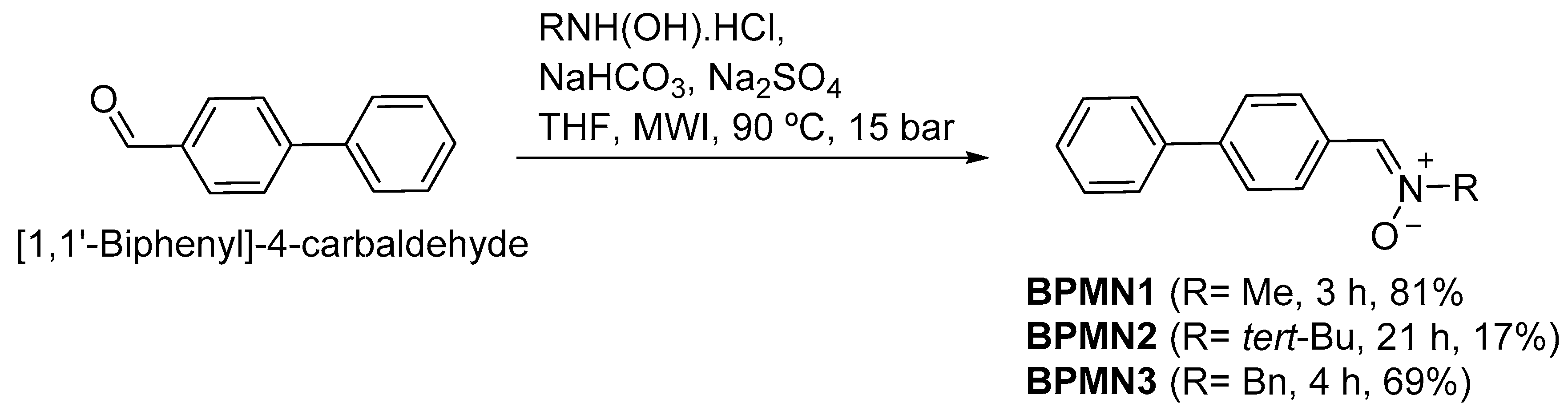
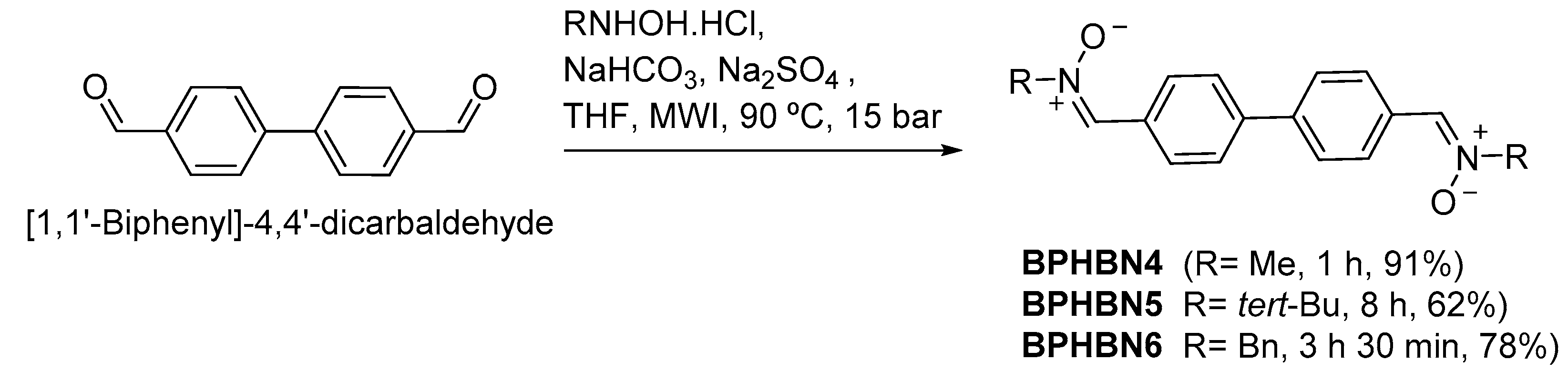
2.1. Chemistry
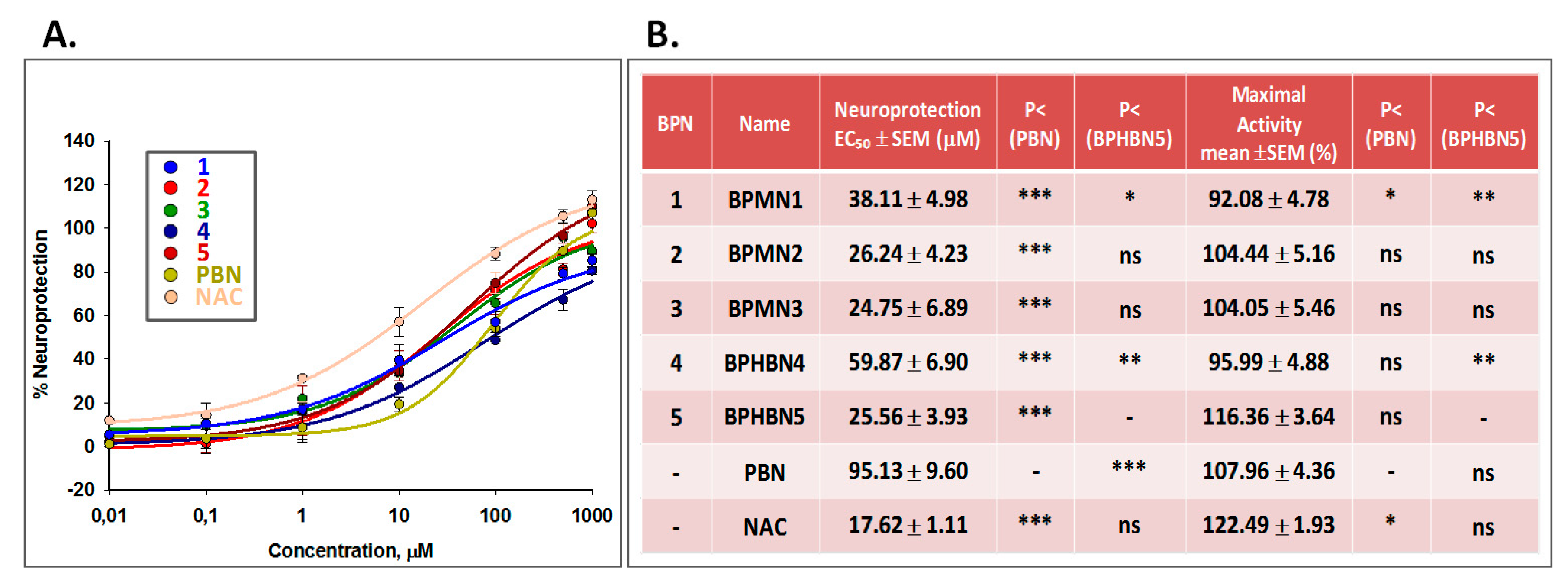
2.2. Neuroprotection Studies of BPNs 1–5
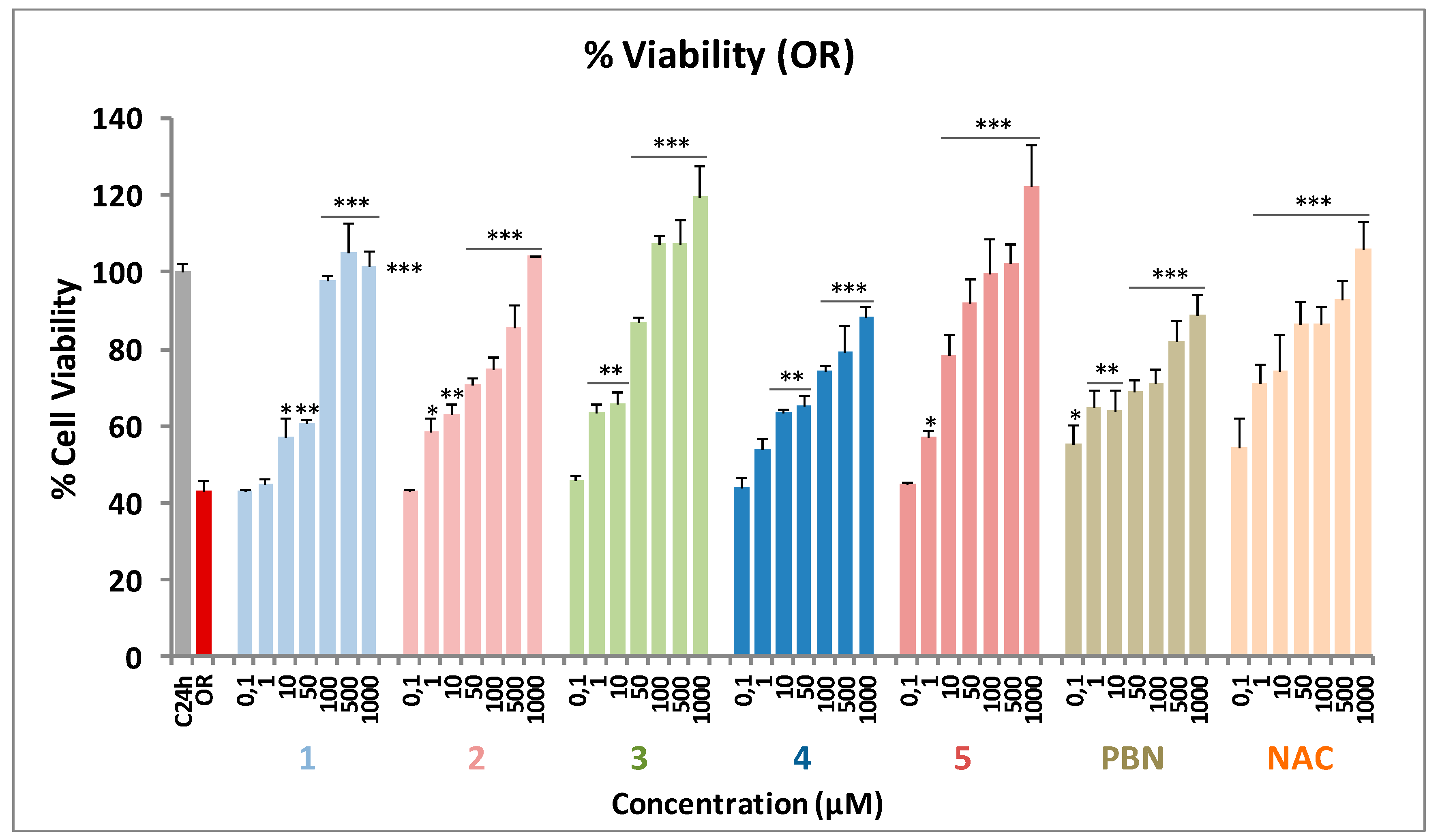
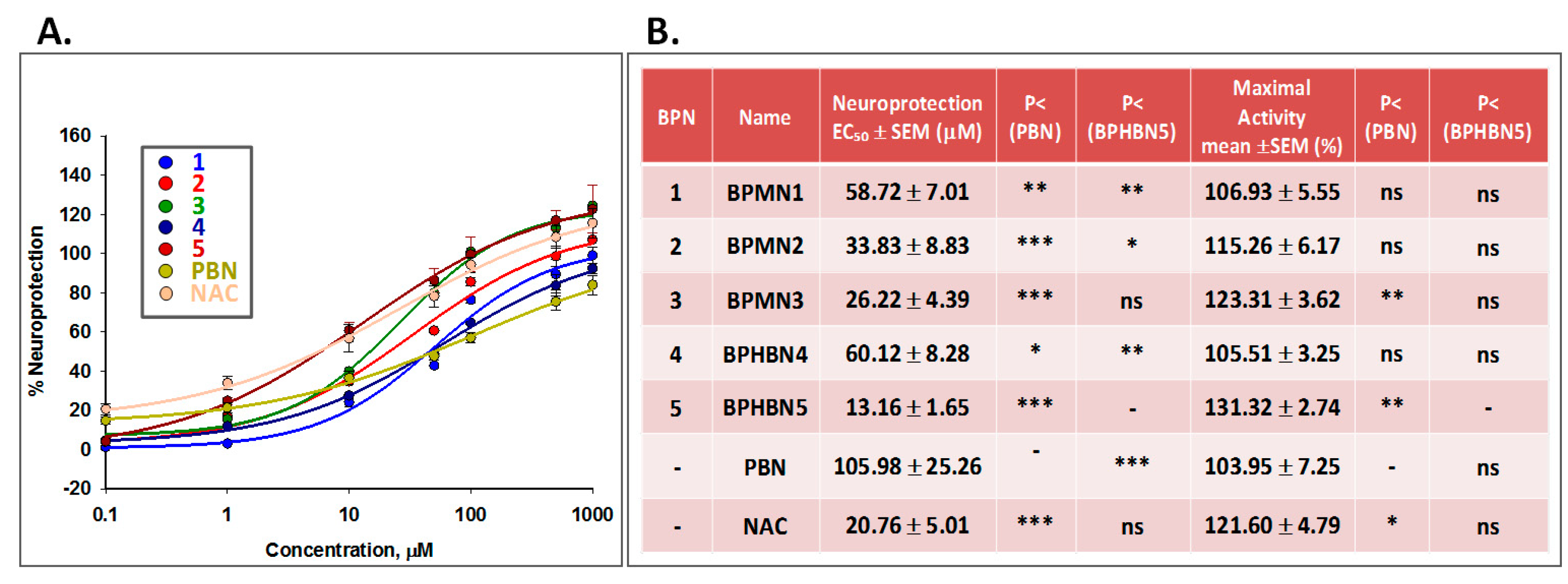
2.2.1. Neuroprotection Analysis in an Oligomycin A/Rotenone (OR) Model
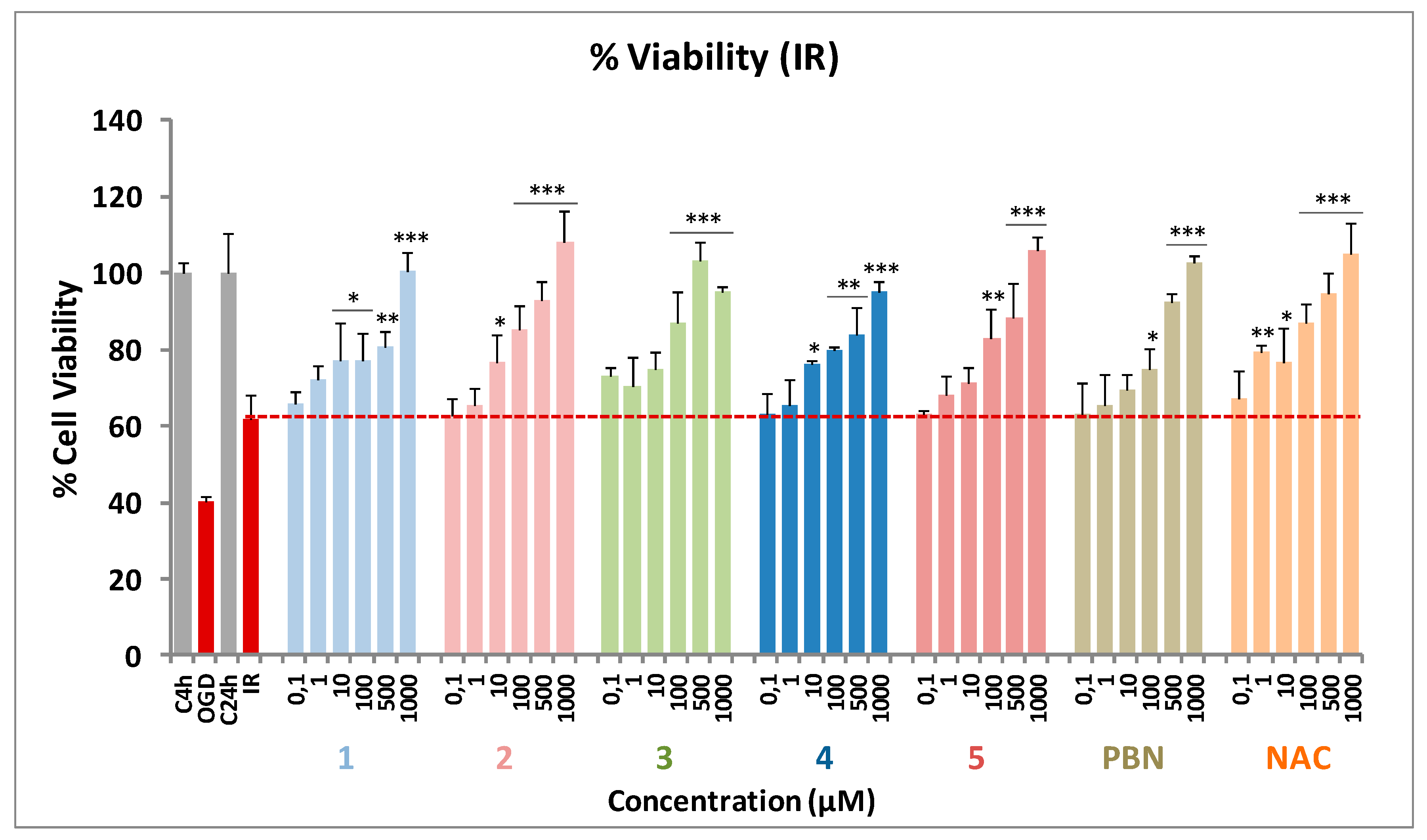
2.2.2. Neuroprotection Analysis in an Oxygen and Glucose Deprivation (OGD) followed by Oxygen and Glucose Resupply (IR) Model
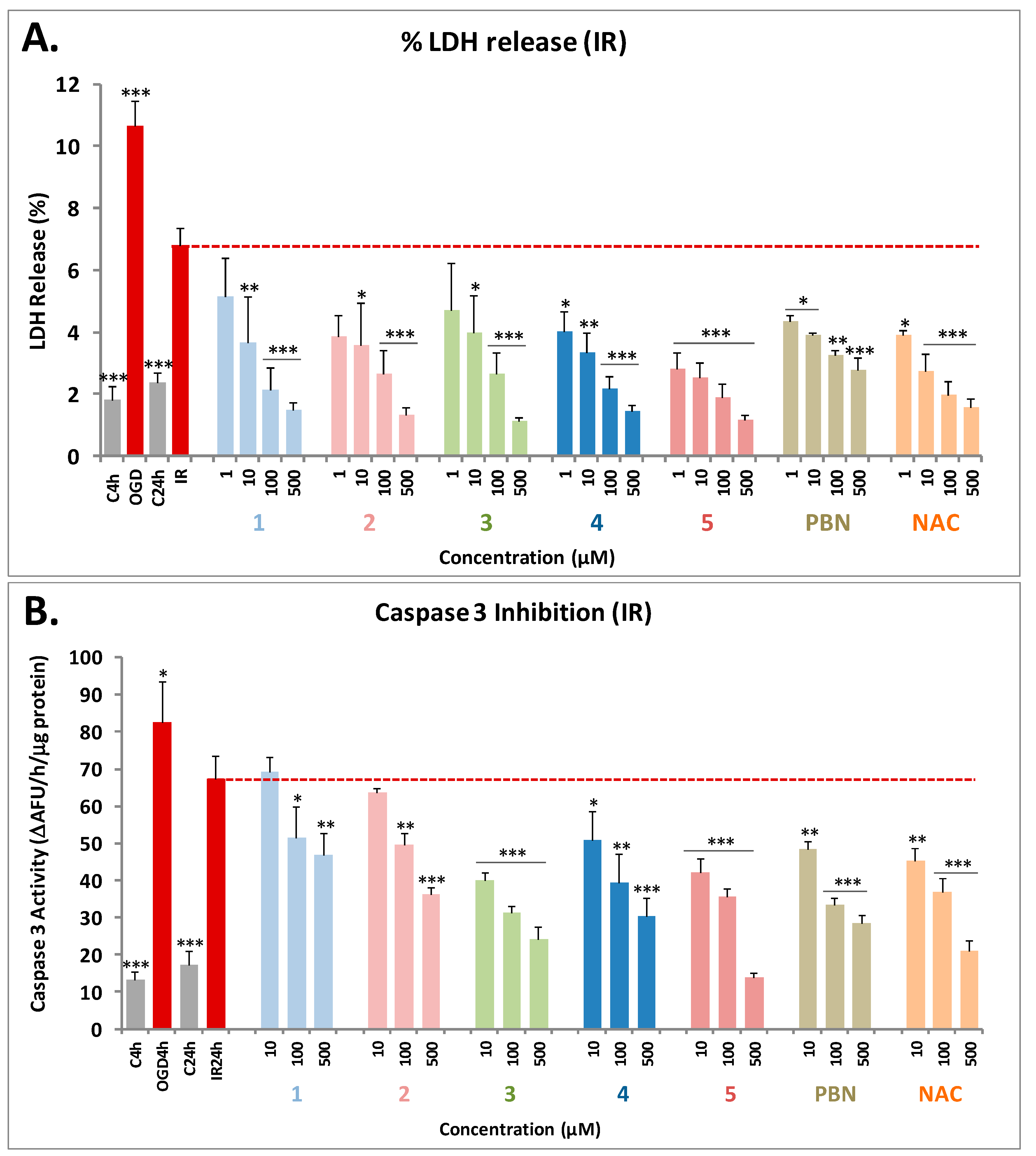
2.2.3. Effect of BPNs 1–5 on Necrotic and Apoptotic Cell Death Induced by Oxygen and Glucose Deprivation followed by Oxygen and Glucose Resupply (IR) Model
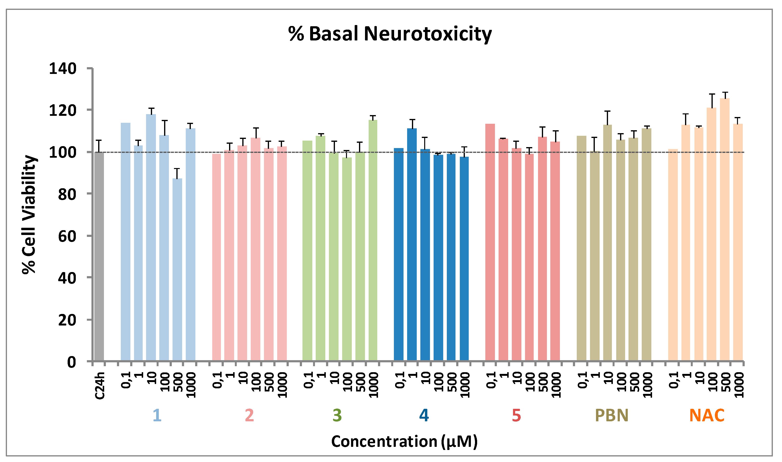
2.3. Basal Neurotoxicity of BPNs 1–5, PBN, and NAC
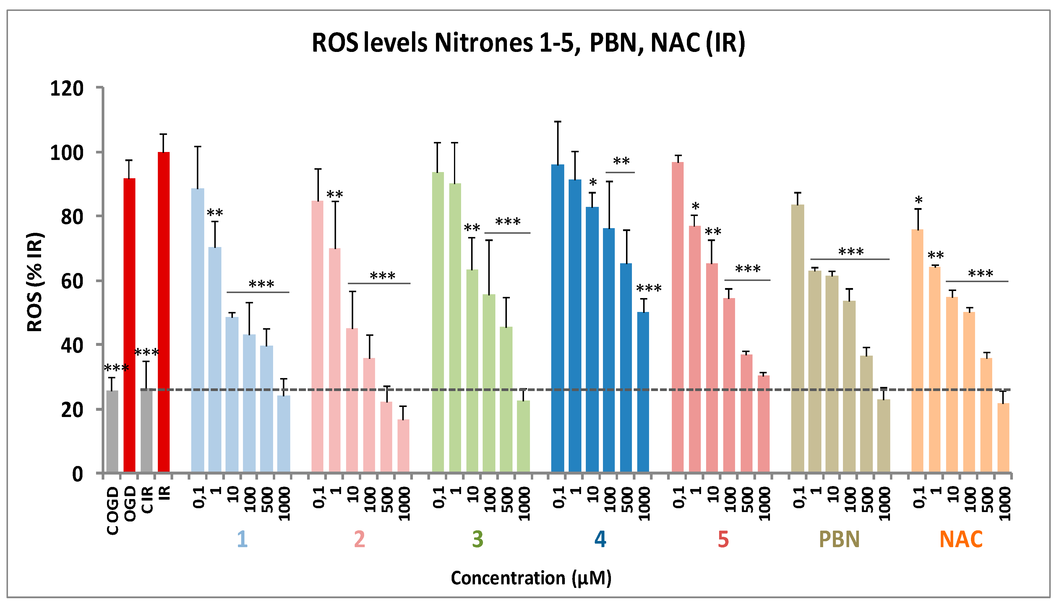
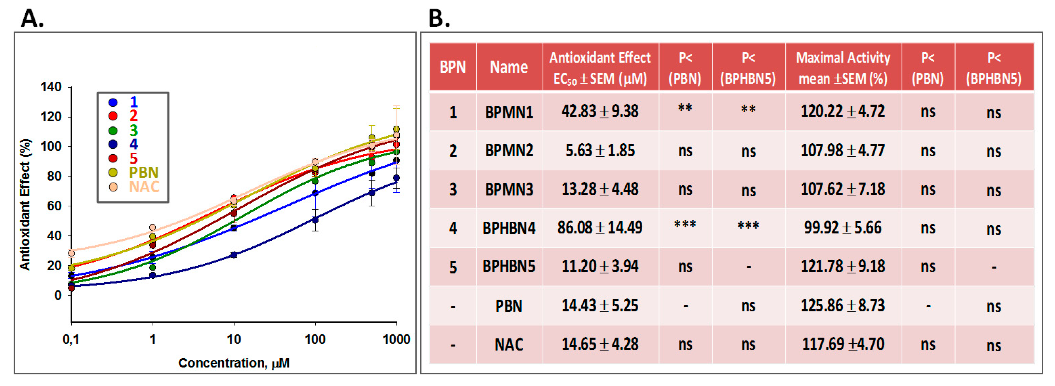
2.4. Antioxidant Capacity of BPNs 1–5, PBN, and NAC: Production and Scavenging of Superoxide Radical in Human Neuroblastoma SH‑SY5Y Cells
3. Materials and Methods
3.1. Chemistry

3.2. Neuroprotection Methods
3.3. Antioxidant Activity Tests of BPNs 1-5, PBN And Standards
4. Conclusions
Author Contributions
Funding
Institutional Review Board Statement
Informed Consent Statement
Data Availability Statement
Conflicts of Interest
Sample Availability
References
- Jenner, P. Oxidative stress in Parkinson’s disease. Ann. Neurol. 2003, 53, S26–S38. [Google Scholar] [CrossRef]
- Sayre, L.M.; Smith, M.A.; Perry, G. Chemistry and biochemistry of oxidative stress in neurodegenerative disease. Curr. Med. Chem. 2001, 8, 721–738. [Google Scholar] [CrossRef] [PubMed]
- Brouns, R.; De Deyn, P.P. The Complexity of Neurobiological Processes in Acute Ischemic Stroke. Clin. Neurol. Neurosurg. 2009, 111, 483–495. [Google Scholar] [CrossRef]
- Chan, P.H. Cellular Antioxidant Defense Mechanisms; Chow, C.K., Ed.; CRC Press: Boca Ratón, FL, USA, 1988; Volume 3, pp. 89–109. [Google Scholar]
- Janzen, E.G.; Blackburn, B.J. Detection and identification of short-lived free radicals by electron spin resonance trapping techniques (spin trapping). Photolysis of organolead, -tin, and -mercury compounds. J. Am. Chem. Soc. 1969, 91, 4481–4490. [Google Scholar] [CrossRef]
- Novelli, G.P.; Angiolini, P.; Tani, R.; Consales, G.; Bordi, L. Phenyl-t-butyl-nitrone is active against traumatic shock in rats. Free Radic. Res. Commun. 1986, 1, 321–327. [Google Scholar] [CrossRef] [PubMed]
- Diener, H.C.; Lees, K.R.; Lyden, P.; Grotta, J.; Dávalos, A.; Davis, S.M.; Shuaib, A.; Ashwood, T.; Wasiewski, W.; Alderfer, V.; et al. SAINT I and II Investigators, NXY-059 for the treatment of acute stroke: Pooled analysis of the SAINT I and II Trials. Stroke 2008, 39, 1751–1758. [Google Scholar] [CrossRef] [Green Version]
- Floyd, R.A.; Kopke, R.D.; Choi, C.H.; Foster, S.B.; Doblas, S.; Towner, R.A. Nitrones as therapeutics. Free Radic. Biol. Med. 2008, 45, 1361–1374. [Google Scholar] [CrossRef] [PubMed] [Green Version]
- Romero, A.; Ramos, E.; Patiño, P.; Oset-Gasque, M.J.; López-Muñoz, F.; Marco-Contelles, J.; Ayuso, M.I.; Alcázar, A. Melatonin and nitrones as potential therapeutic agents for stroke. Frontiers in Aging Neurosci. 2017, 9, 1. [Google Scholar] [CrossRef]
- Samadi, A.; Soriano, E.; Revuelta, J.; Valderas, C.; Chioua, M.; Garrido, I.; Bartolomé, B.; Tomassolli, I.; Ismaili, L.; González-Lafuente, L.; et al. Synthesis, structure, theoretical and experimental in vitro antioxidant/pharmacological properties of -aryl, N-alkyl nitrones, as potential agents for the treatment of cerebral ischemia. Bioorg. Med. Chem. 2011, 19, 951–960. [Google Scholar] [CrossRef] [PubMed]
- Arce, C.; Díaz-Castroverde, S.; Canales, M.J.; Marco-Contelles, J.; Samadi, A.; Oset-Gasque, M.J.; González, M.P. Drugs for stroke: Action of nitrone (Z)-N-(2-bromo-5-hydroxy-4-methoxybenzylidene)-2-methylpropan-2-amine oxide on rat cortical neurons in culture subjected to oxygen-glucose-deprivation. Eur. J. Med. Chem. 2012, 55, 475–479. [Google Scholar] [CrossRef]
- Chioua, M.; Sucunza, D.; Soriano, E.; Hadjipavlou-Litina, D.; Alcázar, A.; Ayuso, I.; Oset-Gasque, M.J.; González, M.P.; Monjas, L.; Rodríguez-Franco, M.I.; et al. Aryl-N-alkyl nitrones, as potential agents for stroke treatment: Synthesis, theoretical calculations, antioxidant, anti-inflammatory, neuroprotective and brain-blood barrier permeability properties. J. Med. Chem. 2012, 55, 153–168. [Google Scholar] [CrossRef] [Green Version]
- Ayuso, M.I.; Martínez-Alonso, E.; Chioua, M.; Escobar-Peso, A.; Gonzalo-Gobernado, R.; Montaner, J.; Marco-Contelles, J.; Alcázar, A. Quinolinylnitrone RP19 Induces Neuroprotection after Transient Brain Ischemia. ACS Chem. Neurosci. 2017, 8, 2202–2213. [Google Scholar] [CrossRef] [PubMed]
- Chioua, M.; Martinez-Alonso, E.; Gonzalo-Gobernado, R.; Ayuso, M.I.; Escobar-Peso, A.; Infantes, L.; Hadjipavlou-Litina, D.; Montoya, J.J.; Montaner, J.; Alcazar, A.; et al. New Quinolylnitrones for stroke therapy: Antioxidant and neuroprotective (z)-n-tert-butyl-1-(2-chloro-6-methoxyquinolin-3-yl) methanimine oxide as a new lead-compound for ischemic stroke treatment. J. Med. Chem. 2019, 62, 2184–2201. [Google Scholar] [CrossRef]
- Chioua, M.; Salgado-Ramos, M.; Diez-Iriepa, D.; Escobar-Peso, A.; Isabel Iriepa, I.; Hadjipavlou-Litina, D.; Martínez-Alonso, E.; Alcázar, A.; Marco-Contelles, J. Novel quinolylnitrones combining neuroprotective and antioxidant properties. ACS Chem. Neurosci. 2019, 10, 2703–2706. [Google Scholar] [CrossRef] [PubMed]
- Ayuso, M.I.; Chioua, M.; Martínez-Alonso, E.; Soriano, E.; Montaner, J.; Masjuán, J.; Hadjipavlou-Litina, D.; Marco-Contelles, J.; Alcázar, A. Cholesteronitrones for stroke. J. Med. Chem. 2015, 58, 6704–6709. [Google Scholar] [CrossRef] [PubMed] [Green Version]
- Jiménez-Almarza, A.; Diez-Iriepa, D.; Chioua, M.; Chamorro, B.; Iriepa, I.; Martínez-Murillo, R.; Hadjipavlou-Litina, D.; Oset-Gasque, M.J.; Marco-Contelles, J. Synthesis, neuroprotective and antioxidant capacity of pbn-related indanonitrones. Bioorg. Chem. 2019, 86, 445–451. [Google Scholar] [CrossRef]
- Klivenyi, P.; Matthews, R.T.; Wermer, M.; Yang, L.; MacGarvey, U.; Becker, D.A.; Natero, R.; Flint, B.M. Azulenyl nitrone spin traps protect against MPTP neurotoxicity. Experimental Neurol. 1998, 152, 163–166. [Google Scholar] [CrossRef]
- Becker, D.A.; Ley, J.J.; Echegoyen, L.; Alvarado, R. Stilbazulenyl nitrone (STAZN): A nitronyl-substituted hydrocarbon with the potency of classical phenolic chain-breaking antioxidants. J. Am. Chem. Soc. 2002, 124, 4678–4684. [Google Scholar] [CrossRef]
- Lapchak, P.A.; Schubert, D.R.; Maher, P.A. De-risking of stilbazulenyl nitrone (STAZN), a lipophilic nitrone to treat stroke using a unique panel of in vitro assays. Trans. Stroke Res. 2011, 2, 209–217. [Google Scholar] [CrossRef] [Green Version]
- Xu, D.-P.; Zhang, K.; Zhang, Z.-J.; Sun, Y.-W.; Guo, B.-J.; Wang, Y.-Q.; Hoi, P.-M.; Han, Y.-F.; Lee, S.M.-Y. A novel tetramethylpyrazine bis-nitrone (TN-2) protects against 6-hydroxyldopamine-induced neurotoxicity via modulation of the NF-κB and the PKCα/PI3-K/Akt pathways. Neurochem. Int. 2014, 78, 76–85. [Google Scholar] [CrossRef]
- Chamorro, B.; Diez-Iriepa, D.; Merás-Sáiz, B.; Chioua, M.; García-Vieira, D.; Iriepa, I.; Hadjipavlou-Litina, D.; López-Muñoz, F.; Martínez-Murillo, R.; González-Nieto, D.; et al. Synthesis, antioxidant properties and neuroprotection of α-phenyl-tert-butylnitrone derived homobisnitrones in in vitro and in vivo ischemia models. Sci. Rep. 2020, 10, 14150. [Google Scholar] [CrossRef]
- Honda, K.; Mikami, K. Asymmetric “acetylenic” [3+2] cycloaddition of nitrones catalyzed by cationic chiral Pd II Lewis acid. Chem. An Asian J. 2018, 13, 2838–2840. [Google Scholar] [CrossRef]
- Pi, C.; Cui, X.; Wu, Y. Iridium-catalyzed direct C-H sulfamidation of aryl Nitrones with Sulfonyl Azides at Room Temperature. J. Org. Chem. 2015, 80, 7333–7339. [Google Scholar] [CrossRef]
- Saito, K.; Kobayashi, C.; Ikeda, M. Effect of radical scavenger N-tert-butyl-α-phenylnitrone on stroke in a rat model using a telemetric system. J. Pharm. Pharm. Sci. 2008, 11, 25–31. [Google Scholar] [CrossRef] [PubMed] [Green Version]
- Diez-Iriepa, D.; Chamorro, B.; Talaván, M.; Chioua, M.; Iriepa, I.; Hadjipavlou-Litina, D.; López-Muñoz, F.; Marco-Contelles, J.; Oset-Gasque, M.J. Homo-tris-nitrones derived from α-phenyl-N-tert-butylnitrone: Synthesis, neuroprotection and antioxidant properties. Int J Mol Sci. 2020, 21, 7949. [Google Scholar] [CrossRef]
- Chan, F.; Moriwaki, K.; De Rosa, M. Detection of necrosis by release of lactate dehydrogenase activity. Methods Mol. Biol. 2013, 979, 65–70. [Google Scholar]
- Vicente, S.; Pérez-Rodríguez, R.; Oliván, A.M.; Martínez-Palacián, A.; González, M.P.; Oset-Gasque, M.J. Nitric Oxide and peroxynitrite induce cellular death in bovine chromaffin cells: Evidence for a mixed necrotic and apoptotic mechanism with caspases activation. J. Neurosci. Res. 2006, 84, 78–96. [Google Scholar] [CrossRef] [PubMed]
- Piotrowska, D.G.; Mediavilla, L.; Cuarental, L.; Głowacka, I.E.; Marco-Contelles, J.; Hadjipavlou-Litina, D.; López-Muñoz, F.; Oset-Gasque, M.J. Synthesis and neuroprotective properties of N-substituted C-dialkoxyphosphorylated nitrones. ACS Omega 2019, 16, 8581–8587. [Google Scholar] [CrossRef] [PubMed] [Green Version]
- Pontiki, E.; Hadjipavlou-Litina, D.; Litinas, K.; Geromichalos, G. Novel cinnamic acid derivatives as antioxidant and anticancer agents: Design, synthesis and modeling studies. Molecules 2014, 19, 9655–9674. [Google Scholar] [CrossRef]


| Nitrones/Standards | ILPOa (%) | LOX Inhibition (IC50 [μM]/ %) a | Scaveging Activitya for •OH (%) | ABTS+•a (%) |
|---|---|---|---|---|
| PBN | 11 | 23 | No | 5 |
| BPMN1 | No | 100 µM | 93 | 4.4 |
| BPMN2 | 46.5 | 27 | 88 | No |
| BPMN3 | 100 | 57.5 µM | 84 | No |
| BPHBN4 | 26.5 | 42 | 93 | 2 |
| BPHBN5 | 68 | 100 µM | 82 | No |
| NDGA | 0.45 µM | |||
| Trolox | 88 | 83 | 91 |
Publisher’s Note: MDPI stays neutral with regard to jurisdictional claims in published maps and institutional affiliations. |
© 2021 by the authors. Licensee MDPI, Basel, Switzerland. This article is an open access article distributed under the terms and conditions of the Creative Commons Attribution (CC BY) license (http://creativecommons.org/licenses/by/4.0/).
Share and Cite
Chamorro, B.; García-Vieira, D.; Diez-Iriepa, D.; Garagarza, E.; Chioua, M.; Hadjipavlou-Litina, D.; López-Muñoz, F.; Marco-Contelles, J.; Oset-Gasque, M.J. Synthesis, Neuroprotection, and Antioxidant Activity of 1,1′-Biphenylnitrones as α-Phenyl-N-tert-butylnitrone Analogues in In Vitro Ischemia Models. Molecules 2021, 26, 1127. https://doi.org/10.3390/molecules26041127
Chamorro B, García-Vieira D, Diez-Iriepa D, Garagarza E, Chioua M, Hadjipavlou-Litina D, López-Muñoz F, Marco-Contelles J, Oset-Gasque MJ. Synthesis, Neuroprotection, and Antioxidant Activity of 1,1′-Biphenylnitrones as α-Phenyl-N-tert-butylnitrone Analogues in In Vitro Ischemia Models. Molecules. 2021; 26(4):1127. https://doi.org/10.3390/molecules26041127
Chicago/Turabian StyleChamorro, Beatriz, David García-Vieira, Daniel Diez-Iriepa, Estíbaliz Garagarza, Mourad Chioua, Dimitra Hadjipavlou-Litina, Francisco López-Muñoz, José Marco-Contelles, and María Jesús Oset-Gasque. 2021. "Synthesis, Neuroprotection, and Antioxidant Activity of 1,1′-Biphenylnitrones as α-Phenyl-N-tert-butylnitrone Analogues in In Vitro Ischemia Models" Molecules 26, no. 4: 1127. https://doi.org/10.3390/molecules26041127