Article

Spectroscopic and Theoretical Study of the Intramolecular π-Type Hydrogen Bonding and Conformations of 2-Cyclopenten-1-ol

1
Department of Chemistry, Texas A&M University, College Station, TX 77843-3255, USA
2
Institute for Quantum Science and Engineering, Texas A&M University, College Station, TX 77843-4242, USA
*
Author to whom correspondence should be addressed.
Molecules 2021, 26(4), 1106; https://doi.org/10.3390/molecules26041106
Submission received: 28 January 2021 / Revised: 12 February 2021 / Accepted: 13 February 2021 / Published: 19 February 2021

Abstract

:
The conformations of 2-cyclopenten-1-ol (2CPOL) have been investigated by high-level theoretical computations and infrared spectroscopy. The six conformational minima correspond to specific values of the ring-puckering and OH internal rotation coordinates. The conformation with the lowest energy possesses intramolecular π-type hydrogen bonding. A second conformer with weaker hydrogen bonding has somewhat higher energy. Ab initio coupled-cluster theory with single and double excitations (CCSD) was used with the cc-pVTZ (triple-ζ) basis set to calculate the two-dimensional potential energy surface (PES) governing the conformational dynamics along the ring-puckering and internal rotation coordinates. The two conformers with the hydrogen bonding lie about 300 cm−1 (0.8 kcal/mole) lower in energy than the other four conformers. The lowest energy conformation has a calculated distance of 2.68 Å from the hydrogen atom on the OH group to the middle of the C=C double bond. For the other conformers, this distance is at least 0.3 Å longer. The infrared spectrum in the O-H stretching region agrees well with the predicted frequency differences between the conformers and shows the conformers with the hydrogen bonding to have the lowest values. The infrared spectra in other regions arise mostly from the two hydrogen-bonded species.

Graphical Abstract

1. Introduction

For many years, our research group has focused on utilizing both experiment and theory to develop a more comprehensive understanding of one-dimensional vibrational potential energy functions (PEFs) and two-dimensional potential energy surfaces (PESs) [1,2,3,4,5,6,7]. In particular, we have investigated the PEFs and PESs which govern the conformational dynamics of small ring molecules as well as those governing internal rotations. In recent years, we have applied our expertise to investigating cyclic molecules that possess intramolecular π-type hydrogen bonding. Among the molecules which we studied are 2-indanol (I) [8], 3-cyclopenten-1-ol (II) [9,10], 2-cyclohexen-1-ol (III) [11], 2-cyclopropen-1-ol (IV) [12], 2-cyclopropen-1-thiol (V) [12], 2-cyclopropen-1-amine (VI) [12], and 3-cyclopenten-1-amine (VII) [13]. These molecules are shown in Scheme 1.
Each of these molecules can exist as several different conformers which are determined by specific coordinate values of the internal rotations of the OH groups and/or the out-of-plane ring bending modes (ring-puckering or ring-twisting). For each of these molecules, the conformer with the intramolecular π-type hydrogen bonding has the lowest energy, and the π bonding stabilizations range from about 2 to 10 kJ/mol. In our present study, we focus our attention on the π-type hydrogen bonding of the OH group to the C=C double bonds of 2-cyclopenten-1-ol (2CPOL).
Other researchers have also reported previous experimental evidence of intramolecular π-type hydrogen bonding between OH groups and C=C double bonds in molecules such as 2-allylphenol [14], 3-buten-2-ol [15], allyl alcohol [16], 1,4-pentadien-3-ol [17], 2-cyclopropylideneethanol [18], 3-buten-1-ol [19], 1-ethenylcyclopropan-1-ol [20], 4-substituted 2-allylphenols [21], allyl-carbinol [22,23], and methallyl-carbinol [23].
In this paper, we report our experimental infrared spectra and computational results for 2CPOL. The focus of the work was to investigate the different conformations of this molecule and to understand the nature of the π-type hydrogen bond involving the OH group. In addition, we wished to investigate the two-dimensional potential energy surface (PES) that governs the conformational changes.

2. Results and Discussion

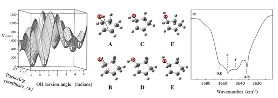
2.1. Calculated Molecular Conformations

The six conformers of 2CPOL with energy minima on the PES result from different values of the ring-puckering coordinate and the internal rotation angle of the OH group. Table 1 presents the key features of these conformations. Figure 1 shows the atomic numbering and the geometrical parameters calculated for the two lowest energy conformers. The lowest energy conformation A clearly possesses π-type hydrogen bonding as the calculated distance from the hydrogen atom on the OH group to the middle of the C=C double bond is 2.68 Å. This is very similar to the distance of 2.75 Å for 3-cyclopenten-1ol (II) and for other cyclic alcohols with this type of π bonding as shown in Table 2. The conformers A and B differ by having carbon atom 4 puckered in opposite directions. According to the CCSD/cc-pVTZ computations, B is only 9 cm−1 higher in energy from A, whereas the other conformers are 293 to 361 cm−1 higher in energy. MP2/cc-pVTZ computations predict B to be 89 cm−1 higher in energy. For B, the calculated distance from the hydrogen atom on the OH group to the middle of the C=C double bond is 3.00 Å, and this is considerably longer than that in the other molecules shown in Table 2, suggesting that the π type hydrogen bonding, if present, is quite small. On the other hand, as discussed below, the calculated and observed O-H stretching frequencies for A and B are both considerably lower than for the other conformers, and this would result from the hydrogen bonding. Figure 2 shows all six calculated structures for the conformational minima. Table 1 presents the ring-puckering angles and OH internal rotation angles, the calculated energy differences, and the calculated relative populations of the six conformers of 2CPOL at 25 °C. Supplementary Table S1 presents the calculated structural parameters for all six conformers from our CCSD/cc-pVTZ computations. Table 1 and Table S1 also show the calculated distances from the hydrogen atoms of the OH group to the center of the C=C bond.
As can be seen in Table 1, the absolute value of the ring-puckering angle is 22° ± 2° for each of the conformers. Thus, the conformational energy differences arise primarily from the internal rotation of the OH group. Conformers A and B differ by about 14° of the OH internal rotational angle, and both may allow the intramolecular hydrogen bonding to occur. However, the A conformer is lower in energy and has a stronger hydrogen bond as reflected by the shorter distance from the OH hydrogen to the center of the C=C bond. For conformer A, this distance is 2.682 Å, whereas for the conformers without hydrogen bonding, the distances are at least 0.3 Å longer.

2.2. Vibrational Spectra

Figure 3 shows a comparison of the experimental liquid phase and vapor-phase infrared spectra. Since intermolecular hydrogen bonding will be much stronger in the liquid phase than the intramolecular hydrogen bonding, bands corresponding to the specific A, B, C, D, E, and F conformers will not be present in the liquid spectra. Table 3 shows the observed vapor-phase and calculated frequencies for selected vibrations of the A conformer of 2CPOL compared to those observed for the related molecules 3-cyclopentenol (II) [9], 3-cyclopenten-1-amine (VII) [13], and cyclopentene [24].
Figure 4 shows the observed infrared band for the conformers of 2CPOL for the O-H stretch region. Based on the calculated frequency differences for the different conformers, the observed IR band at 3632 cm−1 is assigned to both A and B; 3654 cm−1 is assigned to C; 3664 cm−1 is assigned to D; 3664 cm−1 is assigned to E; and 3644 cm−1 is assigned to F. Table 4 shows the observed O-H stretching frequencies for the six conformers of 2CPOL compared to the calculated values. As expected, the weak hydrogen bonding lowers the O-H stretching frequency so that conformers A and B have the lowest value. The weakening of the O-H bond due to its interaction with the C=C double bond is supported by our CCSD/cc-pVTZ computations. The good agreement between observed and calculated frequencies strongly supports the presence of the six expected conformers. Figure 5 shows the vibrational spectra and assignments of 2CPOL in the 400–1700 cm−1 region. Due to the relatively low molecular populations of the other conformers, all of the bands have been assigned to A and B, which make up 69% of the molecular population. The A and B frequencies are predicted to differ very little in this region so each of the bands seen in Figure 5 arises mainly from both conformers.

2.3. Potential Energy Surface

Figure 6 shows the calculated potential energy surface (PES) for 2CPOL in terms of its ring-puckering and OH internal rotation coordinates. This PES was generated by individually computing the energies of more than 150 conformations of 2CPOL using MP2/cc-pVTZ calculations. CCSD/cc-pVTZ calculations for so many conformations would have taken an inordinate amount of computer time. The zero value of the OH internal rotation angle is defined at the position where the hydrogen atom of the hydroxyl group OH points toward the center of the C=C double bond. The PES clearly shows the presence of the six different minima which reside at six different conformational energies. The mathematical function that best satisfies the fit of the calculated data has the form:
V   =   i   =   i 6 a i x i +   n   =   1 6 b n sin n ϕ + n   =   1 6 c n sin n ϕ cos n ϕ + 1 3 d n cos n ϕ + x n   =   1 6 e n sin n ϕ + n   =   1 6 f n cos n ϕ
where x and y refer to the puckering and internal rotation coordinates. This equation consists of a polynomial component dependent on the x term to describe the ring-puckering motion, three sums of trigonometric functions to define the periodicity of the OH internal rotation, and a final set of cross terms, which describe the interaction between the ring-puckering and the internal rotation. Equation (1) is complex due to the lack of symmetry in the 2CPOL molecule. Figure 6 also shows the contour map of the PES calculated for 2CPOL. The calculated interconversion barriers from the MP2/cc-pVTZ computations are also indicated. Table 5 shows a comparison of the calculated results for 2CPOL from CCSD/cc-pVTZ computations to those from MP2/cc-pVTZ computations. We expect the CCSD calculations to be more reliable, but it is gratifying to see that the differences are relatively small.
The PES shows the barrier to ring planarity to be 281 cm−1 and the internal rotation barriers between energy minima to be in the 300 to 1000 cm−1 range.

3. Materials and Methods

2CPOL (95% purity) was purchased from CHEMSAMPCo and was purified by trap-to-trap distillation. Infrared spectra with a spectral resolution of 0.5 cm−1 were recorded using a Bruker Vertex 70 instrument, which was purged by a stream of dry nitrogen gas. The mid-infrared vapor-phase spectrum of 2CPOL was obtained at its vapor pressure at room temperature in a 10 cm cell. 2CPOL has a boiling point of 137 °C and vapor pressure of 3.1 Torr at 25 °C. KBr windows were used for the infrared cell. Spectra of liquid samples were recorded by placing a drop between either KBr plates for the mid-infrared or between CsI plates for the far-infrared. We were not able to record the vapor-phase Raman spectrum of this molecule at the higher temperatures required to obtain sufficient vapor pressure.

3.1. Computations

3.1.1. Structure and Frequency Calculations

The ab initio coupled-cluster theory with single and double excitations (CCSD) method was used with the cc-pVTZ (triple-ζ) basis set to calculate the conformational energies and to compute the geometries for each of the six conformers of 2CPOL. The GAUSSIAN 16 program [25] was used for the computations, and the GaussView 6 program [26] was used to visualize the structures.
Vibrational frequencies were calculated using MP2/cc-pVTZ computations. The scaling factors used for the MP2/cc-pVTZ calculations were 0.970 for frequencies below 1600 cm−1, 0.976 for 1600–2000 cm−1, 0.976 for 1600–2000 cm−1, and 0.948 for higher frequencies except for the O-H stretching region [13]. For the O-H region, 3520–3680 cm−1 the scaling factor of 0.950 was chosen to match the observed value for the lowest energy hydrogen-bonded conformer so that the predicted frequency shifts could readily be calculated.

3.1.2. Potential Energy Surface

The data points required for determining the theoretical PES of 2CPOL and its contour map were obtained from MP2/cc-pVTZ computations. The MAPLE 2015.1 computing environment [27] was used to perform the mathematical fit and plot the data. The PES was generated in terms of the ring-puckering coordinate (x), which is defined in Figure 7a, and the OH internal rotational angle. This angle is defined as the dihedral angle between the OH bond and the black dotted line that connects atom 5 to the mid-point of the C=C double bond as shown in Figure 7b.

4. Conclusions

2CPOL can exist in six different conformers and the one with the lowest energy shows π-type hydrogen bonding. This conformation has an OH hydrogen to the center of the C=C bond distance of 2.68 Å. This value is similar to those we found previously for the related molecules II to IV. The PES for 2CPOL derived in this work shows how the six conformers of this molecule can interconvert from one to another and what are the magnitudes of the barriers that must be overcome. It also shows that the intramolecular π-type hydrogen bonding lowers the conformational energy by approximately 300 cm−1 (0.8 kcal/mole). The experimental infrared spectrum in the O-H stretching region shows evidence for the presence of the predicted conformers.

Supplementary Materials

The following is available online, Table S1: Structural parameters for the six conformers of 2CPOL from CCSD/cc-pVTZ computations.

Author Contributions

E.J.O. and J.L. contributed equally. Both authors have read and agreed to the published version of the manuscript.

Funding

This research was funded by Welch Foundation, grant number A-0396. The APC was funded by MDPI.

Data Availability Statement

The data presented in this study are available in this article.

Acknowledgments

The authors wish to thank the Welch Foundation (Grant A-0396) for financial support and the Laboratory for Molecular Simulation at Texas A&M University, College Station for computational resources. This paper is dedicated to Austin Barnes for his many contributions to the fields of spectroscopy and molecular structure.

Conflicts of Interest

The authors declare no conflict of interest. The funders had no role in the design of the study; in the collection, analyses, or interpretation of data; in the writing of the manuscript, or in the decision to publish the results.

Sample Availability

Not available.

References

  1. Laane, J.; Ocola, E.J.; Chun, H.J. Vibrational Potential Energy Surfaces in Ground and Electronic Excited States. In Frontiers and Advances in Molecular Spectroscopy; Laane, J., Ed.; Elsevier: Amsterdam, The Netherlands, 2017; pp. 101–142. [Google Scholar] [CrossRef]
  2. Laane, J. Vibrational Potential Energy Surfaces in Electronic Excited States. In Frontiers of Molecular Spectroscopy; Laane, J., Ed.; Elsevier: Amsterdam, The Netherlands, 2009; pp. 63–132. [Google Scholar] [CrossRef]
  3. Laane, J. Experimental Determination of Vibrational Potential Energy Surfaces and Molecular Structures in Electronic Excited States. J. Chem. Phys. A 2000, 104, 7715–7733. [Google Scholar] [CrossRef]
  4. Laane, J. Spectroscopic Determination of Ground and Excited State Vibrational Potential Energy Surfaces. Int. Rev. Phys. Chem. 1999, 18, 301–341. [Google Scholar] [CrossRef]
  5. Laane, J. Vibrational Potential Energy Surfaces and Conformations of Molecules in Ground and Excited Electronic States. Annu. Rev. Phys. Chem. 1994, 45, 179–211. [Google Scholar] [CrossRef]
  6. Laane, J. Vibrational Potential Energy Surfaces of Non-Rigid Molecules in Ground and Excited Electronic States. In Structures and Conformations of Non-Rigid Molecules; Laane, J., Dakkouri, M., Eds.; Kluwer Publishing: Amsterdam, The Netherlands, 1993; pp. 65–98. [Google Scholar] [CrossRef]
  7. Laane, J. Determination of Vibrational Potential Energy Surfaces from Raman and Infrared Spectra. J. Pure Appl. Chem. 1987, 59, 1307–1326. [Google Scholar] [CrossRef] [Green Version]
  8. Al-Saadi, A.A.; Wagner, M.; Laane, J. Spectroscopic and Computational Studies of the Intramolecular Hydrogen Bonding of 2-Indanol. J. Phys. Chem. A 2006, 110, 12292–12297. [Google Scholar] [CrossRef]
  9. Ocola, E.J.; Al-Saadi, A.A.; Mlynek, C.; Hopf, H.; Laane, J. Intramolecular π-Type Hydrogen Bonding and Conformations of 3-Cyclopenten-1-ol. 2. Infrared and Raman Spectral Studies at High Temperatures. J. Phys. Chem. A 2010, 114, 7457–7461. [Google Scholar] [CrossRef]
  10. Al-Saadi, A.A.; Ocola, E.J.; Laane, J. Intramolecular π-Type Hydrogen Bonding and Conformations of 3-Cyclopenten-1-ol. 1. Theoretical Calculations. J. Phys. Chem. A 2010, 114, 7453–7456. [Google Scholar] [CrossRef] [PubMed]
  11. Ocola, E.J.; Laane, J. Spectroscopic and Theoretical Study of the Intramolecular π-Type Hydrogen Bonding and Conformations of 2-Cyclohexen-1-ol. J. Phys Chem. A 2016, 120, 74–80. [Google Scholar] [CrossRef] [PubMed]
  12. Ocola, E.J.; Laane, J. Theoretical Investigation of Intramolecular π-Type Hydrogen Bonding and Internal Rotation of 2-Cyclopropen-1-ol, 2-Cyclopropen-1-thiol and 2-Cyclopropen-1-amine. Mol. Phys. 2019, 17, 1404–1412. [Google Scholar] [CrossRef]
  13. Ocola, E.J.; Laane, J. Spectroscopic and Theoretical Study of the Intramolecular π-type Hydrogen Bonding and Conformations of 3-Cyclopentene-1-amine. J. Phys. Chem. 2020, 124, 5907–5916. [Google Scholar] [CrossRef] [PubMed]
  14. Baker, A.W.; Shulgin, A.T. Intramolecular Hydrogen Bonds to π-Electrons and Other Weakly Basic Groups. J. Am. Chem. Soc. 1958, 80, 5358–5363. [Google Scholar] [CrossRef]
  15. Smith, Z.; Carballo, N.; Wilson, E.B.; Marstokk, K.-M.; Møllendal, H. Conformations, Possible H Bonding, and Microwave Spectrum of 3-Buten-2-ol. J. Am. Chem. Soc. 1951, 107, 1951–1957. [Google Scholar] [CrossRef]
  16. Murty, A.N.; Curl, R.F., Jr. Microwave Spectrum of Allyl Alcohol. J. Chem. Phys. 1967, 46, 4176–4180. [Google Scholar] [CrossRef]
  17. Marstokk, K.-M.; Møllendal, H. Microwave Spectrum, Conformation and Intramolecular Hydrogen Bonding of 1,4-Pentadien-3-ol. Acta Chem. Scand. 1990, 44, 18–22. [Google Scholar] [CrossRef]
  18. Bräse, S.; Klæboe, P.; Marstokk, K.-M.; de Meijere, A.; Møllendal, H.; Nielsen, C.J. Conformational Properties of 2-Cyclopropylideneethanol as Studied by Microwave, Infrared and Raman Spectroscopy and by Ab Initio Computations. Acta Chem. Scand. 1998, 52, 1122–1136. [Google Scholar] [CrossRef] [Green Version]
  19. Bakke, J.M.; Bjerkeseth, L.H. The Conformational Composition of 3-Buten-1-ol, the Importance of Intramolecular Hydrogen Bonding. J. Mol. Struct. 1998, 470, 247–263. [Google Scholar] [CrossRef]
  20. Leonov, A.; Marstokk, K.-M.; de Meijere, A.; Møllendal, H. Microwave Spectrum, Conformational Equilibrium, Intramolecular Hydrogen Bonding, Tunneling, and Quantum Chemical Calculations for 1-Ethenylcyclopropan-1-ol. J. Phys. Chem. A 2000, 104, 4421–4428. [Google Scholar] [CrossRef]
  21. Rademacher, P.; Khelashvili, L.; Kowski, K. Spectroscopic and Theoretical Studies on Intramolecular OH–π Hydrogen Bonding in 4-Substituted 2-Allylphenols. Org. Biomol. Chem. 2005, 3, 2620–2625. [Google Scholar] [CrossRef]
  22. Miller, B.J.; Lane, J.R.; Kjaergaard, H.G. Intramolecular OH﮲﮲﮲π Interactions in Alkenols and Alkynols. Phys. Chem. Chem. Phys. 2011, 13, 14183–14193. [Google Scholar] [CrossRef]
  23. Mackeprang, K.; Schrøder, S.D.; Kjaergaard, H.G. Weak Intramolecular OH–π Hydrogen Bonding in Methallyl- and Allyl-Carbinol. Chem. Phys. Lett. 2013, 582, 31–37. [Google Scholar] [CrossRef]
  24. Al-Saadi, A.A.; Laane, J. Ab Initio and DFT Calculations for the Structure and Vibrational Spectra of Cyclopentene and its Isotopomers. J. Mol. Struct. 2007, 830, 46–57. [Google Scholar] [CrossRef]
  25. Frisch, M.J.; Trucks, G.W.; Schlegel, H.B.; Scuseria, G.E.; Robb, M.A.; Cheeseman, J.R.; Scalmani, G.; Barone, V.; Mennucci, B.; Petersson, G.A.; et al. Gaussian 09, Revision A.02; Gaussian, Inc: Wallingford, CT, USA, 2009. [Google Scholar]
  26. Dennington, R.D.; Keith, T.A.; Millam, J.M. GaussView 6.1.1, Graphical Interface; Semichem Inc.: Shawnee, KS, USA, 2000–2019. [Google Scholar]
  27. Waterloo Maple. Maple 2015; Waterloo Maple Inc.: Waterloo, ON, Canada, 2015. [Google Scholar]
Scheme 1. Molecules with intramolecular π-type hydrogen bonding.
Molecules 26 01106 sch001
Figure 1. Calculated structures of the lowest energy conformers of 2CPOL from coupled-cluster theory with single and double excitations (CCSD)-pVTZ computations.
Molecules 26 01106 g001
Figure 2. Calculated conformational minima, relative energies, and calculated abundances at 25 °C of 2CPOL from CCSD/cc-pVTZ computations. Conformers A, C, and F are puckered down, whereas conformers B, D, and E are puckered up.
Molecules 26 01106 g002
Figure 3. Observed infrared spectra of 2CPOL.
Molecules 26 01106 g003
Figure 4. Observed vapor-phase infrared of 2CPOL for the O-H stretch region.
Molecules 26 01106 g004
Figure 5. Observed vapor-phase infrared of 2CPOL for the 400–1700 cm−1 region.
Molecules 26 01106 g005
Figure 6. Potential energy surface (PES) and contour map of 2CPOL in terms of its ring-puckering and the OH internal rotation coordinates.
Molecules 26 01106 g006
Figure 7. (a) Definition of the ring-puckering coordinate (x), where 2x is the distance between the two dotted lines and Ө is the ring puckering angle. (b) 2-cyclopenten-1-ol (2CPOL).
Molecules 26 01106 g007
Table 1. Selected characteristics from CCSD/cc-pVTZ computations for all of the six 2CPOL conformers.
Molecules 26 01106 i001 Molecules 26 01106 i002 Molecules 26 01106 i003 Molecules 26 01106 i004 Molecules 26 01106 i005 Molecules 26 01106 i006 Molecules 26 01106 i007
2CPOL Conformers
ABCDEF
Energy
Energy (cm−1)09293304308361
Energy (kJ/mol)00.113.513.643.684.32
Energy (kcal/mol)00.030.840.870.881.03
Relative population (%)
At 25°35%34%9%8%8%6%
Angles (degrees)
Ring-puckering angle−21.1°22.5°−23.1°22.8°23.7°−22.8°
OH internal rotation angle, ϕ24.8°39.2°−75.7°−91.0°172.3°166.9°
Distance (Å)
O−H140.9600.9600.9580.9580.9580.958
dH14 a2.6823.0013.0193.3353.6803.554
a Distance between H14 of the OH group to the midpoint of the double C=C bond.
Table 2. Calculated H-π (C=C) bonding distances in cyclic molecules.
MoleculeH﮲﮲﮲π (C=C) Distance, ÅMethodReference
2-indanol (I)2.650CCSD/cc-pVTZThis work
2.580MP2/cc-pVTZThis work
3-cyclopenten-1-ol (II)2.744CCSD/cc-pVTZThis work
2.673MP2/cc-pVTZThis work
2-cyclohexen-1-ol (III)2.756CCSD/cc-pVTZThis work
2.737 MP2/cc-pVTZ[11]
2-cyclopropen-1-ol (IV)2.488 CCSD/cc-pVTZ[12]
2.478 MP2/cc-pVTZ[12]
2-cyclopropen-1-thiol (V)2.774 CCSD/cc-pVTZ[12]
2.740 MP2/cc-pVTZ[12]
2-cyclopropen-1-amine (VI)2.583 CCSD/cc-pVTZ[12]
2.571 MP2/cc-pVTZ[12]
3-cyclopenten-1-amine (VII) 2.850 CCSD/cc-pVTZ[13]
2.773MP2/cc-pVTZThis work
2-cyclopenten-1-ol (2CPOL)2.682CCSD/cc-pVTZThis work
2.632MP2/cc-pVTZThis work
Table 3. Observed and calculated frequency differences for selected vibrations conformer A of 2CPOL compared to those observed for other five-membered ring molecules.
2-Cyclopenten-1-ol3-Cyclopenten-1-ol3-Cyclopenten-1-amineCyclopentene
ObservedCalculatedObserved
AAAAPuckered, Cs
Ring-Pseudo C2vFreq.IR aFreq.(IR, R) bFreq. c(IR, R) d,eFreq. f(IR, R) d,fFreq. e(IR, R) b,e
A1
v3C=C stretch1609m1609(3, 80)1607(2, 46) 1613(m, 88)1623(m, 91)
v4CH2 deformation1500s1459(8, 67)1446f(3, 19)1452(m, 17)1471f(vw, 16)
v6=C-H in plane wag1077m1095(10, 100)1108f(4, 95)1109(w, 58) 1101(w, 66)
v8Ring stretch 894m869(13, 60)832(5, 3)804(s, 43)900(m, 100)
v9Ring angle bend694s692(6, 9)745(51, 77)735(w, 9)796g(vw, 1)
B1
v17CH2 deformation1463m1436(7, 77)1446f(17, 19)1437(m, 4)1445f(m, 23)
B2
v25CH2 rock924m971(14, 36) 948f(29, 17) 934(m, 4)1047(s, 1)
v26=C-H out-of-plane wag726s719(20, 15)674(97, 12) 671(s, 11)695(s, 1)
v27Ring puckering------119(2, 11)---(---, ---) ---(---, ---)---(---, ---)
Other vibrations
v OH OH stretch3632w3632(32, 26)3623(41, 9)N. A.N. A.N. A.N. A.
ω CH C-H wag (up and down)1385m1386(80, 53)1395(38, 13)1379(m, 7)N. A.N. A.
ω OH COH wag 1212w1224(13, 14)1275(9, 3)N. A.N. A.N. A.N. A.
  ω CO C-O wag (up and down)462s487(5, 21)------N. A.N. A.N. A.N. A.
ω CO C-O wag (sideways)346 (L)w388(12, 7)444(0.5, 2)N. A.N. A.N. A.N. A.
vCOC-O stretch1032m1045(75, 21)1048(54, 0.7)N. A.N. A.N. A.N. A.
    τ OH OH torsion 217 (L)m313(100, 21)397h(---, 8)N. A.N. A.N. A.N. A.
a IR intensities. s-strong, m-medium, w-weak. b Relative IR and Raman intensities. c From Reference [9], unless indicated. d From Reference [13], unless indicated. e From Reference [24], unless indicated. f Reassigned according to MP2/cc-pVTZ computations.
Table 4. O-H stretching frequencies (cm−1) for the six conformers of 2CPOL.
ConformerObservedCalculated
FrequencyShiftFrequencyShift
A3632036320
B3632036342
C365422364816
D366432365826
E366432365624
F364412364412
Table 5. Comparison of calculated conformational energies and geometrical parameters for 2CPOL from CCSD/cc-pVTZ and MP2/cc-pVTZ computations.
2CPOL Conformers
ABCDEF
Energy, (cm−1)
CCSD/cc-pVTZ09293304308361
MP2/cc-pVTZ089308406406409
Ring-puckering coordinate, (Å)
CCSD/cc-pVTZ−0.1020.109−0.1120.1100.115−0.110
MP2/cc-pVTZ−0.1140.118−0.1240.1180.124−0.122
Ring-puckering angle, (degrees)
CCSD/cc-pVTZ−21.1°22.5°−23.1°22.8°23.7°−22.8°
MP2/cc-pVTZ−23.6°24.3°−25.7°24.4°25.6°−25.3°
OH internal rotation angle, (degrees)
CCSD/cc-pVTZ24.8°39.2°284.3°269.0°172.3°166.9°
MP2/cc-pVTZ22.6°38.7°286.4°268.6°172.3°166.6°
Publisher’s Note: MDPI stays neutral with regard to jurisdictional claims in published maps and institutional affiliations.

