
Stability and Photoisomerization of Stilbenes Isolated from the Bark of Norway Spruce Roots

 ,
,  ,
,
Abstract
:
1. Introduction
2. Results and Discussion
2.1. The Stability of Stilbenes in Solution
2.2. Stability Assessment of Stilbenes in Solid Crude Extract
2.3. UV Stability of Stilbenes
2.4. Isolation and Identification of the New Compounds Formed during UV Irradiation
2.5. The Structural Elucidation of New Peaks Formed from Astringin (and Its Aglycone Piceatannol) and Isorhapontin (and Its Aglycone Isorhapontigenin)
3. Materials and Methods
3.1. Bark Material
3.2. Extraction of Bark Material
3.3. HPLC-DAD Analyses
3.4. Isolation of Stilbenes by Preparative HPLC
3.5. HPLC-DAD/ESI-MS and HPLC-DAD/ESI-MS/MS Analyses
3.6. Stability Assessment of Stilbenes in a Solution and Solid Form
3.7. UV Stability of Isolated Stilbenes and Crude Extracts
3.8. Mass Spectrometry (MS) and Nuclear Magnetic Resonance Spectroscopy (NMR)
4. Conclusions
Supplementary Materials
Author Contributions
Acknowledgments
Conflicts of Interest
References
- Latva-Mäenpää, H.; Laakso, T.; Sarjala, T.; Wähälä, K.; Saranpää, P. Variation of stilbene glucosides in bark extracts obtained from roots and stumps of Norway spruce (Picea abies [L.] Karst.). Trees 2013, 27, 131–139. [Google Scholar] [CrossRef]
- Hammerbacher, A.; Ralph, S.; Boehlmann, J.; Fenning, T.; Gershenzon, J.; Schmidt, A. Biosynthesis of the major tetrahydroxystilbenes in spruce, astringin and isorhapontin, proceeds via resveratrol and is enhanced by fungal infection. Plant. Physiol. 2011, 157, 876–890. [Google Scholar] [CrossRef] [Green Version]
- Franceschi, V.R.; Krokene, P.; Christiansen, E.; Krekling, T. Anatomical and chemical defences of conifer bark against bark beetles and other pests. New Phytol. 2005, 167, 353–375. [Google Scholar] [CrossRef] [PubMed]
- Krokene, P. Conifer defense and resistance to bark beetles. In Biology and Ecology of Native and Invasive Species; Vega, F.E., Hofstetter, R.W., Eds.; Elsevier Academic Press: San Diego, CA, USA, 2015; pp. 177–207. [Google Scholar]
- Gabaston, J.; Richard, T.; Biais, B.; Waffo-Teguo, P.; Pedrot, E.; Jourdes, M.; Corio-Costet, M.-F.; Mérillon, J.-M. Stilbenes from common spruce (Picea abies) bark as natural antifungal agent against downy mildew (Plasmopara viticola). Ind. Crops Prod. 2017, 103, 267–273. [Google Scholar] [CrossRef]
- Freyssin, A.; Page, G.; Fauconneau, B.; Bilan, A.R. Natural stilbenes effects in animal models of Alzheimer’s disease. Neural Regen. Res. 2020, 15, 843–849. [Google Scholar] [CrossRef]
- Latva-Mäenpää, H.; Laakso, T.; Sarjala, T.; Wähälä, K.; Saranpää, P. Root neck of Norway spruce as a source of bioactive lignans and stilbenes. Holzforschung 2014, 68, 1–7. [Google Scholar] [CrossRef]
- Mulat, D.; Latva-Mäenpää, H.; Koskela, H.; Saranpää, P.; Wähälä, K. Rapid chemical characterisation of stilbenes in the root bark of Norway spruce by off-line HPLC/DAD-NMR. Phytochem. Anal. 2014, 25, 529–536. [Google Scholar] [CrossRef]
- Likhtenstein, G. Stilbenes—Applications in Chemistry, Life Sciences and Materials Science; Wiley-VCH: Weinheim, Germany, 2010. [Google Scholar]
- Välimaa, A.-L.; Raitanen, J.-E.; Tienaho, J.; Sarjala, T.; Nakayama, E.; Korpinen, R.; Mäkinen, S.; Eklund, P.; Willför, S.; Jyske, T. Enhancement of Norway spruce bark side-streams: Modification of bioactive and protective properties of stilbenoid-rich extracts by UVA-irradition. Ind. Crops Prod. 2020, 145, 112150. [Google Scholar] [CrossRef]
- Piñeiro, Z.; Palma, M.; Barroso, C.G. Determination of trans-resveratrol in grapes by pressurised liquid extraction and fast high-performance liquid chromatography. J. Chromatogr. A 2006, 1110, 61–65. [Google Scholar] [CrossRef] [PubMed]
- Prokop, J.; Abrman, P.; Seligson, A.L.; Sovak, M. Resveratrol and Its Glycon Piceid Are Stable Polyphenols. J. Med. Food 2006, 9, 11–14. [Google Scholar] [CrossRef]
- Shi, G.; Rao, L.; Yu, H.; Xiang, H.; Yang, H.; Ji, R. Stabilization and encapsulation of photosensitive resveratrol within yeast cell. Int. J. Pharm. 2008, 349, 83–93. [Google Scholar] [CrossRef] [PubMed]
- Kolouchová-Hanzlíková, I.; Melzoch, K.; Filip, V.; Smidrkal, J. Rapid method for resveratrol determination by HPLC with electrochemical and UV detections in wines. Food Chem. 2004, 87, 151–158. [Google Scholar] [CrossRef]
- Trela, B.C.; Waterhouse, A.L. Resveratrol: Isomeric molar absorptivities and stability. J. Agric. Food Chem. 1996, 44, 1253–1257. [Google Scholar] [CrossRef]
- Montsko, G.; Pour Nikfardjam, M.S.; Szabo, Z.; Boddi, K.; Lorand, T.; Ohmacht, R.; Mark, L. Determination of products derived from trans-resveratrol UV photoisomerisation by means of HPLC-APCI-MS. J. Photochem. Photobiol. A 2008, 196, 44–50. [Google Scholar] [CrossRef]
- Tříska, J.; Vrchotová, N.; Olejníčková, J.; Jílek, R.; Sotolář, R. Separation and identification of highly fluorescent compounds derived from trans-resveratrol in the leaves of Vitis vinifera infected by Plasmopara viticola. Molecules 2012, 17, 2773–2783. [Google Scholar] [CrossRef] [Green Version]
- Yang, I.; Kim, E.; Kang, J.; Han, H.; Sul, S.; Park, S.; Kim, S. Photochemical generation of a new, highly fluorescent compound from nonfluorescent resveratrol. Chem. Commun. 2012, 48, 3839–3841. [Google Scholar] [CrossRef] [PubMed]
- Rodríguez, R.A.; Lahoz, I.R.; Faza, O.N.; Cid, M.M.; Lopez, C.S. Theoretical and experimental exploration of the photochemistry of resveratrol: Beyond the simple double bond isomerization. Org. Biomol. Chem. 2012, 10, 9175–9182. [Google Scholar] [CrossRef] [PubMed]
- Francioso, A.; Laštovičková, L.; Mosca, L.; Boffi, A.; Bonamore, A.; Macone, A. Gas chromatographic-mass spectrometric method for the simultaneous determination of resveratrol isomers and 2,4,6-trihydroxyphenantrene in red wines exposed to UV-light. J. Agric. Food Chem. 2019, 67, 11752–11757. [Google Scholar] [CrossRef]
- Latva-Mäenpää, H. Bioactive and Protective Polyphenolics from roots and stumps of Conifer trees (Norway spruce and Scots pine). Academic Dissertation, University of Helsinki, Helsinki, Finland, 2017. [Google Scholar]
- Hu, Y.; Ma, S.; Li, J.; Yu, S.; Qu, J.; Liu, J.; Du, D. Targeted isolation and structure elucidation of stilbene glycosides from the bark of Lysidice brevicalyx Wei guided by biological and chemical screening. J. Nat. Prod. 2008, 71, 1800–1805. [Google Scholar] [CrossRef]
- Jensen, J.S.; Wertz, C.F.; O’Neill, V.A. Preformulation stability of trans-resveratrol and trans-resveratrol glucoside (piceid). J. Agric. Food Chem. 2010, 58, 1685–1690. [Google Scholar] [CrossRef]
- Leong, Y.-W.; Harrison, L.; Powell, A. Phenanthrene and other aromatic constituents of Bulbophyllum vaginatum. Phytochemistry 1999, 50, 1237–1241. [Google Scholar] [CrossRef]
- Moore, W.; Morgan, D.; Stermitz, F. The photochemical conversion of stilbene to phenanthrene. The nature of the intermediate. J. Am. Chem.Soc. 1963, 86, 829–830. [Google Scholar] [CrossRef]
- Sigman, M.; Barbas, J.; Corbett, S.; Chen, Y.; Ivanov, I.; Dabestani, R. Photochemical reactions of trans-stilbene and 1,1-diphenylethylene on silica gel: Mechanisms of oxidation and dimerization. J. Photochem. Photobiol. A: Chemistry 2001, 138, 269–274. [Google Scholar] [CrossRef]
 trans-astringin,
  trans-piceid,
  trans-isorhapontin,
  trans-resveratrol and
  trans-isorhapontigenin.
trans-astringin, trans-piceid, trans-isorhapontin, trans-resveratrol and trans-isorhapontigenin.
trans-isorhapontigenin.
trans-astringin, trans-piceid, trans-isorhapontin, trans-resveratrol and trans-isorhapontigenin. peak derived from cis-astringin, cis-piceid, cis-isorhapontin, cis-resveratrol and cis-isorhapontigenin.
peak derived from cis-astringin, cis-piceid, cis-isorhapontin, cis-resveratrol and cis-isorhapontigenin.
peak derived from cis-astringin, cis-piceid, cis-isorhapontin, cis-resveratrol and cis-isorhapontigenin. trans-astringin, trans-piceid, trans-isorhapontin, trans-resveratrol and trans-isorhapontigenin.
trans-astringin, trans-piceid, trans-isorhapontin, trans-resveratrol and trans-isorhapontigenin.
trans-astringin, trans-piceid, trans-isorhapontin, trans-resveratrol and trans-isorhapontigenin. cis-astringin, cis-piceid, cis-isorhapontin, cis-resveratrol and cis-isorhapontigenin.
cis-astringin, cis-piceid, cis-isorhapontin, cis-resveratrol and cis-isorhapontigenin.
cis-astringin, cis-piceid, cis-isorhapontin, cis-resveratrol and cis-isorhapontigenin.


| Sample Name | Products | tR(min) | UV Profile | [M − H]−, m/z |
|---|---|---|---|---|
| trans-astringin | trans-astringin | 11.8 | trans-stilbene | 405 |
| new peak | 16 | λ max~260 | 403 | |
| trans-piceid | trans-piceid | 16.3 | trans-stilbene | 389 |
| new peak | 27.5 | cis-stilbene | 389 | |
| trans-isorhapontin | trans-isorhapontin | 17.5 | trans-stilbene | 419 |
| new peak | 28.3 | cis-stilbene | 419 | |
| trans-piceatannol | trans-piceatannol | 18.6 | trans-stilbene | 243 |
| no peak detected | ||||
| trans-resveratrol | trans-resveratrol | 25 | trans-stilbene | 227 |
| new peak | 32.3 | cis-stilbene | 227 | |
| trans-isorhapontigenin | trans-isorhapontigenin | 26.4 | trans-stilbene | 257 |
| new peak | 33.1 | cis-stilbene | 257 |
| Sample | Products | Major Compound After UV Irradiation (0 h, 2 h, 24 h) | GC-MS Retention Time (min) | GC-MS (TMSI), m/z | HPLC UV Profile |
|---|---|---|---|---|---|
| trans-astringin | trans-astringin | 0 h | 41.72 | 532 | trans-stilbene |
| cis-astringin | 2 h | 32.04 | 532 | cis-stilbene | |
| new peak | 24 h | 34.48 | 530 | λ max~260 | |
| trans-piceid | trans-piceid | 0 h | 38.42 | 444 | trans-stilbene |
| cis-piceid | 2 h | 30.30 | 444 | cis-stilbene | |
| new peak | 24 h | 31.77 | 442 | λ max~260 | |
| trans-isorhapontin | trans-isorhapontin | 0 h | 42.45 | 474 | trans-stilbene |
| cis-isorhapontin | 2 h/24 h | 31.62 | 474 | cis-stilbene | |
| new peak 1 | 24 h | 30.43 | 472 | λ max~260 | |
| new peak 2 | 24 h | 34.48 | 472 | λ max~260 | |
| new peak 3 | 24 h | 43.54 | 472 | λ max~260 | |
| trans-piceatannol | trans-piceatannol | 0 h | 22.76 | 532 | trans-stilbene |
| new peak 1 | 2 h | 21.57 | 530 | λ max~260 | |
| new peak 1 | 24 h | 21.57 | 530 | λ max~260 | |
| trans-resveratrol | trans-resveratrol | 0 h | 20.00 | 444 | trans-stilbene |
| cis-resveratrol | 2 h | 13.89 | 444 | cis-stilbene | |
| new peak | 24 h | 18.64 | 442 | λ max~260 | |
| trans-isorhapontigenin | trans-isorhapontigenin | 0 h | 22.55 | 474 | trans-stilbene |
| cis-isorhapontigenin | 2 h | 15.62 | 474 | cis-stilbene | |
| new peak | 24 h | 16.36 | 472 | λ max~260 |
| Compound | Stilbene | [M-](m/z) | Calcd. | Formula |
|---|---|---|---|---|
| F1.1 | trans-astringin | 405.1180 | 405.1186 | C20H21O9 |
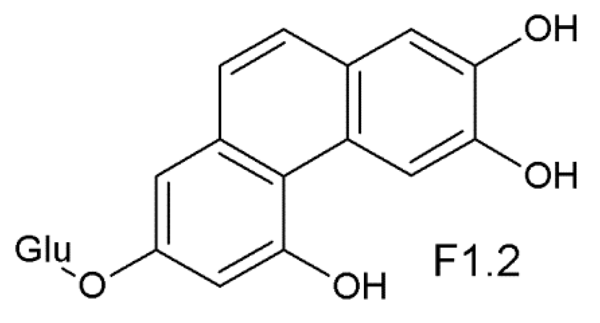
| F1.2 | new peak | 403.1036 | 403.1029 | C20H19O9 |
| F1.3 | cis-astringin | 405.1243 | 405.1186 | C20H21O9 |
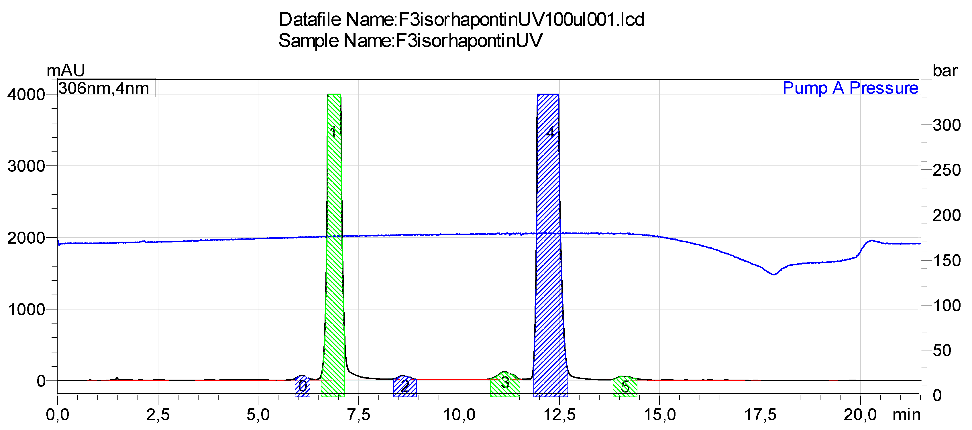
| F3.1 | trans-isorhapontin | 419.1479 | 419.1342 | C21H23O9 |
| F3.2 | new peak | 417.1321 | 417.1186 | C21H21O9 |
| F3.3 | new peak | 417.1327 | 417.1186 | C21H21O9 |
| F3.4 | cis-isorhapontin | 419.1487 | 419.1342 | C21H23O9 |
| F3.5 | new peak | 417.1321 | 417.1186 | C21H21O9 |
| Stability Conditions in Solution Form | Stability CondItions in Solid Form |
|---|---|
| (1) Light protected: The samples were stored in a freezer (−20 °C) with protection from light in glass vials capped under ambient air conditions | (1) Light protected: The solid extract samples in capped glass vials were stored in a freezer (−20 °C) protected from light. |
| (2) Light unprotected: The samples in glass vials (capped under ambient air) were kept on laboratory bench, exposed to continuous fluorescent light. | (2) Light unprotected: The solid extract samples in capped glass vials under ambient air conditions were kept on laboratory bench, exposed to continuous fluorescent light. |
| (3) Light unprotected: Solid extracts samples in uncapped glass vials were kept on laboratory bench, exposed to continuous fluorescent light and permanent contact with air. |
Publisher’s Note: MDPI stays neutral with regard to jurisdictional claims in published maps and institutional affiliations. |
© 2021 by the authors. Licensee MDPI, Basel, Switzerland. This article is an open access article distributed under the terms and conditions of the Creative Commons Attribution (CC BY) license (http://creativecommons.org/licenses/by/4.0/).
Share and Cite
Latva-Mäenpää, H.; Wufu, R.; Mulat, D.; Sarjala, T.; Saranpää, P.; Wähälä, K. Stability and Photoisomerization of Stilbenes Isolated from the Bark of Norway Spruce Roots. Molecules 2021, 26, 1036. https://doi.org/10.3390/molecules26041036
Latva-Mäenpää H, Wufu R, Mulat D, Sarjala T, Saranpää P, Wähälä K. Stability and Photoisomerization of Stilbenes Isolated from the Bark of Norway Spruce Roots. Molecules. 2021; 26(4):1036. https://doi.org/10.3390/molecules26041036
Chicago/Turabian StyleLatva-Mäenpää, Harri, Riziwanguli Wufu, Daniel Mulat, Tytti Sarjala, Pekka Saranpää, and Kristiina Wähälä. 2021. "Stability and Photoisomerization of Stilbenes Isolated from the Bark of Norway Spruce Roots" Molecules 26, no. 4: 1036. https://doi.org/10.3390/molecules26041036