Communication

Nitro-Substituted Dipyrrolyldiketone BF2 Complexes as Electronic-State-Adjustable Anion-Responsive π-Electronic Systems

Department of Applied Chemistry, College of Life Sciences, Ritsumeikan University, Kusatsu 525–8577, Japan
*
Author to whom correspondence should be addressed.
Molecules 2021, 26(3), 595; https://doi.org/10.3390/molecules26030595
Submission received: 6 January 2021 / Revised: 19 January 2021 / Accepted: 20 January 2021 / Published: 23 January 2021
(This article belongs to the Special Issue Supramolecular Anion Binding and Molecular Recognition)

Abstract

:
Nitro-substituted π-electronic molecules are fascinating because of their unique electronic and optical properties and the ease of their transformation into various functional derivatives. Herein, nitro-introduced dipyrrolyldiketone BF2 complexes as anion-responsive π-electronic molecules were synthesized, and their electronic properties and anion-binding abilities were investigated by spectroscopic analyses and theoretical studies. The obtained nitro-substituted derivatives showed solvent-dependent UV/vis spectral changes and high anion-binding affinities due to the easily pyrrole-inverted conformations and polarized pyrrole NH sites upon the introduction of electron-withdrawing moieties.

Graphical Abstract

1. Introduction

Appropriately designed π-electronic systems have unique electronic and optical properties and assembling behavior [1,2,3,4,5,6,7]. As pyrrole-based π-electronic systems, boron-dipyrromethenes (BODIPYs) have been investigated due to high molar absorption coefficients at the visible region, high fluorescence quantum yields, and chemical and photostability. Hence, BODIPYs introducing recognition sites are used for bioimaging, sensors, and drug delivery [4,8,9,10]. Moreover, the properties of π-electronic systems can be modulated by the substituents and resulting structures. The nitro (NO2) group is effective for controlling the electronic states of π-electronic systems owing to its strong electron-withdrawing nature [11,12,13]. The nitro group is also a useful and important substituent in the field of synthetic chemistry. NO2-substituted molecules are easily converted to the corresponding amines, which are the precursors of amides and other derivatives with functional groups. It is also noteworthy that nitroarenes behave as electrophilic coupling partners for Suzuki–Miyaura coupling [14]. As the platforms for NO2 substitution, dipyrrolyldiketone BF2 complexes (e.g., 1a,b, Scheme 1) have been examined as anion-responsive π-electronic molecules [15,16,17,18,19], which formed anion complexes and ion-pairing assemblies in combination with cations [20,21]. Pyrrole β-substituents of 1b enable the facile and selective introduction of substituents at the α-positions, resulting in the synthesis of various derivatives [18,19]. The anion-binding properties depend on the substituents, such as ethyl and fluorine groups, introduced onto the pyrrole rings [16,17]. Compared to 1a, 1b shows less affinities for anions due to the electron-donating β-ethyl substituents. In this study, the introduction of nitro group(s) at the pyrrole α-positions was investigated for modulating the electronic states and anion-binding abilities.

2. Results and Discussion

2.1. Synthesis and Characterization

Nitration of aromatic species using nitric acid proceeds via the mechanism of electrophilic aromatic substitution (SEAr) reactions. Dipyrrolyldiketone BF2 complex 1b showed moderate reactivity for SEAr reactions, such as iodination, at the pyrrole α-positions despite the electron-withdrawing diketone unit [18]. The modified synthesis procedure [22] was adopted to prepare dinitro-substituted 2a as an orange solid in 13% yield from 1b by using concentrated nitric acid and acetic anhydride. Similarly, mononitro-substituted 2b was obtained as a red solid in 48% yield by the treatment of 1b with concentrated nitric acid and acetic acid at −5 °C for 3 h, and subsequently, at r.t. for 1 h (Scheme 2). For the introduction of the functional group for coupling reactions [18], the reaction of 2b with 1.1 equiv. of N-iodosuccinimide at r.t. for 6 h resulted in iodination at the unsubstituted pyrrole α-position to afford reddish orange 2c in 45% yield. The 1H NMR spectrum of 2a in CDCl3 at r.t. showed the NH signal at 10.24 ppm, whereas signals of 2b,c were observed at 10.18/9.59 and 10.14/9.63 ppm, respectively. Given the pyrrole NH signal of 1b at 9.31 ppm [17], the nitro group(s) caused the pyrrole NH signals to shift downfield. Furthermore, the signals of pyrrole β-CH2 in the spectra of 2ac were observed at 2.87/2.83, 2.86/2.81/2.78/2.50, and 2.86/2.83/2.78/2.46 ppm, respectively, while those of 1b appeared at 2.78 and 2.48 ppm, suggesting the electron-withdrawing effect of the nitro groups. The bridging CH signals, 6.83, 6.64, and 6.58 ppm for 2ac, respectively, were also influenced by the nitro substitution in comparison with 1b (6.48 ppm) [17].
Solid-state structures of 2c were revealed by single-crystal X-ray analysis (Figure 1). 2c formed two types of crystal pseudo-polymorphs, both of which were obtained by vapor diffusion of cyclohexane into an EtOAc solution. Polymorph 2c-tri adopted a triclinic crystal system (P 1 ¯ ) containing face-to-face dimers, in which non-inverted pyrrole NH formed intermolecular hydrogen bonding with a disordered EtOAc molecule with the N(−H)···O distance of 2.85 Å (Figure 1a). The 2c molecule in 2c-tri had a planar geometry, whose mean-plane deviation was estimated to be 0.0741 Å (19 atoms). The packing structures included π–π stacking between two pyrrole rings with a stacking distance of 3.34 Å. In contrast, polymorph 2c-mono existed as a monoclinic crystal system (P21/c) containing dimers, in which the pyrrole rings were inverted and the iodopyrrole and nitropyrrole units were in the same plane with multiple intermolecular hydrogen bonds (Figure 1b). The less stable pyrrole-inverted conformation (vide infra) in 2c-mono was stabilized by dimerization with multiple hydrogen bonds of pyrrole NH and bridging CH with the nitro oxygen: the N(–H)···O and C(–H)···O distances were 2.89/2.90/2.97 and 3.41/3.44 Å, respectively. 2c-mono also showed intermolecular hydrogen-bonding interactions at a pyrrolyl N–H···F(–B) with an N(–H)···F distance of 2.88 Å. The 2c molecule in 2c-mono also adopted the planar geometry, whose mean-plane deviations were estimated as 0.129 and 0.239 Å (19 atoms for each), and π–π stacking between the pyrrole ring and the diketone unit with a stacking distance of 3.67 Å.

2.2. Photophysical Properties

The UV/vis absorption spectra of 2a,b revealed characteristic substituent-dependent electronic properties. The UV/vis absorption spectra of 2a showed the maxima at 478 nm (toluene and CH2Cl2) and a broad peak at 526 nm (THF), which were red-shifted compared to those in the case of 1b (452 (CH2Cl2) and 449 nm (toluene and THF)) (Figure 2a,b(i)). On the other hand, the UV/vis absorption spectra of 2b were different from those of 2a, as observed in the maxima at 474 (toluene), 460 (CH2Cl2), and 455 (THF) nm (Figure 2b(i)). The fluorescence emission wavelengths (λem) (and quantum yields, ΦF) of 2a,b, excited at the absorption maxima, in toluene were 495 (0.462) and 498 nm (0.298), respectively, whereas those in CH2Cl2 were 493 (0.460) and 523 nm (0.020), respectively. Furthermore, the fluorescence emission of 2b in THF, excited at 455 nm, was quenched, whereas that of 2a was observed at 491 nm with a ΦF of 0.073. The broad UV/vis absorption spectra of 2b in CH2Cl2 and THF suggested intramolecular charge transfer (ICT). These observations of 2b were consistent with the dipole moments of the solvents: 0.30, 1.14, and 1.74 D for toluene, CH2Cl2, and THF, respectively [23]. On the other hand, the UV/vis absorption spectrum of 2a in THF showed broad and red-shifted bands, probably due to the interactions between the pyrrole NH and solvent molecules. This feature was also observed in the spectral change of 2a upon the addition of CH3CO2 as a tetrabutylammonium (TBA) salt in CH2Cl2 (vide infra). The Stokes shift of 2a was 0.079 eV, which is smaller than 1b (0.100 eV) [17], whereas the Stokes shift of 2b was 0.325 eV due to ICT in CH2Cl2. The UV/vis absorption spectra of 1b and 2a,b were consistent with time-dependent density functional theory (TD-DFT)-based theoretical spectra at CPCM-B3LYP/6-31+G(d,p) in CH2Cl2 (Figure 3) [24]. The main theoretical absorption maxima of 1b and 2a,b were estimated to be 411, 468, and 463 nm, respectively, corresponding to HOMO (highest occupied molecular orbital)-to-LUMO (lowest unoccupied molecular orbital) transitions. Both the HOMO and LUMO of 1b and 2a were distributed over the core π-electronic unit owing to the symmetrical structures. In contrast, in the less symmetric 2b, the HOMO was delocalized at the core π-electronic unit except for the carbonyl oxygen at the α-unsubstituted pyrrole side, whereas the LUMO was more distributed in the nitro-substituted pyrrole and diketone units. Hence, 2b showed ICT, especially in more polar solvents.

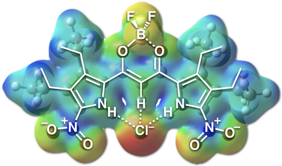
2.3. Anion-Binding Behavior

Pyrrole inversions of dipyrrolyldiketone BF2 complexes, whose pyrrole NH sites are oriented to the side of the carbonyl units, are required for anion binding [15,16,17,18,19]. The theoretical relative stability is correlated with the dipole magnitude and orientation of the pyrrole units. Therefore, the electronic states of pyrrole rings, according to the introduced substituents, modulate the dipole moments and resulting pyrrole-inversion behavior. Moreover, the binding constants (Ka) are determined by the hydrogen-bonding donor strength, depending on the dipole moment of the pyrrole units. Considering these points, the anion-binding properties of 2a,b were examined with UV/vis absorption spectral changes upon the addition of anions as TBA salts in CH2Cl2 solutions (0.01 mM for 2a and 0.02 mM for 2b). CH2Cl2 is used for good solubility for 2a,b and salts and for exhibiting higher Ka values as a less polar solvent. The use of polar solvents decreases the affinities for anions due to the solvation of salts along with receptors. Upon the addition of Cl, the absorption peaks in the spectrum of 2a at 478 and 452 nm were diminished, while the peak at 350 nm became prominent due to the increasing transition dipole moment along the short axis by the conformation change from anion-free to anion-binding states (Figure 4a(i)). Similarly, in the spectrum of 2b, the absorptions at 471 and 460 nm decreased concomitantly with an increase in the absorption of 340 nm (Figure 4a(ii)). On the other hand, the addition of CH3CO2 to 2a induced a spectral change different from those observed upon the addition of Cl, where a new absorption peak around 526 nm increased while those at 478 and 452 nm decreased (Figure 4b(i)). Similar to the case of 2a, drastic spectral changes were observed due to CH3CO2 binding with 2b, leading to new absorption peaks at around 503 and 350 nm, with decreasing peaks at 471 and 460 nm (Figure 4b(ii)). The increasing absorption peaks at 526 and 503 nm for 2a,b, respectively, are derived from the changes in the electronic states. CH3CO2 as a basic anion strongly interacts with pyrrole NH and induces the more polarized structures. The different basicities of Cl and CH3CO2 were sensitive to the interaction strength with the more polarized nitropyrrole-NH of 2a,b, resulting in different absorption spectral changes and electronic states. The Ka values of 2a,b for Cl, Br, and CH3CO2 (Table 1) were estimated to be 190,000, 11,000, and >106 M−1 for 2a and 27,000, 2800, and >106 M−1 for 2b, respectively, which were larger than the corresponding values for 1b (6800, 1200, and 210,000 M−1, respectively) [17]. The Ka values were augmented with the increase in the nitropyrrole-NH units that were more effectively included for hydrogen bonding with anions.
Theoretical studies of 2ac at the B3LYP level by using the 6-31G(d,p) basis set for C, H, B, N, O, and F and the LanL2DZ basis set for I [24] (Figure 5) suggested the stabilities of pyrrole-inverted conformations. Three conformations for 2a and four conformations for 2b,c were examined according to the orientations of pyrrole rings for the discussion of their relative energies. Singly pyrrole-inverted conformation 2a-2 and doubly pyrrole-inverted conformation 2a-3 were less stable than pyrrole-non-inverted conformation 2a-1 by 0.43 and 1.95 kcal/mol, respectively. On the other hand, 2b-2, 2b-2′, and 2b-3 were less stable than 2b-1 by 0.42, 1.96, and 2.87 kcal/mol, respectively, and 2c-2, 2c-2′, and 2c-3 were less stable than 2c-1 by 0.35, 1.62, and 2.64 kcal/mol, respectively. Compared to 1b, with lesser stabilities of 2.20 and 4.98 kcal/mol for the singly and doubly pyrrole-inverted conformations, respectively, 2ac can easily invert their pyrrole rings, especially the nitropyrrole unit(s), due to the decreased dipole moments of the pyrrole rings.
The theoretically estimated electrostatic potential (ESP) maps of 2a,b⋅Cl [24] (Figure 6) were different from that of 1b⋅Cl. The NO2-substituted pyrrole rings in 2a,b⋅Cl are more cationic than the pyrrole rings in 1b⋅Cl, and simultaneously, more cationic BF2 units are observed in 2a,b⋅Cl than in 1b⋅Cl because of the strong electron-withdrawing effect of the NO2 units. Furthermore, Cl in 2a⋅Cl has a smaller electron density due to the electron delocalization onto the dipyrrolyldiketone unit. These electron delocalization properties of 2a,b⋅Cl are consistent with the more effective anion-binding abilities.
The anion-binding modes of 2a,b were investigated based on changes in their 1H NMR spectra upon the addition of Cl as a TBA salt in CD2Cl2 at 20 and −50 °C (Figure 7). In the case of 2a, the addition of small amounts of Cl at −50 °C decreased the signals of pyrrole NH (10.44 ppm) and bridging CH (6.82 ppm). After the addition of 0.43 equiv. of Cl, the signals of pyrrole NH and bridging NH first appeared in the downfield regions. Upon the addition of 1.2 equiv. of Cl, the signals gradually shifted to 13.27 (pyrrole NH) and 9.08 ppm (bridging CH) (Figure 7a). On the other hand, in the case of 2b, the addition of Cl (0.45 equiv.) at −50 °C decreased the signals of the pyrrole NH (10.26/9.63 ppm) and bridging CH (6.61 ppm). Concomitantly, the signals of Cl-binding pyrrole NH and bridging CH appeared in the downfield regions, and the addition of 1.2 equiv. of Cl shifted the Cl-binding pyrrole NH and bridging CH signals to 12.64/12.38 and 7.15 ppm, respectively (Figure 7b). These observations suggested the presence of small amounts of [2+1]-type Cl complexes. In contrast to those that independently appeared as independent signals between anion-free receptors and [1+1]-type complexes [19], the [2+1]-type complexes of 2a,b were in fast equilibria with the corresponding [1+1]-type complexes, resulting in the coalescence into single signals that shifted downfield (Figure 7c).

3. Materials and Methods

3.1. General Procedures

Starting materials were purchased from FUJIFILM Wako Pure Chemical Corp. (Osaka, Japan), Nacalai Tesque Inc. (Kyoto, Japan), Tokyo Chemical Industry Co., Ltd. (Tokyo, Japan), Sigma-Aldrich Co. (Tokyo, Japan), and were used without further purification unless otherwise stated. The spectroscopic data of 1a,b were reported in the precedent reports [15,17]. NMR spectra used in the characterization of products 2ac were recorded on a JEOL ECA-600 600 MHz spectrometer (JEOL Ltd., Tokyo, Japan) and a Bruker AVANCE DRX-600 600 MHz spectrometer (Bruker, Massachusetts, USA). All NMR spectra were referenced to the solvent. UV-visible absorption spectra were recorded on a Hitachi U-3500 spectrometer (Hitachi High-Tech Science Corp., Tokyo, Japan). Fluorescence spectra and quantum yields were recorded on a Hitachi F-4500 fluorescence spectrometer (Hitachi High-Tech Science Corp., Tokyo, Japan) and a Hamamatsu Quantum Yields Measurements System for Organic LED Materials C9920-02 (Hamamatsu Photonics K.K., Hamamatsu, Japan), respectively. Matrix-assisted laser desorption ionization time-of-flight mass spectrometries (MALDI-TOF-MS) were recorded on a Shimadzu Axima-CFRplus (Shimadzu Corp., Kyoto, Japan). TLC analyses were carried out on aluminum sheets coated with silica gel 60 (Merck 5554). Column chromatography was performed on Wakogel C-300.

3.1.1. BF2 Complex of 1,3-bis(3,4-diethyl-5-nitropyrrol-2-yl)-1,3-propanedione, 2a

Similar to the literature procedure [22], concentrated nitric acid (0.04 mL, d = 1.4 g/L, 0.86 mmol) was added in small portions to 0.68 mL of cooled acetic anhydride (−5 °C, brine/ice bath). The mixture was stirred at this temperature for 45 min, after which it was added dropwise to a cooled solution of BF2 complex of 1,3-bis(3,4-diethylpyrrol-2-yl)-1,3-propanedione 1b [17] (77.9 mg, 0.215 mmol) in 1 mL of acetic anhydride. After the addition was completed (ca. 30 min), the mixture was stirred at −5 °C for 3 h, followed by stirring at r.t. for 1 h. The reaction mixture was diluted with ice and was quenched with NaHCO3 aq (pH ~ 5). The aqueous mixture was extracted with Et2O (5 × 15 mL) and the organic layer was neutralized by washing with NaHCO3 aq (pH ~ 8), and was dried with brine and Na2SO4. The solvent was removed under reduced pressure, and the residue was then chromatographed over a silica gel column (eluent: 25% EtOAc/n-hexane) and was recrystallized from CH2Cl2/n-hexane to give 2a (12.6 mg, 27.9 µmol, 13%) as an orange solid. Rf = 0.33 (25% EtOAc/n-hexane). 1H-NMR (600 MHz, CDCl3, 20 °C): δ (ppm) 10.24 (br, 2H, NH), 6.83 (s, 1H, CH), 2.87 (q, J = 7.8 Hz, 4H, CH2CH3), 2.83 (q, J = 7.8 Hz, 4H, CH2CH3), 1.32 (t, J = 7.8 Hz, 6H, CH2CH3), 1.23 (t, J = 7.8 Hz, 6H, CH2CH3). 13C{1H}-NMR (151 MHz, CDCl3, 20 °C): δ (ppm) 171.4, 137.9, 136.4, 129.4, 123.4, 95.8, 19.0, 17.7, 15.5, 14.5. UV/vis (CH2Cl2, λmax[nm] (ε, 104 M−1cm−1)): 473 (10.0). Fluorescence (CH2Cl2 (1 × 10⁻⁵ M), λem[nm] and ΦFex[nm])): 494 and 0.39 (473). MALDI-TOF-MS: m/z (% intensity): 450.2 (60), 451.2 (100), 452.2 (47). Calcd for C19H23BF2N4O6 ([M − H]): 451.16.

3.1.2. BF2 Complex of 1-(3,4-diethyl-5-nitropyrrol-2-yl)-3-(3,4-diethylpyrrol-2-yl)-1,3-propanedione, 2b

Similar to the literature procedure [22], concentrated nitric acid (0.10 mL, d = 1.4 g/L, 0.86 mmol) was added in small portions to 2.0 mL of cooled acetic acid (−5 °C, brine/ice bath). The mixture was stirred at this temperature for 45 min, after which it was added dropwise to a cooled solution of 1b [17] (181 mg, 0.50 mmol) in 5.0 mL acetic acid. After the addition was completed (ca. 30 min), the mixture was stirred at −5 °C for 2 h, followed by stirring at r.t. for 1 h. The reaction mixture was diluted with ice and was quenched with NaHCO3 aq (pH ~ 5). The aqueous mixture was extracted with Et2O (5 × 15 mL), and the organic layer was neutralized by washing with NaHCO3 aq (pH ~ 8) and was dried with brine and Na2SO4. The solvent was removed under reduced pressure and the residue was then chromatographed over a silica gel column (eluent: CH2Cl2) and was recrystallized from CH2Cl2/n-hexane to give 2b (97.3 mg, 23.9 µmol, 48%) as a red solid. Rf = 0.19 (CH2Cl2). 1H-NMR (600 MHz, CDCl3, 20 °C): δ (ppm) 10.18 (br, 1H, NH), 9.59 (br, 1H, NH), 7.14 (d, J = 3.0 Hz, 1H, pyrrole-H), 6.64 (s, 1H, CH), 2.86 (q, J = 7.8 Hz, 2H, CH2CH3), 2.81 (q, J = 7.8 Hz, 2H, CH2CH3), 2.78 (q, J = 7.8 Hz, 2H, CH2CH3), 2.50 (q, J = 7.8 Hz, 2H, CH2CH3), 1.230 (t, J = 7.8 Hz, 3H, CH2CH3), 1.229 (t, J = 7.8 Hz, 3H, CH2CH3), 1.210 (t, J = 7.8 Hz, 3H, CH2CH3), 1.209 (t, J = 7.8 Hz, 3H, CH2CH3). 13C{1H}-NMR (151 MHz, CDCl3, 25 °C): δ (ppm) 170.1, 166.4, 139.2, 136.3, 132.7, 131.2, 129.5, 128.5, 124.6, 123.6, 93.9, 19.4, 18.7, 17.9, 17.8, 15.3, 14.8, 14.5 (the signals of ethyl units are overlapped). UV/vis (CH2Cl2, λmax[nm] (ε, 104 M−1cm−1)): 460 (4.59). Fluorescence (CH2Cl2 (2 × 10⁻⁵ M), λem[nm] and ΦFex[nm])): 494 and 0.39 (473). MALDI-TOF-MS: m/z (% intensity): 405.3 (60), 406.3 (100), 407.3 (46). Calcd for C19H24BF2N3O4 ([M − H]): 406.18.

3.1.3. BF2 Complex of 3-(3,4-diethyl-5-iodopyrrol-2-yl)-1-(3,4-diethyl-5-nitropyrrol-2-yl)-1,3-propanedione, 2c

According to the literature procedure [18], to a CH2Cl2 (50 mL) solution of 2b (40.7 mg, 0.10 mmol) at r.t. was added N-iodosuccinimide (23.6 mg, 0.11 mmol). The mixture was stirred at r.t. for 6 h. The mixture was washed with water and was extracted with CH2Cl2, and it was dried over anhydrous MgSO4 and was evaporated to dryness. The residue was then chromatographed over a silica gel column (eluent: 10% EtOAc/n-hexane) and was recrystallized from CH2Cl2/n-hexane to give 2c (24.2 mg, 25.0 µmol, 45%) as a reddish orange solid. Rf = 0.29 (10% EtOAc/n-hexane). 1H-NMR (600 MHz, CDCl3, 20 °C): δ (ppm) 10.14 (br, 1H, NH), 9.63 (br, 1H, NH), 6.58 (s, 1H, CH), 2.86 (q, J = 7.8 Hz, 2H, CH2CH3), 2.83 (q, J = 7.8 Hz, 2H, CH2CH3), 2.78 (q, J = 7.8 Hz, 2H, CH2CH3), 2.46 (q, J = 7.8 Hz, 2H, CH2CH3), 1.29 (t, J = 7.8 Hz, 6H, CH2CH3), 1.21 (t, J = 7.8 Hz, 3H, CH2CH3), 1.12 (t, J = 7.8 Hz, 3H, CH2CH3). 13C{1H}-NMR (151 MHz, DMSO-d6, 25 °C): δ (ppm) 169.4, 167.8, 139.3, 137.2, 134.3, 133.3, 130.1, 129.0, 127.0, 125.9, 94.5, 19.13, 19.05, 17.7, 17.3, 15.7, 15.3, 14.9, 14.3. MALDI-TOF-MS: m/z (% intensity): 531.2 (45), 532.2 (100), 533.2 (35). Calcd for C19H23BF2IN3O4 ([M − H]): 532.07. This compound was unambiguously characterized by single-crystal X-ray analysis.

3.2. Method for Single-Crystal X-ray Analysis

Crystallographic data are summarized in the Supplementary Materials. A single crystal of 2c-tri was obtained by vapor diffusion of cyclohexane into an EtOAc solution. The data crystal was an orange prism of approximate dimensions 0.130 mm × 0.090 mm × 0.010 mm. A single crystal of 2c-mono as a polymorph was also obtained by vapor diffusion of cyclohexane into an EtOAc solution. The data crystal was an orange prism of approximate dimensions 0.130 mm × 0.090 mm × 0.010 mm. All of the data were collected at 93 K on a Rigaku XtaLAB P200 diffractometer with graphite monochromated Cu-Kα radiation (λ = 1.54184 Å), and the structure was solved by the direct method. In each structure, the non-hydrogen atoms were refined anisotropically. The structure was refined by a full-matrix least-squares method by using a SHELXL 2014 [25] (Yadokari-XG) [26,27]. CIF files (CCDC-2052538–2052539) can be obtained free of charge from the Cambridge Crystallographic Data Centre.

3.3. DFT Caluculations

DFT calculations were carried out using the Gaussian 09 program [24]. Optimized structures were calculated at the B3LYP level by using the 6-31G(d,p) basis set for C, H, B, N, O, and F and the LanL2DZ basis set for I, and TD-DFT-based theoretical spectra were calculated at CPCM-B3LYP/6-31+G(d,p) (CH2Cl2)//B3LYP/6-31G(d,p). Theoretically estimated ESP maps were calculated at B3LYP/6-31+G(d,p)//B3LYP/6-31G(d,p).

4. Conclusions

In this study, mono- and dinitro-substituted dipyrrolyldiketone BF2 complexes were synthesized. One of the obtained nitro-substituted derivatives provided single crystals suitable for X-ray analysis, adopting an unfavorable pyrrole-inverted-conformation in the solid state due to the decreased dipole moment of the pyrrole rings and multiple hydrogen bonds. Theoretical studies revealed that mono-nitro-substituted derivative had localized molecular orbitals, resulting in ICT in more polar solvents. Furthermore, mono- and dinitro-substituted derivatives showed high anion-binding affinities due to the easily pyrrole-inverted conformations preorganized for anion binding and polarized pyrrole NH sites upon the introduction of electron-withdrawing moieties at the pyrrole α-positions. Further modification of the introduced nitro groups would provide a variety of anion-responsive π-electronic molecules that can act as chemical sensors and building units of ion-pairing assemblies.

Supplementary Materials

Figures S1–S3: 1H and 13C{1H}-NMR spectra, Figures S4 and S5: Ortep drawings of single-crystal X-ray structures, Figure S6: Optimized structures, Figure S7: Molecular orbitals (HOMO and LUMO), Figures S8–S10: Theoretical UV/vis absorption spectra, Figures S11 and S12: UV/vis absorption spectral changes and titration plots upon the addition of anions in CH2Cl2, Figure S13: 1H-NMR spectral changes upon the addition of TBACl in CD2Cl2.

Author Contributions

A.K. and H.M. contributed to the writing of the manuscript. A.K. conducted all the experiments. A.K. and H.M. All authors have read and agreed to the published version of the manuscript.

Funding

This work was supported by JSPS KAKENHI Grant Numbers JP18H01968 for Scientific Research (B), JP26107007 for Scientific Research on Innovative Areas “Photosynergetics”, JP20H05863 for Transformative Research Areas (A) “Condensed Conjugation”, and JP20J14132 for JSPS Fellows, and Ritsumeikan Global Innovation Research Organization (R-GIRO) project (2017–22).

Institutional Review Board Statement

Not applicable.

Informed Consent Statement

Not applicable.

Data Availability Statement

The data presented in this study are available in the article and supplementary materials.

Acknowledgments

Theoretical calculations were partially performed at Research Center for Computational Science, Okazaki, Japan. We thank Atsuhiro Osuka, Takayuki Tanaka, and Akito Nakai, Kyoto University, Yohei Haketa, Ritsumeikan University, for single-crystal X-ray analysis, Soichiro Kitazawa and Ryo Kitahara, Ritsumeikan University, for 13C{1H}-NMR measurements, and Hitoshi Tamiaki, Ritsumeikan University, for various measurements.

Conflicts of Interest

The authors declare no conflict of interest.

References

  1. Self-Assembly and Supramolecular Devices. In Supramolecular Chemistry: From Molecules to Nanomaterials; Gale, P.A.; Steed, J.W. (Eds.) Wiley: Hoboken, NJ, USA, 2012; Volume 5. [Google Scholar]
  2. Kokado, K.; Sada, K. Consideration of Molecular Structure in the Excited State to Design New Luminogens with Aggregation-Induced Emission. Angew. Chem. Int. Ed. 2019, 58, 8632–8639. [Google Scholar] [CrossRef] [PubMed]
  3. Haupt, A.; Lentz, D. Corannulenes with Electron-Withdrawing Substituents: Synthetic Approaches and Resulting Structural and Electronic Properties. Chem. Eur. J. 2019, 25, 3440–3454. [Google Scholar] [CrossRef] [PubMed]
  4. Cherumukkil, S.; Vedhanarayanan, B.; Das, G.; Praveen, V.K.; Ajayaghosh, A. Self-Assembly of Bodipy-Derived Extended π-Systems. Bull. Chem. Soc. Jpn. 2018, 91, 100–120. [Google Scholar] [CrossRef]
  5. Lv, X.; Li, W.; Ouyang, M.; Zhang, Y.; Wright, D.S.; Zhang, C. Polymeric electrochromic materials with donor–acceptor structures. J. Mater. Chem. C 2017, 5, 12–28. [Google Scholar] [CrossRef]
  6. An, B.-K.; Gierschner, J.; Park, S.Y. π-Conjugated Cyanostilbene Derivatives: A Unique Self-Assembly Motif for Molecular Nanostructures with Enhanced Emission and Transport. Acc. Chem. Res. 2012, 45, 544–554. [Google Scholar] [CrossRef]
  7. Ajayaghosh, A.; Praveen, V.K. π-Organogels of Self-Assembled p-Phenylenevinylenes: Soft Materials with Distinct Size, Shape, and Functions. Acc. Chem. Res. 2007, 40, 644–656. [Google Scholar] [CrossRef]
  8. Kaur, P.; Singh, K. Recent advances in the application of BODIPY in bioimaging and chemosensing. J. Mater. Chem. C 2019, 7, 11361–11405. [Google Scholar] [CrossRef]
  9. Boens, N.; Leen, V.; Dehaen, W. Fluorescent indicators based on BODIPY. Chem. Soc. Rev. 2012, 41, 1130–1172. [Google Scholar] [CrossRef]
  10. Ulrich, G.; Ziessel, R.; Harriman, A. The Chemistry of Fluorescent Bodipy Dyes: Versatility Unsurpassed. Angew. Chem. Int. Ed. 2008, 47, 1184–1201. [Google Scholar] [CrossRef]
  11. Chen, M.-C.; Chen, D.-G.; Chou, P.-T. Fluorescent Chromophores Containing the Nitro Group: Relatively Unexplored Emissive Properties. ChemPlusChem 2021, 86, 11–27. [Google Scholar] [CrossRef]
  12. Ueta, K.; Tanaka, T.; Osuka, A. Synthesis and Characterizations of meso-Nitrocorroles. Chem. Lett. 2018, 47, 916–919. [Google Scholar] [CrossRef]
  13. Poronik, Y.M.; Baryshnikov, G.V.; Deperasińska, I.; Espinoza, E.M.; Clark, J.A.; Ågren, H.; Gryko, D.T.; Vullev, V.I. Deciphering the unusual fluorescence in weakly coupled bis-nitro-pyrrolo[3,2-b]pyrroles. Commun. Chem. 2020, 3, 190. [Google Scholar] [CrossRef]
  14. Yadav, M.R.; Nagaoka, M.; Kashihara, M.; Zhong, R.-L.; Miyazaki, T.; Sakaki, S.; Nakao, Y. The Suzuki–Miyaura Coupling of Nitroarenes. J. Am. Chem. Soc. 2017, 139, 9423–9426. [Google Scholar] [CrossRef] [PubMed]
  15. Maeda, H.; Kusunose, Y. Dipyrrolyldiketone Difluoroboron Complexes: Novel Anion Sensors with C-H···X Interactions. Chem. Eur. J. 2005, 11, 5661–5666. [Google Scholar] [CrossRef]
  16. Maeda, H.; Ito, Y. BF2 Complex of Fluorinated Dipyrrolyldiketone: A New Class of Efficient Receptor for Acetate Anions. Inorg. Chem. 2006, 45, 8205–8210. [Google Scholar] [CrossRef]
  17. Maeda, H.; Kusunose, Y.; Mihashi, Y.; Mizoguchi, T. BF2 Complexes of β-Tetraethyl-Substituted Dipyrrolyldiketones as Anion Receptors: Potential Building Subunits for Oligomeric Systems. J. Org. Chem. 2007, 72, 2612–2616. [Google Scholar] [CrossRef]
  18. Maeda, H.; Haketa, Y. Selective iodinated dipyrrolyldiketone BF2 complexes as potential building units for oligomeric systems. Org. Biomol. Chem. 2008, 6, 3091–3095. [Google Scholar] [CrossRef]
  19. Yamakado, R.; Sakurai, T.; Matsuda, W.; Seki, S.; Yasuda, N.; Akine, S.; Maeda, H. π-Electronic Systems That Form Planar and Interlocked Anion Complexes and Their Ion-Pairing Assemblies. Chem. Eur. J. 2016, 22, 626–638. [Google Scholar] [CrossRef]
  20. Haketa, Y.; Maeda, H. Dimension-Controlled π-Electronic Ion-Pairing Assemblies. Bull. Chem. Soc. Jpn. 2018, 91, 420–436. [Google Scholar] [CrossRef]
  21. Haketa, Y.; Urakawa, K.; Maeda, H. First decade of π-electronic ion-pairing assemblies. Mol. Syst. Des. Eng. 2020, 5, 757–771. [Google Scholar] [CrossRef]
  22. Pantoş, G.D.; Rodríguez-Morgade, M.S.; Torres, T.; Lyncha, V.M.; Sessler, J.L. 2-Amino-3,4-diethylpyrrole derivatives: New building blocks for coiled structures. Chem. Commun. 2006, 2132–2134. [Google Scholar] [CrossRef] [PubMed]
  23. Appendix A. Properties Purification and Use of Organic Solvents. In Solvents and Solvent Effects in Organic Chemistry; Reichardt, C.; Welton, T. (Eds.) Wiley: Hoboken, NJ, USA, 2010; pp. 549–586. [Google Scholar] [CrossRef]
  24. Frisch, M.J.; Trucks, G.W.; Schlegel, H.B.; Scuseria, G.E.; Robb, M.A.; Cheeseman, J.R.; Scalmani, G.; Barone, V.; Mennucci, B.; Petersson, G.A.; et al. Gaussian 09, Revision D.01; Gaussian, Inc.: Wallingford, CT, USA, 2013. [Google Scholar]
  25. Sheldrick, G.M. A short history of SHELX. Acta Crystallogr. Sect. A 2008, 64, 112–122. [Google Scholar] [CrossRef]
  26. Wakita, K. Yadokari-XG, Software for Crystal Structure Analyses. 2001. Available online: http://chem.s.kanazawa-u.ac.jp/coord/yadokari/y-citation.html (accessed on 6 January 2021).
  27. Kabuto, C.; Akine, S.; Nemoto, T.; Kwon, E. Release of software (Yadokari-XG 2009) for crystal structure analyses. J. Cryst. Soc. Jpn. 2009, 51, 218–224. [Google Scholar] [CrossRef]
Scheme 1. Dipyrrolyldiketone BF2 complexes 1a,b and their anion-binding mode (X: various anions such as halide, acetate, etc.).
Molecules 26 00595 sch001
Scheme 2. Synthesis of nitro-substituted dipyrrolyldiketone BF2 complexes 2ac from 1b.
Molecules 26 00595 sch002
Figure 1. Single-crystal X-ray structures of (a) 2c-tri and (b) 2c-mono as (i) top and (ii) side views and (iii) packing diagrams. Atom color codes: brown, pink, yellow, cyan, red, green, and purple refer to carbon, hydrogen, boron, nitrogen, oxygen, fluorine, and iodine, respectively.
Molecules 26 00595 g001
Figure 2. (i) UV/vis absorption and (ii) fluorescence spectra, with excitation at absorption maxima, of (a) 1b (0.01 mM), (b) 2a (0.01 mM), and (c) 2b (0.02 mM) in toluene (cyan), CH2Cl2 (red), and THF (black).
Molecules 26 00595 g002
Figure 3. TD-DFT-based UV/vis absorption stick spectra of (a) 1b, (b) 2a, and (c) 2b with the transitions correlated with molecular orbitals estimated at CPCM-B3LYP/6-31+G(d,p) (CH2Cl2)//B3LYP/6-31G(d,p).
Molecules 26 00595 g003
Figure 4. UV/vis absorption spectral changes of (a) 2a (0.01 mM) and (b) 2b (0.02 mM) upon the addition of (i) Cl and (ii) CH3CO2 as TBA salts in CH2Cl2.
Molecules 26 00595 g004
Figure 5. Energy diagram of diverse conformations of 2a (left), 2b (middle), and 2c (right) calculated at the B3LYP level by using the 6-31G(d,p) basis set for C, H, B, N, O, and F and the LanL2DZ basis set for I. The relative energies (kcal/mol) are shown, and the energy standards are set to the most stable conformations of respective derivatives (2a-1, 2b-1, and 2c-1).
Molecules 26 00595 g005
Figure 6. ESP mapped on the electron density isosurfaces (δ = 0.01) of 2a,b·Cl and 1b·Cl at the B3LYP-6-31+G(d,p) level.
Molecules 26 00595 g006
Figure 7. 1H-NMR spectral changes of (a) 2a and (b) 2b in CD2Cl2 (1.0 mM) at −50 °C upon the addition of Cl as a TBA salt and (c) possible anion-binding modes of 2a,b (X = NO2 and H, respectively). In (a,b), the signals of anion-free receptors, [1+1]-type, and [2+1]-type complexes are labeled with plus, circle, and asterisk marks, respectively.
Molecules 26 00595 g007
Table 1. Binding constants (Ka, M−1) of 2a,b and 1b [17] as a reference for various anions in CH2Cl2.
2a2b1b
Cl190,00027,0006800
Br11,00028001200
CH3CO2>106>106210,000
Publisher’s Note: MDPI stays neutral with regard to jurisdictional claims in published maps and institutional affiliations.

