Article

Hybrid Multivalent Jack Bean α-Mannosidase Inhibitors: The First Example of Gold Nanoparticles Decorated with Deoxynojirimycin Inhitopes

1
Dipartimento di Chimica “Ugo Schiff”, Università di Firenze, Via della Lastruccia 3-13, 50019 Sesto Fiorentino, Italy
2
Laboratoire d’Innovation Moléculaire et Applications (LIMA), University of Strasbourg | University of Haute-Alsace | CNRS (UMR 7042), Equipe de Synthèse Organique et Molécules Bioactives (SYBIO), ECPM, 25 Rue Becquerel, 67000 Strasbourg, France
3
Soft Matter Nanotechnology Lab, CIC biomaGUNE, Basque Research and Technology Alliance (BRTA), Paseo Miramón 182, 20014 Donostia-San Sebastián, Gipuzkoa, Spain
4
Associated with LENS, Via N. Carrara 1, 50019 Sesto Fiorentino, Italy
*
Authors to whom correspondence should be addressed.
Molecules 2021, 26(19), 5864; https://doi.org/10.3390/molecules26195864
Submission received: 2 September 2021 / Revised: 15 September 2021 / Accepted: 23 September 2021 / Published: 27 September 2021
(This article belongs to the Special Issue Women in Bioorganic Chemistry)

Abstract

:
Among carbohydrate-processing enzymes, Jack bean α-mannosidase (JBα-man) is the glycosidase with the best responsiveness to the multivalent presentation of iminosugar inhitopes. We report, in this work, the preparation of water dispersible gold nanoparticles simultaneously coated with the iminosugar deoxynojirimycin (DNJ) inhitope and simple monosaccharides (β-d-gluco- or α-d-mannosides). The display of DNJ at the gold surface has been modulated (i) by using an amphiphilic linker longer than the aliphatic chain used for the monosaccharides and (ii) by presenting the inhitope, not only in monomeric form, but also in a trimeric fashion through combination of a dendron approach with glyconanotechnology. The latter strategy resulted in a strong enhancement of the inhibitory activity towards JBα-man, with a Ki in the nanomolar range (Ki = 84 nM), i.e., more than three orders of magnitude higher than the monovalent reference compound.

Graphical Abstract

1. Introduction

More than ten years have passed since the first example of a trivalent deoxynojirimycin (DNJ) derivative, that displayed a small, but quantifiable, inhibitory multivalent effect for Jack bean α-mannosidase (JBα-man), was reported [1]. Until then, the multivalency concept was considered an exclusive prerogative of lectin-carbohydrate interactions, and its application to carbohydrate-processing enzymes was almost completely unexplored, being considered extremely challenging, from a practical standpoint, and theoretically arguable. The refutation of such speculations was apparent in 2010, when a fullerene-based 12-valent DNJ compound showed binding enhancements towards JBα-man up to three orders of magnitude over the monovalent counterpart (inhibition constant Ki = 0.15 µM vs. Ki = 188 µM) [2].
After that, a plethora of different scaffolds have been employed for the multimerization of DNJ, ranging from small polyols, β-cyclodextrins, porphyrins, calixarenes, and cyclopeptoids to more complex micellar self-assembled glycopeptides and polymeric dextrans [3,4,5,6,7,8,9].
The largest binding enhancement ever reported for an enzyme inhibitor, so far, was obtained with a cyclopeptoid core, decorated with 36 DNJ units [10], which showed a nanomolar inhibition value (Ki = 0.0011 µM). More importantly, the outstanding multivalent effect observed with this 36-valent cluster has been fully rationalized thanks to the recent achievement of the first high resolution crystal structure of its complex with JBα-man [11].
JBα-man enzyme (220 kDa) is a homodimer (LH)2 bearing two active sites. The crystal structure revealed the formation of a 2:1 JBα-man:inhibitor complex, in which four DNJ inhitopes bind the four active sites of two homodimers [11]. The reported X-ray data of the JBα-man:inhibitor complex did not show any binding to secondary binding sites. Conversely, the observed multivalent effect was rationalized by invoking a chelate binding mode [11]. Through TEM studies, a different homodimer aggregation featuring an S-shape arrangement was proposed for JBα-man in the presence of a tetravalent pyrrolidine iminosugar 1,4-dideoxy-1,4-imino-d-arabinitol (DAB-1) inhibitor [12]. More generally, these data revealed that the binding modes of JBα-man with iminosugar-based multivalent inhibitors strongly depend not only on the bioactive inhitope but also on the size and shape of the prepared multivalent architectures. Surprisingly, glyco-coated multivalent clusters also displayed some inhibition power on glycosidases instead of being hydrolyzed, as shown by the 12-valent mannosylated fullerene (Ki of 320 µM for JBα-man) [13], glyconanodiamonds (Ki between 222 and 517 µM against JBα-man) [14], and perglucosylated or permannosylated cyclodextrins (IC50 of 32–132 µM against Saccharomyces cerevisiae α-glucosidase) [15] probably arising from a lectin-like behavior in those clusters.
In this context, we envisaged that glyco-gold nanoparticles (AuGNPs), decorated with different loading and spatial presentation of DNJ inhitopes, could represent a versatile and useful molecular tool for further mechanistic studies.
AuGNPs, as prepared in our laboratory, are water-dispersible and biocompatible gold nanoparticles coated with a 3D polyvalent carbohydrate shell, having a globular shape, chemically defined composition, and exceptionally small core size (about 2 nm) [16]. It is possible to simultaneously attach more than one kind of ligand onto the gold core (multifunctionality) and to modulate the ligand presentation on the metal surface in order to obtain multifunctional materials for application in nanomedicine (targeting, drug delivery, pathway inhibition, etc.) [17].
Despite the large number of different scaffolds employed for the DNJ multimerization, only three examples of nanoparticles (two inorganic [18,19] and one peptide-based [20]), decorated with this iminosugar, have been reported for different applications, to the best of our knowledge.
Some of us recently found that iminosugar-decorated AuGNPs retain inhibitory activity towards commercially available glycosidases [21]. We also reported that AuGNPs, decorated with DAB-1 iminosugar, were able to efficiently inhibit a therapeutically relevant enzyme [22]. Therein, a clickable iminosugar dendron approach [23] was combined with glyco-gold nanotechnology in order to obtain multivalent DAB-1 nanosystems with a denser iminosugar shell.
In the present work, we report our results, obtained applying this methodology to the preparation and characterization of AuGNPs decorated with simple monosaccharides, heterovalent DNJ-based ligands, either linear or dendritic (Figure 1), and their biological evaluation towards JBα-man.

2. Results and Discussion

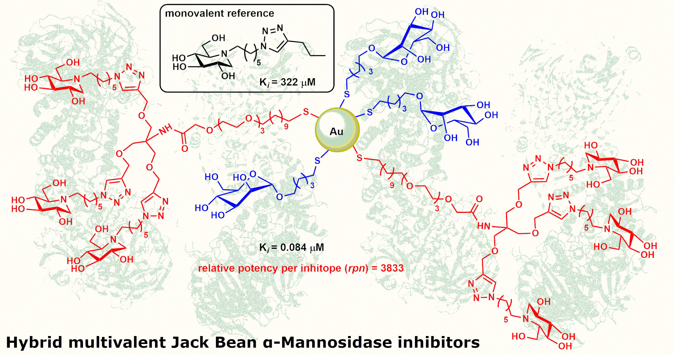
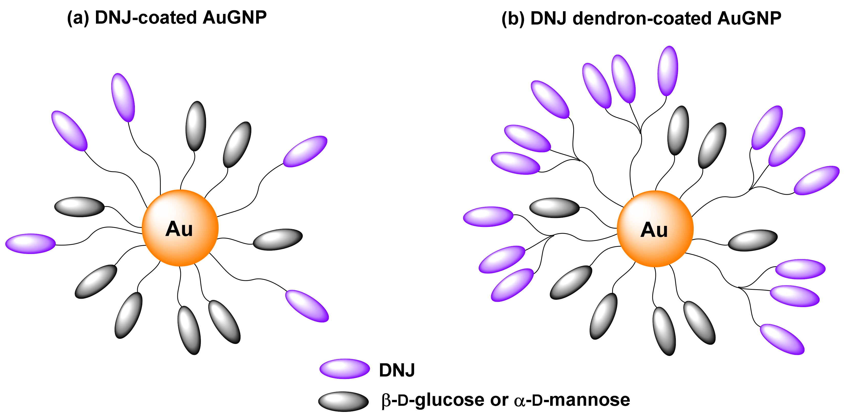
2.1. Preparation of Ligands and Gold Nanoparticles

The gold nanoparticles (15) prepared in this work (Figure 2) are constituted by an approximately 2 nm gold core coated with two components: simple monosaccharides (d-glucose or d-mannose) and DNJ derivatives. We played on the nature of the inner monosaccharide and varied the density of DNJ heads on the periphery of the nanoparticles. Nanoparticles 6 and 7, in Figure 2, bearing 100% monosaccharides with a short and linear five carbons (C5) aliphatic linker, were prepared as control systems. For AuGNPs 15, the carbohydrate moieties guarantee their water dispersibility, while the water soluble and flexible amphiphilic linker was designed to protrude the DNJ heads from the glyco-coated nanoparticle core. In addition, the N-C6 aliphatic linker of the active component DNJ is taking part in the interaction with the catalytic site entry [11,24]. This strategy of ligand presentation was based on our previous experience with similar constructs [21,22,25].
In particular, AuGNPs 6 and 7 were prepared employing, as exclusive components, 5-mercaptopent-1-yl β-d-glucopyranoside (βGlcC5SH, 13) or 5-mercaptopent-1-yl α-d-mannopyranoside (αManC5SH, 14), following well established protocols [25]. AuGNPs 1 and 2 were prepared by employing the monovalent DNJ derivative 12 as the active component and 13 or 14, respectively, as the inner component (Scheme 1).
The preparation of the monovalent DNJ derivative 12 is described in Scheme 1. The peracetylated azido-functionalized DNJ 8 [26] was reduced to the corresponding amino derivative 9 through a standard Staudinger reaction (PPh3, H2O, THF, reflux for 16 h) in 80% yield. The same reaction performed with polymer-bound PPh3 [22,27], and refluxing in THF/H2O, required longer times and provided 9 in lower yields (48%).
The aminoalkyl DNJ 9 was then coupled to the thiol-protected carboxylic acid 10, prepared, as reported in the literature [21], using 1-ethyl-3-(3-dimethylaminopropyl) carbodiimide (EDC) and 1-hydroxybenzotriazole (HOBt) in dry DMF. The amide 11 was obtained in 67% yield after purification by flash column chromatography (FCC). Removal of the acetyl groups of 11 was performed under Zemplén conditions (MeONa) but using CD3OD, instead of MeOH, as solvent to follow the reaction via 1H NMR. Once compound 12 was formed, it was directly used for the next step, avoiding its oxidation to disulfide. AuGNPs 1 and 2 were prepared by mixing 12 with βGlcC5SH (13) or αManC5SH (14), respectively, in the appropriate ratio (40/60) and then adding an Au(III) solution (the total amount of thiols being 3 equiv. with respect to gold) and the reducing agent NaBH4 in excess (Scheme 1). The proportion of the ligands on the gold surface was evaluated by integrating diagnostic signals in the 1H NMR spectrum of the initial mixture and in the 1H NMR spectrum of the supernatant after AuGNPs formation (see Supplementary Materials for 1H NMR spectra). The 40/60 active/inner component ratio allowed to preserve nanoparticle dispersibility in water [22]. After shaking for 2 h at room temperature, the supernatant was removed, and the nanoparticles were washed with methanol. The residue was dispersed in milliQ water, purified by dialysis, and freeze-dried. These nanoparticles were re-dispersed in water after the freeze-drying process, and no flocculation was observed even after several months. Full characterization with different techniques (see below) was carried out before enzymatic evaluation.
To further increase the DNJ loading onto AuGNPs, we combined this strategy with the dendron approach by preparing the trivalent DNJ derivative 18 (Scheme 2). The peracetylated azido-functionalized DNJ 8 was first reacted with the trialkyne 15 [28] to afford the dendron 16 in 85% yield through copper (I) catalyzed azide alkyne cycloaddition (CuAAC) reaction [29,30,31]. Subsequently, introduction of the linker was achieved by coupling the carboxylic acid 10 with 16 in the presence of 3-(diethoxyphosphoryloxy)-1,2,3-benzotriazin-4(3H)-one (DEPBT) as coupling agent affording the final ligand 17 in an excellent 94% yield. In analogy with the incorporation of the monovalent ligand 12 onto AuGNPs, trivalent thiol 18 was generated in situ, and its density on the nanoparticles was modulated with βGlcC5SH (13) (to yield AuGNPs 3) or αManC5SH (14) (to yield AuGNPs 4 and 5), as described above. The mannose-based AuGNPs 4 and 5 shared the same inner component but in different percentages (80% in 4, 60% in 5).
AuGNPs 15 were subjected to quantitative 1H NMR (qNMR) analysis in D2O with 3-(trimethylsilyl)propionic-2,2,3,3-d4 acid (TSP-d4) as an internal standard, following established protocols [21,32,33]. As an example, Figure 3a shows the qNMR for AuGNP 3. In this way, it was possible to quantify the DNJ amount on the nanoparticle by integration of a diagnostic signal (see Supplementary Materials and Table S1) with respect to the internal standard. An average gold core size in 1.6–2.1 nm range was determined by TEM imaging for the AuGNPs (Figure 3c,d) [25,34]. This size was also confirmed by UV-Vis spectra, which did not show an absorption maximum at around 520 nm, typical of gold nanoparticles with a bigger core diameter [35] (Figure 3b).

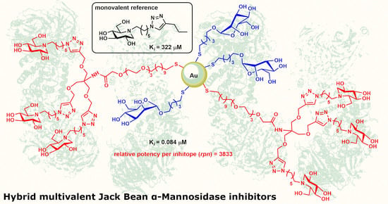
2.2. Biological Evaluation

The DNJ-coated AuGNPs 15 were evaluated against Jack bean α-mannosidase (JBα-man), the glycosidase which showed the largest response to multivalent inhibitor presentation to date [3,4,5,6,7,8,9]. The inhibition mode and constants (Ki) were obtained from Dixon or Lineweaver Burk plots for a scale of inhibitor concentrations reflecting the DNJ concentration directly. The inhibition potency, relative to the corresponding monovalent reference 19 [36] (Figure 4), directly gives the relative potency per inhitope (rpn) (Table 1).
The whole set of newly prepared heterovalent AuGNPs 15 showed good to excellent inhibitory activity towards JBα-man, with Ki values in the low micromolar range to the nanomolar one. All behaved as competitive inhibitors (see Supplementary Materials). Conversely, β-d-gluco- and α-d-mannoside decorated AuGNPs 6 and 7 did not inhibit the enzyme at 0.5 mg mL−1 (This concentration was a good compromise to allow spectrophotometric measurements due to the brown color of AuGNP solutions. The 12-valent mannosylated fullerene from Vincent and Nierengarten et al. (see ref [13]) was active at the hundreds of µM). However, trimerization of DNJ at the gold surface had a major impact: it strongly enhances the affinity towards JBα-man (3 vs. 1, 4 vs. 2, Table 1), with the affinity enhancement per inhitope increasing up to 1840 for AuGNP 4. A further enhancement of the inhibitory properties was reached upon increasing the loading of the trivalent DNJ derivative 18 onto the gold surface from 20% to 40%, with a Ki at 84 nM and an rpn value of 3833 observed for AuGNP 5 (Table 1).
Noticeably, the dendron strategy is a double-edged sword. On the one hand, the intrinsic hindrance of the iminosugar tripod seems to prevent loading as high as linear arms (It is worth noting that for a same percentage loading of d-mannose of 60%, the DNJ concentration on AuGNP 5 is not 3 times higher than that of AuGNP 2, suggesting that the total loading on the particle is lower than for AuGNP 2. However, the ratio between iminosugar DNJ derivatives and monosaccharides still reflects the composition of the preparation mixture, as attested by 1H-NMR spectra recorded before and after (supernatant) the formation of the AuGNPs (see Supplementary Materials)). On the second hand, although the DNJ concentration is almost identical (503 vs. 467 for AuGNP 2 and 5), the inhibition constant of AuGNP 5 is more than 100 times better (Table 1 and Table S1). Despite this lower coating, the multivalent effect, as judged by the rpn, is remarkably increased by two orders of magnitude. The spatial distribution of DNJ at the AuGNP 5, as obtained through the dendron strategy, is highly beneficial to the enzymatic affinity. AuGNPs thus represent an opportunity to compare multivalent systems, having similar concentrations in DNJ inhitopes, but with different local densities. In DNJ dendron-coated AuGNPs, the higher local concentration may favor the bind-and-recapture of the reversible inhibitor heads.

3. Materials and Methods

3.1. General Experimental Procedures for the Syntheses

Commercial reagents were used as received. All reactions were carried out under magnetic stirring and monitored by TLC on 0.25 mm silica gel plates (Merck F254). Column chromatographies were carried out on Silica Gel 60 (32–63 μm) or on silica gel (230–400 mesh, Merck, Darmstadt, Germany). Yields refer to spectroscopically and analytically pure compounds unless otherwise stated. 1H NMR spectra were recorded on a Varian Gemini 200 MHz, a Varian Mercury 400 MHz, or on a Varian INOVA 400 MHz instrument at 25 °C. Additionally, 13C NMR spectra were recorded on a Varian Gemini 200 MHz or on a Varian Mercury 400 MHz instrument. Chemical shifts are reported relative to CDCl3 (13C: δ = 77.0 ppm), or to CD3OD (13C: δ = 49.0 ppm). The following abbreviations were used to designate multiplicities: s = singlet, d = doublet, t = triplet, m = multiplet, br = broad, and dd = double-doublet. Integrals are in accordance with assignments, coupling constants are given in Hz. For detailed peak assignments, 2D spectra were measured (COSY, HSQC). IR spectra were recorded with a IRAffinity-1S SHIMADZU system spectrophotometer. ESI-MS spectra were recorded with a Thermo Scientific™ LCQ fleet ion trap mass spectrometer. Elemental analyses were performed with a Thermo Finnigan FLASH EA 1112 CHN/S analyzer. Optical rotation measurements were performed on a JASCO DIP-370 polarimeter. TEM analysis was performed with a LaB6-TEM of type JEOL JEM-1400PLUS (40 kV–120 kV, HC pole piece) equipped with a GATAN US1000 CCD camera (2 k × 2 k).

3.1.1. Synthesis of Monovalent DNJ Derivative 9

To a solution of 8 (36 mg, 79 μmol) in 1.5 mL of THF 4.6 mL and 3 μL of H2O (145 μmol), PPh3 (25 mg, 87 μmol) was added. The reaction mixture was then refluxed for 16 h, until a TLC analysis (CH2Cl2/MeOH 10: 1) showed the disappereance of the starting material (Rf = 0.91) and the formation of a new product (Rf = 0.10). The solvent was removed under reduced pressure and the crude was purified by FCC (from CH2Cl2/MeOH 10:1 to CH2Cl2/MeOH/NH3 5:1:0.2) affording pure 9 (25 mg, 58.1 μmol, Rf = 0.10 in CH2Cl2/MeOH 10:1) as a colourless oil in 80% yield. [ α ] D 22 = 4.75 (c = 0.59 in CHCl3); 1H-NMR (400 MHz, CDCl3): δ = 5.05 (dt, J = 17.1, 9.2 Hz, 2H, H-3, H-4), 4.94 (td, J = 10.0, 5.0 Hz, 1H, H-2), 4.19–4.09 (m, 2H, H-6), 3.18 (dd, J = 11.5, 5.1 Hz, 1H, Ha-1), 2.75–2.68 (m, 1H, Ha-1′), 2.62 (dt, J = 11.6, 2.5 Hz, 1H, H-5), 2.57–2.50 (m, 1H, Hb-1′), 2.43 (bs, 2H, H-6′), 2.30 (t, J = 10.5 Hz, 1H, Hb-1), 2.06 (s, 3H, OCH3), 2.01 (s, 6H, OCH3), 2.00 (s, 3H, OCH3), 1.48–1.22 (m, 8H, H-2′, H-3′, H-4′, H-5′) ppm; 13C-NMR (100 MHz, CDCl3): δ = 171.0–169.9 (4C, OCOCH3), 74.8 (C-2), 69.7 (C-4), 69.6 (C-3), 61.7 (C-5), 59.7 (C-6), 53.0 (C-1), 51.8 (C-1′), 27.1–26.8 (4C, from C2′ to C5′), 24.9 (C-6′), 21.0–20.8 (4C, COCH3) ppm; MS (ESI): m/z 431.04 (100, H+). IR (CDCl3): ν ~ = 2935, 2860, 2255, 1744, 1664, 1601, 1508, 1369, 1234, 1061, 1032 cm−1. Elemental analysis (%) for C20H34N2O8 (430.49): calcd C, 55.80; H, 7.96; N, 6.51; found: C, 56.34; H, 7.57; N, 6.90.

3.1.2. Synthesis of Monovalent DNJ Based Ligand 11

A solution of EDC.HCl (1-Ethyl-3-(3-dimethylaminopropyl) carbodimide hydrochloride (18.2 mg, 95 µmol), 1-hydroxybenzotriazole (HOBt, 11.7 mg, 87 µmol), and 10 (25.9 mg, 59 µmol) in dry DMF (0.2 mL) was left stirring for 10 min. and then added to a solution of DNJ derivative 9 (17.0 mg, 40 µmol) and N,N-diisopropylethylamine (19 µL, 107 µmol) in DMF (0.9 mL). The reaction mixture was left stirring at room temperature, under nitrogen atmosphere, for 6 h, then diluted with AcOEt (10 mL) and washed with H2O (2 × 3 mL). The organic layer was then washed with a saturated solution of NaHCO3 (1 × 3 mL) and brine (1 × 4 mL), dried over anhydrous Na2SO4 and concentrated under vacuum. The crude was purified by column chromatography (DCM/MeOH from 30:1 to 10:1) affording 22 mg of 11 (26 µmol, 67% yield). Rf = 0.32 (CH2Cl2/MeOH 10:1). [ α ] D 26 = 3.33 (c = 0.69 in CHCl3). 1H-NMR (400 MHz, CDCl3): δ = 5.06 (dt, J = 9.2, 2.8 Hz, 2H, H-3, H-4), 4.99–4.92 (m, 1H, H-2), 4.17–4.13 (m, 2H, H-6), 3.98 (s, 2H, CH2 = CO), 3.69–3.56 (m, 12H), 3.44 (t, J = 6.8 Hz, 2H), 3.29–3.16 (m, 3H, H-6′, Ha-1), 2.85 (t, J = 3.6 Hz, 2H, CH2SAc), 2.77–2.69 (m, 1H, Ha-1′), 2.64–2.61 (m, 1H, H-5), 2.58–2.52 (m, 1H, Hb-1′), 2.32 (bs, 4H, SCOCH3, Hb-1), 2.08 (s, 3H, OCH3), 2.02 (s, 6H, OCH3), 2.01 (s, 3H, OCH3), 1.59–1.45 (m, 4H), 1.36–1.25 (m, 22H) ppm; 13C-NMR (50 MHz, CDCl3): δ = 196.0 (SCOCH3), 170.8-170.0 (4C, −OCOCH3), 169.7 (CONH), 74.7 (C-2), 71.6–70.0 (8C), 69.5 (C-4), 69.4 (d, C-3), 61.5 (C-5), 59.5 (C-6), 52.9 (C-1), 51.7 (C-1′), 38.7 (C-6′), 30.6 (SCOCH3), 29.6–28.8 (11C), 26.9–26.1 (3C, C-2′, C-3′, C-4′), 20.8–20.6 (4C, COCH3) ppm; MS (ESI): m/z 871.35 (100, M+Na+). IR (CDCl3): ν ~ = 3005, 2929, 2856, 1744, 1673, 1540, 1466, 1370, 1237, 1107, 1033 cm−1; Elemental analysis (%) for C41H72N2O14S (849.08): calcd C, 58.00; H, 8.55; N, 3.30; found: C, 56.44; H, 7.90; N, 2.39.

3.1.3. Synthesis of Trivalent DNJ Derivative 16

To a solution of 8 (63 mg, 138 μmol) in 3 mL of THF/H2O = 2:1 CuSO4 (2.1 mg, 13 μmol), sodium ascorbate (5 mg, 25 μmol) and 15 [28] (9.9 mg, 42 μmol) were added. The reaction mixture was stirred in microwave at 80 °C for 45 min, until a TLC analysis (CH2Cl2/MeOH 10: 1) showed the disappearance of the starting material (Rf = 0.46) and the formation of a new product (Rf = 0.00). The solvent was removed under reduced pressure and the crude was purified by FCC (CH2Cl2/MeOH/NH3 from 30:1 to 5: 1: 0.1) affording pure 16 (57 mg, 35.4 μmol, Rf = 0.33 in CH2Cl2/MeOH/NH3 10:1:0.1) as a pale yellow oil, in 85% yield. [ α ] D 22 = 7.47 (c = 0.91 in CHCl3). 1H-NMR (400 MHz, CDCl3): δ = 7.56 (br s, 3H, triazole), 5.09–5.01 (m, 6H, H-3, H-4), 4.97–4.91 (m, 3H, H-2), 4.60 (s, 6H, H-7′), 4.34 (t, J = 7.2 Hz, 6H, H-6′), 4.18–4.10 (m, 6H, H-6), 3.44 (s, 6H, H-8′), 3.18 (dd, J = 11.4, 5.0 Hz, 3 H, Ha-1), 2.76-2.69 (m, 3H, Ha-1′), 2.63–2.60 (m, 3H, H-5), 2.56–2.50 (m, 3H, Hb-1′), 2.29 (t, J = 10.8 Hz, 3H, Hb-1), 2.07 (s, 9H, OCH3), 2.02–2.01 (m, 27H, OCH3), 1.94–1.84 (m, 6H, H-5′), 1.45–1.25 (m, 18H, H-2′, H-3′, H-4′) ppm; 13C-NMR (100 MHz, CDCl3): δ = 170.9-169.9 (12C, OCOCH3), 145.2 (3C, triazole), 122.5 (3C, triazole), 74.8 (3C, C-2), 72.4 (3C, C-8′), 69.7 (3C, C-4), 69.6 (3C, C-3), 65.2 (3C, C-7′), 61.9 (3C, C-5), 59.8 (3C, C-6), 56.3 (NH2C(CH2O-)3), 53.0 (3C, C-1), NH2C(CH2O-)3), 51.7 (3C, C-1′), 50.3 (3C, C-6′), 30.4 (3C, C-5′), 26.8 (3C, C-2′), 26.6 (3C, C-3′), 25.1 (3C, C-4′), 21.0–20.8 (12C, COCH3) ppm; MS (ESI): m/z 1626.58 (100, Na+); IR (CDCl3): ν ~ = 3690, 3606, 2937, 2862, 2257, 1745, 1602, 1438, 1370, 1234, 1097, 1052, 1032 cm−1. Elemental analysis (%) for C73H113N13O27 (1604.75): calcd C, 54.64; H, 7.10; N, 11.35; found: C, 54.60; H, 7.35; N, 10.37.

3.1.4. Synthesis of Trivalent DNJ Based Ligand 17

To a solution of compound 16 (56.8 mg, 35.4 µmol) in dry THF (570 μL) at 0 °C, 3-(diethoxyphosphoryloxy)-1,2,3-benzotriazin-4(3H)-one (DEPBT 21.2 mg, 70.8 μmol) and N,N-diisopropylethylamine (DIPEA 12.0 μL, 70.8 μmol) were added. The mixture was stirred at 25 °C for 15 min under a nitrogen atmosphere, then a solution of 10 (30.0 mg, 21.6 μmol) in dry THF (225 μL) was added and the reaction mixture was stirred at room temperature for 6 days, until a TLC analysis (CH2Cl2/MeOH 6:1) showed the formation of a new product (Rf = 0.7). The reaction mixture was diluted with AcOEt (5 mL), washed with NH4Cl (2 × 3 mL), NaHCO3 (2 × 3 mL) and H2O (2 × 3 mL), dried over anhydrous Na2SO4 and concentrated under vacuum. Purification through gradient column chromatography (CH2Cl2/MeOH from 20:1 to 10:1) afforded pure 17 (61 mg, 30.1 μmol) as a yellow oil, in 94% yield. [ α ] D 18 = 5.29 (c = 0.87 in CHCl3). 1H-NMR (400 MHz, CDCl3): δ = 7.54 (br s, 3H, triazole), 6.81 (s, 1H, CONH), 5.06–4.98 (m, 6H, H-3, H-4), 4.94–4.88 (m, 3H, H-2), 4.57 (s, 6H, H-7′), 4.31 (t, J = 7.2 Hz, 6H, H-6′), 4.15–4.07 (m, 6H, H-6), 3.84 (s, 2H), 3.77 (s, 6H, H-8′), 3.63–3.51 (m, 12 H), 3.39 (t, J = 6.8 Hz, 2H), 3.15 (dd, J = 11.5, 5.1 Hz, 3H, Ha-1), 2.82 (t, J = 7.4 Hz, 2H), 2.74–2.67 (m, 3H, Ha-1′), 2.60–2.58 (m, 3H, H-5), 2.53–2-47 (m, 3H, Hb-1′), 2.28–2.24 (m, 6H, SCOCH3, Hb-1), 2.03 (s, 9H, OCH3), 1.99 (s, 18H, OCH3), 1.98 (s, 9H, OCH3), 1.91–1.84 (m, 6H, H-5′), 1.56–1.48 (m, 4H), 1.33–1.22 (m, 32H) ppm; 13C-NMR (50 MHz, CDCl3): δ = 196.0 (SCOCH3), 170.9–169.7 (12C, OCOCH3), 169.7 (CONH), 144.9 (3C, triazole), 122.6 (3C, triazole), 74.8 (3C, C-2), 71.6–70.1 (8C), 69.6 (3C, C-4), 69.5 (3C, C-3), 68.9 (3C, C-8′), 65.1 (3C, C-7′), 61.8 (3C, C-5), 59.6 (4C, C-6, NHC(CH2O-)3), 53.0 (3C, C-1), 51.6 (3C, C-1′), 50.2 (3C, C-6′), 30.4 (CH2SCOCH3), 29.6–25.0 (22C), 20.9, 20.7 (12C, COCH3) ppm; MS (ESI): m/z 1033.92 (100, (m/z) + 2Na+); IR (CDCl3): ν ~ = 3022, 2932, 2858, 1744, 1675, 1525, 1369, 1220, 1098, 1032 cm−1. Elemental analysis (%) for C94H151N13O33S (2023.34): calcd C, 55.80; H, 7.52; N, 9.00; found: C, 55.76; H, 7.78; N, 8.22.

3.1.5. General Procedure for the In Situ Preparation of Ligands 12 and 18

To a solution of 11 or 17 in CD3OD (10 mg/mL), 30 equivalents of NaOMe were added, and the reaction mixture was left stirring for 2 h at 25 °C under nitrogen atmosphere. The complete disappearance of the starting material was attested via 1H NMR, and the crude, containing compounds 12 or 18, respectively, was directly used for the preparation of AuGNPs.

3.1.6. General Procedure for the Preparation of AuGNPs 15

An aqueous solution of HAuCl4 (25 mM, 1 equiv.) was added to a 12 mM methanolic solution of a suitable mixture of thiol-ending monosaccharide and DNJ ligands (3 equiv. overall). An aqueous solution of NaBH4 (1 M, 27 equiv.) was then added in four portions, with vigorous shaking. The black suspension formed was shaken for 2 h at 25 °C. The residue was washed several times with MeOH. In order to effectively separate the nanoparticles from the supernatant, centrifugation (12,000 rpm, 2 min) was performed. The residue was dissolved in a minimal volume of HPLC gradient grade water and purified by dialysis (SnakeSkin® Pleated Dialysis Tubing, 10,000 MWCO and Slide-A-Lyzer® 10K Dialysis Cassettes, 10,000 MWCO). DNJ-coated AuGNPs were obtained as a dark-brown powder after freeze-drying and characterized via 1H NMR, UV-Vis spectroscopy and TEM analysis (see Supplementary Materials). For the analysis of the ratio between the active component (DNJ-based ligand) and the inner component (monosaccharide ligand), 1H NMR spectra of the initial mixture and the supernatant, after AuGNP formation, were recorded. The DNJ loading on the AuGNPs was evaluated by quantitative NMR (qNMR) using 3-(trimethylsilyl)propionic-2,2,3,3-d4 acid, sodium salt (TSP-d4) as an internal standard in the D2O solution of the AuGNPs. The prepared AuGNPs can be stored at 4 °C for months while maintaining their biophysical properties.

Preparation of 40% monoDNJ-Au-βGlc 1

A 1:1.5 mixture of thiol-ending 12 (4.5 mg, 2.65 μmol) and βGlcC5SH 13 (1.1 mg, 3.89 μmol) in CD3OD (1.1 mL) was used to obtain 0.35 mg of AuGNP 1. TEM (average diameter): 1.8 ± 0.4 nm. Quantitative 1H-NMR (400 MHz, D2O containing 0.05 wt.% of 3-(trimethylsilyl)propionic-2,2,3,3-d4 acid, sodium salt as an internal standard): 0.15 mg of 1 were dissolved in 200 μL of D2O and 15 μL of D2O, containing 0.05 wt.% TSP, were added and 31 nmoles of DNJ conjugate were found. In the quantitative NMR (qNMR) a mediated value of the multiplet corresponding to Ha-1’ proton signal (δ = 2.84 ppm, 1H) of DNJ conjugate and the multiplet corresponding to H-6 (δ = 2.60-2.45 ppm, 2H) was selected for integration as it falls in a spectral region free of other signals. UV-Vis (H2O, 0.1 mg/mL): absence of a maximum band at around 520 nm.

Preparation of 40% monoDNJ-Au-αMan 2

A 1:1.5 mixture of thiol-ending 12 (3.8 mg, 5.89 μmol) and αManC5SH 14 (2.5 mg, 8.84 μmol) in CD3OD (1.2 mL) was used to obtain 1.33 mg of AuGNPs 2. TEM (average diameter): 2.1 ± 0.6 nm. Quantitative 1H-NMR (400 MHz, D2O containing 0.05 wt.% of 3-(trimethylsilyl)propionic-2,2,3,3-d4 acid, sodium salt as an internal standard): 0.60 mg of 2 were dissolved in 200 μL of D2O and 40 μL of D2O containing 0.05 wt.% TSP were added and 140 nmoles of DNJ conjugate were found. Significant peaks: δ = 2.85–2.95 (br signal, 1H, from DNJ conjugate) ppm. UV-Vis (H2O, 0.1 mg/mL): absence of a maximum band at around 520 nm.

Preparation of 20% trisDNJ-Au-βGlc 3

A 1:4 mixture of thiol-ending 18 (10.9 mg, 7.41 μmol) and βGlcC5SH 13 (8.4 mg, 29.64 μmol) in CD3OD (3.1 mL) was used to obtain 4.5 mg of AuGNPs 3. TEM (average diameter): 2.1 ± 0.5 nm. Quantitative 1H-NMR (400 MHz, D2O containing 0.05 wt.% of 3-(trimethylsilyl)propionic-2,2,3,3-d4 acid, sodium salt as an internal standard): 0.60 mg of 3 were dissolved in 180 μL of D2O and 40 μL of D2O containing 0.05 wt.% TSP were added and 170 nmoles of DNJ conjugate were found. Significant peaks: δ = 7.83 (br s, 3H, triazole from DNJ derivative) ppm. UV-Vis (H2O, 0.1 mg/mL): absence of a maximum band at around 520 nm.

Preparation of 20% trisDNJ-Au-αMan 4

A 1:4 mixture of thiol-ending 18 (10.9 mg, 7.41 μmol) and αManC5SH 14 (8.3 mg, 29.64 μmol) in CD3OD (3.1 mL) was used to obtain 3.7 mg of AuGNP 4. TEM (average diameter): 2.0 ± 0.4 nm. Quantitative 1H-NMR (400 MHz, D2O containing 0.05 wt.% of 3-(trimethylsilyl)propionic-2,2,3,3-d4 acid, sodium salt as an internal standard): 0.60 mg of 4 were dissolved in 180 μL of D2O and 40 μL of D2O containing 0.05 wt.% TSP were added and 135 nmoles of DNJ conjugate were found. Significant peaks: δ = 7.84 (br s, 3H, triazole from DNJ derivative) ppm. UV-Vis (H2O, 0.1 mg/mL): absence of a maximum band at around 520 nm.

Preparation of 40% trisDNJ-Au-αMan 5

A 1:1.8 mixture of thiol-ending 18 (6.9 mg, 4.70 μmol) and αManC5SH 14 (2.4 mg, 8.37 μmol) in CD3OD (0.97 mL) was used to obtain 1.7 mg of AuGNP 5. TEM (average diameter): 2.1 ± 0.5 nm. Quantitative 1H-NMR (400 MHz, D2O containing 0.05 wt.% of 3-(trimethylsilyl)propionic-2,2,3,3-d4 acid, sodium salt as an internal standard): 0.60 mg of 5 were dissolved in 180 μL of D2O and 40 μL of D2O containing 0.05 wt.% TSP were added and 151 nmoles of DNJ conjugate were found. Significant peaks: δ = 7.81 (br s, 3H, triazole from DNJ derivative), 2.57-2.44 (m, 6H, from DNJ conjugate) ppm. UV-Vis (H2O, 0.1 mg/mL): absence of a maximum band at around 520 nm.

3.2. Biological Evaluation

p-Nitrophenyl-α-d-mannopyranoside and α-mannosidase (EC 3.2.1.24, from Jack Bean, Km = 2.0 mM pH 5.5) were purchased from Merck (Darmstadt, Germany). Inhibition constants were determined by spectrophotometrically measuring the residual hydrolytic activities of the mannosidase against p-nitrophenyl-α-d-mannopyranoside in the presence and absence of the inhibitor with a VersaMax Microplate Reader. All kinetics were performed at 25 °C and started by substrate addition (20 µL) in a 100 µL assay medium (acetate buffer, 0.2 M, pH = 5) containing α-mannosidase (0.015 U/mL), in presence or absence of various concentrations of inhibitor and substrate (concentrations from km/8 to 2 km). After 15–30 min incubation, the reaction was quenched by the addition of 1M Na2CO3 100 µL). The absorbance of the resulting solution was determined at 405 nm. Under these conditions, the p-nitrophenolate released led to optical densities linear with both reaction time and concentration of the enzyme. Ki values were determined, in duplicate or triplicate, using the Dixon or Lineweaver-Burk graphical methods with Microsoft Excel [37].

4. Conclusions

In conclusion, we report, in this work, the first example of gold nanoparticles decorated with the iminosugar deoxynojirimycin (DNJ). Jack bean α-mannosidase (JBα-man) was chosen as the target enzyme for the biological evaluation due to its well-known responsiveness to the multivalent presentation of iminosugar inhitopes. Monovalent and trivalent DNJ-derivative functionalized with a thiol were synthesized and used in mixture with a glucose or mannose thiol-ending inner component for the in-situ preparation of heterovalent AuGNPs. AuGNPs, bearing only the sugar component, were also prepared as control systems. Biological assays towards JBα-man revealed a competitive inhibition for the whole set of AuGNPs 15, while glyco-coated AuGNPs 67 were inactive, thus demonstrating the fundamental role played by the iminosugar moiety for the inhibition. Comparison with the monovalent reference 19 highlighted affinity enhancements, per inhitope higher than 1600, for the AuGNPs 35 decorated with the trivalent DNJ-derivative, with the best result obtained for AuGNP 5 (Ki = 84 nM, rpn = 3833). Interestingly, AuGNPs 2 and 5 share similar concentration in DNJ but vary by their local inhitope density, which makes them powerful tools to measure the impact of DNJ distribution onto the affinity enhancements. The significant gain of affinity, observed between the DNJ-coated and DNJ dendron-coated AuGNPs, highlights the importance of the bind-and-recapture effect in the complex, interconnected mechanisms underlying the inhibitory multivalent effects. Further investigation with these new nanosystems is currently ongoing in our laboratories.

Supplementary Materials

The following are available online. The 1H and 13C-NMR spectra of DNJ-based ligands 9, 11, 16 and 17; the characterization of AuGNPs 15 (1H-NMR spectra, TEM graphs, UV-Vis spectra); the Dixon and Lineweaver-Burk plots for determination of inhibition constants of AuGNPs 15; Table S1 which contains a resume of DNJ-based AuGNPs 1–5 characterization (average size diameter, calculated amount of iminosugar).

Author Contributions

C.M., F.C. and A.G. planned the synthetic strategy and wrote the manuscript; M.M., M.d.l.A.R. and S.M. characterized the nanosystems; C.V. and J.P.S. made the syntheses; A.B. performed the biological tests, interpreted the results and analyzed them with P.C.; P.C. raised funds. All authors contributed to the writing of the manuscript. All authors have read and agreed to the published version of the manuscript.

Funding

MIUR-Italy (“Progetto Dipartimenti di Eccellenza 2018−2022”) allocated to the Department of Chemistry “Ugo Schiff”, Università di Firenze and Regione Toscana (Bando Salute 2018) for the project: “Late onset Lysosomal Storage Disorders (LSDs) in the differential diagnosis of neurodegenerative diseases: development of new diagnostic procedures and focus on potential pharma-cological chaperones (PCs), Acronym: Lysolate). This work was also funded by the CNRS (UMR 7509), the University of Strasbourg and the Fondation pour la Recherche en Chimie (icFRC Strasbourg).

Acknowledgments

We thank Patrizia Andreozzi for fruitful discussion on experimental data.

Conflicts of Interest

The authors declare no conflict of interest.

Sample Availability

Samples of the compounds reported in the manuscript are not available from the authors.

References

  1. Diot, J.; García-Moreno, M.I.; Gouin, S.G.; Ortiz Mellet, C.; Haupt, K.; Kovensky, J. Multivalent iminosugars to modulate affinity and selectivity for glycosidases. Org. Biomol. Chem. 2009, 7, 357–363. [Google Scholar] [CrossRef]
  2. Compain, P.; Decroocq, C.; Iehl, J.; Holler, M.; Hazelard, D.; Mena Barragán, T.; Ortiz Mellet, C.; Nierengarten, J.-F. Glycosidase Inhibition with Fullerene Iminosugar Balls: A Dramatic Multivalent Effect. Angew. Chem. Int. Ed. 2010, 49, 5753–5756. [Google Scholar] [CrossRef] [PubMed]
  3. Compain, P.; Bodlenner, A. The multivalent effect in glycosidase inhibition: A new, rapidly emerging topic in glycoscience. ChemBioChem 2014, 15, 1239–1251. [Google Scholar] [CrossRef] [PubMed]
  4. Gouin, S.G. Multivalent inhibitors for carbohydrate-processing enzymes: Beyond the “lock-and-key” concept. Chem. Eur. J. 2014, 20, 11616–11628. [Google Scholar] [CrossRef] [PubMed]
  5. Zelli, R.; Longevial, J.-F.; Dumy, P.; Marra, A. Synthesis and biological properties of multivalent iminosugars. New J. Chem. 2015, 30, 5050–5074. [Google Scholar] [CrossRef]
  6. Kanfar, N.; Bartolami, E.; Zelli, R.; Marra, A.; Winum, J.-Y.; Ulrich, S.; Dumy, P. Emerging trends in enzyme inhibition by multivalent nanoconstructs. Org. Biomol. Chem. 2015, 13, 9894–9906. [Google Scholar] [CrossRef]
  7. Matassini, C.; Parmeggiani, C.; Cardona, F.; Goti, A. Are enzymes sensitive to the multivalent effect? Emerging evidence with glycosidases. Tetrahedron Lett. 2016, 57, 5407–5415. [Google Scholar] [CrossRef]
  8. Compain, P. Multivalent effect in glycosidase inhibition: The end of the beginning. Chem. Rec. 2020, 20, 10–22. [Google Scholar] [CrossRef]
  9. González-Cuesta, M.; Ortiz Mellet, C.; García Fernández, J.M. Carbohydrate supramolecular chemistry: Beyond the multivalent effect. Chem. Commun. 2020, 56, 5207–5222. [Google Scholar] [CrossRef] [Green Version]
  10. Lepage, M.L.; Schneider, J.P.; Bodlenner, A.; Meli, A.; De Riccardis, F.; Schmitt, M.; Tarnus, C.; Nguyen-Huynh, N.-T.; Francois, Y.-N.; Leize-Wagner, E.; et al. Iminosugar-Cyclopeptoid Conjugates Raise Multivalent Effect in Glycosidase Inhibition at Unprecedented High Levels. Chem. Eur. J. 2016, 22, 5151–5155. [Google Scholar] [CrossRef]
  11. Howard, E.; Cousido-Siah, A.; Lepage, M.L.; Schneider, J.P.; Bodlenner, A.; Mitschler, A.; Meli, A.; Izzo, I.; Alvarez, A.; Podjarny, A.; et al. Structural Basis of Outstanding Multivalent Effects in Jack Bean α-Mannosidase Inhibition. Angew. Chem. Int. Ed. 2018, 57, 8002–8006. [Google Scholar] [CrossRef] [Green Version]
  12. Mirabella, S.; D’Adamio, G.; Matassini, C.; Goti, A.; Delgado, S.; Gimeno, A.; Robina, I.; Moreno-Vargas, A.J.; Šesták, S.; Jiménez-Barbero, J.; et al. Mechanistic Insight into the Binding of Multivalent Pyrrolidines to α-Mannosidases. Chem. Eur. J. 2017, 23, 14585–14596. [Google Scholar] [CrossRef] [PubMed]
  13. Abellán Flos, M.; García Moreno, M.I.; Ortiz Mellet, C.; García Fernández, J.M.; Nierengarten, J.-F.; Vincent, S.P. Potent Glycosidase Inhibition with Heterovalent Full-erenes: Unveiling the Binding Modes Triggering Multivalent Inhibition. Chem. Eur. J. 2016, 22, 11450–11460. [Google Scholar] [CrossRef] [PubMed]
  14. Siriwardena, A.; Khanal, M.; Barras, A.; Bande, O.; Mena-Barragán, T.; Ortiz Mellet, C.; García Fernández, J.M.; Boukherroub, R.; Szunerits, S. Unprecedented Inhibition of Glyco-sidase-Catalyzed Substrate Hydrolysis by Nanodiamond-Grafted O-Glycosides. RSC Adv. 2015, 5, 100568–100578. [Google Scholar] [CrossRef] [Green Version]
  15. Alali, U.; Vallin, A.; Bil, A.; Khanchouche, T.; Mathiron, D.; Przybylski, C.; Beaulieu, R.; Kovensky, J.; Benazza, M.; Bonnet, V. The Uncommon Strong Inhibition of α-Glucosidase by Multivalent Glycoclusters Based on Cyclodextrin Scaffolds. Org. Biomol. Chem. 2019, 17, 7228–7237. [Google Scholar] [CrossRef]
  16. Marradi, M.; Chiodo, F.; García, I.; Penadés, S. Glyconanoparticles as multifunctional and multimodal carbohydrate systems. Chem. Soc. Rev. 2013, 42, 4728–4745. [Google Scholar] [CrossRef]
  17. Marradi, M.; García, I.; Penadés, S. Carbohydrate-based nanoparticles for potential applications in medicine. In Nanoparticles in Translational Science and Medicine; Villaverde, A., Ed.; Elsevier: Amsterdam, The Netherlands, 2011; Volume 104, pp. 141–173. ISBN 9780124160200. [Google Scholar]
  18. Alvarez-Dorta, D.; Brissonnet, Y.; Saumonneau, A.; Deniaud, D.; Bernard, J.; Yan, X.; Tellier, C.; Daligault, F.; Gouin, S.G. Magnetic Nanoparticles Coated with Thiomannosides or Iminosugars to Switch and Recycle Galactosidase Activity. ChemistrySelect 2017, 2, 9552–9556. [Google Scholar] [CrossRef]
  19. Kleps, I.; Ignat, T.; Miu, M.; Craciunoiu, F.; Trif, M.; Simion, M.; Bragaru, A.; Dinescu, A. Nanostructured Silicon Particles for Medical Applications. J. Nanosci. Nanotechnol. 2010, 10, 2694–2700. [Google Scholar] [CrossRef]
  20. Bonduelle, C.; Huang, J.; Mena-Barragán, T.; Ortiz Mellet, C.; Decroocq, C.; Etamé, E.; Heise, A.; Compain, P.; Lecommandoux, S. Iminosugar-based glycopolypeptides: Glycosidase inhibition with bioinspired glycoprotein analogue micellar self-assemblies. Chem. Commun. 2014, 50, 3350–3352. [Google Scholar] [CrossRef] [PubMed]
  21. Matassini, C.; Marradi, M.; Cardona, F.; Parmeggiani, C.; Robina, I.; Moreno-Vargas, A.J.; Penadés, S.; Goti, A. Gold nanoparticles are suitable cores for building tunable iminosugar multivalency. RSC Adv. 2015, 5, 95817–95822. [Google Scholar] [CrossRef] [Green Version]
  22. Matassini, C.; Vanni, C.; Goti, A.; Morrone, A.; Marradi, M.; Cardona, F. Multimerization of DAB-1 onto Au GNPs affords new potent and selective N-acetylgalactosamine-6-sulfatase (GALNS) inhibitors. Org. Biomol. Chem. 2018, 16, 8604–8612. [Google Scholar] [CrossRef]
  23. Joosten, A.; Schneider, J.P.; Lepage, M.L.; Tarnus, C.; Bodlenner, A.; Compain, P. A Convergent Strategy for the Synthesis of Second-Generation Iminosugar Clusters Using “Clickable” Trivalent Dendrons. Eur. J. Org. Chem. 2014, 1866–1872. [Google Scholar] [CrossRef]
  24. Schneider, J.P.; Tommasone, S.; Della Sala, P.; Gaeta, C.; Talotta, C.; Tarnus, C.; Neri, P.; Bodlenner, A.; Compain, P. Synthesis and Glycosidase Inhibition Properties of Ca-lix[8]Arene-Based Iminosugar Click Clusters. Pharmaceuticals 2020, 13, 366–386. [Google Scholar] [CrossRef] [PubMed]
  25. Martínez-Ávila, O.; Hijazi, K.; Marradi, M.; Clavel, C.; Campion, C.; Kelly, C.; Penadés, S. Gold Manno-Glyconanoparticles: Multivalent Systems to Block HIV-1 gp120 Binding to the Lectin DC-SIGN. Chem. Eur. J. 2009, 15, 9874–9888. [Google Scholar] [CrossRef]
  26. Decroocq, C.; Rodríguez-Lucena, D.; Russo, V.; Mena Barragán, T.; Ortiz Mellet, C.; Compain, P. The Multivalent Effect in Glycosidase Inhibition: Probing the Influence of Architectural Parameters with Cyclodextrin-based Iminosugar Click Clusters. Chem. Eur. J. 2011, 17, 13825–13831. [Google Scholar] [CrossRef]
  27. Ayesa, S.; Samuelsson, B.; Classon, B. A One-Pot, Solid-Phase Synthesis of Secondary Amines from Reactive Alkyl Halides and an Alkyl Azide. Synlett 2008, 97–99. [Google Scholar]
  28. Chabre, Y.M.; Contino-Pépin, C.; Placide, V.; Shiao, T.C.; Roy, R. Expeditive synthesis of glycodendrimer scaffolds based on versatile TRIS and mannoside derivatives. J. Org. Chem. 2008, 73, 5602–5605. [Google Scholar] [CrossRef]
  29. Kolb, H.C.; Finn, M.G.; Sharpless, K.B. Click Chemistry: Diverse Chemical Function from a Few Good Reactions. Angew. Chem. Int. Ed. 2001, 40, 2004–2021. [Google Scholar] [CrossRef]
  30. Rostovtsev, V.C.; Green, L.G.; Fokin, V.V.; Sharpless, K.B. A Stepwise Huisgen Cycloaddition Process: Copper(I)-Catalyzed Regioselective Ligation of Azides and Terminal Alkynes. Angew. Chem. Int. Ed. 2002, 41, 2596–2599. [Google Scholar] [CrossRef]
  31. Tornøe, C.W.; Christensen, C.; Meldal, M. Peptidotriazoles on Solid Phase: [1,2,3]-Triazoles by Regiospecific Copper(I)-Catalyzed 1,3-Dipolar Cycloadditions of Terminal Alkynes to Azides. J. Org. Chem. 2002, 67, 3057–3064. [Google Scholar] [CrossRef]
  32. Manea, F.; Bindoli, C.; Fallarini, S.; Lombardi, G.; Polito, L.; Lay, L.; Bonomi, R.; Mancin, F.; Scrimin, P. Multivalent, Saccharide-Functionalized Gold Nanoparticles as Fully Synthetic Analogs of Type A Neisseria meningitidis Antigens. Adv. Mater. 2008, 20, 4348–4352. [Google Scholar] [CrossRef]
  33. Chiodo, F.; Enríquez-Navas, P.M.; Angulo, J.; Marradi, M.; Penadés, S. Assembling different antennas of the gp120 high mannose-type glycans on gold nanoparticles provides superior binding to the anti-HIV antibody 2G12 than the individual antennas. Carbohydr. Res. 2015, 405, 102–109. [Google Scholar] [CrossRef] [PubMed]
  34. Hostetler, M.J.; Wingate, J.E.; Zhong, C.-J.; Harris, J.E.; Vachet, R.W.; Clark, M.R.; Londono, J.D.; Green, S.J.; Stokes, J.J.; Wignall, G.D.; et al. Alkanethiolate Gold Cluster Molecules with Core Diameters from 1.5 to 5.2 nm: Core and Monolayer Properties as a Function of Core Size. Langmuir 1998, 14, 17–30. [Google Scholar] [CrossRef]
  35. Zhou, M.; Zeng, C.; Chen, Y.; Zhao, S.; Sfeir, M.Y.; Zhu, M.; Jin, R. Evolution from the plasmon to exciton state in ligand-protected atomically precise gold nanoparticles. Nat. Commun. 2016, 7, 13240. [Google Scholar] [CrossRef]
  36. Decroocq, C.; Joosten, A.; Sergent, R.; Mena Barragan, T.; Ortiz Mellet, C.; Compain, P. The Multivalent Effect in Glycosidase Inhibition: Probingthe Influence of Valency, Peripheral Ligand Structure, and Topology with Cyclodextrin-Based Iminosugar ClickClusters. ChemBioChem 2013, 14, 2038–2049. [Google Scholar] [CrossRef] [PubMed]
  37. Segel, I.H. Enzyme Kinetics: Behavior and Analysis of Rapid Equilibrium and Steady-State Enzyme Systems; John Wiley & Sons: New York, NY, USA, 1975. [Google Scholar]
Figure 1. (a) Multivalent presentation of DNJ, using glyco-gold nanoparticles as scaffold and (b) the combination of glyco-gold nanotechnology with the iminosugar dendron approach.
Molecules 26 05864 g001
Figure 2. (a) Schematic representation of the multivalent glyco-gold nanoparticles coated with the iminosugar DNJ derivatives and monosaccharides (β-D-glucose or α-D-mannose derivatives) prepared in this work. (b) Nanoparticles coated with the monosaccharides (control systems).
Molecules 26 05864 g002
Scheme 1. Synthesis of the glyco-gold nanoparticles 1 and 2 decorated with the DNJ monovalent ligand 12.
Molecules 26 05864 sch001
Scheme 2. Synthesis of the trivalent ligand 18 and preparation of AuGNPs 35.
Molecules 26 05864 sch002
Figure 3. Characterization of AuGNP 3 (20% trisDNJ-Au-βGlc): (a) quantitative 1H NMR (D2O) spectrum (400 MHz); (b) UV/Vis spectrum; (c) TEM micrograph in H2O; (d) size-distribution histogram, obtained by measuring 300 nanoparticles (average diameter 2.0 ± 0.2 nm).
Molecules 26 05864 g003
Figure 4. DNJ derivative chosen as the monovalent reference to evaluate the relative potency of the AuGNPs synthesized in this work.
Molecules 26 05864 g004
Table 1. Inhibitory activity of AuGNPs 17 towards Jack bean α-mannosidase (JBα-man).
AuGNPDNJ Conc. (μM)
in 2 mg mL−1 AuGNP 1
Ki (μM) 2rpn3
141316 ± 220
24678 ± 240
35670.198 ± 0.0601626
44500.175 ± 0.1711840
55030.084 ± 0.0663833
60n.i. 4_
70n.i. 4_
1 Estimated by qNMR. 2 The inhibition constant unit directly reflects the concentration of DNJ and not the concentration of nanoparticles. 3 Relative potency per DNJ unit, with respect to the monovalent reference 19; rpn = Ki (monovalent reference 19)/Ki (DNJ in AuGNP 15). 4 n.i. = no inhibition at 0.5 mg mL−1.
Publisher’s Note: MDPI stays neutral with regard to jurisdictional claims in published maps and institutional affiliations.

