Article

Adsorption/Desorption Behaviors and SERS Chemical Enhancement of 6-Mercaptopurine on a Nanostructured Gold Surface: The Au20 Cluster Model

1
Faculty of Food Science and Technology, Thu Dau Mot University, Thu Dau Mot 590000, Vietnam
2
Department of Chemistry, Can Tho University, Can Tho 900000, Vietnam
3
Institute for Computational Science and Technology (ICST), Ho Chi Minh City 700000, Vietnam
*
Authors to whom correspondence should be addressed.
Molecules 2021, 26(17), 5422; https://doi.org/10.3390/molecules26175422
Submission received: 10 August 2021 / Revised: 30 August 2021 / Accepted: 31 August 2021 / Published: 6 September 2021
(This article belongs to the Special Issue Molecular Metal Clusters: Fundamental and Applied Aspects)

Abstract

:
Computational approaches are employed to elucidate the binding mechanism and the SERS phenomenon of 6-mercaptopurine (6MP) adsorbed on the tetrahedral Au20 cluster as a simple model for a nanostructured gold surface. Computations are carried out in both vacuum and aqueous environments using a continuum model. In the gaseous phase and neutral conditions, interaction of 6MP with the gold cluster is mostly dominated by a covalent Au−S bond and partially stabilized by the Au⋅⋅⋅H−N coupling. However, in acidic solution, the nonconventional Au⋅⋅⋅H−S hydrogen-bond becomes the most favorable binding mode. The 6MP affinity for gold clusters decreases in the order of vacuum > neutral solution > acidic medium. During the adsorption, the energy gap of Au20 substantially declines, leading to an increase in its electrical conductivity, which can be converted to an electrical noise. Moreover, such interaction is likely a reversible process and triggered by either the low pH in sick tissues or the presence of cysteine residues in protein matrices. While N−H bending and stretching vibrations play major roles in the SERS phenomenon of 6MP on gold surfaces in neutral solution, the strongest enhancement in acidic environment is mostly due to an Au⋅⋅⋅H−S coupling, rather than an aromatic ring-gold surface π overlap as previously proposed.

Graphical Abstract

1. Introduction

Sulfur-containing compounds with a C=S functional group often exhibit a variety of biological activities and play vital roles in pharmaceutical applications. From a chemical structure viewpoint such a class of drugs can be categorized as either a thiourea S=C(NR2)2 or an artificial DNA nucleobase [1]. Behaving as a nucleobase analogue, the C=S double bond in 6-thiopurine or 6-mercaptopurine (6MP) acts as a surrogate for the natural C=O compounds (Scheme 1). Such a sulfur analogue of adenine is commonly used in the conventional chemotherapy treatment of patients with acute lymphoblastic leukemia, Crohn’s disease, and ulcerative colitis [1,2,3], for its prevention of purine nucleotide biosynthesis [1]. However, the adsorption of 6MP is rather poor, and its therapeutic intake may cause several adverse reactions such as diarrhea, nausea, vomiting, loss of appetite, hair loss, and inflammation of the mouth [4,5]. In this context, it is of great significance to find an appropriate carrier to deliver the drug where it is needed, with the aim of enhancing its therapeutic effects and consequently reducing the unwanted side effects. In addition, development of a simple but powerful method for selective detection of the drug is also of great pharmaceutical interest [6].
In recent decades, nanostructured materials have become more and more popular in biomedical fields in the design of drug delivery systems, diagnosis agents, imaging, and bio-sensors [7,8]. Among the various types of nano-carriers, i.e., dendrimers [9,10], polymers [11,12], liposomes [13,14,15] and metal nanoparticles [16,17,18,19,20], used in drug delivery processes, gold nano-particles (AuNPs) have received a great deal of attention owing to their unique physicochemical properties, high stability, low cytotoxicity, easy functionalization capability, and other relevant features [21,22,23,24,25]. Many valuable outcomes of the use of AuNPs as therapeutic agents, e.g., enhancing the therapeutic effect of the drug, allowing an efficient drug delivery, increasing the therapeutic retention time in the circulation, and improving the target specificity of treatments, have also been well documented in the current literature [26,27,28,29]. Taking advantage of such effects, numerous studies have been carried out to establish suitable techniques for detection of drugs and biomolecules in different media using gold particles [30,31,32,33,34,35]. In particular, the molecular specificity of Raman spectroscopy combined with metallic nano-particles to enhance the signal intensity, which is known as the surface enhanced Raman spectroscopy (SERS) technique, has been used for the study of not only simple molecules such as nucleobases and amino acids, but also for more complex functional structures, i.e., nucleic acids and proteins [36,37,38]. Owing to its efficiency and advantages such as low detection limits, greatly enhanced vibrational positions, and high selectivity for adsorbed molecules, the SERS method has been established as a conventional but highly promising analytical technique for detection of trace amounts of many organic contaminants [39,40].
A number of metals have been identified as capable of producing the SERS phenomenon, including silver, gold, copper, nickel, iron, ruthenium, etc. [41]. Coinage metals turn out to be more suitable overall for use as SERS substrates, since they typically exhibit a localized surface-plasmon resonance in the visible and near-infrared wavelength range where most Raman measurements occur [42]. However, because of several complicating factors including chemical enhancement effects, the Raman band intensity of adsorbates is quite differentiated among specific metal surfaces, and it is rather difficult to predict when a SERS phenomenon can properly be observed. Therefore, a deeper insight into the metal substrate and molecule binding mechanisms is of critical importance for a more comprehensive interpretation of SERS spectra. Several reports on the interactions of 6MP with nanostructured metal surfaces in terms of both experimental and theoretical aspects are available in the recent literature [43,44,45,46]. In order to elucidate the nature of these interactions, validate relevant thermodynamic parameters, as well as interpret or predict spectroscopic properties from the vibrational, electronic absorption spectra, density functional theory (DFT) approaches are commonly selected. Small metallic clusters are typically used as reactant models to investigate the binding mechanism of organic compounds on nanoparticle surfaces. Rashidpour et al. [47] analyzed the adsorption of 6MP on an Au10 surface and found that the conjugation is energetically preferred via the S and S-N sites of the drug molecule. Ren and co-workers [48] also investigated the structures, bonding features, and stability of the complexes of 6MP with an Ag8 cluster by means of DFT (B3LYP) calculations. In a recent study [49] we elucidated the binding mechanism of both 6-mercaptopurine and 6-thioguanine to the Au6 and Au8 clusters. Gold nanoclusters are found to exhibit appropriate characteristics for design of bio-sensing and targeted drug delivery of thione-containing compounds. Irrespective of previous efforts devoted to the study of 6MP binding to gold surfaces [46,49], there are many inherent limitations associated with these studies, and the SERS chemical enhancement of thione-containing drugs by nanostructured gold surfaces is still not fully understood. As a matter of fact, the cluster models used in our recent studies were not large enough to effectively represent the gold nanosurface. Therefore, we set out in the present study to perform DFT calculations to further examine the adsorption/desorption behaviors and the electronic response resulting from the 6MP molecule adsorbed on the larger, stable, and abundant Au20 cluster as a representative gold nanostructured surface. More specifically, the SERS spectrum of the drug adsorbed on the golden surface is generated to interpret available experimental data. A chemical enhancement mechanism is also proposed to provide deeper insights into the SERS phenomenon. As a result of the relative effects of gold, theoretical studies of its derivatives are challenging but important as unexpected phenomena and properties have often been obtained.

2. Computational Methods

Quantum chemical calculations were performed for both binding features and energetic/electronic properties using the Gaussian 16 program package [50]. Geometries of all systems studied were located with the Perdew–Burke–Ernzerhof (PBE) functional [51] in combination with a mixed basis set, i.e., the effective core potential (ECP) cc-pVDZ-PP [52] for the Au element and cc-pVTZ for all atoms in the 6MP molecule. The PBE functional combined with the cc-pVDZ-PP/cc-pVTZ basis sets has been shown to describe relatively well the interactions between gold atoms and sulfur-containing molecules [49,53]. Its use greatly facilitates a direct comparison with previous results. As both the Au20 cluster and all adsorbed species have a closed electron shell in their ground state, we considered only the singlet state. The effect of the solvent, an aqueous solution considered in this study, was simulated using the continuum model, namely, the integral equation formalism-polarizable continuum model (IEF-PCM) [54]. This model has been proved to properly describe both electrostatic and non-electrostatic interactions, and thus could provide us with a sufficient physical description of the solvent environment [55].
Harmonic vibrational frequencies were subsequently computed at the same level to validate the stationary points as equilibrium structures and to estimate the zero-point energy (ZPE) corrections. The Gibbs free energy of the interaction was then calculated with Equation (1):
ΔG° (298 K) = ΔE + ΔZPE + ΔTCG
where ΔE is the relative electronic energy at 0 K, ΔZPE the relative zero-point energy at 0 K, and ΔTCG the relative changes in Gibbs free energy going from 0 to 298 K. The harmonic vibrational frequencies were also employed to simulate the Raman/SERS spectra (without scaling).
The strength of the interactions between both gold cluster and drug molecule was evaluated via the binding energy E b . This parameter is simply defined as the total energy difference between those of the Au20∙6MP complexes and the isolated species by the following Equation (2):
E b = ( E Au 20 + E 6 MP ) E Au 20 · 6 MP
where E Au 20 · 6 MP is the total energy of the Au20∙6MP complexes, whereas E Au 20 and E MP are the energies of the Au20 nanocluster and 6MP molecule, respectively. Thus, a positive value of E b corresponds to an exothermic adsorption, and vice versa. A more positive E b indicates 6MP’s stronger affinity for metals.
Energies of frontier molecular orbitals, i.e., HOMO and LUMO, the HOMO–LUMO energy gap (Eg), and the change of Eg upon binding of the drug to gold clusters were also computed to evaluate the effect of interacting species on each other. The Eg is a useful parameter for determining the kinetic reactivity of materials [56], and its change upon the adsorption process can be used to evaluate the sensitivity of an adsorbent to an adsorbate. Furthermore, the Fermi level energy (EF) and the work function (Φ) of the systems considered were computed by Equations (3) and (4):
E F = E HOMO +   E LUMO 2 ,
Φ = V e ( + ) E F
where E HOMO and E LUMO are HOMO and LUMO energies, respectively; V e ( + ) is the electrostatic potential energy of an electron away from a structured surface.
Φ refers to the energy required for removing an electron from the Fermi level [57]. Thus, we have Φ = E F for V e ( + ) = 0 . Let us mention that Equation (3) is only valid for non-degenerate frontier MOs.
In this study, the impact of 6MP on the E F and Φ values of Au20 was investigated as the main factor in Φ-type sensors using a Kelvin oscillator to quantify Φ during the interaction [58]. Potential changes in the Φ of a sensor arising from the adsorption disturb the gate voltage and generate an electrical noise, which is a suitable indicator for recognizing the presence and attachment of a chemical [59].

3. Results and Discussion

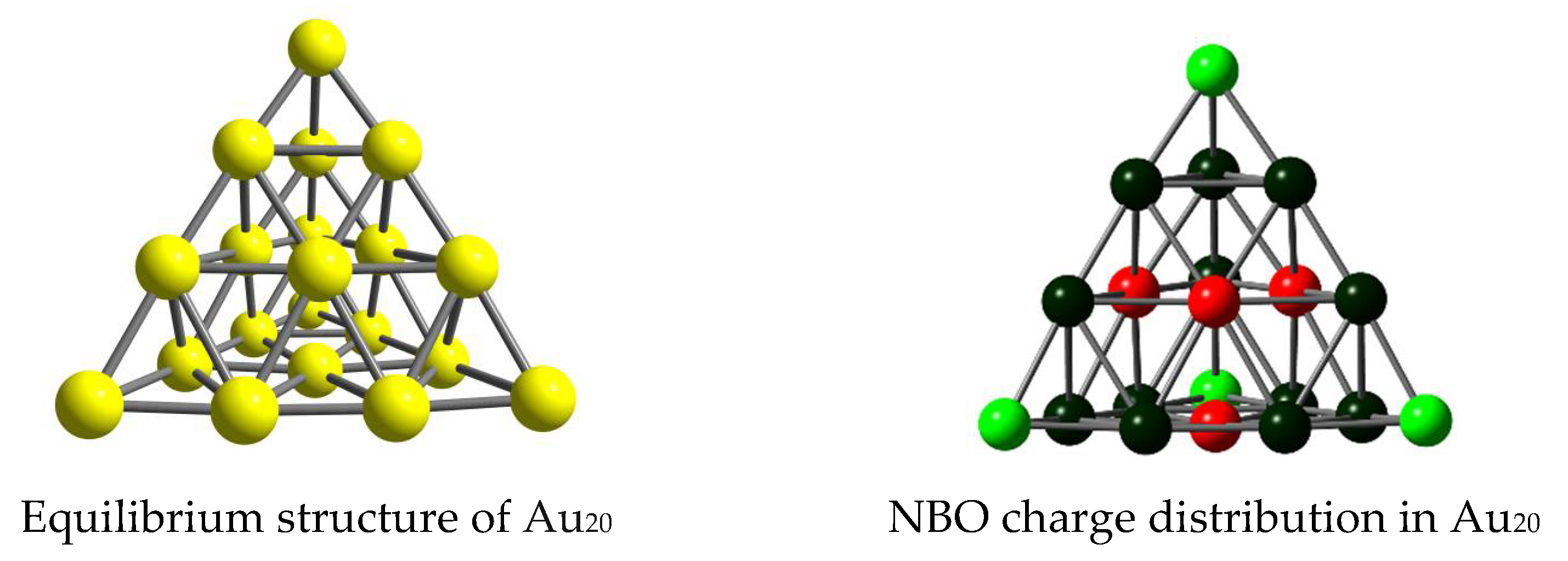
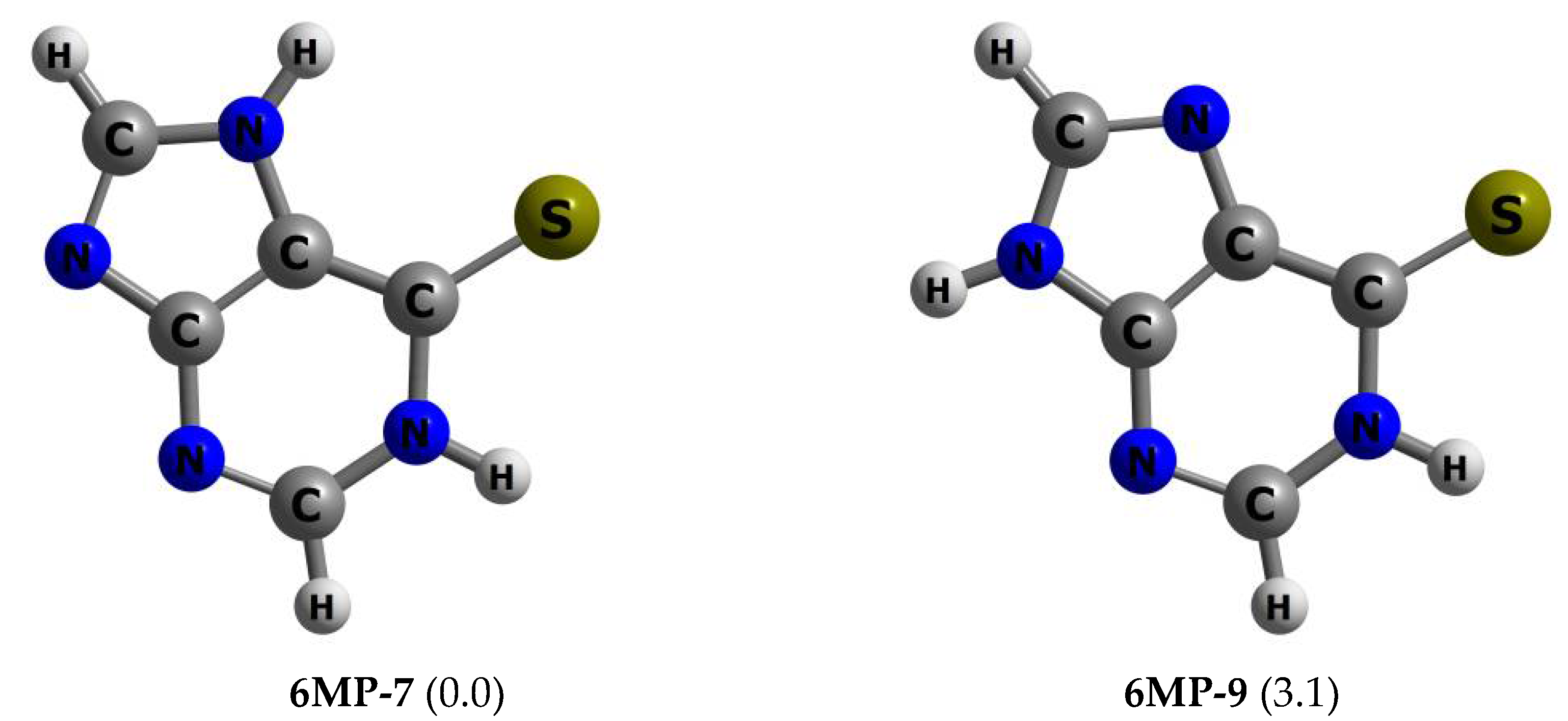
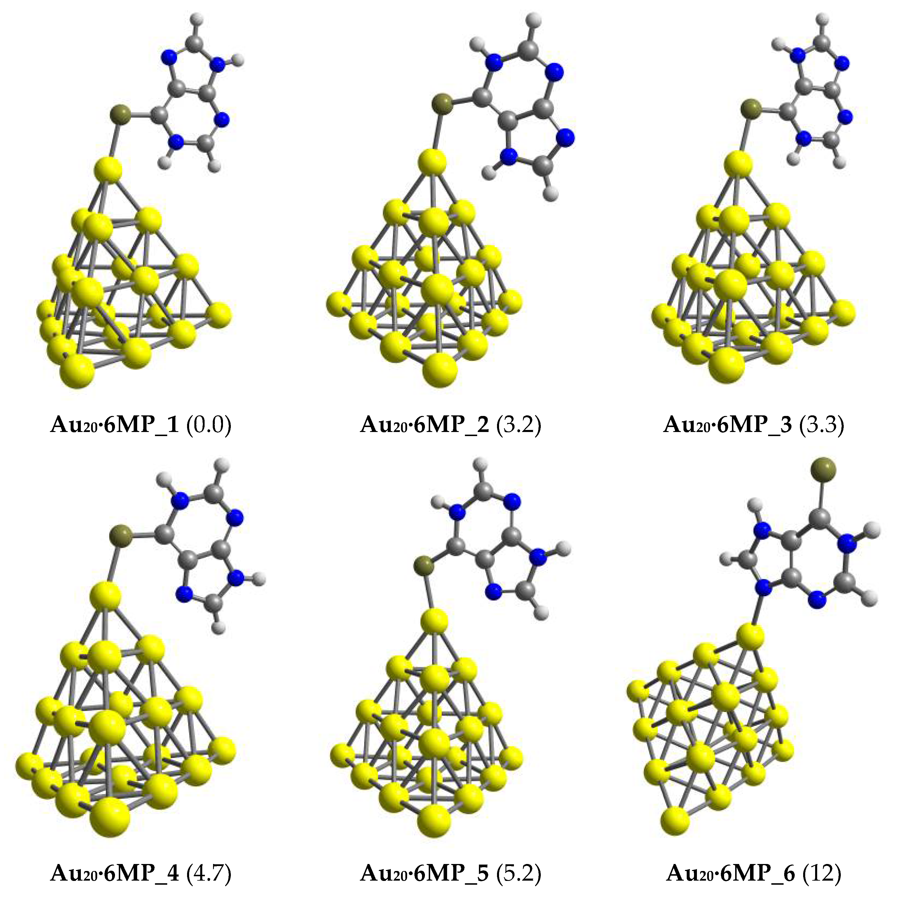
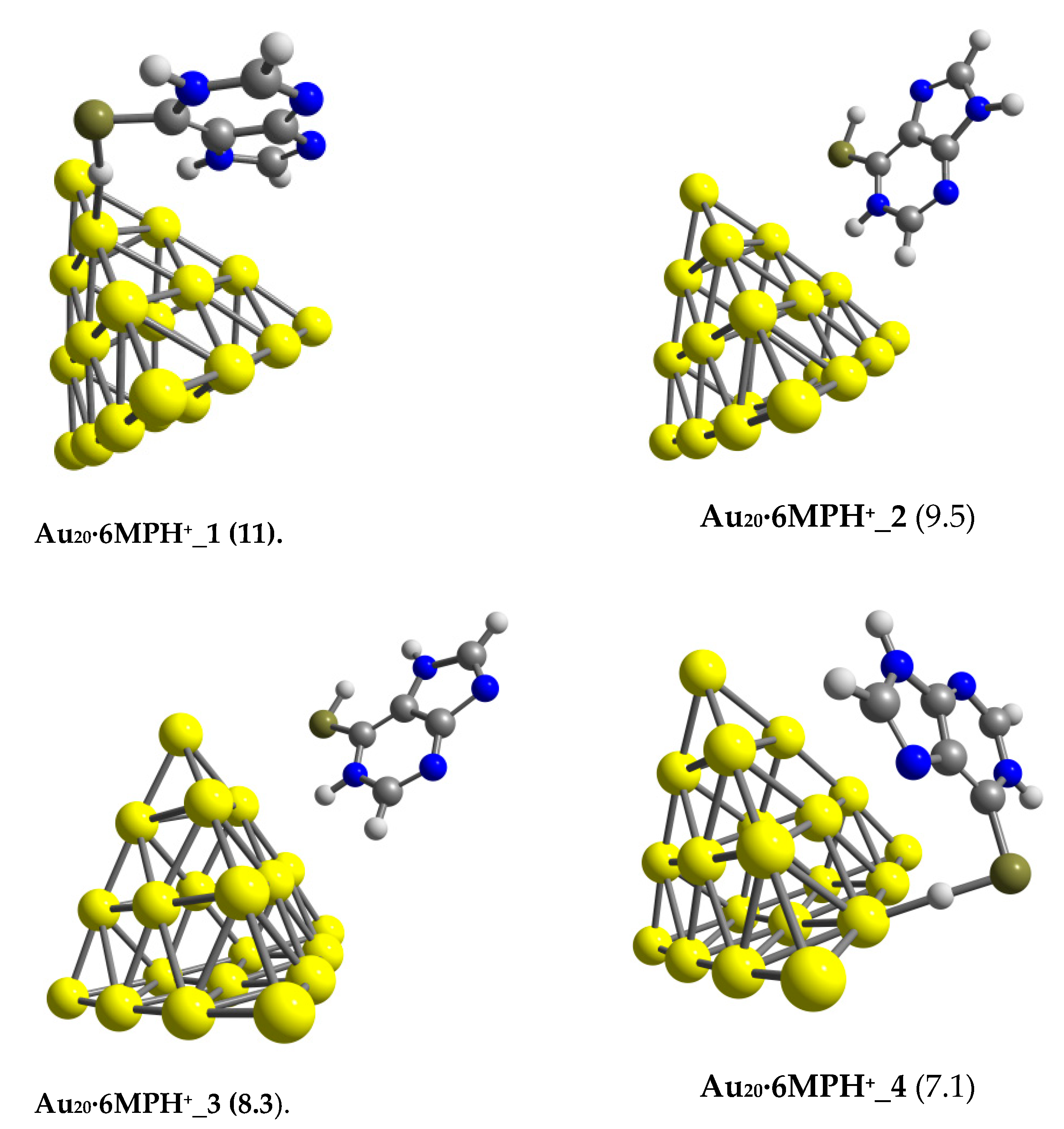
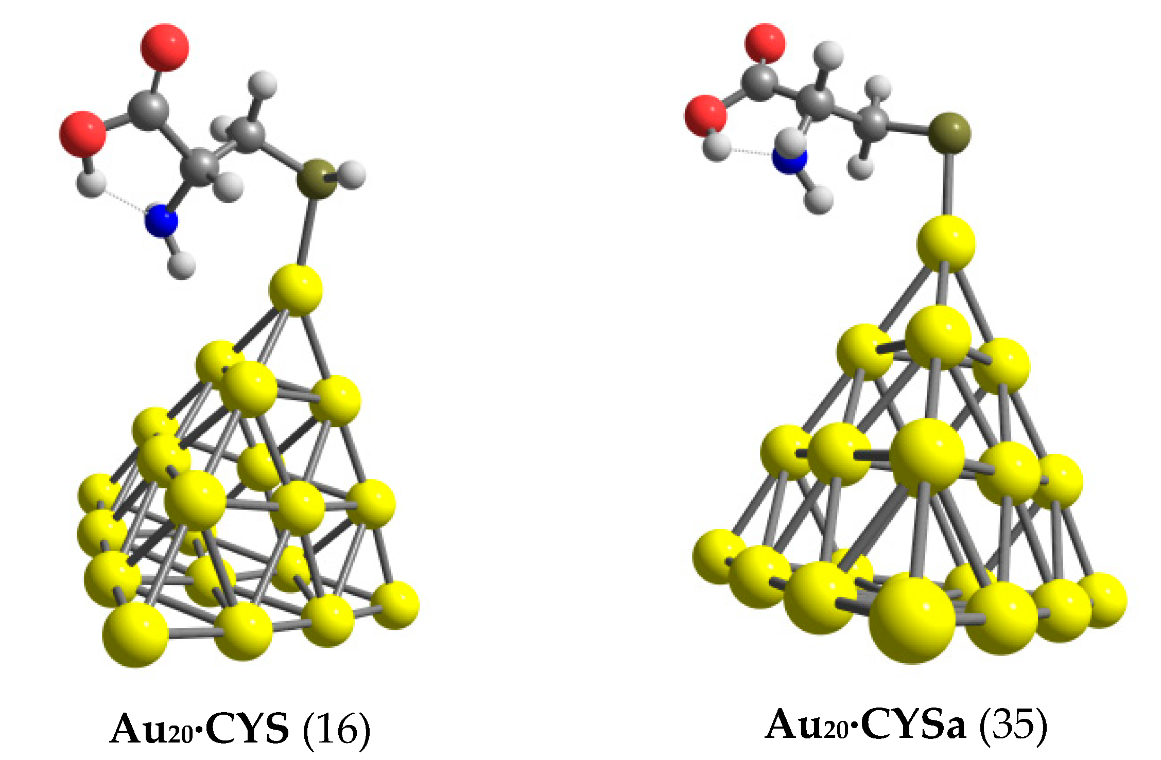
3.1. Structures and Energetics

Numerous experimental and theoretical studies have been devoted to Au20, and the pyramidal structure with a Td symmetry (Figure 1) is unambiguously assigned as its most stable form [60,61]. The Au20 tetrahedron is also well-known as an outstanding indicator in cluster science for its exceptional stability and peculiar 20-electron superatom shell structure [62,63]. Regarding the molecular structure of a 6MP drug, out of several stable configurations [64,65], two planar tautomers—6MP-7 and 6MP-9, presented in Figure 2—tend to dominate its ground state population with a large quantity in both the gas phase and aqueous environments, as reported in recent literature [48,64,66]. In a vacuum, 6MP-7 lies ~3 kcal/mol above 6MP-9, but this energy difference is significantly reduced to ~1 kcal/mol in a water solution (PBE/cc-pVTZ value). Henceforth, both 6MP-7 and 6MP-9 tautomers will be considered for all following calculations and discussion.
As shown in Figure 2, the 6MP molecule exhibits several positions available for binding to the Au20 cluster, i.e., the thione head and the nitrogen atoms unbound to hydrogen. During the interaction, the unoccupied anti-bonding orbitals of 6MP can accept electron density from the Au20 HOMO. The drug is also able to donate electrons on its lone pairs (HOMO) back to the metal LUMO. Moreover, the cluster can play the role of a proton acceptor and form nonconventional Au···H–N hydrogen bonds in which a net charge is transferred from the gold lone pair to the σ*(NH) orbitals [67]. Such an interaction is expected to be an additional factor determining the stability of the resulting products. NBO charges for the Au20 cluster (Figure 1) indicated that the corner Au atoms are positively charged, and thus they are more suitable for a nucleophile attack.
The six stable configurations of the complex formed following binding of 6MP to Au20 are presented in Figure 3, while their optimized coordinates are given in Table S1 in the Supplementary Materials. They were denoted as Au20∙6MP_X with X = 1, 2, 3 … corresponding to an increasing ordering of relative energy (kcal/mol). In general, 6MP prefers to anchor on the vertex of the pyramid, forming a single coordination between the drug and gold cluster (Figure 3). In particular, conformations containing an Au−S bond and a nonconventional hydrogen Au···HN interaction obviously dominate the lowest-lying population of the resulting Au20∙6MP complexes. Such structural motifs, i.e., the Au20∙6MP_1, Au20∙6MP_2, and Au20∙6MP_3 isomers in Figure 3, strongly compete with each other to be the global minimum structure. In the gas phase, Au20∙6MP_1 is considered the most stable form, lying only about 3 kcal/mol below Au20∙6MP_2 and Au20∙6MP_3 (PBE value). Hence, within the expected error margin of the DFT computations, ± 3 kcal/mol on relative energies, they can coexist in subtle experimental conditions, and the adsorbed molecule is likely to rotate around in axing on the C−S bond.
The next lower-energy isomers Au20∙6MP_4 and Au20∙6MP_5 are formed by anchoring the drug on Au20 via the S atom of the pyrimidine ring and the N7 atom of the imidazole moiety. Previously, it was suggested that such interactions are the preferred adsorption modes of 6MP on the gold surface [68]. However, these complexes were computed at the PBE/cc-pVTZ/cc-pVDZ-PP level to be ~5.0 kcal/mol less stable than Au20∙6MP_1 since an N–H⋯Au hydrogen bond was not present, as shown in Figure 3. The remaining structure Au20∙MP_6, which is constructed by directly binding Au20 to a nitrogen atom of the imidazole ring, was computed at 12 kcal/mol less stable. These forms may thus exist only in very small quantities because of their high relative energies. In general, the most favored binding site of 6MP is the S atom, which can be understood on the basis of the hard-soft acid-base (HSAB) theory [69]. Accordingly, the softer S head of 6MP has a higher affinity for soft elements like gold than the harder N head.
For S-bridging complexes, namely Au20∙6MP_1 to Au20∙6MP_5, the S−Au bond distances varied in the range of 2.41 to 2.44 Å (Table 1), being shorter than the corresponding values of 2.52–2.62 Å predicted for the 6MP adsorption on the Au(001) surface [46]. More specifically, the Au–S bond in the lowest energy structure Au20∙6MP_1 was formed with a distance of 2.41 Å, which was slightly shorter than the sum of covalent radii (2.46 Å) of Au (1.44 Å) and S (1.02 Å) [70]. In addition, the chemical bonds S–C and N–H in Au20∙6MP_1 were lengthened to 1.70 and 1.04 Å, respectively, from the corresponding values of 1.66 and 1.02 Å in free 6MP. For the N-bridging complex, the distance of 2.21 Å for the anchoring bonds Au−N in Au20∙MP_6 was somewhat longer than the sum of the covalent radii of nitrogen (0.75 Å) and gold (1.44 Å).
We evaluated the thermodynamic stability of the Au20∙6MP complexes via the binding energy and the changes of Gibbs energy during adsorption. Effects of an aqueous solution on the stability of complexes were also considered using the IEF-PCM model. Table 1 displays the numerical results obtained for the six configurations considered. In the gas phase, the binding energy of Au20∙6MP_1 amounted to 29 kcal/mol and was significantly reduced to 17 kcal/mol for Au20∙6MP_6. The average E b value of 6MP binding to Au20 approximated to ~25 kcal/mol, which was somewhat smaller than the corresponding values obtained for Au6∙6MP (28 kcal/mol) and Au8∙6MP (32 kcal/mol) complexes [49], but much larger than that of 6MP interacting with a B24N24 nanocage (16 kcal/mol) [57]. Moreover, because of the entropic effects the Gibbs free energies of the adsorption process became much smaller. The Gibbs free energy of the most stable complex Au20∙6MP_1 amounted to −20 kcal/mol, whereas that of the least stable isomer Au20∙6MP_6 was only ~9 kcal/mol (Table 1).
When the solvent effect was considered, the absolute values of both E b and Δ G turned out to be smaller, indicating that the interactions became weaker and the resulting complexes more breakable. However, the trend was still similar to the gas phase. For example, the Gibbs energy for forming Au20∙6MP_1 in water was ~ −11 kcal/mol as compared to −20 kcal/mol in the gas phase. In addition, the reaction energies to form S-bridging complexes were larger overall than those of the N-bridging counterparts. In particular, the largest change of Gibbs free energy was predicted to be ~ −12 kcal/mol for Au20∙6MP_3, which was more negative than the value of −3 kcal/mol for the N-bridging complex Au20∙6MP_6. In general, the 6MP adsorption on the Au20 surface is likely to be spontaneous, and the products obtained are thermodynamically stable in both the gas phase and aqueous environments, as the computed Gibbs energies remained negative.

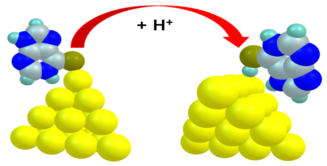
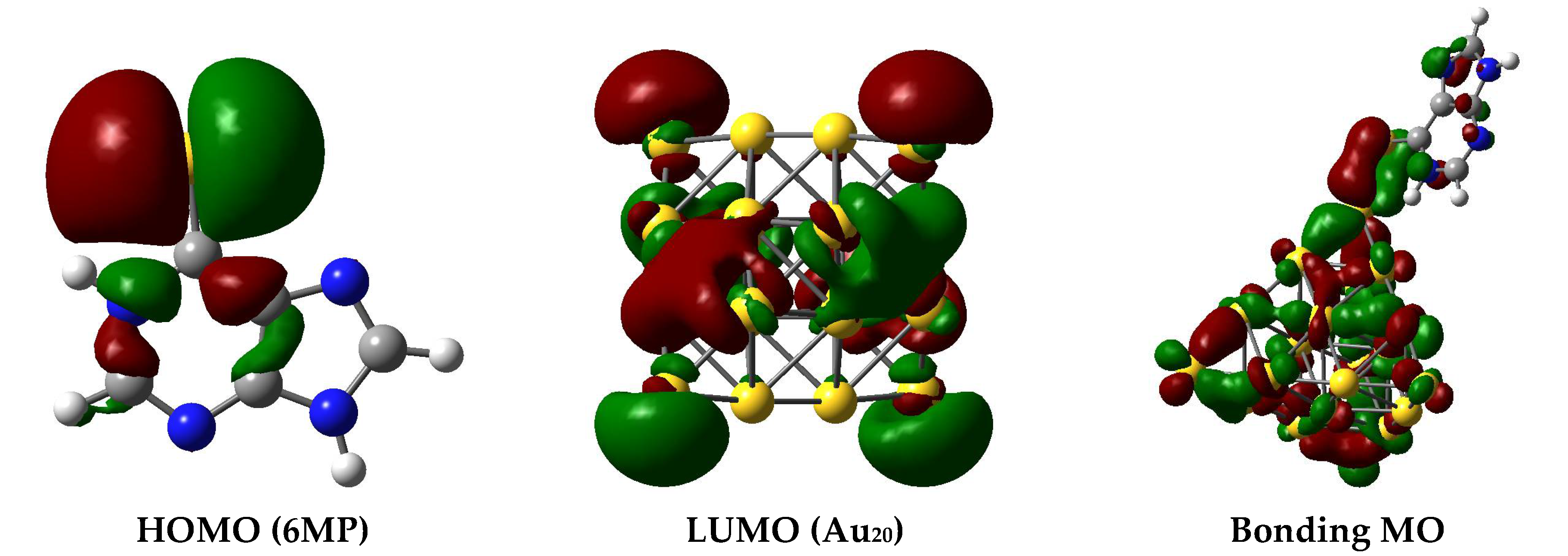
3.2. The Binding Mechanism and Drug Releasing

For deeper insights into the interaction mechanism, we examined the energies of frontier MOs in the 6MP, Au20, and resulting Au20∙6MP complexes. The change in the HOMO-LUMO energy gap ( Δ E g ) was then calculated as follows (Equation (5)):
Δ E g = E g 2 E g 1 E g 1 × 100 %
where E g i is the energy gap E g of Au20 in both the free cluster and the Au20∙6MP complexes.
The change of E g is a proper pointer for identifying the presence and attachment of the drug to a metal surface [71]. In fact, the electrical conductivity ( σ ) of the material is exponentially proportional to a decrease of E g as described by the following Equation (6) [72]:
σ = AT 3 / 2 e E g / 2 k B T ,
where kB and T are the Boltzmann’s constant and the thermodynamic temperature, respectively, and A is a constant. From a 3-parameter Arrhenius-type Equation (6), a small adjustment of Eg due to the adsorption will result in an enormous change of electric conductivity of the system.
Results summarized in Table 2 clearly pointed out that interaction with 6MP tended to alter the electronic properties of the cluster. At the PBE/cc-pvDZ-PP level, the Eg value of Au20 was computed to be ~ −5.7 and −5.1 eV in vacuum and aqueous environments, respectively. During the 6MP adsorption process, such a gap was largely declined up to 31% in the gas phase and 20% when the effect of an aqueous solvent was considered (Table 2). According to Equation (6), the electrical conductivity of Au20 will exponentially increase with respect to the drop in Eg that can be converted to an electrical noise, helping to recognize the drug presence.
The work function Φ, which denotes the energy gap between the Fermi level energy and the void, also plays a crucial role in field emission applications [73]. As a direct result of 6MP attachment, the Fermi level energy decreased from −4.8 eV in the bare Au20 cluster to −4.4 eV in the most stable Au20∙MP_1 complex. Accordingly, the Φ value decreased by nearly 9% (Table 2). This suggested an upgrade in the field emission of the 6MP adsorption and indicated that the gold nanocluster can be used as an Φ–type sensor for detection of 6MP molecules.
Energies of frontier MOs and their energy difference, i.e., HOMO–LUMO gap (Eg), are widely used as indicators for analysis of bonding characteristics between the drug and cluster. As mentioned above, the interactions can be characterized by both forward and backward donation bonds. Depending on the symmetry and energy difference of frontier MOs, the former or the latter distribution will be more predominant. As given in Table 2, the energy gap between LUMO of the 6MP-9 tautomer and HOMO of Au20 cluster was ~3.5 eV in the gas phase. In contrast, the gap between LUMO of Au20 and HOMO of 6MP-9 was significantly reduced to ~1.0 eV. Therefore, the forward donation (6MP→AuN) is likely prevailing, and such an interaction is principally characterized by a charge flow from the drug to the Au atoms. While Au atoms acting as electron acceptors gain more negative charges, the 6MP moiety becomes more positively charged. For example, the net charge of Au20 in Au20-PPX_1 complex was computed to be around −0.4 au (Table S2 of the Supplementary Materials). The nature of Au20−6MP bonding was principally similar to the interaction of lone-pair ligands with gold clusters [74], which were mainly formed via the interaction of the unshared electrons in the HOMO of the 6MP molecule with the LUMO of gold metals. The bonding orbital constructed of an overlap between the HOMO of 6MP and the LUMO of Au20 is illustrated in Figure 4.
We then examined the ability to detach the drug from the gold cluster in target cells, which is considered as the decisive step in a drug delivery process [49,75]. In a human body, the drug can be segregated from the carrier by either external stimuli or internal stimuli operated within a biologically controlled manner such as the pH and glutathione [76]. Because of an exhaustive lactic production, tumor cells are often more acidic than the normal ones. Indeed, the pH of cancer cells is typically below 6 as compared to values above 7 in blood [77]. In an acidic environment, protons can attack any rich-electron site of the 6MP molecule, but the interaction with the S atom may affect the drug release more significantly. Hence, we inspected the effects of protons on the stability of Au20∙6MP complexes by protonation of the thione group, and then carried out geometry optimizations for the resulting Au20∙6MPH+ products.
In an acidic environment, the drug binding to gold metals becomes much more breakable. The interaction is now characterized by a weaker hydrogen bond Au⋅⋅⋅H−X (X = S, N), as demonstrated in Figure 5, instead of a covalent Au−S bond as in Au20∙6MP. Correspondingly, the binding energy of 6MPH+ ions on the Au20 surface was substantially reduced to 11 kcal/mol, which was much smaller than the corresponding value of 22 kcal/mol in the neutral solution. This obviously indicated that under acidic conditions, the 6MP binding to a gold surface was getting weaker, resulting in a faster release.
Another important force contributing to a drug release is an inner inducement related to amino acids in the protein matrices [76]. Thiol-containing amino acids such as cysteine and methionine are usually the most favored binding substrates for noble metals [78,79]. Cysteine is a rather strong acid with dissociation constants of pK1 = 1.7 and pK2 = 8.3 [80]. Thus, in biological systems, the molecule mostly emerges as the deprotonated forms by proton cleavage of either the thiol or carboxyl group [49]. For deeper insights into the drug release mechanism from gold nanoclusters, we examined the following ligand interchange reaction:
Au20∙6MP(aq) + CYSa(aq) → Au20∙CYSa (aq) + 6MP(aq).
The most stable forms of Au20 with cysteine (CYS) and its deprotonated form, denoted as CYSa, are presented in Figure 6. As recently reported [49,53,75], these species tend to adsorb on the gold surface via the S-atom of the thiol group. In an aqueous solution, the binding energies to Au20 were ~35 kcal/mol for CYSa and significantly reduced to ~16 kcal/mol for CYS. These values were much larger than the corresponding values obtained for 6MP adsorption. In fact, the largest binding energy of 6MP to Au20 was ~22 kcal/mol in neutral condition, and it substantially dropped to ~11 kcal/mol in acidic solution. Thus, the affinity of gold nanoparticles for cysteine residues was much higher than its affinity for the 6MP molecule. Hence, drug release from a gold surface in target cells is likely to occur because of this internal force.
Moreover, the complexes were able to undergo a desorption process when exposed to light. In principle, stronger interactions between the adsorbent and adsorbate make the recovery process tougher. According to the transition state theory, the recovery time ( τ ) is exponentially proportional to the binding energy E b as expressed in Equation (7) [81]:
τ = ν 1 e E b   / k T
in which k represents the Boltzman’s constant, ν the attempt frequency, and T the temperature of the system. Equation (7) proves that a larger binding energy Eb will disturb the recovery of the device. In agreement with the larger Eb values (Table 1), the desorption process time of the 6MP molecule from the Au20 surface in a vacuum was generally much longer than that in an aqueous solution.
When applying an attempt frequency of ν = 6.0 × 10 14 Hz (500 nm), the recovery times for Au20∙MP_1, Au20∙MP_2, and Au20∙MP_3 were predicted to be in the range of 2.7 × 10 4 5.7 × 10 6 s, which were significantly reduced to 0.9 × 10 1 4.2 × 10 1 s in aqueous solution. The recovery time of Au20 in the water solution was thus not too long to hinder the desorption process. The cluster also benefited from a rather high sensitivity as its band gap was much modified during the drug binding. Therefore, this nanostructure could be a promising material for designing tiny sensors for 6MP detection.

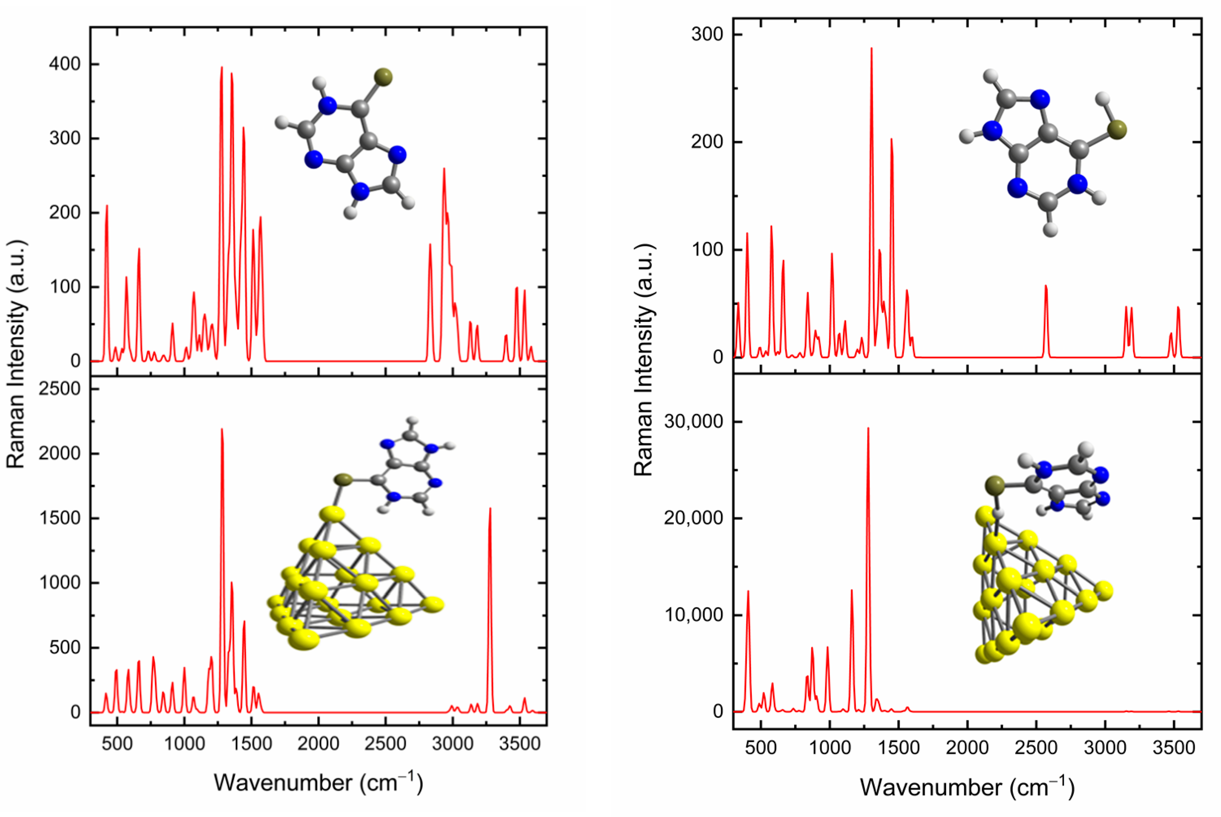
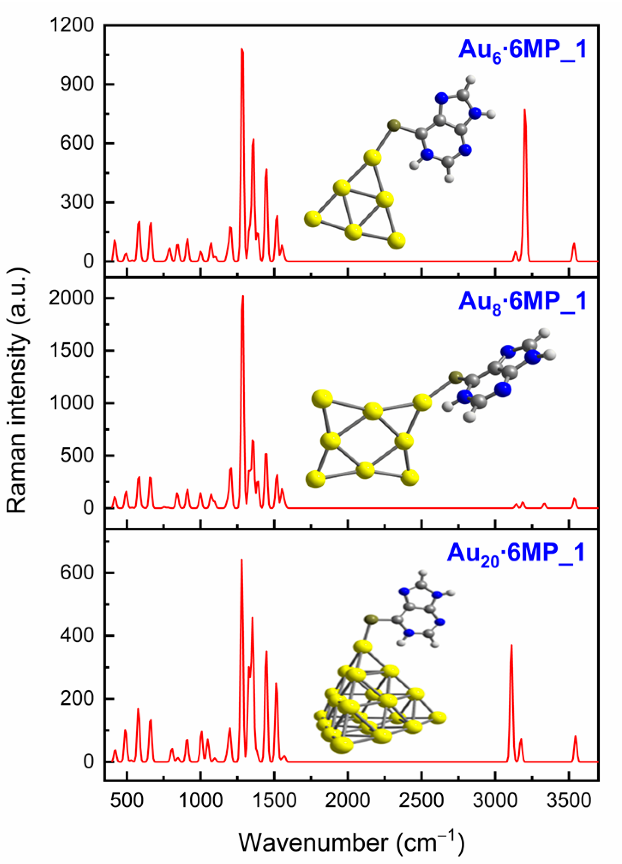
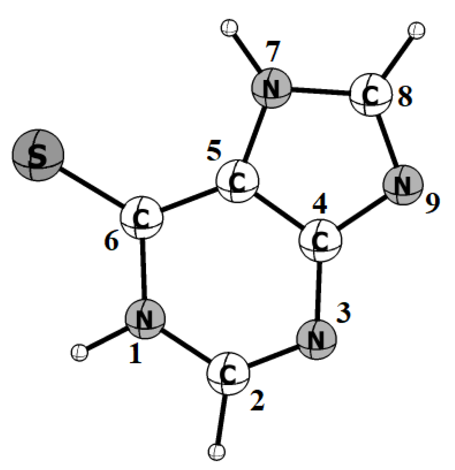
3.3. SERS Spectra of 6MP on Gold Surfaces

For an interpretation of the SERS phenomenon, two operational mechanisms, i.e., the electromagnetic and the chemical enhancements, are frequently taken into consideration [38,82]. Let us briefly recount a few essential characteristics. The former mechanism is controlled by the molecular symmetry and orientation with respect to the metal surface [83]. Certain Raman signals of molecules near the metallic surface become remarkably enhanced as their intensity is proportional to the squared local electromagnetic field intensity. On the contrary, the enhancement does not occur for vibrations parallel to the metal surface [84]. Moreover, the Raman-scattered light results in an extra enhancement when the Raman mode overlaps with a plasmonic resonance [85]. The latter mechanism, in contrast, mostly refers to the chemical interaction and charge transfer between the nanoparticles and adsorbed molecules [86].
Recently, quantum chemical computations have been widely employed for probing the SERS behaviors including the Raman activities and tensor properties that could provide us with valuable information about the interacting centers and the binding mechanism between adsorbed molecules and the metal surface [87]. For example, the Raman enhancements of pyridine on an Ag electrode were measured and then analyzed with the aid of DFT calculations using the tetrahedral Ag20 cluster as a model representing the silver nanoparticles [88]. More recently, similar approaches were also applied to probe the SERS phenomenon of the chlorpyrifos, which is a pesticide using the Ag20 cluster as a model for the silver surface [89]. Likewise, the tetrahedral gold Au20 cluster was also used as a model to validate the SERS spectrum of pramipexole molecules adsorbed on the gold surfaces [75]. Previous reports are also available on the Raman characteristics of 6MP as well as its SERS phenomenon on gold surfaces [68,84,90]. The molecule is well suited for SERS detection, owing to its high affinity for Ag/Au substrates. More importantly, previous studies have revealed for 6MP as well as for other relevant biochemical species that several proposed hypotheses conflict with each other, and numerous interpretative approaches are needed to address the observations.
Normal Raman signatures of 6MP simulated in an aqueous solution are presented in Figure 7. As reported experimentally by Kumar et al. [65], several important signals of the free molecule appeared in the region above 3100 and below 1600 cm−1. In particular, the N−H stretching mode of the imidazole ring was predicted to vibrate at 3532 cm−1, which could be assigned a rather strong peak at 3541 cm−1 in the measured spectrum [65]. However, the corresponding mode of the pyrimidine ring, which was located around 3482 cm−1, was not observed in experimental data. The C−H stretching mode of the imidazole ring was computed to occur at 3185 cm−1, while that of the pyrimidine emerged around 3124 cm−1, which was comparable to the measured result of 3104 cm−1 [65]. Nonetheless, it was rather difficult to unambiguously assign the intense peaks present in the energy region of 1200–1600 cm−1 as they typically resulted from a combination of several vibrations, such as C−X stretching and X−H (X = S, N, C) bending modes. For example, strong signals located around 1360 and 1500 cm−1 likely resulted from a coupling of in-plane ring stretching modes with X−H and C−H in-plane bending vibrations. Experimentally, these modes were observed at 1376 and 1540 cm−1 [65]. Also in line with experimental observations, some weak bands in the lower energy region were likely to arise from a coupling of ring breathing with the N−H and C−H out-of-plane bending modes.
The simulated Raman signatures for the 6MP thiol formed in acidic solution also agreed well with experimental data acquired in HCl solution [68]. Several strong signals in the fingerprint region peaked at 578, 662, 1017, 1304, 1364 and 1450 cm−1, that were comparable to corresponding measured values at 588, 675, 1023, 1330, 1377 and 1449 cm−1. In particular, the appearance of the band at 2570 cm−1 was due to the thiol (S−H) group [91]. The tetrahedron Au20 on the contrary exhibited obvious Raman signals in a narrow frequency range of below 180 cm−1 (Figure S1 of the Supplementary Materials), that were much lower in energy than those of the adsorbed molecules.
The normal Raman and SERS spectra of 6MP adsorbed on the Au20 cluster surface are compared in Figure 7; those simulations were performed in both neutral and acidic conditions. For the neutral environment, the 6MP species binding to Au metals through the S head exhibited a major contribution to the SERS phenomenon. From the simulated spectra, we saw an extraordinary enhancement of vibrations in the energy region of 1250–1500 cm−1. In particular, the strongest enhanced peak at ~1280 cm−1 was mostly produced from the N−H bending mode coupled with the C−H deformation and ring breathing. In the experimental SERS spectrum of 6MP on the gold surface recorded in a KCl solution [68], a very strong band at 1257 cm−1 was also detected. Another noteworthy aspect was the appearance of a prominent band around 3200 cm−1 related to the N−H stretching mode, which was negligible and emerged at higher wavenumbers (3500 cm−1) in the normal Raman spectrum of free 6MP.
A comparison of the SERS spectra in both neutral and acidic environments allowed us to explore important deviations in the band intensities and positions owing to the favorable orientation of the adsorbate. The SERS chemical enhancement of 6MP in acidic solution was much stronger than that recorded in the neutral condition. In addition, we found the disappearance of the S–H band near 2600 cm−1 on the 6MP SERS spectrum, as such vibration is directly oriented to the gold surface. Experimentally, the SERS spectrum of 6MP in a gold colloidal solution (pH = 4.5) recorded by Pannico et al. [84] showed the strongest peak at 1254 cm−1, which was assumed to arise from the ring stretching modes. These authors, moreover, suggested that this enhancement was mainly due to ring-surface π donation and/or surface-ring π* back-donation as a result of a flat orientation on gold surfaces.
In our present calculations, the most enhanced peak was located at 1278 cm−1 and the Au⋅⋅⋅H−S coupling turned out to be a predominant factor leading to the SERS enhancement, rather than an aromatic ring–gold surface π overlap. As a result of this nonconventional hydrogen bond, a charge redistribution emerged, giving rise to an SERS chemical enhancement of the thiol form. A further noteworthy feature was found in the high energy region of the spectrum, where the N/C−H stretching vibrations were located. The SERS spectrum, presented in Figure 7, exhibited a complete absence of signals in the 3000–4000 cm−1 range, which totally agreed with previous experimental observation [84].
A comparison of SERS spectra adsorbed on some small gold clusters is presented in Figure 8. At first glance, we notice that the spectral positions are almost comparable to each other, while the signal intensity is greatly modified with respect to the cluster size. Small Au6 and Au8 systems resulted in higher SERS signals than the larger Au20 counterpart. In particular, the most enhanced SERS signals of 6MP adsorbed on Au8 were primarily related to its lowest HOMO–LUMO energy gap as compared to that of Au6 and Au20 [60]. To simulate the surface of gold nanoparticles, the use of Au20 cluster was expected to provide more reliable results than the smaller sizes Au6 and Au8 as it had a larger number of gold atoms and a more sphere-like structure. Typically, an icosahedral Au nanoparticle with a diameter of 2–10 nm contains several hundreds to thousands of gold atoms [92]. Overall, in considering their characteristics, these small gold clusters behaved quite well as the simplest models for the gold nanoparticles. The smaller sizes Au6 and Au8 allowed us to investigate much larger biomolecular targets having well-defined functional groups.

4. Concluding Remarks

The adsorption and desorption behaviors along with the SERS chemical enhancement of 6-mercaptopurine (6MP) on a gold nanostructured surface were thoroughly investigated by means of DFT computations. The tetrahedral Au20 cluster was used as a simple model to simulate the surface of gold nanoparticles. The effects of an aqueous solution on the structural, energetic, and spectroscopic properties of the resulting complexes were examined with the IEF-PCM model.
In both the gas phase and neutral conditions, the 6MP molecule tended to bind with Au metals via the S-atom of the thione group and was further stabilized by the N−H∙∙∙Au contact. However, in an acidic solution when the drug mainly existed in the thiol form, the interaction was mostly dominated by the weak Au⋅⋅⋅H−S interaction, rather than the stronger covalent Au−S bond. Binding energies were substantially reduced from −29 kcal/mol in a vacuum to −22 and −11 kcal/mol in neutral and acidic solutions, respectively. During the drug adsorption, the energy gap of the adsorbent was greatly modified and likely led to a significant change in the electronic response. Investigation into the frontier MOs also revealed that the forward donation from HOMO(6MP) to LUMO(Au20) was a main ingredient of the 6MP−Au chemical bonding.
A comparison of current computed results with previously measured SERS spectra allowed us to verify the preferable orientation of drug molecules on the gold surface. In a neutral solution, both N−H bending and stretching modes were the predominant factors leading to the SERS enhancement as they were directly oriented to the gold surface. In an acidic environment, the most enhanced peak was found in the fingerprint region and was mainly due to the Au⋅⋅⋅H−S bond rather than the aromatic ring-gold surface π overlap as previously predicted. Desorption of the 6MP molecule from a gold surface was triggered by either the low pH in cancerous tissues or the presence of cysteine residues found in protein matrices. Overall, the results demonstrated the high potentiality of gold nanostructures as drug carriers and detectors, particularly in physiological media.
From a methodological viewpoint, the use of small gold clusters, Aun with n being either 6, 8 or 20, as simple models for nanoparticle surfaces tended to provide similar results for the main features of the complex characteristics and their spectra, with some small differences in spectral intensities. The good behavior of smaller-sized clusters as a surface model opens an avenue for the more systematic theoretical investigations of larger biomolecular targets.

Supplementary Materials

The following are available online. Figure S1: Raman signatures of the tetrahedron Au20, Table S1: Geometries and Cartesian coordinates of Au20⋅6MP complexes, Table S2: Net charges of Au20 moiety in Au20⋅6MP complexes.

Author Contributions

Computations and analysis of results: N.T.N.H. and N.T.S.; Writing and original draft preparation: P.V.N. and M.T.N.; writing, review and editing: all authors. All authors have read and agreed to the published version of the manuscript.

Funding

This research was funded by the Thu Dau Mot University, Vietnam.

Institutional Review Board Statement

Not applicable.

Informed Consent Statement

Not applicable.

Data Availability Statement

Data sharing is not applicable to this article as no new data were created or analyzed in this study.

Conflicts of Interest

The authors declare no conflict of interest. The funders had no role in the design of the study; in the collection, analyses, or interpretation of data; in the writing of the manuscript, or in the decision to publish the results.

Sample Availability

Samples of the compounds in this article are not available from the authors.

References

  1. Scott, K.A.; Njardarson, J.T. Analysis of US FDA-approved drugs containing sulfur atoms. Topics Curr. Chem. 2018, 376, 5. [Google Scholar] [CrossRef]
  2. Parker, W.B. Enzymology of purine and pyrimidine antimetabolites used in the treatment of cancer. Chem. Rev. 2009, 109, 2880. [Google Scholar] [CrossRef] [Green Version]
  3. Sahasranaman, S.; Howard, D.; Roy, S. Clinical pharmacology and pharmacogenetics of thiopurines. Eur. J. Clin. Pharmacol. 2008, 64, 753. [Google Scholar] [CrossRef] [PubMed]
  4. Ensafi, A.A.; Karimi-Maleh, H. Determination of 6-mercaptopurine in the presence of uric acid using modified multiwall carbon nanotubes-TiO2 as a voltammetric sensor. Drug Test. Anal. 2012, 4, 970. [Google Scholar] [CrossRef]
  5. Yang, J.J.; Landier, W.; Yang, W.; Liu, C.; Hageman, L.; Cheng, C.; Pei, D.; Chen, Y.; Crews, K.R.; Kornegay, N. Inherited NUDT15 variant is a genetic determinant of mercaptopurine intolerance in children with acute lymphoblastic leukemia. Int. J. Clin. Oncol. 2015, 33, 1235. [Google Scholar] [CrossRef] [PubMed] [Green Version]
  6. Javarsineh, S.; Vessally, E.; Bekhradnia, A.; Hosseinian, A.; Ahmadi, S. A computational study on the purinethol drug adsorption on the AlN nanocone and nanocluster. J. Clust. Sci. 2018, 29, 767. [Google Scholar] [CrossRef]
  7. Akhter, S.; Ahmad, I.; Ahmad, M.Z.; Ramazani, F.; Singh, A.; Rahman, Z.; Ahmad, F.J.; Storm, G.; Kok, R.J. Nanomedicines as cancer therapeutics: Current status. Curr. Cancer Drug Tar. 2013, 13, 362. [Google Scholar] [CrossRef] [PubMed] [Green Version]
  8. Lah, N.A.C.; Samykano, M.; Trigueros, S. Nanoscale metal particles as nanocarriers in targeted drug delivery system. J. Nanomed. Res. 2016, 4, 00086. [Google Scholar]
  9. Svenson, S.; Tomalia, D.A. Dendrimers in biomedical applications—reflections on the field. Adv. Drug Deliv. Rev. 2012, 64, 102–115. [Google Scholar] [CrossRef]
  10. Kalomiraki, M.; Thermos, K.; Chaniotakis, N.A. Dendrimers as tunable vectors of drug delivery systems and biomedical and ocular applications. Int. J. Nanomed. 2016, 11, 1. [Google Scholar]
  11. Duncan, R. The dawning era of polymer therapeutics. Nat. Rev. Drug Discov. 2003, 2, 347–360. [Google Scholar] [CrossRef]
  12. Vogus, D.R.; Krishnan, V.; Mitragotri, S. A review on engineering polymer drug conjugates to improve combination chemotherapy. Curr. Opin. Colloid Interface Sci. 2017, 31, 75–85. [Google Scholar] [CrossRef]
  13. Allen, T.M.; Cullis, P.R. Liposomal drug delivery systems: From concept to clinical applications. Adv. Drug Deliv. Rev. 2013, 65, 36–48. [Google Scholar] [CrossRef] [PubMed]
  14. Pattni, B.S.; Chupin, V.V.; Torchilin, V.P. New developments in liposomal drug delivery. Chem. Rev. 2015, 115, 10938–10966. [Google Scholar] [CrossRef]
  15. Daraee, H.; Etemadi, A.; Kouhi, M.; Alimirzalu, S.; Akbarzadeh, A. Application of liposomes in medicine and drug delivery. Artif. Cells Nanomed. Biotechnol. 2016, 44, 381–391. [Google Scholar] [CrossRef] [PubMed]
  16. Wei, L.; Lu, J.; Xu, H.; Patel, A.; Chen, Z.-S.; Chen, G. Silver nanoparticles: Synthesis, properties, and therapeutic applications. Drug Discov. Today 2015, 20, 595–601. [Google Scholar] [CrossRef] [PubMed] [Green Version]
  17. Hola, K.; Markova, Z.; Zoppellaro, G.; Tucek, J.; Zboril, R. Tailored functionalization of iron oxide nanoparticles for MRI, drug delivery, magnetic separation and immobilization of biosubstances. Biotechnol. Adv. 2015, 33, 1162–1176. [Google Scholar] [CrossRef]
  18. Kim, C.S.; Tonga, G.Y.; Solfiell, D.; Rotello, V.M. Inorganic nanosystems for therapeutic delivery: Status and prospects. Adv. Drug Deliv. Rev. 2013, 65, 93–99. [Google Scholar] [CrossRef] [PubMed] [Green Version]
  19. Kwon, S.; Singh, R.K.; Perez, R.A.; Abou Neel, E.A.; Kim, H.-W.; Chrzanowski, W. Silica-based mesoporous nanoparticles for controlled drug delivery. J. Tissue Eng. 2013, 4, 2041731413503357. [Google Scholar] [CrossRef] [Green Version]
  20. Sun, X.; Feng, Z.; Hou, T.; Li, Y. Computational simulation of inorganic nanoparticle drug delivery systems at the molecular level. In Computational Pharmaceutics; Ouyang, D., Smith, S.C., Eds.; John Wiley & Sons, Ltd.: Hoboken, NJ, USA, 2015; pp. 149–167. [Google Scholar]
  21. Khan, A.; Rashid, R.; Murtaza, G.; Zahra, A. Gold nanoparticles: Synthesis and applications in drug delivery. Trop. J. Pharm. Res. 2014, 13, 1169–1177. [Google Scholar] [CrossRef]
  22. Bhatia, S. Nanoparticles types, classification, characterization, fabrication methods and drug delivery applications. In Natural Polymer Drug Delivery Systems; Springer: Berlin/Heidelberg, Germany, 2016; pp. 33–93. [Google Scholar]
  23. Bahadar, H.; Maqbool, F.; Niaz, K.; Abdollahi, M. Toxicity of nanoparticles and an overview of current experimental models. Iran. Biomed. J. 2016, 20, 1. [Google Scholar]
  24. Her, S.; Jaffray, D.A.; Allen, C. Gold nanoparticles for applications in cancer radiotherapy: Mechanisms and recent advancements. Adv. Drug Deliv. Rev. 2017, 109, 84–101. [Google Scholar] [CrossRef]
  25. Khodashenas, B.; Ardjmand, M.; Baei, M.S.; Rad, A.S.; Akbarzadeh, A. Conjugation of pectin biopolymer with Au-nanoparticles as a drug delivery system: Experimental and DFT studies. Appl. Organomet. Chem. 2020, 34, e5609. [Google Scholar] [CrossRef]
  26. Demurtas, M.; Perry, C.C. Facile one-pot synthesis of amoxicillin-coated gold nanoparticles and their antimicrobial activity. Gold Bull. 2014, 47, 103. [Google Scholar] [CrossRef] [Green Version]
  27. Austin, L.A.; Mackey, M.A.; Dreaden, E.C.; El-Sayed, M.A. The optical, photothermal, and facile surface chemical properties of gold and silver nanoparticles in biodiagnostics, therapy, and drug delivery. Arch. Toxicol. 2014, 88, 1391–1417. [Google Scholar] [CrossRef] [PubMed] [Green Version]
  28. Torchilin, V.P. Multifunctional, stimuli-sensitive nanoparticulate systems for drug delivery. Nat. Rev. Drug Discov. 2014, 13, 813. [Google Scholar] [CrossRef] [Green Version]
  29. Hainfeld, J.F.; Slatkin, D.N.; Focella, T.M.; Smilowitz, H.M. Gold nanoparticles: A new X-ray contrast agent. Br. J. Radiol. 2005, 79, 248–253. [Google Scholar] [CrossRef] [PubMed]
  30. Ataka, K.; Kottke, T.; Heberle, J. Thinner, smaller, faster: IR techniques to probe the functionality of biological and biomimetic systems. Angew. Chem., Int. Ed. 2010, 49, 5416–5424. [Google Scholar] [CrossRef]
  31. Ma, L.-N.; Zhang, J.; Chen, H.-T.; Zhou, J.-H.; Ding, Y.-Z.; Liu, Y.-S. An overview on ELISA techniques for FMD. Virol. J. 2011, 8, 1–9. [Google Scholar] [CrossRef] [PubMed] [Green Version]
  32. Malou, N.; Raoult, D. Immuno-PCR: A promising ultrasensitive diagnostic method to detect antigens and antibodies. Trends Microbiol. 2011, 19, 295–302. [Google Scholar] [CrossRef] [PubMed]
  33. Seibel, J.; König, S.; Göhler, A.; Doose, S.; Memmel, E.; Bertleff, N.; Sauer, M. Investigating infection processes with a workflow from organic chemistry to biophysics: The combination of metabolic glycoengineering, super-resolution fluorescence imaging and proteomics. Expert Rev. Proteom. 2013, 10, 25–31. [Google Scholar] [CrossRef]
  34. Petryayeva, E.; Algar, W.R.; Medintz, I.L. Quantum dots in bioanalysis: A review of applications across various platforms for fluorescence spectroscopy and imaging. Appl. Spectrosc. 2013, 67, 215–252. [Google Scholar] [CrossRef] [PubMed] [Green Version]
  35. Brauchle, E.; Schenke-Layland, K. Raman spectroscopy in biomedicine–non-invasive in vitro analysis of cells and extracellular matrix components in tissues. Biotechnol. J. 2013, 8, 288–297. [Google Scholar] [CrossRef] [PubMed]
  36. Bantz, K.C.; Meyer, A.F.; Wittenberg, N.J.; Im, H.; Kurtuluş, Ö.; Lee, S.H.; Lindquist, N.C.; Oh, S.-H.; Haynes, C.L. Recent progress in SERS biosensing. Phys. Chem. Chem. Phys. 2011, 13, 11551–11567. [Google Scholar] [CrossRef]
  37. Culha, M. Surface-enhanced raman scattering: An emerging label-free detection and identification technique for proteins. Appl. Spectrosc. 2013, 67, 355–364. [Google Scholar] [CrossRef] [PubMed]
  38. Cialla, D.; Pollok, S.; Steinbrücker, C.; Weber, K.; Popp, J. SERS-based detection of biomolecules. Nanophotonics 2014, 3, 383–411. [Google Scholar] [CrossRef]
  39. Bauman, S.J.; Brawley, Z.T.; Darweesh, A.A.; Herzog, J.B. Substrate oxide layer thickness optimization for a dual-width plasmonic grating for surface-enhanced Raman spectroscopy (SERS) biosensor applications. Sensors 2017, 17, 1530. [Google Scholar] [CrossRef] [Green Version]
  40. Dies, H.; Siampani, M.; Escobedo, C.; Docoslis, A. Direct detection of toxic contaminants in minimally processed food products using dendritic surface-enhanced Raman scattering substrates. Sensors 2018, 18, 2726. [Google Scholar] [CrossRef] [Green Version]
  41. Aikens, C.M.; Schatz, G.C. TDDFT studies of absorption and SERS spectra of pyridine interacting with Au20. J. Phys. Chem. A 2006, 110, 13317–13324. [Google Scholar] [CrossRef]
  42. Sharma, B.; Frontiera, R.R.; Henry, A.-I.; Ringe, E.; Van Duyne, R.P. SERS: Materials, applications, and the future. Mater. Today 2012, 15, 16–25. [Google Scholar] [CrossRef]
  43. Häkkinen, H. The gold–sulfur interface at the nanoscale. Nat. Chem. 2012, 4, 443–455. [Google Scholar] [CrossRef] [PubMed]
  44. Maza, F.L.; Grumelli, D.; Carro, P.; Vericat, C.; Kern, K.; Salvarezza, R.C. The role of the crystalline face in the ordering of 6-mercaptopurine self-assembled monolayers on gold. Nanoscale 2016, 8, 17231–17240. [Google Scholar] [CrossRef] [PubMed]
  45. Fernández, C.C.; Pensa, E.; Carro, P.; Salvarezza, R.; Williams, F.J. Electronic structure of a self-assembled monolayer with two surface anchors: 6-mercaptopurine on Au (111). Langmuir 2018, 34, 5696–5702. [Google Scholar] [CrossRef] [Green Version]
  46. Ren, H.; Cao, X.; Zhang, Y.; Chehelamirani, M.; Salahub, D.R. Theoretical investigation of 6-mercaptopurine isomers’ adsorption on the Au (001) surface: Revealing the fate of different isomers. ACS Omega 2019, 5, 610–618. [Google Scholar] [CrossRef] [Green Version]
  47. Rashidpour, N.; Kashid, V.; Shah, V. Effect of tautomerism on Au-6-mercaptopurine nanocluster stability. AIP Conf. Proc. 2013, 1512, 390–391. [Google Scholar]
  48. Ren, H.; Chen, F.; Li, X.; He, Y. A new insight of structures, bonding and electronic properties for 6-mercaptopurine and Ag 8 clusters configurations: A theoretical perspective. BMC Chem. 2019, 13, 1–10. [Google Scholar] [CrossRef] [PubMed]
  49. Nhat, P.V.; Si, N.T.; Tram, N.T.T.; Duong, L.V.; Nguyen, M.T. Elucidating the binding mechanism of thione-containing mercaptopurine and thioguanine drugs to small gold clusters. J. Comput. Chem. 2020, 41, 1748–1758. [Google Scholar] [CrossRef]
  50. Frisch, M.J.; Trucks, G.W.; Schlegel, H.B.; Scuseria, G.E.; Robb, M.A.; Cheeseman, J.R.; Scalmani, G.; Barone, V.; Petersson, G.A.; Nakatsuji, H.; et al. Gaussian 16; Revision B.01; Software For Theoretical Calculation: Wallingford, CT, USA, 2016; Revision B.01. [Google Scholar]
  51. Perdew, J.P.; Burke, K.; Ernzerhof, M. Generalized gradient approximation made simple. Phys. Rev. Lett. 1996, 77, 3865. [Google Scholar] [CrossRef] [Green Version]
  52. Peterson, K.A. Systematically convergent basis sets with relativistic pseudo-potentials. I. Correlation consistent basis sets for the post-d group 13–15 elements. J. Chem. Phys. 2003, 119, 11099. [Google Scholar] [CrossRef] [Green Version]
  53. Nhat, P.V.; Nguyen, P.T.N.; Si, N.T. A computational study of thiol-containing cysteine amino acid binding to Au6 and Au8 gold clusters. J. Mol. Model. 2020, 26, 58. [Google Scholar] [CrossRef]
  54. Tomasi, J.; Mennucci, B.; Cammi, R. Quantum mechanical continuum solvation models. Chem. Rev. 2005, 105, 2999. [Google Scholar] [CrossRef]
  55. Skyner, R.; McDonagh, J.; Groom, C.; Van Mourik, T.; Mitchell, J. A review of methods for the calculation of solution free energies and the modelling of systems in solution. Phys. Chem. Chem. Phys. 2015, 17, 6174–6191. [Google Scholar] [CrossRef] [PubMed] [Green Version]
  56. Hadipour, N.L.; Ahmadi Peyghan, A.; Soleymanabadi, H. Theoretical study on the Al-doped ZnO nanoclusters for CO chemical sensors. J. Phys. Chem. C 2015, 119, 6398. [Google Scholar] [CrossRef]
  57. Yang, Y.; Ostadhosseini, N. A theoretical investigation on the mercaptopurine drug interaction with boron nitride nanocage: Solvent and density functional effect. Physica E Low-Dimens. Syst. Nanostruct. 2021, 125, 114337. [Google Scholar] [CrossRef]
  58. Baikie, I.D.; Mackenzie, S.; Estrup, P.J.Z.; Meyer, J.A. Noise and the Kelvin method. Rev. Sci. Instrum. 1991, 62, 1326–1332. [Google Scholar] [CrossRef]
  59. Korotcenkov, G. Sensing layers in work-function-type gas sensors. In Handbook of Gas Sensor Materials; Springer: Berlin/Heidelberg, Germany, 2013; pp. 377–388. [Google Scholar]
  60. Nhat, P.V.; Si, N.T.; Leszczynski, J.; Nguyen, M.T. Another look at structure of gold clusters Aun from perspective of phenomenological shell model. Chem. Phys. 2017, 493, 140–148. [Google Scholar] [CrossRef]
  61. Zhao, H.-Y.; Ning, H.; Wang, J.; Su, X.-J.; Guo, X.-G.; Liu, Y. Structural Evolution of Aun (n = 20–32) Clusters: Lowest-Lying Structures and Relativistic Effects. Phys. Lett. A 2010, 374, 1033–1038. [Google Scholar] [CrossRef]
  62. Schwerdtfeger, P. Gold goes nano-from small clusters to low-dimensional assemblies. Angew. Chem. Int. Ed. 2003, 42, 1892–1895. [Google Scholar] [CrossRef] [PubMed]
  63. Gruene, P.; Rayner, D.M.; Redlich, B.; van der Meer, A.F.; Lyon, J.T.; Meijer, G.; Fielicke, A. Structures of neutral Au7, Au19, and Au20 clusters in the gas phase. Science 2008, 321, 674–676. [Google Scholar] [CrossRef] [PubMed] [Green Version]
  64. Bao-Zong, L. DFT calculations on 6-thiopurine tautomers. Acta Chim. Sin. 2004, 62, 1075–1079. [Google Scholar]
  65. Suresh Kumar, S.; Athimoolam, S.; Sridhar, B. XRD, vibrational spectra and quantum chemical studies of an anticancer drug: 6-Mercaptopurine. Spectrochim. Acta A Mol. Biomol. Spectrosc. 2015, 146, 204–213. [Google Scholar] [CrossRef]
  66. Pazderski, L.; Łakomska, I.; Wojtczak, A.; Szłyk, E.; Sitkowski, J.; Kozerski, L.; Kamieński, B.; Koźmiński, W.; Tousek, J.; Marek, R. The studies of tautomerism in 6-mercaptopurine derivatives by 1H-13C, 1H-15N NMR and 13C, 15N CPMAS-experimental and quantum chemical approach. J. Mol. Struct. 2006, 785, 205–215. [Google Scholar] [CrossRef]
  67. Pakiari, A.H.; Jamshidi, Z. Interaction of amino acids with gold and silver clusters. J. Phys. Chem. A 2007, 111, 4391. [Google Scholar] [CrossRef] [PubMed]
  68. Yang, H.; Liu, Y.; Liu, Z.; Yang, Y.; Jiang, J.; Zhang, Z.; Shen, G.; Yu, R. Raman Mapping and In Situ SERS Spectroelectrochemical Studies of 6-Mercaptopurine SAMs on the Gold Electrode. J. Phys. Chem. B 2005, 109, 2739–2744. [Google Scholar] [CrossRef] [PubMed]
  69. Pearson, R.G. Hard and Soft Acids and Bases. J. Am. Chem. Soc. 1963, 85, 3533. [Google Scholar] [CrossRef]
  70. James, A.M.; Lord, M.P. Macmillan’s Chemical and Physical Data; Macmillan: Basingstoke, UK, 1992. [Google Scholar]
  71. Ahmadi Peyghan, A.; Hadipour, N.L.; Bagheri, Z. Effects of Al doping and double-antisite defect on the adsorption of HCN on a BC2N nanotube: Density functional theory studies. J. Phys. Chem. C 2013, 117, 2427. [Google Scholar] [CrossRef]
  72. Ahmadi, A.; Hadipour, N.L.; Kamfiroozi, M.; Bagheri, Z. Theoretical study of aluminum nitride nanotubes for chemical sensing of formaldehyde. Sensor. Actuat. B-Chem. 2012, 161, 1025. [Google Scholar] [CrossRef]
  73. Kim, C.; Kim, B.; Lee, S.M.; Jo, C.; Lee, Y.H. Electronic structures of capped carbon nanotubes under electric fields. Phys. Rev. B 2002, 65, 165418. [Google Scholar] [CrossRef] [Green Version]
  74. Rajsky, T.; Urban, M. Aun (n = 1, 11) clusters interacting with lone-pair ligands. J. Phys. Chem. A 2016, 120, 3938–3949. [Google Scholar] [CrossRef]
  75. Si, N.T.; Nhung, N.T.A.; Bui, T.Q.; Nguyen, M.T.; Nhat, P.V. Gold nanoclusters as prospective carriers and detectors of pramipexole. RSC Adv. 2021, 11, 16619–16632. [Google Scholar] [CrossRef]
  76. Ghosh, P.; Han, G.; De, M.; Kim, C.K.; Rotello, V.M. Gold nanoparticles in delivery applications. Adv. Drug Deliv. Rev. 2008, 60, 1307. [Google Scholar] [CrossRef]
  77. Swietach, P.; Vaughan-Jones, R.D.; Harris, A.L.; Hulikova, A. The chemistry, physiology and pathology of pH in cancer. Philos. Trans. R. Soc. B 2014, 369, 20130099. [Google Scholar] [CrossRef] [Green Version]
  78. Le Guével, X.; Hoötzer, B.; Jung, G.; Hollemeyer, K.; Trouillet, V.; Schneider, M. Formation of fluorescent metal (Au, Ag) nanoclusters capped in bovine serum albumin followed by fluorescence and spectroscopy. J. Phys. Chem. C 2011, 115, 10955–10963. [Google Scholar] [CrossRef]
  79. Eckhardt, S.; Brunetto, P.S.; Gagnon, J.; Priebe, M.; Giese, B.; Fromm, K.M. Nanobio silver: Its interactions with peptides and bacteria, and its uses in medicine. Chem. Rev. 2013, 113, 4708–4754. [Google Scholar] [CrossRef] [PubMed] [Green Version]
  80. O’Neil, M.J. The Merck index: An Encyclopedia of Chemicals, Drugs, and Biologicals; RSC Publishing: London, UK, 2013. [Google Scholar]
  81. Peng, S.; Cho, K.; Qi, P.; Dai, H. Ab initio study of CNT NO2 gas sensor. Chem. Phys. Lett. 2004, 387, 271–276. [Google Scholar] [CrossRef]
  82. Le Ru, E.; Etchegoin, P. Principles of Surface-Enhanced Raman Spectroscopy: And Related Plasmonic Effects; Elsevier: Amsterdam, The Netherlands, 2008. [Google Scholar]
  83. Etchegoin, P.; Cohen, L.; Hartigan, H.; Brown, R.; Milton, M.; Gallop, J. Electromagnetic contribution to surface enhanced Raman scattering revisited. J. Chem. Phys. 2003, 119, 5281–5289. [Google Scholar] [CrossRef]
  84. Pannico, M.; Musto, P. SERS spectroscopy for the therapeutic drug monitoring of the anticancer drug 6-Mercaptopurine: Molecular and kinetic studies. Appl. Surf. Sci. 2021, 539, 148225. [Google Scholar] [CrossRef]
  85. Itoh, T.; Yoshida, K.; Biju, V.; Kikkawa, Y.; Ishikawa, M.; Ozaki, Y. Second enhancement in surface-enhanced resonance Raman scattering revealed by an analysis of anti-Stokes and Stokes Raman spectra. Phys. Rev. B 2007, 76, 129901. [Google Scholar] [CrossRef]
  86. Jensen, L.; Aikens, C.M.; Schatz, G.C. Electronic structure methods for studying surface-enhanced Raman scattering. Chem. Soc. Rev. 2008, 37, 1061–1073. [Google Scholar] [CrossRef]
  87. Yao, G.; Huang, Q. DFT and SERS study of L-Cysteine adsorption on the surface of gold nanoparticles. J. Phys. Chem. C 2018, 122, 15241–15251. [Google Scholar] [CrossRef]
  88. Jeanmaire, D.L.; Van Duyne, R.P. Surface Raman spectroelectrochemistry: Part, I. Heterocyclic, aromatic, and aliphatic amines adsorbed on the anodized silver electrode. J. Electroanal. Chem. Interfacial Electrochem. 1977, 84, 1–20. [Google Scholar] [CrossRef]
  89. Ngo, T.C.; Trinh, Q.T.; Thi Thai An, N.; Tri, N.N.; Trung, N.T.; Truong, D.H.; Huy, B.T.; Nguyen, M.T.; Dao, D.Q. Sers spectra of the pesticide chlorpyrifos adsorbed on silver nanosurface: The Ag20 cluster model. J. Phys. Chem. C 2020, 124, 21702–21716. [Google Scholar] [CrossRef]
  90. Vivoni, A.; Chen, S.P.; Ejeh, D.; Hosten, C.M. Normal-mode analysis of the Raman-active modes of the anti-tumor agent 6-mercaptopurine. J. Raman Spectrosc. 2001, 32, 1–8. [Google Scholar] [CrossRef]
  91. Bazylewski, P.; Divigalpitiya, R.; Fanchini, G. In situ Raman spectroscopy distinguishes between reversible and irreversible thiol modifications in l-cysteine. RSC Adv. 2017, 7, 2964–2970. [Google Scholar] [CrossRef] [Green Version]
  92. Ishida, T.; Murayama, T.; Taketoshi, A.; Haruta, M. Importance of size and contact structure of gold nanoparticles for the genesis of unique catalytic processes. Chem. Rev. 2020, 120, 464–525. [Google Scholar] [CrossRef] [PubMed] [Green Version]
Scheme 1. Chemical structure and atom numbering for 6-mercaptopurine (6MP).
Molecules 26 05422 sch001
Figure 1. Geometry and NBO charge distribution in the tetrahedral Au20 cluster. Au color range.: green, more positive than 0.06 au; red, more negative than −0.06 au.
Molecules 26 05422 g001
Figure 2. Equilibrium structures of two MP lowest-energy tautomers. Values given in parentheses are their relative energies in the gas phase (kcal/mol at the PBE/cc-pVTZ level).
Molecules 26 05422 g002
Figure 3. Lower-lying structures of the Au20∙6MP complex in the gas phase. Values given in parentheses are their relative energies in kcal/mol from PBE computations. The yellow, gray, blue, lime, and white balls represent Au, C, N, S and H atoms, respectively.
Molecules 26 05422 g003
Figure 4. Shapes of frontier orbitals of the Au20 cluster and 6MP molecule before and after interaction (Isovalue for the orbital rendering is 0.02 au).
Molecules 26 05422 g004
Figure 5. Lower-lying structures of the Au20∙6MP complexes in an acidic environment. Values in parentheses are binding energies (kcal/mol, PBE/cc-pVTZ/pVDZ-PP + ZPE). The yellow, gray, blue, lime, and white balls represent Au, C, N, S and H atoms, respectively.
Molecules 26 05422 g005
Figure 6. Equilibrium structures for complexes of Au20 with cysteine (CYS) and its deprotonated form (CYSa). Values in parentheses are binding energies (kcal/mol, PBE/cc-pVTZ/pVDZ-PP + ZPE). The yellow, gray, blue, lime, red, and white balls represent Au, C, N, S, O and H atoms, respectively.
Molecules 26 05422 g006
Figure 7. Computed Raman signatures of 6MP (above) and its SERS spectra adsorbed on the Au20 surface (below). Simulations were performed in both neutral (left) and acidic (right) conditions.
Molecules 26 05422 g007
Figure 8. Comparison of 6MP SERS spectra adsorbed on small Aun clusters with n = 6, 8, 20. Simulations are performed in an aqueous solution.
Molecules 26 05422 g008
Table 1. Relative energy (RE), binding energy ( E b and Gibbs energy ΔG298 in kcal/mol for the 6MP adsorption on Au20, along with Au−X=S, N) bond lengths in the resulting complexes (PBE/cc-pVTZ/cc-pVDZ-PP + ZPE).
ComplexRE E b Δ G 298 r A u X   ( Å )
In a VacuumIn Water In a VacuumIn WaterIn a VacuumIn Water
Au20∙6MP_10.00.029.422.4 −19.6−10.52.41
Au20∙6MP_23.21.026.221.5−16.9−7.822.41
Au20∙6MP_33.31.026.221.4−16.6−11.52.42
Au20∙6MP_44.79.924.712.6−14.7−1.912.41
Au20∙6MP_55.21.424.221.0−15.3−9.642.44
Au20∙6MP_6127.817.314.7−9.08−3.322.21
Table 2. Energies of HOMO and LUMO, Fermi level energy (EF), work function (Φ), energy gap ( E g and change of energy gap ( Δ E g ) upon the 6MP adsorption on the Au20 surface. Values are given in eV, except for Δ E g (%).
Species In a Vacuum In an Aqueous Solution
HOMO E F LUMO E g Δ E g Φ HOMO E F LUMO E g Δ E g Φ
6MP-7−5.37−3.96−2.552.81-3.96 −5.52−4.05−2.582.94-4.05
6MP-9−4.93−3.58−2.232.70-3.58 −5.39−3.91−2.422.98-3.91
Au20−5.73−4.84−3.951.78-4.84 −5.07−4.13−3.181.90-4.13
Au20∙MP_1−5.15−4.42−3.691.47−17.44.42 −4.81−4.01−3.201.60−15.84.01
Au20∙MP_2−5.28−4.52−3.761.52−14.64.52 −4.84−4.03−3.221.62−14.74.03
Au20∙MP_3−5.25−4.50−3.751.49−16.34.50 −4.82−4.02−3.221.60−15.84.02
Au20∙MP_4−4.86−4.30−3.731.13−36.54.30 −4.74−3.98−3.221.52−20.03.98
Au20∙MP_5−4.87−4.26−3.651.23−30.94.26 −4.77−3.98−3.181.60−15.83.98
Au20∙MP_6−5.10−4.35−3.591.51−15.24.35 −4.85−4.01−3.161.68−11.64.01
Publisher’s Note: MDPI stays neutral with regard to jurisdictional claims in published maps and institutional affiliations.

