Review

Biodiversity of Secondary Metabolites Compounds Isolated from Phylum Actinobacteria and Its Therapeutic Applications

by
Muhanna Mohammed Al-shaibani
1,2,
Radin Maya Saphira Radin Mohamed
1,*,
Nik Marzuki Sidik
3,*,
Hesham Ali El Enshasy
4,5,
Adel Al-Gheethi
1,*,
Efaq Noman
6,
Nabil Ali Al-Mekhlafi
7,8 and
Noraziah Mohamad Zin
2
1
Micro-Pollutant Research Centre (MPRC), Faculty of Civil Engineering and Built Environment, Universiti Tun Hussein Onn Malaysia, Parit Raja 86400, Johor, Malaysia
2
Center for Diagnostic, Therapeutic and Investigative Studies, Faculty of Health Sciences, Universiti Kebangsaan Malaysia, Kuala Lumpur 50300, Malaysia
3
Faculty of Agro-Based Industry, Universiti Malaysia Kelantan, Jeli 17600, Kelantan, Malaysia
4
Institute of Bioproducts Development (IBD), Universiti Teknologi Malaysia (UTM), Skudai 81310, Johor, Malaysia
5
City of Scientific Research and Technology Applications (SRTA), 21934 New Burg Al Arab, Alexandria, Egypt
6
Applied Microbiology Department, Faculty of Applied Sciences, Taiz University, Taiz 6803, Yemen
7
Atta-ur-Rahman Institute for Natural Product Discovery, UiTM, Puncak Alam Campus, Bandar Puncak Alam 42300, Selangor, Malaysia
8
Biochemical Technology Program, Department of Chemistry Faculty of Applied Science, Thamar University, Thamar P.O. Box 87246, Yemen
*
Authors to whom correspondence should be addressed.
Molecules 2021, 26(15), 4504; https://doi.org/10.3390/molecules26154504
Submission received: 27 June 2021 / Revised: 14 July 2021 / Accepted: 22 July 2021 / Published: 26 July 2021

Abstract

:
The current review aims to summarise the biodiversity and biosynthesis of novel secondary metabolites compounds, of the phylum Actinobacteria and the diverse range of secondary metabolites produced that vary depending on its ecological environments they inhabit. Actinobacteria creates a wide range of bioactive substances that can be of great value to public health and the pharmaceutical industry. The literature analysis process for this review was conducted using the VOSviewer software tool to visualise the bibliometric networks of the most relevant databases from the Scopus database in the period between 2010 and 22 March 2021. Screening and exploring the available literature relating to the extreme environments and ecosystems that Actinobacteria inhabit aims to identify new strains of this major microorganism class, producing unique novel bioactive compounds. The knowledge gained from these studies is intended to encourage scientists in the natural product discovery field to identify and characterise novel strains containing various bioactive gene clusters with potential clinical applications. It is evident that Actinobacteria adapted to survive in extreme environments represent an important source of a wide range of bioactive compounds. Actinobacteria have a large number of secondary metabolite biosynthetic gene clusters. They can synthesise thousands of subordinate metabolites with different biological actions such as anti-bacterial, anti-parasitic, anti-fungal, anti-virus, anti-cancer and growth-promoting compounds. These are highly significant economically due to their potential applications in the food, nutrition and health industries and thus support our communities’ well-being.

Graphical Abstract

1. Introduction

Global demand for new chemotherapeutic compounds and antibiotics with high bioactivity and low toxicity has increased recently due to the emergence of life-threatening microorganisms and multidrug resistance agents among viruses, bacteria and fungi [1]. Additionally, the detection of secondary metabolites molecules with unique modes of action established various therapeutic agents’ strategies for treating many illnesses [2]. To be more specific, endophytic Actinobacteria are microorganisms that represent a new production source of a large number of secondary metabolites, including alkaloids, beta-lactams, sulfonamides, aminoglycosides, glycopeptides, siderophores, quorum-sensing molecules, immunosuppressants, polyene macrolides, saccharides, pyrazoloisoquinolinones, butenolides, nucleosides and degradative enzymes [3]. In fact, it has been reported that more than 10,000 various bioactive compounds have been discovered from Actinobacteria [4].
Endophytic microbes refer to a group of microorganisms, mostly fungi and bacteria, that exist in the host plant’s intracellular space. It usually causes no obvious harmful effect or symptoms of the disease and could produce various associations such as trophobiotic communalistic, mutualistic and symbiotic co-existence [5]. Endophytes in woody plant hosts could exist within host tissues and protect host plants against herbivores and other pathogenic microorganisms [6]. ActinobacteriaActinobacteria are Gram-positive bacteria with high guanine and cytosine (G + C) content in their genomes, and they are classified into 6 classes, 79 families of 46 orders and 10 fresh families of 16 new orders based on phylogeny using 16S rRNA sequences. The Actinobacterial classes consist of Thermoleophilia, Rubrobacteria Nitriliruptoria, Coriobacteria, Actinomycetia and Acidomicrobiia Salam et. al. [7]. Actinobacteria have ubiquitous characteristics. They are present in diverse ecosystems on the earth such as endophytically with plants and in terrestrial and aquatic environments. An abundance of Actinobacteria species have been recorded in ordinary, extraordinary and extreme environments with high or low temperatures, high radiation, acidic/alkaline pH, salinity, low levels of available moisture and nutrients [8].
The genus Streptomyces is a Gram-positive bacteria. It is the largest genus of the phylum Actinobacteria, which has complex growth and can produce various secondary metabolites [8]. In addition, there are more than 800 Streptomyces species that have been found to date (see http://www.bacterio.net/Streptomyces.html (accessed on 20 August 2020) [9]. Streptomyces is the major microbial genus of the most antibiotic-producing bacteria in the microbial world discovered so far, where streptomycin, gentamycin, rifamycin, chloramphenicol and erythromycin are produced by Streptomyces [10].
Actinobacteria have a large number of secondary metabolite biosynthetic gene clusters. Biosynthetic gene clusters (BGCs) are known as genes comprising locally clustered groups encoding a secondary metabolite biosynthetic pathway. In addition, BGCs contain genes encoding all enzymes required to produce secondary metabolites and pathway-specific regulatory genes. The Actinobacteria have diverse physiology and metabolic flexibility with high potential to produce novel bioactive compounds and enzyme production [11].
The current review aims to summarise the biodiversity, biosynthesis of novel bioactive secondary metabolites, detection of new resources, and strategies to search for potential bioactive compounds producers. In addition, it aims to determine the effects of environments and ecosystem on the phylum Actinobacteria that produce potential bioactive compounds in the pharmaceutical industry market. The VOSviewer software tool was used to visualise bibliometric networks to support Scopus’s prevalent bibliographic databases. This information would be helpful to other scholars who attempt to discover and isolate specific bioactive compounds from phylum Actinobacteria under different ecosystems. Besides their utilisation by people, the perception of the function and distribution of microbial products is regarded as significant to understand microbial families and their impact on biogeochemical cycles.

2. Methods and Protocol

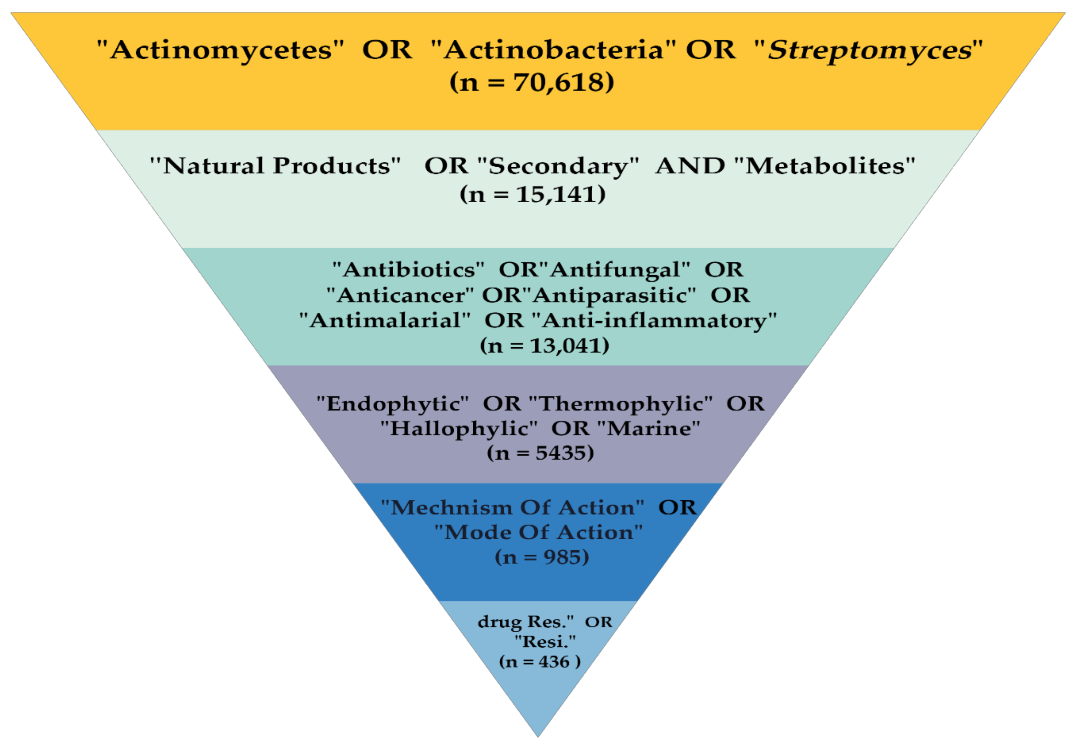
2.1. Study Design and Search Strategy
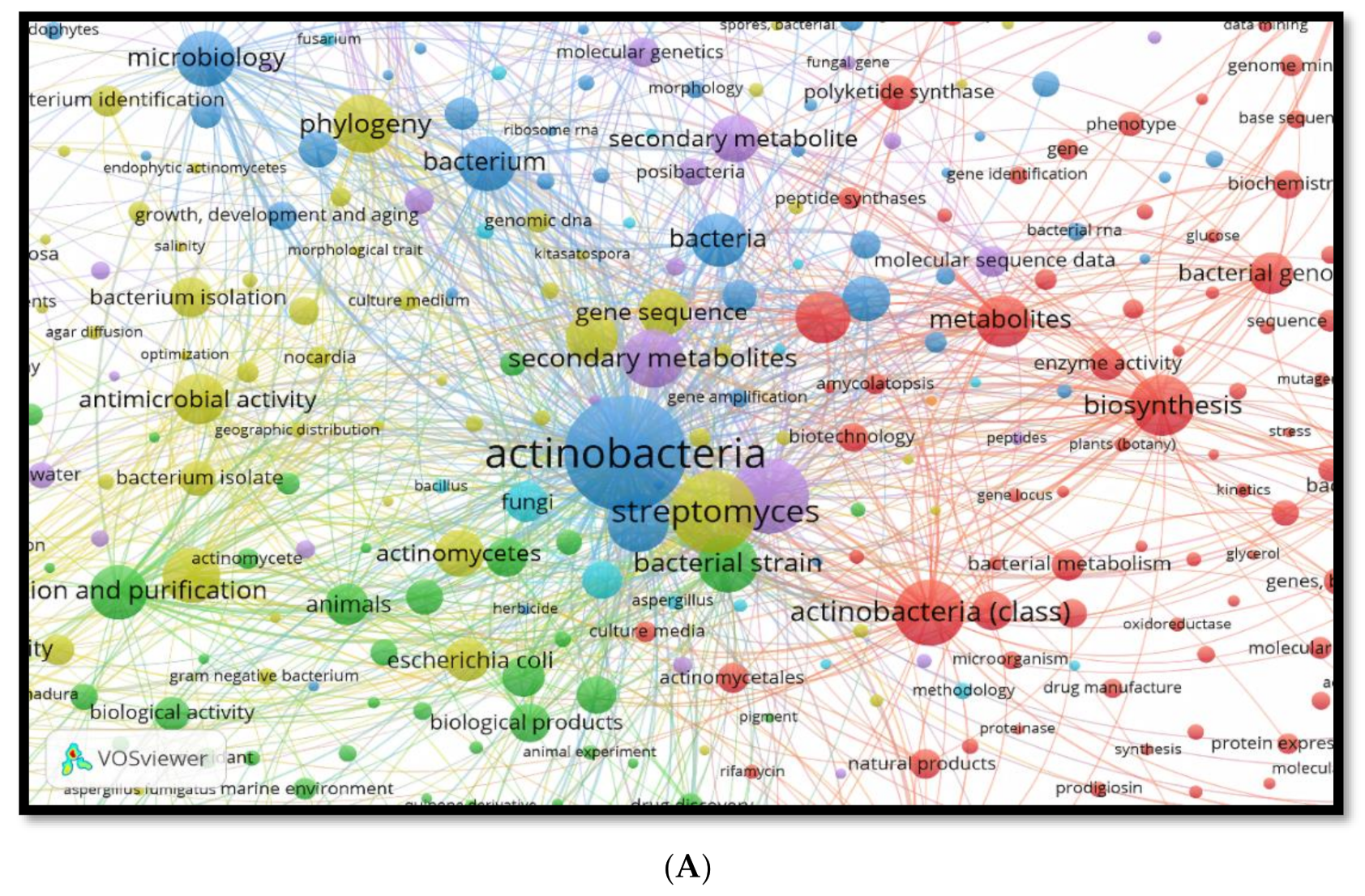
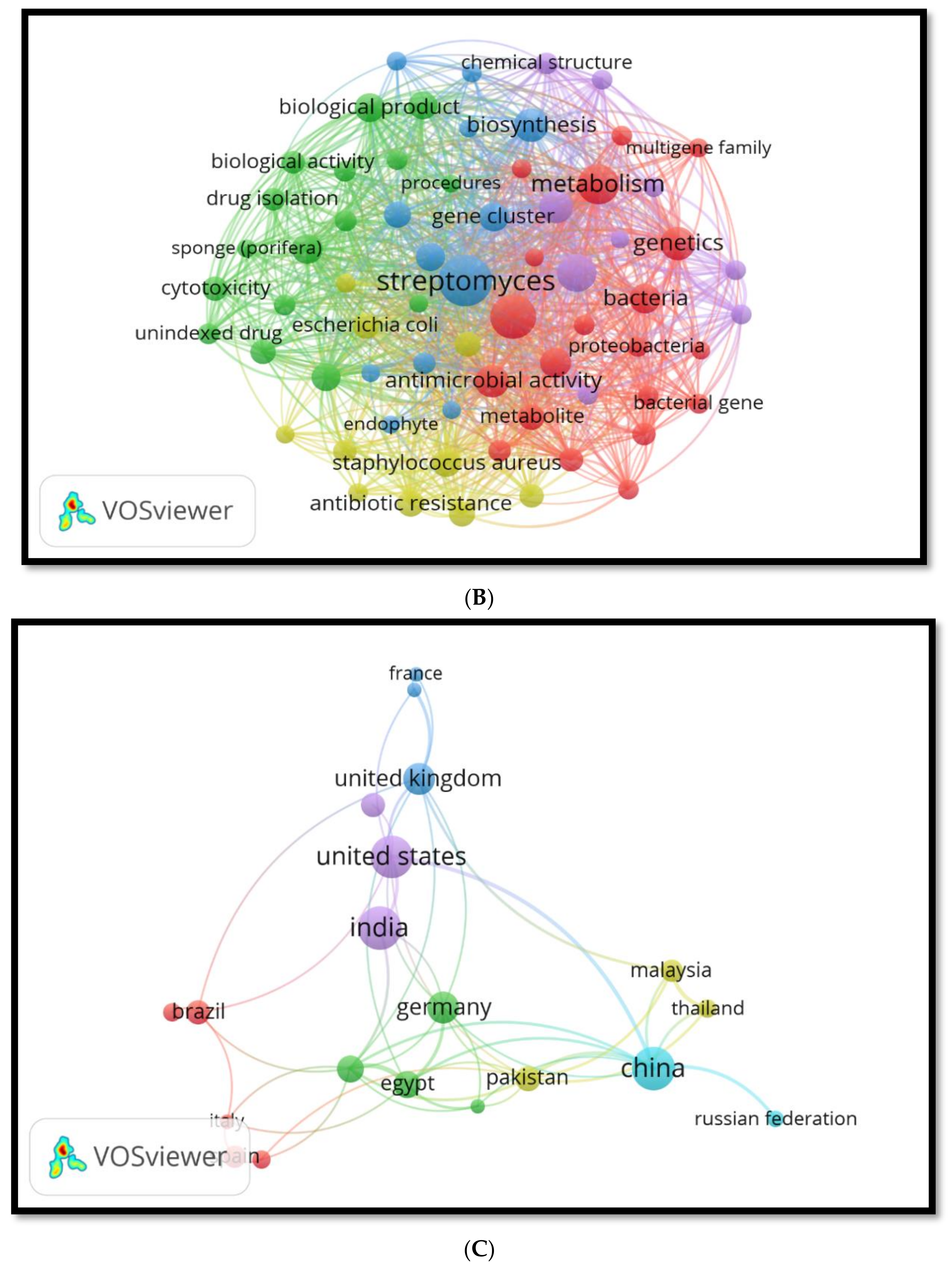
The methodology of this review was conducted as demonstrated in Figure 1. The process involved six main steps: Step 1 was an identification of the target articles through the Scopus database in the period between 2010 and 22 March 2021 using the general keywords “Actinomycetes” OR “Actinobacteria,” OR “Streptomyces”. The number of articles obtained was n = 70,618. In Step 2, the resulting articles were further screened using more specific keywords, including “Natural Products” OR “Secondary Metabolites” (n = 15,141). Then, in Step 3, from that number of articles, further screening was carried out using more specific keywords, which included antibiotics, anti-fungal, anti-cancer, anti-parasitic, “antimalarial,” OR anti-inflammatory (n = 13,041). In Step 4, from the preceding number of articles, screening was implemented using more precise keywords, such as endophytic, thermophilic, halophilic OR marine (n = 5435 was extracted). Subsequently, in Step 5, from the total number of articles, further screening was considered using more specific keywords, including “mechanism of action” OR “mode of action” (n = 985). Finally, in Step 6, from these articles, the final screening was executed using more specific keywords “drug-resistance” OR “resistant”. The number of articles based on keywords is (n = 436), extracted studies based on related titles was n = 336; reviewed studies out of limitations were n = 31; records excluded based on the validity of the study data and clear contributions were n = 260, and out of limitations were n = 46. Finally, the number of studies included in this review was n = 116. A review scheme was conducted to determine all research documents published only in the English language, presented in Figure 1.

2.2. Data Analysis

The VOSviewer software was used to visualise the bibliometric networks to build assistance for common bibliographic databases from the Scopus database. The most important journals, articles, authors, organisations and states among such articles were identified. Besides this, bibliographic coupling and most utilised keywords in the abstracts with keywords and titles were identified.
The majority of diversity keywords from the reviewed and cited papers based on the Scopus database were Streptomyces, Actinobacteria, natural products, primary, secondary metabolites, habitat effects of environments, pharmaceutical industry, as shown in Figure 2A.
This analysis includes diverse secondary metabolite molecules induced by environmental factors such as antibiotic agents, biological products, antineoplastic agent, anti-fungal, and agent with antimalarial activity, as shown in Figure 2B. The reviewed and cited papers based on the scattered keywords of the antimicrobial isolated from 2010 to 2021 from phylum Actinobacteria were vancomycin, polyketide, tetracycline, cyclopeptide, erythromycin, streptomycin, macrolides and amphotericin B, as represented in Figure S1. The percentage of keywords diversification from the Scopus database for Streptomyces, Actinobacteria, natural products, primary, secondary metabolites, habitat effects of environments, pharmaceutical industry and anti-infective agents occurrence using VOSviewer software tool to analyse and visualise scientific literature is shown in Figures S2 and S3. The most reviewed and cited papers based on the scattered keywords of the countries for the publication and citation were USA (46 %), UK and India (13% each), Germany (11%), China (6%) and Thailand (3%), while KSA, Egypt, Pakistan, Malaysia had only 2% each, as shown in Figure 2C and Figure S4.

3. Primary and Secondary Metabolites Natural Products

Actinobacteria synthesise diverse metabolite molecules that have key roles in their heterogeneous and complex microenvironments. Natural products, also known as sec-ondary metabolites, are useful compounds developed by microbes. These are not usually needed for natural cell development, but they provide advantages to the cells in other ways [12]. Such compounds could play roles in inhibition, communication, nutrient ac-quisition, or other associations with nearby environments or organisms.
Natural products have been utilised for a long time. In fact, the Chinese are amongst users of traditional medicines with more than five thousand plants and microbes’ products in their pharmacopoeia [13]. Therapeutic plant species have been, and are still being, used in traditional medicine in several countries [12]. Primary metabolites are chemicals required for ordinary growth, development and reproduction of organisms as well as maintenance of cellular function, representing the key role in the survival of organisms. Besides this, the primary metabolite plays an active role in the anabolic and catabolic processes in many organisms or cells [14]. The secondary metabolites are substances formed during the end or near the stationary stage of organisms’ development. They are very significant for nutrition and health and are, therefore, economically important [14]. Even though they serve different survival actions in nature, they do not necessarily play a critical role in the growth and development of the organism producing them [15].

4. History of Isolation of Secondary Metabolites from Actinobacteria

Historically, in (1940), Waksman and Woodruff isolated actinomycin D from soil bacteria [16]. Then, Schatz et al. in 1944 [17] isolated streptomycin, an effective antibiotic against tuberculosis. Furthermore, hundreds of various antibiotics were reported from the genus Streptomyces [18]. Actinobacteria are among the secondary metabolites producers and hold high pharmacological and commercial interest. It has great capability to produce secondary metabolites such as immunomodulators [19], antibiotics [20], anti-cancer drugs [21], growth factors [22], anthelminthic enzymes and herbicides [23]. Supplementary Table S1 describes the historical isolation of bioactive compounds from Actinobacteria from the first isolation by Selman Waksman [20].
Several anti-fungal, anti-parasitic, bioactive compounds, growth-promoting compounds and anti-cancer compounds with their chemical classification and application isolated from Actinobacteria and Streptomyces sp. are represented in Supplementary Table S2. The molecular structures of several bioactive compounds isolated from Actinobacteria and Streptomyces are demonstrated in Figure 3, Figure 4 and Figure 5.

5. Microbial Ecology of Actinobacteria

The diversity of Actinobacteria has been investigated in several special or extreme environments, such as Actinobacteria in terrestrial environments, Actinobacteria in aquatic and marine environments, and thermophilic as well as alkaliphilic and haloalkaliphilic Actinobacteria.

5.1. Actinobacteria in Terrestrial Environments

Terrestrial Actinobacteria have several antimicrobial capabilities, including three active compounds determined as 2,3-heptanedione, butyl propyl ester and cyclohexane with an antimalarial activity isolated from Streptomyces SUK 08 [24].
The diketopiperazines compounds and chloramphenicol were isolated from Streptomyces SUK 25 isolated from the Zingiber spectabile plant in Malaysia [25]. Meanwhile, Streptomyces sp. CAH29 isolated from the rhizosphere environment can produce tetrangomycin, possessing potent anti-bacterial and anti-fungal action against Candida albicans, methicillin-resistant Staphylococcus aureus, Streptococcus pyogenes and Staphylococcus aureus having 14, 10, 12 and 8 mm inhibition zones, respectively [26]. Besides this, the isolate revealed a marked anti-tumour action with 1.1 μg/mL and IC50 3.3 against colorectal (HCT116) and hepatocellular (HepG2) carcinoma cell lines, respectively [27]. Actinomycin D, which has anti-tumour and anti-bacterial activity, was extracted from Streptosporangium sp. (AI-21) at Hardwar district in Uttarakhand state, India [28]. The list of the terrestrial and rhizosphere Actinobacteria isolation and screening for their antimicrobial activity are shown in Table S3.

5.2. Actinobacteria in Aquatic and Marine Environments

Marine Actinobacteria are abundant sources for marine drug discovery causal for many bioactive compounds of biomedical appearance. Almost 30 strains of actinomycetes were separated and determined from various families of genus Streptomyces. The Streptomyces isolates of M93, W108, W38, M72, M71 and M1 are amongst all the chosen strains revealed to show potent antimicrobial action against the multidrug-resistant bacteria (MDRB) [29]. Furthermore, antitumor compounds produced by marine Actinobacteria is reported [30]. Moreover, antimalarial activity was also described from marine Actinobacteria as bioactive compounds [28]. Additionally, from freshwater sediments, 84 Actinobacteria were separated and isolated into a prevalent genus Streptomyces as well as eight uncommon genera such as Micrococcus, Kocuria, Nocardiopsis, Promicromonospora, Saccharopolyspora, Amycolatopsis, Prauserella, and Rhodococcus. All strains revealed significant inhibition potentials against yeast pathogens, Gram-negative bacteria and Gram-positive bacteria [5].
Moreover, Saccharopolyspora sp. IMA1 was separated from the coral reef environment. Its metabolites existed in aquatic bacterial pathogens Vibrio vulnificus, Vibrio parahemolyticus and Vibrio harveyi. The IMA1 crude extract revealed excellent antioxidant activity, where 2,2′-azino-bis 3-ethylbenzothiazoline-6-sulfonic acid (ABTS) has a radical scavenging activity [31]. Almost 52 strains of Actinobacteria were isolated from the marine sediment of the Portuguese coast. The isolates’ greatest fraction were from the genus Micromonospora, from which six Streptomyces strains were active against Candida. albicans, with minimum inhibitory concentration (MIC) values ranging from 3.90 to 125 µg mL−1 [2].
The list of bioactive compounds isolated from aquatic and marine Actinobacteria are demonstrated in Table 1.

5.3. Thermophilic Actinobacteria

Thermophilic Actinobacteria can withstand temperatures in the range of 40–80 °C [8]. They thrive on organic debris that has decomposed (dead plant and animal materials). Thermophilic Actinobacteria are divided into two types: moderately thermophilic and strictly thermophilic. Certain strains of moderately thermophilic Actinobacteria may develop at temperatures ranging from 37 to 65 °C, whereas some grow between 28 and 60 °C and require 45–55 °C for maximal growth. However, maximum proliferation happens at 55–60 °C.
Thermotolerant Actinobacteria, on the other hand, can withstand temperatures as high as 50 °C [38]. Determining fresh bioactive compounds from taxonomically sole strains of extremotrophic or extremophilic Actinobacteria resulted in the anticipation that mining such groups might provide an alternate dimension to the route of subordinate metabolite resources [39]. Thermotolerant and thermophilic Actinobacteria are unique for having specific metabolic rates and physical features, which are useful in several ecological functions. It was also reported that thermotolerant Actinobacteria like Streptosporangium sp., S. lanatus, S. coeruleorubidis, S. toxytricini and Streptomyces tauricus, are claimed to inhibit the rhizosphere of several plants in the Kuwait desert during hot seasons [11]. Thermophilic Actinobacteria have not been widely investigated. However, they have developed significant antibiotics like thermomycin from Streptomyces thermophilus and anthramycin, an anti-tumour drug created by Streptomyces refuineus [40].
The Streptomyces strain Al-Dhabi-2 found in Saudi Arabia’s thermophilic region indicated antimicrobial capabilities against pathogenic microorganism [41]. Three strains identified as DJT 15 Streptomyces thermoviolaceus subsp. apingens, DJT 32 Saccharomonospora viridis, and DJT 36 Saccharomonospora glauca exposed inhibitory activity in bioassays with respect to the Enterobacter species (ESKAPE) pathogens, Pseudomonas aeruginosa, Acinetobacter baumannii Klebsiella pneumoniae, Staphylococcus aureus and Enterococcus faecium. Apart from that, DJT 32 and 36 prevented the growth of Aspergillus fumigatus, a filamentous fungus isolated from the same compost. Azole-resistant human pathogenic strains of Aspergillus fumigatus were also shown to be inhibited by strain DJT 32 [42].

5.4. Alkaliphilic and Haloalkaliphilic Actinobacteria

An alkaliphilic and mildly halophilic bacterial strain develops optimally at pH ranging from 9.0 to 10.0, with 5–7% (w/v) NaCl [43]. Haloalkaliphilic ActinobacteriaActinobacteria are found in pristine coastal habitat in the saline soil. These unique ActinobacteriaActinobacteria are hereditarily different, developing at high salt concentrations [44]. Alkaliphilic ActinobacteriaActinobacteria are, thus, classified into three main categories: alkali-tolerant ActinobacteriaActinobacteria (developed in the pH range between 6 and 11, abstemiously alkaliphilic (developed in a pH ranging from 7 to 10 but display weak development at pH 7.0), and alkaliphilic (grow optimally at pH 10–11) [11].
Moreover, Streptomyces clavuligerus (strain Mit-1) was isolated from Mithapur (Western Coast, Gujarat, India) and is described as a salt-tolerant alkaliphilic actinomycete. Note that this strain secreted alkaline protease [45]. Besides this, Thakrar et al. [46] issued the capability to produce protease from the two halo-tolerant and alkaliphilic actinomycetes extracted from the salt-enriched soil of Coastal Gujarat in India. These strains are identified as Nocardiopsis alba OM-4 and Nocardiopsis alba TATA-13. In addition, the phylogeny and multiplicity of the haloalkaliphilic ActinobacteriaActinobacteria study represent a concise and comprehensive account utilising conventional and molecular techniques. This includes the occurrence and cultivation of the marine actinomycetes from the seawater and sediments of pristine coastal habitat in Gujarat (India).
They found that the ActinobacteriaActinobacteria belonged to Prauseria, Streptomyces, Brachybacterium and Nocardiopsis [47]. Besides this, the multiplicity of alkali tolerant ActinobacteriaActinobacteria in numerous soils is gathered from various Algerian Sahara areas. Almost 29 alkali-tolerant ActinobacteriaActinobacteria strains were separated by utilising a complex agar medium. The strains were then tested for non-ribosomal peptide synthetases and genes encoding polyketide synthases, as well as antibiotic behaviour against a broad microorganisms’ variety. They found that some strains can create subordinate metabolites against several pathogenic microbes [19].
A list of bioactive compounds isolated from thermophilic, alkaliphilic and haloalkaliphilic ActinobacteriaActinobacteria is shown in Table 2.

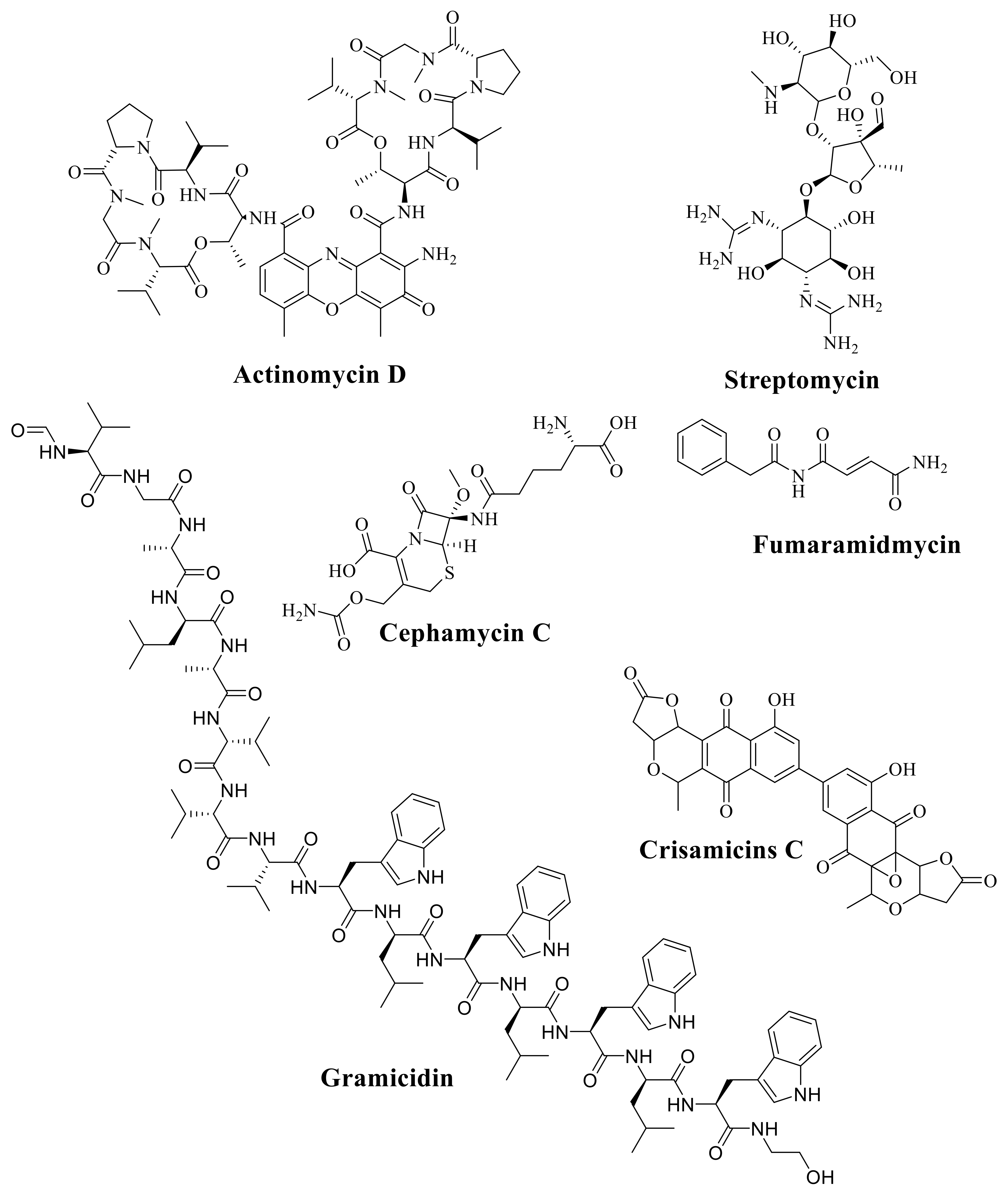
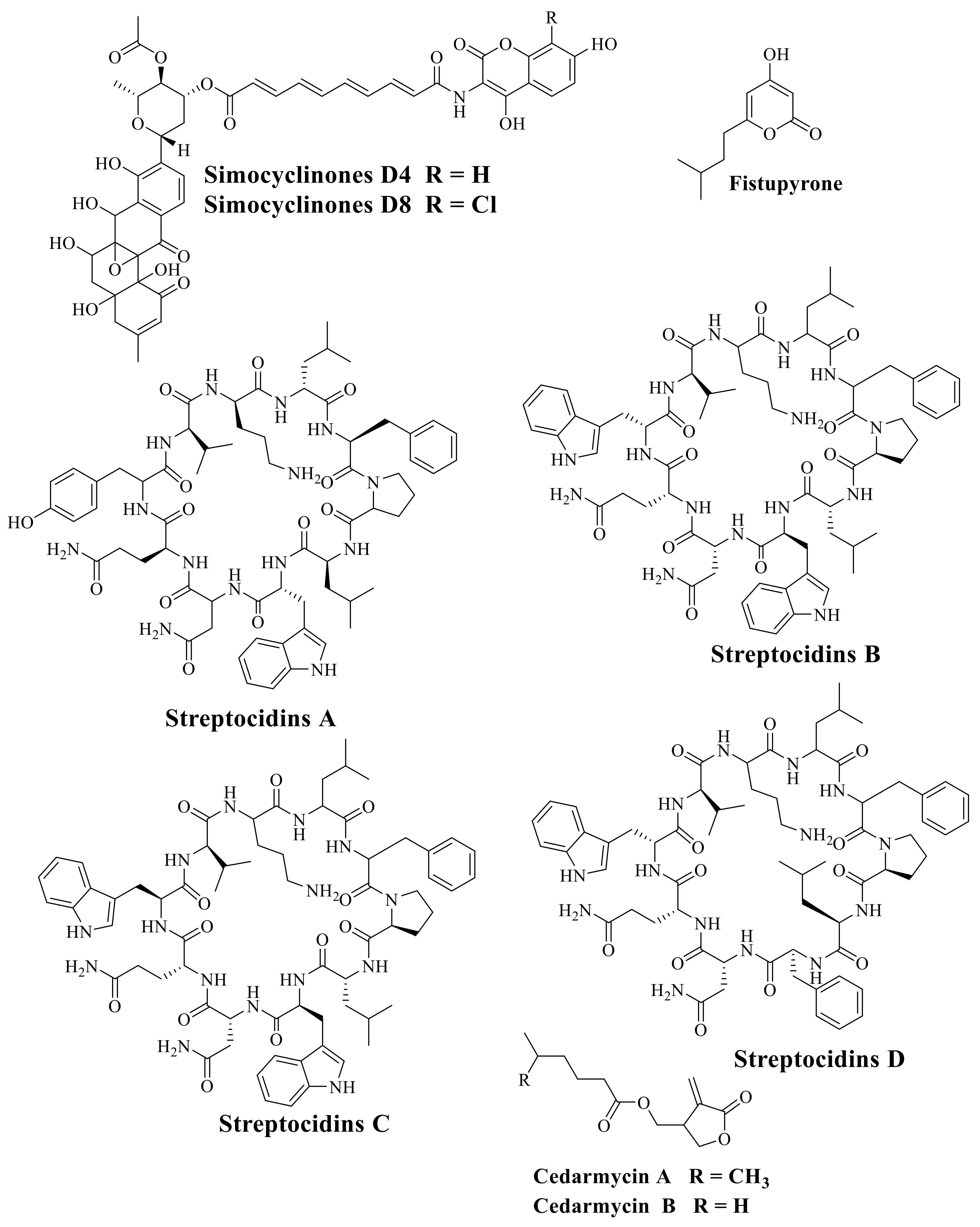
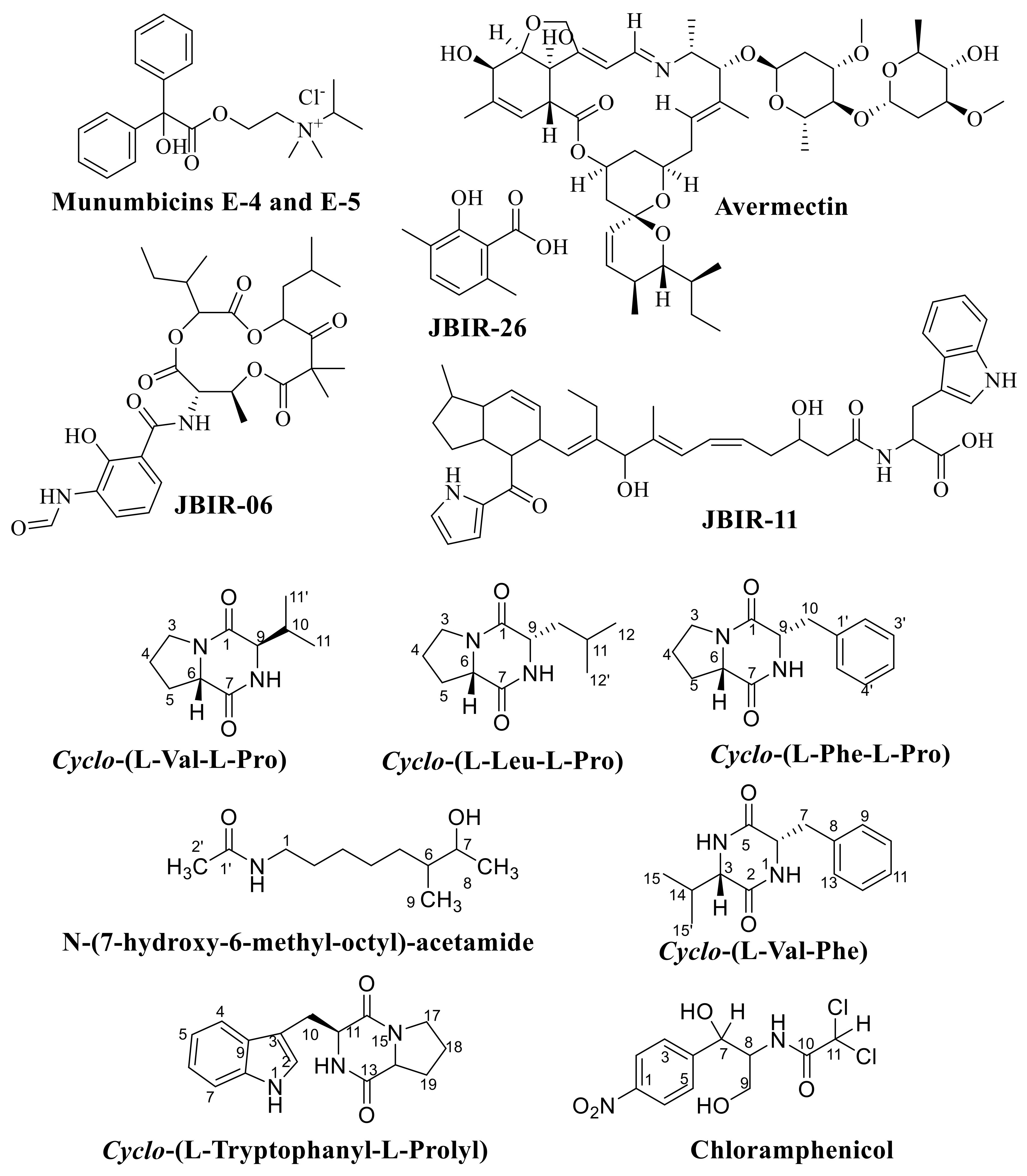
6. The Important Antibiotics Isolated from Actinobacteria against Drug-Resistant Pathogens

Yücel and Yamaç [50] showed that extracts from Streptomyces sp. 1492 exhibited antimicrobial activity against MRSA, Vancomycin-resistant Enterobacter faecium (VRE) and Acinetobacter baumanii with 125 µg/mL and 250 to 1000 µg/mL of MICs and MBCs, respectively. From Verrucosispora sp. strains isolated from the Japan Sea’s sediment, Goodfellow and Fiedler [51] identified atrop-abyssomicin C and proximicins A, B and C. It inhibits the formation of p-aminobenzoate during tetrahydrofolate synthesis in Gram-positive bacteria with methicillin-resistant Staphylococcus aureus (MRSA). The Streptomyces malachitofuscus has anti-fungal action against Candida albicans and Mucor miehei, as Sajid et al. [52] reported. The spoxazomicins was extracted from Streptosporangium oxazolinicum sp. nov., obtained from the root of numerous orchids at Okinawa. The study also isolated spoxazomicins A-C from Streptosporangium oxazolinicum K07-0460 (T) with antitrypanosomal activity [53]. Besides this, the arylomycine isolated from Streptomyces sp. HCCB10043 show activity against Gram-positive bacteria, among which included Staphylococcus aureus [54]. The chlorocatechelins A and B extracted from Streptomyces sp. are fresh siderophores encompassing an acylguanidine structure and chlorinated catecholate sets that inhibit various bacteria and fungi [55]. An extremely potent subordinate metabolite formed by endophytic strain is known as Streptomyces sp. HUST012. It showed antimicrobial and anti-tumour activities separated from the medicinal plant stems, Dracaena cochinchinensis Lour [56]. The updated list of antibiotics separated from ActinobacteriaActinobacteria and Streptomyces is given in Table 3.
The existing literature reported 124 compounds from Actinobacteria with anti-MRSA activity. Several bioactive pharmaceutical compounds generated by Actinobacteria was found to be effective against Vancomycin-resistant Enterococcus (VRE), Methicillin-resistant Staphylococcus aureus (MRSA) and other drug-resistant bacterial pathogens strains. Kakadumycin A was isolated from Streptomyces sp. NRRL 30566 inhibited the growth of MRSA American Type Culture Collection (ATCC) 33591 four times lower in MIC than vancomycin at a concentration of 0.5 µg/mL compared to the concentration of vancomycin of 2.0 μg/mL, as reported by Castillo et. al. [57]. In addition, polyketide antibiotic SBR-22 comes from a fresh actinomycetes strain, labelled as BT-408, a novel strain of Streptomyces psammoticus. The strain showed anti-bacterial activity against MRSA, as documented by Sujatha et. al. [58]. Besides this, Yoo et. al. [59] isolated laidlomycin from Streptomyces CS684 obtained from the soil at Jeonnam, Korea, with potential activity against VRE. Meanwhile, Laidi et. al. [60] isolated a new actinomycetes strain designated as SK4-6 from Egyptian soil that demonstrated strong activity against bacteria, including MRSA and Micrococcus luteus. Furthermore, Streptomyces sp. PVRK-1 was isolated from the Manakkudy mangroves in Tamil Nadu, South India. This Streptomyces has been able to produce many small molecules exhibiting potent anti-MRSA activity [43].
Table 3. The list of important producers and chemical classification of antibiotics isolated from Actinobacteria.
AntibioticProducerChemical ClassReference
Cephamycin CNocardia lactamduransB- Lactam[61]
ChlortetracyclineS. aureofaciensTetracycline[62]
Clavulanic acidS. clavuligerusB- Lactam[63]
CycloserineS. orchidaceusPeptide[64]
DaptomycinS. rodeosporusLipopeptide[65]
DaunorubicinS. PeucetiusPeptide[66]
FK506S.tubercidicusMacrolide[67]
FortimicinMicromonospora olivasterosporaAminoglycoside[68]
FosfomycinS. fradiaePhosphoric acid[69]
FumaramidmycinS. kurssanoviiAlkaloids[70]
GentamycinMicromonospora sppAminoglycoside[71]
KanamycinS. kanamyceticusAminoglycoside[72]
LincomycinnS. lincolnensisSugar—amide[73]
NeomycinS. fradiaeAminoglycoside[74]
NikkomycinS. tendaeNucleoside[75]
NocardicinNocardia uniformisB- Lactam[76]
NovobiocinS. neveusAminocoumarin[77]
OleandomycinS. antibioticusMacrolide[78]
OxytetracyclineS. rimosusTetracycline[79]
ParomomycinS.rimosus formaAminoglycoside[80]
RifamycinAmycolatopsis AnsamycinRNA polymerase (PK)[81]
SpiramycinS. ambofaciensMacrolide (PK)[82]
StreptomycinS. griseusAminoglycoside[83]
TetracyclineS. aureofaciensTetracycline (PK)[84]
ThienamycinS. cattleyaβ-Lactam Peptidoglycan[85]
TobramycinS. tenebrariusAminoglycoside[86]
VancomycinS.orientalisPeptidoglycan[87]
Abbreviation: S. = (Streptomyces).
In addition, Choi et. al. [88] claimed that Streptomyces sp. CS392 have anti-bacterial activity against VRE and MRSA. Additionally, the strain Streptomyces sp (VITBRK2) can yield strong antimicrobial activity against VRE and MRSA strains [89]. Moreover, Streptomyces rubrolavendulae ICN3 strain displayed potent antimicrobial activity towards MRSA, with a 42 mm inhibition zone. The MIC was 500 μg/mL from the crude extracts, while the cleansed compound was identified as C23 with MIC 2.5 μg/mL in the in vitro assay [90]. Similarly, from a mangrove forest soil on Peninsular Malaysia’s east coast, the MUSC 135T strain was isolated. It can produce broad-spectrum antibiotic bacitracin against MRSA strain ATCC BAA-44 [91]. Correspondingly, from the stems of Dracaena Cochinchinensis Lour plant, Streptomyces sp. HUST012 was isolated, producing two potent antimicrobial compounds towards MRSA ATCC 25923 with MIC of 62.5 and 0.04 µg/mL, identified as SPE-B11.8-B5.4 [56]. Streptomyces sp. KB1 from Ao-Nang, Krabi province, Thailand, contains a compound known as 2,4-Di-tert-butylphenol, an anti-MRSA MIC of 31.15 µg/mL in comparison to vancomycin, with a MIC value of 1.56 µg/mL [92]. Diketopiperazines and chloramphenicol extracted from Streptomyces SUK 25, derived from the Zingiber spectabile root, seem effective towards MRSA [25]. Moreover, Bhakyashree and Kannabiran [93] reported the separation of anti-MRSA compounds from Streptomyces sp. VITBKA3 strain proteins and cell membrane biosynthesis inhibitors. The strains of Streptomyces sp. derived from different environments with potentials in production of anti-MRSA bioactive compounds from 2014 until 2020 is given in Table 4.

7. Production of Enzymes from Actinobacteria

Both marine and terrestrial Actinobacteria in Table 5 produce a wide range of biologically active enzymes. They can secrete protease, cellulases, lipase, xylanase, pectinase and amylase. These enzymes are vitally important in textile or paper industries, the food industry and fermentation, besides biotechnological application. Consequently, Actinobacteria have been discovered to be a good L-asparaginase source. Various Actinobacteria often those extracted from soils, for example, Nocardia sp Streptomyces albidoflavus, S. griseus and Streptomyces karnatakensis produce several enzymes [102]. Enzymes like urease, chitinase and catalase are also generated from Actinobacteria [103]. Surprisingly, keratinase, an enzyme that destroys the feathers of poultry chickens, has been successfully produced from Nocradiopsis sp. [104]. Likewise, Actinobacteria separated from goat and chicken gut revealed the existence of numerous enzymes like lipase, phytase, protease and amylase [105].

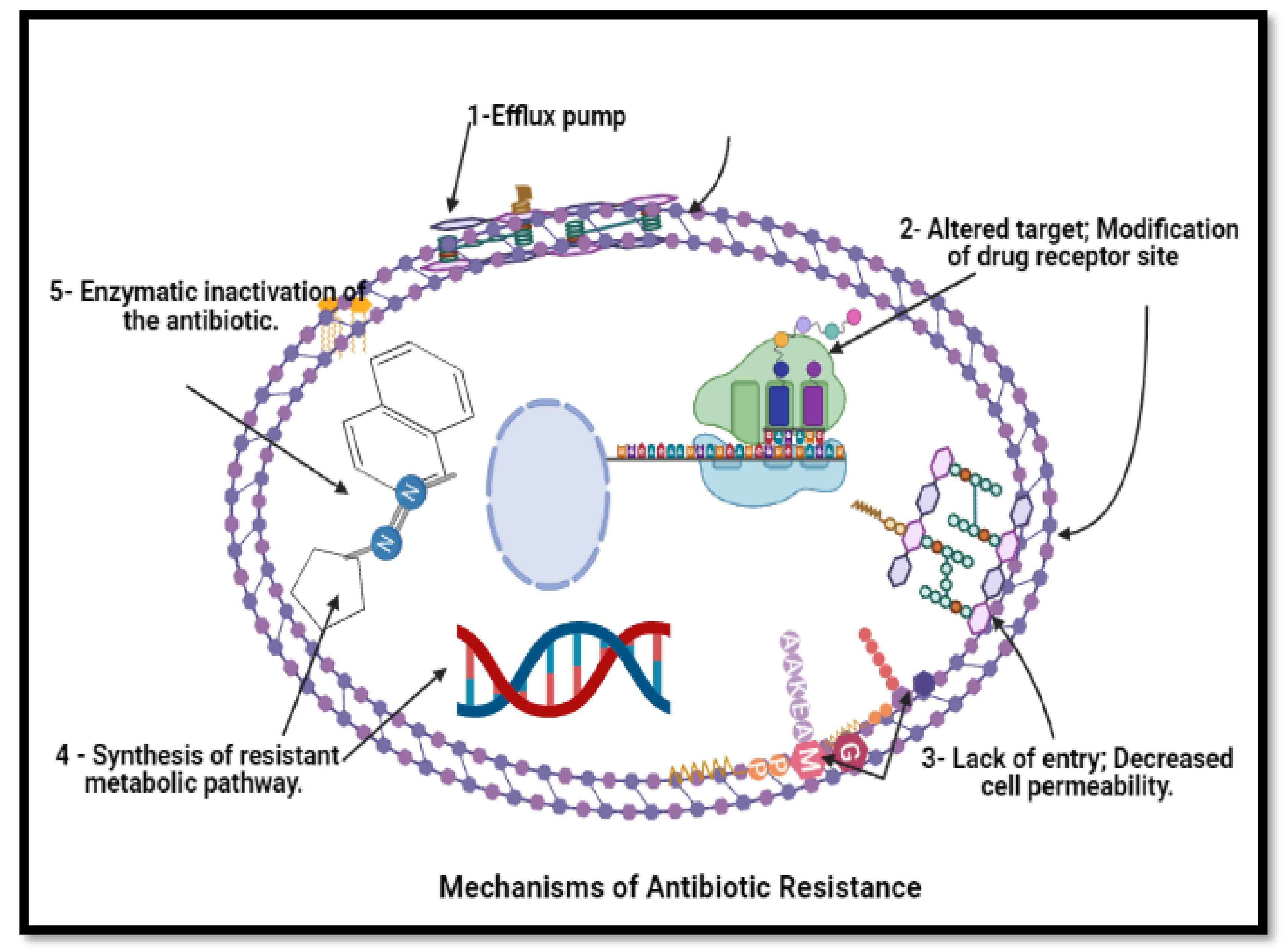
8. Mechanism of Bioactive Compounds from Actinobacteria against Drug-Resistant Pathogens

Once scientists provide new antimicrobial drugs, the microorganism starts to adapt themselves against these drugs, which become ineffective at some points. This is primarily due to changes that occur inside the microorganism, especially bacteria, due to the interaction of numerous organisms through their environment and surroundings. These changes may occur for various reasons: mutations, selective pressure, gene transfer and phenotypic change [65]. For example, gene mutation occurs when bacteria reproduce, leading to the development of bacteria with genes that help them resist antibiotics. In addition, the mutation leads to alter the target and modification of the drug-receptor site in the target site [66].
Moreover, selective pressure means that bacteria carrying resistance genes hold up and multiply so that new resistance bacteria become the predominant type. The selective pressure leads to a lack of entry and decreased cell permeability. In addition, it leads to greater exit and active efflux pump [67]. Bacteria unnecessarily replicate to transmit their antibiotic resistance gene. Instead, it is passed across various types of bacteria resistance determinants through horizontal gene transfer making the bacterium resistant [68]. Besides this, phenotypic change suggests that the bacteria can change some of their properties to become more resistant to common antibiotics using the enzymatic inactivation of the antibiotics or by synthesising resistant metabolic pathways [69]. Other mechanisms, by modulation of methicillin-resistance of penicillin-binding protein (PBP2a) synthesis, were regulated by two genes known as MecI and MecR1 proteins. When existing, the signalling or regulatory proteins of the plasmid-mediated staphylococcal β-lactamase gene bla-Z system are working.
Furthermore, homogeneous tolerance is based on mutations at a different locus of genetics. Furthermore, other external and internal causes affect the development of methicillin resistance [115]. The mechanism of anti-bacterial resistance is demonstrated in Figure 6.

9. Conclusions and Future Prospects

There is a global demand for new chemotherapeutic agents and antibiotics that are extremely active and have low toxicity and environmental effects. The drug resistance in viruses, fungal and bacteria, as well as the emergence of life-threatening microorganisms, have become higher than before. This is due to the wrong usage of dose and time of medication administration, increasing the requirement of new and active compounds that assist and relieve all the aspects of the human condition. Actinobacteria have been isolated from different ecosystems, including several medicinal plants from the terrestrial and rhizosphere environment, hot springs as thermophilic Actinobacteria, deep-sea sediments, marine sponges and alkalines line soil. Several previous published works reported that Actinobacteria are understudied phylogenetic groups with high biosynthetic potential. This type of bacteria was found to have the greatest number of biosynthetic gene clusters in its genomes. There are more opportunities to investigate new resources and other biological characteristics of previously inaccessible natural products extracted from Actinobacteria as interest grows in bioactive molecules from drug formulation with minimal side effects. This group represents the new resource of bioactive compounds due to its ability to grow in multisectoral environments.
This review focused on the emphasis and the connection between finding new resources and novel strategies to search for potential bioactive compounds isolated from phylum Actinobacteria. In addition, this review highlighted some limitations in today’s research regarding a new source for bioactive compounds isolated from Actinobacteria. For example, most previous publications concentrated only on the endophytic Actinobacteria, excluding other environments such as thermophilic, alkaliphilic and haloalkaliphilic Actinobacteria. Note that many other biodiversities and multidisciplinary environments represent important and new resources of potentially bioactive compounds. As a result, it appears that certain new techniques and methodologies for a thorough investigation of bioactive natural compounds are required. This includes, for example, the discovery of novel structure–activity relationships in nature, which has become increasingly important for the synthesis inspiration of natural bioactive product compounds. This results in increased diversity with less complexity and a good knowledge of isolation processes.
To address the challenges of biodiversity and promote future sustainable use of natural resources, a multidisciplinary perspective is required to find, describe and convey nature’s richness. This is necessary for identifying novel bioactive chemicals from Actinobacteria. Moreover, this review summarised the new bioactive compounds isolated from Actinobacteria and their applications in industrial, agricultural and environmental protection, pharmaceutical bioactive compounds and pharmaceutically related biomolecules. This includes superordinate metabolites that act as inhibitory or killing agents against pathogens that affect humans and animals, including resistant bacteria, fungi, viruses and several protozoa. They can also produce an anti-cancer and several enzymes for active degradation and meeting industrial demands worldwide. Therefore, continuous selective isolation and screening studies on the characterisation and identification of novel potential bioactive compounds from Actinobacteria are required. This is to create commercially viable, long-term and cost-effective production methods. Their metabolic flexibility and abundance also provide a novel, strong pathway for the bioremediation of organic wastes and contaminants.

Supplementary Materials

Table S1: Historically isolation of bioactive compounds from Actinobacteria. Table S2: List of antifungal, growth promoting, antitumor and antiparasitic bioactive compounds, chemical classification and their application which isolated from Actinobacteria. Table S3: Terrestrial and rhizosphere Actinobacteria isolation and screening for their antimicrobial activity. Figure S1: The spread of reviewed and cited papers based on the scattered keywords of the antimicrobial isolated from 2010-2020 from phylum Actinobacteria. Figure S2: Percentage of diversity of keywords from the Scopus database for Actinobacteria, Streptomyces, natural products, primary, secondary metabolites, habitat effects of environments, pharmaceutical industry using VOSviewer software tool to analyse and visualise scientific literature. Figure S3: Spread of reviewed and cited papers based on the dispersed keywords of anti-inflective agents’ occurrence. Data was extracted using VOSviewer software to analyse and visualise scientific literature. Figure S4: Spread of reviewed and cited papers based on the scattered keywords of the countries for the publication and citation. Data was extracted using VOSviewer software.

Author Contributions

Conceptualisation, methodology and writing—review preparation, M.M.A.-s., R.M.S.R.M. and N.M.S.; data validation, H.A.E.E. and N.M.Z.; formal analysis and data curation, E.N., N.A.A.-M. and A.A.-G. All authors have read and agreed to the published version of the manuscript.

Funding

This research was funded by the FRGS/1/2020/WAB02/UTHM/03/1, FRGS/1/2017/STG05/UMK/01/1 and Financial support from industrial grant No. R.J130000.7609.4C284 and R.J130000.7609.4C240.

Institutional Review Board Statement

Not applicable.

Informed Consent Statement

Not applicable.

Data Availability Statement

Supplementary Material related to this article can be found in the Supplementary data file.

Acknowledgments

The authors wish to thank The Ministry of Higher Education Malaysia under Fundamental Research Grant Scheme (FRGS) (Ref: FRGS/1/2020/WAB02/UTHM/03/1) under postdoc scheme research for supporting this research. The authors would like to thank the Center for Diagnostic, Therapeutic and Investigative Studies, Faculty of Health Sciences, Universiti Kebangsaan Malaysia and to the Faculty of Agro-based Industry, Universiti Malaysia Kelantan for financial support. H.A.E. would like to thank RMC, Universiti Teknologi Malaysia (UTM), Financial support from industrial grant No. R.J130000.7609.4C284 and R.J130000.7609.4C240.

Conflicts of Interest

The authors declare no conflict of interest.

References

  1. Carvalho, I.T.; Santos, L. Antibiotics in the aquatic environments: A review of the European scenario. Environ. Int. 2016, 94, 736–757. [Google Scholar] [CrossRef]
  2. Ribeiro, I.; Girão, M.; Alexandrino, D.; Ribeiro, T.; Santos, C.; Pereira, F.; Mucha, A.; Urbatzka, R.; Leão, P.; Carvalho, M. Diversity and Bioactive Potential of Actinobacteria Isolated from a Coastal Marine Sediment in Northern Portugal. Microorganisms 2020, 8, 1691. [Google Scholar] [CrossRef] [PubMed]
  3. Qi, D.; Zou, L.; Zhou, D.; Chen, Y.; Gao, Z.; Feng, R.; Zhang, M.; Li, K.; Xie, J.; Wang, W. Taxonomy and Broad-Spectrum Antifungal Activity of Streptomyces sp. SCA3-4 Isolated From Rhizosphere Soil of Opuntia stricta. Front. Microbiol. 2019, 10, 1390. [Google Scholar] [CrossRef]
  4. Girão, M.; Ribeiro, I.; Ribeiro, T.; Azevedo, I.C.; Pereira, F.; Urbatzka, R.; Leão, P.; Carvalho, M.F. Actinobacteria Isolated From Laminaria ochroleuca: A Source of New Bioactive Compounds. Front. Microbiol. 2019, 10, 683. [Google Scholar] [CrossRef] [PubMed] [Green Version]
  5. Kandel, S.L.; Joubert, P.M.; Doty, S.L. Bacterial Endophyte Colonization and Distribution within Plants. Microorganisms 2017, 5, 77. [Google Scholar] [CrossRef] [Green Version]
  6. Frank, A.C.; Guzmán, J.P.S.; Shay, J.E. Transmission of Bacterial Endophytes. Microorganisms 2017, 5, 70. [Google Scholar] [CrossRef] [Green Version]
  7. Salam, N.; Jiao, J.-Y.; Zhang, X.-T.; Li, W.-J. Update on the classification of higher ranks in the phylum Actinobacteria. Int. J. Syst. Evol. Microbiol. 2020, 70, 1331–1355. [Google Scholar] [CrossRef]
  8. Passari, A.K.; Chandra, P.; Leo, V.V.; Mishra, V.K.; Kumar, B.; Singh, B.P. Production of potent antimicrobial compounds from Streptomyces cyaneofuscatus associated with fresh water sediment. Front Microbiol. 2017, 8, 68. [Google Scholar]
  9. Li, L.-Y.; Yang, Z.-W.; Asem, M.D.; Fang, B.-Z.; Salam, N.; Alkhalifah, D.H.M.; Hozzein, W.N.; Nie, G.-X.; Li, W.-J. Streptomyces desertarenae sp. nov., a novel actinobacterium isolated from a desert sample. Antonie Leeuwenhoek 2019, 112, 367–374. [Google Scholar] [CrossRef] [PubMed]
  10. Bérdy, J. Bioactive Microbial Metabolites. J. Antibiot. 2005, 58, 1–26. [Google Scholar] [CrossRef] [PubMed] [Green Version]
  11. Shivlata, L.; Satyanarayana, T. Thermophilic and alkaliphilic Actinobacteria: Biology and potential applications. Front. Microbiol. 2015, 6, 1014. [Google Scholar] [CrossRef]
  12. Xin, T.; Su, C.; Lin, Y.; Wang, S.; Xu, Z.; Song, J. Precise species detection of traditional Chinese patent medicine by shotgun metagenomic sequencing. Phytomedicine 2018, 47, 40–47. [Google Scholar] [CrossRef]
  13. Ji, H.F.; Li, X.J.; Zhang, H.Y. Natural products and drug discovery. EMBO Rep. 2009, 10, 194–200. [Google Scholar] [CrossRef] [PubMed] [Green Version]
  14. Chaudhary, A.K.; Dhakal, D.; Sohng, J.K. An insight into the “-omics” based engineering of Streptomycetes for second-ary metabolite overproduction. BioMed Res. Int. 2013, 2013, 968518. [Google Scholar] [CrossRef] [PubMed] [Green Version]
  15. Demain, A.L.; Fang, A. The natural functions of secondary metabolites. In History of Modern Biotechnology I; Springer: Cham, Switzerland, 2000; pp. 1–39. [Google Scholar] [CrossRef]
  16. Waksman, S.A.; Woodruff, H.B. The Soil as a Source of Microorganisms Antagonistic to Disease-Producing Bacteria. J. Bacteriol. 1940, 40, 581–600. [Google Scholar] [CrossRef] [PubMed] [Green Version]
  17. Schatz, A.; Bugle, E.; Waksman, S.A. Streptomycin, a Substance Exhibiting Antibiotic Activity Against Gram-Positive and Gram-Negative Bacteria. Exp. Biol. Med. 1944, 55, 66–69. [Google Scholar] [CrossRef]
  18. Woodruff, H.B.; Selman, A. Waksman, Winner of the 1952 Nobel Prize for Physiology or Medicine. Appl. Environ. Microbiol. 2014, 80, 2–8. [Google Scholar] [CrossRef] [Green Version]
  19. Meklat, A.; Bouras, N.; Mokrane, S.; Zitouni, A.; Djemouai, N.; Klenk, H.-P.; Sabaou, N.; Mathieu, F. Isolation, Classifica-tion and Antagonistic Properties of Alkalitolerant Actinobacteria from Algerian Saharan Soils. Geomicrobiol. J. 2020, 37, 826–836. [Google Scholar] [CrossRef]
  20. Hoskisson, P.; Fernández-Martínez, L.T. Regulation of specialised metabolites in Actinobacteria—Expanding the paradigms. Environ. Microbiol. Rep. 2018, 10, 231–238. [Google Scholar] [CrossRef] [Green Version]
  21. Silva, L.J.; Crevelin, E.J.; Souza, D.T.; Júnior, G.L.; De Oliveira, V.M.; Ruiz, A.L.T.G.; Rosa, L.H.; Moraes, L.A.B.; Melo, I.S. Actinobacteria from Antarctica as a source for anticancer discovery. Sci. Rep. 2020, 10, 13870. [Google Scholar] [CrossRef]
  22. Bibb, M.J. Regulation of secondary metabolism in streptomycetes. Curr. Opin. Microbiol. 2005, 8, 208–215. [Google Scholar] [CrossRef]
  23. Busi, S.; Pattnaik, S.S. Current status and applications of Actinobacteria in the production of anticancerous compounds. In New and Future Developments in Microbial Biotechnology and Bioengineering; Elsevier: Amsterdam, The Netherlands, 2018; pp. 137–153. [Google Scholar]
  24. Zin, N.M.; Remali, J.; Nasrom, M.N.; Ishak, S.A.; Baba, M.S.; Jalil, J. Bioactive compounds fractionated from endophyte Streptomyces SUK 08 with promising ex-vivo antimalarial activity. Asian Pac. J. Trop. Biomed. 2017, 7, 1062–1066. [Google Scholar] [CrossRef]
  25. Alshaibani, M.; Jalil, J.; Sidik, N.; Edrada-Ebel, R.; Zin, N.M. Isolation and characterization of cyclo-(tryptophanyl-prolyl) and chloramphenicol from Streptomyces sp. SUK 25 with antimethicillin-resistant Staphylococcus aureus activity. Drug Des. Dev. Ther. 2016, 10, 1817–1827. [Google Scholar] [CrossRef] [Green Version]
  26. Ozakin, S.; Davis, R.W.; Umile, T.P.; Pirinccioglu, N.; Kizil, M.; Celik, G.; Sen, A.; Minbiole, K.P.C.; Ince, E. The isolation of tetrangomycin from terrestrial Streptomyces sp. CAH29: Evaluation of antioxidant, anticancer, and anti-MRSA activity. Med. Chem. Res. 2016, 25, 2872–2881. [Google Scholar] [CrossRef]
  27. Ahmad, R.; El-Gendy, A.; Ahmed, R.R.; Hassan, H.; El-Kabbany, H.M.; Merdash, A.G. Exploring the Antimicrobial and Antitumor Potentials of Streptomyces sp. AGM12-1 Isolated from Egyptian Soil. Front. Microbiol. 2017, 8, 438. [Google Scholar] [CrossRef] [Green Version]
  28. Dimri, A.G.; Chauhan, A.; Aggarwal, M. In vitro antibacterial characterization of Streptosporangium sp. (AI-21) a new soil isolate against food borne bacteria. Sci. Arch. 2020, 1, 25–34. [Google Scholar] [CrossRef]
  29. Cheema, M.T.; Fatima, A.; Sajid, I. Molecular Identification, Bioactivity Screening and Metabolic Fingerprinting of the Actinomycetes of Chenab River Sediments. Br. Microbiol. Res. J. 2016, 17, 1–13. [Google Scholar] [CrossRef]
  30. Kim, J.; Shin, D.; Kim, S.-H.; Park, W.; Shin, Y.; Kim, W.K.; Lee, S.K.; Oh, K.-B.; Shin, J.; Oh, D.-C. Borrelidins C–E: New Antibacterial Macrolides from a Saltern-Derived Halophilic Nocardiopsis sp. Mar. Drugs 2017, 15, 166. [Google Scholar] [CrossRef] [PubMed] [Green Version]
  31. Krishnamoorthy, M.; Dharmaraj, D.; Rajendran, K.; Karuppiah, K.; Balasubramanian, M.; Ethiraj, K. Pharmacological activities of coral reef associated actinomycetes, Saccharopolyspora sp. IMA1. Biocatal. Agric. Biotechnol. 2020, 28, 101748. [Google Scholar] [CrossRef]
  32. Paulus, C.Y.; Rebets, B.; Tokovenko, S.; Nadmid, L.P.; Terekhova, M.; Myronovskyi, S.B.; Zotchev, C.; Rückert, S.B.; Zahler, S. New natural products identified by combined genomics-metabolomics profiling of marine Streptomyces sp. MP131-18. Sci. Rep. 2017, 7, 1–11. [Google Scholar] [CrossRef] [PubMed]
  33. Zhang, Y.-M.; Liu, B.-L.; Zheng, X.-H.; Huang, X.-J.; Li, H.-Y.; Zhang, Y.; Zhang, T.-T.; Sun, D.-Y.; Lin, B.-R.; Zhou, G.-X. Anandins A and B, two rare steroidal alkaloids from a marine Streptomyces anandii H41-59. Mar. Drugs 2017, 15, 355. [Google Scholar] [CrossRef] [PubMed] [Green Version]
  34. Sarmiento-Vizcaíno, A.; Braña, A.F.; Pérez-Victoria, I.; Martín, J.; De Pedro, N.; De la Cruz, M.; Díaz, C.; Vicente, F.; Acuña, J.L.; Reyes, F.; et al. A new natural product with cytotoxic activity against tumor cell lines produced by deep-sea sediment derived Micromonospora matsumotoense M-412 from the Avilés Canyon in the Cantabrian Sea. Mar. Drugs. 2017, 15, 271. [Google Scholar] [CrossRef] [Green Version]
  35. Yang, N.; Song, F. Bioprospecting of novel and bioactive compounds from marine actinomycetes isolated from South China Sea sediments. Curr. Microbiol. 2018, 75, 142–149. [Google Scholar] [CrossRef]
  36. Waters, A.L.; Peraud, O.; Kasanah, N.; Sims, J.W.; Kothalawala, N.; Anderson, M.A.; Abbas, S.H.; Rao, K.V.; Jupally, V.R.; Kelly, M. An analysis of the sponge Acanthostrongylophora igens’ microbiome yields an Actinomycete that produces the natural product manzamine A. Front. Mar. Sci. 2014, 1, 54. [Google Scholar] [CrossRef] [PubMed] [Green Version]
  37. Mahapatra, G.P.; Raman, S.; Nayak, S.; Gouda, S.; Das, G.; Patra, J.K. Metagenomics approaches in discovery and development of new bioactive compounds from marine actinomycetes. Curr. Microbiol. 2020, 77, 645–656. [Google Scholar] [CrossRef]
  38. Nandimath, A.P.; Karad, D.D.; Gupta, S.G.; Kharat, A.S. Consortium inoculum of five thermo-tolerant phosphate solubilising Actinomycetes for multipurpose biofertiliser preparation. Iran J. Microbiol. 2017, 9, 295–304. [Google Scholar] [PubMed]
  39. Mohammadipanah, F.; Wink, J. Actinobacteria from Arid and Desert Habitats: Diversity and Biological Activity. Front. Microbiol. 2016, 6, 1541. [Google Scholar] [CrossRef] [Green Version]
  40. Verma, E.; Chakraborty, S.; Tiwari, B.; Mishra, A.K. Antimicrobial Compounds from Actinobacteria: Synthetic Pathways and Applications. In New and Future Developments in Microbial Biotechnology and Bioengineering; Elsevier: Amsterdam, The Netherlands, 2018. [Google Scholar] [CrossRef]
  41. Al-Dhabi, N.A.; Esmail, G.A.; Duraipandiyan, V.; Arasu, M.V. Chemical profiling of Streptomyces sp. Al-Dhabi-2 recovered from an extreme environment in Saudi Arabia as a novel drug source for medical and industrial applications. Saudi J. Biol. Sci. 2019, 26, 758–766. [Google Scholar] [CrossRef]
  42. Halket, G.; Herron, P.; Munro, C. Isolation of thermophilic Actinobacteria from compost and identification of bioactive compounds with antimicrobial properties. Access Microbiol. 2020, 2, 954. [Google Scholar] [CrossRef]
  43. Borsodi, A.K.; Szili-Kovács, T.; Schumann, P.; Spröer, C.; Márialigeti, K.; Tóth, E. Nesterenkonia pannonica sp. nov., a novel alkaliphilic and moderately halophilic actinobacterium. Int. J. Syst. Evol. Microbiol. 2017, 67, 4116–4120. [Google Scholar] [CrossRef]
  44. Arayes, M.A.; Mabrouk, M.E.M.; Sabry, S.A.; Abdella, B. Diversity and characterization of culturable haloalkaliphilic bacteria from two distinct hypersaline lakes in northern Egypt. Biologia 2021, 76, 751–761. [Google Scholar] [CrossRef]
  45. Thumar, J.T.; Singh, S.P. Repression of alkaline protease in salt-tolerant alkaliphilic Streptomyces clavuligerus strain Mit-1 under the influence of amino acids in minimal medium. Biotechnol. Bioprocess Eng. 2011, 16, 1180–1186. [Google Scholar] [CrossRef]
  46. Thakrar, F.; Kikani, B.; Sharma, A.; Singh, S. Stability of Alkaline Proteases from Haloalkaliphilic Actinobacteria Probed by Circular Dichroism Spectroscopy. Appl. Biochem. Micro. 2018, 54, 591–602. [Google Scholar] [CrossRef]
  47. Gohel, S.D.; Singh, S.P. Molecular Phylogeny and Diversity of the Salt-Tolerant Alkaliphilic Actinobacteria Inhabiting Coastal Gujarat, India. Geomicrobiol. J. 2018, 35, 775–789. [Google Scholar] [CrossRef]
  48. Nithya, K.; Muthukumar, C.; Biswas, B.; Alharbi, N.S.; Kadaikunnan, S.; Khaled, J.M.; Dhanasekaran, D. Desert Actinobacteria as a source of bioactive compounds production with a special emphases on Pyridine-2,5-diacetamide a new pyridine alkaloid produced by Streptomyces sp. DA3-7. Microbiol. Res. 2018, 207, 116–133. [Google Scholar] [CrossRef]
  49. Li, R.; Liu, Y.; Sheng, Y.; Xiang, Q.; Zhou, Y.; Cizdziel, J.V. Effect of prothioconazole on the degradation of microplastics derived from mulching plastic film: Apparent change and interaction with heavy metals in soil. Environ. Pollut. 2020, 260, 113988. [Google Scholar] [CrossRef]
  50. Yücel, S.; Yamaç, M. Selection of Streptomyces isolates from Turkish karstic caves against antibiotic resistant microorganisms. Pak. J. Pharm. Sci. 2010, 23, 1–6. [Google Scholar] [PubMed]
  51. Goodfellow, M.; Fiedler, H.-P. A guide to successful bioprospecting: Informed by Actinobacterial systematics. Antonie Leeuwenhoek 2010, 98, 119–142. [Google Scholar] [CrossRef]
  52. Sajid, I.; Shaaban, K.A.; Hasnain, S. Identification, isolation and optimization of antifungal metabolites from the Streptomyces Malachitofuscus ctf9. Braz. J. Microbiol. 2011, 42, 592–604. [Google Scholar] [CrossRef] [PubMed] [Green Version]
  53. Inahashi, Y.; Matsumoto, A.; Omura, S.; Takahashi, Y. Streptosporangium oxazolinicum sp. nov., a novel endophytic actinomycete producing new antitrypanosomal antibiotics, spoxazomicins. J. Antibiot. 2011, 64, 297–302. [Google Scholar] [CrossRef]
  54. Rao, M.; Wei, W.; Ge, M.; Chen, D.; Sheng, X. A new antibacterial lipopeptide found by UPLC-MS from an actinomycetes Streptomycessp. HCCB10043. Nat. Prod. Res. 2013, 27, 2190–2195. [Google Scholar] [CrossRef]
  55. Kishimoto, S.; Nishimura, S.; Hattori, A.; Tsujimoto, M.; Hatano, M.; Igarashi, M.; Kakeya, H. Chlorocatechelins A and B fromStreptomycessp.: New Siderophores Containing Chlorinated Catecholate Groups and an Acylguanidine Structure. Org. Lett. 2014, 16, 6108–6111. [Google Scholar] [CrossRef]
  56. Khieu, T.-N.; Liu, M.-J.; Nimaichand, S.; Quach, N.-T.; Chu-Ky, S.; Phi, Q.-T.; Vu, T.-T.; Nguyen, T.-D.; Xiong, Z.; Prabhu, D.M.; et al. Characterization and evaluation of antimicrobial and cytotoxic effects of Streptomyces sp. HUST012 isolated from medicinal plant Dracaena cochinchinensis Lour. Front. Microbiol. 2015, 6, 574. [Google Scholar] [CrossRef] [Green Version]
  57. Castillo, U.; Harper, J.K.; Strobel, G.A.; Sears, J.; Alesi, K.; Ford, E.; Lin, J.; Hunter, M.; Maranta, M.; Ge, H.; et al. Kakadumycins, novel antibiotics fromStreptomycessp. NRRL 30566, an endophyte ofGrevillea pteridifolia. FEMS Microbiol. Lett. 2003, 224, 183–190. [Google Scholar] [CrossRef] [Green Version]
  58. Sujatha, P.; Raju, K.B.; Ramana, T. Studies on a new marine streptomycete BT-408 producing polyketide antibiotic SBR-22 effective against methicillin resistant Staphylococcus aureus. Microbiol. Res. 2005, 160, 119–126. [Google Scholar] [CrossRef]
  59. Yoo, J.-C.; Kim, J.-H.; Ha, J.-W.; Park, N.-S.; Sohng, J.-K.; Lee, J.-W.; Park, S.-C.; Kim, M.-S.; Seong, C.-N. Production and bi-ological activity of laidlomycin, anti-MRSA/VRE antibiotic from Streptomyces sp. CS684. J. Microbiol. 2007, 45, 6–10. [Google Scholar] [PubMed]
  60. Laidi, R.F.; Sifour, M.; Sakr, M.; Hacène, H. A new actinomycete strain SK4-6 producing secondary metabolite effective against methicillin-resistant Staphylococcus aureus. World J. Microbiol. Biotechnol. 2008, 24, 2235–2241. [Google Scholar] [CrossRef]
  61. Iacovelli, R.; Zwahlen, R.D.; Bovenberg, R.A.; Driessen, A.J. Biochemical characterization of the Nocardia lactamdurans ACV synthetase. PLoS ONE 2020, 15, e0231290. [Google Scholar] [CrossRef]
  62. Kannan, R.R.; Iniyan, A.M.; Prakash, V.S.G. Isolation of a small molecule with anti–MRSA activity from a mangrove symbiont Streptomyces sp. PVRK–1 and its biomedical studies in Zebrafish embryos. Asian Pac. J. Trop. Biomed. 2011, 1, 341–347. [Google Scholar] [CrossRef] [Green Version]
  63. Ser, H.-L.; Law, J.W.-F.; Chaiyakunapruk, N.; Jacob, S.A.; Palanisamy, U.D.; Chan, K.-G.; Goh, B.-H.; Lee, L.-H. Fermentation conditions that affect clavulanic acid production in Streptomyces clavuligerus: A systematic review. Front. Microbiol. 2016, 7, 522. [Google Scholar] [CrossRef] [PubMed]
  64. Schade, S.; Paulus, W. D-cycloserine in neuropsychiatric diseases: A systematic review. Int. J. Neuropsychopharmacol. 2016, 19. [Google Scholar] [CrossRef] [Green Version]
  65. Yamaguchi, M.; Goto, K.; Hirose, Y.; Yamaguchi, Y.; Sumitomo, T.; Nakata, M.; Nakano, K.; Kawabata, S. Identification of evolutionarily conserved virulence factor by selective pressure analysis of Streptococcus pneumoniae. Commun. Biol. 2019, 2, 96. [Google Scholar] [CrossRef] [Green Version]
  66. Lee, W.; Do, T.; Zhang, G.; Kahne, D.; Meredith, T.C.; Walker, S. Antibiotic Combinations That Enable One-Step, Targeted Mutagenesis of Chromosomal Genes. ACS Infect. Dis. 2018, 4, 1007–1018. [Google Scholar] [CrossRef]
  67. Schwarz, S.; Loeffler, A.; Kadlec, K. Bacterial resistance to antimicrobial agents and its impact on veterinary and human medicine. Adv. Vet. Dermatol. 2017, 8, 95–100. [Google Scholar] [CrossRef] [Green Version]
  68. Touchon, M.; Sousa, J.A.M.D.; Rocha, E.P. Embracing the enemy: The diversification of microbial gene repertoires by phage-mediated horizontal gene transfer. Curr. Opin. Microbiol. 2017, 38, 66–73. [Google Scholar] [CrossRef]
  69. Kapoor, G.; Saigal, S.; Elongavan, A. Action and resistance mechanisms of antibiotics: A guide for clinicians. J. Anaesthesiol. Clin. Pharmacol. 2017, 33, 300–305. [Google Scholar] [CrossRef] [PubMed]
  70. Bello, I.A.; Ndukwe, G.I.; Amupitan, J.O.; Ayo, R.G.; Shode, F.O. Syntheses and biological activity of some derivatives of C-9154 antibiotic. Int. J. Med. Chem. 2012, 2012, 148235. [Google Scholar] [CrossRef]
  71. Boumehira, A.; El Enshasy, H.A.; Hacene, H.; Elsayed, E.A.; Aziz, R.; Park, E.Y. Recent Progress on the Development of Antibiotics from the Genus Micromonospora. Biotechnol. Bioprocess Eng. 2016, 21, 199–223. [Google Scholar] [CrossRef] [Green Version]
  72. Nelson, R.A.; Pope, J.A., Jr.; Luedemann, G.M.; McDaniel, L.E.; Schaffner, C.P. Crisamicin A, a new antibiotic from Micromonospora. I. Taxonomy of the producing strain, fermentation, isolation, physico-chemical characterization and antimicrobial properties. J. Antibiot. 1986, 39, 335–344. [Google Scholar] [CrossRef] [PubMed] [Green Version]
  73. Soliveri, J.; Laborda, F. PA-5 and PA-7, pentaene and heptaene macrolide antibiotics produced by a new isolate of Streptoverticillium from Spanish soil. Appl. Microbiol. Biotechnol. 1987, 25, 366–371. [Google Scholar] [CrossRef]
  74. Ozasa, T.; Suzuki, K.; Sasamata, M.; Tanaka, K.; Kobori, M.; Kadota, S.; Nagai, K.; Saito, T.; Watanabe, S.; Iwanami, M. Novel antitumor antibiotic phospholine. 1. Production, isolation and characterization. J. Antibiot. 1989, 42, 1331–1338. [Google Scholar] [CrossRef] [Green Version]
  75. Schimana, J.; Fiedler, H.-P.; Groth, I.; Submuth, R.; Beil, W.; Walker, M.; Zeeck, A. Simocyclinones, Novel Cytostatic Angucyclinone Antibiotics Produced by Streptomyces antibioticus Tue 6040. I. Taxonomy, Fermentation, Isolation and Biological Activities. J. Antibiot. 2000, 53, 779–787. [Google Scholar] [CrossRef] [PubMed] [Green Version]
  76. Igarashi, Y.; Ogawa, M.; Sato, Y.; Saito, N.; Yoshida, R.; Kunoh, H.; Onaka, H.; Furumai, T. Fistupyrone, a novel inhibitor of the infection of Chinese cabbage by Alternaria brassicicola, from Streptomyces sp. TP-A0569. J. Antibiot. 2000, 53, 1117–1122. [Google Scholar] [CrossRef] [Green Version]
  77. Flinspach, K.; Rückert, C.; Kalinowski, J.; Heide, L.; Apel, A.K. Draft genome sequence of Streptomyces niveus NCIMB 11891, producer of the aminocoumarin antibiotic novobiocin. Genome Announc. 2014, 2, e011461-3. [Google Scholar] [CrossRef] [Green Version]
  78. Choi, S.H.; Ryu, M.; Yoon, Y.J.; Kim, D.-M.; Lee, E.Y. Glycosylation of various flavonoids by recombinant oleandomycin glycosyltransferase from Streptomyces antibioticus in batch and repeated batch modes. Biotechnol. Lett. 2012, 34, 499–505. [Google Scholar] [CrossRef]
  79. Yin, S.; Wang, W.; Wang, X.; Zhu, Y.; Jia, X.; Li, S.; Yuan, F.; Zhang, Y.; Yang, K. Identification of a cluster-situated activator of oxytetracycline biosynthesis and manipulation of its expression for improved oxytetracycline production in Streptomyces rimosus. Microb. Cell Fact. 2015, 14, 46. [Google Scholar] [CrossRef] [Green Version]
  80. Ogawara, H. Comparison of antibiotic resistance mechanisms in antibiotic-producing and pathogenic bacteria. Molecules 2019, 24, 3430. [Google Scholar] [CrossRef] [PubMed] [Green Version]
  81. Ye, F.; Shi, Y.; Zhao, S.; Li, Z.; Wang, H.; Lu, C.; Shen, Y. 8-Deoxy-Rifamycin Derivatives from Amycolatopsis mediterranei S699 ΔrifT Strain. Biomolecules 2020, 10, 1265. [Google Scholar] [CrossRef]
  82. Thibessard, A.; Haas, D.; Gerbaud, C.; Aigle, B.; Lautru, S.; Pernodet, J.-L.; Leblond, P. Complete genome sequence of Streptomyces ambofaciens ATCC 23877, the spiramycin producer. J. Biotechnol. 2015, 214, 117–118. [Google Scholar] [CrossRef] [PubMed]
  83. Ishigaki, Y.; Akanuma, G.; Yoshida, M.; Horinouchi, S.; Kosono, S.; Ohnishi, Y. Protein acetylation involved in streptomycin biosynthesis in Streptomyces griseus. J. Proteom. 2017, 155, 63–72. [Google Scholar] [CrossRef]
  84. Vastrad, B.M.; Neelagund, S.E. Optimizing the medium conditions for production of tetracycline by solid state fermentation of Streptomyces aureofaciens NCIM 2417 using statistical experimental methods. J. Biomed. Eng. 2018, 1, 29–44. [Google Scholar]
  85. Braña, A.F.; Rodríguez, M.; Pahari, P.; Rohr, J.; García, L.A.; Blanco, G. Activation and silencing of secondary metabolites in Streptomyces albus and Streptomyces lividans after transformation with cosmids containing the thienamycin gene cluster from Streptomyces cattleya. Arch. Microbiol. 2014, 196, 345–355. [Google Scholar] [CrossRef] [PubMed] [Green Version]
  86. Hong, W.; Yan, S. Engineering Streptomyces tenebrarius to synthesize single component of carbamoyl tobramycin. Lett. Appl. Microbiol. 2012, 55, 33–39. [Google Scholar] [CrossRef]
  87. Abdullah, K.G.; Chen, H.I.; Lucas, T.H. Safety of topical vancomycin powder in neurosurgery. Surg. Neurol. Int. 2016, 7, S919–S926. [Google Scholar] [CrossRef] [PubMed] [Green Version]
  88. Choi, H.J.; Kim, D.W.; Choi, Y.W.; Lee, Y.G.; Lee, Y.-I.; Jeong, Y.K.; Joo, W.H. Broad-spectrum In vitro antimicrobial activities of Streptomyces sp. strain BCNU 1001. Biotechnol. Bioprocess Eng. 2012, 17, 576–583. [Google Scholar] [CrossRef]
  89. Rajan, B.M.; Kannabiran, K. Extraction and identification of anti-bacterial secondary metabolites from marine Streptomyces sp. VITBRK2. Int. J. Mol. Cell Med. 2014, 3, 130–137. [Google Scholar] [PubMed]
  90. Kannan, R.R.; Iniyan, A.M.; Vincent, S.G.P. Production of a compound against methicillin resistant Staphylococcus aureus (MRSA) from Streptomyces rubrolavendulae ICN3 & its evaluation in zebrafish embryos. Indian J. Med. Res. 2014, 139, 913–920. [Google Scholar] [PubMed]
  91. Ser, H.-L.; Ab Mutalib, N.-S.; Yin, W.-F.; Chan, K.-G.; Goh, B.H.; Lee, L.-H. Evaluation of Antioxidative and Cytotoxic Activities of Streptomyces pluripotens MUSC 137 Isolated from Mangrove Soil in Malaysia. Front. Microbiol. 2015, 6, 1398. [Google Scholar] [CrossRef] [Green Version]
  92. Chawawisit, K.; Bhoopong, P.; Phupong, W.; Lertcanawanichakul, M. 2, 4-Di-tert-butylphenol, the bioactive compound produced by Streptomyces sp. KB1. J. Appl. Pharm. Sci. 2015, 5, 7–12. [Google Scholar] [CrossRef]
  93. Bhakyashree, K.; Kannabiran, K. Actinomycetes mediated targeting of drug resistant MRSA pathogens. J. King Saud Univ. Sci. 2020, 32, 260–264. [Google Scholar] [CrossRef]
  94. Kemung, H.M.; Tan, L.T.-H.; Khan, T.M.; Chan, K.-G.; Pusparajah, P.; Goh, B.-H.; Lee, L.-H. Streptomyces as a prominent resource of future anti-MRSA drugs. Front. Microbiol. 2018, 9, 2221. [Google Scholar] [CrossRef]
  95. Djinni, I.; Defant, A.; Kecha, M.; Mancini, I. Metabolite profile of marine--derived endophytic Streptomyces sundarbansensis WR 1 L 1 S 8 by liquid chromatography–mass spectrometry and evaluation of culture conditions on antibacterial activity and mycelial growth. J. Appl. Microbiol. 2014, 116, 39–50. [Google Scholar] [CrossRef] [PubMed]
  96. Yuan, G.; Lin, H.; Wang, C.; Hong, K.; Liu, Y.; Li, J. 1H and 13C assignments of two new macrocyclic lactones isolated from Streptomyces sp. 211726 and revised assignments of azalomycins F3a, F4a and F5a. Magn. Reason. Chem. 2011, 49, 30–37. [Google Scholar] [CrossRef]
  97. Rho, J.-R.; Subramaniam, G.; Choi, H.; Kim, E.-H.; Ng, S.P.; Yoganathan, K.; Ng, S.; Buss, A.D.; Butler, M.S.; Gerwick, W.H. Gargantulide A, a complex 52-membered macrolactone showing antibacterial activity from Streptomyces sp. Org. Lett. 2015, 17, 1377–1380. [Google Scholar] [CrossRef] [Green Version]
  98. Lacret, R.; Oves-Costales, D.; Gómez, C.; Díaz, C.; De la Cruz, M.; Pérez-Victoria, I.; Vicente, F.; Genilloud, O.; Reyes, F. New ikarugamycin derivatives with antifungal and antibacterial properties from Streptomyces zhaozhouensis. Mar. Drugs. 2015, 13, 128–140. [Google Scholar] [CrossRef] [Green Version]
  99. Norouzi, H.; Khorasgani, M.R.; Danesh, A. Anti-MRSA activity of a bioactive compound produced by a marine Streptomyces and its optimization using statistical experimental design. Iran. J. Basic Med. Sci. 2019, 22, 1073–1084. [Google Scholar] [CrossRef] [PubMed]
  100. Sharma, M.; Manhas, R.K. Purification and characterization of actinomycins from Streptomyces strain M7 active against methicillin resistant Staphylococcus aureus and vancomycin resistant Enterococcus. BMC Microbiol. 2019, 19, 44. [Google Scholar] [CrossRef] [PubMed] [Green Version]
  101. Haste, N.M.; Thienphrapa, W.; Tran, D.N.; Loesgen, S.; Sun, P.; Nam, S.-J.; Jensen, P.; Fenical, W.; Sakoulas, G.; Nizet, V.; et al. Activity of the thiopeptide antibiotic nosiheptide against contemporary strains of methicillin-resistant Staphylococcus aureus. J. Antibiot. 2012, 65, 593–598. [Google Scholar] [CrossRef] [Green Version]
  102. Lewin, G.R.; Carlos, C.; Chevrette, M.G.; Horn, H.A.; McDonald, B.R.; Stankey, R.J.; Fox, B.G.; Currie, C.R. Evolution and ecology of Actinobacteria and their bioenergy applications. Annu. Rev. Microbiol. 2016, 70, 235–254. [Google Scholar] [CrossRef] [Green Version]
  103. Kaur, T.; Rani, R.; Manhas, R.K. Biocontrol and plant growth promoting potential of phylogenetically new Streptomyces sp. MR14 of rhizospheric origin. AMB Express 2019, 9, 1–14. [Google Scholar] [CrossRef] [PubMed] [Green Version]
  104. Verma, A.; Singh, H.; Anwar, S.; Chattopadhyay, A.; Tiwari, K.K.; Kaur, S.; Dhilon, G.S. Microbial keratinases: Industrial enzymes with waste management potential. Crit. Rev. Biotechnol. 2017, 37, 476–491. [Google Scholar] [CrossRef]
  105. Karimi, S.; Soofiani, N.M.; Lundh, T.; Mahboubi, A.; Kiessling, A.; Taherzadeh, M.J. Evaluation of Filamentous Fungal Biomass Cultivated on Vinasse as an Alternative Nutrient Source of Fish Feed: Protein, Lipid, and Mineral Composition. Fermentation 2019, 5, 99. [Google Scholar] [CrossRef] [Green Version]
  106. Ding, Y.; Yang, Y.; Ren, Y.; Xia, J.; Liu, F.; Li, Y.; Tang, X.-F.; Tang, B. Extracellular production, characterization, and engineering of a polyextremotolerant subtilisin-like protease from feather-degrading Thermoactinomyces vulgaris strain CDF. Front. Microbiol. 2020, 11, 605771. [Google Scholar] [CrossRef] [PubMed]
  107. Gadde, U.D.; Oh, S.; Lillehoj, H.S.; Lillehoj, E.P. Retracted Article: Antibiotic growth promoters virginiamycin and bacitracin methylene disalicylate alter the chicken intestinal metabolome. Sci. Rep. 2018, 8, 3592. [Google Scholar] [CrossRef] [PubMed] [Green Version]
  108. Jiang, C.; Wang, H.; Kang, Q.; Liu, J.; Bai, L. Cloning and Characterization of the Polyether Salinomycin Biosynthesis Gene Cluster of Streptomyces albus XM211. Appl. Environ. Microbiol. 2012, 78, 994–1003. [Google Scholar] [CrossRef] [Green Version]
  109. Vishnupriya, B.; Sundaramoorthi, C.; Kalaivani, M.; Selvam, K. Production of lipase from Streptomyces griseus and evaluation of Bioparameters. Int. J. Chem. Tech. Res. 2010, 2, 1380–1383. [Google Scholar] [CrossRef]
  110. Taibi, Z.; Saoudi, B.; Boudelaa, M.; Trigui, H.; Belghith, H.; Gargouri, A.; Ladjama, A. Purification and biochemical characterization of a highly thermostable xylanase from Actinomadura sp. strain Cpt20 isolated from poultry compost. Appl. Biochem. Biotechnol. 2012, 166, 663–679. [Google Scholar] [CrossRef] [PubMed]
  111. Prakash, D.; Nawani, N.; Prakash, M.; Bodas, M.; Mandal, A.; Khetmalas, M.; Kapadnis, B. Actinomycetes: A repertory of green catalysts with a potential revenue resource. Biomed Res. Int. 2013, 2013, 264020. [Google Scholar] [CrossRef]
  112. Jacob, N.; Poorna, C.A.; Prema, P. Purification and partial characterization of polygalacturonase from Streptomyces lydicus. Bioresour. Technol. 2008, 99, 6697–6701. [Google Scholar] [CrossRef] [PubMed]
  113. Govindaraji, P.K.; Vuppu, S. Characterisation of pectin and optimization of pectinase enzyme from novel Streptomyces fumigatiscleroticus VIT-SP4 for drug delivery and concrete crack-healing applications: An eco-friendly approach. Saudi J. Biol. Sci. 2020, 27, 3529–3540. [Google Scholar] [CrossRef] [PubMed]
  114. Nithya, K.; Muthukumar, C.; Kadaikunnan, S.; Alharbi, N.S.; Khaled, J.M.; Dhanasekaran, D. Purification, characterization, and statistical optimization of a thermostable α-amylase from desert actinobacterium Streptomyces fragilis DA7-7. 3 Biotech 2017, 7, 350. [Google Scholar] [CrossRef] [PubMed]
  115. Uma Maheswari, R. Phenotypic and Genotypic Characterisation of Vancomycin Resistant Staphylococcus aureus among MRSA Isolates in a Tertiary Care Hospital. Master’s Thesis, Tirunelveli Medical College, Tirunelvel, India, 2019. Available online: http://repository-tnmgrmu.ac.in/id/eprint/11129 (accessed on 23 August 2019).
Figure 1. Phases of the review protocol. The process involved six main steps using the general keywords “Actinomycetes” OR “Actinobacteria,” OR “Streptomyces”.
Molecules 26 04504 g001
Figure 2. VOSviewer software tool to analyse and visualise scientific literature from 2010 to 2021 for phylum Actinobacteria. (A) Diversity of keywords from the Scopus database for Streptomyces, Actinobacteria, natural products, primary, secondary metabolites, habitat effects of environments, pharmaceutical industry; (B) Spread of reviewed and cited papers based on the dispersed keywords of anti-infective agents and occurrence. (C) The countries with the highest numbers of publication and citation. N.B: The name of countries in small letters from the VOSviewer software itself.
Molecules 26 04504 g002aMolecules 26 04504 g002b
Figure 3. The molecular structure of Actinomycin D; Streptomycin; Gramicidin; Cephamycin C; Fumaramidmycin; and Crisamicins C.
Molecules 26 04504 g003
Figure 4. The molecular structure of Simocyclinones D4 and D8; Fistupyrone; Streptocidins A–D; Cedarmycin A and B.
Molecules 26 04504 g004
Figure 5. The molecular structure of Munumbicins E-4 and E-5; Avermectin; JBIR-06; JBIR-11; and JBIR-26. Cyclo-(L-Val-L-Pro), Cyclo-(L-Leu-L-Pro), Cyclo-(L-Phe-L-Pro), N-(7-Hydroxy-6-Methyl-Octyl)-Acetamide, Cyclo-(L-Val-L-Phe), Cyclo-(L-Tryptophanyl-L-Prolyl) and Chloramphenicol.
Molecules 26 04504 g005
Figure 6. Mechanism of anti-bacterial resistance to avoid killing by antimicrobial molecules. N.B.: BioRender was used to draw these scientific figures.
Molecules 26 04504 g006
Table 1. List of bioactive compounds, producers, source of isolation and screening for their bioactivity isolated from aquatic and marine Actinobacteria.
Bioactive CompoundProducerChemical GroupBioactivitySource of IsolationReferences
UnknownM72, M1, M71, W38, W108 and M93N/AAnti-bacterialChenab River Sediments[29]
Lynamicins, spiroindimicinsStreptomyces sp.Bisindole pyrroleAnti-bacterialDeep sea marine sediment[32]
AnandinsStreptomyces anandiiSteroidal AlkaloidsCytotoxicMarine sediments from
mangrove zone
[33]
Paulomycin GMicromonospora matsumotoensePaulomycin derivativesAnti-tumor propertiesDeep sea marine sediment[34]
Rifamycin BSalinispora sp.PolyketidesAnti-bacterialSediment[35]
Manzamine AMicromonospora sp.AlkaloidAntimalarialSymbiont to sponge Acanthostrongylophora[36]
Violapyrone BStreptomyces somaliensisα-pyroneAnti-bacterialDeep sea marine sediment[37]
Abbreviation: S. = (Streptomyces), N/A; not applicable.
Table 2. List of bioactive compounds from thermophilic, alkaliphilic and haloalkaliphilic Actinobacteria.
Bioactive CompoundProducerChemical ClassReferences
ThermomycinStreptomycesthermophilusPolyketide Antibiotic[40]
AnthramycinStreptomycesrefuineusBenzodiazepine
Alkaloid
[40]
Pyridine-2,5-diacetamideStreptomyces sp. DA3-7Antimicrobial[48]
1, 4-butanediol, adipic acid, & terephthalic acidThermomonospora fuscaaliphatic-aromatic copolyesters[49]
Table 4. List of anti-MRSA activity of bioactive compounds, producers and their chemical classes isolated from Actinobacteria.
AntibioticProducerChemical ClassReference
Angumicynones A (1); Angumicynones B (2);
Angucyclinones analogues compounds 3–8
Streptomyces sp. MC004Angucyclic quinones[94]
Watasemycin A (3)Thiazostatins
Pulicatin G (4) and
aerugine (5)
Benzyl thiazole and thiazoline
Polyketide [2-hydroxy-5-((6-hydroxy-4-oxo-4H-pyran-2-yl) methyl)-2-propylchroman-4-one]Streptomyces
sundarbansensis
WR1L1S8
N/A[95]
Azalomycin F5a (1) and its four derivative compounds:Streptomyces hygroscopicus
var. azalomyceticus
Polyhydroxy macrolide[96]
Gargantulide AStreptomyces sp. A42983Macrolactone[97]
New Ikarugamycins:
Compound 1: Isoikarugamycin;
Compound 2:
28-N-methylikarugamycin;
Compound 3:
30-oxo-28-N-methyl-ikarugamycin;
Compound 4: Ikarugamycin;
Compound 5: MKN-003B;
Compound 6:
1 H-indole-3-carboxaldehyde;
Compound 7: Phenylethanoic acid
Streptomyces zhaozhouensis
CA-185989
Compounds 1- 4: Pentacyclic tetramic acid macrolactams; Compound 5: Butenolide; Compound 6: Indole;
Compound 7:
Acetic acid
[98]
Pyrrole-Like StructureStreptomyces sp. MN41pyrrole[99]
actinomycins V, X2 and D.Streptomyces antibioticus
NBRC 12838T
Actinomycins[100]
Abyssomicin CActinobacteriapolyketide[93]
laidlomycinStreptomyces sp. CS684affecting the metabolism[89]
Neocitreamicins I and IINocardia [93]
EtamycinActinomycetes strains CNS-575cyclic peptide[101]
DichloromethaneActinobacteria (I-400A, B1-T61, M10-77)N/A[93]
2, 4-dichloro-5-sulfamoyl benzoic acid (DSBA)Streptomyces sp. VITBRK2N/A[93]
Abbreviation: S. = (Streptomyces), N/A; not applicable.
Table 5. List of enzymes, their producers, uses and application in industry isolated from Actinobacteria.
EnzymeProducerUseApplication in IndustryReferences
ProteaseThermoactinomyces sp.,DetergentsDetergent[106]
Nocardiopsissp.,SCheese makingFood[106]
Pactum, StreptomycesClarification—low-calorie beerBrewing[107]
Hermoviolaceus, SSp.DehairingLeather[107]
CellulaseS. ThermobifidaRemoval of stainsDetergent[108]
Halotolerans, S. Sp., RuberDenim finishing, softening of cottonTextile[106]
Deinking, modification of fibresPaper and pulp[108]
LipaseS. griseusRemoval of stainsDetergent[109]
Stability of dough and conditioningBaking[109]
Cheese flavouringDairy[110]
XylanaseActinomadura Sp.Conditioning of doughBaking[110]
DigestibilityAnimal feed[111]
Bleach boostingPaper and pulp[111]
PectinaseS. lydicusClarification, mashingBeverage[112]
ScouringTextile[113]
AmylaseS. erumpensDeinking, drainagePaper and pulp[114]
Removal of stainsDetergent[114]
Abbreviation: S. = (Streptomyces).
Publisher’s Note: MDPI stays neutral with regard to jurisdictional claims in published maps and institutional affiliations.

