Article

Synthesis and Biological Assessment of 4,1-Benzothiazepines with Neuroprotective Activity on the Ca2+ Overload for the Treatment of Neurodegenerative Diseases and Stroke

by
Lucía Viejo
1,2,
Marcos Rubio-Alarcón
1,
Raquel L. Arribas
1,
Manuel Moreno-Castro
1,
Raquel Pérez-Marín
1,
María Braun-Cornejo
1,
Martín Estrada-Valencia
1,*,† and
Cristóbal de los Ríos
1,2,*
1
Instituto-Fundación Teófilo Hernando and Departamento de Farmacología y Terapéutica, Universidad Autónoma de Madrid, C/Arzobispo Morcillo, 4, 28029 Madrid, Spain
2
Instituto de Investigación Sanitaria, Servicio de Farmacología Clínica, Hospital Universitario de La Princesa, 28006 Madrid, Spain
*
Authors to whom correspondence should be addressed.
Present address: Leeds Institute of Cardiovascular and Metabolic Medicine, School of Medicine, University of Leeds, Leeds LS2 9JT, UK.
Molecules 2021, 26(15), 4473; https://doi.org/10.3390/molecules26154473
Submission received: 9 June 2021 / Revised: 14 July 2021 / Accepted: 20 July 2021 / Published: 24 July 2021
(This article belongs to the Special Issue Featured Papers in Medicinal Chemistry)

Abstract

:
In excitable cells, mitochondria play a key role in the regulation of the cytosolic Ca2+ levels. A dysregulation of the mitochondrial Ca2+ buffering machinery derives in serious pathologies, where neurodegenerative diseases highlight. Since the mitochondrial Na+/Ca2+ exchanger (NCLX) is the principal efflux pathway of Ca2+ to the cytosol, drugs capable of blocking NCLX have been proposed to act as neuroprotectants in neuronal damage scenarios exacerbated by Ca2+ overload. In our search of optimized NCLX blockers with augmented drug-likeness, we herein describe the synthesis and pharmacological characterization of new benzothiazepines analogues to the first-in-class NCLX blocker CGP37157 and its further derivative ITH12575, synthesized by our research group. As a result, we found two new compounds with an increased neuroprotective activity, neuronal Ca2+ regulatory activity and improved drug-likeness and pharmacokinetic properties, such as clog p or brain permeability, measured by PAMPA experiments.

Graphical Abstract

1. Introduction

Calcium ions act as very versatile intracellular second messengers. Calcium originates a plethora of cell responses, such as neurotransmitters exocytosis, muscle contraction or gene transcription activation [1]. The cytosolic Ca2+ concentration ([Ca2+]c) is an essential parameter that needs to be finely regulated. Calcium homeostasis depends on the quantity of Ca2+ uptake through plasmalemmal transporters and its release from intracellular organelles [1]. The Ca2+ entry to the cytosol mainly occurs via ligand or voltage-gated ionic channels. The first ones comprise the N-methyl-d-aspartate-sensitive glutamate receptors (NMDAR) [2], the α-amino-3-hydroxy-5-methyl-4-isoxazolpropionic acid-sensitive glutamate receptors (AMPAR) [3], and some subtypes of the acetylcholine nicotinic receptors [4]. Among the second ones, voltage-gated Ca2+ channels (VGCC) outstand [5]. Ca2+ ions can be released to cytosol from endoplasmic reticulum (ER) through inositol-1,4,5-triphosphate receptors (IP3R) or ryanodine receptors [6], and from mitochondria through both mitochondrial Na+/Ca2+ (NCLX) and H+/Ca2+ exchangers [7]. Cells are capable of decreasing [Ca2+]c with Ca2+-chelating proteins, such as Calbindin-D28K and parvalbumin, or with plasmalemmal transporters like the Na+/Ca2+ exchanger [8]. Furthermore, ER and mitochondria uptake Ca2+ through the sarcoplasmic/endoplasmic reticulum Ca2+ ATPase [1] and the mitochondrial Ca2+ uniporter [9]. The malfunction of any of these mechanisms gives rise to cell Ca2+ overload or deficit, in sensitive areas of excitable cells, which promote situations of cell morbidity [10].
The impairment of the [Ca2+]c balance has been widely documented in several central and vascular diseases. In particular, this is a common pathological event in neurodegenerative diseases, where Ca2+ dyshomeostasis is more exacerbated than the minor Ca2+ deregulation observed in a normal aging. In Alzheimer’s disease (AD), the Ca2+ dyshomeostasis hypothesis points out that Ca2+ dysregulation in intracellular stores, plasma membrane transporters, and buffering mechanisms accelerate the progression of the disease. Therefore, the Ca2+ dysregulation evidenced in AD causes functional impairments, leading to brain damage and cognitive deficit [11]. Moreover, Ca2+ imbalance has been strongly correlated with several hallmarks of AD, as amyloid β-peptide (Aβ) oligomers can form pores in the membranes permeable to Ca2+ [12]. In addition, soluble Aβ oligomers induce Ca2+ dyshomeostasis through NMDAR overstimulation, driving to synapsis loss in mice brains [13]. Interestingly, the first approved drug for AD within the last 18 years (approved in 7 June 2021 by FDA), aducanumab [14], reduced Ca2+ overload in animal models [15]. Besides, the other non-anticholinesterasic drug approved for AD is other Ca2+ regulator, the NMDAR blocker memantine [16]. Various Ca2+ permeable channels such as NMDAR [17], AMPAR [18], VGCC [19] or ionotropic purinergic receptors 7 [20,21] are also implicated in the progression of the amyotrophic lateral sclerosis. These Ca2+ channels participate in the amplification of the neuronal lesion, for instance, triggered by the glutamate released by activated microglia, and where the mitochondrial dysfunction seems to be the main target [22,23]. The α-synuclein, chief component of the Lewy bodies of dopaminergic neurons compromised in Parkinson’s disease, forms small aggregates (protofibrils) able to generate ionic pores in the lipid membrane [24], and to induce an uncontrolled Ca2+ entry to neurons [25]. Additionally, the continuous firing exerted by dopaminergic neurons needs high fluxes of Ca2+, what makes these neurons particularly vulnerable to Ca2+ dyshomeostasis [26]. The polyglutamined huntingtin protein, characteristic of the Huntington’s disease, binds directly to IP3R, and induces a massive Ca2+ release from the endoplasmic reticulum [27]. The pathological huntingtin protein also affects both NMDA and VGCC permeability to Ca2+ [28,29]. As far as stroke, it is considered as an “acute” neurodegeneration process, where cerebral ischemic injury shares physiopathological mechanisms of neuronal death with neurodegenerative diseases. Many of these mechanisms control Ca2+ dysregulation as a key event generated by the large exposure to glutamate [30] and the subsequent recruitment of NMDAR and AMPAR. Thus, compounds studied for ischemic brain injury, such as melatonin, have featured their neuroprotective actions through the expression of Ca2+-binding proteins [31].
In summary, all these observations reinforce the clinical study of Ca2+ channels antagonists and the search of new drugs capable of regulating [Ca2+]c. These remain a therapeutic challenge, where innovative approaches focused on interacting with intracellular organelles buffering Ca2+ are getting more attention every day. Under this premise, our research group studies new 4,1-benzothiazepine derivatives analogues to CGP37157 (Figure 1) [32] with important neuroprotective actions in several in vitro models of neurodegeneration and stroke. Their pharmacological activity involves biological targets handling [Ca2+]c, as VGCC [33], NCLX [34], or the Ca2+ homeostasis modulator 1 [35]. Hence, these compounds can be considered as a privileged family of Ca2+ modulators. Our main goal aimed to improve their pharmacokinetic properties, increasing their aqueous solubility while keeping their neuroprotective profile. Then, through some isosteric replacements, compounds with better Lipinsky’s rules of five parameters were obtained without compromising pharmacodynamics (Table S1, Supplementary Material) [36].
Figure 1 shows lead compounds of previously described families of 4,1-benzothiazepines. Based on these results, we herein described a newly-synthesized CGP37157 analogues presenting chemical descriptors that afforded the best pharmacological activities in previous studies, as well as the replacement by pyridine of the pending phenyl ring, in order to supply less lipophilic drugs. Alternatively, isomers of ITH12575 were also prepared with the goal of assessing the effect of shifting the position of the isopropyl group on the pharmacological activity. In addition, the use of dimethylamine as an isoster of chlorine has been probed, which would also enhance pharmacokinetic profile of derivatives.

2. Results and Discussion
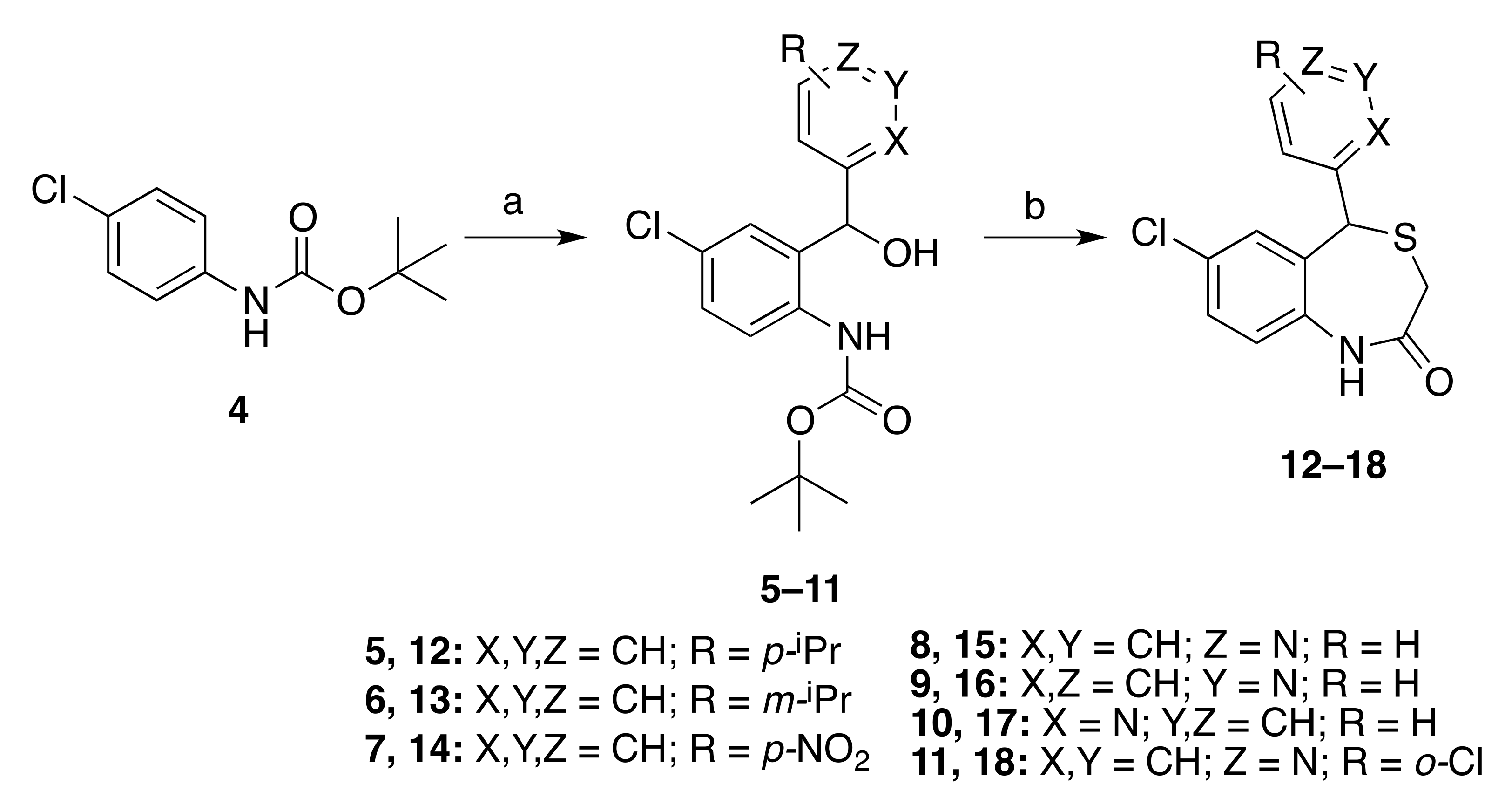
2.1. Chemistry

The preparation of 4,1-benzothiazepine derivatives was achieved through two main reaction steps, namely o-lithiation (Scheme 1, step a) and S-alkylation (Scheme 1, step b), following experimental procedures previously reported with slight modifications [34].
Non-commercial isopropylbenzaldehydes, necessary to prepare intermediates 5 and 6, were synthesized from the corresponding bromoisopropylbenzene by metal–halogen exchange with nBuLi in presence of DMF as a source of carbonyl (Supplementary Material). Boc-protected p-chloroaniline [35] suffered NH–deprotonation by tBuLi dropwise addition at −78 °C. To promote the subsequent ortho-metallation process, temperature was elevated up to −20 °C. Then, the resulting organolithium intermediates underwent nucleophilic addition on the selected aromatic aldehydes to produce diarylcarbinol derivatives 57 in medium to low yields. The addition of aryllithium species over arylaldehydes was affected by various obstacles, being the main one product degradation by an intramolecular nucleophilic attack of the generated alcohol over the carbamate, similarly to what was previously reported [38]. With the aim of increasing polarity and aqueous solubility by the incorporation of aromatic nitrogen atoms, some pyridylaldehydes were used as substrates for the addition reaction, which produced intermediates 811. Next, preparation of compounds 1218 was executed by treatment with an excess of methyl thioglycolate in refluxing of trifluoroacetic acid (TFA; 85 °C). In this acidic environment, several reactions proceed in one pot. A SN1 reaction occurs due to removal of H2O and attack of the thiol reagent over the stabilized carbocation, along with the release of the Boc-protected amine. Thus, the newly free amine attacks the pending methyl ester in an intramolecular fashion, to yield the corresponding benzothiazepines 1218 (Scheme 1) in moderate to excellent yields. In previous works, substrates possessing aromatic nitrogen atoms needed the use of alternative reagents and additional reaction steps [36]. By contrast, the 5-(pyridin-N-yl)benzothiazepines 1518 were obtained in good yields and under similar conditions to those for the synthesis of the 5-arylbenzothiazepines 1214. As shown in Table S1 (Supplementary Material), pyridine-bearing derivatives 1518 present a better aqueous solubility, according to their calculated partition coefficient logarithm (clog p) value that are between 2.5 and 3.2.
A second strategy to increase polarity of 4,1-benzothiazepines was explored. It consisted of replacing the chlorine atom at position 7 by a dimethylamino group. The Boc-protected N,N-dimethyl-p-phenylenediamine 19 (Scheme 2) (Supplementary Material) was subjected to the same conditions of ortho-lithiation, and the nucleophilic addition over o-isopropylbenzaldehyde led to the formation of intermediate 20 in moderated yield (33%). In this case, the ortho-lithiation reaction followed by the nucleophilic addition gave lower yields with several other tested aromatic aldehydes (e.g., with o-chlorobenzaldehyde, yielding the corresponding intermediate in a 12% yield), presumably due to an enhanced intramolecular addition of carbinol to the carbamate, with removal of tert-butanol. Then, the reaction of 20 with methyl thioglycolate in TFA at reflux afforded the corresponding 7-(dimethylamino)-5-(2-(propan-2-yl)phenyl-1,5-dihydro-4,1-benzothiazepin-2(3H)-one 21 with a 38% yield (Scheme 2).
Attempting to increase yields, we replaced the Boc- group with pivaloyl (2,2-dimethylpropanoyl) as a protecting group, which had offered a reliable alternative for the preparation of pyridothiazepines [36]. Thus, N,N-dimethyl-p-phenylenediamine was protected at the free amine with pivaloyl chloride using triethylamine as a base in excellent yields. The subsequent ortho-lithiation reaction of 22 was promoted with tBuLi at −20 °C as commented above, and its addition to selected aromatic aldehydes furnished the corresponding open intermediates 2325 in low to good yields, but with a more straightforward purification than that experimented with the Boc-protected intermediate 20 and analogues. Formation of the benzothiazepine scaffold needed higher temperatures, i.e., 110 °C, to avoid the isolation of S-alkylated non-cycled intermediate. Thus, we obtained in one-pot products 2628 in low yields (Scheme 3). Nevertheless, this entails a significant optimization since the removal of pivaloyl required in previous reports a three-step protocol [36] (removal of pivaloyl protecting group, S-alkylation, and finally, a LiHMDS-catalyzed cyclization).
The replacement of chlorine by dimethylamine at C7 afforded a slight reduction of the predicted clog p (Table S1, Supplementary Material). Some other derivatives proposed to be tested were benzothiazepines unsubstituted at C7. These new analogues were expected to be more lipophilic than the head CGP37157, but their clog p also lowered, about one unit. Consequently, we synthesized new derivatives starting from aniline, which was firstly protected with pivaloyl chloride, and the subsequent addition of aromatic aldehydes via ortho-lithiation provided the expected intermediates 30 and 31. With the goal of probing alternative methodologies, preparation of fluorinated intermediate 30 was carried out through ortho-lithiation with nBuLi in smooth conditions. Although yield was not disappointing in this example (43%), the reaction with other aldehydes were unsuccessful. Moreover, the disturbing presence of the by-product derived from the addition of n-butyllithium to p-fluorobenzaldehyde in the crude product moved us back to use tBuLi as metallating agent. We achieved addition to the bulky o-anisaldehyde by inducing ortho-metallation of 29 with tBuLi, affording intermediate 31 in low yield. The treatment with methyl thioglycolate at 100 °C underwent deprotection, S-alkylation, and cyclization in one pot; however, this treatment produced very low yields (Scheme 4).
The alternative three-independent-steps methodology was carried out as described [36]. Nevertheless, it did not entail an improvement in terms of overall chemical yield compared with the one-pot preparation, in addition to increasing the time required and expenses.
Finally, we appreciated that 4-aminopyridine could become an eligible starting point for the synthesis of new pyridothiazepines with enhanced polarity, different to the previously described pyrido [2,3-e][1,4] thiazepines [36], as they would give rise to interesting pyrido[4,3-e][1,4] thiazepine bicycles. Protection of 4-aminopyridine with pivaloyl chloride was conducted in good yield at lower temperature (−20 °C), to avoid the formation of diacylated product, observed at rt. Regular ortho-lithiation conditions and the subsequent addition to benzaldehyde, conducted this time at −20 °C unlike previous reactions, provided the corresponding intermediate 35 in excellent yield (90%). Then, the one-pot reaction using methyl thioglycolate in TFA at high temperature (up to 115 °C, including the use of sealed tube) failed. Alternatively, we recruited the three steps plan. Hence, pivaloyl group was removed under basic aqueous conditions (KOH in ethanol/H2O reflux) and the free amino alcohol 36 suffered nucleophilic substitution of methyl thioglycolate aided by TFA under CH2Cl2 reflux, obtaining 36 in low yield, presumably due to the scarce contribution of the pyridine ring to the stability of the intermediate carbocation. Lastly, cyclization to form the desired pyrido[4,3-e][1,4] thiazepine 38 was accomplished by deprotonation of the amine with LiHMDS in THF (Scheme 5). The global yield of these three chemical steps reached a decent 22% (13% from aniline, five reaction steps). Intriguingly, the incorporation of an aromatic nitrogen at position 7 gave rise to a pyridothiazepine derivative with a very good clog p value (1.93, Table S1, Supplementary Material), much better than those of the previously reported pyrido[2,3-e][1,4] thiazepines [36].

2.2. Pharmacology

2.2.1. Assessment of per se Toxicity of the New Derivatives in Cell Cultures

Firstly, we aimed to ensure that the new synthesized compounds had no negative effect over the viability of our cellular models. Thus, SH-SY5Y neuroblastoma cells and rat cortical neurons were exposed to compounds at increasing concentrations (1, 3, 10, and 30 μM) (Figure 2). First, experiments of viability in SH-SY5Y cells in presence of tested compounds evidenced that compound 13, bearing an isopropyl group at meta position, showed a remarkable cytotoxic profile at 30 μM (Figure 2), reason why 13 was discontinued from the pharmacological analysis. Despite compound 12 did not produce a dramatic loss of cell viability, this new molecule was not further evaluated on the grounds that some solubility problems were appreciated. Indeed, both compounds are highly lipophilic benzothiazepines analogues to ITH12575, as confirmed by their clog p (Table S1, Supplementary Material). The other compounds herein described did not affect cell viability in a statistically significant fashion. Regarding cortical neurons, compounds 18, 26, 27, 28, and 33 slightly reduced cell viability in a significant manner at 30 μM. Thus, we paid attention to their therapeutic range in experiments using cortical neurons.
Overall, these experiments helped us to stablish proper testing concentrations. For instance, considering that the neuroprotective experiments need long incubation period, the selected concentration was 10 μM. In the case of the modulation of neuronal Ca2+ oscillations, assays to evaluate IC50 in the micromolar range seemed reliable, as they are acute assays.

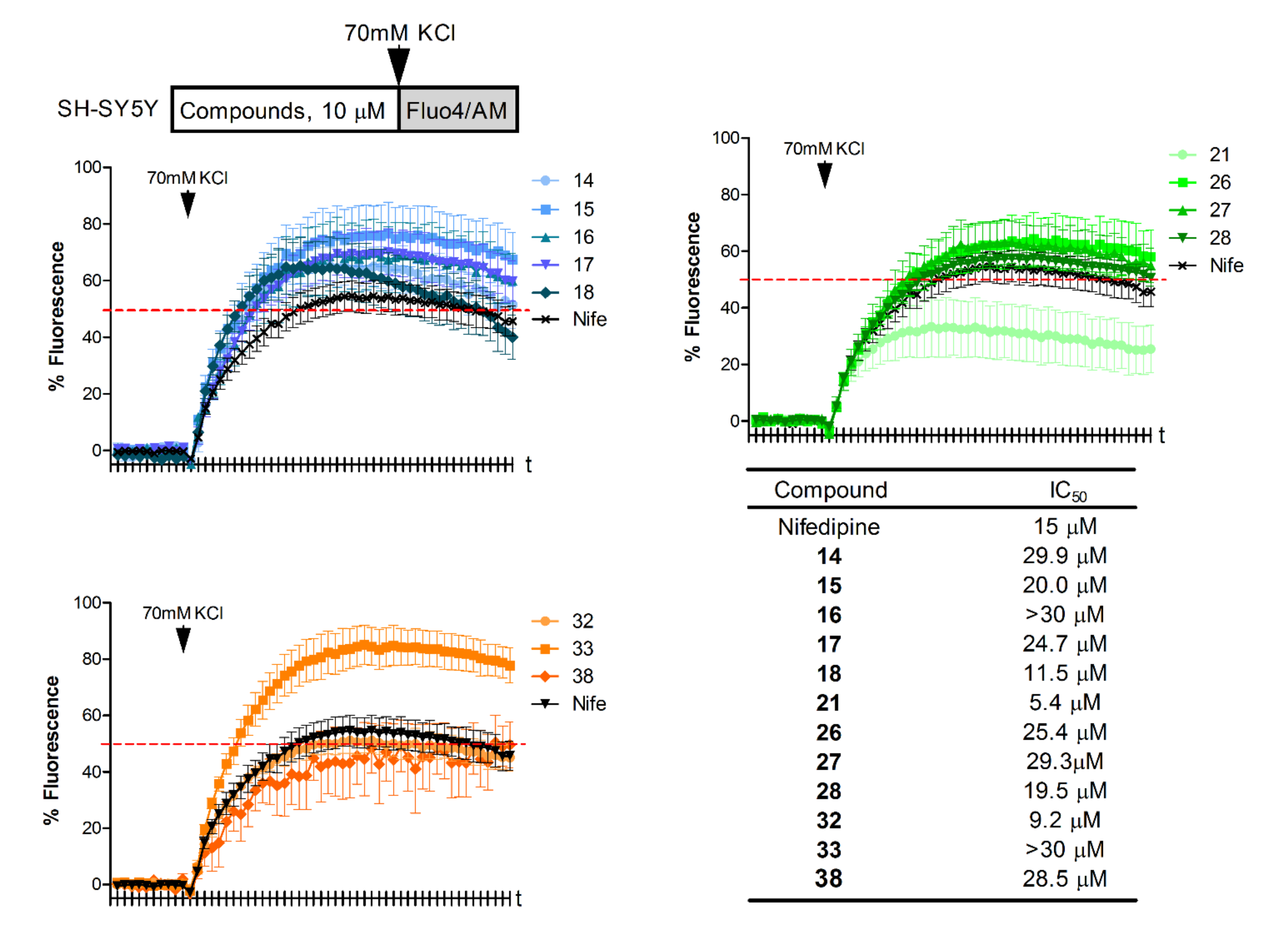
2.2.2. Effect of Compounds on the Membrane Depolarization-Induced and NMDA-Induced Ca2+ Influx

Subsequently, we assessed the ability of the compounds to minimize altered Ca2+ elevations into cells in our two cellular models. On one hand, we subjected SH-SY5Y neuroblastoma cells to a high extracellular K+ concentration (70 mM), which evokes cell depolarization and VGCC opening [33]. This experimental model mimics what occurs in a plethora of neurodegenerative diseases, where Ca2+ overload is a common event and mitochondria play a valuable role to buffer such Ca2+ elevations. Should our compounds slow the rate of Ca2+ release from mitochondria, they would improve the capacity of neurons to handle the neurotoxic cytosolic Ca2+ accumulation. Thus, most of the synthesized compounds, preincubated to SH-SY5Y cells at the concentration of 10 μM, showed an amenable mitigating effect of the depolarization-stimulated [Ca2+]c elevation in SH-SY5Y cells by about 50% (Figure 3). Compounds 18, 21, and 32 decreased Ca2+ signaling at 10 μM in a similar manner or even better than the reference compound nifedipine (10 μM) (Figure 3). This appreciable Ca2+ antagonist feature allowed us to calculate their IC50, defined as the concentration of compound capable of halving the 70 mM K+-evoked Ca2+ elevation. For that purpose, SH-SY5Y cells were incubated with the test compounds at several concentrations from 1 to 30 μM and then stimulated with high K+ to induce depolarization. In this situation, increasing concentrations of compounds produced a proportional decline of Ca2+ uptake, allowing us to draw concentration–response curves (Figure 3). Those compounds prevailing in the single concentration experiments, 18, 21, and 32, presented the lowest IC50 of the family, 11.5, 5.4, and 9.2 μM, respectively.
On the other hand, NMDAR is one the principal pathways of Ca2+ overload reported in neurodegenerative diseases [39]. Indeed, a unique non-anticholinesterasic drug prescribed for AD is the NMDAR antagonist memantine [16]. For this reason, we considered of note to assess whether these potential NCLX blockers could also mitigate the altered Ca2+ fluctuations derived from NMDAR stimulation. To achieve this aim, we chose the primary culture of the rat cortical embryonic neurons, as SH-SY5Y neuroblastoma cells have not operative NMDAR [40]. In this protocol, NMDA was administered at the concentration of 10 μM to rat cortical embryonic neurons to induce Ca2+ entry through the NMDAR. The subsequent [Ca2+]c elevation was slightly reduced by compounds exposed to neurons at 10 μM (Figure 4), mostly by about 25%, except 21 and 27, which showed a noticeable mitigation of the NMDA-elicited Ca2+ increase, even higher than that exerted by the reference compound memantine at 10 μM. Therefore, only compounds 21 and 27 offered IC50 values below 30 μM, being 6.2 and 18.5 μM, respectively.

2.2.3. Effect of Compounds against Toxic Stimuli Related to Neurodegeneration

The neuroprotective effects of these compounds in in vitro models of neurodegeneration were evaluated against a cocktail of rotenone and oligomycin A (R/O, 30 μM/10 μM) and the glutamate exposure (50 μM). For the former, SH-SY5Y neuroblastoma cells were subjected to oxidative stress by administration of the stressor cocktail R/O, which disrupts the correct function of the mitochondrial electron transport chain by inhibiting complex I and V, respectively. It is worthwhile mentioning that this oxidative stress model related to aging produces, in terms of the mitochondrial impairment, neuronal Ca2+ dishomeostasis [41]. Hence, a potential NCLX partial blocker should efficiently counteract the neurotoxicity elicited by R/O. Thus, the effect of the compounds administered at 10 μM against the R/O damage was evaluated by the MTT reduction method. As it can be appreciated in Figure 5, dimethylamino analogues 21, 26, 27, and 28 present a clear trend to protect against oxidative stress, as these compounds are able to reduce the cell death induced by R/O. Beside this family, pyridothiazepine 38 also showed a neuroprotective profile against oxidative stress.
Regarding the other neurodegeneration model, the effect against the excitotoxicity produced by high glutamate concentration was selected, considering that during the progression of neurodegenerative diseases, neurons lose their ability to control glutamate concentrations, while this neurotransmitter keeps in the synaptic cleft, producing cell death by over-activating NDMAR [42]. Similar to the fluorescence experiments of NMDA-evoked [Ca2+]c oscillations, the cell culture used for these assays were rat cortical embryonic neurons, considering their susceptibility to glutamate administration. In these experiments, compounds elicited a different pattern of neuroprotection. For instance, compounds 21, 32, and 33 presented a statistically significant dissipation of the glutamate-elicited neurotoxicity. Again, compound 38 nicely recovered cell viability of the glutamate-challenged cortical neurons.

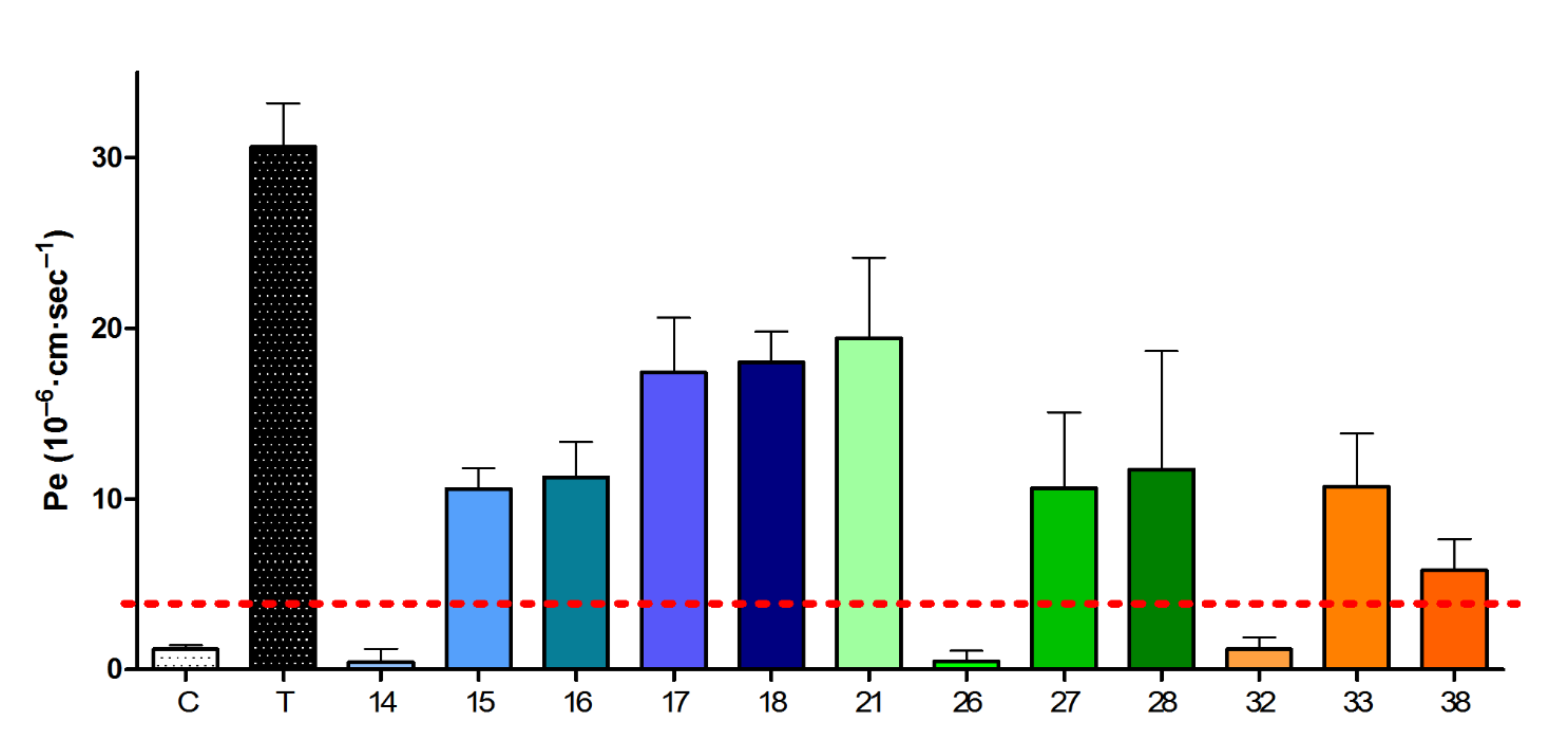
2.2.4. Blood–Brain Barrier Crossing Ability

Considering the potential use of these compounds as drugs targeting the central nervous system (CNS), we evaluated their ability to penetrate the blood–brain barrier (BBB) using the parallel artificial membrane permeation assay (PAMPA) prediction method. The in vitro permeabilities (Pe) of compounds to CNS were obtained by executing these experiments in the presence of the lipid extract of a porcine brain as a model of human BBB, as described in the experimental section. Negative and positive reference drugs were used to assess the correct development of each assay. Thus, most of the benzothiazepine derivatives described in this work were capable of crossing the BBB by passive diffusion, showing Pe values higher than 4, a widely accepted value that defines the limit for passive diffusion [43]. As it can be appreciated in Figure 6, only compounds 14, 26, and 32 did not reach this Pe value. In the case of 14, the presence of the highly charged nitro group presumably hinders its passive diffusion.

3. Materials and Methods

3.1. General

Commercially available reagents for synthesis or biological experiments were purchased from Sigma-Aldrich/Merck (Madrid, Spain). Solvents, with analytical grade, were obtained from VWR/Avantor (Barcelona, Spain) and freshly distilled when used for reactions. Thin layer chromatography (TLC) under silica gel was used to monitor the evolution of reactions by UV light exposure at 254 nm. Reactions were carried out under Schlenk conditions (vacuum/argon purges and argon atmosphere). Further purifications of compounds were conducted by automatized flash chromatography ISOLERA One (Biotage, Uppsala, Sweden), using pre-charged “SNAP” columns. Melting points, without correction, were obtained with a SMP-10 apparatus (Stuart, Stone, Staffordshire, UK). MS spectra were acquired in a QSTAR ABSciex apparatus (Applied Biosystems, Foster City, CA, USA). We acquired 1H and 13C NMR spectra in a AVANCE 300 MHz station (Bruker, Billerica, MA, USA), shown as parts per million (ppm) calibrated by the residual proton signal from the deuterated solvents. The purity of the final compounds was evaluated by elemental analysis in a CHNS-932 station (Leco, St Joseph, MI, USA), where mass percentages were within the 0.4% compared with the calculated value, or by HRMS in an ABSciex QSTAR spectrometer under the high-resolution configuration with electrospray as the ionization source, where m/z values were within the 5 ppm compared with the calculated value.

3.2. General Procedure for the Synthesis of Intermediates 511, 20, 2628, 31, and 35

General procedure was similar to what has been described, ref. [34] with slight modifications. To a solution of the Boc- or Piv-protected anilines 4 [35], 19, 22, and 29 (Supplementary Material) (1 equiv) in dry THF (4–8 mL/mmol) at −78 °C under Ar atmosphere, tBuLi (1.7 M in hexanes, 2.7 equiv) was added dropwise. When the addition was complete, the mixture was stirred for 15 min at −78 °C, then it was warmed up to −20 °C and stirred for 2 h more. After this time, the reaction mixture was cooled down to −78 °C and a solution of the corresponding arylaldehyde (1.1 equiv) in THF (5 mL) was slowly added, stirring the resulting mixture at −78 °C until no longer evolution was observed (2–5 h) by TLC. Reaction was interrupted by slow addition of H2O (15 mL) and allowed to reach rt. Afterwards, it was extracted with diethyl ether (3 × 30 mL) and combined organic layer was washed with brine (3 × 50 mL), dried over anhydrous MgSO4, filtered, and evaporated, obtaining an oil that was purified by automated flash chromatography using ethyl acetate/n-hexane mixtures as eluent.

3.2.1. 4-Chloro-2-[hydroxy(4′-isopropylphenyl)methyl]-N-tert-butoxycarbonylaniline (5)

Following the General procedure 4.2, reaction of 4-chloro-N-tert-butoxycarbonylaniline 4 [35] (2.28 mmol, 519 mg) with p-isopropylbenzaldehyde (Supplementary Material) (2.51 mmol, 372 mg) [tBuLi (6.16 mmol)] yielded 5 (341 mg, 40%). 1H NMR (300 MHz, CDCl3) δ 7.81 (d, J = 8.70 Hz, 1H, Ar), 7.66 (s, 1H, NH), 7.30 (m, 6H, Ar), 7.08 (d, J = 2.5 Hz, 1H, H3), 5.84 (s, 1H, CHOH), 3.60 (bs, 1H, OH), 2.94 [m, J = 6.9 Hz, 1H, CH(CH3)2], 1.47 [s, 9H, C(CH3)3], 1.28 [d, J = 6.9 Hz, 6H, CH(CH3)2].

3.2.2. 4-Chloro-2-[hydroxy(3′-isopropylphenyl)methyl]-N-tert-butoxycarbonylaniline (6)

Following the General procedure 4.2, reaction of 4-chloro-N-tert-butoxycarbonylaniline 4 [35] (0.66 mmol, 150 mg) with m-isopropylbenzaldehyde (Supplementary Material) (0.72 mmol, 107 mg) [tBuLi (1.78 mmol)] yielded 6 (72 mg, 29%). 1H NMR (300 MHz, CDCl3) δ 7.84 (d, J = 8.9 Hz, 1H, Ar), 7.55 (s, 1H, NH), 7.35–7.15 (m, 4H, Ar), 7.10–7.05 (m, 2H, Ar), 5.85 (s, 1H, CHOH), 3.50 (bs, 1H, OH), 2.92 [m, J = 6.8 Hz, 1H, CH(CH3)2], 1.45 [s, 9H, C(CH3)3], 1.25 [d, J = 6.8 Hz, 6H, CH(CH3)2].

3.2.3. 4-Chloro-2-[hydroxy(4′-nitrophenyl)methyl]-N-tert-butoxycarbonylaniline (7)

Following the General procedure 4.2, reaction of 4-chloro-N-tert-butoxycarbonylaniline 4 [35] (3.01 mmol, 685 mg) with p-nitrobenzaldehyde (3.31 mmol, 500 mg) [tBuLi (8.13 mmol)] yielded 7 (264 mg, 23%). 1H NMR (300 MHz, CDCl3) δ 8.20 (d, J = 9.0 Hz, 2H, Ar), 7.69 (d, J = 8.6 Hz, 1H, Ar), 7.55 (d, J = 9.0 Hz, 2H, Ar), 7.48 (s, 1H. NH), 7.31 (m, 1H, Ar), 7.09 (d, J = 2.5 Hz, 1H, H3), 5.93 (s, 1H, CHOH), 4.80 (bs, 1H, OH), 1.43 [s, 9H, C(CH3)3].

3.2.4. 4-Chloro-2-[hydroxy(pyridin-4-yl)methyl]-N-tert-butoxycarbonylaniline (8)

Following the General procedure 4.2, reaction of 4-chloro-N-tert-butoxycarbonylaniline 4 [35] (4.24 mmol, 965 mg) with isonicotinaldehyde (4.67 mmol, 500 mg) [tBuLi (11.45 mmol)] yielded 8 (618 mg, 44%). 1H NMR (300 MHz, CDCl3) δ 8.29 (d, J = 6.2 Hz, 2H, Ar), 8.04 (s, 1H, NH), 7.77 (d, J = 8.8 Hz, 1H, Ar), 7.34 (d, J = 6.2 Hz, 2H, Ar), 7.28 (dd, J = 8.7, 2.4 Hz, 1H, Ar), 7.10 (d, J = 2.4 Hz, 1H, H3), 5.78 (s, 1H, CHOH), 4.70 (bs, 1H, OH), 1.41 [s, 9H, C(CH3)3].

3.2.5. 4-Chloro-2-[hydroxy(pyridin-3-yl)methyl]-N-tert-butoxycarbonylaniline (9)

Following the General procedure 4.2, reaction of 4-chloro-N-tert-butoxycarbonylaniline 4 [35] (4.24 mmol, 965 mg) with nicotinaldehyde (4.67 mmol, 500 mg) [tBuLi (11.45 mmol)] yielded 9 (718 mg, 51%). 1H NMR (300 MHz, CDCl3) δ 8.42 (s, 1H, Ar), 8.30 (m, 1H, Ar), 8.18 (s, 1H, NH), 7.75 (m, 2H, Ar), 7.24 (m, 2H, Ar), 7.10 (d, J = 2.3 Hz, 1H, H3), 5.86 (s, 1H, CHOH), 4.90 (bs, 1H, OH), 1.43 [s, 9H, C(CH3)3].

3.2.6. 4-Chloro-2-[hydroxy(pyridin-2-yl)methyl]-N-tert-butoxycarbonylaniline (10)

Following the General procedure 4.2, reaction of 4-chloro-N-tert-butoxycarbonylaniline 4 [35] (4.24 mmol, 965 mg) with picolinaldehyde (4.67 mmol, 500 mg) [tBuLi (11.45 mmol)] yielded 10 (394 mg, 28%). 1H NMR (300 MHz, CDCl3) δ 8.52 (d, J = 6.0 Hz, 1H, Ar), 8.38 (s, 1H, NH), 7.75 (d, J = 8.6 Hz, 1H, Ar), 7.67 (m, 1H, Ar), 7.24 (m, 4H, Ar), 5.79 (s, 1H, CHOH), 4.93 (bs, 1H, OH), 1.42 [s, 9H, C(CH3)3].

3.2.7. 4-Chloro-2-[(3-chloropyridin-4-yl)hydroxymethyl]-N-tert-butoxycarbonylaniline (11)

Following the General procedure 4.2, reaction of 4-chloro-N-tert-butoxycarbonylaniline 4 [35] (1.93 mmol, 439 mg) with 3-isonicotinaldehyde (2.11 mmol, 300 mg) [tBuLi (5.21 mmol)] yielded 11 (144 mg, 20%). 1H NMR (300 MHz, CDCl3) δ 8.44 (d, J = 5.1 Hz, 1H, Ar), 8.38 (s, 1H, Ar), 7.67 (d, J = 5.1 Hz, 1H, Ar), 7.55 (s, 1H, NH), 7.53 (d, J = 8.7 Hz, 1H, Ar), 7.22 (dd, J = 8.7, 2.5 Hz, 1H, Ar), 6.89 (d, J = 2.5 Hz, 1H, H3), 6.03 (s, 1H, CHOH), 5.48 (bs, 1H, OH), 1.47 [s, 9H, C(CH3)3].

3.2.8. 4-Dimethylamino-2-[hydroxy(2′-Isopropylphenyl)methyl]-N-tert-butoxycarbonylaniline (20)

Following the General procedure 4.2, reaction of 4-dimethylamino-N-tert-butoxycarbonylaniline 19 (Supplementary Material) (4.23 mmol, 1 g) with o-isopropylbenzaldehyde (4.65 mmol, 690 mg) [tBuLi (11.42 mmol)] yielded 20 (537 mg, 33%). 1H NMR (300 MHz, acetone-d6) δ 7.83 (bs, 1H, NH), 7.45 (m, 2H, Ar), 7.34–7.15 (m, 3H, Ar), 6.65 (dd, 1H, J = 8.7, 3.0 Hz, H5), 6.42 (s, 1H, CHOH), 6.22 (d, 1H, J = 3.0 Hz, H3), 4.94 (s, 1H, OH), 3.16 [m, J = 6.0 Hz, 1H, CH(CH3)2], 2.76 [s, 6H, N(CH3)2], 1.46 [s, 9H, C(CH3)3], 1.20 [d, J = 6.0 Hz, 3H, CH(CH3)], 0.98 [d, J = 6.0 Hz, 3H, CH(CH3)].

3.2.9. 2-[(2′-Chlorophenyl)hydroxymethyl]-4-dimethylamino-N-tert-butylcarbonylaniline (23)

Following the General procedure 4.2, reaction of N-[4-(dimethylamino)phenyl]-2,2-dimethylpropanamide 22 (Supplementary Material) (2.26 mmol, 0.5 g) with o-chlorobenzaldehyde (0.28 mL, 318 mg, 2.26 mmol) [tBuLi (6.1 mmol)] yielded 23 (284 mg, 35%). 1H NMR (300 MHz, CDCl3) δ 7.96 (bs, 1H, NH), 7.57 (bd, J = 6.0, 1H, Ar), 7.4 (d, J = 9.0 Hz, 1H, H6), 7.3–7.20 (m, 3H, Ar), 6.65 (dd, J = 9.0, 3.0 Hz, H5), 6.38 (d, J = 3.0 Hz, 1H, H3), 6.05 (d, J = 2.7 Hz, 1H, CHOH), 4.11 (d, J = 2.7 Hz, 1H, OH), 2.81 [s, 6H, N(CH3)2], 1.25 [s, 9H, C(CH3)3].

3.2.10. 4-Dimethylamino-2-[hydroxy(2′-Methoxyphenyl)methyl]-N-tert-butylcarbonylaniline (24)

Following the General procedure 4.2, reaction of N-[4-(dimethylamino)phenyl]-2,2-dimethylpropanamide 22 (Supplementary Material) (3.40 mmol, 750 mg) with o-methoxybenzaldehyde (0.45 mL, 509 mg, 3.74 mmol) [tBuLi (9.18 mmol)] yielded 24 (865 mg, 71%). 1H NMR (300 MHz, CDCl3) δ 8.79 (s, 1H, NH), 7.97 (d, 1H, J = 8.4 Hz, H6), 7.30 (m, 1H, Ar), 6.95 (d, J = 8.0 Hz, 1H, Ar), 6.87 (m, 2H, Ar), 6.74 (bd, J = 8.4 Hz, 1H, H5), 6.51 (d, J = 2.7 Hz, 1H, H3), 6.04 (d, J = 3.6 Hz, 1H, CHOH), 4.04 (d, 1H, J = 3.6 Hz, OH), 3.93 (s, 3H, OCH3), 2.86 [s, 6H, N(CH3)2], 1.06 [s, 9H, C(CH3)3].

3.2.11. 4-Dimethylamino-2-[(4′-fluorophenyl)hydroxymethyl]-N-tert-butylcarbonylaniline (25)

Following the General procedure 4.2, reaction of N-[4-(dimethylamino)phenyl]-2,2-dimethylpropanamide 22 (Supplementary Material) (2.26 mmol, 0.5 g) with p-fluorobenzaldehyde (0.27 mL, 309 mg, 2.49 mmol) [tBuLi (6.10 mmol)] yielded 25 (390 mg, 50%). 1H NMR (300 MHz, CDCl3) δ 8.21 (s, 1H, NH), 7.82 (d, J = 9.0 Hz, 1H, H6), 7.27 (m, 2H, Ar), 6.98 (m, 2H, Ar), 6.72 (dd, J = 9.0, 3.0 Hz, 1H, H5), 6.50 (d, J = 3.0 Hz, 1H, H3), 5.75 (s, 1H, CHOH), 3.77 (s, 1H, OH), 2.88 [s, 6H, N(CH3)2], 1.04 [s, 9H, C(CH3)3].

3.2.12. 2-[Hydroxy(2′-methoxyphenyl)methyl]-N-tert-butylcarbonylaniline (31)

Following the General procedure 4.2, reaction of N-phenyl-2,2-dimethylpropanamide 29 (Supplementary Material) (2.80 mmol, 0.5 g) with o-methoxybenzaldehyde (0.88 mL, 780 mg, 5.60 mmol) [tBuLi (7.00 mmol)] yielded 31 (215 mg, 25%). 1H NMR (300 MHz, CDCl3) δ 9.53 (s, 1H, NH), 8.48 (d, J = 8.4 Hz, 1H, H6), 7.67–7.06 (m, 7H, Ar), 6.31 (d, J = 4.3 Hz, 1H, CHOH), 4.46 (d, 1H, J = 4.3 Hz, OH), 4.17 (s, 3H, OCH3), 1.35 [s, 9H, C(CH3)3].

3.2.13. N-[3-[hydroxy(phenyl)methyl]pyrydin-4-yl]-2,2-dimethylpropanamide (35)

Following the General procedure 4.2, reaction of 2,2-dimethyl-N-(4-pyridinyl)propanamide 34 (Supplementary Material) (4.21 mmol, 750 mg) benzaldehyde (0.47 mL, 491 mg, 4.62 mmol) [tBuLi (11.37 mmol)] yielded 35 (1.08 g, 90%). 1H NMR (300 MHz, CDCl3) δ 9.37 (s, 1H, NH), 8.37 (d, J = 5.7 Hz, 1H, Ar), 8.31 (d, J = 5.7 Hz, 1H, Ar), 7.99 (s, 1H, Ar), 7.36–7.30 (m, 5H, Ar), 5.85 (s, 1H, CHOH), 3.72 (s, 1H, OH), 1.07 [s, 9H, C(CH3)3].

3.3. Synthesis of 2-[(4′-Fluorophenyl)hydroxymethyl]-N-tert-butylcarbonylaniline (30)

Similar to what has been described for the use of tBuLi [34], with modifications. To a solution of the Piv-protected aniline 29 (Supplementary Material) (2.82 mmol, 0.5 g) in dry THF (12 mL) at −78 °C under Ar atmosphere, nBuLi (1.9 M in hexanes, 2.5 equiv, 7.05 mmol, 3.7 mL) was added dropwise. When the addition was complete, the mixture was stirred for 15 min at −78 °C, then it was warmed up to 0 °C and stirred for 4 h more. After this time, the reaction mixture was cooled down to −78 °C and a solution of p-fluorobenzaldehyde (0.62 mL, 0.7 g, 5.64 mmol) in THF (5 mL) was slowly added, and the reaction mixture was allowed to reach 0 °C for 2 h. Reaction was interrupted by slow addition of HCl 1N (12 mL). Afterwards, it was extracted with ethyl acetate (3 × 20 mL) and the combined organic layer was dried over anhydrous Na2SO4, filtered, and evaporated, obtaining an oil that was purified by automatized flash chromatography using ethyl acetate/n-hexane mixtures as eluent, yielding 30 as a yellow oil (360 mg, 43%). 1H NMR (300 MHz, CDCl3) δ 9.15 (s, 1H, NH), 8.37 (d, J = 8.1 Hz, 1H, Ar), 7.62–7.27 (m, 5H, Ar), 7.23 (m, 2H, Ar), 6.09 (s, 1H, CHOH), 4.22 (s, 1H, OH), 1.31 [s, 9H, C(CH3)3].

3.4. General Procedure for the Synthesis of 4,1-Benzothiazepines 1218, 21, 2628

Similar to what has been described [34], with slight modifications. Methyl thioglycolate (6 equiv) and trifluoroacetic acid (TFA, 14 equiv) were added to the intermediates 511 or 20 under Ar. The mixture was stirred at 85–110 °C for 24 h. Then, it was dissolved in CH2Cl2 (30 mL) and washed subsequently with brine (30 mL), NaOH (1N, 30 mL), and brine (30 mL). The organic layer was dried over Na2SO4, filtered, and evaporated. The crude material was purified by automatized flash chromatography using ethyl acetate/n-hexane mixtures as eluent to give pure compounds.

3.4.1. 7-Chloro-5-(4′-isopropylphenyl)-3,5-dihydro-4,1-benzothiazepin-2-(1H)-one (12)

Following the General procedure 4.4, reaction of 5 (0.66 mmol, 248 mg) [methyl thioglycolate (3.96 mmol, 420 mg), TFA (9.24 mmol, 1.05 g, 707 μL)] yielded 12 (140 mg, 64%) as a white solid. Mp: 240–243 °C. 1H NMR (300 MHz, CDCl3) δ 8.20 (s, 1H, NH), 7.39 (d, J = 8.1 Hz, 2H, Ar), 7.27 (m, 3H, Ar), 7.04 (d, J = 8.4 Hz, 1H, H9), 6.98 (d, J = 2.3 Hz, 1H, H6), 5.64 (s, 1H, H5), 3.34 (d, J = 12.2 Hz, 1H, H3a), 2.99 (d, J = 12.2 Hz, 1H, H3b), 2.94 [m, J = 6.9 Hz, 1H, CH(CH3)2], 1.28 [d, J = 6.9 Hz, 6H, CH(CH3)2]. 13C NMR (75.4 MHz, CDCl3) δ 170.6, 149.4, 136.9, 134.8, 133.4, 133.2, 129.4, 128.9, 128.7, 127.2, 125.4, 47.2, 34.0, 31.7, 24.1, 24.0. MS (API-ES+): m/z 332.09 [(M + H)+], 663.18 [(2M + Na)+]. Anal. C18H18ClNOS (C,H,N).

3.4.2. 7-Chloro-5-(3′-isopropylphenyl)-3,5-dihydro-4,1-benzothiazepin-2-(1H)-one (13)

Following the General procedure 4.4, reaction of 6 (0.19 mmol, 71 mg) [methyl thioglycolate (1.15 mmol, 122 mg), TFA (2.66 mmol, 303 mg, 204 μL)] yielded 13 (21 mg, 33%) as a white solid. Mp: 218–220 °C. 1H NMR (300 MHz, CDCl3) δ 8.01 (s, 1H, NH), 7.30 (m, 2H, Ar), 7.25 (m, 3H, Ar), 6.98 (d, J = 8.4 Hz, 1H, H9), 6.90 (d, J = 2.3 Hz, 1H, H6), 5.59 (s, 1H, H5), 3.28 (d, J = 12.2 Hz, 1H, H3a), 2.92 (d, J = 12.2 Hz, 1H, H3b), 2.86 [m, J = 6.9 Hz, 1H, CH(CH3)2], 1.21 [d, J = 6.9 Hz, 3H, CH(CH3)], 1.19 [d, J = 6.9 Hz, 3H, CH(CH3)]. 13C NMR (75.4 MHz, CDCl3) δ 149.8, 136.8, 136.0, 134.8, 133.3, 129.0, 128.9, 128.7, 127.8, 127.0, 126.8, 125.4, 47.6, 34.2, 31.7, 24.2, 24.0. MS (API-ES+): m/z 332.10 [(M + H)+], 663.20 [(2M + Na)+]. HRMS: m/z (M + H)+ calcd. for C18H18ClNOS 332.0870, found, 332.0869.

3.4.3. 7-Chloro-5-(4′-nitrophenyl)-3,5-dihydro-4,1-benzothiazepin-2-(1H)-one (14)

Following the General procedure 4.4, reaction of 7 (0.70 mmol, 265 mg) [methyl thioglycolate (4.20 mmol, 446 mg), TFA (9.80 mmol, 1.12 g, 750 μL)] yielded 14 (234 mg, >99%) as a white solid. Mp: 289–291 °C. 1H NMR (300 MHz, DMSOd6) δ 9.52 (s, 1H, NH), 8.25 (d, J = 8.8 Hz, 2H, Ar), 7.74 (d, J = 8.8 Hz, 2H, Ar), 7.45 (dd, J = 8.3, 2.4 Hz, 1H, Ar), 7.29 (d, J = 2.4 Hz, 1H, H6), 7.06 (d, J = 8.4 Hz, 1H, H9), 5.78 (s, 1H, H5), 3.14 (d, J = 12.2 Hz, 1H, H3a), 3.07 (d, J = 12.2 Hz, 1H, H3b). 13C NMR (75.4 MHz, DMSOd6) δ 168.4, 146.8, 146.4, 136.6, 135.6, 130.3, 129.8, 128.8, 128.7, 126.5, 123.5, 46.3, 30.5. MS (API-ES+): m/z 335.02 [(M + H)+]. Anal. C15H11ClN2O3S (C,H,N).

3.4.4. 7-Chloro-5-(pyridin-4-yl)-3,5-dihydro-4,1-benzothiazepin-2-(1H)-one (15)

Following the General procedure 4.4, reaction of 8 (1.85 mmol, 619 mg) [methyl thioglycolate (11.10 mmol, 1.18 g), TFA (25.90 mmol, 2.95 g, 1.98 mL)] yielded 15 (369 mg, 69%) as a white solid. Mp: 224–226 °C. 1H NMR (300 MHz, DMSOd6) δ 9.51 (s, 1H, NH), 8.58 (m, 2H, Ar), 7.46 (m, 3H, Ar), 7.33 (d, J = 2.4 Hz, 1H, H6), 7.07 (d, J = 8.4 Hz, 1H, H9), 5.64 (s, 1H, H5), 3.15 (d, J = 12.2 Hz, 1H, H3a), 3.04 (d, J = 12.2 Hz, 1H, H3b). 13C NMR (75.4 MHz, DMSOd6) δ 168.4, 149.7, 147.8, 136.6, 135.3, 130.3, 128.9, 128.8, 126.5, 123.4, 45.9, 30.3. MS (API-ES+): m/z 291.03 [(M + H)+]. Anal. C14H11ClN2OS (C,H,N).

3.4.5. 7-Chloro-5-(pyridin-3-yl)-3,5-dihydro-4,1-benzothiazepin-2-(1H)-one (16)

Following the General procedure 4.4, reaction of 9 (2.14 mmol, 716 mg) [methyl thioglycolate (12.84 mmol, 1.36 g), TFA (29.96 mmol, 3.42 g, 2.29 mL)] yielded 16 (476 mg, 76%) as a white solid. Mp: 224–226 °C. 1H NMR (300 MHz, DMSOd6) δ 9.62 (s, 1H, NH), 8.62 (d, J = 2.4 Hz, 1H, H2′), 8.55 (m, 1H, Ar), 7.93 (m, 1H, Ar), 7.43 (m, 2H, Ar), 7.13 (d, J = 2.4 Hz, 1H, H6), 7.07 (d, J = 8.4 Hz, 1H, H9), 5.70 (s, 1H, H5), 3.10 (AA’BB’, J = 12.5 Hz, 2H, H3). 13C NMR (75.4 MHz, DMSOd6) δ 168.5, 149.7, 149.0, 136.6, 136.5, 135.5, 133.8, 130.3, 128.7, 128.3, 126.3, 123.5, 44.2, 30.7. HRMS: m/z (M + H)+ calcd. for C14H12ClN2OS 291.0353, found, 291.0347.

3.4.6. 7-Chloro-5-(pyridin-2-yl)-3,5-dihydro-4,1-benzothiazepin-2-(1H)-one (17)

Following the General procedure 4.4, reaction of 10 (1.18 mmol, 395 mg) [methyl thioglycolate (7.08 mmol, 751 mg), TFA (16.52 mmol, 1.88 g, 1.26 mL)] yielded 17 (200 mg, 58%) as a white solid. Mp: 215–217 °C. 1H NMR (300 MHz, CDCl3) δ 8.65 (m, 1H, Ar), 7.78 (dt, J = 7.7, 1.9 Hz, 1H, Ar), 7.65 (s, 1H, NH), 7.60 (d, J = 8.9 Hz, 1H, H6), 7.27 (m, 2H, Ar), 7.00 (m, 2H, Ar), 5.72 (s, 1H, H5), 3.26 (d, J = 12.5 Hz, 1H, H3a), 3.08 (d, J = 12.5 Hz, 1H, H3b). 13C NMR (75.4 MHz, CDCl3) δ 169.9, 156.6, 149.9, 137.3, 136.2, 134.8, 133.4, 129.0, 125.5, 123.9, 123.2, 49.3, 31.2. MS (API-ES+): m/z 291.03 [(M + H)+]. HRMS: m/z (M + H)+ calcd. for C14H12ClN2OS 291.0353, found, 291.0342.

3.4.7. 7-Chloro-5-(3′-chloropyridin-4-yl)-3,5-dihydro-4,1-benzothiazepin-2-(1H)-one (18)

Following the General procedure 4.4, reaction of 11 (0.39 mmol, 144 mg) [methyl thioglycolate (2.34 mmol, 248 mg), TFA (5.46 mmol, 1.88 g, 1.26 mL)] yielded 18 (80 mg, 63%) as a white solid. Mp: 226–229 °C. 1H NMR (300 MHz, CDCl3) δ 8.64 (d, J = 6.0 Hz, 1H, H6′), 8.61 (s, 1H, H2′), 7.83 (d, J = 5.1 Hz, 1H, H5′), 7.51 (s, 1H, NH), 7.31 (dd, J = 2.4, 8.4 Hz, 1H, H8), 7.07 (d, J = 8.4 Hz, 1H, H9), 6.78 (d, J = 2.4 Hz, 1H, H6), 5.94 (s, 1H, H5), 3.28 (d, J = 12.3 Hz, 1H, H3a), 3.10 (d, J = 12.3 Hz, 1H, H3b). 13C NMR (75.4 MHz, CDCl3) δ 169.2, 150.0, 148.2, 148.1, 135.3, 135.0, 133.9, 133.7, 129.6, 128.5, 125.8, 125.4, 43.9, 31.2. HRMS: m/z (M + H)+ calcd. for C14H11Cl2N2OS 324.9964, found, 324.9953.

3.4.8. 7-Dimethylamino-5-(2′-isopropylphenyl)-3,5-dihydro-4,1-benzothiazepin-2-(1H)-one (21)

Following the General procedure 4.4, reaction of 20 (0.26 mmol, 100 mg) [methyl thioglycolate (1.56 mmol, 165 mg), TFA (3.64 mmol, 415 mg, 279 μL)] yielded 21 (29 mg, 38%) as a white solid. Mp: 108–112 °C. 1H NMR (300 MHz, acetone-d6) δ 8.49 (s, 1H, NH), 7.78 (m, 1H, Ar), 7.40–7.25 (m, 3H), 7.00 (d, J = 8.4 Hz, 1H, H9), 6.66 (dd, J = 3.0, 8.4 Hz, 1H, H8), 6.09 (m, 2H, H6, H5), 3.33 (d, J = 12.0 Hz, 1H, H3a), 2.93 [m, J = 6.9 Hz, 1H, CH(CH3)2], 2.83 (d, J = 12.0 Hz, 1H, H3b), 2.72 [s, 6H, N(CH3)2], 1.19 [d, J = 6.9 Hz, 3H, CH(CH3)], 0.86 [d, J = 6.9 Hz, 3H, CH(CH3)]; 13C NMR (75.4 MHz, CDCl3) δ 169.8, 150.7, 148.0, 136.4, 135.4, 130.6, 129.3, 127.5, 127.0, 126.6, 125.8, 113.0, 112.4, 44.0, 40.6, 32.4, 24.5, 23.7. HRMS: m/z (M + H)+ calcd. for C20H25N2OS 341.1682, found, 341.1671.

3.4.9. 5-(2′-Chlorophenyl)-7-dimethylamino-3,5-dihydro-4,1-benzothiazepin-2-(1H)-one (26)

Following the General procedure 4.4, reaction of 23 (0.39 mmol, 140 mg) [methyl thioglycolate (2.33 mmol, 247 mg), TFA (5.43 mmol, 619 mg, 415 μL)] yielded 26 (36 mg, 27%) as a white solid. Mp: 178–181 °C. 1H NMR (300 MHz, DMSOd6) δ 9.44 (s, 1H, NH), 7.81 (d, J = 7.5 Hz, 1H, Ar), 7.49 (m, 2H, Ar), 7.45 (m, 1H, Ar), 6.93 (d, J = 8.7 Hz, 1H, Ar), 6.66 (dd, J = 11.4, 5.4 Hz, 1H, H8), 5.95 (m, 2H, H6, H5), 3.16 (d, 1H, H3a), 2.90 (d, 1H, H3b), 2.75 [s, 6H, N(CH3)2]. 13C NMR (75.4 MHz, DMSOd6): δ 168.6, 148.9, 134.9, 132.9, 132.8, 131.0, 129.9, 129.8, 127.3, 126.3, 125.0, 112.0, 110.3, 43.5, 31.1. HRMS: m/z (M + H)+ calcd. for C17H18ClN2OS 333.0822, found, 333.0829. Anal. C17H17ClN2OS (C,H,N).

3.4.10. 7-Dimethylamino-5-(2′-methoxyphenyl)-3,5-dihydro-4,1-benzothiazepin-2-(1H)-one (27)

Following the General procedure 4.4, reaction of 24 (0.70 mmol, 250 mg) [methyl thioglycolate (4.18 mmol, 444 mg), TFA (9.76 mmol, 1.11 g, 750 μL)] yielded 27 (24 mg, 10%) as a white solid. Mp: 233–236 °C. 1H NMR (300 MHz, CDCl3) δ 7.75 (d, J = 9.0 Hz, 1H, Ar), 7.33 (m, 1H, Ar), 7.11 (s, 1H, NH), 7.05 (m, 1H, Ar), 6,90 (m, 2H, Ar), 6.56 (dd, J = 9.0, 3.0 Hz, 1H, H8), 6.25 (d, J = 3.0 Hz, 1H, H6), 6.17 (s, 1H, H5), 3.70 (s, 3H, OCH3), 3.35 (d, J = 12.0 Hz, 1H, H3a), 2.95 (d, J = 12.0 Hz, 1H, H3b), 2.79 [s, 6H, N(CH3)2]. 13C NMR (75.4 MHz, DMSOd6): δ 170.6, 157.0, 150.0, 135.7, 130.8, 129.2, 125.7, 125.2, 125.2 120.4, 111.8, 111.1, 55.7, 40.6, 40.4, 31.7. Anal. C18H21N2O2S·H2O (C,H,N).

3.4.11. 7-Dimethylamino-5-(4′-fluorophenyl)-3,5-dihydro-4,1-benzothiazepin-2-(1H)-one (28)

Following the General procedure 4.4, reaction of 25 (0.43 mmol, 150 mg) [methyl thioglycolate (2.61 mmol, 277 mg), TFA (6.09 mmol, 694 mg, 466 μL)] yielded 28 (25 mg, 18%) as a white solid. Mp: 219–222 °C. 1H NMR (300 MHz, DMSOd6) δ 9.14 (s, 1H, NH), 7.52 (m, 2H, Ar), 7.21 (m, 2H, Ar), 6.87 (d, J = 9.0 Hz, 1H, H9), 6.65 (dd, J = 9.0 Hz, J = 3.0 Hz, 1H, H8), 6.33 (d, J = 3.0, 1H, H6), 5.54 (s, 1H, H5), 3.05 (d, J = 12.0 Hz, 1H, H3a), 2.93 (d, J = 12.0 Hz, 1H, H3b), 2.78 [s, 6H, N(CH3)2]. 13C NMR (75.4 MHz, DMSOd6): δ 168.8, 148.9, 134.7, 134.5, 130.8 (JC-F = 8.3 Hz), 126.1, 125.2, 115.2 (JC-F = 21.3 Hz), 111.9, 111.6, 46.7, 40.0, 30.9. HRMS: m/z (M + H)+ calcd. for C17H18FN2OS 317.1120, found, 317.1120.

3.4.12. 5-(4′-Fluorophenyl)-3,5-dihydro-4,1-benzothiazepin-2-(1H)-one (32)

Following the General procedure 4.4, reaction of 30 (0.36 mmol, 109 mg) [methyl thioglycolate (2.16 mmol, 229 mg), TFA (5.04 mmol, 575 mg, 388 μL)] yielded 32 (12 mg, 12%) as a white solid. Mp: 251–254 °C. 1H NMR (300 MHz, DMSOd6) δ 7.47 (m, 2H, Ar), 7.46–7.23 (m, 3H, Ar), 7.08 (m, 3H, Ar), 6.98 (m, 1H, Ar), 5.66 (s, 1H, H5), 3.29 (d, J = 12.2 Hz, 1H, H3a), 3.02 (d, J = 12.2 Hz, 1H, H3b). 13C NMR (75.4 MHz, CDCl3): δ 170.0, 157.6 (d, J = 245 Hz), 136.2, 134.9, 133.2, 131.1 (d, J = 8.0 Hz), 128.9, 128.8, 127.9, 124.3, 115.8 (d, J = 21.0 Hz), 47.2, 31.5. HRMS: m/z (M + Na)+ calcd. for C15H12FNNaOS 296.0621, found, 296.0621.

3.4.13. 5-(2′-Methoxyphenyl)-3,5-dihydro-4,1-benzothiazepin-2-(1H)-one (33)

Following the General procedure 4.4, reaction of 31 (0.98 mmol, 308 mg) [methyl thioglycolate (5.88 mmol, 624 mg), TFA (13.72 mmol, 1.56 g, 1.06 mL)] yielded 33 (23 mg, 8%) as a white solid. Mp: 216–218 °C. 1H NMR (300 MHz, CDCl3) δ 7.76 (d, J = 9.2 Hz, 1H, Ar), 7.63–6.79 (m, 8H, Ar), 6.20 (s, 1H, H5), 3.68 (s, 3H, OCH3), 3.33 (d, J = 12.1 Hz, 1H, H3a), 2.99 (d, J = 12.1 Hz, 1H, H3b). 13C NMR (75.4 MHz, CDCl3): δ 170.3, 156.7, 136.4, 134.9, 130.8, 129.4, 128.4, 128.4, 127.8, 125.4, 124.0, 120.6, 111.1, 55.7, 40.1, 31.6. Anal. C16H15NO2S (C,H,N).

3.5. Synthesis of (4-Aminopyridin-3-yl)(phenyl)methanol (36)

Similar to what we have described [36]. To a solution of intermediate 35 (0.63 mmol, 179 mg) in 1,4-dioxane (18 mL), KOHaq 2 M (18 mL) was added, and the mixture was refluxed for 24 h. Then, reaction mixture was cooled down to rt and extracted with ethyl acetate (3 × 50 mL). The combined organic layer was dried over anhydrous Na2SO4, filtered, and evaporated, obtaining 36 as a yellow solid (132 mg, >99%). 1H NMR (300 MHz, CDCl3) δ 8.00 (d, J = 5.7 Hz, 1H, Ar), 7.86 (s, 1H, Ar), 7.37–7.30 (m, 5H, Ar), 6.42 (d, J = 5.7 Hz, 1H, Ar), 5.78 (s, 1H, CHOH), 4.76 (s, 3H, OH, NH2).

3.6. Synthesis of Methyl 2-[(4-Aminopyridin-3-yl)(phenyl)methylthio]acetate (37)

Similar to what we have described [36]. To a solution of intermediate 36 (445 mg, 2.22 mmol) in dry CH2Cl2 (25 mL), methyl thioglycolate (13.3 mmol, 1.41 g) and TFA (10 mL) were added under Ar. The mixture was stirred at 45 °C for 22 h. Then, reaction mixture was concentrated, dissolved in CH2Cl2 (50 mL), and washed subsequently with saturated NaHCO3 (50 mL), NaOH (1N, 50 mL), and brine (50 mL). The organic layer was dried over Na2SO4, filtered, and evaporated. The crude was purified by automatized flash chromatography using ethyl acetate/n-hexane mixtures as eluent to give 37 (144 mg, 25%). 1H NMR (300 MHz, CDCl3) δ 8.12 (d, J = 5.7 Hz, 1H, Ar), 7.89 (s, 1H, Ar), 7.51 (m, 2H, Ar), 7.41–7.32 (m, 3H, Ar), 6.56 (d, J = 5.7 Hz, 1H, Ar), 5.35 (s, 1H, CH), 5.08 (s, 2 H, NH2), 3.74 (s, 3H, OCH3), 3.22 (d, J = 16.8 Hz, 1H, H2a), 3.14 (d, J = 16.8 Hz, 1H, H2b).

3.7. Synthesis of 5-Phenyl-3,5-dihydropyrido[4,3-e][1,4]thiazepin-2-(1H)-one (38)

Similar to what we have described [36]. To a solution of intermediate 37 (150 mg, 0.52 mmol) in dry THF (23 mL), LiHMDS (1.04 mmol, 1.0 M in hexane, 1.04 mL) was added dropwise at −78 °C under Ar and stirred at this temperature for 15 min. Then, reaction mixture was allowed to warm up to rt for 24 h. Reaction was terminated by adding a saturated solution of NH4Cl (50 mL) and extracted with CH2Cl2 (3 × 30 mL). The combined organic layer was washed with brine (3 × 50 mL), dried over anhydrous Na2SO4, filtered and evaporated, to obtain an orange solid (135 mg, 90%). Mp: 138–143 °C. 1H NMR (300 MHz, CDCl3) δ 8.52 (d, J = 5.7 Hz, 1H, Ar), 8.30 (s, 1H, Ar), 7.48–7.37 (m, 5H, Ar), 6.93 (d, J = 5.7 Hz, 1H, Ar), 5.63 (s, 1H, H5), 3.30 (d, J = 13.2 Hz, 1H, H3a), 3.15 (d, J = 13.2 Hz, 1H, H3b). 13C NMR (75.4 MHz, CDCl3): δ 170.4, 151.0, 150.0, 144.0, 136.4, 129.3, 129.0, 128.9, 128.4, 117.4, 46.3, 31.6. HRMS: m/z (M + H)+ calcd. for C14H13N2OS 257.0743, found, 257.0742.

3.8. Experimental Use of Animals

Maximum efforts were made to reduce the number of pregnant rats sacrificed for culturing cortical embryonic neurons and their suffering, following the guidelines of the EU Council Directive. Experiments were approved by the Ethics Committee of the Universidad Autónoma de Madrid.

3.9. Cell Cultures

The neuroblastoma SH-SY5Y cell line was obtained from the American Type Culture Collection (ATCC-CRL-2266), maintained and cultured in a 1:1 mixture of MEM and F12 media with 10% of fetal bovine serum (FBS), according to procedures previously described [36]. For fluorescence-based [Ca2+]c measurements, SH-SY5Y cells were seeded in clear-bottomed, 96-well black plates at a density of 40,000 cells/well. For viability assays, SH-SY5Y cells were seeded in 48-well plates at a density of 70,000 cells/well following the procedure recently reported [44]. Rat cortical embryonic neurons were prepared from 19-day-old embryos directly seeded in the plate well treated with poly-d-lysine in neurobasal medium (Gibco 21103, Madrid, Spain) supplemented with 1% of l-glutamine (Sigma, G6392), 0.5% of penicillin/streptomycin and 0.1% of gentamincine. In fluorescence-based [Ca2+]c measurement, neurons were seed at clear-bottomed, 96-well black plates at a density of 30,000 cells/well. For viability assays, the rat cortical neurons were also seeded in 48-well plates at a density of 40,000 cells/well. All the biological assays were conducted under sterile conditions.

3.10. Per se Toxicity Assay

The per se toxicity of the new synthetized compounds was assessed in both cell cultures by the method of MTT reduction. After 24 h of seeding, SH-SY5Y cells were preincubated with compounds at different concentrations (1, 3, 10, and 30 μM) for 24 h. Afterwards, MEM/F12 medium was replaced by one fresh with 1% FBS with the compounds at the same concentration. The viability of cultures was measured 20 h later, following a procedure recently described [44]. In the case of rat cortical neurons, 7 days after seeding, compounds were added to the wells to reach a final concentration of 1, 3, 10, and 30 μM. Forty-eight hours later, neuron viability was measure by the same procedure as the one described for SH-SY5Y cells.

3.11. Blockade of Cytosolic Ca2+ Fluctuations Assay

To assess the blocking effect of compounds on the VGCC, we used SH-SY5Y cells charged with the Ca2+-sensitive fluorescent dye Fluo-4/AM at 10 μM in Krebs-HEPES buffer, following a protocol recently described [44]. Compounds were incubated at the desired concentrations (1, 3, 10, and 30 μM). The Ca2+ entry was favored by addition of K+ 70 mM to the extracellular medium. To evaluate the blocking effect of compounds on NMDA receptors, we have followed a protocol recently reported by our research group [45]. Rat cortical neurons were charged with Fluo-4/AM at 3 μM in Krebs-HEPES buffer and compounds incubated at same concentrations than before. To stimulate Ca2+ entry through the NMDA-sensitive glutamate receptors, NMDA was applied at 10 μM. In both cases, fluorescence oscillations as result of the [Ca2+]c increases were real-time monitored in a multi-well fluorescence plate reader (FluoStar Optima, BMG, Germany), with excitation and emission wavelengths of 485 and 520 nm, respectively. Data were calculated as fluorescence increase (ΔF), according to the formula:
Δ F = F x F o   F m a x F m i n
where Fx is the maximum fluorescence obtained after Ca2+ entry stimulation, F0 is the averaged fluorescence before, Fmax is the maximum fluorescence of the well obtained by treating cells with 5% Triton X-100, and the Fmin is the minimum fluorescence of well obtained by adding MnCl2 1 M to the previously Triton-lysed cells. The ΔF data were normalized respect to the control (ΔFc). Data are presented as percentage of blockade (100% ΔF) at the concentration of 10 μM, or as IC50, i.e., the concentration of compound capable of halving the ΔFc (%ΔF = 50).

3.12. Neuroprotection Assays

The effect of compounds on the SH-SY5Y cells and rat cortical neurons viability compromised of different toxics was also evaluated by method of MTT reduction [46]. Twenty-four hours after seeding, SH-SY5Y cells were preincubated with compounds at the concentration of 10 μM for 24 h. Afterwards, MEM/F12 medium was replaced by one fresh with 1% FBS, compounds at the same concentration, and rotenone/oligomycin A administered to media at the concentration of 30/10 μM, respectively. The cultures’ viability was measured 20 h later, following the MTT procedure recently described [44]. As reference neuroprotective compound, melatonin 10 nM was selected. In the case of the rat cortical neurons, 7 days after seeding in 48-well plates, neurons were preincubated adding the compounds at a final concentration of 10 μM for 24 h. Then, toxic stimuli of 50 µM of glutamate was added to the wells. After 20 h, cell viability was measured by the MTT procedure [44]. Here, the reference protective compound used was memantine (30 nM).

3.13. Blood–Brain Barrier Permeation Assay (PAMPA)

The parallel artificial membrane permeation assay (PAMPA) was used to predict the capacity of compounds to cross the blood–brain barrier (BBB) by passive diffusion, in a similar manner as previously described [43]. Compounds were dissolved in 10 mM phosphate-buffered saline (PBS) buffer (pH 7.4) to the concentration of 100 µM. The filter membrane of the donor 96-well plate (Multiscreen IP sterile clear plate PDVF membrane, pore size 0.45 μM) was impregnated with 4 µL of porcine brain lipid (PBL) (Avanti Polar Lipids, Inc., Alabaster, AL, USA) in dodecane (20 mg/mL), and after 5 min, 180 µL of each compound solution was added. The acceptor 96-well plate (Multiscreen, Millipore Corp. Burlington, MA, USA) was filled with 180 µL of PBS and 1% of DMSO. Finally, the donor filter plate was carefully put on the acceptor plate to form a “sandwich”, which was left undisturbed for 4 h at rt. After incubation, an UV plate reader determined the concentration of compounds in both acceptor and donor wells (150 µL/well) as the maximum absorption wavelength of each compound. Concentration of the compounds in the donor and acceptor well, and equilibrium concentration were calculated from the standard curve and expressed as the permeability (Pe), according to the following formula:
log Pe = log C × ln 1 drug acceptor drug equilibrium
where C = Vdonor × Vaceptor Vdonor + Vaceptor × Area × Time )
Results are expressed as mean ± SEM of three different experiments in duplicate.

3.14. Data Analysis

To calculate the necessary sample size (n) for each experiment, taking a confidence interval (Z) of 95% and an error margin admitted (e) of 5%, we applied the formula n = Z2·s2/e2, where the averaged standard deviation (s) in our similar previous experiments using neuroblastoma cells was 0.04. Thus, a sample size of a minimum of three experiments was necessary. Comparisons between control and test groups were executed by a one-way analysis of variance (ANOVA), followed by Dunnett or Newman–Keuls post hoc test, using the GraphPad Prism 5.0 for Mac OSX software, and considered statistical different when p < 0.05.

4. Conclusions

A series of new CGP37157 derivatives with improved physicochemical properties were synthesized, and their pharmacological properties were evaluated. Their ability to buffer the neuronal Ca2+ by controlling the mitochondrial regulatory machinery, and thus protecting against neurotoxic stimuli derived from Ca2+ overload, make them promising potential neuroprotective drugs. Despite their modest capacity to mitigate neuronal Ca2+ elevation, the first subgroup of new 4,1-benzothiazepines present a significant protection against oxidative stress, and a slight capacity to mitigate neuronal Ca2+ elevation. Among these derivatives, compound 18 stands out thanks to its ability to reduce Ca2+ overload induced by both K+-evoked depolarization and NMDA stimulation, as well as its neuroprotective profile against both R/O-induced oxidative stress and high glutamate-exerted excitotoxicity. Notably, compound 18 is the closest analogue to CGP37157, with an added aromatic nitrogen in the para position of the pending phenyl ring. This slight modification allowed the compound to overcome the solubility problems of CGP37157, keeping its pharmacological properties similar. Regarding to the family of 7-dimethilaminobenzothiazepines, the derivative 21, which possesses an o-isopropyl substituent at the pending phenyl ring, showed the most promising regulatory capacity of [Ca2+]c levels, correlating with an interesting reduction of the neuronal death in an excitotoxicity scenario. Again, compound 21 is the closest analogue to our previous head compound ITH12575 [35], which is currently under investigation for several pathologies affecting mitochondrial function, such as acute traumatic cerebral damage. In the case of the new derivatives lacking substitution at C7, compounds 32 and 33, although they were not good at regulating Ca2+ overload, they have shown a significant neuroprotection against glutamate. Finally, the 4-aminopyridine-fused thiazepine 38, has a very interesting protective profile against the neurotoxic stimuli used, even without presenting good regulatory capability of neuronal Ca2+. Almost all the new compounds feature a highly probable passive diffusion through the BBB, which is a crucial fact for any drug targeting the CNS. In fact, the most promising compounds, 18 and 21, have a Pe value higher than the threshold value of 40 nm/s (Figure 6). Excluding compounds 13 and 14, which surprisingly were slightly soluble and toxic, the rest of the new compounds presenting diverse substitution have shown interesting neuroprotective profiles. Overall, we highlight compounds 18 and 21, as promising optimized benzothiazepine derivatives for further studies to define their neuroprotective properties.

Supplementary Materials

The following are available online. Synthesis of precursors and intermediates and Table S1, Lipinski’s parameters.

Author Contributions

Conceptualization, L.V. and C.d.l.R.; methodology, L.V., C.d.l.R. and M.E.-V.; formal analysis, L.V.; investigation, L.V., M.R.-A., R.L.A., M.M.-C., R.P.-M., and M.B.-C.; data curation, C.d.l.R. and L.V.; writing—original draft preparation, L.V. and C.d.l.R.; writing—review and editing, C.d.l.R., M.E.-V., R.L.A., and L.V.; visualization, C.d.l.R., M.E.-V., R.L.A., and L.V.; supervision, C.d.l.R. and M.E.-V.; project administration, C.d.l.R.; funding acquisition, C.d.l.R. All authors have read and agreed to the published version of the manuscript.

Funding

This research was funded by Acciones Estratégicas en Salud, Proyectos de Investigación en Salud (ISCIII/FEDER, PI16/01041, PI19/01724).

Institutional Review Board Statement

The study was conducted according to the guidelines of the Declaration of Helsinki (2010/63/EU; Spanish legislation: Articles 33.1 and 33.8, RD53/2013), and approved by the Ethics Committee of the Universidad Autónoma de Madrid (registry number ES-28079-0000097).

Informed Consent Statement

Not applicable.

Data Availability Statement

Data supporting obtained results can be obtained from the authors upon request.

Acknowledgments

We thank Antonio G. García (UAM) for continuous support and advice. We thank Fundación Teófilo Hernando for technical support.

Conflicts of Interest

The authors declare no conflict of interest.

Sample Availability

Samples of the compounds 1218, 21, 2628, 32, 33, and 38 are available from the authors.

References

  1. Berridge, M.J.; Bootman, M.D.; Roderick, H.L. Calcium Signalling: Dynamics, Homeostasis and Remodelling. Nat. Rev. Mol. Cell Biol. 2003, 4, 517–529. [Google Scholar] [CrossRef] [PubMed] [Green Version]
  2. Morris, R.G.M. NMDA Receptors and Memory Encoding. Neuropharmacology 2013, 74, 32–40. [Google Scholar] [CrossRef]
  3. Chang, P.K.; Verbich, D.; McKinney, R.A. AMPA Receptors as Drug Targets in Neurological Disease—Advantages, Caveats, and Future Outlook. Eur. J. Neurosci. 2012, 35, 1908–1916. [Google Scholar] [CrossRef]
  4. Shen, J.; Yakel, J.L. Nicotinic Acetylcholine Receptor-Mediated Calcium Signaling in the Nervous System. Acta. Pharmacol. Sin. 2009, 30, 673–680. [Google Scholar] [CrossRef] [Green Version]
  5. Zamponi, G.W.; Striessnig, J.; Koschak, A.; Dolphin, A.C. The Physiology, Pathology, and Pharmacology of Voltage-Gated Calcium Channels and their Future Therapeutic Potential. Pharmacol. Rev. 2015, 67, 821–870. [Google Scholar] [CrossRef] [Green Version]
  6. Carafoli, E. Calcium--a Universal Carrier of Biological Signals. Delivered on 3 July 2003 at the Special FEBS Meeting in Brussels. FEBS J. 2005, 272, 1073–1089. [Google Scholar] [CrossRef]
  7. Carafoli, E. The Interplay of Mitochondria with Calcium: An Historical Appraisal. Cell Calcium 2012, 52, 1–8. [Google Scholar] [CrossRef] [PubMed]
  8. Guerini, D.; Coletto, L.; Carafoli, E. Exporting Calcium from Cells. Cell Calcium 2005, 38, 281–289. [Google Scholar] [CrossRef]
  9. De Stefani, D.; Raffaello, A.; Teardo, E.; Szabò, I.; Rizzuto, R. A Forty-Kilodalton Protein of the Inner Membrane is the Mitochondrial Calcium Uniporter. Nature 2011, 476, 336–340. [Google Scholar] [CrossRef]
  10. Fernández-Morales, J.; Arranz-Tagarro, J.; Calvo-Gallardo, E.; Maroto, M.; Padín, J.; García, A.G. Stabilizers of Neuronal and Mitochondrial Calcium Cycling as a Strategy for Developing a Medicine for Alzheimer’s Disease. ACS Chem. Neurosci. 2012, 3, 873–883. [Google Scholar] [CrossRef] [PubMed] [Green Version]
  11. Agostini, M.; Fasolato, C. When, Where and how? Focus on Neuronal Calcium Dysfunctions in Alzheimer’s Disease. Cell Calcium 2016, 60, 289–298. [Google Scholar] [CrossRef] [Green Version]
  12. Di Scala, C.; Yahi, N.; Boutemeur, S.; Flores, A.; Rodriguez, L.; Chahinian, H.; Fantini, J. Common Molecular Mechanism of Amyloid Pore Formation by Alzheimer’s Β-Amyloid Peptide and A-Synuclein. Sci. Rep. 2016, 6, 28781. [Google Scholar] [CrossRef] [PubMed] [Green Version]
  13. Arbel-Ornath, M.; Hudry, E.; Boivin, J.R.; Hashimoto, T.; Takeda, S.; Kuchibhotla, K.V.; Hou, S.; Lattarulo, C.R.; Belcher, A.M.; Shakerdge, N.; et al. Soluble Oligomeric Amyloid-Β Induces Calcium Dyshomeostasis that Precedes Synapse Loss in the Living Mouse Brain. Mol. Neurodegener. 2017, 12, 27. [Google Scholar] [CrossRef] [PubMed] [Green Version]
  14. Sevigny, J.; Chiao, P.; Bussière, T.; Weinreb, P.H.; Williams, L.; Maier, M.; Dunstan, R.; Salloway, S.; Chen, T.; Ling, Y.; et al. The Antibody Aducanumab Reduces Aβ Plaques in Alzheimer’s Disease. Nature 2016, 537, 50–56. [Google Scholar] [CrossRef]
  15. Gamage, K.K.; Kumar, S. Aducanumab Therapy Ameliorates Calcium Overload in a Mouse Model of Alzheimer’s Disease. J. Neurosci. 2017, 37, 4430–4432. [Google Scholar] [CrossRef] [PubMed]
  16. Alam, S.; Lingenfelter, K.S.; Bender, A.M.; Lindsley, C.W. Classics in Chemical Neuroscience: Memantine. ACS Chem. Neurosci. 2017, 8, 1823–1829. [Google Scholar] [CrossRef]
  17. Urushitani, M.; Nakamizo, T.; Inoue, R.; Sawada, H.; Kihara, T.; Honda, K.; Akaike, A.; Shimohama, S. N-Methyl-D-Aspartate Receptor-Mediated Mitochondrial Ca2+ Overload in Acute Excitotoxic Motor Neuron Death: A Mechanism Distinct from Chronic Neurotoxicity After Ca2+ Influx. J. Neurosci. Res. 2001, 63, 377–387. [Google Scholar] [CrossRef]
  18. Van Den Bosch, L.; Vandenberghe, W.; Klaassen, H.; Van Houtte, E.; Robberecht, W. Ca(2+)-Permeable AMPA Receptors and Selective Vulnerability of Motor Neurons. J. Neurol. Sci. 2000, 180, 29–34. [Google Scholar] [CrossRef]
  19. Chang, Q.; Martin, L.J. Voltage-Gated Calcium Channels are Abnormal in Cultured Spinal Motoneurons in the G93A-SOD1 Transgenic Mouse Model of ALS. Neurobiol. Dis. 2016, 93, 78–95. [Google Scholar] [CrossRef] [Green Version]
  20. Calzaferri, F.; Ruiz-Ruiz, C.; de Diego, A.M.G.; de Pascual, R.; Méndez-López, I.; Cano-Abad, M.F.; Maneu, V.; de Los Ríos, C.; Gandía, L.; García, A.G. The Purinergic P2X7 Receptor as a Potential Drug Target to Combat Neuroinflammation in Neurodegenerative Diseases. Med. Res. Rev. 2020, 40, 2427–2465. [Google Scholar] [CrossRef]
  21. Liu, J.; Prell, T.; Stubendorff, B.; Keiner, S.; Ringer, T.; Gunkel, A.; Tadic, V.; Goldhammer, N.; Malci, A.; Witte, O.W.; et al. Down-Regulation of Purinergic P2X7 Receptor Expression and Intracellular Calcium Dysregulation in Peripheral Blood Mononuclear Cells of Patients with Amyotrophic Lateral Sclerosis. Neurosci. Lett. 2016, 630, 77–83. [Google Scholar] [CrossRef]
  22. Damiano, M.; Starkov, A.A.; Petri, S.; Kipiani, K.; Kiaei, M.; Mattiazzi, M.; Flint Beal, M.; Manfredi, G. Neural Mitochondrial Ca2+ Capacity Impairment Precedes the Onset of Motor Symptoms in G93A Cu/Zn-Superoxide Dismutase Mutant Mice. J. Neurochem. 2006, 96, 1349–1361. [Google Scholar] [CrossRef]
  23. Carriedo, S.G.; Sensi, S.L.; Yin, H.Z.; Weiss, J.H. AMPA Exposures Induce Mitochondrial Ca(2+) Overload and ROS Generation in Spinal Motor Neurons in Vitro. J. Neurosci. 2000, 20, 240–250. [Google Scholar] [CrossRef] [Green Version]
  24. Volles, M.J.; Lee, S.J.; Rochet, J.C.; Shtilerman, M.D.; Ding, T.T.; Kessler, J.C.; Lansbury, P.T. Vesicle Permeabilization by Protofibrillar Alpha-Synuclein: Implications for the Pathogenesis and Treatment of Parkinson’s Disease. Biochemistry 2001, 40, 7812–7819. [Google Scholar] [CrossRef] [PubMed]
  25. Danzer, K.M.; Haasen, D.; Karow, A.R.; Moussaud, S.; Habeck, M.; Giese, A.; Kretzschmar, H.; Hengerer, B.; Kostka, M. Different Species of Alpha-Synuclein Oligomers Induce Calcium Influx and Seeding. J. Neurosci. 2007, 27, 9220–9232. [Google Scholar] [CrossRef]
  26. Surmeier, D.J. Calcium, Ageing, and Neuronal Vulnerability in Parkinson’s Disease. Lancet Neurol. 2007, 6, 933–938. [Google Scholar] [CrossRef]
  27. Tang, T.; Tu, H.; Chan, E.Y.W.; Maximov, A.; Wang, Z.; Wellington, C.L.; Hayden, M.R.; Bezprozvanny, I. Huntingtin and Huntingtin-Associated Protein 1 Influence Neuronal Calcium Signaling Mediated by Inositol-(1,4,5) Triphosphate Receptor Type 1. Neuron 2003, 39, 227–239. [Google Scholar] [CrossRef] [Green Version]
  28. Zeron, M.M.; Hansson, O.; Chen, N.; Wellington, C.L.; Leavitt, B.R.; Brundin, P.; Hayden, M.R.; Raymond, L.A. Increased Sensitivity to N-Methyl-D-Aspartate Receptor-Mediated Excitotoxicity in a Mouse Model of Huntington’s Disease. Neuron 2002, 33, 849–860. [Google Scholar] [CrossRef] [Green Version]
  29. Kaltenbach, L.S.; Romero, E.; Becklin, R.R.; Chettier, R.; Bell, R.; Phansalkar, A.; Strand, A.; Torcassi, C.; Savage, J.; Hurlburt, A.; et al. Huntingtin Interacting Proteins are Genetic Modifiers of Neurodegeneration. PLoS Genet. 2007, 3, e82. [Google Scholar] [CrossRef] [PubMed] [Green Version]
  30. Sattler, R.; Charlton, M.P.; Hafner, M.; Tymianski, M. Distinct Influx Pathways, Not Calcium Load, Determine Neuronal Vulnerability to Calcium Neurotoxicity. J. Neurochem. 1998, 71, 2349–2364. [Google Scholar] [CrossRef]
  31. Koh, P. Melatonin Regulates the Calcium-Buffering Proteins, Parvalbumin and Hippocalcin, in Ischemic Brain Injury. J. Pineal. Res. 2012, 53, 358–365. [Google Scholar] [CrossRef]
  32. Cox, D.A.; Conforti, L.; Sperelakis, N.; Matlib, M.A. Selectivity of Inhibition of Na(+)-Ca2+ Exchange of Heart Mitochondria by Benzothiazepine CGP-37157. J. Cardiovasc. Pharmacol. 1993, 21, 595–599. [Google Scholar] [CrossRef]
  33. González-Lafuente, L.; Egea, J.; León, R.; Martínez-Sanz, F.J.; Monjas, L.; Perez, C.; Merino, C.; García-De Diego, A.M.; Rodríguez-Franco, M.I.; Garcia, A.G.; et al. Benzothiazepine CGP37157 and its Isosteric 2′-Methyl Analogue Provide Neuroprotection and Block Cell Calcium Entry. ACS Chem. Neurosci. 2012, 3, 519–529. [Google Scholar] [CrossRef] [Green Version]
  34. Martínez-Sanz, F.J.; Lajarín-Cuesta, R.; Moreno-Ortega, A.J.; González-Lafuente, L.; Fernández-Morales, J.C.; López-Arribas, R.; Cano-Abad, M.F.; de los Ríos, C. Benzothiazepine CGP37157 Analogues Exert Cytoprotection in various in Vitro Models of Neurodegeneration. ACS Chem. Neurosci. 2015, 6, 1626–1636. [Google Scholar] [CrossRef]
  35. Moreno-Ortega, A.J.; Martínez-Sanz, F.J.; Lajarín-Cuesta, R.; de Los Rios, C.; Cano-Abad, M.F. Benzothiazepine CGP37157 and its 2’-Isopropyl Analogue Modulate Ca2+ Entry through CALHM1. Neuropharmacology 2015, 95, 503–510. [Google Scholar] [CrossRef]
  36. Martínez-Sanz, F.J.; Lajarín-Cuesta, R.; González-Lafuente, L.; Moreno-Ortega, A.J.; Punzón, E.; Cano-Abad, M.F.; de los Ríos, C. Neuroprotective Profile of Pyridothiazepines with Blocking Activity of the Mitochondrial Na(+)/Ca(2+) Exchanger. Eur. J. Med. Chem. 2016, 109, 114–123. [Google Scholar] [CrossRef]
  37. López-Gil, A.; Nanclares, C.; Méndez-López, I.; Martínez-Ramírez, C.; de Los Rios, C.; Padín-Nogueira, J.F.; Montero, M.; Gandía, L.; García, A.G. The Quantal Catecholamine Release from Mouse Chromaffin Cells Challenged with Repeated ACh Pulses is Regulated by the Mitochondrial Na+ /Ca2+ Exchanger. J. Physiol. 2017, 595, 2129–2146. [Google Scholar] [CrossRef] [Green Version]
  38. Pei, Y.; Lilly, M.J.; Owen, D.J.; D’Souza, L.J.; Tang, X.; Yu, J.; Nazarbaghi, R.; Hunter, A.; Anderson, C.M.; Glasco, S.; et al. Efficient Syntheses of Benzothiazepines as Antagonists for the Mitochondrial Sodium-Calcium Exchanger: Potential Therapeutics for Type II Diabetes. J. Org. Chem. 2003, 68, 92–103. [Google Scholar] [CrossRef]
  39. Hardingham, G.E.; Bading, H. Synaptic Versus Extrasynaptic NMDA Receptor Signalling: Implications for Neurodegenerative Disorders. Nature reviews. Neuroscience 2010, 11, 682–696. [Google Scholar] [CrossRef] [Green Version]
  40. Kulikov, A.V.; Rzhaninova, A.A.; Goldshtein, D.V.; Boldyrev, A.A. Expression of NMDA Receptors in Multipotent Stromal Cells of Human Adipose Tissue Under Conditions of Retinoic Acid-Induced Differentiation. Bull. Exp. Biol. Med. 2007, 144, 626–629. [Google Scholar] [CrossRef]
  41. Mattson, M.P.; Cheng, B.; Davis, D.; Bryant, K.; Lieberburg, I.; Rydel, R.E. Β-Amyloid Peptides Destabilize Calcium Homeostasis and Render Human Cortical Neurons Vulnerable to Excitotoxicity. J. Neurosci. 1992, 12, 376–389. [Google Scholar] [CrossRef] [Green Version]
  42. Lewerenz, J.; Maher, P. Chronic Glutamate Toxicity in Neurodegenerative Diseases-what is the Evidence? Front. Neurosci. 2015, 9, 469. [Google Scholar] [CrossRef]
  43. Di, L.; Kerns, E.H.; Fan, K.; McConnell, O.J.; Carter, G.T. High Throughput Artificial Membrane Permeability Assay for Blood-Brain Barrier. Eur. J. Med. Chem. 2003, 38, 223–232. [Google Scholar] [CrossRef]
  44. Lajarín-Cuesta, R.; Nanclares, C.; Arranz-Tagarro, J.; González-Lafuente, L.; Arribas, R.L.; Araujo de Brito, M.; Gandía, L.; de los Ríos, C. Gramine Derivatives Targeting Ca2+ Channels and Ser/Thr Phosphatases: A New Dual Strategy for the Treatment of Neurodegenerative Diseases. J. Med. Chem. 2016, 59, 6265–6280. [Google Scholar] [CrossRef] [Green Version]
  45. Espadinha, M.; Dourado, J.; Lajarin-Cuesta, R.; Herrera-Arozamena, C.; Gonçalves, L.M.D.; Rodríguez-Franco, M.I.; de los Rios, C.; Santos, M.M.M. Optimization of Bicyclic Lactam Derivatives as NMDA Receptor Antagonists. ChemMedChem 2017, 12, 537–545. [Google Scholar] [CrossRef] [Green Version]
  46. Denizot, F.; Lang, R. Rapid Colorimetric Assay for Cell Growth and Survival: Modifications to the Tetrazolium Dye Procedure Giving Improved Sensitivity and Reliability. J. Immunol. Methods 1986, 89, 271–277. [Google Scholar] [CrossRef]
Figure 1. Chemical structures of the hit CGP37157, and the leads ITH12575 [34,35] and ITH12662 [36,37], as the best compounds of the previously evaluated 4,1-benzothiazepines synthesized by our research group.
Molecules 26 04473 g001
Scheme 1. (a) (i) tBuLi, THF, −78 °C to −20 °C, 2 h; (ii) ArCHO, THF, −78 °C, 2 h; (b) methyl thioglycolate, TFA, 85 °C, 24 h.
Molecules 26 04473 sch001
Scheme 2. (a) Boc2O, Et3N, THF, rt, 2 h; (b) (i) tBuLi, THF, −78 °C to −20 °C, 2 h; (ii) ortho-(iso-propyl)benzaldehyde, THF, −78 °C, 2 h; (c) methyl thioglycolate, TFA, 90 °C, 24 h.
Molecules 26 04473 sch002
Scheme 3. (a) tBuCOCl, Et3N, CH2Cl2, 0 °C to rt, 3 h; (b) (i) tBuLi, THF, −78 °C to −20 °C, 2 h; (ii) ArCHO, THF, −78 °C, 2 h; (c) methyl thioglycolate, TFA, 110 °C, 24 h.
Molecules 26 04473 sch003
Scheme 4. (a) tBuCOCl, Et3N, CH2Cl2, rt, 3 h; (b) for R = p-F: (i) nBuLi, THF, 0 °C to rt, 4 h; (ii) p-fluorobenzaldehyde, THF, −78 °C to rt, 2 h; (c) for R = o-OCH3 or 3′-pyridylaldehyde: (i) tBuLi, THF, −78 °C to −10 °C, 2–3 h; (ii) ArCHO, THF, −78 °C to −20 °C, 2 h; (d) methyl thioglycolate, TFA, 100 °C, 24 h.
Molecules 26 04473 sch004
Scheme 5. (a) tBuCOCl, Et3N, CH2Cl2, rt, 3 h; (b) for R = p-F: (i) nBuLi, THF, 0 °C to rt, 4 h; (ii) p-fluorobenzaldehyde, THF, −78 °C to rt, 2 h; (c) for R = o-OCH3 or 3′-pyridylaldehyde: (i) tBuLi, THF, −78 °C to −10 °C, 2–3 h; (ii) ArCHO, THF, −78 °C to −20 °C, 2 h; (d) methyl thioglycolate, TFA, 100 °C, 24 h; (e) LiHMDS, THF, −78 °C to rt, 24 h.
Molecules 26 04473 sch005
Figure 2. Effect of compounds on the cell viability of SH-SY5Y cells and rat cortical neurons, measured by the MTT method. Data are means ± SEM of at least three independent experiments, normalized over the maximum cell viability (basal = 100%), obtained from SH-SY5Y cells or rat cortical neurons not exposed to any compound.*** p < 0.001, ** p < 0.01, * p < 0.05, compared with basal group.
Molecules 26 04473 g002
Figure 3. Representative fluorescent curve of [Ca2+]c oscillations for each test compound and the standard nifedipine (Nife), at the concentration of 10 μM. The curves represent the % of fluorescence compared to a control situation, i.e., cells just stimulated with 70 mM K+, and they are the mean ± SEM of at least 3 independent experiments by triplicate. Bottom right, table with the IC50 data calculated from experiments using compounds concentrations from 1 to 30 μM.
Molecules 26 04473 g003
Figure 4. Representative fluorescent curves of [Ca2+]c oscillations for each test compound and the standard memantine (Mem), at the concentration of 10 μM. The curves represent the % of fluorescence compared to a control situation, i.e., cells just stimulated with NMDA 10 μM, and they are the mean ± SEM of at least 3 independent experiments by triplicate. Bottom right, table with the IC50 data calculated from experiments using compounds concentrations from 1 to 30 μM.
Molecules 26 04473 g004
Figure 5. Effect of compounds on cell viability compromised by rotenone and oligomycin A (R/O) in SH-SY5Y cells (A) or glutamate 50 μM in rat cortical neurons (B) measured by the MTT method. Data are means ± SEM of at least three independent experiments in triplicate, normalized over the maximum viability (control) obtained from cells not exposed to the toxic stimulus. Melatonin (Mela, 10 nM) or memantine (Mem, 30 nM) were used as standard compounds, respectively. ### p < 0.001, compared with control group (white bars), *** p < 0.001, ** p < 0.01, * p < 0.05 compared with cells stimulated with toxic stimulus in absence of compounds (black bars).
Molecules 26 04473 g005
Figure 6. Blood–brain barrier (BBB) permeability evaluated by the parallel artificial membrane permeation assay (PAMPA) method. Permeability is represented as means ± SEM of three independent experiments by triplicate. C (caffeine) and T (testosterone) are negative and positive control, respectively. A value of Pe ≥ 4 indicates ability to cross the BBB passively.
Molecules 26 04473 g006
Publisher’s Note: MDPI stays neutral with regard to jurisdictional claims in published maps and institutional affiliations.

