
Shoot Multiplication and Callus Induction of Labisia pumila var. alata as Influenced by Different Plant Growth Regulators Treatments and Its Polyphenolic Activities Compared with the Wild Plant


Abstract
:1. Introduction
2. Results
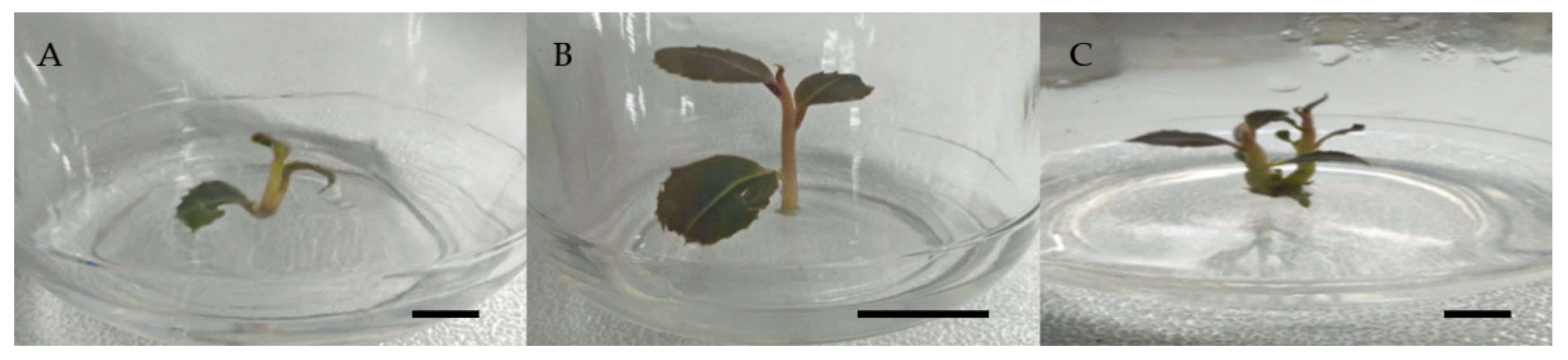
2.1. Shoot Multiplication
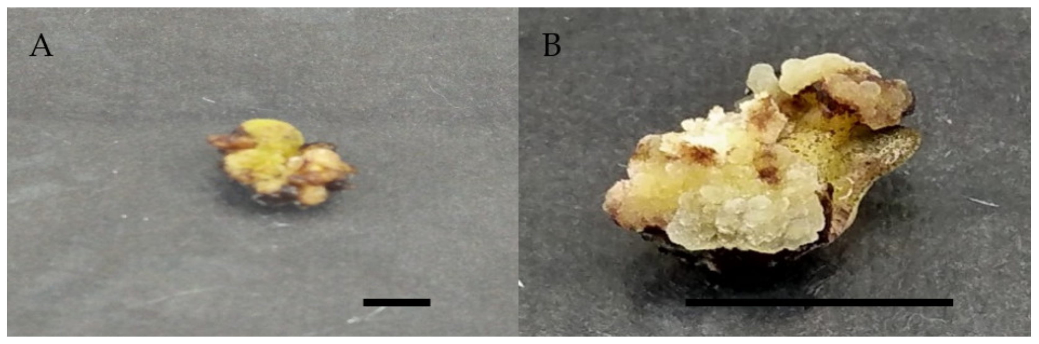
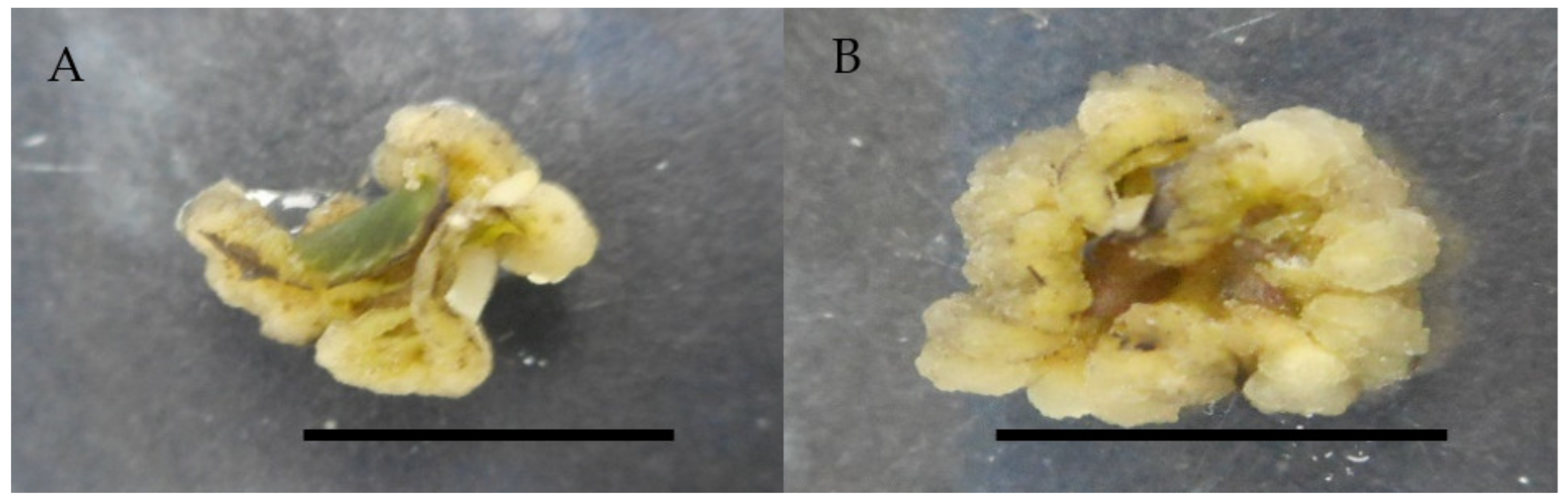
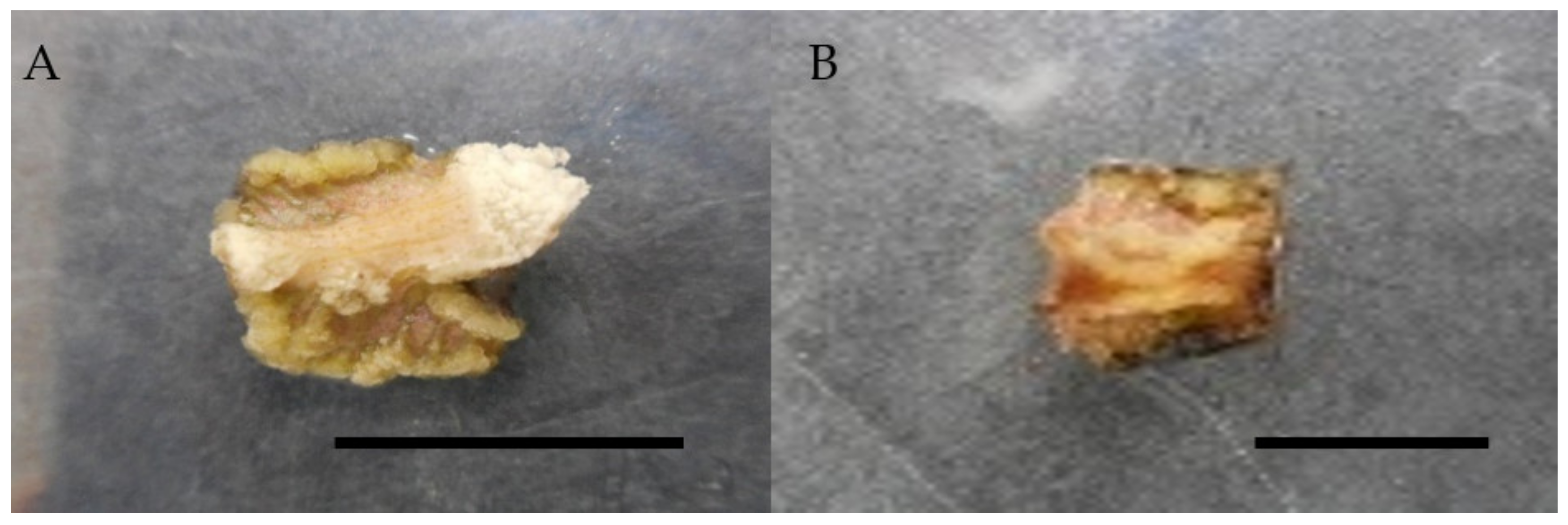
2.2. Callus Induction
2.2.1. Callus Induction Using a Single Concentration of 2,4-D and Picloram
2.2.2. Callus Induction using Combination of 0.5 mg/L 2,4-D and 0.25 mg/L Different Cytokinins
2.2.3. Callus Induction Using a Combination of 1.0 mg/L Picloram and 0.5 mg/L Cytokinins
2.3. Total Antioxidant, Phenolic, and Flavonoid Content in Wild Plant and In Vitro Culture of Labisia pumila var. alata
2.3.1. Total Phenolic and Flavonoid Content in Wild Plant, In Vitro-Derived Plantlet, and Callus of Labisia pumila var. alata
2.3.2. Total Antioxidant (DPPH and FRAP Methods) in Wild Plant, In Vitro-Derived Plantlet, and Callus of Labisia pumila var. alata
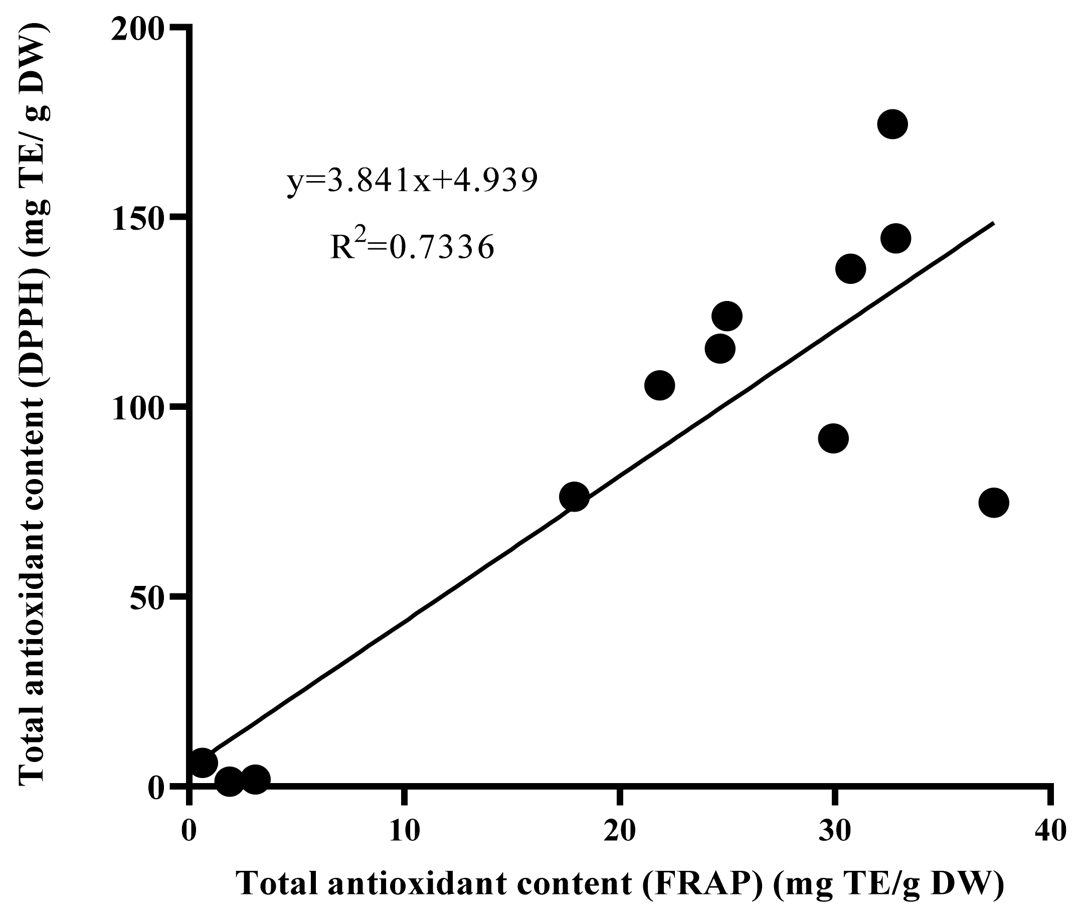
2.3.3. Correlation between Antioxidant Activities and Total Phenolic and Flavonoid Content in Wild Plant, In Vitro-Derived Plantlet, and Callus of Labisia pumila var. alata
3. Discussion
4. Materials and Methods
4.1. Plant Materials and Maintenance
4.2. Media Preparation
4.3. Shoot Multiplication
4.4. Callus Induction and Maintenance
4.5. Measurement of Total Antioxidant Activity
4.5.1. Extract Preparation
4.5.2. Ferric Reducing Antioxidant Potential Assay (FRAP)
4.5.3. 2,2-Diphenyl-1-picrylhydrazyl (DPPH) Free Radical Scavenging Assay
4.5.4. Total Phenolic Content Determination
4.5.5. Total Flavonoid Content Determination
4.6. Experimental Design and Data Analysis
5. Conclusions
Author Contributions
Funding
Institutional Review Board Statement
Informed Consent Statement
Data Availability Statement
Acknowledgments
Conflicts of Interest
References
- Abdullah, N.; Chermahini, S.H.; Suan, C.L.; Sarmidi, M.R. Labisia pumila: A review on its traditional, phytochemical and biological uses. World Appl. Sci. J. 2013, 27, 1297–1306. [Google Scholar]
- Chua, L.S.; Lee, S.Y.; Abdullah, N.; Sarmidi, M.R. Review on Labisia pumila (Kacip Fatimah): Bioactive phytochemicals and skin collagen synthesis promoting herb. Fitoterapia 2012, 83, 1322–1335. [Google Scholar] [CrossRef]
- Ibrahim, M.H.; Jaafar, H.Z. The relationship of nitrogen and C/N ratio with secondary metabolites levels and antioxidant activities in three varieties of Malaysian Kacip Fatimah (Labisia pumila Blume). Molecules 2011, 16, 5514–5526. [Google Scholar] [CrossRef] [PubMed]
- Ling, A.P.K.; Tan, K.P.; Hussein, S. Comparative effects of plant growth regulators on leaf and stem explants of Labisia pumila var. alata. J. Zhejiang Univ. Sci. B. 2013, 14, 621–631. [Google Scholar] [CrossRef] [Green Version]
- Hasan, N.A.; Hussein, S.B.; Ibrahim, R. Effect of medium pH and sucrose concentrations on adventitious roots induction of Labisia pumila. In Proceedings of the IRES 14th International Conference, Paris, France, 7 November 2015; pp. 44–48. [Google Scholar]
- Norhaiza, M.; Maziah, M.; Hakiman, M. Antioxidative properties of leaf extracts of a popular Malaysian herb, Labisia pumila. J. Med. Plants Res. 2009, 3, 217–223. [Google Scholar]
- Pihie, L.; Hawariah, A.; Zakaria, Z.A.; Othman, F. Antiproliferative and proapoptotic effects of Labisia pumila ethanol extract and its active fraction in human melanoma HM3KO cells. Evid. Based Compl. Alt. Med. 2012, 2012, 1–13. [Google Scholar] [CrossRef] [PubMed] [Green Version]
- Karimi, E.; Jaafar, H.Z.; Ahmad, S. Antifungal, anti-inflammatory and cytotoxicity activities of three varieties of Labisia pumila Benth: From microwave obtained extracts. BMC Compl. Altern. Med. 2013, 13, 20. [Google Scholar] [CrossRef] [PubMed] [Green Version]
- Ibrahim, M.H.; Jaafar, H.Z.E. Increased carbon dioxide concentration improves the antioxidative properties of the Malaysian herb, Kacip Fatimah (Labisia pumila Blume). Molecules 2011, 16, 6068–6081. [Google Scholar] [CrossRef] [Green Version]
- Mohd Hanafi, M.M.; Yaakob, H.; Sarmidi, M.R.; Aziz, R.; Prieto, J.M. Marantodes pumilum L. plant extracts induce apoptosis, cell cycle arrest and inhibit cell migration and invasion on prostate cancer cell lines. Planta Med. 2016, 82, 381. [Google Scholar] [CrossRef]
- Ahmad, S.U.; Azam, A.; Shuid, A.N.; Mohamed, I.N. Phyto-estrogenic effects of Marantodes pumilum (Blume) Kuntze syn. Labisia pumila (Blume) Fern.-Vill. for the prevention and treatment of post-menopausal diseases. Indian J. Tradit. Knowl. 2017, 16, 208–215. [Google Scholar]
- Madzuki, I.N.; Lau, S.F.; Tantowi, N.A.C.A.; Ishak, N.I.M.; Mohamed, S. Labisia pumila prevented osteoarthritis cartilage degeneration by attenuating joint inflammation and collagen breakdown in postmenopausal rat model. Inflammopharmacology 2018, 26, 1207–1217. [Google Scholar] [CrossRef]
- Ibrahim, M.H.; Jaafar, H.Z.E. Reduced photoinhibition under low irradiance enhanced Kacip Fatimah (Labisia pumila Benth) secondary metabolites, phenyl alanine lyase and antioxidant activity. Int. J. Mol. Sci. 2012, 13, 5290–5306. [Google Scholar] [CrossRef]
- Fazwa, F.; Syafiqah Nabilah, S.B.; Norhayati, S.; Norwati, M.; Marzalina, M. Rapid mass production of elite clone of Labisia pumila var. alata (KFeFRIM01) for sustainable supply of high-quality planting materials. Int. J. Agric. For. Plant. 2018, 6, 66–72. [Google Scholar]
- Moscatiello, R.; Baldan, B.; Navazio, L. Plant cell suspension cultures. Met. Mol. Biol. 2013, 953, 77–93. [Google Scholar]
- Bilušić Vundać, V.; Pfeifhofer, W.; Brantner, A.; Males, Z. Essential oils of seven Stachys taxa from Croatia. Biochem. Syst. Ecol. 2006, 34, 875–881. [Google Scholar] [CrossRef]
- Perrino, E.V.; Valerio, F.; Gannouchi, A.; Trani, A.; Mezzapesa, G. Ecological and plant community implication on essential oils composition in useful wild Officinal species: A pilot case study in Apulia (Italy). Plants 2021, 10, 574. [Google Scholar] [CrossRef] [PubMed]
- Karimi, E.; Jaafar, H.Z.E.; Ahmad, S. Phytochemical analysis and antimicrobial activities of methanolic extracts of leaf, stem and root from different varieties of Labisia pumila Benth. Molecules 2011, 16, 4438–4450. [Google Scholar] [CrossRef]
- Gupta, D. Methods for determination of antioxidant capacity: A review. Int. J. Pharm. Sci. Res. 2015, 6, 546–566. [Google Scholar]
- Pisoschi, A.M.; Negulescu, G.P. Methods for total antioxidant activity determination: A review. Biochem. Anal. Biochem. 2011, 1, 106. [Google Scholar] [CrossRef] [Green Version]
- Stoker, H.S. General, Organic and Biological Chemistry Canada; Nelson Education, Ltd: Toronto, Canada, 2013. [Google Scholar]
- Dontha, S. A review on antioxidant methods. Asian J. Pharm. Clin. Res. 2016, 9, 14–32. [Google Scholar]
- Hussein, R.A.; El-Anssary, A.A. Plant secondary metabolites: The key drivers of the pharmacological actions of medicinal plants. In Herbal Medicine; Builders, P.F., Ed.; IntechOpen: London, UK, 2018; pp. 1–20. [Google Scholar]
- Herrmann, K.M. The shikimate pathway as an entry to aromatic secondary metabolism. Plant Physiol. 1995, 107, 7–12. [Google Scholar] [CrossRef] [Green Version]
- Mendoza, N.; Silva, E.M.E. Introduction to phytochemicals: Secondary metabolites from plants with active principles for pharmacological importance. In Phytochemicals: Source of Antioxidants and Role in Disease Prevention; Asao, T., Asaduzzaman, M., Eds.; IntechOpen: London, UK, 2018; pp. 25–47. [Google Scholar]
- Manach, C.; Scalbert, A.; Morand, C.; Remesy, C.; Jimenez, L. Polyphenols: Food sources and bioavailability. Am. J. Clin. Nutr. 2004, 79, 727–747. [Google Scholar] [CrossRef] [PubMed] [Green Version]
- Pott, D.M.; Osorio, S.; Vallarino, J.G. From central to specialized metabolism: An overview of some secondary compounds derived from the primary metabolism for their role in conferring nutritional and organoleptic characteristics to fruit. Front. Plant Sci. 2019, 10, 1–19. [Google Scholar] [CrossRef] [PubMed] [Green Version]
- Upadhyay, R.; Chaurasia, J.K.; Tiwari, K.N.; Singh, K. Comparative antioxidant study of stem and stem induced callus of Phyllanthus fraternus Webster—an important antiviral and hepatoprotective plant. Appl. Biochem. Biotechnol. 2013, 171, 2153–2164. [Google Scholar] [CrossRef] [PubMed]
- Nabilah, S.; Fazwa, F.; Suhaila, S.; Norhayati, N.; Zaki, M.; Masitah, M. Acclimatization of KFeFRIM01: A superior clone of Labisia pumila var. alata. Int. J. Environ. Agric. Res. 2017, 3, 9–13. [Google Scholar]
- Marbawi, H.; Cyril, O.; David, D.; Gansau, J.A. In vitro multiple shoot regeneration from stem explant of commercially important medicinal herb Labisia pumila var. pumila. ASM Sci. J. 2018, 11, 171–180. [Google Scholar]
- Tekdal, D.; Çetiner, S. The determination of self-compatibility status of Thermopsis turcica through histological analysis. J. Appl. Biol. Sci. 2014, 8, 64–67. [Google Scholar]
- Li, J.Z.; Jing, T.Y.; Qing, G.W. A protocol for rapid and high-frequency in vitro propagation of Solanum nigrum L. Sains Malays. 2017, 46, 1183–1189. [Google Scholar]
- Chen, H.Y.; Liu, J.; Pan, C.; Yu, J.W.; Wang, Q.C. In vitro regeneration of adventitious buds from leaf explants and their subsequent cryopreservation in highbush blueberry. Plant Cell Tissue Organ. Cult. 2018, 134, 193–204. [Google Scholar] [CrossRef]
- Nikolić, R.; Mitić, N.; Miletić, R.; Nešković, M. Effects of cytokinins on in vitro seed germination and early seedling morphogenesis in Lotus corniculatus L. J. Plant Growth Regul. 2006, 25, 187–194. [Google Scholar] [CrossRef]
- Masekesa, T.R.; Gasura, E.; Ngadze, E.; Icishahayoa, D.; Kujeke, G.T.; Chidzwondob, F.; Robertsona, I. Efficacy of zeatin, kinetin and thidiazuron in induction of adventitious root and shoot from petiole explants of sweet potato cv. Brondal. S. Afr. J. Bot. 2016, 104, 1–5. [Google Scholar] [CrossRef]
- Nisha Rani, D.; Nair, G.M. Effects of plant growth regulators on high frequency shoot multiplication and callus regeneration of an important Indian medicinal plant, Nirgundi (Vitex negundo L.). In Vitro Cell. Dev. Biol. Plant 2006, 42, 69–73. [Google Scholar] [CrossRef]
- Sahoo, Y.; Chand, P.K. Micropropagation of Vitex negundo L., a woody aromatic medicinal shrub, through high-frequency axillary shoot proliferation. Plant Cell Rep. 1998, 18, 301–307. [Google Scholar] [CrossRef] [PubMed]
- Chandramu, C.; Rao, M.; Reddy, V.D. High frequency induction of multiple shoots from nodal explants of Vitex negundo L. using sodium sulphate. J. Plant Biotechnol. 2003, 5, 107–113. [Google Scholar]
- Jebakumar, M.; Jayabalan, M. An efficient method for regeneration of plantlets from nodal explants of Prosalea corydifolia Linn. Plant Cell Biotechnol. Mol. Biol. 2000, 1, 37–40. [Google Scholar]
- Husain, M.K.; Anis, M. Rapid in vitro propagation of Eclipta alba (L.) Hassk. through high frequency axillary shoot proliferation. Acta Physiol. Plant. 2006, 28, 325–330. [Google Scholar] [CrossRef]
- Raja, H.D.; Arockiasamy, D.I. In vitro propagation of Mentha viridis L. from nodal and shoot tip explants. Plant Tiss. Cult. Biotechnol. 2008, 18, 1–6. [Google Scholar] [CrossRef]
- Manivannan, A.; Soundararajan, P.; Park, Y.G.; Jeong, B.R. In vitro propagation, phytochemical analysis, and evaluation of free radical scavenging property of Scrophularia kakudensis Franch tissue extracts. BioMed Res. Int. 2015, 2015, 480564. [Google Scholar] [CrossRef] [Green Version]
- Varghese, T.; Rema Shree, A.B.; Naheesa, E.; Neelakandan, N.; Nandakumar, S. In vitro propagation of Terminalia arjuna Roxb. multipurpose tree. Plant Cell Biotechnol. Mol. Biol. 2003, 4, 95–98. [Google Scholar]
- Ch, B.; Rao, K.; Gandi, S.; Giri, A. Abiotic elicitation of gymnemic acid in the suspension cultures of Gymnema sylvestre. World J. Microbiol. Biotechnol. 2012, 28, 741–747. [Google Scholar] [CrossRef] [PubMed]
- Abdelmageed, A.H.A.; Faridah, Q.Z.; Nor Shuhada, K.; Julia, A.A. Callus induction and plant regeneration of Michelia champaca (Magnoliaceae): A multipurpose tree. J. Med. Plants Res. 2012, 6, 3338–3344. [Google Scholar] [CrossRef] [Green Version]
- Ikeuchi, M.; Sugimoto, K.; Iwase, A. Plant callus: Mechanisms of induction and repression. Plant Cell 2013, 25, 3159–3173. [Google Scholar] [CrossRef] [PubMed] [Green Version]
- Cunha, A.C.G.D.; Ferreira, M.F. Somatic embryogenesis, organogenesis and callus growth kinetics of flax. Plant Cell Tissue Organ Cult. 1996, 47, 1–8. [Google Scholar] [CrossRef]
- Sami, A.M.; Hashish, K.I.; Sawsan, S.S.; Lobna, S.T. In vitro propagation protocol of Hibiscus syriacus L. plants. Int. J. PharmTech Res. 2016, 9, 178–186. [Google Scholar]
- Oi, K.; Samuel, K.; Modeste, K.K.; Oumar, S.; Edmond, K.; Hilaire, K.T. Improved callogenesis and somatic embryogenesis using amino acids and plant growth regulators combination in pineapple [Ananas comosus (L.) Merr (Bromeliaceae)]. Eur. J. Biotechnol. Biosci. 2017, 5, 6–16. [Google Scholar]
- Haida, Z.; Nakasha, J.J.; Hakiman, M. In vitro responses of plant growth factors on growth, yield, phenolics content and antioxidant activities of Clinacanthus nutans (Sabah snake grass). Plants 2020, 9, 1030. [Google Scholar] [CrossRef] [PubMed]
- Zahid, N.A.; Jaafar, H.Z.E.; Hakiman, M. Micropropagation of ginger (Zingiber officinale Roscoe) ‘Bentong’ and evaluation of its secondary metabolites and antioxidant activities compared with the conventionally propagated plant. Plants 2021, 10, 630. [Google Scholar] [CrossRef]
- Rameshkumar, R.; Satish, L.; Pandian, S.; Rathinapriya, P.; Rency, A.S.; Shanmugaraj, G.; Pandian, S.K.; Leung, D.W.; Ramesh, M. Production of squalene with promising antioxidant properties in callus cultures of Nilgirianthus ciliatus. Ind. Crop Prod. 2018, 126, 357–367. [Google Scholar] [CrossRef]
- Muthukrishnan, S.; Kumar, T.S.; Gangaprasad, A.; Maggi, F.; Rao, M.V. Phytochemical analysis, antioxidant and antimicrobial activity of wild and in vitro derived plants of Ceropegia thwaitesii Hook–An endemic species from Western Ghats, India. J. Genet. Eng. Biotechnol. 2018, 16, 621–630. [Google Scholar] [CrossRef]
- Hinneburg, I.; Dorman, H.D.; Hiltunen, R. Antioxidant activities of extracts from selected culinary herbs and spices. Food Chem. 2006, 97, 122–129. [Google Scholar] [CrossRef]
- Kousalya, L.; Narmatha Bai, V. (2016). Effect of growth regulators on rapid micropropagation and antioxidant activity of Canscora decussata (Roxb.) Roem. & Schult.—A threatened medicinal plant. Asian Pac. J. Reprod. 2016, 5, 161–170. [Google Scholar]
- Song, H.; Kumar, P.; Arivazhagan, G.; Lee, S.I.; Yoon, H.M.; Kim, I.H.; Kwon, H.J.; Kim, J.M.; Hakkim, F.L. Antioxidant property of leaves and calluses extracts of in-vitro grown 5 different Ocimum species. J. Plant Biotechnol. 2012, 39, 146–153. [Google Scholar] [CrossRef]
- Mustapha, Z.; Harun, H. Phytochemical constituents in leaves and callus of Ficus deltoidea Jack var. kunstleri (King) Corner. Walailak J. Sci. Technol. 2015, 12, 431–439. [Google Scholar]
- Verpoorte, R.; Alfermann, A.W. (Eds.) Metabolic Engineering of Plant Secondary Metabolism; Springer Science & Business Media: Berlin, Germany, 2000. [Google Scholar]
- Grąbkowska, R.; Matkowski, A.; Grzegorczyk-Karolak, I.; Wysokińska, H. Callus cultures of Harpagophytum procumbens (Burch.) DC. ex Meisn.; Production of secondary metabolites and antioxidant activity. South Afr. J. Bot. 2016, 103, 41–48. [Google Scholar] [CrossRef]
- Abd Samat, N.M.A.; Ahmad, S.; Awang, Y.; Bakar, R.A.H.; Hakiman, M. Alterations in herbage yield, antioxidant activities, phytochemical contents, and bioactive compounds of sabah snake grass (Clinacanthus nutans L.) with regards to harvesting age and harvesting frequency. Molecules 2020, 25, 2833. [Google Scholar] [CrossRef] [PubMed]
- Huda-Faujan, N.; Noriham, A.; Norrakiah, A.S.; Babji, A.S. Antioxidant activity of plants methanolic extracts containing phenolic compounds. Afr. J. Biotechnol. 2009, 8, 484–489. [Google Scholar]
- Sun, T.; Ho, C.T. Antioxidant activities of buckwheat extracts. Food Chem. 2005, 90, 743–749. [Google Scholar] [CrossRef]
- Hönig, M.; Plíhalová, L.; Husičková, A.; Nisler, J.; Doležal, K. Role of cytokinins in senescence, antioxidant defence and photosynthesis. Int. J. Mol. Sci. 2018, 19, 4045. [Google Scholar] [CrossRef] [Green Version]
- Fazal, H.; Abbasi, B.H.; Ahmad, N.; Noureen, B.; Shah, J.; Ma, D.; Chuanliang, L.; Akbar, F.; Uddin, M.N.; Khan, H.; et al. Biosynthesis of antioxidative enzymes and polyphenolics content in calli cultures of Prunella vulgaris L. in response to auxins and cytokinins. Artif. Cells Nanomed. Biotechnol. 2020, 48, 893–902. [Google Scholar] [CrossRef]
- Piotrowska-Niczyporuk, A.; Bajguz, A. The effect of natural and synthetic auxins on the growth, metabolite content and antioxidant response of green alga Chlorella vulgaris (Trebouxiophyceae). Plant Growth Regul. 2014, 73, 57–66. [Google Scholar] [CrossRef] [Green Version]
- Lukmanul, H.; Gowri Shankar, C.; Girija, S. Chemical composition and antioxidant property of holy basil (Ocimum sanctum L.) leaves, stems, and inflorescence and their in vitro callus cultures. J. Agric. Food Chem. 2007, 55, 9109–9117. [Google Scholar]
- Paula, C.; Santos, G.; Rosa, M.; Seabra, P.B.; Andrade, M.; Fernades, F. Phenolic anti-oxidant compounds produced by in vitro shoots of sage (Salvia officinalis L.). Plant Sci. 2002, 162, 981–987. [Google Scholar]
- Barz, W. Catabolism of endogenous and exogenous compounds by plant cell cultures. In Plant Tissue Culture and Its Biotechnological Application. Proceedings in Life Sciences; Barz, W., Reinhard, E., Zenk, M.H., Eds.; Springer-Verlag: Berlin, Germany, 1977; pp. 153–171. [Google Scholar]
- Sargent, J.A.; Skoog, F. Effects of indoleacetic acid and kinetin on scopoletin and scopolin levels in relation to growth of tobacco tissue in vitro. Plant Physiol. 1960, 35, 934–941. [Google Scholar] [CrossRef] [PubMed] [Green Version]
- Skoog, F.; Montaldi, E. Auxin–kinetin interaction regulating the scopoletin and scopolin levels in tobacco tissue cultures. Proc. Natl. Acad. Sci. USA 1961, 47, 36–49. [Google Scholar] [CrossRef] [PubMed] [Green Version]
- Murashige, T.; Skoog, F. A revised medium for rapid growth and bio assays with tobacco tissue cultures. Physiol. Plant. 1962, 15, 473–497. [Google Scholar] [CrossRef]
- Wong, S.P.; Lai, P.L.; Jen, H.W.K. Antioxidant activities of aqueous extracts of selected plants. Food Chem. 2006, 99, 775–783. [Google Scholar] [CrossRef]
- Benzie, I.F.; Strain, J.J. The ferric reducing ability of plasma (FRAP) as a measure of “antioxidant power”: The FRAP assay. Anal. Biochem. 1996, 239, 70–76. [Google Scholar] [CrossRef] [PubMed] [Green Version]
- Singleton, V.L.; Rossi, J.A. Colorimetry of total phenolics with phosphomolybdic-phosphotungstic acid reagents. Am. J. Enol. Viticult. 1965, 16, 144–158. [Google Scholar]
- Marinova, D.; Ribarova, F.; Atanassova, M. Total phenolics and total flavonoids in Bulgarian fruits and vegetables. J. Univ. Chem. Technol. Metal. 2005, 40, 255–260. [Google Scholar]


| Growth Regulators (mg/L) | No. of Shoots | Length of Shoot (cm) | No. of Leaves | ||
|---|---|---|---|---|---|
| BAP | Kin | Zea | |||
| 0 | 0 | 0 | 5.56 abcd | 3.89 ab | 2.1 bc |
| 1 | 0 | 0 | 6.15 abc | 3.55 abc | 1.6 bcdef |
| 2 | 0 | 0 | 2.03 g | 2.15 efg | 1.0 def |
| 3 | 0 | 0 | 3.92 bcdef | 2.51 defg | 1.73 bcde |
| 4 | 0 | 0 | 4.06 abcdef | 2.49 defg | 1.4 cdef |
| 5 | 0 | 0 | 3.25 efg | 2.37 defg | 1.42 cdef |
| 0 | 0 | 0 | 4.19 abcdef | 3.19 abcd | 1.8 bcd |
| 0 | 1 | 0 | 3.32 efg | 2.61 cdefg | 0.93 ef |
| 0 | 2 | 0 | 3.13 fg | 2.54 defg | 0.93 ef |
| 0 | 3 | 0 | 3.61 def | 2.26 efg | 1.1 def |
| 0 | 4 | 0 | 3.75 cdef | 2.71 cdefg | 1.4 cdef |
| 0 | 5 | 0 | 2.12 g | 2.13 fg | 0.8 f |
| 0 | 0 | 0 | 2.74 fg | 2.10 g | 1.4 cdef |
| 0 | 0 | 1 | 5.36 abcde | 4.32 a | 2.4 b |
| 0 | 0 | 2 | 6.53 ab | 3.50 abc | 3.13 a |
| 0 | 0 | 3 | 6.64 a | 2.95 bcde | 2.27 b |
| 0 | 0 | 4 | 5.29 abcde | 2.93 bcdef | 1.0 def |
| 0 | 0 | 5 | 6.10 abc | 2.89 bcdefg | 1.0 def |
| Growth Regulators (mg/L) | Callus Induction Percentage (%) | Callus Score | Morphology, Texture | |
|---|---|---|---|---|
| 2,4-D | Picloram | |||
| 0 | 0 | NC | - | NC |
| 0.5 | 0 | 60 a | ++ | Greenish, compact |
| 1 | 0 | 6.7 c | ++ | Greenish, compact |
| 1.5 | 0 | 6.7 c | + | Friable, yellowish to greenish |
| 2 | 0 | NC | - | NC |
| 2.5 | 0 | NC | - | NC |
| 0 | 0.5 | 6.7 c | + | Friable, yellowish |
| 0 | 1 | 50 a | +++ | Friable, whitish |
| 0 | 1.5 | 30 b | ++ | Friable, whitish |
| 0 | 2 | 27 b | ++ | Friable, yellowish to whitish |
| 0 | 2.5 | 27 b | + | Friable, yellowish to whitish |
| Growth Regulators (mg/L) | Callus Formation (%) | Callus Score | Morphology, Texture |
|---|---|---|---|
| 0.5 2,4-D + 0.25 zeatin | 100 a | +++ | Compact, yellowish to greenish |
| 0.5 2,4-D + 0.25 kinetin | 73.4 ab | ++ | Friable, yellowish to greenish |
| 0.5 2,4-D + 0.25 BAP | 46.6 b | + | Friable, yellowish to greenish |
| 0.5 2,4-D + 0.25 TDZ | 6.6 c | + | Friable, whitish |
| Growth Regulators (Mg/L) | Callus Formation Percentage (%) | Callus Score | Morphology, Texture |
|---|---|---|---|
| 1.0 picloram + 0.5 zeatin | 33.4 ab | + | Greenish, compact |
| 1.0 picloram + 0.5 kinetin | 80 a | +++ | Whitish, friable |
| 1.0 picloram + 0.5 BAP | 66.6 ab | ++ | Whitish, compact |
| 1.0 picloram + 0.5 TDZ | 20 b | + | Whitish to dark brown, compact |
| Source of Sample | Total Phenolic Content (mg GAE/g DW) | Total Flavonoid Content (mg QE/g DW) |
|---|---|---|
| Wild leaves | 1.01 ± 0.07 c | 1.67 ± 0.02 c |
| In vitro plantlets | 1.4 ± 0.07 b | 1.41 ± 0.68 b |
| Callus | 1.9 ± 0.14 a | 2.38 ± 0.18 a |
| Total Antioxidant Content (mg TE/g DW) | ||
|---|---|---|
| Source of Sample | DPPH | FRAP |
| Wild plant | 40.45 ± 2.69 c | 81 ± 5.39 c |
| In vitro plantlet | 57.47 ± 2.63 b | 115 ± 5.25 b |
| Callus | 75.88 ± 5.80 a | 152 ± 11.59 a |
| Variables | 1 | 2 | 3 | 4 | |
|---|---|---|---|---|---|
| 1 | TPC | 1 | |||
| 2 | TFC | 0.86 * | 1 | ||
| 3 | DPPH | 0.81 * | 0.91 * | 1 | |
| 4 | FRAP | 0.98 * | 0.91 * | 0.86 * | 1 |
Publisher’s Note: MDPI stays neutral with regard to jurisdictional claims in published maps and institutional affiliations. |
© 2021 by the authors. Licensee MDPI, Basel, Switzerland. This article is an open access article distributed under the terms and conditions of the Creative Commons Attribution (CC BY) license (https://creativecommons.org/licenses/by/4.0/).
Share and Cite
Najhah, M.Y.; Jaafar, H.Z.E.; Nakasha, J.J.; Hakiman, M. Shoot Multiplication and Callus Induction of Labisia pumila var. alata as Influenced by Different Plant Growth Regulators Treatments and Its Polyphenolic Activities Compared with the Wild Plant. Molecules 2021, 26, 3229. https://doi.org/10.3390/molecules26113229
Najhah MY, Jaafar HZE, Nakasha JJ, Hakiman M. Shoot Multiplication and Callus Induction of Labisia pumila var. alata as Influenced by Different Plant Growth Regulators Treatments and Its Polyphenolic Activities Compared with the Wild Plant. Molecules. 2021; 26(11):3229. https://doi.org/10.3390/molecules26113229
Chicago/Turabian StyleNajhah, Mat Yunus, Hawa Z. E. Jaafar, Jaafar Juju Nakasha, and Mansor Hakiman. 2021. "Shoot Multiplication and Callus Induction of Labisia pumila var. alata as Influenced by Different Plant Growth Regulators Treatments and Its Polyphenolic Activities Compared with the Wild Plant" Molecules 26, no. 11: 3229. https://doi.org/10.3390/molecules26113229