Article

Pestalotiones A–D: Four New Secondary Metabolites from the Plant Endophytic Fungus Pestalotiopsis Theae

1
State Key Laboratory of Mycology, Institute of Microbiology, Chinese Academy of Sciences, Beijing 100101, China
2
University of Chinese Academy of Sciences, Beijing 100039, China
3
Jiangsu Key Laboratory for Biofunctional Molecules, College of Life Science and Chemistry, Jiangsu Second Normal University, Nanjing 210003, China
4
School of Biological Medicine, Beijing City University, Beijing 100083, China
*
Author to whom correspondence should be addressed.
These authors contributed equally to this work.
Molecules 2020, 25(3), 470; https://doi.org/10.3390/molecules25030470
Submission received: 7 January 2020 / Revised: 19 January 2020 / Accepted: 20 January 2020 / Published: 22 January 2020
(This article belongs to the Collection Bioactive Compounds)

Abstract

:
Two new xanthone derivatives, pestalotiones A (1) and B (2), one new diphenyl ketone riboside, pestalotione C (7), and one new diphenyl ether, pestalotione D (8), along with five known compounds isosulochrin dehydrate (3), 3,8-dihydroxy-6-methyl-9-oxo-9H-xanthene-1-carboxylate (4), isosulochrin (5), chloroisosulochrin (6), and pestalotether D (9), were isolated from the crude extract of the plant endophytic fungus Pestalotiopsis theae (N635). The structures of the new compounds were unambiguously deduced by HRESIMS and 1D/2D-NMR spectroscopic data. Compound 6 showed modest cytotoxicity against the HeLa cell line with an IC50 value of 35.2 μM. Compound 9 also showed cytotoxic to the HeLa and MCF-7 cell lines, with IC50 values of 60.8 and 22.6 μM, respectively. Additionally, compounds 1 and 2 exhibited antioxidant activity in scavenging DPPH radical with IC50 values of 54.2 and 59.2 μg/mL, respectively.

1. Introduction

Fungi are capable of producing a variety of bioactive secondary metabolites [1,2,3]. Endophytic fungi inhabiting the normal tissues of healthy plants have attracted considerable attention due to their ecological and biotechnological potential [4,5]. Special environments and selective pressures have an influence on the metabolic process of endophytes, leading to their enormous biological diversity and a variety of biosynthetic capabilities [6,7,8,9]. The widely distributed endophytic fungi, Pestalotiopsis spp., has attracted much attention owing to the discovery of structurally diverse and biologically active secondary metabolites [10,11,12,13,14,15], including the anticancer drug, paclitaxel, which was isolated from P. guepinii and P. microspora [16,17]. Chemical investigations of the fungus P. theae have also yielded bioactive compounds such as cytosporins, phytotoxins, pestalotheols, pestalazines, and pestalamides [18,19,20,21]. In a search for new bioactive natural products from this fungal genus, a strain of P. theae (N635), isolated from the branches of the tea plant Camellia sinensis (Theaceae) in the suburb of Hangzhou, P. R. China, was grown in different solid-substrate fermentation cultures. Chemical studies of the resulting crude extracts had afforded structurally unique compounds showing an antiproliferative effect against the human tumor cell lines HeLa and MCF-7, including two spiroketals chlorotheolides A and B possessing the unique [4,7] methanochromene and dispirotrione skeletons, and their putative biosynthetic precursors [22]. In addition, two new xanthone derivatives, pestalotiones A (1) and B (2), one new diphenyl ketone riboside, pestalotione C (7), and one new diphenyl ether, pestalotione D (8), along with five known compounds isosulochrin dehydrate (3) [23] 3,8-dihydroxy-6-methyl-9-oxo-9H-xanthene-1-carboxylate (4) [24], isosulochrin (5) [23], chloroisosulochrin (6) [23], and pestalotether D (9) [25] (Figure 1), were isolated from the crude extract. All compounds were evaluated for cytotoxicity against a panel of four human tumor cell lines that are commonly used in our laboratory. Meanwhile, their antioxidant activities were also evaluated. Details of the isolation, structure elucidation, biological activities and proposed biosynthetic pathway of these metabolites are reported herein.

2. Results and Discussion

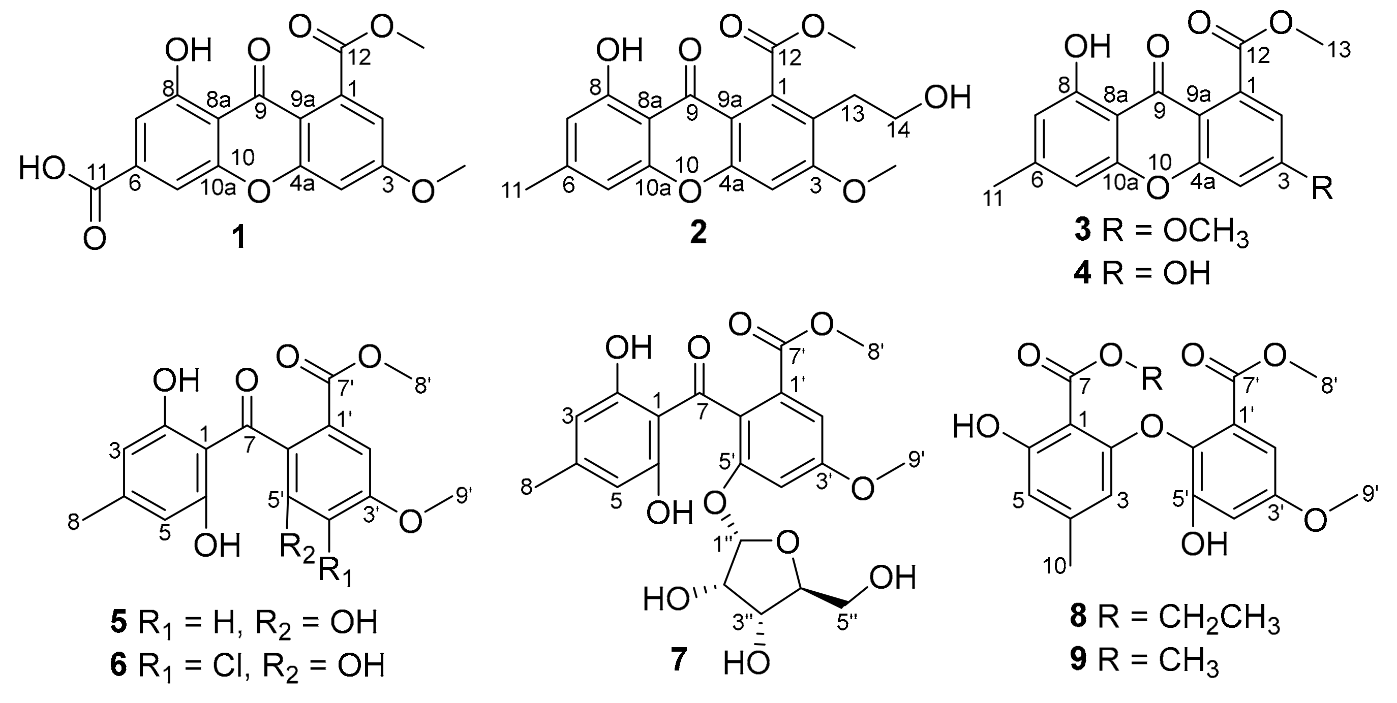
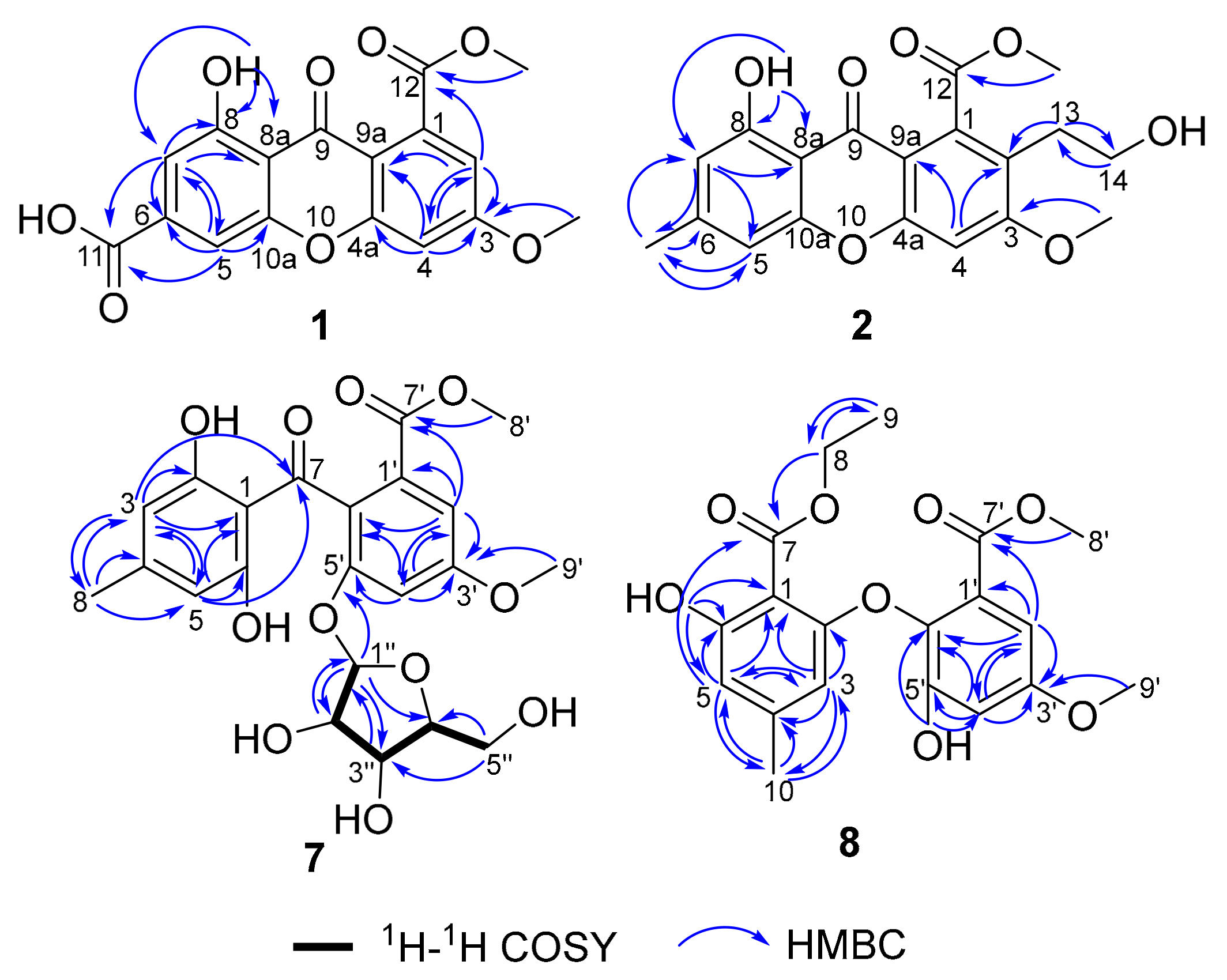
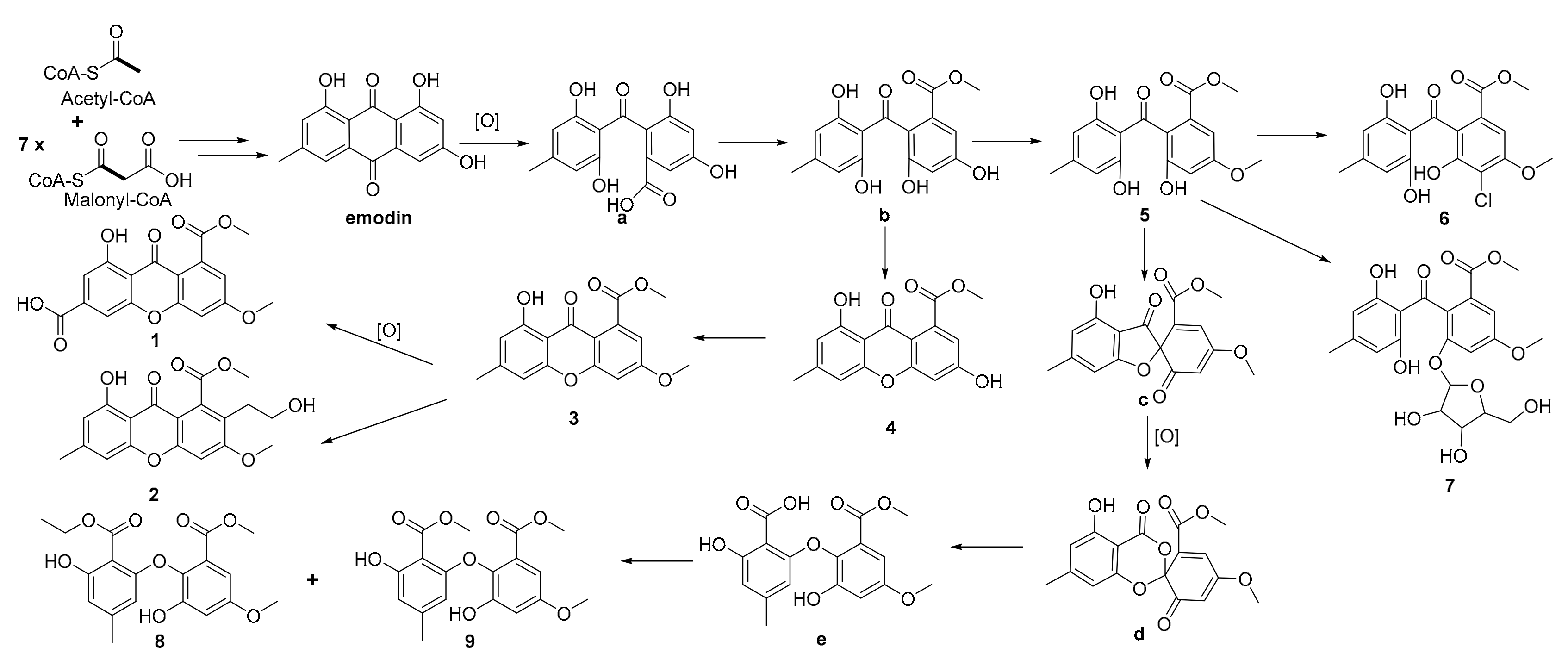
2.1. Isolation and Structure Elucidation

Pestalotione A (1) was assigned the molecular formula C17H12O8 (12 degrees of unsaturation) on the basis of HRESIMS data. The UV spectrum of the yellow powder showed four maxima (234, 254, 310, and 369 nm), suggesting a xanthone chromophore [24]. Analysis of its NMR data (Table 1) revealed the presence of one exchangeable proton (δH 12.3), two methoxy groups, 12 aromatic carbons (the carbon C-6 signal resonated at δC 139.4 was determined by HMBC correlations (Figure 2) from H-5 and H-7 to C-6), including four protonated, two carboxylic carbons (δC 165.8, 167.9), and one conjugated ketone carbon (δC 179.5). These data accounted for all the NMR resonances of 1 except for one unobserved exchangeable proton and required 1 to be a tricyclic compound. A detailed NMR data comparison with xanthone derivative isosulochrin dehydrate (3) revealed the similarity of them. The 1H–1H coupling patterns for the four aromatic protons also revealed two m-substituted aryl rings. HMBC correlations (Figure 2) from H-2 to C-3, C-4, C-9a, and the carboxylic carbon C-12 (δC 167.9), H-4 to C-2, C-3, C-4a, the ketone carbon C-9 (δC 179.5) and C-9a, H-5 to C-6, C-7, C-9, C-10a, and the carboxylic carbon C-11 (δC 165.8), and from H-7 to C-5, C-6, C-8, C-8a, C-9, and C-11 permitted completion of the xanthone core structure with two carboxylic carbons C-11 and C-12 located at C-6 and C-1, respectively. The cross-peaks from the phenolic proton OH-8 (δH 12.3) to C-7, C-8, and C-8a led to the attachment of the hydroxy groups to C-8. HMBC correlations from two methoxy groups to C-3 and C-12, established the locations of these methoxy groups. The remaining one exchangeable proton was located at C-11 by default. Therefore, the planar structure of compound 1 was established as 1-hydroxy-6-methoxy-8-(methoxycarbonyl)-9-oxo-9H-xanthene-3-carboxylic acid, named pestalotione A (Figure 1).
The molecular formula of pestalotione B (2) was established as C19H18O7 (11 degrees of unsaturation) on the basis of the HRESIMS. The 1H and 13C-NMR spectrum (Table 1) of 2 exhibited one exchangeable proton at δH 12.3, two oxygenated methyls, one methyl, two methylenes (one oxygenated), twelve olefinic/aromatic carbons (three of which were protonated), one carboxylic carbon (δC 168.4), and one conjugated ketone carbon (δC 180.3). These data accounted for all the resonances observed in the NMR spectra of 2 except for one unobserved exchangeable proton. The 1H- and 13C-NMR spectra of 2 displayed signals for structural features similar to 1, except that the aromatic proton H-2 (δH 7.13) and the carboxylic carbon (δC 165.8) in 1 were replaced by the 2-hydroxyethyl unit (δH 2.87, 3.00, 3.50, 3.56, δC 38.0, 61.5, respectively) and a methyl group (δH/C 2.43/22.4), respectively. This was further confirmed by HMBC correlations (Figure 2) from H3-11 to C-5, C-6, and C-7, H2-13 to C-2 and C-14, and from H2-14 to C-13. Accordingly, compound 2, namely pestalotione B, was identified as methyl 8-hydroxy-2-(2-hydroxyethyl)-3-methoxy-6-methyl-9-oxo-9H-xanthene-1-carboxylate.
The molecular formula of pestalotione C (7) was established as C22H24O11 (11 degrees of unsaturation) on the basis of HRESIMS data. Its 1H, 13C, and HSQC NMR spectroscopic data (Table 2) showed resonances for two exchangeable protons (δH 10.9 and 3.3, respectively), three methyl groups with two oxygenated, one methylene unit, four oxymethines, twelve olefinic/aromatic carbons including four protonated, one carboxylic carbon (δC 167.7), and one conjugated ketone carbon (δC 201.1). Interpretation of these data revealed structural features similar to those presented in isosulochrin (5) [23] except for the presence of a furanose unit. Interpretation of the 1H–1H COSY NMR data (Figure 2) led to the identification of one isolated proton spin-system corresponding to the C-1′′-C-2′′-C-3′′-C-4′′-C-5′′ subunit of structure 7. The ribose residue was confirmed by comparing the 13C-NMR data with those of several furanoside, such as isotorachrysone-6-O-α-d-ribofuranoside, and asperflavin ribofuranoside [26,27]. The sugar moiety was further determined as α-form by comparison of the coupling constant (J1′′2′′ = 4.4 Hz) of the anomeric proton with those of the methyl-α-d-ribofuranoside (J1,2 = 4.3 Hz) and methyl-β-d-ribofuranoside (J1,2 = 1.2 Hz) [28]. The key HMBC correlations (Figure 2) from the anomeric proton H-1′′ to C-4′′ (δC 88.1) and C-5′ (δC 155.7) determined the ribose moiety, which was linked to C-5′ through oxygen bond. Upon acid hydrolysis of 7 with methanol/HCl, the liberated sugar was identified as d-ribose by measurement of its specific rotation ( [ α ] D 25 −16.0, c 0.2, H2O) [29]. Thus, compound 7 was elucidated as isosulochrin-5′-O-α-d-ribofuranoside, named pestalotione C (7).
The molecular formula of pestalotione D (8) was established as C19H20O8 (10 degrees of unsaturation) on the basis of HRESIMS data. The overall appearance of 1H and 13C-NMR spectra (Table 3) of 8 are highly similar to those of 9 except that signals for the methoxyl group (CH3O-7) were replaced by those for the ethoxyl group (δH/δC 4.47/62.4, 1.40/14.3) in the spectra of 8, which were supported by the HMBC correlations (Figure 2) from H2-8 to C-7 and C-9 and from H3-9 to C-8. The chemical structure of compound 8 was elucidated as methyl 2-(2-(ethoxycarbonyl)-3-hydroxy-5-methylphenoxy)-3-hydroxy-5-methoxybenzoate, named pestalotione D (8).
Biogenetically, emodin, biosynthesized from one molecule of acetyl-CoA and seven molecules of malonyl-CoA [30], could be the biosynthetic precursor not only for compounds 14, but also for 59, first via oxidation and methylation to form the key intermediate b, and then followed by a series of reactions through different routes to form 19. The proposed precursor and the reaction cascades leading to the generation of these metabolites are illustrated in Figure 3.

2.2. Bioactivities

Compounds 19 were tested for cytotoxicity against a panel of four human tumor cancer cell lines, HeLa (human cervical carcinoma cell line), MCF-7 (human breast cancer cell line), HepG2 (human hepatoma cell line), and ACHN (human renal carcinoma cell line). Compounds 6 and 9 showed cytotoxic to the HeLa cell line, with IC50 values of 35.2 and 60.8 μM, respectively, whereas the positive control cisplatin showed IC50 values of 15.1 and 5.5 μM, respectively. Compound 9 also showed cytotoxic to the MCF-7 cell line, with IC50 value of 22.6 μM, whereas the positive control cisplatin showed an IC50 value of 5.8 μM. Other compounds did not show detectable inhibitory effects on the cell lines tested at 100 μM. Meanwhile, their antioxidant activity was also evaluated by the DPPH (2,2-diphenyl-1-picrylhydrazyl radical) scavenging method with ascorbic acid as positive control (IC50 = 6.0 μg/mL). Only compounds 1 and 2 exhibited weak DPPH scavenging activity with respective IC50 values of 54.2 and 59.2 μg/mL.
Xanthone derivatives were found to display diverse activities, such as tumor cytotoxic activity, antivirus, antibacterial, antifungal, and antimalaria activities [31,32,33,34]. Pestalotiopsis sp. was an interesting producer of bioactive metabolites. In our previous study, two spiroketals chlorotheolides A and B from P. theae showed an antiproliferative effect against the human tumor cell lines HeLa and MCF-7 [22]. In the current study, new structural metabolites with cytotoxic and antioxidant activities were identified from the same fungus. This highlighted the high potential of bioprospecting larvicides from the endophytic fungi.

3. Experimental Section

3.1. General Experimental Procedures

Optical rotations were measured on an Anton Paar MCP 200 Automatic Polarimeter and UV data were obtained on a Thermo Genesys-10S UV/Vis spectrophotometer. IR data were recorded using a Nicolet IS5 FT-IR spectrophotometer. 1H and 13C-NMR data were acquired with Bruker Avance-400 and -500 spectrometer using solvent signals (acetone-d6: δH 2.05/δC 29.8, 206.3; DMSO-d6: δH 2.50/δC 39.5; methanol-d4: δH 3.31/δC 49.0; CDCl3: δH 7.26/δC 77.2 ppm) as references. The HSQC and HMBC experiments were optimized for 145.0 and 8.0 Hz, respectively. ESIMS and HRESIMS data were obtained using an Agilent Accurate-Mass-Q-TOF LC/MS 6520 instrument equipped with an electrospray ionization (ESI) source. The fragmentor and capillary voltages were kept at 125 and 3500 V, respectively. Nitrogen was supplied as the nebulizing and drying gas. The temperature of the drying gas was set at 300 °C. The flow rate of the drying gas and the pressure of the nebulizer were 10 L/min and 10 psi, respectively. All MS experiments were performed in positive ion mode. Full-scan spectra were acquired over a scan range of m/z 100–1000 at 1.03 spectra/s. HPLC separations were performed on an Agilent 1260 instrument equipped with a variable-wavelength UV detector.

3.2. Fungal Material

The culture of P. theae (N635) was isolated from Camellia sinensis (Theaceae) in Hangzhou, People’s Republic of China. The isolate was identified based on sequence analysis of the ITS region of the rDNA (GenBank Accession No. KF641183). Firstly, the strain was cultured on potato dextrose agar (PDA) at 25 °C for 10 days. Secondly, agar plugs were cut into small pieces (about 0.5 × 0.5 × 0.5 cm3) under aseptic conditions, and every five pieces were inoculated into an Erlenmeyer flask (250 mL) containing 50 mL of media (0.4% glucose, 1% malt extract, and 0.4% yeast extract) with final pH 6.5. The flasks inoculated with the media were used as seed cultures after incubating at 25 °C on a rotary shaker at 170 rpm for 5 days. Spore inoculum was prepared by suspension in sterile, distilled H2O, resulting in a final spore/cell suspension of 1 × 10⁶/mL. Thirdly, each Fernbach flask (500 mL) containing 80 g of rice and 120 mL of distilled H2O was then sealed, soaked overnight, and autoclaved at 15 psi for 30 min. After cooling to room temperature, 5.0 mL of the spore inoculum obtained from liquid phase cultivation was added to each flask and incubated at 25 °C for 40 days.

3.3. Extraction and Isolation

The fermented rice material was extracted several times with EtOAc (4 × 4.0 L), and the organic solvent was evaporated to dryness by vacuum steamer to obtain the crude extract (15 g), which was fractionated by silica gel vacuum liquid chromatography (VLC) using petroleum ether−EtOAc gradient elution. The fraction (1.5 g) eluted with 35%–55% EtOAc were combined and separated by ODS CC using MeOH–H2O gradient elution. A 230 mg subfraction eluted with 60% MeOH was separated by Sephadex LH-20 CC eluting with MeOH, and the resulting subfraction were purified by RP HPLC (Agilent Zorbax SB-C18 column, 5 μm; 9.4 × 250 mm, 65%–75% MeOH in H2O for 30 min, 2.0 mL/min) to afford 1 (2.1 mg, tR 20.82 min) and 2 (2.0 mg, tR 26.0 min). The remaining subfractions eluted with 40%, 50%, and 80% MeOH were separated by Sephadex LH-20 CC eluting with MeOH, and the resulting subfractions were purified by RP HPLC to afford 3 (4.6 mg, tR 32.51 min; 65%–78% MeOH in H2O for 35 min, 2.0 mL/min), 4 (3.5 mg, tR 21.02 min, 60%–80% MeOH in H2O for 35 min, 2.0 mL/min), 5 (7.2 mg, tR 12.32 min, 50%–68% MeOH in H2O for 15 min, 2.0 mL/min), 6 (6.3 mg, tR 23.12 min, 60%–90% MeOH in H2O for 30 min, 2.0 mL/min). The fraction (236 mg) eluted with 90% EtOAc was combined and separated by Sephadex LH-20 using CH2Cl2–MeOH = 1:1 gradient elution. The resulting subfraction was purified by RP HPLC to afford 7 (10.0 mg, tR 26.5 min, 43% MeOH in H2O for 30 min, 2.0 mL/min). The fraction (2.7 g) eluted with 15% EtOAc was combined and separated by a normal pressure columnar using petroleum ether−EtOAc gradient elution. The resulting subfraction (1.0 g) was separated by Sephadex LH-20 CC eluting with CH2Cl2–MeOH = 1:1 gradient elution, and was purified by RP HPLC to afford 8 (1.5 mg, tR 36.1 min, 59% MeOH in H2O for 40 min, 2.0 mL/min). The fraction (4.7 g) eluted with 20% EtOAc was combined and separated by normal pressure columnar using petroleum ether−EtOAc gradient elution. The resulting subfraction (0.8 g) was separated by medium pressure column eluting with 55%–95% MeOH gradient elution for 80 min, 10 mL/min, and the fraction collected for 40–43 min was purified by RP HPLC to afford 9 (1.5 mg, tR 34.0 min, 56% MeOH in H2O for 50 min, 2.0 mL/min).

3.4. Spectroscopic Data (uv and IR, ms)

Pestalotione A (1): yellow powder; UV(MeOH) λmax (log ε) 234 (3.8), 254 (3.6), 310 (3.5), 369 (3.1) nm; IR (neat) νmax 3091, 2947, 1734, 1697, 1648, 1602, 1562, 1414, 1322, 1252, 1146, 1038, 1013 and 771 cm−1; 1H-NMR, 13C-NMR, and HMBC data see Table 1 (see Supplementary Materials); HRESIMS m/z 345.0623 (calcd for C17H13O8, 345.0605).
Pestalotione B (2): yellow powder; UV(MeOH) λmax (log ε) 242 (4.2), 271 (4.3), 302 (3.9), 360 (3.6) nm; IR (neat) νmax 3503, 2948, 1731, 1652, 1614, 1588, 1488, 1422, 1368, 1264, 1213, 1030 cm−1; 1H-NMR, 13C-NMR, and HMBC data see Table 1; HRESIMS m/z 359.1121 (calcd for C19H19O7, 359.1125).
Pestalotione C (7): yellow oil; [ α ] D 25 +25.0 (c 0.1, MeOH); UV (MeOH) λmax (log ε) 214 (3.0), 286 (1.7) nm; IR (neat) νmax 3423, 2952, 2846, 1721, 1635, 1607, 1445, 1326, 1250, 1045, 1025, 830, 791 cm–1; 1H-NMR, 13C-NMR and HMBC data see Table 2; HRESIMS m/z 487.1217 (calcd for C22H24O11 Na, 487.1216).
Hydrolysis of pestalotione C (7): The compound 7 (1 mg) was dissolved in acetone (300 μL) and added to 700 μL 6 M HCl. After hydrolysis at 100 °C for 48 h, and adding double distilled water (3 mL) into the reaction bulb, the aglycone was extracted with CH2Cl2 (3 × 10 mL). The aqueous was rotary evaporate-dried, dissolved in water (1 mL), then the specific rotation was measured. The rotation recorded for the ribose isolated was [ α ] D 25 −16.0 (c 0.2, H2O), which closely matched that for the d-ribose [ α ] D 25 −23.0 (c 0.02, H2O) [35].

3.5. MTS Assay

The assay plate was read at 490 nm using a microplate reader. The assay was run in triplicate. In a 96-well plate, each well was plated with (2–5) × 103 cells (depending on the cell multiplication rate). After cell attachment overnight, the medium was removed, and each well was treated with 100 µL of medium containing 0.1% DMSO, or appropriate concentrations of the test compounds and the positive control cisplatin (100 mM as stock solution of a compound in DMSO and serial dilutions; the test compounds showed good solubility in DMSO and did not precipitate when added to the cells). The plate was incubated at 37 °C for 48 h in a humidified, 5% CO2 atmosphere. Proliferation was assessed by adding 20 μL of MTS (Promega) to each well in the dark, followed by incubation at 37 °C for 90 min. The assay plate was read at 490 nm using a microplate reader. The assay was run in triplicate [36].

3.6. Antioxidant Assay

The DPPH scavenging assay was performed according to the former reported method [37]. The DPPH radical scavenging test was conducted in a 96-well plate. The tested compounds 19 were added to 50 µL (0.34 mmol/L) DPPH solution in ethanol solutions at a range of 50 µL solutions of different concentrations (12.5, 25, 50, 100, and 200 μM). After 30 min of incubation at 37 °C in the dark environment, the absorbance was read at 517 nm using a microplate reader, employing distilled water as a blank for baseline correction. The data that represent three independent experiments was calculated, and ascorbic acid was used as a positive control.

4. Conclusions

In summary, nine polyketides including four new ones were isolated from the crude extract of the fungus P. theae. Compounds 1 and 2 exhibited antioxidant activity, while compounds 6 and 9 showed moderate cytotoxic to the human tumor cells. The discovery of these secondary metabolites further expanded the structural diversity of the natural products produced by the fungal genus Pestalotiopsis.

Supplementary Materials

Supplementary materials can be accessed at: https://www.mdpi.com/1420-3049/25/3/470/s1.

Author Contributions

L.L. designed the research. L.G., J.L., S.N. and S.L. performed the experiments and analyzed the data. L.G. and J.L. wrote the paper. All authors read and approved the final manuscript.

Funding

This work was supported by the National Key R&D Program of China (2018YFC0311002) and the National Natural Science Foundation of China (21772228, 31700009, 31800017), the Natural Science Fund for Colleges and Universities in Jiangsu Province (17KJB350003), the Youth Innovation Promotion Association of Chinese Academy of Sciences (2011083).

Conflicts of Interest

The authors declare no conflicts of interest.

References

  1. Jiang, C.S.; Zhou, Z.F.; Yang, X.H.; Lan, L.F.; Gu, Y.C.; Ye, B.P.; Guo, Y.W. Antibacterial sorbicillin and diketopiperazines from the endogenous fungus Penicillium sp. GD6 associated Chinese mangrove Bruguiera gymnorrhiza. Chin. J. Nat. Med. 2018, 16, 358–365. [Google Scholar] [CrossRef]
  2. Wu, Z.H.; Liu, D.; Xu, Y.; Chen, J.L.; Lin, W.H. Antioxidant xanthones and anthraquinones isolated from a marine-derived fungus Aspergillus versicolor. Chin. J. Nat. Med. 2018, 16, 219–224. [Google Scholar] [CrossRef]
  3. Zhang, X.; Li, Z.; Gao, J. Chemistry and biology of secondary metabolites from Aspergillus Genus. Nat. Prod. J. 2018, 8, 275–304. [Google Scholar] [CrossRef]
  4. Aly, A.H.; Debbab, A.; Proksch, P. Fungal endophytes-secret producers of bioactive plant metabolites. Pharmazie 2013, 68, 499–505. [Google Scholar] [PubMed]
  5. Kusari, S.; Spiteller, M. Are we ready for industrial production of bioactive plant secondary metabolites utilizing endophytes? Nat. Prod. Rep. 2011, 28, 1203–1207. [Google Scholar] [CrossRef] [PubMed]
  6. Yang, Z.N.; Rogers, L.M.; Song, Y.D.; Guo, W.J.; Kolattukudy, P.E. Homoserine and asparagine are host signals that trigger in planta expression of a pathogenesis gene in Nectria haematococca. P. Natl. Acad. Sci. USA 2005, 102, 4197–4202. [Google Scholar] [CrossRef] [Green Version]
  7. Young, C.A.; Felitti, S.; Shields, K.; Spangenberg, G.; Johnson, R.D.; Bryan, G.T.; Saikia, S.; Scott, B. A complex gene cluster for indole-diterpene biosynthesis in the grass endophyte Neotyphodium lolii. Fungal Genet. Biol. 2006, 43, 679–693. [Google Scholar] [CrossRef]
  8. Zhang, H.W.; Song, Y.C.; Tan, R.X. Biology and chemistry of endophytes. Nat. Prod. Rep. 2006, 23, 753–771. [Google Scholar] [CrossRef]
  9. Tan, R.X.; Zou, W.X. Endophytes: a rich source of functional metabolites. Nat. Prod. Rep. 2001, 18, 448–459. [Google Scholar] [CrossRef]
  10. Strobel, G.; Yang, X.S.; Sears, J.; Kramer, R.; Sidhu, R.S.; Hess, W.M. Taxol from Pestalotiopsis microspora, an endophytic fungus of Taxus wallachiana. Microbiology 1996, 142, 435–440. [Google Scholar] [CrossRef] [Green Version]
  11. Ding, G.; Zheng, Z.H.; Liu, S.C.; Zhang, H.; Guo, L.D.; Che, Y.S. Photinides A-F, cytotoxic benzofuranone-derived α-lactones from the plant endophytic fungus Pestalotiopsis photiniae. J. Nat. Prod. 2009, 72, 942–945. [Google Scholar] [CrossRef] [PubMed]
  12. Li, J.; Li, L.; Si, Y.K.; Jiang, X.J.; Guo, L.D.; Che, Y.S. Virgatolides A-C, benzannulated spiroketals from the plant endophytic fungus Pestalotiopsis virgatula. Org. Lett. 2011, 13, 2670–2673. [Google Scholar] [CrossRef] [PubMed]
  13. Li, J.; Wu, X.F.; Ding, G.; Feng, Y.; Jiang, X.J.; Guo, L.D.; Che, Y.S. α-Pyrones and pyranes from the plant pathogenic fungus Pestalotiopsis scirpina. Eur. J. Org. Chem. 2012, 2445–2452. [Google Scholar] [CrossRef]
  14. Yang, X.L.; Zhang, J.Z.; Luo, D.Q. The taxonomy, biology and chemistry of the fungal Pestalotiopsis genus. Nat. Prod. Rep. 2012, 29, 622–641. [Google Scholar] [CrossRef] [PubMed]
  15. Xu, J.; Yang, X.B.; Lin, Q. Chemistry and biology of Pestalotiopsis-derived natural products. Fungal Divers. 2014, 66, 37–68. [Google Scholar] [CrossRef]
  16. Strobel, G.A.; Hess, W.M.; Li, J.Y.; Ford, E.; Sears, J.; Sidhu, R.S.; Summerell, B. Pestalotiopsis guepinii, a taxol-producing endophyte of the Wollemi pine, Wollemia nobilis. Aust. J. Bot. 1997, 45, 1073–1082. [Google Scholar] [CrossRef]
  17. Metz, A.M.; Haddad, A.; Worapong, J.; Long, D.M.; Ford, E.J.; Hess, W.M.; Strobel, G.A. Induction of the sexual stage of Pestalotiopsis microspora, a taxol-producing fungus. Microbiology 2000, 146, 2079–2089. [Google Scholar] [CrossRef] [Green Version]
  18. Akone, S.H.; El Amrani, M.; Lin, W.H.; Lai, D.W.; Proksch, P. Cytosporins F-K, new epoxyquinols from the endophytic fungus Pestalotiopsis theae. Tetrahedron Lett. 2013, 54, 6751–6754. [Google Scholar] [CrossRef]
  19. Nagata, T.; Ando, Y.; Hirota, A. Phytotoxins from tea gray blight fungi, Pestalotiopsis longiseta and Pestalotiopsis theae. Biosci. Biotech. Bioch. 1992, 56, 810–811. [Google Scholar] [CrossRef] [Green Version]
  20. Li, E.W.; Tian, R.R.; Liu, S.C.; Chen, X.L.; Guo, L.D.; Che, Y.S. Pestalotheols A-D, bioactive metabolites from the plant endophytic fungus Pestalotiopsis theae. J. Nat. Prod. 2008, 71, 664–668. [Google Scholar] [CrossRef]
  21. Ding, G.; Jiang, L.H.; Guo, L.D.; Chen, X.L.; Zhang, H.; Che, Y.S. Pestalazines and pestalamides, bioactive metabolites from the plant pathogenic fungus Pestalotiopsis theae. J. Nat. Prod. 2008, 71, 1861–1865. [Google Scholar] [CrossRef] [PubMed]
  22. Liu, L.; Han, Y.; Xiao, J.H.; Li, L.; Guo, L.D.; Jiang, X.J.; Kong, L.Y.; Che, Y.S. Chlorotheolides A and B, spiroketals generated via Diels-Alder Reactions in the endophytic fungus Pestalotiopsis theae. J. Nat. Prod. 2016, 79, 2616–2623. [Google Scholar] [CrossRef] [PubMed]
  23. Shimada, A.; Takahashi, I.; Kawano, T.; Kimura, Y. Chloroisosulochrin, chloroisosulochrin dehydrate, and pestheic acid, plant growth regulators, produced by Pestalotiopsis theae. Z. Naturforsch B. 2001, 56, 797–803. [Google Scholar] [CrossRef]
  24. Hamasaki, T.; Kimura, Y. Isolation and structures of four new metabolites from Aspergillus wentii. Agric. Bioi. Chem. 1983, 47, 163–165. [Google Scholar] [CrossRef] [Green Version]
  25. Klaiklay, S.; Rukachaisirikul, V.; Tadpetch, K.; Sukpondma, Y.; Phongpaichit, S.; Buatong, J.; Sakayaroj, J. Chlorinated chromone and diphenyl ether derivatives from the mangrove-derived fungus Pestalotiopsis sp. PSU-MA69. Tetrahedron 2012, 68, 2299–2305. [Google Scholar] [CrossRef]
  26. Du, L.; Zhu, T.; Liu, H.; Fang, Y.; Zhu, W.; Gu, Q. Cytotoxic polyketides from a marine-derived fungus Aspergillus glaucus. J. Nat. Prod. 2008, 71, 1837–1842. [Google Scholar] [CrossRef]
  27. Li, Y.; Li, X.; Lee, U.; Kang, J.S.; Choi, H.D.; Sona, B.W. A new radical scavenging anthracene glycoside, asperflavin ribofuranoside, and polyketides from a marine isolate of the fungus microsporum. Chem. Pharm. Bull. 2006, 54, 882–883. [Google Scholar] [CrossRef] [Green Version]
  28. Serianni, A.S.; Barker, R. [13C]-Enriched tetroses and tetrofuranosides: An evaluation of the relationship between NMR parameters and furanosyl ring conformation. J. Org. Chem. 1984, 49, 3292–3300. [Google Scholar] [CrossRef]
  29. Ness, R.K.; Diehl, H.W.; Fletcher, H.G., Jr. New benzoyl derivatives of d-ribofuranose and aldehydo-d-ribose. The preparation of crystalline 2, 3, 5-Tri-O-benzoyl-β-d-ribose from d-Ribose1. J. Am.Chem. Soc. 1954, 76, 763–767. [Google Scholar] [CrossRef]
  30. Xu, X.X.; Liu, L.; Zhang, F.; Wang, W.Z.; Li, J.Y.; Guo, L.D.; Che, Y.S.; Liu, G. Identification of the first diphenyl ether gene cluster for pestheic acid biosynthesis in plant endophyte Pestalotiopsis fici. Chembiochem 2014, 15, 284–292. [Google Scholar] [CrossRef]
  31. Gobbi, S.; Zimmer, C.; Belluti, F.; Rampa, A.; Hartmann, R.W.; Recanatini, M.; Bisi, A. Novel highly potent and selective nonsteroidal aromatase inhibitors: Synthesis, biological evaluation and structure-activity relationships investigation. J. Med. Chem. 2010, 53, 5347–5351. [Google Scholar] [CrossRef] [PubMed]
  32. Palmeira, A.; Paiva, A.; Sousa, E.; Seca, H.; Almeida, G.M.; Lima, R.T.; Fernandes, M.X.; Pinto, M.; Vasconcelos, M.H. Insights into the in vitro antitumor mechanism of action of a new pyranoxanthone. Chem. Biol. Drug Des. 2010, 76, 43–58. [Google Scholar] [CrossRef] [PubMed]
  33. Honda, N.; Pavan, F.R.; Coelho, R.; de Andrade Leite, S.; Micheletti, A.; Lopes, T.; Misutsu, M.; Beatriz, A.; Brum, R.; Leite, C.Q.F. Antimycobacterial activity of lichen substances. Phytomedicine 2010, 17, 328–332. [Google Scholar] [CrossRef] [PubMed]
  34. Pinto, M.; Sousa, M.; Nascimento, M. Xanthone derivatives: New insights in biological activities. Curr. Med. Chem. 2005, 12, 2517–2538. [Google Scholar] [CrossRef] [PubMed]
  35. Shi, T.; Qi, J.; Shao, C.L.; Zhao, D.L.; Hou, X.M.; Wang, C.Y. Bioactive Diphenyl Ethers and Isocoumarin Derivatives from a Gorgonian-Derived Fungus Phoma sp. (TA07-1). Mar. Drugs 2017, 15, 146. [Google Scholar] [CrossRef] [PubMed] [Green Version]
  36. Liu, L.; Chen, X.Y.; Li, D.; Zhang, Y.; Li, L.; Guo, L.D.; Cao, Y.; Che, Y.S. Bisabolane sesquiterpenoids from the plant endophytic fungus Paraconiothyrium brasiliense. J. Nat. Prod. 2015, 78, 746–753. [Google Scholar] [CrossRef]
  37. Tan, C.B.; Liu, Z.M.; Chen, S.H.; Huang, X.S.; Cui, H.; Long, Y.H.; Lu, Y.J.; She, Z.G. Antioxidative polyketones from the mangrove-derived fungus Ascomycota sp. SK2YWS-L. Sci. Rep. 2016, 6, 36609. [Google Scholar] [CrossRef] [Green Version]
Sample Availability: Not available.
Figure 1. Chemical structures of compounds 19.
Molecules 25 00470 g001
Figure 2. HMBC correlations of compounds 1, 2, 7, and 8 and selected 1H–1H COSY of compound 7.
Molecules 25 00470 g002
Figure 3. Proposed biosynthetic pathways of compounds 19.
Molecules 25 00470 g003
Table 1. NMR data for compounds 1 (DMSO-d6) and 2 (acetone-d6).
Pos.12
δHa (J in Hz)δCb HMBCcδHa (J in Hz)δCb HMBCc
1 134.7, qC 141.2, qC
27.13, d (2.2)112.9, CH3, 4, 9a, 12 118.5, qC
3 165.3, qC 166.7, qC
47.30, d (2.2)101.9, CH2, 3, 4a, 9, 9a7.22, s101.3, CH2, 3, 4a, 9, 9a
4a 158.1, qC 159.5, qC
57.45, s107.5, CH6, 7, 9, 10a, 11 6.83, s108.2, CH7, 8a, 9, 10a, 11
6 139.4, qC 150.3, qC
77.23, s110.6, CH5, 6, 8, 8a, 9, 116.63, s112.4, CH5, 8, 8a, 11,
8 160.7, qC 162.3, qC
8a 110.1, qC 107.1, qC
9 179.5, qC 180.3, qC
9a 110.3, qC 111.7, qC
10a 155.3, qC 156.8, qC
11 165.8, qC 2.43, s22.4, CH35, 6, 7
12 167.9, qC 168.4, qC
13a 3.00, m38.0, CH22, 14
13b 2.87, m 2, 14
14a 3.56, m61.5, CH213
14b 3.50, m 13
CH3O-33.98, s56.8, CH334.14, s57.9, CH33
CH3O-123.89, s52.8, CH3123.97, s53.1, CH312
HO-812.3, s 7, 8, 8a12.3, s 7, 8, 8a
a Recorded at 500 MHz. b Recorded at 125 MHz. cHMBC correlations, optimized for 8 Hz, are from proton(s) stated by the indicated carbon
Table 2. NMR data for compound 7 (methanol-d4).
Pos.δHa (J in Hz)δCb HMBCc
1 110.9, qC
2 163.2, qC
36.13, s109.0, CH1, 2, 5, 7, 8
4 149.6, qC
56.13, s109.0, CH1, 3, 6, 7, 8
6 163.2, qC
7 201.1, qC
82.20, s22.1, CH33, 4, 5
1′ 130.6, qC
2′7.17, d (2.4)109.0, CH1′, 3′, 4′, 6′, 7′
3′ 161.6, qC
4′7.03, d (2.4)108.2, CH2′, 3′, 5′, 6′
5′ 155.7, qC
6′ 130.4, qC
7′ 167.7, qC
8′3.70, s52.6, CH3 7′
9′3.86, s56.2, CH33′
1′′5.56, d (4.4)103.3, CH5′, 2′′, 3′′, 4′′
2′′4.00, dd (6.4, 4.4)73.3, CH1′′
3′′3.90, dd (6.4, 3.2)70.7, CH1′′
4′′3.96, dd (6.8, 3.2)88.1, CH
5′′3.55, m63.0, CH23′′, 4′′
a Recorded at 400 MHz. b Recorded at 100 MHz. c HMBC correlations, optimized for 8 Hz, are from proton(s) stated by the indicated carbon.
Table 3. NMR data for compound 8 (CDCl3).
Pos.δHa (J in Hz)δCb HMBCc
1 102.1, qC
2 158.2, qC
35.94, d (1.2)107.1, CH1, 2, 4, 5, 7, 10
4 146.6, qC
56.49, d (1.2)112.5, CH1, 3, 6, 7, 10
6 162.1, qC
7 169.7, qC
84.47, q (7.2)62.4, CH27, 9
91.40, t (7.2)14.3, CH38
102.16, s22.2, CH33, 4, 5
1′ 125.5, qC
2′7.05, d (3.2)107.3, CH1′, 3′, 4′, 6′, 7′
3′ 157.5, qC
4′6.81, d (3.2)107.3, CH2′, 3′, 5′, 6′
5′ 150.7, qC
6′ 135.4, qC
7′ 165.4, qC
8′3.74, s52.5, CH37′
9′3.84, s55.9, CH33′
OH-610.54, s 1, 5, 6
OH-5′6.84, br s 4′, 6′
a Recorded at 400 MHz. b Recorded at 100 MHz. c HMBC correlations, optimized for 8 Hz, are from proton(s) stated by the indicated carbon.

