Article

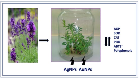
Effect of AuNPs and AgNPs on the Antioxidant System and Antioxidant Activity of Lavender (Lavandula angustifolia Mill.) from In Vitro Cultures

by
Paula Jadczak
1,*,
Danuta Kulpa
1,
Radosław Drozd
2,
Włodzimierz Przewodowski
3 and
Agnieszka Przewodowska
3
1
Department of Plant Genetics, Breeding and Biotechnology, Faculty of Environmental Management and Agriculture, West Pomeranian University of Technology in Szczecin Słowackiego 17, 71-434 Szczecin, Poland
2
Department of Microbiology and Biotechnology, Faculty of Biotechnology and Animal Husbandry, West Pomeranian University of Technology in Szczecin, Piastów 45, 70-311 Szczecin, Poland
3
Plant Breeding and Acclimation Institute in Bonin National Research Institute, Bonin 3, 73-009 Bonin, Poland
*
Author to whom correspondence should be addressed.
Molecules 2020, 25(23), 5511; https://doi.org/10.3390/molecules25235511
Submission received: 27 August 2020 / Revised: 18 November 2020 / Accepted: 19 November 2020 / Published: 25 November 2020
(This article belongs to the Special Issue The Antioxidant Capacities of Natural Products 2020)

Abstract

:
The aim of this study was to determine the effect of gold and silver nanoparticles on the activity of antioxidant enzymes (ascorbate peroxidase (APX), superoxide dismutase (SOD), guaiacol peroxidase (POX), and catalase (CAT)), the free radical scavenging capacity, and the total polyphenol capacity of lavender (Lavandula angustifolia Mill.) cultivar “Munstead” propagated in vitro. In the experiment, fragments of lavender plants were cultivated in vitro on medium with the addition of 1, 2, 5, 10, 20, and 50 mg∙dm−3 of AgNPs or AuNPs (particle sizes 24.2 ± 2.4 and 27.5 ± 4.8 nm, respectively). It was found that the nanoparticles increase the activity of the antioxidant enzymes APX and SOD; however, the reaction depends on the NP concentration. The highest APX activity is found in plants propagated on media with 2 and 5 mg∙dm−3 of AgNPs. AuNPs significantly increase the APX activity when added to media with a concentration of 10 mg∙dm−3. The highest SOD activity is recorded at 2 and 5 mg∙dm−3 AgNP and AuNP concentrations. The addition of higher concentrations of nanoparticles to culture media results in a decrease in the APX and SOD activity. The addition of AuNPs to culture media at concentrations from 2 to 50 mg∙dm−3 increases the POX activity in comparison to its activity when AgNPs are added to the culture media. No significant influence of NPs on the increase in CAT activity was demonstrated. AgNPs and AuNPs increased the free radical scavenging capacity (ABTS•+). The addition of NPs at concentrations of 2 and 5 mg∙dm−3 increased the production of polyphenols; however, in lower concentrations it decreased their content in lavender tissues.

Graphical Abstract

1. Introduction

True lavender (Lavandula angustifolia), classified in the mint family (Lamiaceae), is an evergreen shrub native to southern Europe, primarily the Mediterranean region. It is commercially cultivated in, among other places, France, Portugal, Poland, Spain, Hungary, the UK, Bulgaria, Australia, China, and the USA [1,2,3]. Essential oil with the characteristic aroma is one of the most important commercial components of true lavender, as it is used in the food, perfume, and the cosmetic industry [4,5,6]. Lavender oil is a mixture of chemical substances comprising primarily mono- and sesquiterpene compounds, exhibiting a complex therapeutic effect [7,8]. It is commonly used individually or as an additive in aromatherapy and folk and conventional medicine [9,10,11,12]. Numerous studies have confirmed the bactericidal and fungicidal properties of lavender oil toward Staphylococcus aureus, Enterococcus, Aspergillus nidulans and even Trichophyton metangrophytes, which is considered to be resistant to certain synthetic antibiotics [1]. In addition, lavender oil is characterized by analgesic, sedative, anticancer, and anti-inflammatory properties. It is a perfect agent to control digestive disorders and helps to combat skin disorders of various origins [10]. A study has also proven its effectiveness in alleviating depression symptoms [13].
Nowadays, the production of high-quality propagation materials, free of viruses and identical to parent plants, is ensured by biotechnological methods, including in vitro plant culture. Plants propagated in vitro are genetically uniform, which is of special importance for the production of secondary metabolites [14,15]. The large-scale production of lavender requires efficient in vitro propagation techniques to avoid the overexploitation of natural populations and allow the application of biotechnology-based approaches for plant improvement and the production of valuable secondary metabolites [16].
A series of studies have confirmed that medicinal plants propagated in vitro are characterized by different contents and compositions of essential oils [16,17,18,19]. The composition of secondary metabolites can also be changed using biotic and abiotic elicitors. These elicitors induce a series of physiological reactions in the plant and stimulate the production of secondary metabolites [20,21].
Nanotechnology has been applied in many fields of science and industry as a matter of specific qualities that have been identified by nanoparticles [22]. Nanometals are increasingly used in commercial products, including in the production of drugs and cosmetics and as transporting molecules, biosensors, or drug delivery agents [23,24,25]. They are also used in optoelectronics, nano-devices, and nanoelectronics. Their antibacterial properties have found application in medicine, water treatment, and food processing [26]. Resent research describe the wide use of nanosilver and gold in medicine [27,28,29,30].
They are valued for their unique physical and chemical properties (e.g., a higher surface to volume ratio, atypical surface structure, increased reactivity), and for their specific impact on biological tissues [31,32,33,34,35,36].
In recent times, particular attention has been paid to the use of nanotechnology in in vitro cultures. Nanoparticles have been mainly used as antimicrobial agents for sterilization and prevention of contamination of plant cultures in vitro [37]. In recent years, their influence on seed germination and plant growth has been increasingly studied [38,39,40]. The influences of copper (Cu), zinc (Zn), iron (Fe), titanium (Ti), and aluminium (Al) nanoparticles on plants are widely described among other things in Phaseolus radiatus [41], Stevia rebaudiana [42], Sinapis alba, Lepidium sativum, [43], Cicer arietinum [44], and Nicotiana tabacum [45]. However, the influence of nanoparticles of two metals, silver and gold, is most often studied. Ag and Au nanoparticles have been selected because of the features related to their commercial applicability—their low price, easy availability, and high stability in aqueous solutions—and the numerous studies indicating their low toxicity [46]. The effect of nanoparticles on plant development has been determined, such as the influence of silver nanoparticles on Solanum tuberosum [47] and Eichhornia crassipes [48] and gold nanoparticles on Brassica juncea [49], Artemisia absinthium, and Vigna radiata [50].
These nanoparticles are among the latest currently studied elicitors [51,52,53]. It has been proven that metal nanoparticles are highly reactive toward plants. Due to their small size, they can easily penetrate the cell membrane and accumulate inside the cell [54]. The properties of nanoparticles are strongly dependent on many factors, such as their method of preparation, the temperature at which they are prepared, and the surfactants used for capping [55].
Certain metal nanoparticles have a considerable impact on the physiological processes of plants, such as seed germination, growth, and metabolism [56,57,58,59], and scientific research indicates that their impact can be both positive and negative. The majority of studies describe that they exhibit a destructive influence that results in damage at the genomic level, chromosomal aberrations, cell growth inhibition, and programmed cell death-apoptosis [60,61,62]. Numerous studies have demonstrated the phytotoxicity of metal nanoparticles leading to the activation of the antioxidative system in plants [63,64]. Antioxidative enzymes, including guaiacol peroxidase (POX), superoxide dismutase (SOD), catalase (CAT), ascorbate peroxidase (APX), and alternative oxidase, and other low-molecular compounds in the form of polyphenols, vitamins, and minerals, are responsible for the inhibition of formation or the removal of excessive reactive oxygen species (ROS) from plant organisms [65,66].
The increase in the production levels of ROS under the influence of nanoparticles has been reported in many plants, such as Oryza sativa [67,68], Alium cepa [69], Brassica juncea [70,71], Arabidopsis Tthaliana [72], and Nicotina tabacum [73].
The oxidative stress induced by the metal nanoparticles may influence the production and qualitative and quantitative composition of secondary metabolites in plants [40,74]. The favorable influence of elicitation by metal nanoparticles on the production of biologically active compounds in plants has been confirmed by Zhang et al. [41,75] for Artemisia anuua, Sharafi et al. [42,76] for Hypericum perforatum, and Oloumi et al. [43,77] for Glycyrrhiza glabra. Silver and gold nanoparticles have been used as elicitors in cultures of Prunella vulgaris [78], Stevia glycosides [52], Cucumis anguria [79], and Caralluma tuberculata [80]. The latest scientific reports indicate significant changes in the metabolite profile of Arabidopsis thaliana under the influence of silver nanoparticles [81].
This paper is part of a cycle of research aimed at developing an efficient method of production of secondary metabolites, especially essential oils, by changing the profile of the secondary metabolites produced by Lavandula angustifolia plants from in vitro cultures. According to our previous studies, the addition to growth media of 1 to 50 mg∙dm−3 of AgNPs and AuNPs has a significant impact on plant development [82]. Our research has also shown that the addition of 10 or 50 mg∙dm−3 of AgNPs and AuNPs changes the composition of essential oils produced by lavender plants [83]. However, it is not clear to us whether this phenomenon is a result of stress or a change in the production of one of the endogenous plant growth regulators—e.g., ethylene, which modulates the endogenous production of other regulators. According to Akasaka-Kennedy [84], the addition of silver nanoparticles reduces the production of endogenous ethylene in Brassica sp tissues. Therefore, the main aim of the study is to answer the question of whether the addition of nanoparticles, which has been shown earlier, modifies their growth, affects the production of secondary metabolites, or activates ROS in plants grown on media in in vitro cultures. So far, there have been no scientific reports on the influence of AuNPs and AgNPs on the activation of the antioxidant system in L. angustifolia propagated in vitro.
The aim of the study was to analyze the influence of the addition to an in vitro culture of lavender of AgNPs and AuNPs on elements of the enzymatic antioxidant defences system, POX, APX, SOD, and CAT in plant tissue.

2. Results

2.1. Antioxidative Enzyme Activity

2.1.1. APX

Silver nanoparticles added to the propagation medium at the lowest concentration used (1 mg·dm−3) did not affect the APX enzyme activity, which remained at the level of control plants (Figure 1). However, the addition of higher AgNP concentrations to the medium resulted in increasing the APX activity. The highest APX activity was determined in plants propagated on media with the addition of 2 and 5 mg·dm−3 of AgNPs, at 26.01 and 18.93 U·mg−1 DW, respectively. With the increased AgNP concentration in culture media in the range of 10 to 50 mg·dm−3, the enzyme activity decreased, but it was at a significantly higher level (p ≤ 0.05) compared to the activity of control plants. The addition of AuNPs to the culture media had a significant effect on the increase in APX activity in relation to the control, but only for AuNP concentrations equal to 10 mg·dm−3 (9.28 U·mg−1 DW).

2.1.2. POX

The POX activity varied depending on the nanoparticle type applied. The POX activity was close to that of the control when plants were propagated on media with an AuNP addition at concentrations between 2 and 50 mg·dm−3 (Figure 1). A reduction in activity in relation to the control was observed solely for the lowest of the concentrations used (1 mg·dm−3). The propagation in media with the addition of AgNPs resulted in a dramatic reduction in the POX activity, relative to both the control as well as plants propagated on media with the addition of AuNPs. A particularly low POX activity was determined for plants propagated on media with an addition of 5 to 50 mg·dm−3 (0.30 to 0.57 U·mg−1 DW).

2.1.3. SOD

The activity of SOD above the control was recorded for the concentrations 2 and 5 mg·dm−3 of AgNPs (866.22 and 1276.34 U·mg−1 DW, respectively). The addition of higher AgNP concentrations to the culture media (10, 20, 50 mg·dm−3) resulted in the decreased activity of the enzyme, thus it remained at the control level. Gold nanoparticles resulted in a decreased SOD activity (p ≤ 0.05) only at the highest concentrations applied—20 and 50 mg·dm−3 (407.36 and 312.28 U·mg−1 DW). The activity of SOD for the lowest AuNPs concentrations remained at the control level (Figure 1).

2.1.4. CAT

The conducted study revealed that no significant impact of gold and silver nanoparticles could be found for the activity of catalase determined in the studied tissues (Figure 1).

2.2. Free Radical Scavenging Activity and Total Polyphenols Content

2.2.1. The Total Polyphenol Content

Polyphenols content varied depending on the nanoparticle type and its concentration in the medium (Figure 2). Increasing polyphenol contents relative to the control were determined for lavender cultivated on media with the addition of 2 and 5 mg·dm−3 of AuNPs and 5 to 50 mg·dm−3 of AgNPs (0.214 and 0.217, respectively, and from 0.212 to 0.318 mg of TAE/100 gDW). On the other hand, the lavender cultivated on media with the addition of the highest AuNP concentrations (20 and 50 mg·dm−3) and lowest AgNPs concentrations (1 and 2 mg·dm−3) was characterized by significantly lower polyphenol contents.

2.2.2. Free Radical Scavenging Activity (ABTS•+)

The presented outcomes suggest that gold and silver nanoparticles added to culture media in in vitro cultures have a positive impact on the free radical scavenging activity of true lavender (Figure 3). An exception here was the lowest AgNP concentration (1 mg·dm−3–197.77 μM AAE/100 gDW) and highest AuNP concentration used (50 mg·dm−3–158.86 μM AAE/100 gDW). The highest free radical scavenging capacity values were determined for plants cultivated on media with the addition of 5 mg·dm−3 of AgNPs and AuNPs, at 330.70 and 309.62 μM AAE/100 gDW, respectively. The addition of NPs in 1, 2, and 5 mg·dm−3 concentrations to the culture media resulted in a gradual increase in the antioxidant activity of lavender, whereas a reduced scavenging capacity was observed for higher concentrations (10, 20, and 50 mg·dm−3).

3. Discussion

Gold and silver nanoparticles are used on a large scale in many industries. Due to their antibacterial, anti-angiogenic, and anti-inflammatory properties, silver nanoparticles are used in antibacterial coatings for coating medical devices, wound and burn dressings, numerous cosmetic products, and food packaging [85,86,87]. Gold nanoparticles are used in molecular imaging, targeted drug delivery, gene therapy, cancer treatment, and radiation [88]. Prociak et al. [89] presented the potential use of gold and silver nanocolloids in cosmetic production. Microbiological tests performed in this study, showed that emulsion with silver and gold nanocolloids possessed satisfactory fungicidal properties and had a positive effect on the characteristics of the creams such as their consistency and absorption. Ag and Au nanocolloids can also be used in medicine, e.g., for the treatment of wounds, including the most difficult to heal burn and post-surgery wounds [90,91,92] or in medical imaging when diagnosing cancer [93,94]. It has been also confirmed the use of nanocolloids in the food industry, mainly in food packaging or prevention of contamination [95,96].
In view of the wide range of possibilities for the use of nanoparticles, it is important to determine the toxic properties of nanoparticles for humans and the environment. The results of most published scientific studies exclude the toxicity of nanoparticles [85,97,98]. However, there are also studies showing their adverse effects on living organisms and the environment, with this harmfulness depending on many factors, such as their physicochemical properties (shape, size, load) or coating agents [99,100]. Most of the studies show that nanoparticles are toxic to plants at higher concentrations. In addition, the type of target cells, tissues, or organisms or the type of test itself should be considered [100]. Therefore, it is vital to precisely determine the effect of nanoparticles on living organisms, including plants [100].
In response to oxidative damage, plants have developed a defense system which manifests itself, among other things, through the increased activity of antioxidant enzymes [101,102].
The activity of antioxidative enzymes may be a good marker of the toxic influence of the external environment on an organism. Mehrian et al. [103] studied the influence of AgNPs on the activity of antioxidant enzymes in Lycopersicon esculentum. It was found that, with the increase in the concentration of silver nanoparticles in the plant tissue medium, the activity of SOD, CAT, and POX in the shoots and roots increased. The increased activity of CAT, SOD, and POX was also observed in the callus tissue of sugar cane (Saccharum spp. cv CP-77,400) propagated on media with the addition of 20 to 60 AgNPs [104] (Barbasz et al.]. Iqbal et al. [105] carried out a study to investigate the effect of silver nanoparticles (AgNP) on the physiological, biochemical, and antioxidant parameters of wheat (Triticum aestivum L.) under heat stress conditions. It was found that the addition of AgNPs had a protective effect on plant tissues under stress conditions—wheat plants treated with AgNPs showed a significant increase in dry matter, with a simultaneous increase in SOD, POX, CAT, APX, and GPX activity under heat stress conditions. In the conducted study, we analysed the impact of the addition of gold or silver nanoparticles to culture media on the activity of CAT, APX, POX, and SOD antioxidative enzymes. The conducted study revealed a significant impact of the applied nanoparticles on the activity of all the tested antioxidative enzymes, with the exception of CAT. The observed effect was variable depending on the nanoparticle type and concentration. Lavender exposed to stress caused by AgNPs or AuNPs primarily activated the antioxidative system, consisting of APX and SOD. Increased activity of CAT, SOD, and POD due to Cu2O and Zn nanoparticles has also been confirmed for cucumber Cucumis sativus L. in the study of Kim and Lee [106]. These authors, using low concentrations of both kinds of nanoparticles, observed a significant increase in the activity of these three antioxidative enzymes. However, with the increased concentration of metals nanoparticles in the culture media (above 100 mg dm−1), the activity of these enzymes was markedly reduced. The concentration of the analyzed AgNPs which significantly reduced the activity of APX and SOD was 5 mg·dm−3. The experiment carried out by Gunjan et al. [107] examined the impact of gold nanoparticles on the activity of antioxidative enzymes in Brassica juncea seedlings. It was established that the glutathione reductase (GR), ascorbate peroxidase (APX), and glutathione peroxidase (GPX) activity increases with high AuNP concentrations (200 mg·dm−3), and that they are significantly higher than the CAT activity. These results suggest that the enzymatic complex in the form of APX, GPX, and GR forms part of the defensive mechanism against oxidative disturbance produced by nanometals in Brassica juncea, and that CAT does not have a substantial role in this case. In the study of Tripathi et al. [108], it was noted that silver nanoparticles (at concentrations from 100 to 300 µM) markedly stimulate superoxide dismutase and ascorbate peroxidase activity, while inhibiting the glutathione reductase and dehydroascorbate reductase activity in Pisum sativum.
Our study revealed that the true lavender antioxidative activity under in vitro cultures depends on the nanoparticle type and its concentration in the medium. Lavandula angustifolia is known for its health-promoting properties, resulting from the presence of antioxidative substances in its tissues (Miliasukas et al.) [109]. The chemical composition of plant metabolites and their antioxidative properties are influenced by various factors, including the i.a. cultivation method, growing location, climatic conditions, and plant genotype [2,110,111]. In our study, the free radical ABTS•+ scavenging analysis revealed a significant increase in the true lavender antioxidative activity after the addition of gold and silver nanoparticles to all culture media. This result is similar to the study of Chung et al. [79], where increased antioxidative activity was observed in transformed Corylus avellana roots under the impact of AgNPs. Fazal et al. [78] tested AuNPs and AgNPs as elicitors of Prunella vulgaris callus cultures and observed a marked increase in antioxidative activity.
Increased antioxidant activity may be associated, for example, with an increase in the content of substances with antioxidant activity, including polyphenols, in plant tissues. In our research we also examined the influence of the addition of AgNPs and AuNPs to the culture media of in vitro cultures on the polyphenol content. Polyphenols are secondary plant metabolites with strong antioxidants properties and are produced by plant cells for protective purposes against pathogens and also contribute in adaptation process to the external environment. Polyphenol compounds have also been suggested to have the ability to support activity of the cellular enzymatic antioxidative system, as well as modulate low-molecular antioxidant concentrations in the form of ascorbic acid (vitamin C) and alpha-tocopherol (vitamin E) in plant organisms [112]. In conducted study for determination of total phenolic compounds content in Lavandula angustifolia has been used the Folin-Ciocalteu assay. The sensitivity of this method can be affected by other cellular compounds such as sugars, proteins and lipids. However, depend on the plant organs, the total phenolic content can differ. Leaves of many plants are especially rich in this compounds and eventual interference other co-extracted substances do not affect the quality of assay [113]. Despite its limitations, Folin-Ciocalteu method is widely used to determine the content of polyphenols in the tissues of plants grown in in vitro cultures [114]. It was used, inter alia, by Tian et al., (2018) [115] determining the content of polyphenols in Atropa bellladonna tissues treated with Mn2O3 NPs, callus tissues of Caralluma tuberculata treated with AgNPs [80] or Stevia rebaudiana shoots treated with ZnONPs [42]. Previous research has stated that the influence of metal nanoparticles on the content of polyphenols in vegetable plants is variable. An increased content of polyphenols under the impact of AgNPs has been determined for Bacopa moonnieri [116] and Solanum tuberosum [117] and a reduction for castor [118]. We obtained similar results in our research. The polyphenol content was higher than in control plants when the plants grew on media with the addition of 10–50 mg·dm−3 AgNPs and 2–5 AuNPs. The study of Jamshidi and Ghanati [119] provided evidence of the significant effect of silver nanoparticle elicitation on the increase in phenol and flavonoid contents in hazel plants. In their experiment, they utilized low AgNP concentrations (2, 5, 10 mg·dm−3), indicating their clear impact on the content of the tested antioxidants in cell cultures. Ghorbanpour and Hadian [120] showed the significant influence of MWCTs (multi-walled carbon nanotubes) in the culture of the callus tissue of Satureja khuzestanica on the total flavonoid content in callus extracts for all MWCNT concentrations used. However, the analysis of the polyphenol content showed that its increase was observed only for some of the concentrations applied (50, 100, and 200 μg mL−1 MWCNTs). Their experiment further revealed that the maximum values for flavonoids and phenols were obtained using a MWCNT concentration of 100 μg·mL−1. In the present study, the nanoparticle concentrations used in culture media were lower. Tian et al. [115] demonstrated the significant impact of Mn2O3NPs at the concentration 25 mg·dm−3 on the increase in the alkaloid content in Atropa balladona L. tissues.
Production of bioactive secondary metabolites is generally associated with plant defense mechanisms. However, the mechanism of action of nanoparticles and their influence on ROS activation and the production of secondary metabolites is still poorly understood. According to Saha and Gupta (2018) [121], who studied the influence of silver nanoparticles on the development of Swertia chirata in in vitro cultures, ROS generated in presence of AgNP triggered an array of antioxidative enzymes which later on balanced the ROS content in the treated plant system. These enzymes also stimulated the shoot proliferations and helped in maximization of generation of number of shoots per explant considered. According to the authors, the reduction of ethylene production due to AgNPs may have a stimulating effect on plant development. Amir et al., (2019) [80] suggest that NPs might act as signal compounds, those like in nature to the chemical elicitors and influence cellular growth and secondary metabolism. Upon exposure to application of NPs, plant cell goes through a series of events in a cascade manner, resulting in oxidative outburst and generation of reactive oxygen species (ROS) in the surrounding environment of the plant cell. The ROS in turn can damage cell membrane and nuclei. To cope with the intense stress situation and to scavenge the ROS, plants activate their metabolic pathways including the notable mitogen-activated protein kinase (MAPK) pathway. MAPK activation propels the plant antioxidant elements to come in contact with ROS in a cascade fashion. Several studies have shown that nanoparticles, especially AgNPs, affect the up-regulation of genes related to the response to abiotic stress, including the production of enzymes such as SOD, CAT, AXP [122,123]. Nair and Chung, (2014) [124] when examining the toxicity of AgNPs on rice, found a significant up-regulation of SOD gene. In their opinion, induction of SOD gene in response to AgNPs stress might be to maintain the redox homeostasis of the cell.
Our results suggest that gold and silver nanoparticles added to in vitro culture media have a significant impact on the antioxidative potential (free radical scavenging capacity) and activation of antioxidative enzymes of in vitro true lavender cultures. The defence mechanism of lavender exposed to stress caused by AgNPs and AuNPs primarily consists of the APX-SOD enzymatic complex, with CAT remaining insignificant in this case. It can additionally be stated that AgNPs at the concentration of 5 mg·dm−3 result in the highest antioxidative activity of APX and SOD, and higher concentrations in the culture medium inhibit their activity. AgNPs have a more toxic impact on lavender plants cultivated in vitro than AuNPs. Silver and gold nanoparticles added to culture media have significant impacts on the total polyphenol contents in true lavender cultivated in vitro and its free radical scavenging capacity ABTS•+, however this depends on the applied nanoparticle concentration.

4. Materials and Study Methods

4.1. Plant Culture under In Vitro Conditions

The study material comprised L. angustifolia cultivar “Munstead” plants growing under in vitro conditions. The explants used for the experiment were single-node shoot fragments and apical shoot fragments with several leaves and the apical meristem. They were placed on the Murashige and Skoog (MS) media [125] with the addition of 2 mg dm−3 of kinetin and 0.2 mg dm−3 of indole-3-acetic acid [126] and 1, 2, 5, 10, 20, or 50 mg dm−3 of AgNPs or AuNPs. The medium pH was set at 5.7 using 0.1 M solutions of HCl and NaOH. The culture medium was hardened with 7 g·dm−3 of agar and was subject to a 20-min sterilization in an autoclave at 121 °C and a 1 ATM pressure. Propagation was carried out in jars of 200 mL volume, containing 20 mL of medium. Eight explants were placed in each culture vessel. The experiment was established in 10 replications for each type and concentration of the nanoparticles used. The jars were placed for 28 days in a growth chamber at a temperature of 24 °C and relative air humidity of 70–80%. The cultures were illuminated with fluorescent light at an intensity of 40 PAR (μE·m−2·s−1) for 16 h per day. After 28 days of culture, the study material was collected and subjected to analyses.

4.2. Nanoparticles

In the experiment, aqueous solutions of AuNPs measuring 24.2 ± 2.4 nm and AgNPs measuring 27.5 ± 4.8 nm were used. The aqueous suspensions were synthesized using the methods of Turkevich et al. [127] and Liu et al. [128] with modified synthesis conditions and two-stage microwave-convection heating. For this purpose, aqueous mixtures of 3.5 mM sodium citrate with 7.0 mM tetrachloroauric acid (HAuCl4) and 7.0 mM silver nitrate (AgNO3), respectively, were prepared. Once their spectra were plotted with a UV-Vis Epoch Microplate Spectrophotometer (BioTek, Winooski, VT, USA), the optical density of the fractions obtained was adjusted to a common DEV value using the spectra absorbance maxima (λmax = 520 nm for gold colloid and λmax = 445 nm for silver colloid). The similarities in the morphology, shape, and size of the synthesized and prepared AuNPs and AgNPs were assessed through analyzing their images obtained using a transmission electron microscope (TEM) JEM-2100 (JOEL ltd., Tokyo, Japan) (Figure 4).

4.3. Antioxidant Enzymes Activity Assay

4.3.1. Sample Preparation

All the steps of the protein extraction were carried out at 4 °C on a cooled plate. Fresh leaf samples weighing approximately 0.2 g were previously powdered in liquid nitrogen using a cold mortar and pestle. The obtained powder was resuspended in 1 mL of cold 50 mmol L−1 phosphate buffer (pH 7.0) enriched with 1 mmol L−1 of phenylmethylsulfonyl fluoride, 2 mmol L−1 of EDTA, and 1% polyvinylpyrrolidone. The resulting solution was centrifuged at 15,000 rpm for 10 min at 4 °C. The extracts were immediately used for the determination of the enzyme activity or stored at −80 °C for further analysis.

4.3.2. Guaiacol Peroxidase Assay (POX)

The activity of POX was determined according to the protocol of Chance and Maehly [129], which was adapted to a microplate reader, by monitoring the rate of tetraguaiacol formation from guaiacol by an increase in absorbance at 470 nm (ε = 26.6 mM−1 cm−1). One unit of POX is defined as the amount of enzyme that forms 1 µmol of tetraguaiacol per minute. The results were expressed in units per mg of the extracted protein.

4.3.3. Ascorbate Peroxidase Assay (APX)

The activity of APX was measured according to the protocol of Nakano and Asada [130] by monitoring the decrease in absorbance at 290 nm (ε = 2.8 mM−1 cm−1). The measurements were performed using a microplate reader. One unit of APX is defined as the amount of enzyme that oxidizes 1 µmol of ascorbate per minute. The results were expressed in the units per mg of the extracted protein.

4.3.4. Dismutase Assay (SOD)

The total SOD activity was determined based on the inhibition of the photochemical reduction of nitroblue tetrazolium (NBT) according to the protocol of Beauchamp and Fridovich [131], which was adapted to a microplate reader. One unit of SOD is defined as the amount of enzyme that inhibits the NBT reduction by 50%. The results were expressed in units per mg of the extracted protein.

4.3.5. Catalase Assay (CAT)

The activity of CAT was determined according to the protocol of Li and Schellhorn [132] based on the rate of decomposition of hydrogen peroxide by measuring the decrease in absorbance at 240 nm (ε = 43.6 mM−1 cm−1). One unit of CAT is defined as the amount of enzyme that decomposes 1 µmol of hydrogen peroxide per minute. The results were expressed in units per mg of the extracted protein.

4.4. Total Phenol Content and Free-Radical ABTS•+ Scavenging Ability Assay

4.4.1. Tissue Extract Preparation

A total of 100 mg of tissue (previously frozen at −80 °C) was powdered in liquid nitrogen using a cooled laboratory mortar and pestle. The obtained powder was transferred to plastic tubes, mixed with 5 mL of cooled methanol, and left for 1 h in the dark. During extraction, the samples were mixed periodically. Then, the samples were centrifuged at 15,000 rpm and the obtained extract was used for further analysis.

4.4.2. Total Polyphenol Content Assay

The total content of polyphenolic acid in the tissue extracts was determined using Folin-Ciocalteu reagent according to the method of Anastasiadi et al. [133] which was modified to a microplate reader scale. The concentration of polyphenolic compounds was expressed as the mg of tannic acid equivalent (TEA) per 100 g of dry weight of sample (mg TAE/100 gDW).

4.4.3. Free Radical ABTS•+ Scavenging Ability Assay

The free radical scavenging activity was assessed in the tissue extracts using the cation radical discoloration of the 2,2′-azino-bis (3-ethylbenzothiazoline-6-sulfonic acid) diammonium salt ABTS•+ assay adapted to a microplate reader scale, according to the method of Shi et al. [134]. Ascorbic acid (AAE) was used as a standard for calibration. The results were expressed in the μM of ascorbic acid equivalent per 100 g of dry weight of sample (μM AAE/100 gDW).

4.5. Statistical Analysis

The experiment was performed in three replications. An analysis of variance was performed, followed by Tukey’s test (p ≤ 0.05). Homogenous groups were labeled with successive letters of the alphabet.

Author Contributions

P.J. performed the experiments in in vitro cultures, analyzed the data, and wrote the manuscript; D.K. contributed to the revisions of the manuscript; W.P. developed, prepared, and standardized the solutions of nanoparticles. A.P. measured and analyzed the solutions of nanoparticles; R.D. manuscript editing and revision, enzymatic assay supervising. All the authors were responsible for processing information and manuscript writing. All the authors read and approved the final manuscript. All authors have read and agreed to the published version of the manuscript.

Funding

This research received no external funding.

Conflicts of Interest

The authors declare no conflict of interest.

References

  1. Cavanagh, H.M.A.; Wilkinson, J.M. Lavender essential oil: A review. Healthc. Infect. 2005, 10, 35–38. [Google Scholar] [CrossRef] [Green Version]
  2. Verma, R.S.; Rahman, L.U.; Chanotiya, C.S.; Verma, R.K.; Chauhan, A.; Yadav, A.; Singh, A.; Yadav, A.K. Essential oil composition of Lavandula angustifolia Mill. Cultivated in mild hills of Uttarahand, India. J. Serb. Chem. Soc. 2010, 75, 343–348. [Google Scholar] [CrossRef]
  3. Brailko, V.A.; Mitrofanova, O.; Lesnikowa-Sedoshenko, N.; Chelombit, S.; Mitrofanova, I.V. Anatomy features of Lavandula angustifolia Mill. and Lavandula hybrida rev. plants in vitro. J. Agric. Food Chem. 2017, 63, 111–117. [Google Scholar] [CrossRef] [Green Version]
  4. Da Porto, C.; Decorti, D.; Kikic, I. Flavour compounds of Lavandula angustifolia L. to use in food manufacturing: Comparison of three different extraction methods. Food Chem. 2009, 112, 1072–1078. [Google Scholar] [CrossRef]
  5. Hamad, K.J.; Al-Shaheen, S.J.A.; Kaskoos, R.A.; Ahamad, J.; Jameel, M.; Mir, S.R. Essential oil composition and antioxidant activity of Lavandula angustifolia from Iraq. Int. Res. J. Pharm. 2013, 4, 117–120. [Google Scholar]
  6. Waithaka, P.; Gathuru, E.; Githaiga, B.; Kwoko, J. Making of perfumes from essential oils extracted from lavender plant collected from Egerton University, Main Campus Njoro, Kenya. Afr. J. Biomed. Res. 2016, 2, 35–40. [Google Scholar]
  7. Landmann, C.; Fink, B.; Festner, M.; Dregus, M.; Engel, K.H.; Schwab, W. Cloning and functional characterization of three terpene sythases from lavender (Lavandula angustifolia). Arch. Biochem. Biophys. 2007, 465, 417–429. [Google Scholar] [CrossRef] [Green Version]
  8. Mahmoudi, R.; Nostratpour, S. Teuricum polium L. essential oil: Phytochemical component and antioxidant properties. Int. Food. Res. J. 2013, 4, 1697–1701. [Google Scholar]
  9. Gören, A.; TopÇu, G.; Bilsel, M.; AydoĞmuŞ, Z.; Pezzuto, J.M. The chemical contituents and biological activity of Lavandula stocheas ssp. stocheas. Z. Nat. C 2002, 57, 797–800. [Google Scholar]
  10. Hajhashemi, V.; Ghannadi, A.; Sharif, B. Anti-inflammatory and analgesic properties of the leaf extracts and essential oil of Lavandula angustifolia Mill. J. Ethnopharmacol. 2003, 89, 67–71. [Google Scholar] [CrossRef]
  11. Raut, S.J.; Karruppayil, M.S. A status review on the medicinal properties of essential oils. Ind. Crops. Prod. 2014, 62, 250–264. [Google Scholar] [CrossRef]
  12. Prasad, A.; Shukla, S.P.; Mathur, A.; Singh, C.C.; Kumar, A.M. Genetic fidelity of long-term micropropagated Lavandula officinalis Chaix: An important aromatic plant. Plant. Cell Tiss. Org. 2015, 120, 803–811. [Google Scholar] [CrossRef]
  13. Akhondzadeh, S.; Kashan, L.; Fotouhi, A.; Jarvandi, S.; Mobaseri, M.; Moin, M.; Khani, M.; Amir, H.J.; Baghalian, K.; Taghizadeh, M. Comparison of Lavandula angustifolia Mill. tincture and imipramine in the treatment of mild to moderate depression: A double-blind, randomized trial. Prog. Neuropsychopharmacol. Biol. Psychiatry 2003, 27, 123–127. [Google Scholar] [CrossRef]
  14. Al-Qudah, T.; Shibli, R.A.; Alali, F.Q. In vitro propagation and secondary metabolites production in wild germander (Teucrium polium L.). Vitr. Cell. Dev. Biol. -Plant 2011, 47, 496–505. [Google Scholar] [CrossRef]
  15. Frabetti, M.; Gutiérrez-Pesce, P.; Mendoza-de, G.E.; Rugini, E. Micropropagation of Teucrium fruticans L. an ornamental and medicinal plant. Vitr. Cell. Dev. Biol. -Plant. 2009, 45, 129–134. [Google Scholar] [CrossRef]
  16. Gonçalves, S.; Romano, A. In vitro culture of lavenders (Lavandula spp.) and the production of secondary metabolites. Biotechnol. Adv. 2013, 31, 166–174. [Google Scholar]
  17. Amoo, S.O.; Aremu, A.O.; Van Staden, J. In vitro plant regeneration, secondary metabolite production and antioxidant activity of micropropagated Aloe arborescens Mill. Plant Cell Tiss. Org. 2012, 111, 345–358. [Google Scholar] [CrossRef]
  18. Jakovljević, D.Z.; Vasić, S.M.; Stanković, M.S.; Čomić, L.R.; Topuzović, M.D. Secondary metabolite content and in vitro biological effects of Ajuga chamaepitys (L.) Schreb subsp. chamaepitys. Arch. Biol. Sci. 2015, 67, 1195–1202. [Google Scholar]
  19. Makowczyńska, J.; Sliwinska, E.; Kalemba, D.; Piątczak, E.; Wysokińska, H. In vitro propagation, DNA content and essential oil composition of Teucrium scorodonia L. ssp. scorodonia. Plant Cell Tiss. Org. 2016, 127, 1–13. [Google Scholar]
  20. Vanisree, M.; Hsin-Sheng, T. Plant cell cultures—An alternative and efficient source for the production of biologically important secondary metabolites. Int. J. Appl. Eng. Res. 2004, 2, 29–48. [Google Scholar]
  21. Shakeran, Z.; Keyhanfar, M.; Asghari, G.; Ghanadian, M. Improvement of atropine production by different biotic and abiotic elicitors in hairy root cultures of Datura metel. Turk. J. Biol. 2015, 39, 111–118. [Google Scholar] [CrossRef]
  22. Rajak, A. Nanotechnology and its application. J. Nanomed. Nanotechnol. 2018, 9, 502. [Google Scholar] [CrossRef]
  23. Das, M.; Shim, K.H.; An, S.S.A.; Yi, D. Review on gold nanoparticles and their applications. J. Toxiol. Env. 2012, 3, 193–205. [Google Scholar] [CrossRef]
  24. Roco, M.C. Nanotechnology: Convergence with modern biology and medicine. Curr. Opin. Biotechnol. 2003, 3, 337–346. [Google Scholar] [CrossRef] [Green Version]
  25. Nowack, B.; Bucheli, T.D. Occurrence, behavior and effects of nanoparticles in the environment. Env. Pollut. 2007, 1, 5–22. [Google Scholar] [CrossRef]
  26. Rajmedevi, J.; Jeyasubramanian, K.; Marikani, A.; Rajakumar, G.; Rahuman, A.A. Synthesis and antimicrobial activity of copper nanoparticles. Mater. Lett. 2012, 71, 114–116. [Google Scholar]
  27. Ahmed, J.; Gultekinoglu, M.; Edirsinghe, M. Bacterial cellulose micro-nano fibres for wound healing applications. Biotechnol. Adv. 2020, 41, 107549. [Google Scholar] [CrossRef]
  28. Matharu, R.K.; Porwal, H.; Chen, B.; Ciric, L.; Edirsinghe, M. Viral filtration using carbon-based materials. Med. Devices. Sens. 2020, 3, e10107. [Google Scholar] [CrossRef]
  29. Matharu, R.K.; Ciric, L.; Ren, G.; Edirsinghe, M. Comparative study of the antimicrobial effects of tungsten nanoparticles and tungsten nanocomposite fibres on hospital acquired bacterial and viral pathogens. J. Nanomater. 2020, 10, 1017. [Google Scholar] [CrossRef]
  30. Panyala, N.R.; Pena-Mendez, E.M.; Havel, J. Gold and nano-gold in medicine: Overview, toxicology and perspectives. J. Appl. Biomed. 2009, 7, 75–91. [Google Scholar] [CrossRef] [Green Version]
  31. Neira, T.R.; Carmora, E.; Recio, G.; Nesi, A.N.; Diaz, M.R.; Alberdi, M.; Rengel, Z.; Blancheteau, A.I. Metallic nanoparticles influence the structure and function of the photosynthetic apparatus in plants. Plant. Psychiol. Biochem. 2018, 130, 408–417. [Google Scholar] [CrossRef]
  32. Alexis, F.; Pridgen, E.; Molnar, L.K.; Farokhzad, O.C. Factors affectin the clearance and biodistibution of polymeric nanoparticles. Mol. Pharm. 2008, 5, 505–515. [Google Scholar] [CrossRef] [Green Version]
  33. Attarad, A.; Hira, Z.; Muhammad, Z.; Ihsan, U.H.; Abdul, R.P.; Joham, S.A.; Altaf, H. Sythesis, characterization, applications and challenges of iron oxide nanoparticles. Nanotechnol. Sci. Appl. 2019, 9, 49–67. [Google Scholar]
  34. Jain, P.K.; Lee, K.S.; El-Sayed, I.H.; El-Sayed, M.A. Calculated absorption and acattering properties of gold nanoparticles of different size, shape, and composition: Applications in biological imaging and biomedicine. J. Phys. Chem. B 2006, 110, 7238–7248. [Google Scholar] [CrossRef] [Green Version]
  35. Cheun-Yi, Y.; Ceran, B.; Rotello, V.M. Gold nanoparticles: Preparation, properties and applications in biotechnology. Nanoscale 2012, 4, 1871–1880. [Google Scholar]
  36. Bhattacharya, R.; Mukherjee, P. Biological properties of “naked” metal nanoparticles. Adv. Drug. Deliv. Rev. 2008, 11, 1289–1306. [Google Scholar] [CrossRef]
  37. Abdi, G.; Selehi, H.; Kosh-Khui, M. Noano silver: A novel nanomaterial for removal of bacterial contaminations in valerian (Valeriana officinalis L.) tissue culture. Acta Physiol. Plant. 2008, 30, 709–714. [Google Scholar] [CrossRef]
  38. Mahna, N.; Vahed, S.Z.; Khani, S. Plant in vitro culture goes nano: Nanosilver-mediated decontamination of ex vitro explants. J. Nanomed. Nanotechnol. 2013, 4, 1. [Google Scholar] [CrossRef] [Green Version]
  39. Wang, P.; Lombi, E.; Zhao, F.J.; Kopittke, P.M. Nanotechnology: A new opportunity in plant sciences. Trends Plant. Sci. 2016, 21, 699–721. [Google Scholar] [CrossRef]
  40. Hussain, M.; Raja, N.I.; Mashwani, Z.; Iqbal, M.; Sabir, S.; Yasmeen, F. In vitro seed germination and biochemical profiling of Artemisia absinthium exposed to various metallic nanoparticles. 3 Biotech 2018, 130, 408–417. [Google Scholar] [CrossRef] [Green Version]
  41. Lee, W.M.; An, Y.J.; Yoo, H.; Kweon, H.S. Toxicity and bioavailibity of copper nanoparticles to the terrestrial plants mung been (Phaseolus radiatus) and wheat (Triticum aestivum): Plant agar test for water-insoluble nanoparticles. Env. Toxicol. Chem. 2008, 27, 1915–1921. [Google Scholar] [CrossRef] [PubMed]
  42. Javed, R.; Yucesan, B.; Zia, M.; Gurel, E. Elicitation of secondary metabolites in callus cultures of Stevia rebaudiana Bertoni grown under ZnO and CuO nanoparticles stress. Sugar Tech. 2018, 20, 194–201. [Google Scholar] [CrossRef]
  43. Asztemborska, M.; Steborowski, R.; Kowalska, J.; Bystrzejewska-Piotrowska, G. Accumulation of platinum nanoparticles by Sinapis alba and Lepidium sativum plants. Water Air Soil Pollut. 2015, 226, 126. [Google Scholar] [CrossRef] [PubMed] [Green Version]
  44. Burman, U.; Saini, M.; Kumar, P. Effect of zinc oxide nanoparticles on growth and antioxidant system of chickpea seedlings. Toxicol. Env. Chem. 2013, 95, 605–612. [Google Scholar] [CrossRef]
  45. Fraizer, T.P.; Burklew, C.E.; Zhang, B. Titanium dioxide nanoparticles affect the growth and microRNA expression of tobacco (Nicotiana Tab.). Funct. Integr. Genom. 2014, 14, 75–83. [Google Scholar] [CrossRef]
  46. Sarmast, M.K.; Salehi, H. Silver nanoparticles: An influential element in plant nanobiotechnology. Mol. Biotechnol. 2016, 58, 441–449. [Google Scholar] [CrossRef]
  47. Homaee, M.B.; Ehsanpour, A.A. Physiological and biochemical responses of potato (Solanum tuberosum) to silver nanoparticles and silver nitrate treatments under in vitro conditions. Ind. J. Plant. Physiol. 2015, 20, 353–359. [Google Scholar] [CrossRef]
  48. Rani, P.U.; Yasur, J.; Loke, K.S.; Dutta, D. Effect of synthetic and biosynthesized silver nanoparticles on growth, physiology and oxidative stress of water hyacinth: Eichhornia crassipes (Mart) Solms. Acta Physiol. Plant. 2016, 38, 58. [Google Scholar] [CrossRef]
  49. Arora, S.; Sharma, P.; Kumar, S.; Nayan, R.; Khanna, P.K.; Zaidi, M.G.H. Gold-nanoparticle induced enhancement in growth and seed yield of Brassica Juncea. Plant. Growth Regul. 2012, 66, 303–310. [Google Scholar] [CrossRef]
  50. Nair, P.M.G.; Chung, I.M. Physiological and molecular level studies on the toxicity of silver nanoparticles in germinating seedlings of mung bean (Vigna Radiata L.). Acta Physiol. Plant. 2015, 37, 1719. [Google Scholar] [CrossRef]
  51. Vecerova, K.; Vecera, Z.; Docekal, B.; Oravec, M.; Pompeiano, A.; Tríska, J.; Urban, O.; Pompiano, J. Changes of primary and secondary metabolites in barley plants exposed to CdO nanoparticles. Environ. Pollut. 2016, 218, 207–218. [Google Scholar] [CrossRef]
  52. Golkar, P.; Moradi, M.; Garousi, A.G. Elicitation of stevia glycosides using salicylic acid and silver nanoparticles under callus culture. Sugar Tech. 2018, 21, 569–577. [Google Scholar] [CrossRef]
  53. Jamshidi, M.; Ghanti, F.; Razaei, A.; Bemani, E. Change of antioxidant enzymes activity of hazel (Corylus avellana L.) cells by AgNPs. Cytotechnology 2014, 68, 525–530. [Google Scholar] [CrossRef] [Green Version]
  54. Javed, R.M.; Usman, M.; Tabassum, S.; Zia, M. Effect of capping agents: Structural, optical and biological properties of ZnO nanoparticles. Appl. Surf. Sci. 2016, 386, 319–326. [Google Scholar] [CrossRef]
  55. Lu, C.M.; Zhang, C.Y.; Wen, J.Q.; Tao, M.X. Research of the effect of nanometer materials on germination and growth enhancement of Glycine max and its mechanism. Mater. Sci. 2002, 21, 168–171. [Google Scholar]
  56. Dimkpa, C.O.; McLean, J.E.; Latta, D.E.; Manangón, E.; Britt, D.W.; Johnson, W.P.; Boyanov, M.I.; Anderson, A.J. Cuo and ZnO nanoparticles: Phytotoxicity, metal speciation, and induction of oxidative stress in sand-grown wheat. J. Nanopart. Res. 2012, 14, 1–15. [Google Scholar] [CrossRef]
  57. Shaw, A.K.; Hossain, Z. Impact of nano-Cuo stress on rice (Oryza sativa L.) seedlings. Chemosphere 2013, 96, 906–915. [Google Scholar] [CrossRef]
  58. Gopinath, K.; Gowri, S.; Karthika, V.; Arumugam, A. Green synthesis of gold nanoparticles from fruit extract of Terminalia arjuna for the enhanced seed germination activity of Gloriosa superb. J. Nanostructure Chem. 2014, 4, 1–11. [Google Scholar] [CrossRef] [Green Version]
  59. Ghosh, M.; Bandyopadhyay, M.; Mukherjee, A. Genotoxicity of titanium dioxide (TiO2) nanoparticles at two trophic levels: Plant and human lymphocytes. Chemosphere 2010, 81, 1253–1262. [Google Scholar] [CrossRef]
  60. Kumbhakar, D.V.; Datta, A.K.; Mandal, A.; Das, D.; Gupta, S.; Ghosh, B.; Halder, S.; Dey, S. Effectivity of copper and cadmium sulfide nanoparticles in mitotic and meiotic cells of Nigella sativa L. (black cumin)–can nanoparticles act as mutagenic agents? J. Exp. Nanosci. 2016, 11, 823–829. [Google Scholar] [CrossRef] [Green Version]
  61. Pramanik, A.; Datta, A.K.; Gupta, S.; Ghosh, B.; Das, D.; Kumbhakar, D.V. Assessment of genotoxicity of engineered nanoparticles (cadmium sulphide-CdS and copper oxide-CuO) using plant model (Coriandrum sativum L.). Int. J. Res. Pharm. Sci. 2017, 8, 741–753. [Google Scholar]
  62. Oukarroum, A.; Barhoumi, L.; Pirastru, L.; Dewez, D. Silver nanoparticle toxicity effect on growth and cellular viability of the aquatic plant Lemna gibba. Env. Toxicol. Chem. 2013, 32, 902–907. [Google Scholar] [CrossRef] [PubMed]
  63. Jiang, H.S.; Qiu, X.N.; Li, G.B.; Li, W.; Yin, L.Y. Silver nanoparticles induced accumulation of reactive oxygen species and alteration of antioxidant systems in the aquatic plant Spirodela polyrhiza. Env. Toxicol. Chem. 2014, 33, 1398–1405. [Google Scholar] [CrossRef] [PubMed]
  64. Gill, S.S.; Tuteja, N. Reactive oxygen species and antioxidant machinery in abiotic stress tolerance in crop plants. Plant. Physiol. Bioch. 2010, 48, 909–930. [Google Scholar] [CrossRef]
  65. Hatami, M.; Ghorbanpour, M. Defense enzymes activity and biochemical variations of Pelargonium zonale in response to nanosilver particles and dark storage. Turk. J. Biol. 2014, 38, 130–139. [Google Scholar] [CrossRef]
  66. Chew, B.P.; Park, J.S. Carotenoid action on the immune response. J. Nutr. 2004, 134, 257S–261S. [Google Scholar] [CrossRef]
  67. Mirzajani, F.; Askari, H.; Hamzelou, S.; Farzaneh, M.; Ghassempour, A. Effect of silver nanoparticles on Oryza sativa L. and its rhizosphere bacteria. Ecotoxicol. Env. Saf. 2013, 88, 48–54. [Google Scholar] [CrossRef]
  68. Panda, K.K.; Achary, V.M.M.; Krishnaveni, R.; Padhi, B.K.; Sarangi, S.N.; Sahu, S.N.; Panda, B.B. In vitro biosynthesis and genotoxicity bioassay of silver nanoparticles using plants. Toxicol. Vitr. 2011, 25, 1097–1105. [Google Scholar] [CrossRef]
  69. De, A.; Chakrabarti, M.; Ghosh, I. Evaluation of genotoxicity and oxidative stress of aluminium oxide nanoparticles and its bulk form in Allium cepa. Nucleus 2016, 59, 219–225. [Google Scholar] [CrossRef]
  70. Rao, S.; Shekhawat, G.S. Phytotoxicity and oxidative stress perspective of two selected nanoparticles in Brassica juncea. 3 Biotech 2016, 6, 244. [Google Scholar] [CrossRef] [Green Version]
  71. Sharma, P.; Bhatt, D.; Zaidi, M.G.H.; Zaidi, M.G.H.; Saradhi, P.P.; Khanna, P.K.; Arora, S. Silver nanoparticle-mediated enhancement in growth and antioxidant status of Brassica juncea. Appl. Biochem. Biotechnol. 2012, 167, 2225–2233. [Google Scholar] [CrossRef]
  72. Liu, F.; Jiang, H.; Ye, S.; Chen, W.P.; Liang, W.; Xu, Y. The Arabidopsis P450 protein CYP82C2 modulates jasmonate-induced root growth inhibition, defense gene expression and indole glucosinolate biosynthesis. Cell Res. 2010, 20, 539–552. [Google Scholar] [CrossRef] [PubMed]
  73. Dai, Y.; Wang, Z.; Zhao, J.; Xu, L.; Yu, X.; Wei, Y.; Xing, B. Interaction of CuO nanoparticles with plant cells: Internalization, oxidative stress, electron transport chain disruption, and toxicogenomic responses. Environ. Sci. Nano 2018, 5, 2269–2281. [Google Scholar] [CrossRef]
  74. Moharrami, F.; Hosseini, B.; Sharafi, A.; Farjaminezhad, M. Enhanced production of hyoscyamine and scopolamine from genetically trans-formed root culture of Hyoscyamus reticulatus L. elicited by iron oxide nanoparticles. Vitr. Cell. Dev. Biol.-Plant 2017, 53, 104–111. [Google Scholar] [CrossRef] [PubMed] [Green Version]
  75. Zhang, B.; Zheng, L.P.; Li, W.Y.; Wang, J.W. Stimulation of artemisinin production in Artemisia annua hairy roots by Ag-SiO2 core-shell nanoparticles. Curr. Nanosci. 2013, 9, 363–370. [Google Scholar] [CrossRef]
  76. Sharafi, E.; Nekoei, S.M.K.; Fotokian, M.H.; Davoodi, D.; Mirzaei, H.H.; Hasanloo, T. Improvement of hypericin and hyperforin production using zinc and iron nano-oxides as elicitors in cell suspension culture of St John’s wort (Hypericum perforatum L.). J. Med. Plants By-Prod. 2013, 2, 177–184. [Google Scholar]
  77. Oloumi, H.; Soltaninejad, R.; Baghizadeh, A. The comparative effects of nano and bulk size particles of CuO and ZnO on glycyrrhizin and phenolic compounds contents in Glycyrrhiza glabra L. seedlings. Indian J. Plant. Physiol. 2015, 20, 157–161. [Google Scholar] [CrossRef]
  78. Fazal, H.; Abbasi, B.H.; Ahmed, N.; Ali, S.S.; Shujait, A.S.; Akbar, F.; Kanwal, F. Correlation of different spectral lights with biomass accumulation and production of antioxidant secondary metabolites in callus cultures of medicinally important Prunella vulgaris L. J. Photochem. Photobiol. B Bio. 2016, 159, 1–7. [Google Scholar] [CrossRef]
  79. Chung, I.; Rajakumar, G.; Thiruvengadam, M. Effect of silver nanoparticles on phenolic compounds production and biological activities in hairy root cultures of Cucumis Anguria. Acta Biol. Hung. 2018, 69, 97–109. [Google Scholar] [CrossRef] [Green Version]
  80. Ali, A.; Mohamed, S.; Khan, M.A.; Raja, N.I.; Arif, M.; Kamil, A.; Mashwani, Z. Silver nanoparticles elicited in vitro callus cultures for accumulation of biomass and secondary metabolites in Caralluma Tuberculate. Artif. Cells Nanomed. Biotechnol. 2019, 47, 715–724. [Google Scholar] [CrossRef] [Green Version]
  81. Kruszka, D.; Sawikowska, A.; Selvakesavan, R.K.; Krajewski, P.; Kachlicki, P.; Franklin, G. Silver nanoparticles affect phenolic and phytoalexin composition of Arab Thaliana. Sci. Total Env. 2020, 716, 135361. [Google Scholar] [CrossRef] [PubMed]
  82. Jadczak, P.; Kulpa, D.; Bihun, M.; Przewodowski, W. Positive effect of AgNps and AuNps in in vitro cultures of Lavandula angustifolia Mill. Tissue Organ. Cult. 2019, 139, 191–197. [Google Scholar] [CrossRef] [Green Version]
  83. Wesołowska, A.; Jadczak, P.; Kulpa, D.; Przewodowski, W. Gas chromatography mass spectrometry (GC-MS) analysis of essential oils from AgNps and AuNps elicited Lavandula angustifolia in vitro cultures. Molecules 2019, 24, 606. [Google Scholar] [CrossRef] [PubMed] [Green Version]
  84. Akasaka-Kennedy, Y.; Yoshida, H.; Takathaka, Y. Efficient plant regeneration from leaves of rapeseed (Brassica napus L.): The influence of AgNO3 and genotype. Plant. Cell Rep. 2006, 24, 649–654. [Google Scholar] [CrossRef]
  85. Devi, G.H.; Suruthi, P.; Veerakumar, R.; Vinoth, S.; Subbaiya, R.; Chozhavendahn, S. A review on metallic gold and silver nanoparticles. J. Pharm. Tech. 2019, 12, 935–943. [Google Scholar] [CrossRef]
  86. Aziz, S.G.G.; Aziz, S.G.G.; Akbarzadeh, A. Advances in silver nanotechnology: An update on biomedical applications and future perspectives. Drug Res. 2017, 67, 198–203. [Google Scholar] [CrossRef]
  87. Colvin, V.L. The potential environmental impact of engineered nanomaterials. Nat. Biotechnol. 2003, 21, 1166–1170. [Google Scholar] [CrossRef]
  88. Austin, L.A.; Mackey, M.A.; Dreaden, E.C.; El-Sayed, A.M. The optical, photothermal, and facile surface chemical properties of gold and silver nanoparticles in biodiagnostics, therapy, and drug delivery. Arch. Toxicol. 2014, 88, 1391–1417. [Google Scholar] [CrossRef] [Green Version]
  89. Prociak, J.; Grabowska, A.; Chwastowski, J.; Majka, T.; Banach, M. Safety of the application of nanosilver and nanogold in topical cosmetic preparations. Colloids Surf. B Biointerfaces 2019, 183, 110416. [Google Scholar] [CrossRef]
  90. Ovais, M.; Khalil, A.T.; Raza, A.; Ayaz, M.; Saravanan, M.; Ali, M.; Ahmed, I.; Shahid, M.; Shinwari, Z.K. Multifunctional theranostic applications of biocompatible green-synthesized colloidal nanoparticles. Appl. Microbiol. Biotechnol. 2018, 102, 4393–4408. [Google Scholar] [CrossRef]
  91. Dhas, S.P.; Anbarasan, S.; Mukherjee, A.; Chandrasekaran, N. Biobased silver nanocolloid coating on silk fibers for prevention of post-surgical wound infections. Int. J. Nanomed. 2015, 10, 159–170. [Google Scholar]
  92. Baygar, T. Characterization of silk sutures coated with propolis and biogenic silver nanoparticles (AgNPs); an eco-friendly solution with wound healing potential against surgical site infections (SSIs). Turk. J. Med. Sci. 2020, 13, 258–266. [Google Scholar]
  93. Doughton, J.A.; Hofman, M.S.; Eu, P.; Hicks, R.J.; Williams, S. A first-in-human study of 68Ga-nanocolloid PET/CT sentinel lymph node imaging in prostate cancer demonstrates aberrant lymphatic drainage pathways. J. Nucl. Med. 2018, 59, 1837–1842. [Google Scholar] [CrossRef] [PubMed] [Green Version]
  94. Rasilla, J.M.; Balbín, L.F.; Arboniés, J.C.; Elola-Olaso, A.M.; Delgado-Bolton, R.; Pereyra, L.I.; Rey, C.R.; Gutiérrez, L.L.; Maté, A.G.; Santamaría, J.M.R.; et al. SPECT-TAC: A new tool for localisation of sentinel lymph nodes in breast cancer patients. Rev. Española Med. Nucl. 2008, 27, 183–190. [Google Scholar]
  95. Velmurugan, P.; Anbalagan, K.; Manosathyadevan, M.; Lee, K.J.; Cho, M.; Lee, S.M.; Park, J.H.; Oh, S.G.; Bang, K.-S.; Oh, B.T. Green synthesis of silver and gold nanoparticles using Zingiber officinale root extract and antibacterial activity of silver nanoparticles against food pathogens. Bioprocess. Biosyst. Eng. 2014, 37, 1935–1943. [Google Scholar] [CrossRef]
  96. Carrillo-Inungaray, M.L.; Trejo-Ramirez, J.A.; Reyes-Munguia, A.; Carranza-Alvarez, C. Use of nanoparticles in the food industry: Advances and perspectives. In Impact of Nanoscience in the Food Industry; Academic Press: Cambridge, MA, USA, 2018; pp. 419–444. [Google Scholar]
  97. Connor, E.E.; Mwamuka, J.; Gole, A.; Murphy, J.M.; Wyatt, M.D. Gold nanoparticles are taken up by human cells but do not cause acute cytotoxicity. Small 2005, 1, 325–327. [Google Scholar] [CrossRef]
  98. Tiedemann, D.; Taylor, U.; Rehbock, C.; Jakobi, J.; Klein, S.; Kues, W.A.; Barcikowski, S.; Rath, D. Reprotoxicity of gold, silver, and gold-silver alloy nanoparticles on mammalian gametes. Analyst 2014, 7, 931–942. [Google Scholar] [CrossRef]
  99. Sung, J.H.; Ji, J.H.; Park, J.D.; Song, M.Y.; Song, K.S.; Ryu, H.R.; Yoon, J.U.; Jeon, K.S.; Jeong, J.; Han, B.S.; et al. Subchronic inhalation toxicity of gold nanoparticles. Part. Fibre Toxicol. 2011, 8, 16. [Google Scholar] [CrossRef] [Green Version]
  100. Pan, Y.; Neuss, S.; Leifert, A.; Fischler, M.; Wen, F.; Simon, U.; Schmid, G.; Brandau, W.; Jahnen-Dechent, W. Size-dependent cytotoxicity of gold nanoparticles. Small 2007, 3, 1941–1949. [Google Scholar] [CrossRef]
  101. Sharma, V.K.; Siskova, K.M.; Zboril, R.; Gardea-Torresdey, J.L. Organic-coated silver nanoparticles in biological and environmental conditions: Fate, stability and toxicity. Adv. Colloid Interface Sci. 2014, 204, 15–34. [Google Scholar] [CrossRef]
  102. Harish, K.K.; Nagasmay, V.; Himangshu, B.; Anuttam, K. Metallic nanoparticles: A review. Biomed. J. Sci. Tech. Res. 2018, 4, 3765–3775. [Google Scholar]
  103. Mehrian, S.K.; Heidari, R.; Rahmani, F. Effect of chemical synthesis silver nanoparticles on germination indices and seedlings growth in seven varieties of Lycopersicon esculentum mill (tomato) plants. J. Clust. Sci. 2016, 27, 327–340. [Google Scholar] [CrossRef]
  104. Barbasz, A.; Kreczmer, B.; Ocwieja, M. Effects of exposure of callus cells of two wheat varieties to silver nanoparticles and silver salt (AgNO3). Acta Physiol. Plant. 2016, 38, 76. [Google Scholar] [CrossRef] [Green Version]
  105. Iqbal, M.; Raja, N.I.; Ali, A.; Rashid, H.; Hussain, M.; Ejaz, M.; Iqbal, R.; Khan, U.A.; Shaheen, N.; Rauf, A.; et al. Effect of silver nanoparticles on growth of wheat under heat stress. Iran. J. Sci. Technol. Trans. A Sci. 2017, 43, 387–395. [Google Scholar] [CrossRef]
  106. Kim, S.; Lee, I. Alteration of phytotoxicity and oxidant stress potential by metal oxide nanoparticles in Cucumis sativus. Water Air Soil Pollut. 2012, 223, 2799–2806. [Google Scholar] [CrossRef]
  107. Gunjan, B.; Zaidi, M.G.H.; Sandeep, A. Impact of gold nanoparticles on physiological and biochemical characteristics of Brarica juncea. J. Plant. Physiol. Biochem. 2014, 2, 133. [Google Scholar]
  108. Tripathi, D.K.; Singh, S.; Singh, S.; Srivastava, P.K.; Singh, V.P.; Singh, S. Nitric oxide alleviates silver nanoparticles (AgNps)-induced phytotoxicity in Pisum sativum seedlings. Plant. Physiol. Biochem. 2017, 110, 167–177. [Google Scholar] [CrossRef]
  109. Miliauskas, G.; Venskutonis, P.R.; Beek, T.A. Screening of radical scavenging activity of some medicinal and aromatic plant extracts. Food Chem. 2004, 85, 231–237. [Google Scholar] [CrossRef]
  110. Demissie, Z.A.; Sarker, L.S.; Mahmoud, S.S. Cloning and functional characterization of β-phellandrene synthase from Lavandula angustifolia. Planta 2011, 233, 685–696. [Google Scholar] [CrossRef]
  111. Wornouk, G.; Demisse, Z.; Rheut, M.; Mahmoud, S. Biosythesis and therapuetic properties of lavandula essential oil constituent. Planta Med. 2011, 77, 7–15. [Google Scholar] [CrossRef] [Green Version]
  112. Boudet, A.M. Evolution and current status of research in phenolic compounds. Phytochemistry 2007, 68, 2722–2735. [Google Scholar] [CrossRef] [PubMed]
  113. Ainsworth, E.; Gillespie, K. Estimation of total phenolic content and other oxidation substrates in plant tissues using Folin-Ciocalteu reagent. Nat. Protoc. 2007, 2, 875–877. [Google Scholar] [CrossRef] [PubMed]
  114. Sanchezz-Rangel, J.C.; Benavides, J.; Heredia, J.B.; Cisneros-Zevallos, L.; Jacobo-Velazquez, D.A. The Folin-Ciocalteu assay revisited: Improvement of its specificity for total phenolic content determination. Anal. Methods 2013, 5, 5990–5999. [Google Scholar] [CrossRef]
  115. Tian, H.; Ghorbanpour, M.; Kariman, K. Manganese oxide nanoparticle-induced changes in growth, redox reactions and elicitation of antioxidant metabolites in deadly nightshade (Atropa Belladonna L.). Ind. Crop. Prod. 2018, 126, 403–414. [Google Scholar] [CrossRef]
  116. Krishnaraj, C.; Geneshan, J.E.; Rajan, R.; Abirami, S.M.; Mohan, N.; Kalaichelvan, P.T. Effect of biologically synthesized silver nanoparticles on Bacopa monnieri (Linn.) Wettst. Plant growth metabolism. Process. Biochem. 2012, 47, 651–658. [Google Scholar] [CrossRef]
  117. Homaee, M.B.; Ehsanpour, A.A. Silver nanoparticles and silver ions: Oxidative stress responses and toxicity in potato (Solanum tuberosum L.) grown in vitro. Hortic. Environ. Biotechnol. 2015, 57, 544–553. [Google Scholar] [CrossRef]
  118. Yasur, J.; Rani, P.U. Environmental effects of nanosilver: Impact on castor seed germination, seedling growth, and plant physiology. Environ. Sci. Pollut. Res. 2013, 20, 8636–8648. [Google Scholar] [CrossRef]
  119. Jamshidi, M.; Ghanati, F. Taxanes content and cytotoxicity of hazel cells extract after elicitation with silver nanoparticles. Plant. Physiol. Biochem. 2017, 110, 178–184. [Google Scholar] [CrossRef]
  120. Ghorbanpour, M.; Hadian, J. Multi-walled carbon nanotubes stimulate callus induction, secondary metabolites biosynthesis and antioxidant capacity in medicinal plant Satureja khuzestanica grown in vitro. Carbon 2015, 94, 749–759. [Google Scholar] [CrossRef]
  121. Saha, N.; Gupta, S.D. Promotion of shoot regeneration of Swertia chirata by biosynthesized silver nanoparticles and their involvement in ethylene interceptions and activation of antioxidant activity. Plant. Cell Tissue Organ. Cult. 2018, 134, 89–300. [Google Scholar] [CrossRef]
  122. García-Sánchez, S.; Bernales, I.; Cristobal, S. Early response to nanoparticles in the Arabidopsis transcriptome compromises plant defence and root-hair development through salicylic acid signaling. BMC Genom. 2015, 16, 341. [Google Scholar] [CrossRef] [PubMed] [Green Version]
  123. Kaveh, R.; Li, Y.S.; Ranjbar, S.; Tehrani, R.; Brueck, C.L.; Van Aken, B. Changes in Arabidopsis thaliana gene expression in response to silver nanoparticles and silver ions. Environ. Sci. Technol. 2013, 18, 10637–10644. [Google Scholar] [CrossRef] [PubMed]
  124. Nair, P.M.G.; Chung, I.M. Physiological and molecular level effects of silver nanoparticles exposure in rice (Oryza sativa L.) seedlings. Chemosphere 2014, 112, 105–113. [Google Scholar] [CrossRef]
  125. Murashige, T.; Skoog, F. A revised medium for rapid growth and bioassays with tobacco tissue cultures. Physiol. Plant. 1962, 15, 473–497. [Google Scholar] [CrossRef]
  126. Andrys, D.; Kulpa, D. In vitro propagation affects the composition of narrow-leaved lavender essential oils. Acta Chromat. 2018, 30, 225–230. [Google Scholar] [CrossRef]
  127. Turkevich, J.; Stevenson, P.C.; Hillier, J. A study of the nucleation and growth processes in the synthesis of colloidal gold. Discuss. Faraday Soc. 1951, 11, 55–75. [Google Scholar] [CrossRef]
  128. Liu, F.K.; Ker, C.J.; Chang, Y.C.; Ko, F.H.; Chu, T.C.; Dai, B.T. Microwave heating for the preparation of nanometer gold particles. J. Appl. Physic. 2003, 42, 4152–4158. [Google Scholar] [CrossRef]
  129. Chance, B.; Maehly, A.C. Assay of catalase and peroxidases. Method Enzym. 1995, 2, 764–775. [Google Scholar]
  130. Nakano, Y.; Asada, K. Hydrogen peroxide is scavenged by ascorbate specific peroxidase in spinach chloroplasts. Plant. Cell Physiol. 1981, 22, 867–880. [Google Scholar]
  131. Beauchamp, C.; Fridovich, I. Superoxide dismutase: Improved assay and an assay applicable to acrylamide gels. Anal. Biochem. 1971, 44, 276–287. [Google Scholar] [CrossRef]
  132. Li, Y.; Schellhorn, H.E. Rapid kinetic microassay for catalase activity. J. Biomol. Tech. 2007, 18, 185–187. [Google Scholar] [PubMed]
  133. Anastasiadi, M.; Pratsinis, H.; Kletsas, D.; Skaltsounis, A.L.; Haroutounian, S.A. Bioactive non-coloured polyphenols content of grapes, wines and vinification by-products: Evaluation of the antioxidant activities of their extracts. Food Res. Int. 2010, 43, 805–813. [Google Scholar] [CrossRef]
  134. Shi, F.; Jia, X.; Zhao, C.; Chen, Y. Antioxidant activities of various extracts from Artemisisa selengensis turcz (LuHao). Molecules 2010, 15, 4934–4946. [Google Scholar] [CrossRef] [PubMed] [Green Version]
Figure 1. Effect of AgNPs and AuNPs (1–50 mg∙dm−3) on the antioxidant enzymes activity in Lavandula angustifolia. a-ascorbate peroxidase (APX), b-guaiacol peroxidase (POX), c-catalase (CAT), and d-superoxide dismutase (SOD). Values represent the means of three replications ± SE. Different colors of bars indicate significant statistical differences between the treatments and control samples according to Tukey’s Test (p < 0.05).
Molecules 25 05511 g001
Figure 2. Effect of AgNPs and AuNPs (1–50 mg·dm−3) on the total polyphenol content (PP) of Lavandula angustifolia (±SE). Different colors of bars indicate significant statistical difference between the treatments and control samples according to Tukey’s Test (p < 0.05).
Molecules 25 05511 g002
Figure 3. Effect of AgNPs and AuNPs (mg∙dm−3) on the antioxidant activity of Lavandula angustifolia in vitro cultures measured by the ABTS•+ radicals scavenging assay. Values represent the means of three replications ±SE. Different colors of bars indicate significant statistical differences between the treatments and control samples according to Tukey’s test (p < 0.05).
Molecules 25 05511 g003
Figure 4. TEM image of AgNPs suspended in water; scale bar: 200 nm.
Molecules 25 05511 g004
Sample Availability: Samples of the compounds are not available from the authors.
Publisher’s Note: MDPI stays neutral with regard to jurisdictional claims in published maps and institutional affiliations.

