
Covalent Organic Framework Composites: Synthesis and Analytical Applications


Abstract
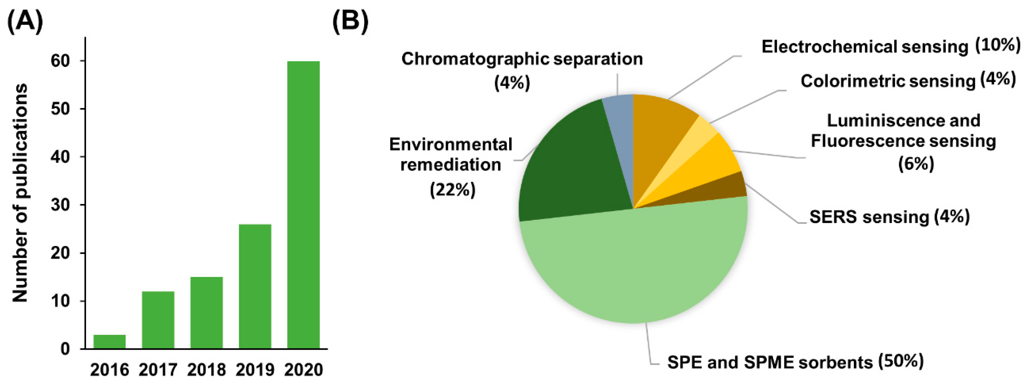
:1. Introduction
2. Synthesis of COF Composites
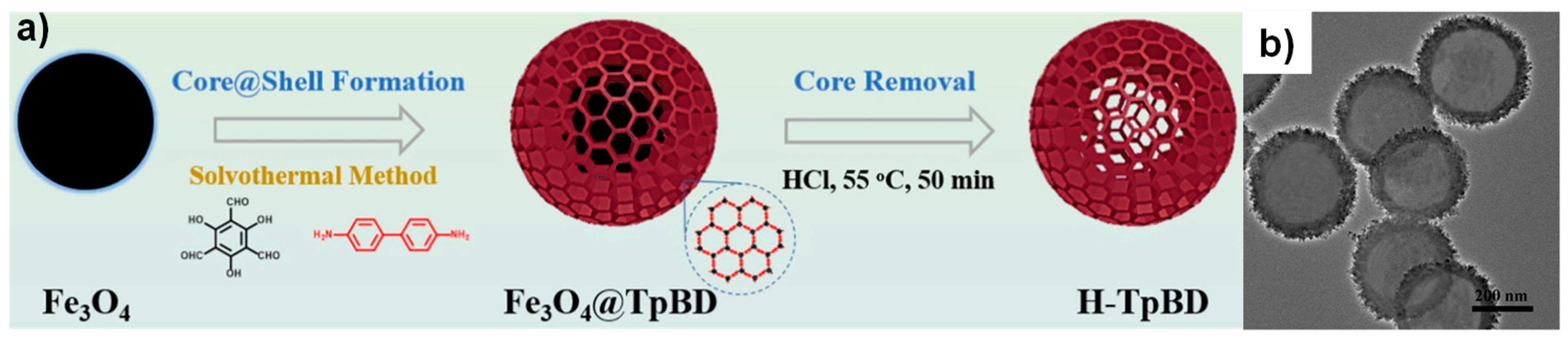
2.1. Iron Oxide—COF Composites
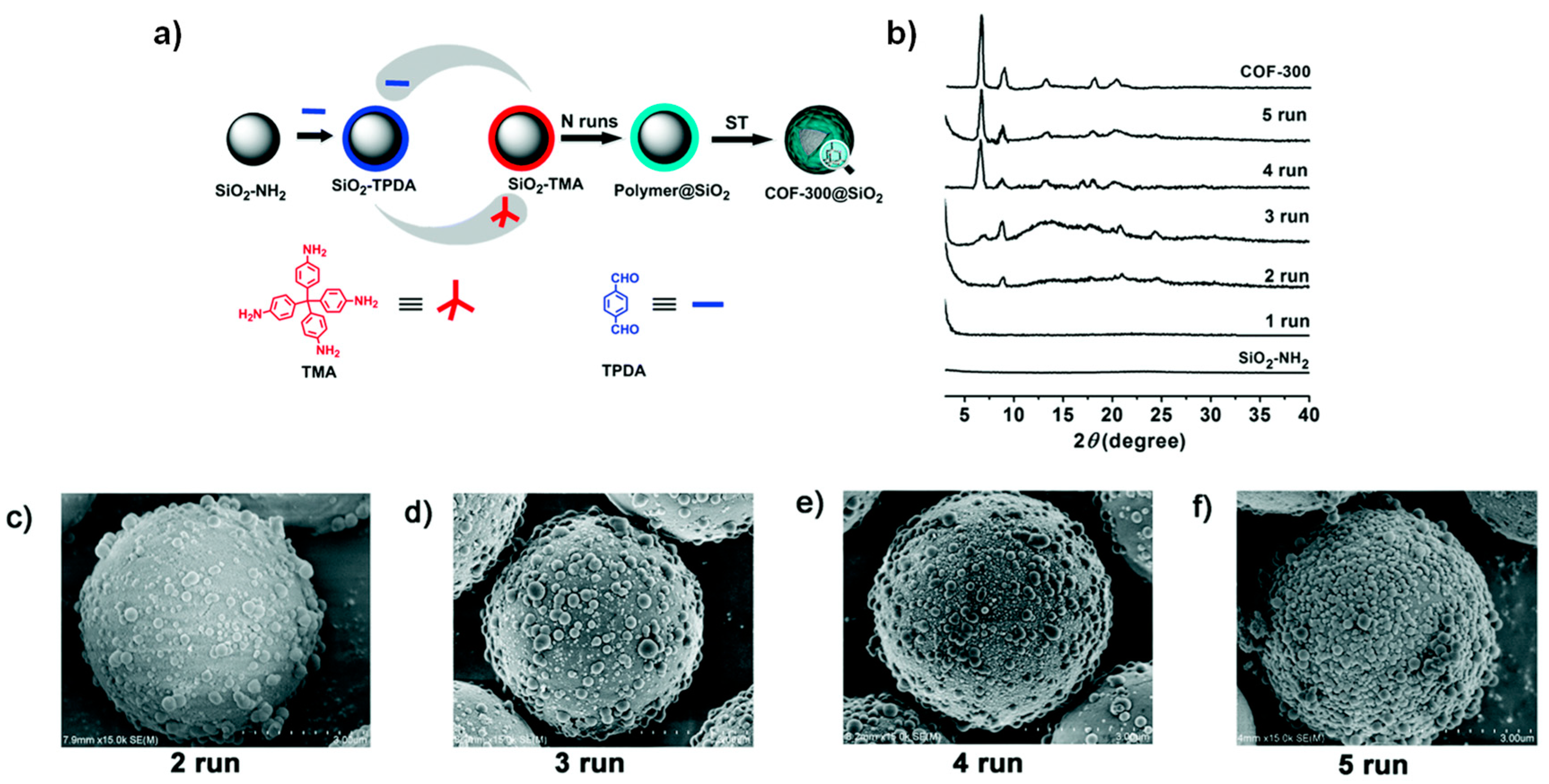
2.2. Silicon Dioxide–COF Composites
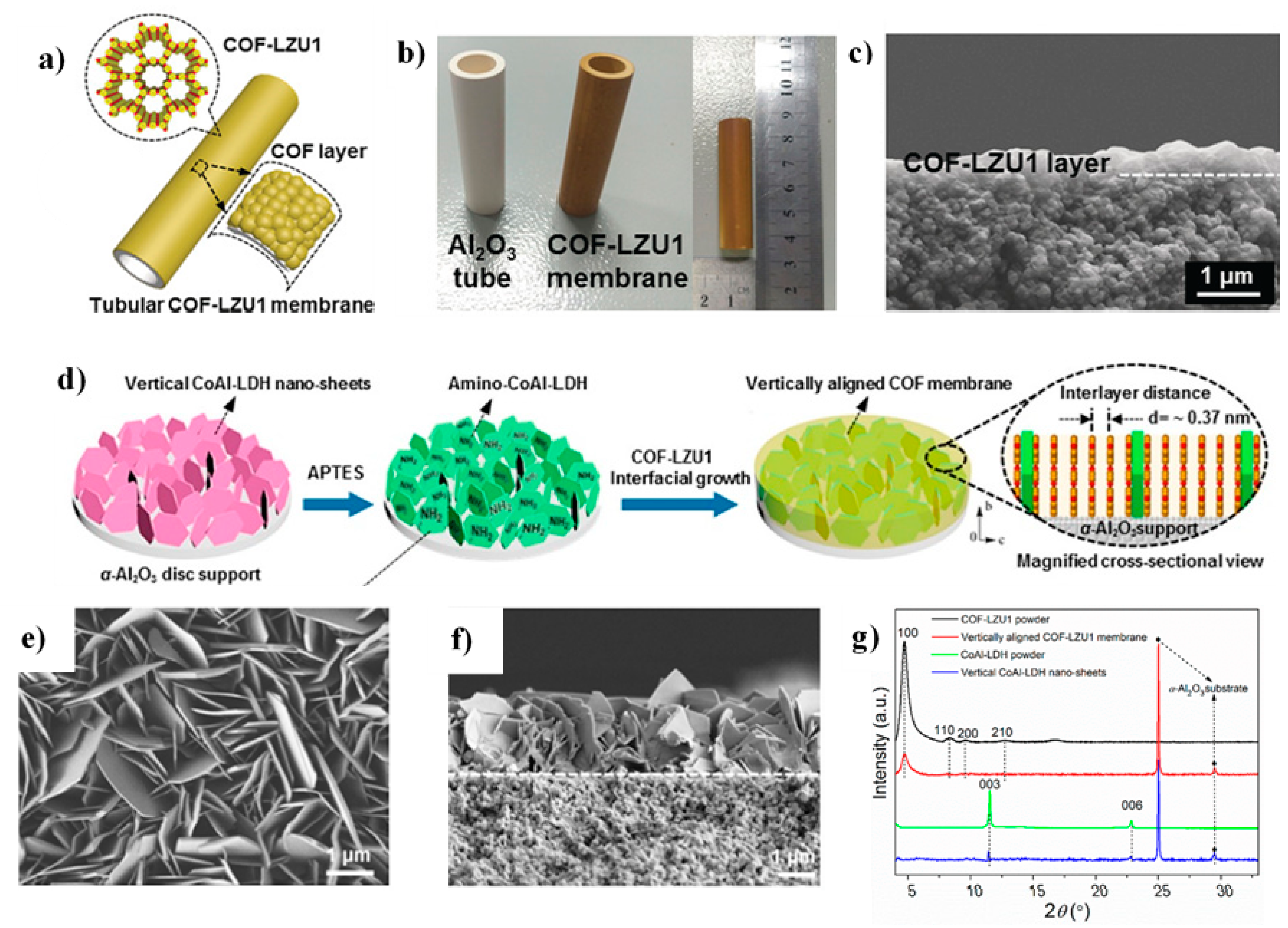
2.3. Aluminum Oxide–COF Composites
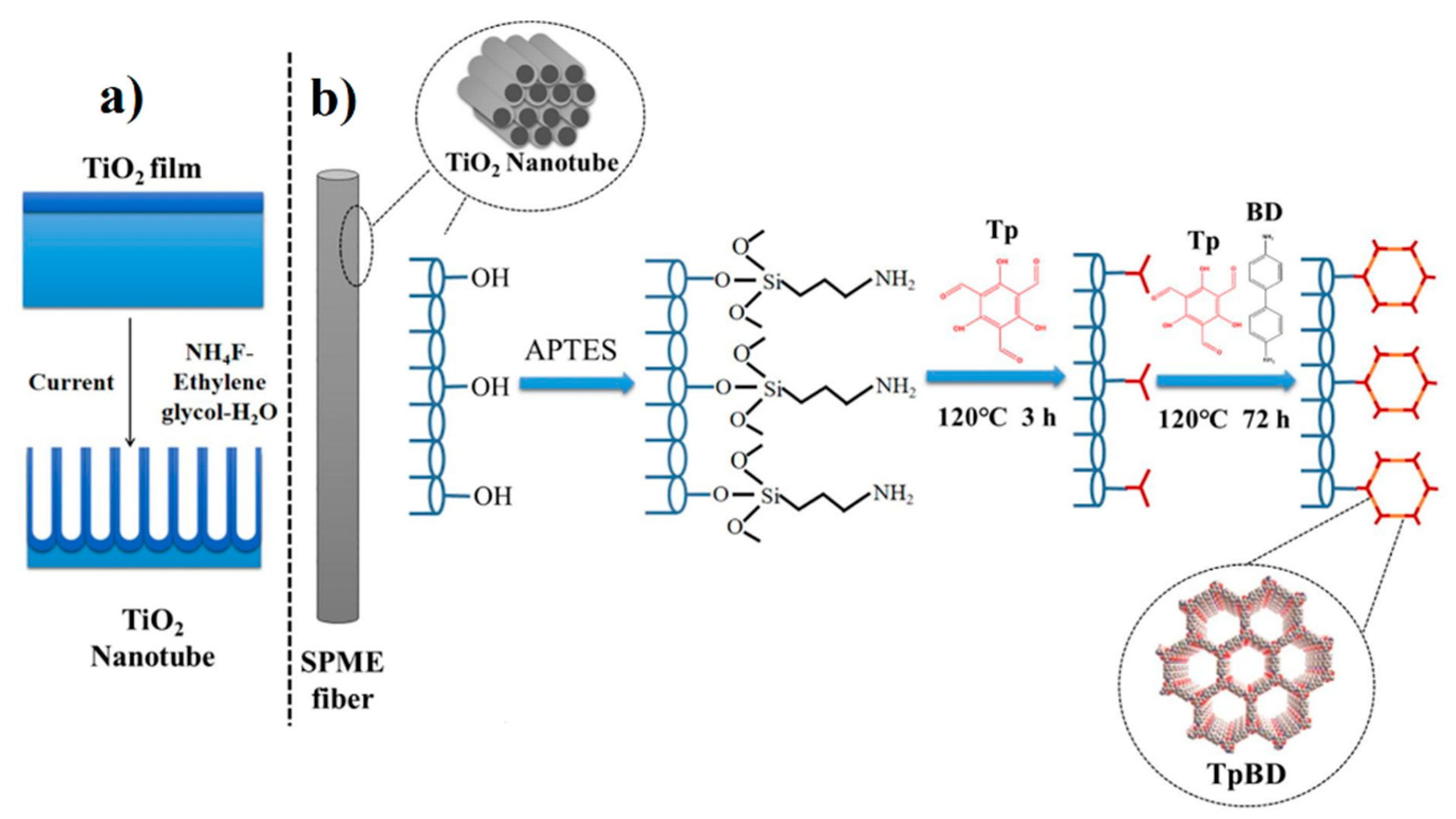
2.4. Titanium Dioxide–COF Composites
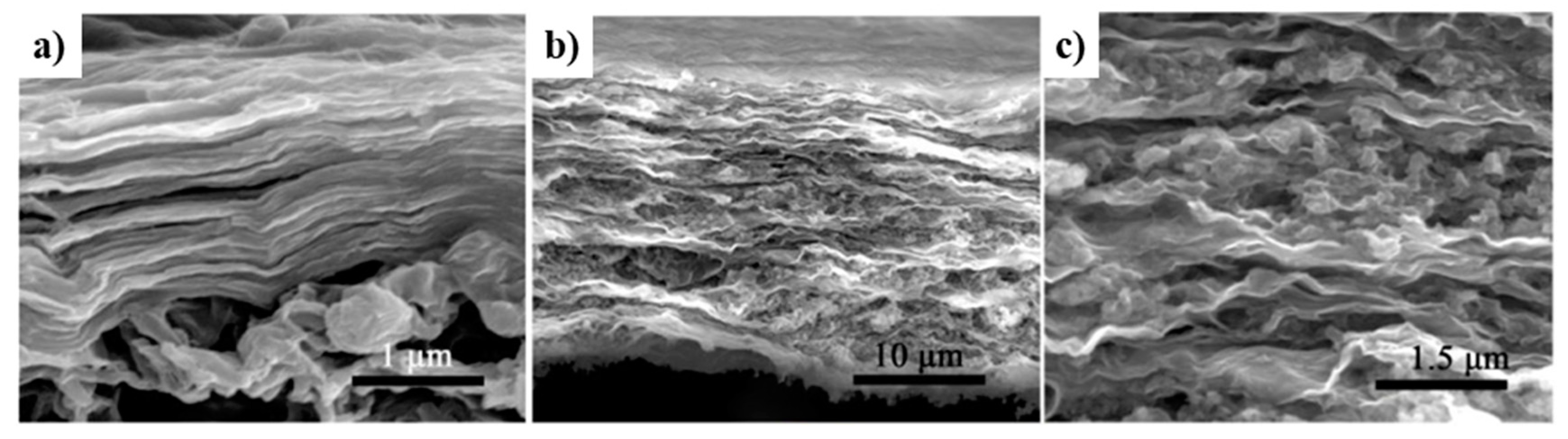
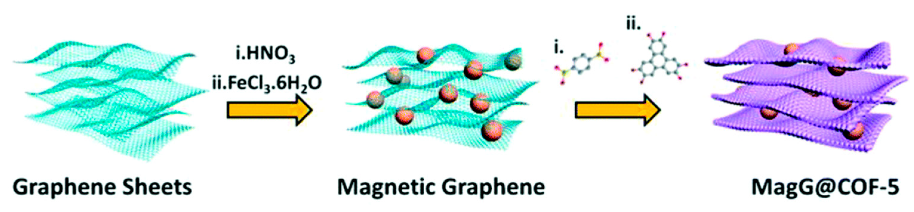
2.5. Graphene-Based COF Composites
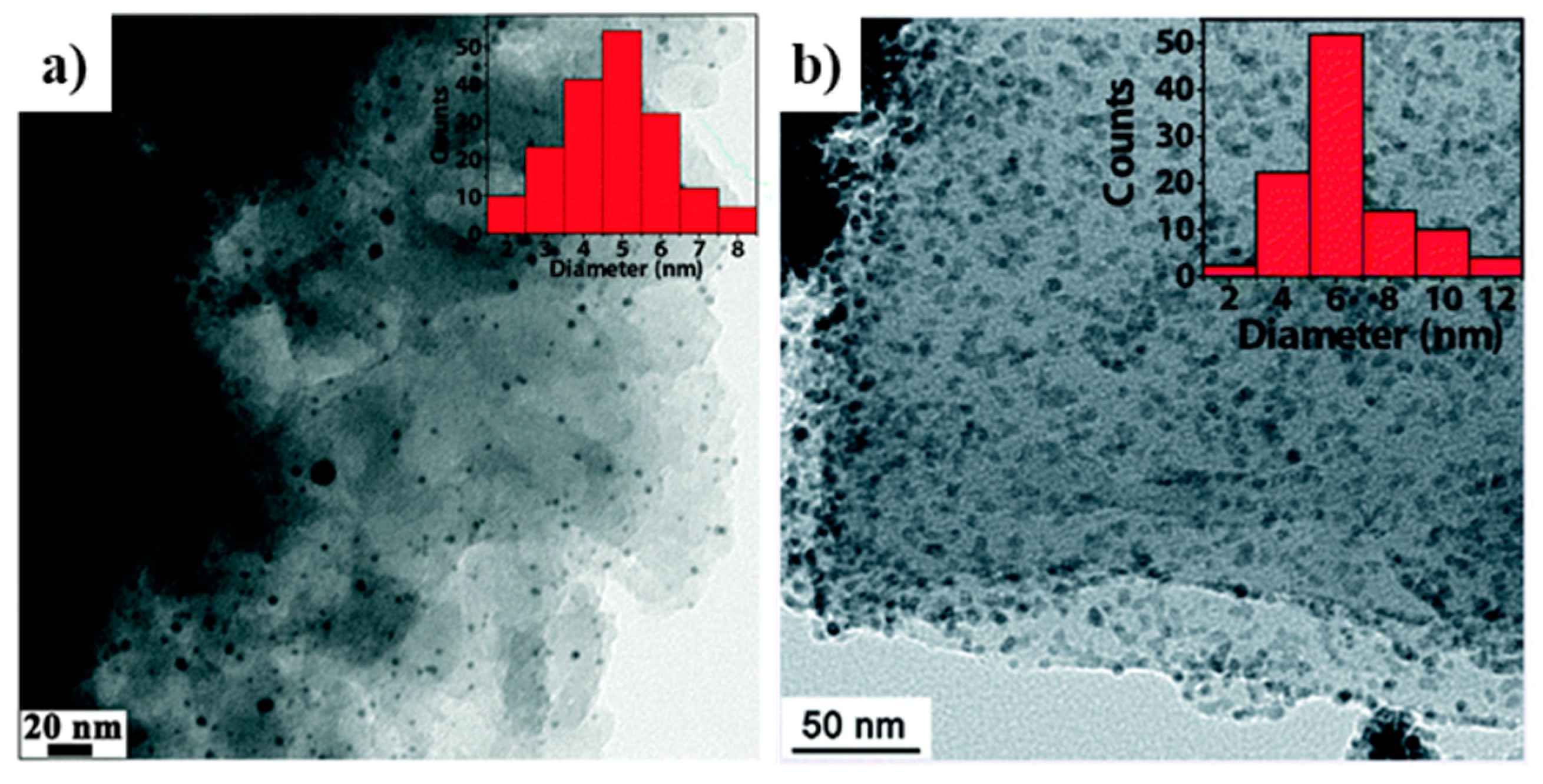
2.6. Metal Nanoparticle-COF Composites
2.7. Stainless-Steel COF Composites
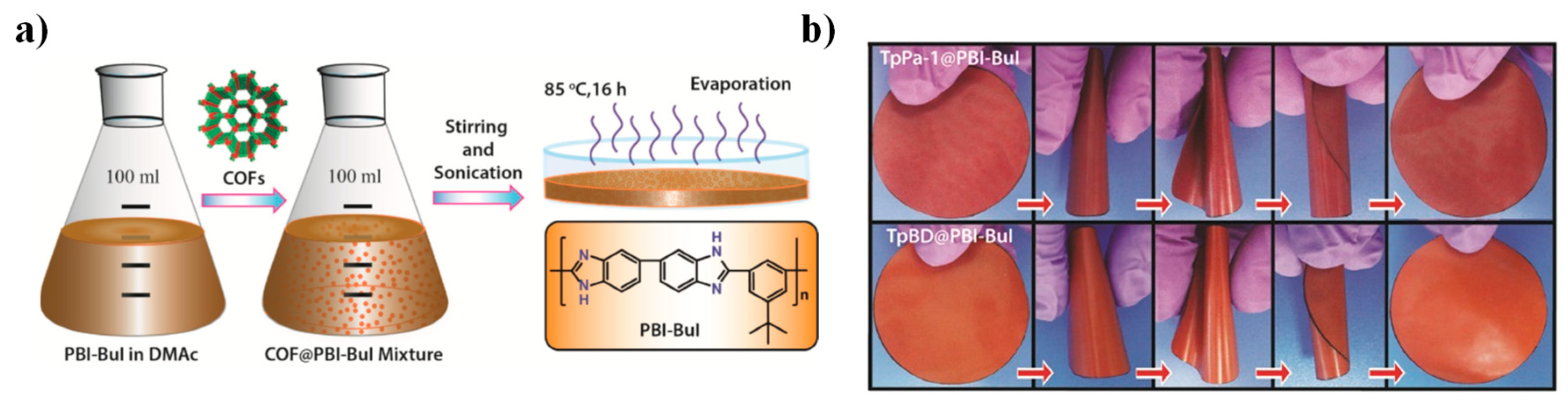
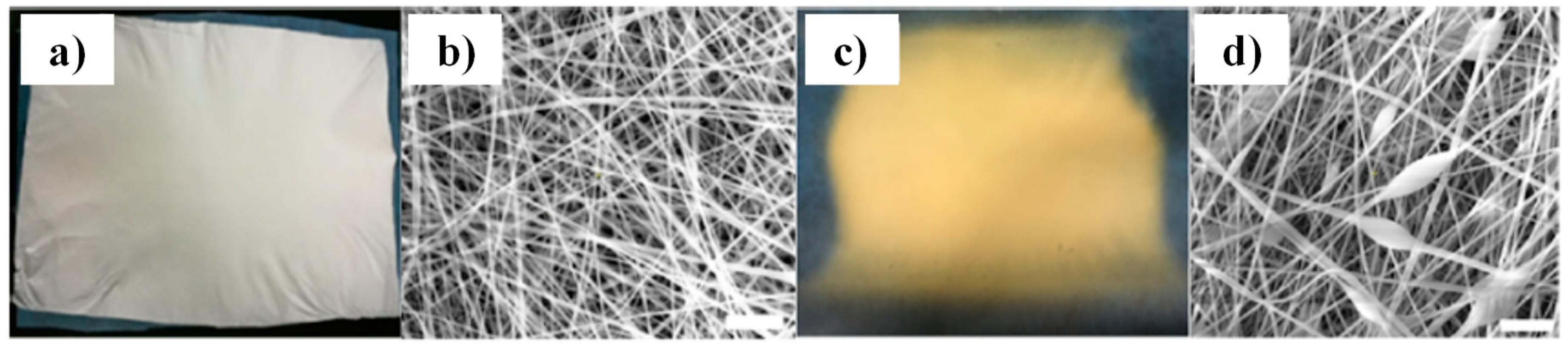
2.8. Others: COF Composites with Polymeric Substrates and MOFs
3. Analytical Applications
3.1. Applications for Separation
3.1.1. Chromatographic Separation
3.1.2. Environmental Remediation
3.1.3. Extraction and Microextraction Strategies for Sample Preparation
3.2. Applications for Sensing
3.2.1. Electrochemical Sensing
3.2.2. Luminescent and Colorimetric Sensing
3.2.3. Surface-Enhanced Raman Scattering
4. Conclusions and Outlook
Author Contributions
Funding
Acknowledgments
Conflicts of Interest
References
- Lohse, M.S.; Bein, T. Covalent Organic Frameworks: Structures, Synthesis, and Applications. Adv. Funct. Mater. 2018, 28. [Google Scholar] [CrossRef] [Green Version]
- Kandambeth, S.; Dey, K.; Banerjee, R. Covalent Organic Frameworks: Chemistry beyond the Structure. J. Am. Chem. Soc. 2019, 141, 1807–1822. [Google Scholar] [CrossRef]
- Chen, X.; Geng, K.; Liu, R.; Tan, K.T.; Gong, Y.; Li, Z.; Tao, S.; Jiang, Q.; Jiang, D. Covalent Organic Frameworks: Chemical Approaches to Designer Structures and Built-In Functions. Angew. Chem. Int. Ed. 2020, 59, 5050–5091. [Google Scholar] [CrossRef]
- Li, Y.; Chen, W.; Xing, G.; Jiang, D.; Chen, L. New synthetic strategies toward covalent organic frameworks. Chem. Soc. Rev. 2020, 49, 2852–2868. [Google Scholar] [CrossRef]
- Martínez-Abadía, M.; Mateo-Alonso, A. Structural Approaches to Control Interlayer Interactions in 2D Covalent Organic Frameworks. Adv. Mater. 2020, 32, 2002366. [Google Scholar] [CrossRef]
- Zhang, X.; Li, G.; Wu, D.; Zhang, B.; Hu, N.; Wang, H.; Liu, J.; Wu, Y. Recent advances in the construction of functionalized covalent organic frameworks and their applications to sensing. Biosens. Bioelectron. 2019, 145, 111699. [Google Scholar] [CrossRef] [PubMed]
- Liu, X.; Huang, D.; Lai, C.; Zeng, G.; Qin, L.; Wang, H.; Yi, H.; Li, B.; Liu, S.; Zhang, M.; et al. Recent advances in covalent organic frameworks (COFs) as a smart sensing material. Chem. Soc. Rev. 2019, 48, 5266–5302. [Google Scholar] [CrossRef] [PubMed]
- Samanta, P.; Desai, A.V.; Let, S.; Ghosh, S.K. Advanced Porous Materials for Sensing, Capture and Detoxification of Organic Pollutants toward Water Remediation. ACS Sustain. Chem. Eng. 2019, 7, 7456–7478. [Google Scholar] [CrossRef]
- Xue, R.; Guo, H.; Wang, T.; Gong, L.; Wang, Y.; Ai, J.; Huang, D.; Chen, H.; Yang, W. Fluorescence properties and analytical applications of covalent organic frameworks. Anal. Methods 2017, 9, 3737–3750. [Google Scholar] [CrossRef]
- Guo, H.; Zhang, L.; Xue, R.; Ma, B.; Yang, W. Eyes of covalent organic frameworks: Cooperation between analytical chemistry and COFs. Rev. Anal. Chem. 2019, 38. [Google Scholar] [CrossRef]
- González-Sálamo, J.; Jiménez-Skrzypek, G.; Ortega-Zamora, C.; González-Curbelo, M.Á.; Hernández-Borges, J. Covalent organic frameworks in sample preparation. Molecules 2020, 25, 3288. [Google Scholar] [CrossRef] [PubMed]
- Chen, L.; Wu, Q.; Gao, J.; Li, H.; Dong, S.; Shi, X.; Zhao, L. Applications of covalent organic frameworks in analytical chemistry. TrAC Trends Anal. Chem. 2019, 113, 182–193. [Google Scholar] [CrossRef]
- Maciel, E.V.S.; de Toffoli, A.L.; Neto, E.S.; Nazario, C.E.D.; Lanças, F.M. New materials in sample preparation: Recent advances and future trends. TrAC Trends Anal. Chem. 2019, 119, 115633. [Google Scholar] [CrossRef]
- Wang, J.; Zhuang, S. Covalent organic frameworks (COFs) for environmental applications. Coord. Chem. Rev. 2019, 400, 213046. [Google Scholar] [CrossRef]
- Li, N.; Du, J.; Wu, D.; Liu, J.; Li, N.; Sun, Z.; Li, G.; Wu, Y. Recent advances in facile synthesis and applications of covalent organic framework materials as superior adsorbents in sample pretreatment. TrAC Trends Anal. Chem. 2018, 108, 154–166. [Google Scholar] [CrossRef]
- Zheng, J.; Huang, J.; Yang, Q.; Ni, C.; Xie, X.; Shi, Y.; Sun, J.; Zhu, F.; Ouyang, G. Fabrications of novel solid phase microextraction fiber coatings based on new materials for high enrichment capability. TrAC Trends Anal. Chem. 2018, 108, 135–153. [Google Scholar] [CrossRef]
- Wang, J.; Li, J.; Gao, M.; Zhang, X. Recent advances in covalent organic frameworks for separation and analysis of complex samples. TrAC Trends Anal. Chem. 2018, 108, 98–109. [Google Scholar] [CrossRef]
- Zhang, J.; Chen, J.; Peng, S.; Peng, S.; Zhang, Z.; Tong, Y.; Miller, P.W.; Yan, X.-P. Emerging porous materials in confined spaces: From chromatographic applications to flow chemistry. Chem. Soc. Rev. 2019, 48, 2566–2595. [Google Scholar] [CrossRef]
- Qian, H.-L.; Yang, C.-X.; Wang, W.-L.; Yang, C.; Yan, X.-P. Advances in covalent organic frameworks in separation science. J. Chromatogr. A 2018, 1542. [Google Scholar] [CrossRef]
- Wang, X.; Ye, N. Recent advances in metal-organic frameworks and covalent organic frameworks for sample preparation and chromatographic analysis. Electrophoresis 2017, 38, 3059–3078. [Google Scholar] [CrossRef]
- Wang, Z.; Zhang, S.; Chen, Y.; Zhang, Z.; Ma, S. Covalent organic frameworks for separation applications. Chem. Soc. Rev. 2020, 49, 708–735. [Google Scholar] [CrossRef]
- Fernandes, S.P.S.; Romero, V.; Espiña, B.; Salonen, L.M. Tailoring Covalent Organic Frameworks to Capture Water Contaminants. Chem. Eur. J. 2019. [Google Scholar] [CrossRef] [PubMed]
- Li, Y.; Yang, C.X.; Yan, X.P. Controllable preparation of core-shell magnetic covalent-organic framework nanospheres for efficient adsorption and removal of bisphenols in aqueous solution. Chem. Commun. 2017, 53, 2511–2514. [Google Scholar] [CrossRef] [PubMed]
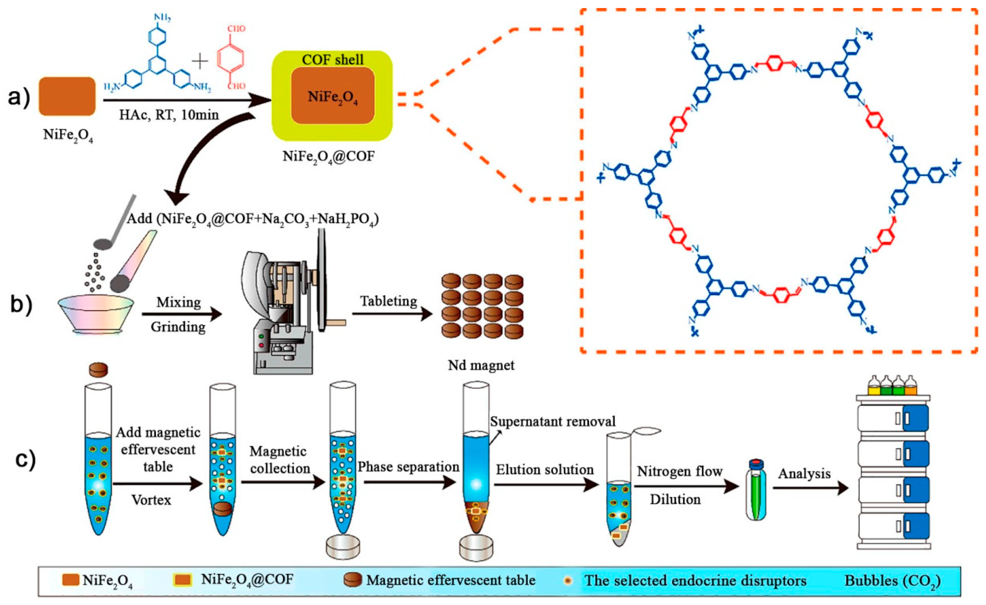
- Tan, C.; Li, J.; Liu, W.; Zhao, Q.; Wang, X.; Li, Y. Core-shell magnetic covalent organic framework nanocomposites as an adsorbent for effervescent reaction-enhanced microextraction of endocrine disruptors in liquid matrices. Chem. Eng. J. 2020, 396, 125191. [Google Scholar] [CrossRef]
- Hou, C.; Zhao, D.; Chen, W.; Li, H.; Zhang, S.; Liang, C. Covalent organic framework-functionalized magnetic CuFe2O4/Ag nanoparticles for the reduction of 4-nitrophenol. Nanomaterials 2020, 10, 426. [Google Scholar] [CrossRef] [PubMed] [Green Version]
- Arora, S.W. Superparamagnetic iron oxide nanoparticles: Magnetic nanoplatforms as drug carriers. Int. J. Nanomed. 2012, 7, 3445–3471. [Google Scholar] [CrossRef] [Green Version]
- He, S.; Zeng, T.; Wang, S.; Niu, H.; Cai, Y. Facile synthesis of magnetic covalent organic framework with three-dimensional bouquet-like structure for enhanced extraction of organic targets. ACS Appl. Mater. Interfaces 2017, 9, 2959–2965. [Google Scholar] [CrossRef]
- Wang, L.; Bao, J.; Wang, L.; Zhang, F.; Li, Y. One-pot synthesis and bioapplication of amine-functionalized magnetite nanoparticles and hollow nanospheres. Chem. Eur. J. 2006, 12, 6341–6347. [Google Scholar] [CrossRef]
- Romero, V.; Fernandes, S.P.S.; Rodriguez-Lorenzo, L.; Kolen’ko, Y.V.; Espiña, B.; Salonen, L.M. Recyclable magnetic covalent organic framework for the extraction of marine biotoxins. Nanoscale 2019, 11, 6072–6079. [Google Scholar] [CrossRef]
- Romero, V.; Fernandes, S.P.S.; Kovář, P.; Pšenička, M.; Kolen’ko, Y.V.; Salonen, L.M.; Espiña, B. Efficient adsorption of endocrine-disrupting pesticides from water with a reusable magnetic covalent organic framework. Microporous Mesoporous Mater. 2020, 307. [Google Scholar] [CrossRef]
- Liao, Y.; Li, J.; Thomas, A. General Route to High Surface Area Covalent Organic Frameworks and Their Metal Oxide Composites as Magnetically Recoverable Adsorbents and for Energy Storage. ACS Macro Lett. 2017, 6, 1444–1450. [Google Scholar] [CrossRef]
- Huang, L.; Mao, N.; Yan, Q.; Zhang, D.; Shuai, Q. Magnetic Covalent Organic Frameworks for the Removal of Diclofenac Sodium from Water. ACS Appl. Nano Mater. 2020, 3, 319–326. [Google Scholar] [CrossRef] [Green Version]
- Zhang, J.; Zhai, S.; Li, S.; Xiao, Z.; Song, Y.; An, Q.; Tian, G. Pb(II) removal of Fe3O4@SiO2-NH2 core-shell nanomaterials prepared via a controllable sol-gel process. Chem. Eng. J. 2013, 215, 461–471. [Google Scholar] [CrossRef]
- Pang, Y.H.; Yue, Q.; Huang, Y.; Yang, C.; Shen, X.F. Facile magnetization of covalent organic framework for solid-phase extraction of 15 phthalate esters in beverage samples. Talanta 2020, 206, 120194. [Google Scholar] [CrossRef] [PubMed]
- Rodríguez-San-Miguel, D.; Yazdi, A.; Guillerm, V.; Pérez-Carvajal, J.; Puntes, V.; Maspoch, D.; Zamora, F. Confining Functional Nanoparticles into Colloidal Imine-Based COF Spheres by a Sequential Encapsulation–Crystallization Method. Chem. Eur. J. 2017, 23, 8623–8627. [Google Scholar] [CrossRef] [PubMed]
- Tan, J.; Namuangruk, S.; Kong, W.; Kungwan, N.; Guo, J.; Wang, C. Manipulation of Amorphous-to-Crystalline Transformation: Towards the Construction of Covalent Organic Framework Hybrid Microspheres with NIR Photothermal Conversion Ability. Angew. Chem. Int. Ed. 2016, 55, 13979–13984. [Google Scholar] [CrossRef]
- Ma, W.F.; Zhang, Y.; Li, L.L.; You, L.J.; Zhang, P.; Zhang, Y.T.; Li, J.M.; Yu, M.; Guo, J.; Lu, H.J.; et al. Tailor-made magnetic Fe3O4@mTiO2 microspheres with a tunable mesoporous anatase shell for highly selective and effective enrichment of phosphopeptides. ACS Nano. 2012, 6, 3179–3188. [Google Scholar] [CrossRef]
- Xie, Y.; Zhang, T.; Chen, Y.; Wang, Y.; Wang, L. Fabrication of core-shell magnetic covalent organic frameworks composites and their application for highly sensitive detection of luteolin. Talanta 2020, 213, 120843. [Google Scholar] [CrossRef]
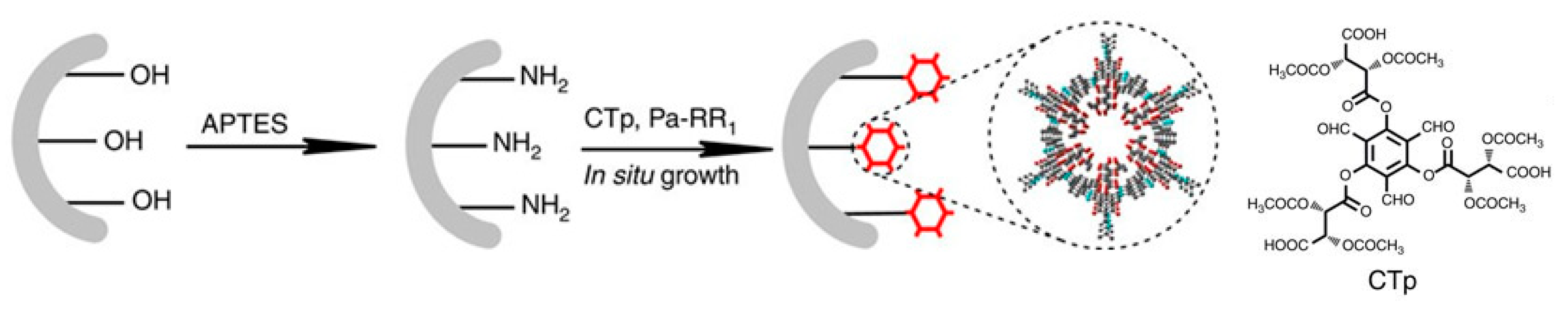
- Wang, L.L.; Yang, C.X.; Yan, X.P. In situ growth of covalent organic framework shells on silica microspheres for application in liquid chromatography. ChemPlusChem 2017, 82, 933–938. [Google Scholar] [CrossRef]
- Qian, H.L.; Yang, C.; Yan, X.P. Layer-by-layer preparation of 3D covalent organic framework/silica composites for chromatographic separation of position isomers. Chem. Commun. 2018, 54, 11765–11768. [Google Scholar] [CrossRef]
- Qian, H.L.; Yang, C.X.; Yan, X.P. Bottom-up synthesis of chiral covalent organic frameworks and their bound capillaries for chiral separation. Nat. Commun. 2016, 7. [Google Scholar] [CrossRef] [PubMed]
- Hao, D.; Zhang, J.; Lu, H.; Leng, W.; Ge, R.; Dai, X.; Gao, Y. Fabrication of a COF-5 membrane on a functionalized α-Al2O3 ceramic support using a microwave irradiation method. Chem. Commun. 2014, 50, 1462–1464. [Google Scholar] [CrossRef] [PubMed]
- Lu, H.; Wang, C.; Chen, J.; Ge, R.; Leng, W.; Dong, B.; Huang, J.; Gao, Y. A novel 3D covalent organic framework membrane grown on a porous α-Al2O3 substrate under solvothermal conditions. Chem. Commun. 2015, 51, 15562–15565. [Google Scholar] [CrossRef] [PubMed]
- Fan, H.; Gu, J.; Meng, H.; Knebel, A.; Caro, J. High-Flux Membranes Based on the Covalent Organic Framework COF-LZU1 for Selective Dye Separation by Nanofiltration. Angew. Chem. Int. Ed. 2018, 57, 4083–4087. [Google Scholar] [CrossRef] [PubMed]
- Fan, H.; Peng, M.; Strauss, I.; Mundstock, A.; Meng, H.; Caro, J. High-Flux Vertically Aligned 2D Covalent Organic Framework Membrane with Enhanced Hydrogen Separation. J. Am. Chem. Soc. 2020, 142, 6872–6877. [Google Scholar] [CrossRef]
- Yue, Q.; Huang, Y.Y.; Shen, X.F.; Yang, C.; Pang, Y.H. In situ growth of covalent organic framework on titanium fiber for headspace solid-phase microextraction of 11 phthalate esters in vegetables. Food Chem. 2020, 318, 126507. [Google Scholar] [CrossRef] [PubMed]
- Wang, C.; Gao, W.; Liu, N.; Xin, Y.; Liu, X.; Wang, X.; Tian, Y.; Chen, X.; Hou, B. Covalent Organic Framework Decorated TiO2 Nanotube Arrays for Photoelectrochemical Cathodic Protection of Steel. Corros. Sci. 2020, 176, 108920. [Google Scholar] [CrossRef]
- Wen, R.; Li, Y.; Zhang, M.; Guo, X.; Li, X.; Li, X.; Han, J.; Hu, S.; Tan, W.; Ma, L.; et al. Graphene-synergized 2D covalent organic framework for adsorption: A mutual promotion strategy to achieve stabilization and functionalization simultaneously. J. Hazard. Mater. 2018, 358, 273–285. [Google Scholar] [CrossRef]
- Kinloch, I.A.; Suhr, J.; Lou, J.; Young, R.J.; Ajayan, P.M. Composites with carbon nanotubes and graphene: An outlook. Science 2018, 362, 547–553. [Google Scholar] [CrossRef] [Green Version]
- Zhang, X.; Li, H.; Wang, J.; Peng, D.; Liu, J.; Zhang, Y. In-situ grown covalent organic framework nanosheets on graphene for membrane-based dye/salt separation. J. Membr. Sci. 2019, 581, 321–330. [Google Scholar] [CrossRef]
- Chen, L.; Wang, W.; Fang, Q.; Zuo, K.; Hou, G.; Ai, Q.; Li, Q.; Ci, L.; Lou, J. High performance hierarchically nanostructured graphene oxide/covalent organic framework hybrid membranes for stable organic solvent nanofiltration. Appl. Mater. Today 2020, 20. [Google Scholar] [CrossRef]
- Wang, H.; Jiao, F.; Gao, F.; Zhao, X.; Zhao, Y.; Shen, Y.; Zhang, Y.; Qian, X. Covalent organic framework-coated magnetic graphene as a novel support for trypsin immobilization. Anal. Bioanal. Chem. 2017, 409, 2179–2187. [Google Scholar] [CrossRef] [PubMed]
- Wang, J.; Li, J.; Gao, M.; Zhang, X. Self-assembling covalent organic framework functionalized magnetic graphene hydrophilic biocomposites as an ultrasensitive matrix for N-linked glycopeptide recognition. Nanoscale 2017, 9, 10750–10756. [Google Scholar] [CrossRef] [PubMed] [Green Version]
- Lu, Y.; Wang, B.; Wang, C.; Yan, Y.; Wu, D.; Liang, H.; Tang, K. A Covalent Organic Framework-Derived Hydrophilic Magnetic Graphene Composite as a Unique Platform for Detection of Phthalate Esters from Packaged Milk Samples. Chromatographia 2019, 82, 1089–1099. [Google Scholar] [CrossRef]
- Wang, L.; Xie, Y.; Yang, Y.; Liang, H.; Wang, L.; Song, Y. Electroactive Covalent Organic Frameworks / Carbon Nanotubes Composites for Electrochemical Sensing. ACS Appl. Nano Mater. 2020, 3, 1412–1419. [Google Scholar] [CrossRef]
- Pachfule, P.; Kandambeth, S.; Díaz Díaz, D.; Banerjee, R. Highly stable covalent organic framework-Au nanoparticles hybrids for enhanced activity for nitrophenol reduction. Chem. Commun. 2014, 50, 3169–3172. [Google Scholar] [CrossRef]
- Pachfule, P.; Panda, M.K.; Kandambeth, S.; Shivaprasad, S.M.; Díaz, D.D.; Banerjee, R. Multifunctional and robust covalent organic framework-nanoparticle hybrids. J. Mater. Chem. A 2014, 2, 7944–7952. [Google Scholar] [CrossRef]
- Shi, X.; Yao, Y.; Xu, Y.; Liu, K.; Zhu, G.; Chi, L.; Lu, G. Imparting Catalytic Activity to a Covalent Organic Framework Material by Nanoparticle Encapsulation. ACS Appl. Mater. Interfaces 2017, 9, 7481–7488. [Google Scholar] [CrossRef]
- Bhadra, M.; Sasmal, H.S.; Basu, A.; Midya, S.P.; Kandambeth, S.; Pachfule, P.; Balaraman, E.; Banerjee, R. Predesigned Metal-Anchored Building Block for in Situ Generation of Pd Nanoparticles in Porous Covalent Organic Framework: Application in Heterogeneous Tandem Catalysis. ACS Appl. Mater. Interfaces 2017, 9, 13785–13792. [Google Scholar] [CrossRef]
- Kalidindi, S.B.; Oh, H.; Hirscher, M.; Esken, D.; Wiktor, C.; Turner, S.; Van Tendeloo, G.; Fischer, R.A. Metal@COFs: Covalent organic frameworks as templates for pd nanoparticles and hydrogen storage properties of Pd@COF-102 hybrid material. Chem. Eur. J. 2012, 18, 10848–10856. [Google Scholar] [CrossRef]
- Wang, R.L.; Li, D.P.; Wang, L.J.; Zhang, X.; Zhou, Z.Y.; Mu, J.L.; Su, Z.M. The preparation of new covalent organic framework embedded with silver nanoparticles and its applications in degradation of organic pollutants from waste water. Dalton Trans. 2019, 48, 1051–1059. [Google Scholar] [CrossRef] [PubMed]
- Chen, G.J.; Li, X.B.; Zhao, C.C.; Ma, H.C.; Kan, J.L.; Xin, Y.B.; Chen, C.X.; Dong, Y.B. Ru Nanoparticles-Loaded Covalent Organic Framework for Solvent-Free One-Pot Tandem Reactions in Air. Inorg. Chem. 2018, 57, 2678–2685. [Google Scholar] [CrossRef] [PubMed]
- Lu, S.; Hu, Y.; Wan, S.; McCaffrey, R.; Jin, Y.; Gu, H.; Zhang, W. Synthesis of Ultrafine and Highly Dispersed Metal Nanoparticles Confined in a Thioether-Containing Covalent Organic Framework and Their Catalytic Applications. J. Am. Chem. Soc. 2017, 139, 17082–17088. [Google Scholar] [CrossRef] [PubMed]
- Wang, L.; Xu, H.; Qiu, Y.; Liu, X.; Huang, W.; Yan, N.; Qu, Z. Utilization of Ag nanoparticles anchored in covalent organic frameworks for mercury removal from acidic waste water. J. Hazard. Mater. 2020, 389, 121824. [Google Scholar] [CrossRef]
- Hu, C.; Zhang, Z.; Liu, S.; Liu, X.; Pang, M. Monodispersed CuSe Sensitized Covalent Organic Framework Photosensitizer with an Enhanced Photodynamic and Photothermal Effect for Cancer Therapy. ACS Appl. Mater. Interfaces 2019, 11, 23072–23082. [Google Scholar] [CrossRef]
- Koczkur, K.M.; Mourdikoudis, S.; Polavarapu, L.; Skrabalak, S.E. Polyvinylpyrrolidone (PVP) in nanoparticle synthesis. Dalton Trans. 2015, 44, 17883–17905. [Google Scholar] [CrossRef] [Green Version]
- Zhang, T.; Chen, Y.; Huang, W.; Wang, Y.; Hu, X. A novel AuNPs-doped COFs composite as electrochemical probe for chlorogenic acid detection with enhanced sensitivity and stability. Sens. Actuators B 2018, 276, 362–369. [Google Scholar] [CrossRef]
- Wen, L.; Wu, P.; Wang, L.-L.; Chen, L.-Z.; Wang, M.-L.; Wang, X.; Lin, J.-M.; Zhao, R.-S. Solid-phase microextraction using a β-ketoenamine-linked covalent organic framework coating for efficient enrichment of synthetic musks in water samples. Anal. Methods 2020, 12, 2434–2442. [Google Scholar] [CrossRef]
- Guo, J.-X.; Qian, H.-L.; Zhao, X.; Yang, C.; Yan, X.-P. In situ room-temperature fabrication of a covalent organic framework and its bonded fiber for solid-phase microextraction of polychlorinated biphenyls in aquatic products. J. Mater. Chem. A 2019, 7, 13249–13255. [Google Scholar] [CrossRef]
- Tian, Y.; Hou, Y.; Yu, Q.; Wang, X.; Tian, M. Layer-by-layer self-assembly of a novel covalent organic frameworks microextraction coating for analyzing polycyclic aromatic hydrocarbons from aqueous solutions via gas chromatography. J. Sep. Sci. 2020, 43, 896–904. [Google Scholar] [CrossRef]
- Li, W.; Huang, L.; Guo, D.; Zhao, Y.; Zhu, Y. Self-assembling covalent organic framework functionalized poly (styrene-divinyl benzene-glycidylmethacrylate) composite for the rapid extraction of non-steroidal anti-inflammatory drugs in wastewater. J. Chromatogr. A 2018, 1571, 76–83. [Google Scholar] [CrossRef] [PubMed]
- Biswal, B.P.; Chaudhari, H.D.; Banerjee, R.; Kharul, U.K. Chemically Stable Covalent Organic Framework (COF)-Polybenzimidazole Hybrid Membranes: Enhanced Gas Separation through Pore Modulation. Chem. Eur. J. 2016, 22, 4695–4699. [Google Scholar] [CrossRef] [PubMed]
- Wang, R.; Shi, X.; Zhang, Z.; Xiao, A.; Sun, S.P.; Cui, Z.; Wang, Y. Unidirectional diffusion synthesis of covalent organic frameworks (COFs) on polymeric substrates for dye separation. J. Membr. Sci. 2019, 586, 274–280. [Google Scholar] [CrossRef]
- Li, W.; Jiang, H.X.; Geng, Y.; Wang, X.H.; Gao, R.Z.; Tang, A.N.; Kong, D.M. Facile Removal of Phytochromes and Efficient Recovery of Pesticides Using Heteropore Covalent Organic Framework-Based Magnetic Nanospheres and Electrospun Films. ACS Appl. Mater. Interfaces 2020, 12, 20922–20932. [Google Scholar] [CrossRef]
- Xu, L.; Xu, J.; Shan, B.; Wang, X.; Gao, C. TpPa-2-incorporated mixed matrix membranes for efficient water purification. J. Membr. Sci. 2017, 526, 355–366. [Google Scholar] [CrossRef]
- Fu, J.; Das, S.; Xing, G.; Ben, T.; Valtchev, V.; Qiu, S. Fabrication of COF-MOF Composite Membranes and Their Highly Selective Separation of H2/CO2. J. Am. Chem. Soc. 2016, 138, 7673–7680. [Google Scholar] [CrossRef]
- Yang, H.; Cheng, X.; Cheng, X.; Pan, F.; Wu, H.; Liu, G.; Song, Y.; Cao, X.; Jiang, Z. Highly water-selective membranes based on hollow covalent organic frameworks with fast transport pathways. J. Membr. Sci. 2018, 565, 331–341. [Google Scholar] [CrossRef]
- Ma, M.; Du, Y.; Zhang, L.; Gan, J.; Yang, J. β-Cyclodextrin covalent organic framework–modified organic polymer monolith as a stationary phase for combined hydrophilic and hydrophobic aqueous capillary electrochromatographic separation of small molecules. Microchim. Acta 2020, 187, 385. [Google Scholar] [CrossRef]
- Wang, S.; Zhang, L.; Xiao, R.; Chen, H.; Chu, Z.; Zhang, W.; Liu, F. Fabrication of SiO2@COF5 microspheres and their application in high performance liquid chromatography. Anal. Methods 2018, 10, 1968–1976. [Google Scholar] [CrossRef]
- Zhang, K.; Cai, S.-L.; Yan, Y.-L.; He, Z.-H.; Lin, H.-M.; Huang, X.-L.; Zheng, S.-R.; Fan, J.; Zhang, W.-G. Construction of a hydrazone-linked chiral covalent organic framework–silica composite as the stationary phase for high performance liquid chromatography. J. Chromatogr. A 2017, 1519, 100–109. [Google Scholar] [CrossRef]
- Singh, J.; Dutta, T.; Kim, K.-H.; Rawat, M.; Samddar, P.; Kumar, P. “Green” synthesis of metals and their oxide nanoparticles: Applications for environmental remediation. J. Nanobiotechnol. 2018, 16, 84. [Google Scholar] [CrossRef]
- Baby, R.; Saifullah, B.; Hussein, M.Z. Carbon Nanomaterials for the Treatment of Heavy Metal-Contaminated Water and Environmental Remediation. Nanoscale Res. Lett. 2019, 14, 341. [Google Scholar] [CrossRef] [Green Version]
- Wang, Y.; Pan, C.; Chu, W.; Vipin, A.K.; Sun, L. Environmental remediation applications of carbon nanotubes and graphene oxide: Adsorption and catalysis. Nanomaterials 2019, 9, 439. [Google Scholar] [CrossRef] [Green Version]
- Wang, D.; Pillai, S.C.; Ho, S.-H.; Zeng, J.; Li, Y.; Dionysiou, D.D. Plasmonic-based nanomaterials for environmental remediation. Appl. Catal. B Environ. 2018, 237, 721–741. [Google Scholar] [CrossRef]
- Kumar, A.; Rana, A.; Sharma, G.; Sharma, S.; Naushad, M.; Mola, G.T.; Dhiman, P.; Stadler, F.J. Aerogels and metal–organic frameworks for environmental remediation and energy production. Environ. Chem. Lett. 2018, 16, 797–820. [Google Scholar] [CrossRef]
- Mahfoudhi, N.; Boufi, S. Nanocellulose as a novel nanostructured adsorbent for environmental remediation: A review. Cellulose 2017, 24, 1171–1197. [Google Scholar] [CrossRef]
- Zhang, N.; Ishag, A.; Li, Y.; Wang, H.; Guo, H.; Mei, P.; Meng, Q.; Sun, Y. Recent investigations and progress in environmental remediation by using covalent organic framework-based adsorption method: A review. J. Clean. Prod. 2020, 277, 123360. [Google Scholar] [CrossRef]
- Li, L.; Chen, R.; Li, Y.; Xiong, T.; Li, Y. Novel cotton fiber-covalent organic framework hybrid monolith for reversible capture of iodine. Cellulose 2020, 27, 5879–5892. [Google Scholar] [CrossRef]
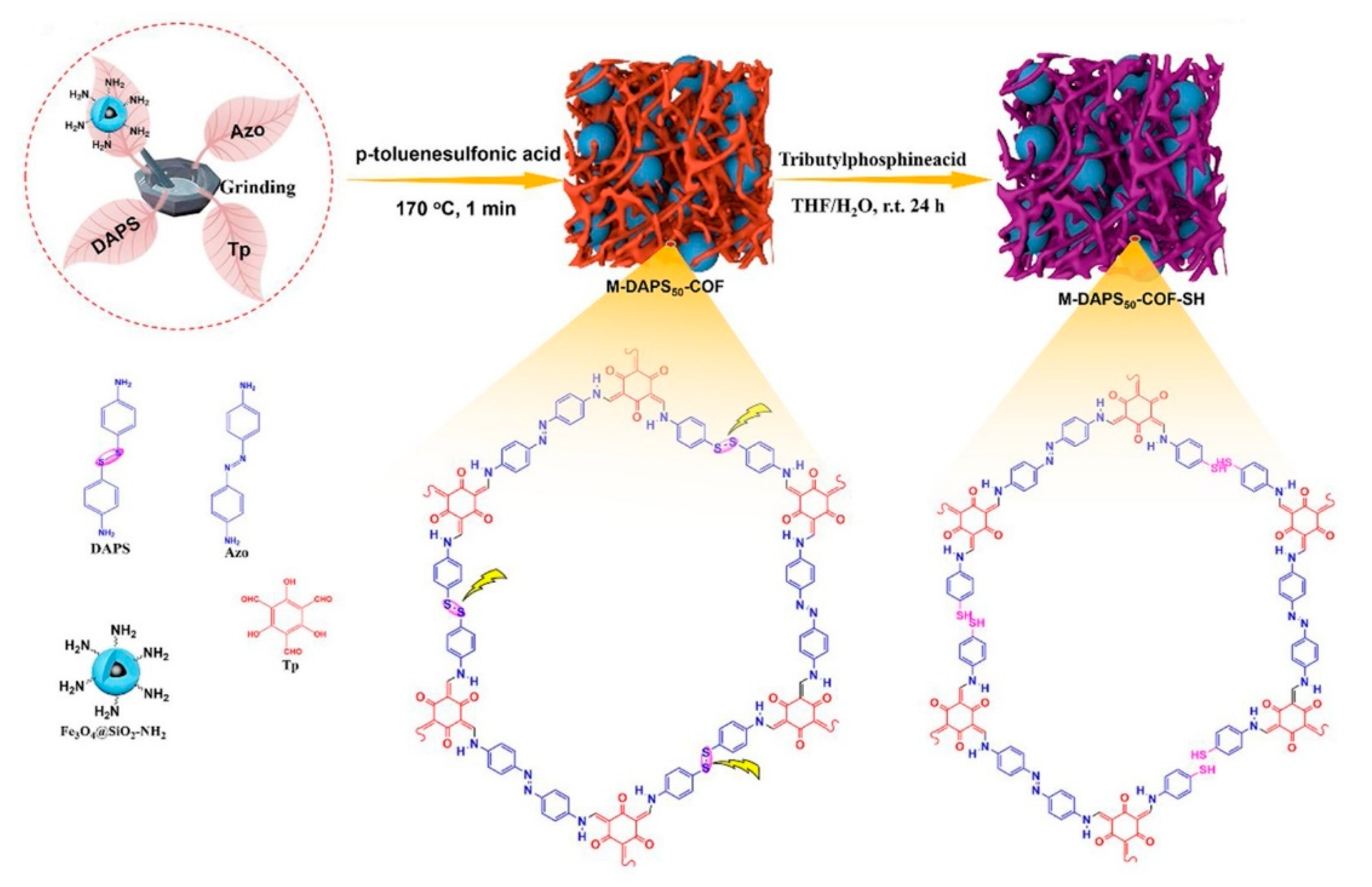
- Huang, L.; Shen, R.; Liu, R.; Shuai, Q. Thiol-functionalized magnetic covalent organic frameworks by a cutting strategy for efficient removal of Hg2+ from water. J. Hazard. Mater. 2020, 392, 122320. [Google Scholar] [CrossRef]
- Liu, X.; Xu, H.; Wang, L.; Qu, Z.; Yan, N. Surface nano-traps of Fe0/COFs for arsenic(III) depth removal from wastewater in non-ferrous smelting industry. Chem. Eng. J. 2020, 381, 122559. [Google Scholar] [CrossRef]
- Zhong, X.; Lu, Z.; Liang, W.; Hu, B. The magnetic covalent organic framework as a platform for high-performance extraction of Cr(VI) and bisphenol a from aqueous solution. J. Hazard. Mater. 2020, 393, 122353. [Google Scholar] [CrossRef] [PubMed]
- Mi, X.; Zhou, S.; Zhou, Z.; Vakili, M.; Qi, Y.; Jia, Y.; Zhu, D.; Wang, W. Adsorptive removal of diclofenac sodium from aqueous solution by magnetic COF: Role of hydroxyl group on COF. Colloids Surf. A 2020, 603, 125238. [Google Scholar] [CrossRef]
- Zhuang, S.; Chen, R.; Liu, Y.; Wang, J. Magnetic COFs for the adsorptive removal of diclofenac and sulfamethazine from aqueous solution: Adsorption kinetics, isotherms study and DFT calculation. J. Hazard. Mater. 2020, 385, 121596. [Google Scholar] [CrossRef] [PubMed]
- Zhang, J.; Chen, Z.; Tang, S.; Luo, X.; Xi, J.; He, Z.; Yu, J.; Wu, F. Fabrication of porphyrin-based magnetic covalent organic framework for effective extraction and enrichment of sulfonamides. Anal. Chim. Acta 2019, 1089, 66–77. [Google Scholar] [CrossRef]
- Li, Y.; Zhang, H.; Chen, Y.; Huang, L.; Lin, Z.; Cai, Z. Core-Shell Structured Magnetic Covalent Organic Framework Nanocomposites for Triclosan and Triclocarban Adsorption. ACS Appl. Mater. Interfaces 2019, 11, 22492–22500. [Google Scholar] [CrossRef]
- Firoozi, M.; Rafiee, Z.; Dashtian, K. New MOF/COF Hybrid as a Robust Adsorbent for Simultaneous Removal of Auramine O and Rhodamine B Dyes. ACS Omega 2020, 5, 9420–9428. [Google Scholar] [CrossRef] [Green Version]
- Sun, Q.; Aguila, B.; Perman, J.; Earl, L.D.; Abney, C.W.; Cheng, Y.; Wei, H.; Nguyen, N.; Wojtas, L.; Ma, S. Postsynthetically Modified Covalent Organic Frameworks for Efficient and Effective Mercury Removal. J. Am. Chem. Soc. 2017, 139, 2786–2793. [Google Scholar] [CrossRef]
- Mellah, A.; Fernandes, S.P.S.; Rodríguez, R.; Otero, J.; Paz, J.; Cruces, J.; Medina, D.D.; Djamila, H.; Espiña, B.; Salonen, L.M. Adsorption of Pharmaceutical Pollutants from Water Using Covalent Organic Frameworks. Chem. Eur. J. 2018, 24, 10601–10605. [Google Scholar] [CrossRef]
- Fernandes, S.P.S.; Mellah, A.; Kovář, P.; Sárria, M.P.; Pšenička, M.; Djamila, H.; Salonen, L.M.; Espiña, B. Extraction of Ibuprofen from Natural Waters Using a Covalent Organic Framework. Molecules 2020, 25, 3132. [Google Scholar] [CrossRef]
- Chandra, S.; Kandambeth, S.; Biswal, B.P.; Lukose, B.; Kunjir, S.M.; Chaudhary, M.; Babarao, R.; Heine, T.; Banerjee, R. Chemically stable multilayered covalent organic nanosheets from covalent organic frameworks via mechanical delamination. J. Am. Chem. Soc. 2013, 135, 17853–17861. [Google Scholar] [CrossRef]
- Salonen, L.M.; Pinela, S.R.; Fernandes, S.P.S.; Louçano, J.; Carbó-Argibay, E.; Sarriá, M.P.; Rodríguez-Abreu, C.; Peixoto, J.; Espiña, B. Adsorption of marine phycotoxin okadaic acid on a covalent organic framework. J. Chromatogr. A 2017, 1525, 17–22. [Google Scholar] [CrossRef] [Green Version]
- Pawliszyn, J.; Lord, H.L. Handbook of Sample Preparation; Wiley: Hoboken, NJ, USA, 2010; ISBN 9780813823621. [Google Scholar]
- Wang, H.; Li, Z.; Feng, W.; Jia, Q. Polymer monolith containing an embedded covalent organic framework for the effective enrichment of benzophenones. New J. Chem. 2017, 41, 13043–13050. [Google Scholar] [CrossRef]
- Yan, Z.; Hu, B.; Li, Q.; Zhang, S.; Pang, J.; Wu, C. Facile synthesis of covalent organic framework incorporated electrospun nanofiber and application to pipette tip solid phase extraction of sulfonamides in meat samples. J. Chromatogr. A 2019, 1584, 33–41. [Google Scholar] [CrossRef]
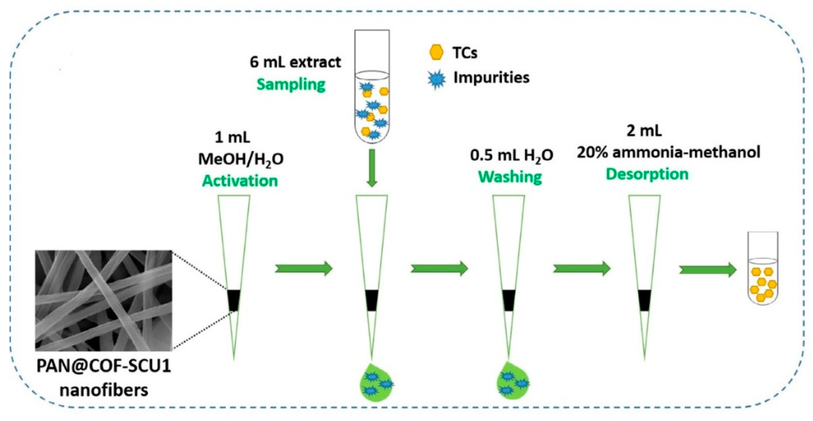
- Wang, R.; Li, C.; Li, Q.; Zhang, S.; Lv, F.; Yan, Z. Electrospinning fabrication of covalent organic framework composite nanofibers for pipette tip solid phase extraction of tetracycline antibiotics in grass carp and duck. J. Chromatogr. A 2020, 1622, 461098. [Google Scholar] [CrossRef]
- Chen, Y.; Chen, Z. COF-1-modified magnetic nanoparticles for highly selective and efficient solid-phase microextraction of paclitaxel. Talanta 2017, 165, 188–193. [Google Scholar] [CrossRef]
- Wang, R.; Chen, Z. A covalent organic framework-based magnetic sorbent for solid phase extraction of polycyclic aromatic hydrocarbons, and its hyphenation to HPLC for quantitation. Microchim. Acta 2017, 184, 3867–3874. [Google Scholar] [CrossRef]
- Li, N.; Wu, D.; Hu, N.; Fan, G.; Li, X.; Sun, J.; Chen, X.; Suo, Y.; Li, G.; Wu, Y. Effective Enrichment and Detection of Trace Polycyclic Aromatic Hydrocarbons in Food Samples based on Magnetic Covalent Organic Framework Hybrid Microspheres. J. Agric. Food Chem. 2018, 66, 3572–3580. [Google Scholar] [CrossRef]
- Yan, Y.; Lu, Y.; Wang, B.; Gao, Y.; Zhao, L.; Liang, H.; Wu, D. Self-Assembling Hydrophilic Magnetic Covalent Organic Framework Nanospheres as a Novel Matrix for Phthalate Ester Recognition. ACS Appl. Mater. Interfaces 2018, 10, 26539–26545. [Google Scholar] [CrossRef]
- Chen, L.; He, Y.; Lei, Z.; Gao, C.; Xie, Q.; Tong, P.; Lin, Z. Preparation of core-shell structured magnetic covalent organic framework nanocomposites for magnetic solid-phase extraction of bisphenols from human serum sample. Talanta 2018, 181, 296–304. [Google Scholar] [CrossRef]
- Chen, L.; Zhang, M.; Fu, F.; Li, J.; Lin, Z. Facile synthesis of magnetic covalent organic framework nanobeads and application to magnetic solid-phase extraction of trace estrogens from human urine. J. Chromatogr. A 2018, 1567, 136–146. [Google Scholar] [CrossRef]
- Deng, Z.-H.; Wang, X.; Wang, X.-L.; Gao, C.-L.; Dong, L.; Wang, M.-L.; Zhao, R.-S. A core-shell structured magnetic covalent organic framework (type Fe3O4@COF) as a sorbent for solid-phase extraction of endocrine-disrupting phenols prior to their quantitation by HPLC. Microchim. Acta 2019, 186, 108. [Google Scholar] [CrossRef] [PubMed]
- Zhang, W.; Lan, C.; Zhang, H.; Zhang, Y.; Zhang, W.; Zhao, W.; Johnson, C.; Hu, K.; Xie, F.; Zhang, S. Facile Preparation of Dual-Shell Novel Covalent-Organic Framework Functionalized Magnetic Nanospheres Used for the Simultaneous Determination of Fourteen Trace Heterocyclic Aromatic Amines in Nonsmokers and Smokers of Cigarettes with Different Tar Yields. J. Agric. Food Chem. 2019, 67, 3733–3743. [Google Scholar] [CrossRef] [PubMed]
- Liang, R.; Hu, Y.; Li, G. Photochemical synthesis of magnetic covalent organic framework/carbon nanotube composite and its enrichment of heterocyclic aromatic amines in food samples. J. Chromatogr. A 2020, 1618, 460867. [Google Scholar] [CrossRef] [PubMed]
- Wang, M.; Gao, M.; Zhang, K.; Wang, L.; Wang, W.; Fu, Q.; Xia, Z.; Gao, D. Magnetic covalent organic frameworks with core-shell structure as sorbents for solid phase extraction of fluoroquinolones, and their quantitation by HPLC. Microchim. Acta 2019, 186. [Google Scholar] [CrossRef] [PubMed]
- Wen, A.; Li, G.; Wu, D.; Yu, Y.; Yang, Y.; Hu, N.; Wang, H.; Chen, J.; Wu, Y. Sulphonate functionalized covalent organic framework-based magnetic sorbent for effective solid phase extraction and determination of fluoroquinolones. J. Chromatogr. A 2020, 1612, 460651. [Google Scholar] [CrossRef]
- Guan, S.; Wu, H.; Yang, L.; Wang, Z.; Wu, J. Use of a magnetic covalent organic framework material with a large specific surface area as an effective adsorbent for the extraction and determination of six fluoroquinolone antibiotics by HPLC in milk sample. J. Sep. Sci. 2020, 43, 3775–3784. [Google Scholar] [CrossRef]
- Fan, J.; Liu, Z.; Li, J.; Zhou, W.; Gao, H.; Zhang, S.; Lu, R. PEG-modified magnetic Schiff base network-1 materials for the magnetic solid phase extraction of benzoylurea pesticides from environmental water samples. J. Chromatogr. A 2020, 1619, 460950. [Google Scholar] [CrossRef]
- Lu, J.; Wang, R.; Luan, J.; Li, Y.; He, X.; Chen, L.; Zhang, Y. A functionalized magnetic covalent organic framework for sensitive determination of trace neonicotinoid residues in vegetable samples. J. Chromatogr. A 2020, 1618, 460898. [Google Scholar] [CrossRef]
- Liu, J.; Li, G.; Wu, D.; Yu, Y.; Chen, J.; Wu, Y. Facile preparation of magnetic covalent organic framework–metal organic framework composite materials as effective adsorbents for the extraction and determination of sedatives by high-performance liquid chromatography/tandem mass spectrometry in meat samp. Rapid Commun. Mass Spectrom. 2020, 34, e8742. [Google Scholar] [CrossRef]
- Wang, W.; Wang, J.; Zhang, S.; Cui, P.; Wang, C.; Wang, Z. A novel Schiff base network-1 nanocomposite coated fiber for solid-phase microextraction of phenols from honey samples. Talanta 2016, 161, 22–30. [Google Scholar] [CrossRef]
- Wang, M.; Zhou, X.; Zang, X.; Pang, Y.; Chang, Q.; Wang, C.; Wang, Z. Determination of pesticides residues in vegetable and fruit samples by solid-phase microextraction with a covalent organic framework as the fiber coating coupled with gas chromatography and electron capture detection. J. Sep. Sci. 2018, 41, 4038–4046. [Google Scholar] [CrossRef] [PubMed]
- Liu, L.; Meng, W.-K.; Zhou, Y.-S.; Wang, X.; Xu, G.J.; Wang, M.-L.; Lin, J.-M.; Zhao, R.-S. Β-Ketoenamine-linked covalent organic framework coating for ultra-high-performance solid-phase microextraction of polybrominated diphenyl ethers from environmental samples. Chem. Eng. J. 2019, 356, 926–933. [Google Scholar] [CrossRef]
- Hou, Y.-J.; Deng, J.; He, K.; Chen, C.; Yang, Y. Covalent Organic Frameworks-Based Solid-Phase Microextraction Probe for Rapid and Ultrasensitive Analysis of Trace Per- and Polyfluoroalkyl Substances Using Mass Spectrometry. Anal. Chem. 2020, 92, 10213–10217. [Google Scholar] [CrossRef] [PubMed]
- Zang, X.; Pang, Y.; Li, H.; Chang, Q.; Zhang, S.; Wang, C.; Wang, Z. Solid phase microextraction of polycyclic aromatic hydrocarbons from water samples by a fiber coated with covalent organic framework modified graphitic carbon nitride. J. Chromatogr. A 2020, 1628, 461428. [Google Scholar] [CrossRef]
- Wu, M.; Chen, G.; Liu, P.; Zhou, W.; Jia, Q. Polydopamine-based immobilization of a hydrazone covalent organic framework for headspace solid-phase microextraction of pyrethroids in vegetables and fruits. J. Chromatogr. A 2016, 1456, 34–41. [Google Scholar] [CrossRef]
- Wang, Q.; Wu, H.; Lv, F.; Cao, Y.; Zhou, Y.; Gan, N. A headspace sorptive extraction method with magnetic mesoporous titanium dioxide@covalent organic frameworks composite coating for selective determination of trace polychlorinated biphenyls in soils. J. Chromatogr. A 2018, 1572, 1–8. [Google Scholar] [CrossRef]
- Yang, X.; Wang, J.; Wang, W.; Zhang, S.; Wang, C.; Zhou, J.; Wang, Z. Solid phase microextraction of polycyclic aromatic hydrocarbons by using an etched stainless-steel fiber coated with a covalent organic framework. Microchim. Acta 2019, 186, 145. [Google Scholar] [CrossRef]
- Khataei, M.M.; Yamini, Y.; Ghaemmaghami, M. Reduced graphene-decorated covalent organic framework as a novel coating for solid-phase microextraction of phthalate esters coupled to gas chromatography-mass spectrometry. Microchim. Acta 2020, 187, 256. [Google Scholar] [CrossRef]
- Wu, T.; Zang, X.; Wang, M.; Chang, Q.; Wang, C.; Wu, Q.; Wang, Z. Covalent Organic Framework as Fiber Coating for Solid-Phase Microextraction of Chlorophenols Followed by Quantification with Gas Chromatography-Mass Spectrometry. J. Agric. Food Chem. 2018, 66, 11158–11165. [Google Scholar] [CrossRef]
- Zhang, G.; Ding, T.; Shi, Q.; Jiang, Z.; Niu, Y.; Zhang, M.; Tong, L.; Chen, Z.; Tang, B. Covalent organic frameworks-based paper solid phase microextraction combined with paper spray mass spectrometry for highly enhanced analysis of tetrabromobisphenol A. Analyst 2020. [Google Scholar] [CrossRef]
- Lin, X.; Wang, X.; Wang, J.; Yuan, Y.; Di, S.; Wang, Z.; Xu, H.; Zhao, H.; Qi, P.; Ding, W. Facile synthesis of a core-shell structured magnetic covalent organic framework for enrichment of organophosphorus pesticides in fruits. Anal. Chim. Acta 2020, 1101, 65–73. [Google Scholar] [CrossRef] [PubMed]
- Lin, X.-P.; Wang, X.-Q.; Wang, J.; Yuan, Y.-W.; Di, S.-S.; Wang, Z.-W.; Xu, H.; Zhao, H.-Y.; Zhao, C.-S.; Ding, W.; et al. Magnetic covalent organic framework as a solid-phase extraction absorbent for sensitive determination of trace organophosphorus pesticides in fatty milk. J. Chromatogr. A 2020, 1627, 461387. [Google Scholar] [CrossRef] [PubMed]
- Li, N.; Wu, D.; Li, X.; Zhou, X.; Fan, G.; Li, G.; Wu, Y. Effective enrichment and detection of plant growth regulators in fruits and vegetables using a novel magnetic covalent organic framework material as the adsorbents. Food Chem. 2020, 306, 125455. [Google Scholar] [CrossRef]
- Li, W.-T.; Shi, W.; Hu, Z.-J.; Yang, T.; Chen, M.-L.; Zhao, B.; Wang, J.-H. Fabrication of magnetic Fe3O4@metal organic framework@covalent organic framework composite and its selective separation of trace copper. Appl. Surf. Sci. 2020, 530, 147254. [Google Scholar] [CrossRef]
- Tan, X.; Fan, Y.; Wang, S.; Wu, Y.; Shi, W.; Huang, T.; Zhao, G. Ultrasensitive and highly selective electrochemical sensing of sodium picrate by Dihydroxylatopillar[6]arene-Modified gold nanoparticles and cationic Pillar[6]arene functionalized covalent organic framework. Electrochim. Acta 2020, 335, 135706. [Google Scholar] [CrossRef]
- Tan, X.; Zhang, Z.; Cao, T.; Zeng, W.; Huang, T.; Zhao, G. Control Assembly of Pillar[6]arene-Modified Ag Nanoparticles on Covalent Organic Framework Surface for Enhanced Sensing Performance toward Paraquat. ACS Sustain. Chem. Eng. 2019, 7, 20051–20059. [Google Scholar] [CrossRef]
- Zhang, C.; Cui, M.; Ren, J.; Xing, Y.; Li, N.; Zhao, H.; Liu, P.; Ji, X.; Li, M. Facile synthesis of novel spherical covalent organic frameworks integrated with Pt nanoparticles and multiwalled carbon nanotubes as electrochemical probe for tanshinol drug detection. Chem. Eng. J. 2020, 401, 126025. [Google Scholar] [CrossRef]
- Wang, Q.; Li, R.; Zhao, Y.; Zhe, T.; Bu, T.; Liu, Y.; Sun, X.; Hu, H.; Zhang, M.; Zheng, X.; et al. Surface morphology-controllable magnetic covalent organic frameworks: A novel electrocatalyst for simultaneously high-performance detection of p-nitrophenol and o-nitrophenol. Talanta 2020, 219, 121255. [Google Scholar] [CrossRef]
- Ma, B.; Guo, H.; Wang, M.; Wang, Q.; Yang, W.; Wang, Y.; Yang, W. Electrocatalysis and simultaneous determination of hydroquinone and acetaminophen using PN‒COF/graphene oxide modified electrode. Microchem. J. 2020, 155, 104776. [Google Scholar] [CrossRef]
- Sun, Y.; Waterhouse, G.I.N.; Xu, L.; Qiao, X.; Xu, Z. Three-dimensional electrochemical sensor with covalent organic framework decorated carbon nanotubes signal amplification for the detection of furazolidone. Sens. Actuators B. 2020, 321, 128501. [Google Scholar] [CrossRef]
- Sahiner, N.; Demirci, S. The use of covalent organic frameworks as template for conductive polymer synthesis and their sensor applications. J. Porous Mater. 2019, 26, 481–492. [Google Scholar] [CrossRef]
- Liang, C.; Lin, H.; Wang, Q.; Shi, E.; Zhou, S.; Zhang, F.; Qu, F.; Zhu, G. A redox-active covalent organic framework for the efficient detection and removal of hydrazine. J. Hazard. Mater. 2020, 381, 120983. [Google Scholar] [CrossRef] [PubMed]
- Li, J.; Zhang, C.; Yin, M.; Zhang, Z.; Chen, Y.; Deng, Q.; Wang, S. Surfactant-Sensitized Covalent Organic Frameworks-Functionalized Lanthanide-Doped Nanocrystals: An Ultrasensitive Sensing Platform for Perfluorooctane Sulfonate. ACS Omega 2019, 4, 15947–15955. [Google Scholar] [CrossRef] [PubMed] [Green Version]
- Guo, L.; Song, Y.; Cai, K.; Wang, L. “On-off” ratiometric fluorescent detection of Hg2+ based on N-doped carbon dots-rhodamine B@TAPT-DHTA-COF. Spectrochim. Acta Part A 2020, 227, 117703. [Google Scholar] [CrossRef] [PubMed]
- Tian, Y.; Lu, Q.; Guo, X.; Wang, S.; Gao, Y.; Wang, L. Au nanoparticles deposited on ultrathin two-dimensional covalent organic framework nanosheets for: In vitro and intracellular sensing. Nanoscale 2020, 12, 7776–7781. [Google Scholar] [CrossRef]
- Dong, J.; Li, X.; Peh, S.B.; Yuan, Y.D.; Wang, Y.; Ji, D.; Peng, S.; Liu, G.; Ying, S.; Yuan, D.; et al. Restriction of Molecular Rotors in Ultrathin Two-Dimensional Covalent Organic Framework Nanosheets for Sensing Signal Amplification. Chem. Mater. 2019, 31, 146–160. [Google Scholar] [CrossRef]
- Liu, H.; Zhang, Y.; Zhang, D.; Zheng, F.; Huang, M.; Sun, J.; Sun, X.; Li, H.; Wang, J.; Sun, B. A fluorescent nanoprobe for 4-ethylguaiacol based on the use of a molecularly imprinted polymer doped with a covalent organic framework grafted onto carbon nanodots. Microchim. Acta 2019, 186. [Google Scholar] [CrossRef]
- Wang, M.; Gao, M.; Deng, L.; Kang, X.; Zhang, K.; Fu, Q.; Xia, Z.; Gao, D. A sensitive and selective fluorescent sensor for 2,4,6-trinitrophenol detection based on the composite material of magnetic covalent organic frameworks, molecularly imprinted polymers and carbon dots. Microchem. J. 2020, 154, 104590. [Google Scholar] [CrossRef]
- Sun, P.; Hai, J.; Sun, S.; Lu, S.; Liu, S.; Liu, H.; Chen, F.; Wang, B. Aqueous stable Pd nanoparticles/covalent organic framework nanocomposite: An efficient nanoenzyme for colorimetric detection and multicolor imaging of cancer cells. Nanoscale 2020, 12, 825–831. [Google Scholar] [CrossRef]
- Cui, W.-R.; Zhang, C.-R.; Jiang, W.; Liang, R.-P.; Wen, S.-H.; Peng, D.; Qiu, J.-D. Covalent Organic Framework Nanosheet-Based Ultrasensitive and Selective Colorimetric Sensor for Trace Hg2+ Detection. ACS Sustain. Chem. Eng. 2019, 7, 9408–9415. [Google Scholar] [CrossRef]
- Li, W.; Li, Y.; Qian, H.-L.; Zhao, X.; Yang, C.-X.; Yan, X.-P. Fabrication of a covalent organic framework and its gold nanoparticle hybrids as stable mimetic peroxidase for sensitive and selective colorimetric detection of mercury in water samples. Talanta 2019, 204, 224–228. [Google Scholar] [CrossRef]
- He, J.; Xu, F.; Chen, Z.; Hou, X.; Liu, Q.; Long, Z. AuNPs/COFs as a new type of SERS substrate for sensitive recognition of polyaromatic hydrocarbons. Chem. Commun. 2017, 53, 11044–11047. [Google Scholar] [CrossRef] [PubMed]
- Su, Y.; Wu, D.; Chen, J.; Chen, G.; Hu, N.; Wang, H.; Wang, P.; Han, H.; Li, G.; Wu, Y. Ratiometric Surface Enhanced Raman Scattering Immunosorbent Assay of Allergenic Proteins via Covalent Organic Framework Composite Material Based Nanozyme Tag Triggered Raman Signal “turn-on” and Amplification. Anal. Chem. 2019, 91, 11687–11695. [Google Scholar] [CrossRef] [PubMed]
- Yao, D.; Li, C.; Wang, H.; Wen, G.; Liang, A.; Jiang, Z. A new dual-mode SERS and RRS aptasensor for detecting trace organic molecules based on gold nanocluster-doped covalent-organic framework catalyst. Sens. Actuators B 2020, 319, 128308. [Google Scholar] [CrossRef]
- Deblase, C.R.; Silberstein, K.E.; Truong, T.-T.; Abruña, H.D.; Dichtel, W.R. Β-Ketoenamine-Linked Covalent Organic Frameworks Capable of Pseudocapacitive Energy Storage. J. Am. Chem. Soc. 2013, 135, 16821–16824. [Google Scholar] [CrossRef] [PubMed]
- Wen, W.; Song, Y.; Yan, X.; Zhu, C.; Du, D.; Wang, S.; Asiri, A.M.; Lin, Y. Recent advances in emerging 2D nanomaterials for biosensing and bioimaging applications. Mater. Today 2018, 21, 164–177. [Google Scholar] [CrossRef]
- Liu, B.; Zhuang, J.; Wei, G. Recent advances in the design of colorimetric sensors for environmental monitoring. Environ. Sci. Nano 2020, 7, 2195–2213. [Google Scholar] [CrossRef]
- Prakash, J.; Kumar, V.; Kroon, R.E.; Asokan, K.; Rigato, V.; Chae, K.H.; Gautam, S.; Swart, H.C. Optical and surface enhanced Raman scattering properties of Au nanoparticles embedded in and located on a carbonaceous matrix. Phys. Chem. Chem. Phys. 2016, 18, 2468–2480. [Google Scholar] [CrossRef]
- Wang, H.; Jiang, X.; Lee, S.-T.; He, Y. Silicon nanohybrid-based surface-enhanced raman scattering sensors. Small 2014, 10, 4455–4468. [Google Scholar] [CrossRef]
- Zhang, N.; Tong, L.; Zhang, J. Graphene-based enhanced raman scattering toward analytical applications. Chem. Mater. 2016, 28, 6426–6435. [Google Scholar] [CrossRef]
- Lai, H.; Li, G.; Xu, F.; Zhang, Z. Metal-organic frameworks: Opportunities and challenges for surface-enhanced Raman scattering-a review. J. Mater. Chem. C 2020, 8, 2952–2963. [Google Scholar] [CrossRef]
Sample Availability: Samples of the compounds are not available from the authors. |


| Abbreviation | Name |
|---|---|
| BD | benzidine |
| BD-Me2 | o-tolidine |
| BPDA | [1,1′-biphenyl]-4,4′-dicarbaldehyde |
| Bpy | 5,5′-diamino-2,2′-bipyridine |
| DAAQ | 2,6-diaminoanthraquinone |
| DAB | 1,4-diaminobenzene |
| DHTA | 2,5-dihydroxyterephthalaldehyde |
| DMTP | 2,5-dimethoxyterephthalaldehyde |
| ETTA | 4,4′,4″,4‴-(ethane-1,1,2,2-tetrayl)tetraaniline |
| Pa-1 | p-phenylenediamine |
| Tp | 1,3,5-triformylphloroglucinol |
| TAM | tetra(4-anilyl)methane |
| TAPB | 1,3,5-tris(4-aminophenyl)benzene |
| TAPT | 2,4,6-tris(4-aminophenyl)-1,3,5-triazine |
| TFB, Tb | 1,3,5-triformylbenzene |
| TFPB | 1,3,5-tris(p-formylphenyl)benzene |
| TFTA | 2,3,5,6-tetrafluoroterephthalaldehyde |
| TMC | trimesoyl chloride |
| TPA | terephthaldicarboxaldehyde |
| TPDA | terephthalaldehyde |
| TPDH | terephthalic dihydrazide |
| COF Composite | Surface Area (m2·g‒1) | Analyte | Maximum Adsorption Capacity (mg·g‒1) | Contact Time (min) | Ref |
|---|---|---|---|---|---|
| Fe3O4/TpBD | 273 | Bisphenol-A | 161 | 5 | [23] |
| Bisphenol-AF | 237 | ||||
| Fe3O4/TpBD-Me2 | 538 | Okadaic acid | 812 | 120 | [29] |
| DTX-1 | 830 | ||||
| Fe3O4/TpBD-Me2 | 538 | Atrazine | 54 | 120 | [30] |
| Chlorpyrifos | 270 | ||||
| Fe3O4/TFBPa-1 COF | 872 | Iodine | 797 | n.d. | [31] |
| Fe3O4/TpPa-1 | 352 | Diclofenac | 565 | 20 | [32] |
| Al2O3/LZU-1 COF | n.d. | Organic dyes | n.d. | n.d. | [44] |
| Oxime-functionalized GS/TpDAAQ COF | 51 | Uranium | 144 | 20 | [48] |
| Au NPs/TpPa-1 | 339 | Nitrophenol | n.d. | 13 | [56] |
| Au NPs/TAPB-DMTP COF | 2388 | Nitrophenol | n.d. | 10 | [58] |
| Ag NPs/TPHH COF | 143 | Nitroaromatic compounds and organic dyes | n.d. | 2‒6 | [61] |
| Pt NPs/ThioCOF | 50 | Nitrophenol | n.d. | 8 | [63] |
| AgNPs/LZU-1 COF | 230 | Mercury | 113 | 120 | [64] |
| PVDF/TpPa-1 | n.d. | Organic dyes | n.d. | n.d. | [73] |
| Cotton fiber/TAPB-PDA COF | 166 | Iodine | 824 | 720 | [88] |
| Fe3O4/DAPS-SH COF | 182 | Mercury | 383 | 20 | [89] |
| Fe0/TAPB-PDA | 132 | Arsenic | 136 | 720 | [90] |
| Fe3O4/TpPa-1 | 485 | Chromium | 245 | 60 | [91] |
| Fe3O4/TAB-DHA | 212 | Diclofenac | 203 | 30 | [92] |
| Fe3O4/TAPB-DMTA | 2245 | Diclofenac | 40 | 50 | [93] |
| Sulfonamides | 55 | 80 | |||
| Fe3O4/pCOF | 31 | Sulfonamides | 2‒12 | 20 | [94] |
| Fe3O4/TPA-TAPB | 56 | Triclosan | 2 | 20 | [95] |
| Triclocarban | 1.5 | ||||
| MOF-5/melamine-based COF | 7 | Auramine-O | 18 | 8 | [96] |
| Rhodamine-B | 16 |
| COF Composite | Surface Area (m2·g‒1) | Analyte | Sample | Extraction Approach | Analytical Technique | LOD | Ref |
|---|---|---|---|---|---|---|---|
| NiFe2O4/TAPB-TPA COF | 52 | Endocrine disruptors | Urine and serum | Effervescent SPE | HPLC-FLD | 0.019–0.096 µg·L‒1 | [24] |
| Fe3O4/TpPa-1 | 248 | PAHs | Water | mSPE | HPLC-FLD | 0.24–1.01 ng L‒1 | [27] |
| Fe3O4/TpBD COF | n.d. | Phthalate esters | Beverages | mSPE | GC-MS/MS | 0.005–2.75 µg·L‒1 | [34] |
| TiO2/TpBD | 899 * | Phthalate esters | Food | HS-SPME | GC-MS/MS | 0.001–0.430 µg·L‒1 | [46] |
| Fe3O4/Graphene/COF-5 | 201 | Glycopeptides | Serum | mSPE | MALDI-TOF-MS | 0.5 fmol·µL‒1 | [53] |
| Fe3O4/TFBBD COF | 176 | Phthalate esters | Milk | mSPE | GC-MS | 4–20 ng·L‒1 | [54] |
| ss/TpPa-1 COF | 468 * | Synthetic musks | Water | DI-SPME | GC-MS/MS | 0.04–0.31 ng·L‒1 | [68] |
| ss/TFPB-BD COF | 286 * | Polychlorinated biphenyls | Food | HS-SPME | GC-MS/MS | 0.07–0.35 µg·L‒1 | [69] |
| ss/TFPA-TAPP COF | n.d. | PAHs | Water | HS-SPME | GC-FID | 6.1–24 ng·L‒1 | [70] |
| PS-GMA/TFBDABP COF | 404 | NSAIDs | Water | Syringe SPE | HPLC-UV | 0.13–0.82·µg L‒1 | [71] |
| PAN/BPDA-ETTA COF | 17 | Phytochromes | Food | TFME | HPLC-MS/MS | n.d. | [74] |
| Poly(GMA-DMA)/TMCPa-1 COF | n.d. | Benzophenones | Urine and serum | Microlumn SPE | HPLC-UV | 0.4–0.7 µg·L‒1 | [103] |
| PAN/SNW-1 COF | 205 | Sulfonamides | Food | PT-SPE | HPLC-PAD | 1.7–2.7 µg·L‒1 | [104] |
| PAN/TFBPa-1 COF | 47 | Tetracycline antibiotics | Food | PT-SPE | HPLC-PAD | 0.6–3 µg·L‒1 | [105] |
| Fe3O4/COF-1 | n.d. | Paclitaxel | Plasma | mSPE | HPLC-UV | 0.02 µg·L‒1 | [106] |
| Fe3O4/LZU-1 COF | n.d. | PAHs | Soil | mSPE | HPLC-UV | 0.2–20 ng·L‒1 | [107] |
| Fe3O4/TpBD COF | 114 | PAHs | Food | mSPE | HPLC-PAD | 0.83–11.7 ng·L‒1 | [108] |
| Fe3O4/TFBBD COF | 147 | Phthalate esters | Plasma | mSPE | GC-MS | 2.5–10 ng·L‒1 | [109] |
| Fe3O4/TAPB-TPA COF | 181 | Bisphenols | Serum | mSPE | HPLC-MS | 1–78 ng·L‒1 | [110] |
| Fe3O4/TFBBD COF | 202 | Estrogens | Urine | mSPE | HPLC-MS | 0.2–7.7 ng·L‒1 | [111] |
| Fe3O4/TAPB-TPA COF | 179 | BPA and BPAF | Tea | mSPE | HPLC-FLD | 0.08–0.21 µg·L‒1 | [112] |
| Fe3O4/TpBD COF | 171 | Heterocyclic aromatic amines | Urine | mSPE | HPLC-MS/MS | 0.14–0.46 ng·L‒1 | [113] |
| CNTs/CTC-COF | 430 | Heterocyclic aromatic amines | Food | dSPE | HPLC-MS/MS | 5.8–25 ng·kg‒1 | [114] |
| Fe3O4/TFPB-DATP COF | 124 | Fluoroquinolone antibiotics | Food and serum | mSPE | HPLC-PAD | 0.25–0.5 µg·kg‒1 | [115] |
| Fe3O4/TpBD-Au NPs | 70 | Fluoroquinolone antibiotics | Food | mSPE | HPLC-MS/MS | 0.1–1 µg·kg‒1 | [116] |
| Fe3O4/TpPa-1 COF | 506 | Fluoroquinolone antibiotics | Milk | mSPE | HPLC-UV | 0.05–0.20 µg·L‒1 | [117] |
| Fe3O4/SNW-1 COF | 157 | Benzoylurea pesticides | Wastewaters | mSPE | HPLC-PAD | 0.4–1 µg·L‒1 | [118] |
| Fe3O4/TpBD-NO2 COF | 255 | Neonicotinoid pesticides | Food | mSPE | HPLC-UV | 0.02–0.05 µg·L‒1 | [119] |
| Fe3O4-ZIF8/TFBBD COF | 84 | Sedatives | Meat | mSPE | HPLC-MS/MS | 0.04–0.2 µg·kg‒1 | [120] |
| ss/SNW1 COF | 668 * | Phenols | Honey | DI-SPME | GC-MS | 0.04–0.5 µg·kg‒1 | [121] |
| ss/TpPa-NO2 COF | n.d. | Pesticides | Food | DI-SPME | GC-ECD | 0.04–0.25 µg·kg‒1 | [122] |
| ss/TpPa-1 COF | 625 * | Polybrominated diphenyl ethers | Water | DI-SPME | GC-NCI-MS | 5.8–22 µg·L‒1 | [123] |
| ss/TFTA-TAPT COF | n.d. | Polyfluoroalkyl substances | Water | DI-SPME | nanoESI-MS | 0.02–0.8 ng·L‒1 | [124] |
| Carbon nitride/TpBD | 136 | PAHs | Water | DI-SPME | GC/MS | 0.02–0.05 µg·L‒1 | [125] |
| ss/TFBTPDH COF | 722 * | Pyrethroids | Food | HS-SPME | GC-ECD | 0.11–0.23 µg·kg‒1 | [126] |
| Fe3O4-TiO2/TAPB-TPA COF | 167 | Polychlorinated biphenyls | Soils | HS-SE | GC-MS | 3–6 ng·kg‒1 | [127] |
| ss/TAPB-TMC COF | 73 * | PAHs | Water | HS-SPME | GC-MS | 0.21–0.94 ng·L‒1 | [128] |
| ss/rGO-SNW1 COF | 116 | Phthalate esters | Water | HS-SPME | GC-MS | 0.01–0.5 µg·L‒1 | [129] |
| ss/TpBD | 967 * | Chlorophenols | Food | HS-SPME | GC-MS | 0.3–1.8 µg·kg‒1 | [130] |
| Cellulose paper/TpBD COF | 248 * | Tetrabromobisphenol-A | Water | TFME | PS-MS | 0.54 µg·L‒1 | [131] |
| COF Composite | Synthesis | Sample | Analyte | Sensing Technique | LOD | Ref |
|---|---|---|---|---|---|---|
| TAPB-DMTP COF/Fe3O4 NPs | Synthesis of COF in the presence of Fe3O4 nanoparticles | Food | Luteolin | Electrochemical | 0.0072 µM | [38] |
| Thi-TFPB-COF/CNTs | Synthesis of Thi-TFPB-COF in the presence of CNTs. | Drugs | Ascorbic acid | Electrochemical | 16.68 µM | [55] |
| TAPB-DMTP COF/Au NPs | In situ synthesis of Au NPs on TAPB-DMTP COF surface. | Food | Chlorogenic acid | Electrochemical | 0.0095 µM | [67] |
| 2HP6@Au-CP6@TpPa-1 | Synthesis of TpPa-1 and functionalization with CP6. Assembly of 2HP6@Au NPs on the COF surface. | Water and soil | Sodium picrate | Electrochemical | 0.0017 µM | [136] |
| TpPa-1/WP6@AgNPs | Synthesis of the TpPa-1 and WP6@AgNPs followed assembling of the nanoparticles on the COF surface. | Water | Paraquat | Electrochemical | 0.014 µM | [137] |
| PtNPs@TpBD/MWCNTs | Synthesis of Pt NPs inside the TpBD COF pores, followed by loading onto MWCNTs surface by sonication. | Drugs | Tanshinol | Electrochemical | 0.018 µM | [138] |
| TpTAPB COF/Fe3O4 NPs | Synthesis of COF in the presence of Fe3O4 nanoparticles | Water | p-nitrophenol | Electrochemical | 0.2361 µM | [139] |
| o-nitrophenol | 0.6568 µM | |||||
| PN-COF/GO | Synthesis of PN-COF followed by the mixture with GO in DMF | Drugs | Acetaminophen | Electrochemical | 0.011 µM | [140] |
| Cosmetics | Hydroquinone | 0.009 µM | ||||
| TAPB-TPA COF/NH2-CNTs | Synthesis of COF in the presence of NH2-CNTs. | Food | Furazolidone | Electrochemical | 0.0775 µM | [141] |
| COF-1,4/PANi | Loading of monomers ANi and Py inside COF pores followed in situ polymerization. | Air | HCl and NH3 gases | Electrochemical | n.d. | [142] |
| COF-1,4/PPy | ||||||
| TpDAAQ/Ni | Loading of DQ-COF onto Ni matrix by drop-casting | Water | Hydrazine | Electrochemical | 0.07 µM | [143] |
| UCNPs@LZU1 COF | Synthesis of COF-LZU1 on the surface of amine functionalized UCNPs | Water and food packing | PFOS | Fluorescence | 0.15 pM | [144] |
| CDs/RhB@PT-DHTA COF | First, synthesis of PT-DHTA COF functionalized with rhodamine B. Second grafting of CDs onto the COF surface. | Water and cosmetics | Hg (II) | Fluorescence | 0.016 µM | [145] |
| TFPA-TAPA COF/Au NPs | In situ synthesis of Au NPs on TFPA-TAPA COF nanosheets surface. | n.d. | Hepatitis A virus | Fluorescence | 15 pM | [146] |
| PVDF/NUS-30 COF | Fabrication of PVDF/NUS 30 mixed membrane by electrospinning. | n.d. | Amino acids | Fluorescence | n.d. | [147] |
| CDs-MIP/TpPa-1 COF | Coating of CNs with a MIP shell and simultaneous loading on COF surface. | Wine | 4-ethylguaiacol | Fluorescence | 0.11 µM | [148] |
| Fe3O4/TFTA-TAPB COF/MIP/CDs | Loading of CDs on the magnetic COF followed in situ polymerization for coating with the MIP (reverse microemulsion method). | Water | 2,4,6-trinitrophenol | Fluorescence | 0.0001 µM | [149] |
| FA-Pd NPs/CMC-LZU1 COF | First synthesis of COF-LZU1 in the presence of CMC. Second, in situ synthesis of Pd NPs on the CMC-LZU1 composite. Finally, functionalization of the immobilized Pd NPs with FA. | Serum | HeLa cancer cells | Colorimetric | 100 HeLa cells per mL | [150] |
| Au NPs/TpBpy COF nanosheets | Synthesis of TpBpy COF followed by in situ synthesis Au NPs on the COF surface. | Water | Hg(II) | Colorimetric | 0.33 nM | [151] |
| Au NPs/PTAzo COF | Synthesis of PTAzo COF followed by in situ synthesis Au NPs on the COF surface. | Water | Hg (II) | Colorimetric | 0.75 nM | [152] |
| Au NPs/SNW-1 COF | Self-assembly by mixing the obtained stock solution of SNW-1 and Au NPs under ultrasound. | Water | PAHs | SERS | 1–0.1 µM | [153] |
| Au NPs/TAPB-DMTP COF | TAPB-DMTP COF synthesis followed by in situ growth of Au NPs on the COF. | Food | β-lactoglobulin | SERS | 0.28 nM | [154] |
| Au NPs/BTAPa-1 COF | BTAPA-1 COF synthesis followed by in situ growth of Au NPs on the COF. | Urine | ATP | SERS | 0.004 nM | [155] |
Publisher’s Note: MDPI stays neutral with regard to jurisdictional claims in published maps and institutional affiliations. |
© 2020 by the authors. Licensee MDPI, Basel, Switzerland. This article is an open access article distributed under the terms and conditions of the Creative Commons Attribution (CC BY) license (http://creativecommons.org/licenses/by/4.0/).
Share and Cite
Jarju, J.J.; Lavender, A.M.; Espiña, B.; Romero, V.; Salonen, L.M. Covalent Organic Framework Composites: Synthesis and Analytical Applications. Molecules 2020, 25, 5404. https://doi.org/10.3390/molecules25225404
Jarju JJ, Lavender AM, Espiña B, Romero V, Salonen LM. Covalent Organic Framework Composites: Synthesis and Analytical Applications. Molecules. 2020; 25(22):5404. https://doi.org/10.3390/molecules25225404
Chicago/Turabian StyleJarju, Jenni J., Ana M. Lavender, Begoña Espiña, Vanesa Romero, and Laura M. Salonen. 2020. "Covalent Organic Framework Composites: Synthesis and Analytical Applications" Molecules 25, no. 22: 5404. https://doi.org/10.3390/molecules25225404