Article

Chemical Constituent of β-Glucuronidase Inhibitors from the Root of Neolitsea acuminatissima

1
Botanical Drug Technology Division, Biomedical Technology and Device Research Laboratories, Industrial Technology Research Institute, Hsinchu 30011, Taiwan
2
Department of Fragrance and Cosmetic Science, College of Pharmacy, Kaohsiung Medical University, Kaohsiung 80708, Taiwan
3
School of Pharmacy, College of Pharmacy, Kaohsiung Medical University, Kaohsiung 80708, Taiwan
4
Drug Development and Value Creation Center, Kaohsiung Medical University, Kaohsiung 80708, Taiwan
5
Department of Biomedical Science and Environmental Biology, Kaohsiung Medical University, Kaohsiung 80708, Taiwan
6
Department of Chinese Pharmaceutical Sciences and Chinese Medicine Resources, China Medical University, Taichung 40402, Taiwan
7
Chinese Medicine Research Center, China Medical University, Taichung 40402, Taiwan
8
Department of Biotechnology, Asia University, Taichung 41354, Taiwan
*
Authors to whom correspondence should be addressed.
These authors contributed equally to this work.
Molecules 2020, 25(21), 5170; https://doi.org/10.3390/molecules25215170
Submission received: 10 October 2020 / Revised: 3 November 2020 / Accepted: 4 November 2020 / Published: 6 November 2020
(This article belongs to the Collection Bioactive Compounds)

Abstract

:
Neolitsea acuminatissima (Lauraceae) is an endemic plant in Taiwan. One new carboline alkaloid, demethoxydaibucarboline A (1), two new eudesmanolide-type sesquiterpenes, methyl-neolitacumone A (2), neolitacumone E (3), and twelve known compounds (415) were isolated from the root of Neolitsea acuminatissima. Their structures were elucidated by spectroscopic analysis. Glucuronidation represents a major metabolism process of detoxification for carcinogens in the liver. However, intestinal bacterial β-Glucuronidase (βG) has been considered pivotal to colorectal carcinogenesis. To develop specific bacterial-βG inhibitors with no effect on human βG, methanolic extract of roots of N. acuminatissima was selected to evaluate their anti-βG activity. Among the isolates, demethoxydaibucarboline A (1) and quercetin (8) showed a strong bacterial βG inhibitory effect with an inhibition ratio of about 80%. Methylneolitacumone A (2) and epicatechin (10) exhibited a moderate or weak inhibitory effect and the enzyme activity was less than 45% and 74%, respectively. These four compounds specifically inhibit bacterial βG but not human βG. Thus, they are expected to be used for the purpose of reducing chemotherapy-induced diarrhea (CID). The results suggest that the constituents of N. acuminatissima have the potential to be used as CID relief candidates. However, further investigation is required to determine their mechanisms of action.

1. Introduction

The Neolitsea genus (Lauraceae) is an important component with approximately 85 species and includes evergreen shrubs and small trees in the tropical and subtropical region of Asia [1]. Some plants of the genus Neolitsea have been used as folk/herbal medicine, such as treatment of rheumatic arthralgia, furuncle and carbuncle, and edema [2]. Moreover, crude extracts and some pure chemical constituents of the Neolitsea species exhibited antioxidant, antiaging, antimicrobial [2,3], anti-inflammation [4,5,6], tyrosinase inhibition [7], and antitumor activity [8,9]. Phytochemical studies of Neolitsea have revealed the presence of alkaloids, benzenoids, flavonoids, lignans, quinone, sesquiterpenes, steroids, terpenoids, and others [2,4,5,6,7,8,9,10]. This success has spurred the continuing search for more bioactive constituents of Formosan Neolitsea plants. Neolitsea acuminatissima (Hayata) Kanehira & Sasaki is an endemic species and evergreen small tree distributed in broad-leaved forests at high altitude throughout Taiwan [11]. An alkaloid, (+)-laurotetanine, of the stem bark of this plant was first published in 1965 by Tomita et al. [12]. Twenty compounds have further been isolated from the stem bark and among them, eudesmanolide sesquiterpenes, such as neolitacumones B and C, are known to have cytotoxicity against Hep 2,2,15 cells [8]. However, as study of the root of N. acuminatissima has not been performed, an investigation was carried out to search for additional valuable bioactivity from N. acuminatissima roots.
Irinotecan (CPT-11), a first-line chemotherapeutic agent, is essential for treating malignancies, such as brain, lung, colorectal, and pancreatic cancers [13]. One of the major dose-limiting toxicities of CPT-11 regimen is unpredictable and causes severe diarrhea: more than 80% of patients suffer from delayed-onset diarrhea, and with 30% to 40% of them having severe diarrhea (grade 3 to 5) [14,15]. This toxicity significantly affects quality of life and may threaten the success of cancer chemotherapy, thus resulting in decreasing the drug dose or even discontinuation of treatment. Therefore, treating CPT-11-induced diarrhea is a significant clinical need [16,17]. Microflora in the intestine play a pivotal role in CPT-11-induced diarrhea. As a prodrug, CPT-11 is converted by carboxylesterase to SN-38, the active metabolite responsible for both toxicity and antitumor activity [18]. SN-38 is further catalyzed to inactive SN-38 glucuronide (SN-38G) by UDP-glucuronosyltransferase in the liver and excreted into the bile with other major components, CPT-11 and SN-38 by P-glycoprotein [19]. However, bacterial β-Glucuronidase (βG) enzymes in intestinal microflora, such as Escherichia coli, may reconvert nontoxic SN-38G to toxic metabolite SN-38 and lead to damage of intestine epithelia cells and cause severe diarrhea [18,19]. Therefore, inhibiting intestinal E. coli βG (eβG) activity is expected to protect the intestines from injury and thus alleviate chemotherapy-induced diarrhea (CID), even enhancing the therapeutic index. Despite some antidiarrheal agents already used to treat CID clinically [14,15], these approaches have several drawbacks. Thus, development of eβG inhibitors from natural products are valuable and expected to mitigate CID.
In a preliminary anti-eβG screening assay, sixty-five species of Formosan lauraceous plants were selected to evaluate their anti-eβG activity. Among them, the methanolic extract of the root of Neolitsea acuminatissima (NARM) showed a strong inhibitory effect on eβG with an inhibition ratio of 68%, without affecting the activity of human βG (hβG). (The details are shown in the Supporting Information, Figure S1.) According to preliminary anti-eβG data, we proposed the existence of active constituents in N. acuminatissima, since the chemical constituents and biological activity of roots of this plant have seldom been investigated previously. It is worthy to verify the phytochemistry and medicinal treatment of NARM. Thus, NARM was selected as the candidate to investigate secondary metabolites for their anti-eβG activity. Here in this article, the structure elucidation of three new compounds and results of anti-eβG activity are reported.

2. Results and Discussion

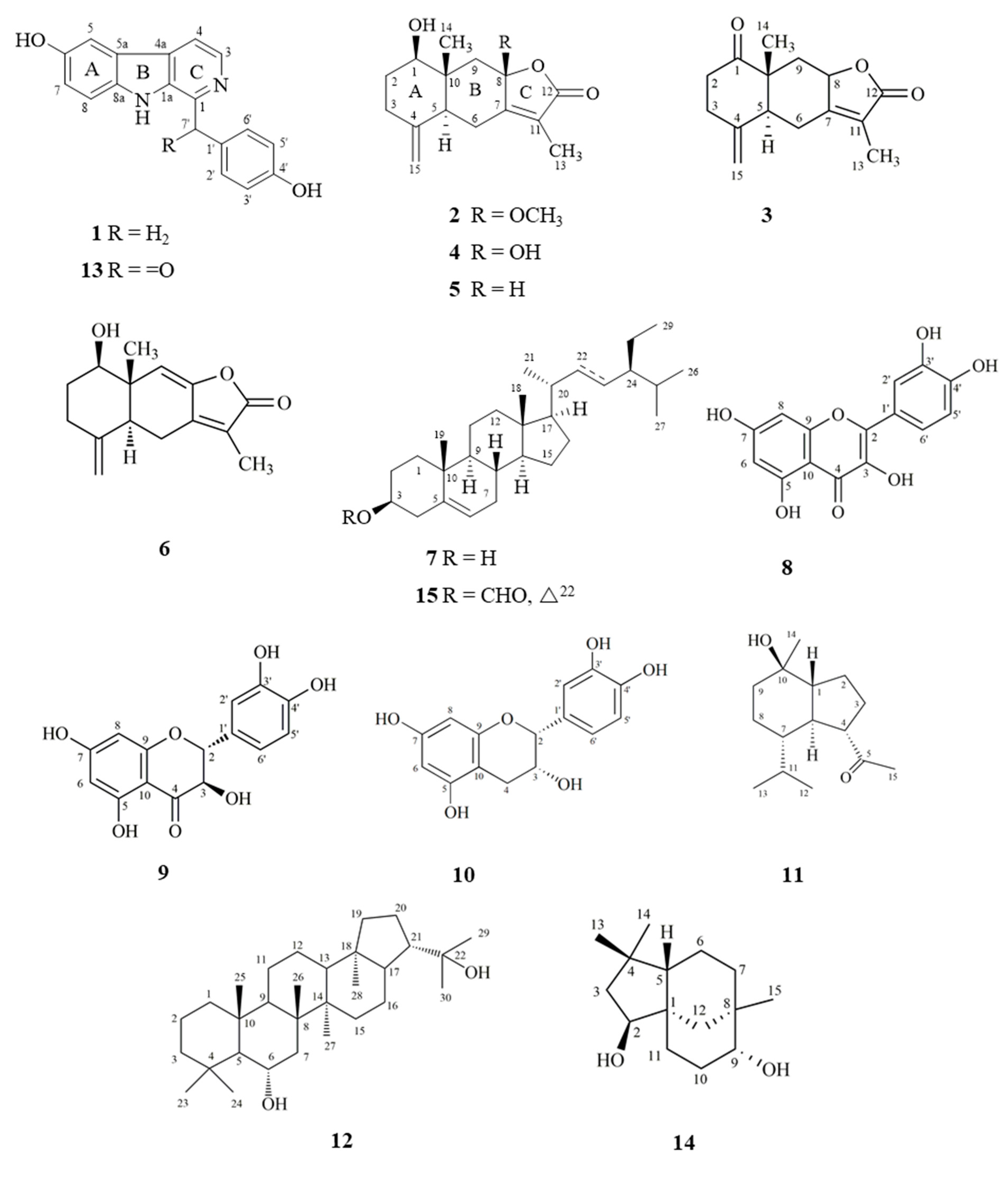
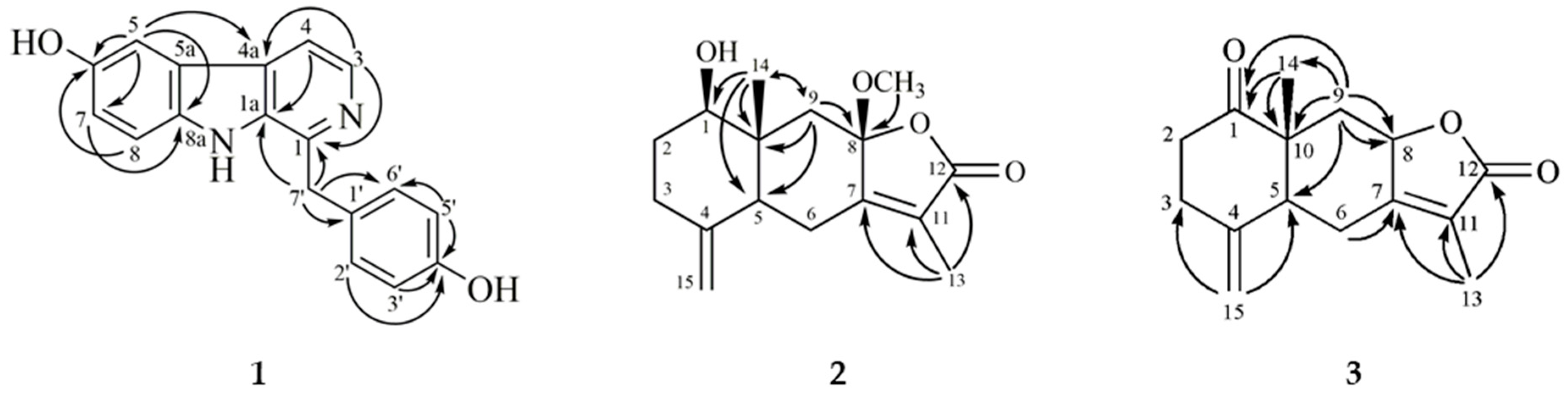
2.1. Structure Elucidation of Compounds 13

Fifteen compounds (115), including three new compounds, were isolated from dichloromethane (NARD) and ethyl acetate (NARE) soluble layers of NARM. Their structures (Figure 1) were spectroscopically determined by FTIR and 1D and 2D NMR, and through comparison with those of reported analogs. The structure determination of three new compounds was described in the present paper.
Compound 1 was isolated as a yellowish powder. The molecular formula C18H14N2O2 was determined by ESIMS and HR-ESIMS (291.1127 [M + H]+). The IR spectrum exhibited absorption peaks attributable to a hydroxy group (3277 cm−1) and an aromatic ring (1610, 1583, and 1512 cm−1).
The UV spectrum of 1 was similar to that of daibucarboline A [4] and cecilin [20] and was consistent with a carboline moiety [20]. The 1H-NMR spectrum of 1 (Table 1) showed a trisubstituted aromatic ABX system (δ 7.10 (1H, dd, J = 8.8, 2.4 Hz, H-7), 7.40 (1H, d, J = 8.8 Hz, H-8), 7.56 (1H, d, J = 2.4 Hz, H-5)) on ring A, an NH proton (δ 10.32 (1H, br s)) on ring B, a pair of mutually coupled protons (δ 7.82 (1H, d, J = 5.2 Hz, H-4) and 8.24 (1H, d, J = 5.2 Hz, H-3)), and a p-hydroxybenzyl group (δ 4.37 (2H, s, H-7′), 6.70 (2H, d, J = 8.8 Hz, H-3′ and H-5′), and 7.20 (2H, d, J = 8.8 Hz, H-2′ and H-6′)) on ring C. The HMBC correlations (Figure 2) from H-7′ to C-1, C-1a, and C-1′ further confirmed the p-hydroxybenzyl group located at C-1 on ring C. A comparison of 1H and 13C data of 1 with those of cecilin [20] suggested that their structures are closely related, except that the hydroxy group of 1 replaced the methoxy group at C-6 of cecilin. This was supported by HMBC correlation from H-5 to C-6, H-8 to C-6, and the ESIMS of 1 contained less 14 amu [CH2] than cecilin. Meanwhile, the NMR spectra of 1 closely resembled those of daibucarboline A [4], except for the absence of a methoxy substituent at C-3 position. From these data, the structure of 1 was determined to be demethoxydaibucarboline A, which was further confirmed by HSQC, COSY, NOESY, and HMBC experiments.
Compound 2 was obtained as colorless needles with [α] D 25 +148.6° (c 0.27, CHCl3). The molecular formula C16H22O4 was deduced from the HRESIMS ion peak at m/z 301.1409 [M + Na]+ (calcd for C16H22O4Na 301.1410) with six degrees of unsaturation. Its IR spectrum exhibited absorption peaks corresponding to a hydroxy group (3419 cm−1) and an α,β-unsaturated lactone functional group (1733 cm−1). The UV spectrum showed absorbance maxima at 219 nm. The 1H-NMR spectrum (Table 1) exhibited signals for a tertiary methyl (δ 0.94 (3H, s, H-14)), a vinylic methyl (δ 1.86 (3H, d, J = 1.6 Hz, H-13)), and two terminal methylene protons (δ 4.64 (1H, d, J = 1.6 Hz) and 4.90 (1H, d, J = 1.6 Hz)), which are similar to those of known eudesmanolides, 1β-acetoxy-8β-hydroxyeudesman-4(15),7(11)-dien-8α,12-olide [21]. The 13C-NMR spectrum and DEPT experiments displayed 15 carbon signals that correspond to an eudesmanolide containing a conjugated γ-lactone carbonyl (δ 171.7), a terminal double bond (δ 108.1), a set of tetrasubstituted double bond (C-7 and -11), an acetal (δ 106.2, C-8), two methyls (C-13 and -14), four methylenes (C-2, -3, -6, and -9), two methines (C-1 and -5), and two quaternary carbons (C-4 and -10), respectively. The signals at δ 8.3 (C-13), 106.2 (C-8), 124.3 (C-11), 158.8 (C-7), and 171.7 (C-12) are characteristic of a 5-oxygenated-3-methyl-5-hydrofuran-2-one functional moiety of eudesmanolides [21]. The 1H and 13C-NMR data of 2 were similar to those of neolitacumone A (5) [8], except 2 has a methoxy group instead of a hydroxy group at C-8 of neolitacumone A (5), and the ESIMS of 2 had greater 14 amu than 5. In addition, the location of the methoxy group of 2 at C-8 was further confirmed by HMBC correlation from OCH3-8 to C-8. The HMBC correlations from H-1 to C-2 (δ 30.8), C-9 (δ 46.4), and C-10 (δ 41.1); from H-13 to C-7 (δ 158.8), C-11 (δ 124.3), and C-12; from H-14 to C-1 (δ 78.8), C-5 (δ 49.6), C-9, and C-10; and from H-15a and H-15b to C-3 (δ 33.6) and C-5 further confirmed the planar structure of 2. In addition, the trans-A/B ring junction of 2 was confirmed by observation of NOESY cross-peaks between δ 0.94 (H-14)/1.57 (H-2β), 0.94 (H-14)/2.47 (H-6β), and 2.78 (H-9β) and between δ 1.57 (H-2β)/2.31 (H-3β) [8]. Thus, the stereochemistry of 2 was confirmed as 1β-OH, 5α-H and 10β-CH3. In addition, NOESY correlations between δ 3.16 (8-OCH3)/0.94 (H-14) and 2.78 (H-9β) suggested a β-orientation of the methoxy group at C-8. From the above data, the structure of 2 was further confirmed by DEPT, HSQC, COSY, NOESY, and HMBC experiments. Hence, it was determined to be 1β-hydroxy-8β-methoxy-eudesman-4(15), 7(11)-dien-8α,12-olide and was named as methylneolitacumone A.
Compound 3 was isolated as a white powder with [α] D 25 +225.2° (c 0.04, CHCl3). The molecular formula was obtained as C15H18O3 with ESIMS and HRESIMS analysis, which is two hydrogens less than neolitacumone B (5). Its IR and UV spectra exhibited absorption peaks attributable to an α,β-unsaturated lactone moiety [8,21]. According to 1H, 13C-NMR, and DEPT spectra, 3 was suggested to share a similar skeleton with neolitacumones A (4), B (5), and methylneolitacumone A (2) as an eudesmane-type sesquiterpenoid [8,21]. Comparison of compound 3 with 5 displayed similarities in both the physical data and the 1H and 13C-NMR spectra while the difference appeared at C-1 with a carbonyl group (IR: 1749 cm−1 and δc 211.3) replacing a hydroxy group in 5. Meanwhile, an 0.29 ppm downfield shift of the characteristic methyl signals at C-14 of eudesmanolide was due to a carbonyl inductive effect. The structure was also supported by HMBC correlations from H-9α (δH 1.44) and H-9β (δH 2.63) to C-1 and from H-14 (δH 1.17) to C-1. In addition, HMBC correlations from H-2α/H-2β and H-3α/H-3β to C-1 (δ 211.3); from H-13 to C-7 (δ 160.3), C-11 (δ 121.4), and C-12 (δ 174.5); from H-14 to C-1 (δ 211.3), C-5 (δ 48.2), C-9 (δ 40.0), and C-10 (δ 49.4); from H-15a and H-15b to C-3 (δ 34.4) and C-5 in 3 were employed for determining the planar structure of 3. The β-orientation of H-8 and H-14 of 3 was due to NOESY cross-peaks between δ 4.80 (H-8)/1.17 (H-14) and 2.63 (H-9β), and δ 1.17 (H-14)/2.50 (H-6β) and 2.63 (H-9β). Accordingly, relative configurations of 3 were confirmed as 5α-H, 8β-H, and 10β-CH3. Meanwhile, optical rotation of 3 [α] D 25 +225.2° was similar to other eudesman-type compounds 2 and 46 as a positive endpoint, suggesting their relative configurations are similar [8]. As determined by the above observations, the structure of 3 was determined to be 1-oxoeudesman-4(15),7(11)-dien-8α,12-olide and was named neolitacumone E.
The known compounds, neolitacumones A–C (46) [8], β-sitosterol (7) [22], quercetin (8) [22], dihydroquercetin (9) [23], epicatechin (10) [22], oplopanone (11) [24], zeorin (12) [25], linderaggrine A (13) [26], clovane-2β,9α-diol (14) [27], and stigmast-5-ene-3β-yl formate (15) [28] were identified by comparison of their physical and spectroscopic data with literature values.

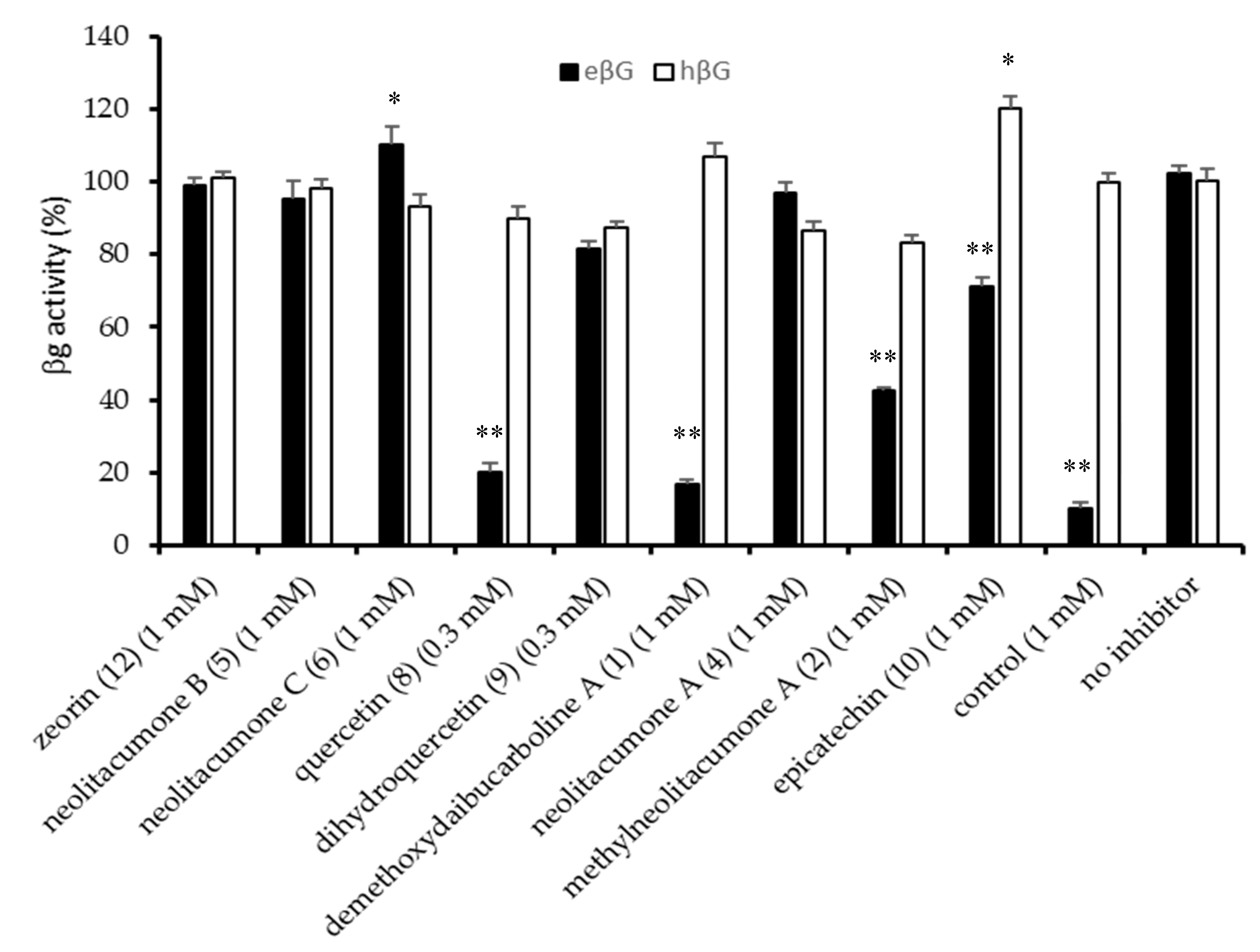
2.2. Anti-E. Coli β-Glucuronidase Activity of Compounds Isolated from N. acuminatissima

Bioassay-guided isolation of NARD and NARE led to purification of 15 compounds. Among them, compounds 1, 2, 46, 810, and 12 were examined for their specific inhibition for eβG versus hβG by in vitro βG-based activity assays. The result showed that compounds 1 (1 mM) and 8 (0.3 mM) exhibited strong anti-eβG activity comparable to the positive control (1-((6,8-dimethyl-2-oxo-1,2-dihydroquinolin-3-yl)methyl)-3-(4-ethoxyphenyl)-1-(2-hydroxyethyl)thiourea, 1mM) [29], and led eβG activity below 20%, respectively (Figure 3). Compounds 2 and 10 had moderate or weak anti-eβG activity. Compounds 1 and 8 showed significant anti-eβG activity without affecting the enzyme activity of normal human intestinal (hβG > 90%); therefore, they do not influence normal function of human intestines. They may provide as a specific inhibitor to reduce eβG-induced intestinal injury and CID. Comparing the anti-eβG effect of flavone-quercetin (8), flavanone-dihydroquercetin (9), and flavan-epicatechin (10), these similar flavonoids with different saturations in the C-ring showed different behavior; compounds 9 and 10 had slight or weak anti-eβG effect, whereas 8 with a double bond between C-2/C-3 in conjugation with a 4-carbonyl group in ring C significantly decreased eβG activity. The result suggests that the flavone co-planar skeleton of 8 is better stabilized in the enzymes’ binding pocket than 9 and 10 [30]. Since the inhibitory effect of β-Glucuronidase of eudesmanolide-type sesquiterpenes has never been reported, in the present paper, the anti-eβG result of methylneolitacumone A (2), neolitacumone A (4), neolitacumone B (5), and neolitacumone C (6) with the same eudesmanolide skeleton were discussed. Compound 2 exhibited moderate anti-eβG activity, while compounds 46 did not affect the enzyme effect. Comparison of these compounds showed that compound 2 has a methoxy group instead of a hydroxy group at C-8 and showed a better inhibitory effect than 4. Thus, it appears that presence of the lipophilic 8-methoxy group decreases eβG activity.
β-Glucuronidase is a lysosomal enzyme and has been found in animals, plants, and bacteria [31]. This enzyme is responsible for hydrolysis of β-glucuronide conjugates of endogenous and exogenous compounds in the body, such as benzo[a]pyrene glucuronides and natural plant glucuronides. It has been found that βG released from macrophages and neutrophils is necessary for bioactivation of glucuronide conjugates into the aglycone [30,32], and that the enzyme requires acidic conditions for its catalytic activity. For hβG, it exhibits maximal catalytic activity at pH 4–4.5, while eβG shows optimal activity at neutral pH [32,33,34]. Inhibition or dysfunction of hβG may disturb glycosaminoglycan degradation and cause mucopolysaccharidosis (MPS), affecting appearance, physical abilities, organ and system functioning, and mental development [34,35]. Thus, it is worthy to figure out candidates that can specifically inhibit eβG activity but not hβG.
CID is a common side effect experienced by cancer patients; it affects quality of life and may result in early death either directly from life-threatening sequelae or indirectly from adjustments in cancer treatment that result in suboptimal therapy [14,15,16]. Many studies indicate that inhibition of βG activity can reduce CPT-11-induced intestine mucosal damage and CID [19,20,33]. Although use of antibiotics against intestinal βG could relieve CPT-11-induced diarrhea, allergies and resistance effects must be consulted or monitored for choosing an antibiotic; meanwhile, antibiotics will kill all native gut floras, including probiotics within the digestive tract, which is not recommended for long-term use in chemotherapeutic patients [14,34]. Development of eβG-specific inhibitors can be used as a chemotherapy adjuvant to reduce CID [33].
To sum up these results, demethoxydaibucarboline A (1), methylneolitacumone A (2), and quercetin (8) isolated from N. acuminatissima may be candidate-specific eβG inhibitors to reduce CID and intestinal injury. The best inhibitor among these isolates is quercetin (8). As we know, quercetin is the major flavonoid in our diet. Diets rich in quercetin and other flavonoids may relate to decreasing incidence of cardiovascular, neoplastic, and neurodegenerative diseases [30,32,36]. Thus, a specific eβG inhibitor, such as quercetin, could be used as a nutrient supplement for chemoprevention and health promotion. Further experiments are needed to pinpoint their mechanisms of action.

3. Materials and Methods

3.1. General

Melting points were determined on an Electrothermal MEL-TEMP 3.0 apparatus (manufacturer, city, state abbrev. if USA, country) and were uncorrected. Optical rotations were recorded on a Jasco P2000 digital polarimeter. UV spectra were obtained in methanol on a Beckman Coulter TM-DU 800 UV–visible spectrophotometer. IR spectra were measured on a Perkin Elmer system 2000 FTIR spectrophotometer. 1D (1H, 13C, DEPT) and 2D (COSY, NOESY, HSQC, HMBC) NMR spectra using CDCl3, acetone-d6, or methanol-d4 as solvents were recorded on Varian Gemini-2000 200 MHz, Varian Unity Plus 400 MHz, Mercury Plus 400 MHz, and Varian VNMRS 600 MHz FT-NMR spectrometers. Deuterated solvents were purchased from Sigma-Aldrich (St. Louis, MO, USA). Low-resolution ESIMS was obtained with Waters ZQ 4000 and JEOL-JMS-HX 100 mass spectrometers. High-resolution ESIMS was recorded on JEOL JMS-SX102A GC/LC/MS and Finnigan MAT-95XL mass spectrometers. Silica gel (70–230 Mesh; Merck, Darmstadt, Germany) and spherical C18 100 Å reversed-phase silica gel (RP-18; particle size 40–63 μm; Silicycle, Quebec, Canada) were used for column chromatography, and silica gel 60 F254 (Merck) and RP-18 F254S (Merck) were used for TLC and preparative TLC. Further purification was performed by HPLC (Chrom Tech P230 HPLC pump, Phenomenex Luna 5u Silica 250 × 10.00 mm, 5 micron, refractive index detector IOTA2). The reagents in the β-Glucuronidase activity assay, e.g., bovine serum albumin, p-nitrophenyl-β-d-glucopyranoside, and 1-((6,8-dimethyl-2-oxo-1,2-dihydroquinolin-3-yl)methyl)-3-(4-ethoxyphenyl)-1-(2-hydroxyethyl)thiourea were all purchased from Sigma-Aldrich (St. Louis, MO, USA).

3.2. Plant Material

Roots of N. acuminatissima were collected in July 2012 from Yilan County, Taiwan and positively identified by one of the authors, Prof. I. S. Chen. A voucher specimen (2012-07-NA) was deposited at the Herbarium of the Department of Fragrance and Cosmetic Science, Kaohsiung Medical University, Kaohsiung, Taiwan.

3.3. Extraction and Isolation

Air-dried roots of N. acuminatissima (7.4 kg) were extracted three times with MeOH (20 L) at room temperature. The extracts were filtered and concentrated under reduced pressure, and the residue (NARM, 440 g) was successively partitioned with dichloromethane, ethyl acetate, n-butanol, and water to obtain four different soluble fractions, named NARD (87.7 g), NARE (30.3 g), NARB (198.0 g), and NARW (68.8 g), respectively. All were assessed by anti-eβG activity. Active NARD and NARE layers were further purified using silica gel column chromatography (CC).
NARD (87.7 g) was subjected to silica gel CC with a gradient of n-hexane/acetone/MeOH to obtain fractions 1–14. Fractions 9, 10, 12, and 14 showed anti-eβG activity. Fraction 3 (26.5 g) was purified by silica gel CC, eluting with n-hexane/CH2Cl2/acetone (2:2:1), MeOH/H2O (10:0.1), and n-hexane/acetone (3:1) to individually yield 12 (120.5 mg), 7 (165.0 mg) and 15 (2.0 mg). Fraction 4 (3.2 g) was subjected to CC, eluting with n-hexane/EtOAc and n-hexane/acetone (6:1) then recrystallized to yield 6 (74.0 mg). In addition, 11 (1.2 mg) was also obtained with eluting n-hexane/EtOAc and cyclohexane/CH2Cl2/acetone (13:1:1). Fraction 5 (2.3 g) was purified by silica gel CC eluting with CH2Cl2/acetone (15:1) and preparative HPLC eluting with n-hexane/acetone (3:1) to obtain 3 (1.1 mg). Fraction 7 (5.6 g) was subjected to CC eluting with n-hexane/CH2Cl2/acetone (1:3:2) and MeOH/H2O (1:1) to afford 4 (206.0 mg). In addition, 14 (2.0 mg) was also obtained with eluting n-hexane/CH2Cl2/acetone (1:3:1), n-hexane/CH2Cl2/acetone (2:2:1), MeOH/H2O (1:1), and n-hexane/acetone (3:1). Active fraction 9 (3.2 g) was chromatographed on CC, eluting with MeOH/H2O (1:1), n-hexane/acetone (6:1), and Sephadex LH-20 to afford 1 (12.1 mg). Active fraction 10 (6.4 g) was subjected to CC and recrystallized to obtain 5 (25.0 mg).
NARE (30.3 g) was subjected to CC with a gradient of MeOH/H2O (1:2–1:1–100% MeOH) to obtain fractions 1–10. Fractions 5–7 showed anti-eβG activity. Fraction 3 (0.8 g) was purified by CC, eluting with CH2Cl2/acetone (1:1) and MeOH/H2O (1:1) to yield 9 (53.6 mg). In addition, 10 (40.3 mg) and 1 (4.4 mg) were also obtained by silica gel CC individually eluting with CH2Cl2/acetone (2:1) and MeOH/H2O (1:1). Active fraction 5 (2.3 g) was purified by CC, eluting with n-hexane/acetone (1:2) and CH2Cl2/acetone (3:1) to obtain 13 (1.5 mg). Sequentially, 4 (14.0 mg) was afforded by silica gel CC eluting with n-hexane/EtOAc (1:1) and CH2Cl2/EtOAc (1:1). Active fraction 6 (2.0 g) was subjected to Sephadex LH-20 and silica gel CC, eluting with n-hexane/EtOAc (1:3), n-hexane/acetone (3:1), and MeOH/H2O (1:1) to obtain 2 (6.8 mg). Fraction 9 (0.2 g) was purified by Sephadex LH-20 and CC, eluting with n-hexane/acetone (2:1) to yield 8 (18.7 mg).

3.3.1. Demethoxydaibucarboline A (1)

Yellowish powder; UV (MeOH) λmax (log ε) 228 (4.22), 241 (4.03), 297 (3.96), 356 (3.65), and 385 (sh, 3.42) nm; IR (KBr) νmax 3277 (OH), 1610, 1583, and 1512 (aromatic ring) cm−1; 1H (400 MHz) and 13C (100 MHz) NMR spectral data, see Table 1; HRESIMS [M + H]+ m/z 291.1127 (calculated for C18H15N2O2, 291.1128).

3.3.2. Methylneolitacumone A (2)

Colorless needles (acetone/water); m.p.: 112–114 °C; [α] D 25 + 148.6° (c 0.27, CHCl3); UV (MeOH) λmax (log ε) 219 (3.94) nm; IR (KBr) νmax 3419 (OH), 1733 (C=O), 1643 (C=CH2) cm−1; 1H (400 MHz) and 13C (100 MHz) NMR spectral data, see Table 1; HRESIMS [M + Na]+ m/z 301.1409 (calculated for C16H22O4Na, 301.1410).

3.3.3. Neolitacumone E (3)

White powder; [α] D 25 +225.2° (c 0.04, CHCl3); UV (MeOH) λmax (log ε) 218 (3.96) nm; IR (KBr) νmax 1749, 1708 (C=O) cm−1; 1H (500 MHz) and 13C (125 MHz) NMR spectral data, see Table 1; HRESIMS [M + Na]+ m/z 269.1146 (calculated for C15H18O3Na, 269.1148).

3.4. In Vitro β-Glucuronidase Activity Assay

The effect of isolates of N. acuminatissima to inhibit β-Glucuronidase activity was determined using purified eβG and hβG enzymes obtained as described [34,35]. The enzymes were prepared in a reaction buffer (10% DMSO + 0.05% bovine serum albumin in PBS) at pH 7.5 and 4.5 for eβG and hβG, respectively. The test samples or vehicle control (10 μL) were mixed with 40 μL βG enzyme (3.75 ng/well) into a 96-well plate at 37 °C for 30 min and then 50 μL p-nitrophenyl-β-d-glucopyranoside (pNPG) (10 mM) was added and incubated at 37 °C for 1 h. Finally, 5 μL 2N NaOH was added to stop the reaction. Optical densities were determined at 405 nm using a microplate spectrophotometer. Results are displayed as percent of βG activity compared with the untreated control. 1-((6,8-dimethyl-2-oxo-1,2-dihydroquinolin-3-yl)methyl)-3-(4-ethoxyphenyl)-1-(2-hydroxy- ethyl)thiourea was used as a positive control. Data are shown as the mean ± standard deviation (SD) of three measurements. Groups were compared using the Student’s t-test (SPSS 18 Inc., UAS). Differences between groups were determined to be significant when * p < 0.05 and ** p < 0.01.

4. Conclusions

In this study, one new carboline alkaloid, demethoxydaibucarboline A (1), two new eudesmanolide-type sesquiterpenes, methylneolitacumone A (2), neolitacumone E (3), and twelve known compounds (415) were isolated from the root of N. acuminatissima. These compounds were investigated by using an anti-eβG assay. The results indicated that two isolated compounds, demethoxydaibucarboline A (1) and quercetin (8) showed significant anti-eβG activity with an inhibition ratio of approximately 80%, respectively. Methylneolitacumone A (2) exhibited a moderate inhibitory effect and eβG activity was less than 45%. Compounds 1 at 1 mM and 8 at a lower concentration of 0.3 mM exhibited specific inhibition of eβG activity but not hβG, suggesting that active secondary metabolites of N. acuminatissima are potential β-Glucuronidase inhibitors. These secondary metabolites will be potential β-Glucuronidase inhibitors that protect intestines from injury and thus relieve chemotherapy-induced diarrhea (CID). Although the detailed mechanism of action of these compounds remains to be determined, the results confirmed that N. acuminatissima is a valuable source from which natural product-based supplements and medicinal products can be derived.

Supplementary Materials

The following are available online. Figure S1: Preliminary anti-eβG screening assay of partial Lauraceae plants. Figures S2–S28: The 1D- and 2D-NMR spectra of compounds 13.

Author Contributions

Conceptualization, I.-S.C., H.-S.C. and H.-H.K.; methodology, T.-L.C. and H.-H.K.; formal analysis, C.-H.L., H.-J.C., C.-C.C., T.-L.C. and H.-H.K.; investigation, C.-H.L., H.-J.C. and C.-C.C.; resources, I.-S.C., H.-S.C., T.-L.C. and H.-H.K.; data curation, T.-L.C. and H.-H.K.; writing—original draft preparation, C.-H.L., H.-J.C. and C.-C.C.; writing—review and editing, I.-S.C., Y.-H.K. and H.-H.K.; visualization, H.-S.C. and H.-H.K.; supervision, I.-S.C. and Y.-H.K.; project administration, H.-H.K.; funding acquisition, Y.-H.K. and H.-H.K. All authors have read and agreed to the published version of the manuscript.

Funding

This research was funded by the Ministry of Science and Technology of Taiwan (MOST 108-2320-B-037-023) and in part by the Aim for the Top Universities Grant, grant number KMU-TP104H01, and KMU-TC108A03-9 from the Drug Development and Value Creation Research Center, Kaohsiung Medical University, Taiwan. This work was also supported partially by the China Medical University grant in Taiwan (10959189) and the Chinese Medicine Research Center, China Medical University from The Featured Areas Research Center Program within the framework of the Higher Education Sprout Project by the Ministry of Education (MOE) in Taiwan (CMRC-CHM-2-1).

Acknowledgments

We thank C.C. Wang and M.Y. Hung of the Center for Research Resources and Development of Kaohsiung Medical University for NMR and ESI-MS measurements, respectively, and S.C. Yu of the Department of Chemistry of National Sun Yat-Sen University, Kaohsiung, Taiwan, for ESI-MS and HRESIMS measurements.

Conflicts of Interest

The authors declare no conflict of interest.

References

  1. Li, L.; Li, J.; Conran, J.G.; Li, X.W. Phylogeny of Neolitsea (Lauraceae) inferred from Bayesian analysis of nrDNA ITS and ETS sequences. Pl. Syst. Evol. 2007, 269, 203–221. [Google Scholar] [CrossRef]
  2. Wang, W.Y.; Ma, P.; Xu, L.J.; Peng, Y.; Xiao, P.G. Chemical constituents and biological activities of plants from Genus Neolitsea. Chem. Biodivers. 2014, 11, 55–72. [Google Scholar] [CrossRef]
  3. Joshi, S.C.; Verma, A.R.; Mathela, C.S. Antioxidant and antibacterial activities of the leaf essential oils of Himalayan Lauraceae species. Food Chem. Toxicol. 2010, 48, 37–40. [Google Scholar] [CrossRef] [PubMed]
  4. Wong, S.L.; Chang, H.S.; Wang, G.J.; Chiang, M.Y.; Huang, H.Y.; Chen, C.H.; Tsai, S.C.; Lin, C.H.; Chen, I.S. Secondary metabolites from the roots of Neolitsea daibuensis and their anti-inflammatory activity. J. Nat. Prod. 2011, 74, 2489–2496. [Google Scholar] [CrossRef] [PubMed]
  5. Kim, S.S.; Kim, J.E.; Lee, C.G.; Lee, N.H. Neolitsea aciculata essential oil inhibits drug-resistant skin pathogen growth and Propionibacterium acnes-induced inflammatory effects of human monocyte leukemia. Nat. Prod. Commun. 2011, 6, 1193–1198. [Google Scholar] [CrossRef] [Green Version]
  6. Chen, K.S.; Hsieh, P.W.; Hwang, T.L.; Chang, F.R.; Wu, Y.C. Anti-inflammatory furanogermacrane sesquiterpenes from Neolitsea parvigemma. Nat. Prod. Res. 2005, 19, 283–286. [Google Scholar] [CrossRef]
  7. Kim, S.S.; Hyun, C.G.; Choi, Y.H.; Lee, N.H. Tyrosinase inhibitory activities of the compounds isolated from Neolitsea aciculata (Blume) Koidz. J. Enzyme Inhib. Med. Chem. 2013, 28, 685–689. [Google Scholar] [CrossRef]
  8. Chang, F.R.; Hsieh, T.J.; Huang, T.L.; Chen, C.Y.; Kuo, R.Y.; Chang, Y.C.; Chiu, H.F.; Wu, Y.C. Cytotoxic constituents of the stem bark of Neolitsea acuminatissima. J. Nat. Prod. 2002, 65, 255–258. [Google Scholar] [CrossRef]
  9. Li, W.S.; Duh, C.Y. Sesquiterpene lactones from Neolitsea villosa. Phytochemistry 1993, 32, 1503–1507. [Google Scholar]
  10. Chang, H.S.; Chen, I.S. Chemical constituents and bioactivity of Formosan lauraceous plants. J. Food Drug Anal. 2016, 24, 247–263. [Google Scholar] [CrossRef] [Green Version]
  11. Liao, J.C. Lauraceae. In Flora of Taiwan, 2nd ed.; Editorial Committee of the Flora of Taiwan: Taipei, Taiwan, 1996; Volume 2, pp. 484–496. [Google Scholar]
  12. Tomita, M.; Lu, S.T.; Fu, S.C.; Lin, Y.M. Studies on the alkaloids of Formosan lauraceous plants. VI. alkaloids of Neolitsea acuminatissima (Hayata) Kanehira et Sasaki. Yakugaku Zasshi 1965, 85, 662–664. [Google Scholar] [CrossRef] [PubMed]
  13. Kong, R.; Liu, T.; Zhu, X.; Ahmad, S.; Williams, A.; Phan, A.T.; Zhao, H.; Scott, J.E.; Yeh, L.A.; Wong, S.T.C. Old drug new use—Amoxapine and its metabolites as potent bacterial β-Glucuronidase inhibitors for alleviating cancer drug toxicity. Clin. Cancer Res. 2014, 20, 3521–3530. [Google Scholar] [CrossRef] [Green Version]
  14. Richardson, G.; Dobish, R. Chemotherapy induced diarrhea. J. Oncol. Pharm. Pract. 2007, 13, 181–198. [Google Scholar] [CrossRef] [Green Version]
  15. Benson, A.B., 3rd; Ajani, J.A.; Catalano, R.B.; Engelking, C.; Kornblau, S.M.; Martenson, J.A., Jr.; McCallum, R.; Mitchell, E.P.; O’Dorisio, T.M.; Vokes, E.E.; et al. Recommended guidelines for the treatment of cancer treatment-induced diarrhea. J. Clin. Oncol. 2004, 22, 2918–2926. [Google Scholar] [CrossRef] [Green Version]
  16. Sakai, H.; Sagara, A.; Matsumto, K.; Jo, A.; Hirosaki, A.; Takase, K.; Sugiyama, R.; Sato, K.; Ikegami, D.; Horie, S.; et al. Neutrophil recruitment is critical for 5-fluorouracil-induced diarrhea and the decrease in aquaporins in the colon. Pharmacol. Res. 2014, 87, 71–79. [Google Scholar] [CrossRef]
  17. Bhatt, A.P.; Pellock, S.J.; Biernat, K.A.; Walton, W.G.; Wallace, B.D.; Creekmore, B.C.; Letertre, M.M.; Swann, J.R.; Wilson, I.D.; Roques, J.R.; et al. Targeted inhibition of gut bacterial β-Glucuronidase activity enhances anticancer drug efficacy. Proc. Natl. Acad. Sci. USA 2020, 117, 7374–7381. [Google Scholar] [CrossRef] [Green Version]
  18. Xue, H.; Sawyer, M.B.; Field, C.J.; Dieleman, L.A.; Baracos, V.E. Nutritional modulation of antitumor efficacy and diarrhea toxicity related to irinotecan chemotherapy in rats bearing the ward colon tumor. Clin. Cancer Res. 2007, 13, 7146–7154. [Google Scholar] [CrossRef] [Green Version]
  19. Takasuna, K.; Hagiwara, T.; Watanabe, K.; Onose, S.; Yoshida, S.; Kumazawa, E.; Nagai, E.; Kamataki, T. Optimal antidiarrhea treatment for antitumor agent irinotecan hydrochloride (CPT-11)-induced delayed diarrhea. Cancer Chemother. Pharmacol. 2006, 58, 494–503. [Google Scholar] [CrossRef]
  20. Aguiar, L.M.G.; Filho, R.B.; Gottlieb, O.R.; Maia, J.G.S.; Pinho, S.l.V.; De Souse, J.R. Cecilin, a 1-benzyl-β-carboline from Aniba santalodora. Phytochemistry 1980, 19, 1859–1860. [Google Scholar] [CrossRef]
  21. Ulubelen, A.; Gören, N.; Bohlmann, F.; Jakupovic, J.; Grenz, M.; Tanker, N. Sesquiterpene lactones from Smyrnium cordifolium. Phytochemistry 1985, 24, 1305–1308. [Google Scholar] [CrossRef]
  22. Wei, X.H.; Yang, S.J.; Liang, N.; Hu, D.Y.; Jin, L.H.; Wei, X.; Yang, S. Chemical constituents of Caesalpinia decapetala (Roth) Alston. Molecules 2013, 18, 1325–1336. [Google Scholar] [CrossRef]
  23. Nifant’ev, E.E.; Kukhareva, T.S.; Dzgoeva, Z.M.; Vasyanina, L.K.; Koroteev, M.P.; Kaziev, G.Z. Selective phosphorylation of dihydroquercetin with trivalent phosphorus reagents. Heteroatom Chem. 2003, 14, 339–403. [Google Scholar]
  24. Alberto Marco, J.; Sanz-Cervera, J.F.; Sancenon, F.; Jakupovic, J.; Rustaiyan, A.; Mohamadi, F. Oplopanone derivatives monoterpene glycosides from Artemisia sieberi. Phytochemistry 1993, 34, 1061–1065. [Google Scholar] [CrossRef]
  25. Ahmad, S.; Riyanto, S.; Sukari, M.A.; Rahmani, M.; Ali, A.M. Hopane and lupane triterpenes from leaves and stem bark of Aegle marmelos (Rutaceae). Asian, J. Chem. 2013, 25, 4591–4594. [Google Scholar] [CrossRef]
  26. Kuo, P.C.; Li, Y.C.; Hwang, T.L.; Ma, G.H.; Yang, M.L.; Lee, E.J.; Wu, T.S. Synthesis and structural characterization of an anti-inflammatory principle purified from Lindera aggregata. Tetrahedron Lett. 2014, 55, 108–110. [Google Scholar] [CrossRef]
  27. Heymann, H.; Tezuka, Y.; Kikuchi, T.; Supriyatna, S. Constituents of Sindora sumatrana Miq. I. isolation and NMR spectral analysis of sesquiterpenes from the dried pods. Chem. Pharm. Bull. 1994, 42, 138–146. [Google Scholar] [CrossRef] [Green Version]
  28. Chumkaew, P.; Kato, S.; Chantrapromma, K. New cytotoxic steroids from the fruits of Syzygium siamense. J. Asian Nat. Prod. Res. 2010, 12, 424–428. [Google Scholar] [CrossRef]
  29. Cheng, K.W.; Tseng, C.H.; Yang, C.N.; Tzeng, C.C.; Cheng, T.C.; Leu, Y.L.; Chuang, Y.C.; Wang, J.Y.; Lu, Y.C.; Chen, Y.L.; et al. Specific inhibition of bacterial β-Glucuronidase by pyrazolo [4,3-c]quinoline derivatives via a pH-dependent manner to suppress chemotherapy-induced intestinal toxicity. J. Med. Chem. 2017, 60, 9222–9238. [Google Scholar] [CrossRef]
  30. Untergehrer, M.; Bücherl, D.; Wittmann, H.-J.; Strasser, A.; Heilmann, J.; Jürgenliemk, G. Structure-dependent deconjugation of flavonoid glucuronides by human β-Glucuronidase—In vitro and in silico analyses. Planta Med. 2015, 81, 1182–1189. [Google Scholar] [CrossRef] [Green Version]
  31. Stahl, P.D.; Fishman, W.H. β-Glucuronidase. In Method of Enzymatic Analysis, 3rd ed.; Bergmeyer, H.U., Ed.; Verlag Chemie GmbH: Weinheim, Germany, 1983; Volume 4, pp. 246–256. [Google Scholar]
  32. Kawai, Y. β-Glucuronidase activity and mitochondrial dysfunction: The sites where flavonoid glucuronides act as anti-inflammatory agents. J. Clin. Biochem. Nutr. 2014, 54, 145–150. [Google Scholar] [CrossRef] [PubMed] [Green Version]
  33. Kim, D.H.; Jin, Y.H. Intestinal bacterial β-Glucuronidase activity of patients with colon cancer. Arch. Pharmacal. Res. 2001, 24, 564–567. [Google Scholar] [CrossRef] [PubMed]
  34. Cheng, T.C.; Chuang, K.H.; Roffler, S.R.; Cheng, K.W.; Leu, Y.L.; Chuang, C.H.; Huang, C.C.; Kao, C.H.; Hsieh, Y.C.; Chang, L.S.; et al. Discovery of specific inhibitors of intestinal E. coli beta-glucuronidase through in silico virtual screening. Sci. World J. 2015, 2015, 740815. [Google Scholar] [CrossRef] [Green Version]
  35. Cheng, K.W.; Tseng, C.H.; Tzeng, C.C.; Leu, Y.L.; Cheng, T.C.; Wang, J.Y.; Chang, J.M.; Lu, Y.C.; Cheng, C.M.; Chen, I.J.; et al. Pharmacological inhibition of bacterial β–glucuronidase prevents irinotecan-induced diarrhea without impairing its antitumor efficacy in vivo. Pharmacol. Res. 2019, 139, 41–49. [Google Scholar] [CrossRef]
  36. Hertog, M.G.; Hollman, P.C. Potential health effects of the dietary flavonol quercetin. Eur. J. Clin. Nutr. 1996, 50, 63–71. [Google Scholar]
Figure 1. Structures of compounds (115) isolated from the root of Neolitsea acuminatissima.
Molecules 25 05170 g001
Figure 2. Key HMBC (H→C) correlations of 13.
Molecules 25 05170 g002
Figure 3. Anti-E. coli β-Glucuronidase (anti-eβG) activity of isolates of the root of N. acuminatissima. Each column represents the mean ± SD of three experiments. * p < 0.05, ** p < 0.01 indicates significant differences from the no inhibitor group.
Molecules 25 05170 g003
Table 1. 1H- and 13C-NMR spectroscopic data of compounds 13.
Position1 (Acetone-d6)Position2 (CDCl3)3 (CDCl3)
δH (J in Hz)δCδH (J in Hz)δCδH (J in Hz)δC
1 146.613.36, dd (11.2, 4.0)78.8 211.3
38.24, d (5.2)139.01.80, ddd (13.6, 4.8, 2.0)30.82.73, td (16.1, 7.3)37.3
47.82, d (5.2)114.31.57, ddd (13.6, 4.8, 2.0) 2.47, m
57.56, d (2.4)107.22.05, td (13.8, 4.8)33.62.67, td (14.6, 5.0)34.4
6 152.82.31, td (13.8, 4.8) 2.47, m
77.10, dd (8.8, 2.4)119.44 146.6 144.6
87.40, d (8.8)113.951.88, m49.62.22, dt (13.7, 1.6)48.2
1a 131.32.38, dd (13.0, 3.2)24.22.86, dd (13.7, 3.6)25.0
4a 129.72.47, t (13.0) 2.50, td (13.7, 1.2)
5a 123.97 158.8 160.3
8a 136.68 106.24.80, dd (11.8, 6.3)78.2
1′ 131.61.38, d (13.6)46.41.44, dd (13.2, 11.8)40.0
2′, 6′7.20, d (8.8)113.32.78, d (13.6) 2.63, td (13.2, 6.3)
3′, 5′6.70, d (8.8)116.610 41.1 49.4
4′ 157.311 124.3 121.4
7′4.37, s40.812 171.7 174.5
NH10.32, br s 131.86, d (1.6)8.31.83, t (1.6)8.6
140.94, s10.31.17, s17.0
15a4.64, d (1.6)108.14.92, s110.8
15b4.90, d (1.6) 5.16, s
OCH33.16, s50.4
1H- (400 MHz) and 13C-NMR (100 MHz) data for compounds 1 and 2; 1H- (500 MHz) and 13C-NMR (125 MHz) data for compound 3.
Sample Availability: Samples of the compounds are available from the authors.
Publisher’s Note: MDPI stays neutral with regard to jurisdictional claims in published maps and institutional affiliations.

