Chemical Composition and Bioactivity of Essential Oil of Ten Labiatae Species


 and
and
Abstract
:1. Introduction
2. Results and Discussion
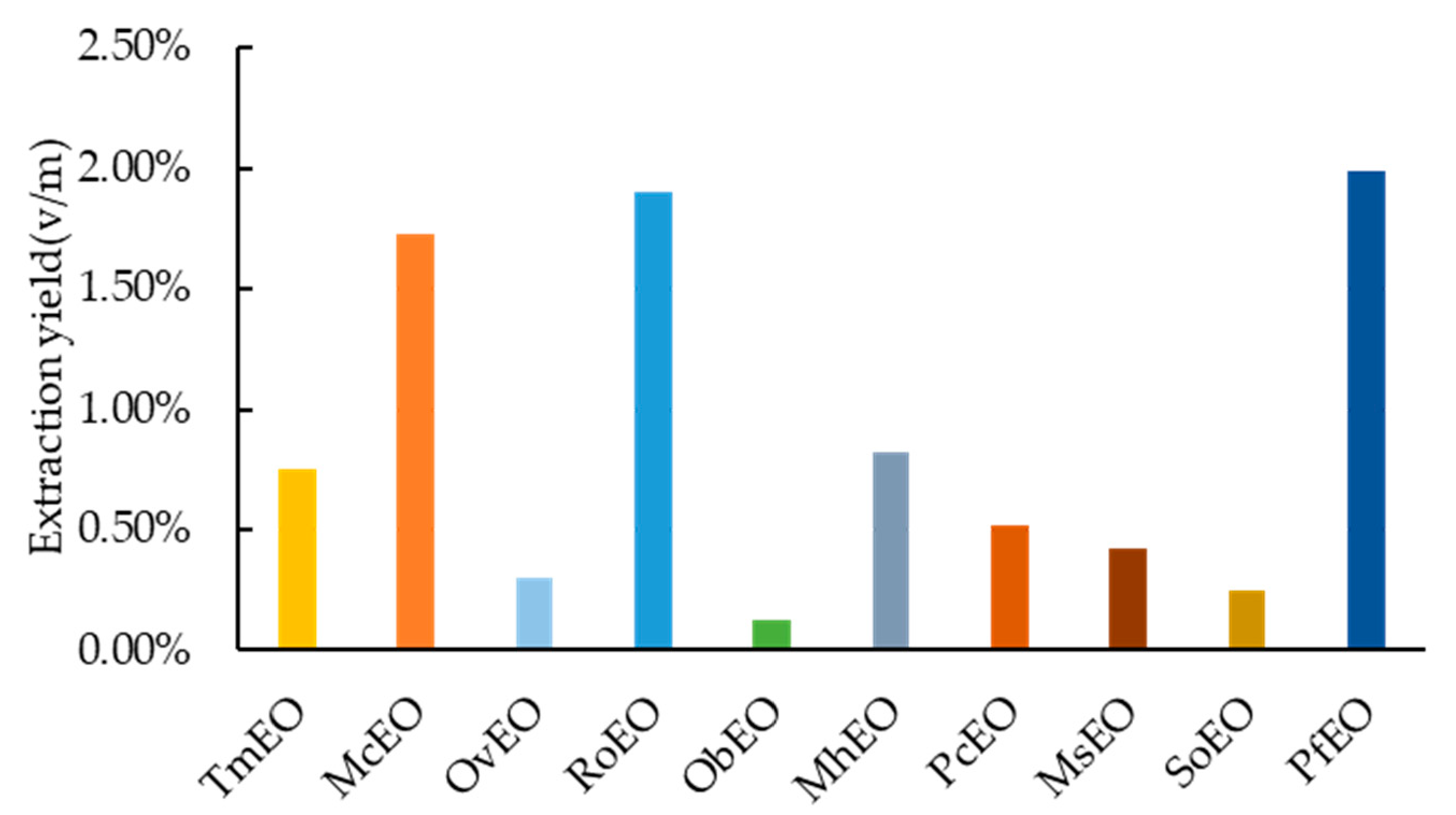
2.1. Extraction and Yield
2.2. Chemical Composition
2.3. Antioxidant Activity
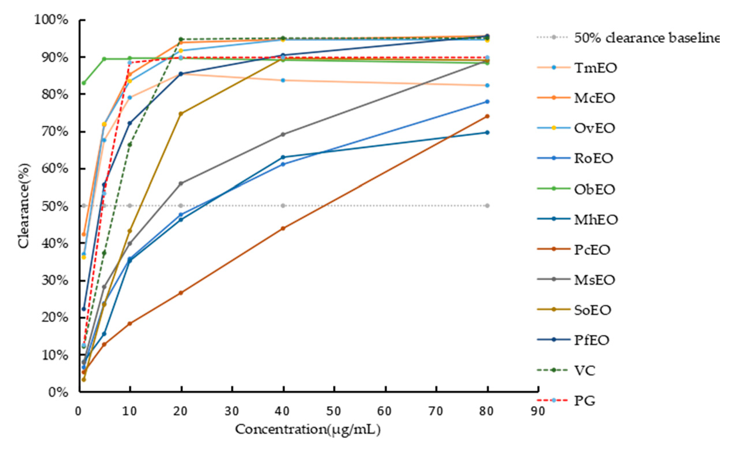
2.3.1. DPPH Free Radical Scavenging Activity
2.3.2. Ferric Reducing Antioxidant Power (FRAP)
2.4. Antibacterial Activity
Results of IZD, MIC and MBC
2.5. Antibacterial Stability of Essential Oils
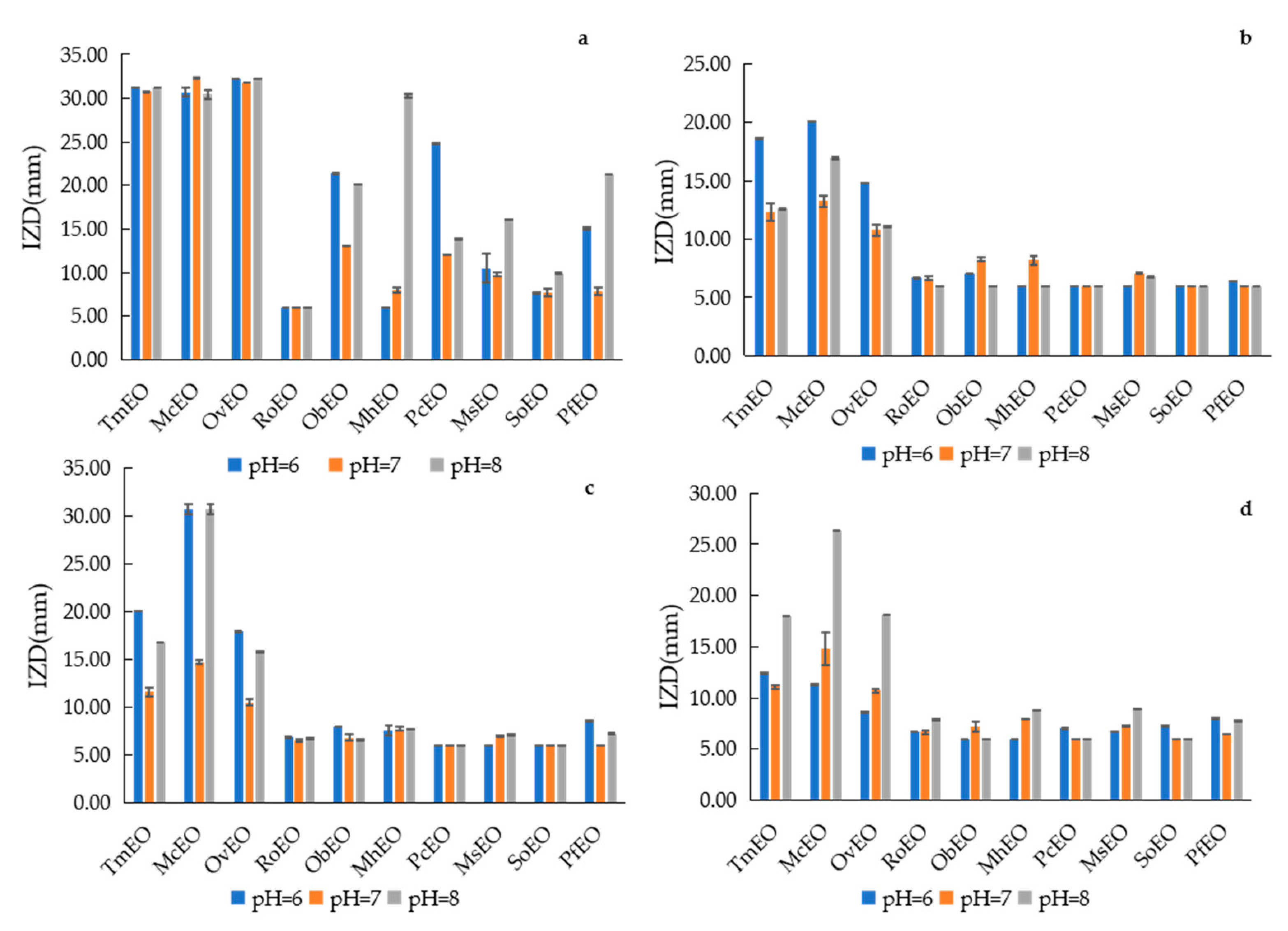
2.5.1. Effects of Different pH on Antibacterial Stability of EOs
2.5.2. Effects of Different Temperature on Antibacterial Stability of EOs
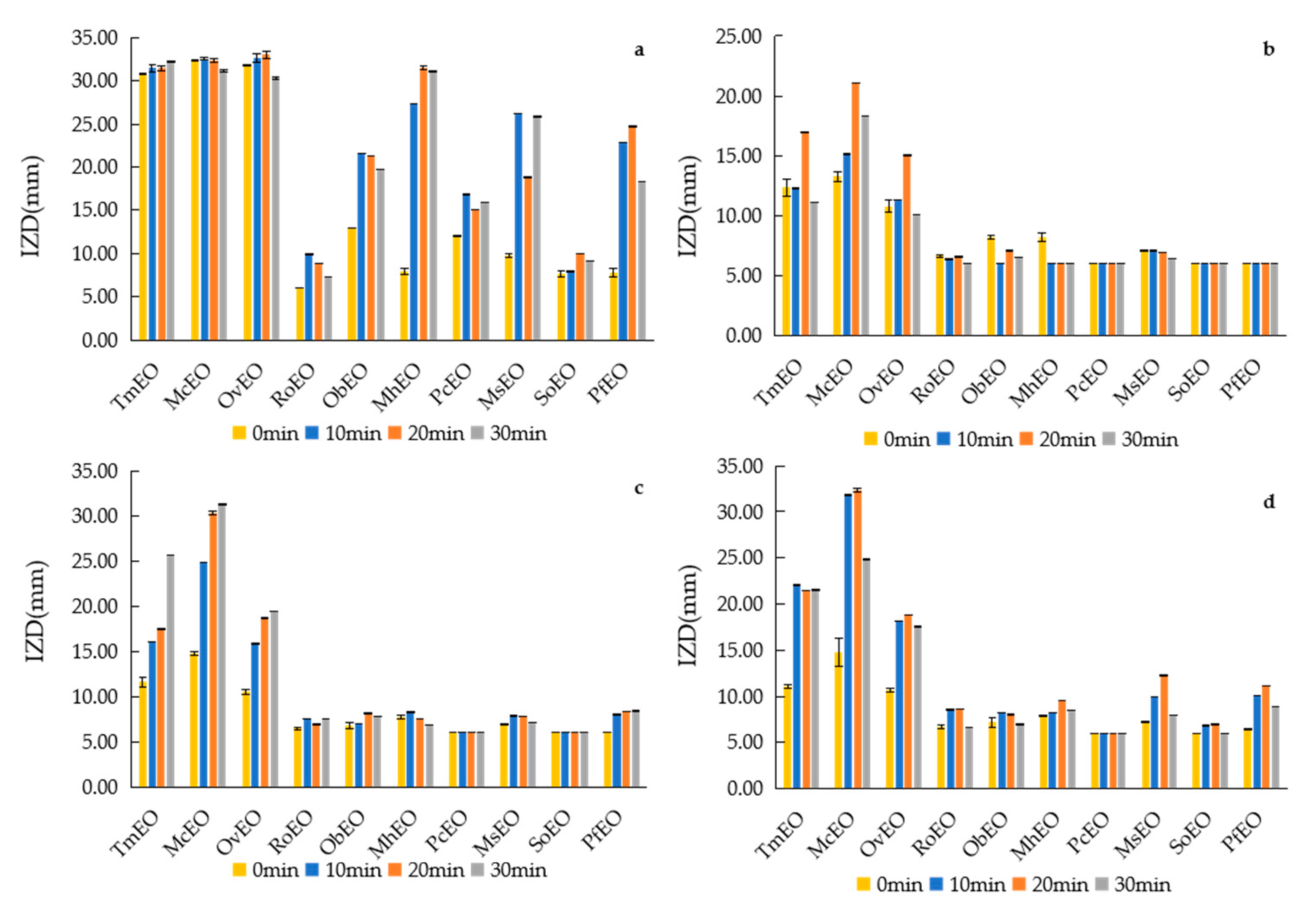
2.5.3. Effects of UV Irradiation Time on Antibacterial Stability of EOs
3. Materials and Methods
3.1. Plant Materials Collection and EOs Extraction
3.2. Identification of the Chemical Components of the EOs
3.3. Determination of Antioxidant Activity
3.3.1. Antioxidant Activity Determined by DPPH
3.3.2. Antioxidant Activity Determined by FRAP
3.4. Determination of Antibacterial Activity
3.4.1. Test Microorganisms
3.4.2. Inhibition Zone Diameter
3.4.3. Minimal Inhibitory Concentrations (MIC) and Minimal Bactericidal Concentration (MBC)
3.4.4. Antibacterial Stability of EOs
3.5. Statistical Analysis
4. Conclusions
Supplementary Materials
Author Contributions
Funding
Conflicts of Interest
References
- Bjornerot, L.; Franklin, A.; Tysen, E. Usage of antibacterial and antiparasitic drugs in animals in Sweden between 1988 and 1993. Veter- Rec. 1996, 139, 282–286. [Google Scholar] [CrossRef] [PubMed]
- Sui, Q.W.; Zhang, J.Y.; Wei, Y.S.; Chen, M.X.; Dong, H.M.; Xiong, J.H. Veterinary antibiotics use, occurrence of antibiotic resistance pathogen and its antibiotic resistance genes in animal production: An overview. Asian J. Ecotoxicol. 2015, 10, 20–34. [Google Scholar] [CrossRef]
- Chen, X.; Zhao, X.; Deng, Y.; Bu, X.; Ye, H.; Guo, N. Antimicrobial potential of myristic acid against Listeria monocytogenes in milk. J. Antibiot. 2019, 72, 298–305. [Google Scholar] [CrossRef] [PubMed]
- Guo, S.; Ma, J.; Xing, Y.; Xu, Y.; Jin, X.; Yan, S.; Shi, B. Artemisia annua L. aqueous extract as an alternative to antibiotics improving growth performance and antioxidant function in broilers. Ital. J. Anim. Sci. 2020, 19, 399–409. [Google Scholar] [CrossRef] [Green Version]
- Bakkali, F.; Averbeck, S.; Idaomar, M. Biological effects of essential oils—A review. Food Chem. Toxicol. 2008, 46, 446–475. [Google Scholar] [CrossRef]
- Burt, S. Essential oils: Their antibacterial properties and potential applications in foods—A review. Int. J. Food Microbiol. 2004, 94, 223–253. [Google Scholar] [CrossRef]
- Baker, D.H.A.; Al-Moghazy, M.; Elsayed, A. The in vitro cytotoxicity, antioxidant and antibacterial potential of Satureja hortensis L. essential oil cultivated in Egypt. Bioorganic Chem. 2020, 95, 103559. [Google Scholar] [CrossRef]
- Al-Mariri, A.; Safi, M. The Antibacterial Activity of Selected Labiatae (Lamiaceae) Essential Oils against Brucella melitensis. Iran. J. Med Sci. 2013, 38, 44–50. [Google Scholar]
- Nabavi, S.M.; Marchese, A.; Izadi, M.; Curti, V.; Daglia, M. Plants belonging to the genus Thymus as antibacterial agents: From farm to pharmacy. Food Chem. 2015, 173, 339–347. [Google Scholar] [CrossRef]
- Xie, Q.; Liu, Z.; Li, Z. Chemical Composition and Antioxidant Activity of Essential Oil of Six Pinus Taxa Native to China. Molecules 2015, 20, 9380–9392. [Google Scholar] [CrossRef] [Green Version]
- Della Cuna, F.S.R.; Calevo, J.; Bari, E.; Caser, M.; Boselli, C.; Tava, A.; Della Cuna, F.R. Characterization and Antioxidant Activity of Essential Oil of Four Sympatric Orchid Species. Molecules 2019, 24, 3878. [Google Scholar] [CrossRef] [PubMed] [Green Version]
- Zhang, Z.; Shen, P.; Liu, J.; Gu, C.; Lu, X.; Li, Y.; Cao, Y.; Liu, B.; Fu, Y.; Zhang, N. In Vivo Study of the Efficacy of the Essential Oil of Zanthoxylum bungeanum Pericarp in Dextran Sulfate Sodium-Induced Murine Experimental Colitis. J. Agric. Food Chem. 2017, 65, 3311–3319. [Google Scholar] [CrossRef] [PubMed]
- Lai, F.; Wissing, S.A.; Müller, R.H.; Fadda, A.M. Artemisia arborescens L essential oil-loaded solid lipid nanoparticles for potential agricultural application: Preparation and characterization. AAPS PharmSciTech 2006, 7, E10–E18. [Google Scholar] [CrossRef] [Green Version]
- González, J.O.W.; Gutiérrez, M.M.; Murray, A.P.; Ferrero, A. Composition and biological activity of essential oils from Labiatae against Nezara viridula (Hemiptera: Pentatomidae) soybean pest. Pest Manag. Sci. 2011, 67, 948–955. [Google Scholar] [CrossRef] [PubMed]
- Ju, J.; Xie, Y.; Guo, Y.; Cheng, Y.; Qian, H.; Yao, W. Application of edible coating with essential oil in food preservation. Crit. Rev. Food Sci. Nutr. 2018, 59, 2467–2480. [Google Scholar] [CrossRef]
- Andrys, D.; Adaszyńska-Skwirzyńska, M.; Kulpa, D. Essential oil obtained from micropropagated lavender, its effect on HSF cells and application in cosmetic emulsion as a natural protective substance. Nat. Prod. Res. 2017, 32, 849–853. [Google Scholar] [CrossRef]
- Li, G.P.; Dudai, N.; Li, J.J.; Lin, L.J. Comparison of volatile composition of essential oils from three different Lamiaceae plants. Chin. J. Trop. Crops 2018, 39, 1644–1650. [Google Scholar] [CrossRef]
- Wang, D.; Cheng, B.; Ding, L.; Ren, M.S. Extraction Technology and Chemical Composition Analysis of Thyme Essential Oil. China Condiment 2019, 44, 76–80. [Google Scholar] [CrossRef]
- Cao, L.; Si, J.Y.; Liu, Y.; Sun, H.; Jin, W.; Li, Z.; Zhao, X.H.; Le Pan, R. Essential oil composition, antimicrobial and antioxidant properties of Mosla chinensis Maxim. Food Chem. 2009, 115, 801–805. [Google Scholar] [CrossRef]
- La Pergola, A.; Restuccia, C.; Napoli, E.; Bella, S.; Brighina, S.; Russo, A.; Suma, P. Commercial and wild SicilianOriganum vulgareessential oils: Chemical composition, antimicrobial activity and repellent effects. J. Essent. Oil Res. 2017, 29, 451–460. [Google Scholar] [CrossRef]
- Bouyahya, A.; Et-Touys, A.; Bakri, Y.; Talbaui, A.; Fellah, H.; Abrini, J.; Dakka, N. Chemical composition of Mentha pulegium and Rosmarinus officinalis essential oils and their antileishmanial, antibacterial and antioxidant activities. Microb. Pathog. 2017, 111, 41–49. [Google Scholar] [CrossRef] [PubMed]
- Kathirvel, P.; Ravi, S. Chemical composition of the essential oil from basil (Ocimum basilicumLinn.) and itsin vitrocytotoxicity against HeLa and HEp-2 human cancer cell lines and NIH 3T3 mouse embryonic fibroblasts. Nat. Prod. Res. 2012, 26, 1112–1118. [Google Scholar] [CrossRef]
- Li, G.M.; Li, S.L.; Bai, Y.B.; Hu, Y.L. Analysis of Chemical Components of Essential Oil from Mentha piperita Grown in Ruilli by GC-MS. Chin. J. Trop. Agric. 2017, 37, 84–88. [Google Scholar] [CrossRef]
- Pang, Y.X.; Zhang, Y.B.; Wu, M.; Yuan, Y.; Yu, F.L.; Chen, X.L.; Hu, X. Comparative Analysis of Patchouli Essential Oils with Singlecropping and Intercropping with Rubber Tree. Biomass Chem. Eng. 2014, 48, 15–18. [Google Scholar] [CrossRef]
- Almeida, P.P.; Mezzomo, N.; Ferreira, S.R. Extraction of Mentha spicata L. Volatile Compounds: Evaluation of Process Parameters and Extract Composition. Food Bioprocess. Technol. 2010, 5, 548–559. [Google Scholar] [CrossRef]
- Kulak, M.; Gul, F.; Sekeroglu, N. Changes in growth parameter and essential oil composition of sage (Salvia officinalis L.) leaves in response to various salt stresses. Ind. Crop. Prod. 2020, 145, 112078. [Google Scholar] [CrossRef]
- You, C.X.; Yang, K.; Wu, Y.; Zhang, W.J.; Wang, Y.; Geng, Z.F.; Chen, H.P.; Jiang, H.Y.; Du, S.S.; Deng, Z.W.; et al. Chemical composition and insecticidal activities of the essential oil of Perilla frutescens (L.) Britt. aerial parts against two stored product insects. Eur. Food Res. Technol. 2014, 239, 481–490. [Google Scholar] [CrossRef]
- Aruoma, O.I. Methodological considerations for characterizing potential antioxidant actions of bioactive components in plant foods. Mutat. Res. Mol. Mech. Mutagen. 2003, 9–20. [Google Scholar] [CrossRef]
- Baharfar, R.; Azimi, R.; Mohseni, M. Antioxidant and antibacterial activity of flavonoid-, polyphenol- and anthocyanin-rich extracts from Thymus kotschyanus boiss & hohen aerial parts. J. Food Sci. Technol. 2015, 52, 6777–6783. [Google Scholar] [CrossRef] [Green Version]
- Skrypnik, L.; Novikova, A.; Tokupova, E. Improvement of Phenolic Compounds, Essential Oil Content and Antioxidant Properties of Sweet Basil (Ocimum basilicum L.) Depending on Type and Concentration of Selenium Application. Plants 2019, 8, 458. [Google Scholar] [CrossRef] [Green Version]
- Zhang, Z.; Zhang, S.; Su, R.; Xiong, D.; Feng, W.; Chen, J. Controlled Release Mechanism and Antibacterial Effect of Layer-By-Layer Self-Assembly Thyme Oil Microcapsule. J. Food Sci. 2019, 84, 1427–1438. [Google Scholar] [CrossRef] [PubMed]
- Assiri, A.M.A.; Elbanna, K.; Al-Thubiani, A.; Ramadan, M.F. Cold-pressed oregano (Origanum vulgare) oil: A rich source of bioactive lipids with novel antioxidant and antimicrobial properties. Eur. Food Res. Technol. 2015, 242, 1013–1023. [Google Scholar] [CrossRef]
- Niu, F.; Pan, W.; Su, Y.; Yang, Y. Physical and antimicrobial properties of thyme oil emulsions stabilized by ovalbumin and gum arabic. Food Chem. 2016, 212, 138–145. [Google Scholar] [CrossRef]
- Dutra, T.V.; Castro, J.C.; Menezes, J.L.; Ramos, T.R.; Prado, I.N.D.; Machinski, M.; Mikcha, J.M.G.; Filho, B.A.D.A. Bioactivity of oregano (Origanum vulgare) essential oil against Alicyclobacillus spp. Ind. Crop. Prod. 2019, 129, 345–349. [Google Scholar] [CrossRef]
- Hussain, A.I.; Anwar, F.; Chatha, S.A.S.; Jabbar, A.; Mahboob, S.; Nigam, P.S. Rosmarinus officinalis essential oil: Antiproliferative, antioxidant and antibacterial activities. Braz. J. Microbiol. 2010, 41, 1070–1078. [Google Scholar] [CrossRef] [Green Version]
- Saha, S.; Dhar, T.; Sengupta, C.; Ghosh, P. Biological activities of essential oils and methanol extracts of five Ocimum species against pathogenic bacteria. Czech., J. Food Sci. 2013, 31, 195–202. [Google Scholar] [CrossRef] [Green Version]
- Delamare, A.P.L.; Moschen-Pistorello, I.T.; Artico, L.; Atti-Serafini, L.; Echeverrigaray, S. Antibacterial activity of the essential oils of Salvia officinalis L. and Salvia triloba L. cultivated in South Brazil. Food Chem. 2007, 100, 603–608. [Google Scholar] [CrossRef]
- Chowdhury, S.; Mandal, G.P.; Patra, A.K.; Kumar, P.; Samanta, I.; Pradhan, S.; Samanta, A.K. Different essential oils in diets of broiler chickens: 2. Gut microbes and morphology, immune response, and some blood profile and antioxidant enzymes. Anim. Feed. Sci. Technol. 2018, 236, 39–47. [Google Scholar] [CrossRef]
- Zeng, Z.; Zhang, S.; Wang, H.; Piao, X. Correction to: Essential oil and aromatic plants as feed additives in non-ruminant nutrition: A review. J. Anim. Sci. Biotechnol. 2020, 11, 1. [Google Scholar] [CrossRef] [PubMed]
- Hu, W.; Feng, K.; Xiu, Z.; Jiang, A.; Lao, Y. Efficacy of thyme oil-alginate-based coating in reducing foodborne pathogens on fresh-cut apples. Int. J. Food Sci. Technol. 2019, 54, 3128–3137. [Google Scholar] [CrossRef]
- Wang, S.P. Study on Antimicrobial Activity of Several Kinds of Aromatic Essential Oil. Anhui Agric. Bull. 2018, 24, 20–24. [Google Scholar] [CrossRef]
- Polatoğlu, K.; Sen, A.; Kandemir, A.; Gören, N. Essential Oil Composition and DPPH Scavenging Activity of EndemicTanacetum mucroniferumHub.—Mor. & Grierson from Turkey. J. Essent. Oil Bear. Plants 2012, 15, 66–74. [Google Scholar] [CrossRef]
- Thaipong, K.; Boonprakob, U.; Crosby, K.; Cisneros-Zevallos, L.; Byrne, D.H. Comparison of ABTS, DPPH, FRAP, and ORAC assays for estimating antioxidant activity from guava fruit extracts. J. Food Compos. Anal. 2006, 19, 669–675. [Google Scholar] [CrossRef]
- Chen, Y.X.; Liu, J.H.; Lin, F.; Du, X.D. Determination of Antioxidative Activity of 41 Kinds of Chinese Herbal Medicines by Using DPPH and FRAP Methods. Res. Explor. Lab. 2011, 30, 11–14. [Google Scholar]
- Deng, R.N.; Zhang, D.Q. Comparative Study on Determination Methods of Bacteriostasis of Lavender Essential Oil in Field. Environ. Environ. Sci. Manag. 2017, 42, 102–105. [Google Scholar]
- Ding, W.L.; Wang, R.; Hao, D.L.; Zhang, X.Y.; Li, Y. Evaluation of Antagonistic Activity of Biocontrol Microbial Agents against Ginseng Diseases. Mod. Chin. Med. 2018, 20, 1122–1125. [Google Scholar]
- Yang, Y.D.; Lu, D.H.; Yang, D.M.; Yuan, X.J.; Qin, H.Y. Antibacterial Activity and Mechanism of Essential Oil from Cinamomum camphora on Staphylococcus aureus. Mod. Chin. Med. 2017, 19, 372–376. [Google Scholar] [CrossRef]
- Duan, W.L.; Liu, Y.Q.; Bao, Y.H. Study on Antimicrobial Activities and Stability of Essential Oil from Artemisia argyi. J. Food Sci. Biotechnol. 2015, 34, 1332–1337. [Google Scholar]


| No | Molecular Formula | Compounds | Relative Peak Area (%) | |||||||||
|---|---|---|---|---|---|---|---|---|---|---|---|---|
| TmEO | McEO | OvEO | RoEO | ObEO | MhEO | PcEO | MsEO | SoEO | PfEO | |||
| 1 | C10H16 | α-Pinene | 0.46 | 0.13 | - | 26.46 | 0.09 | 0.4 | - | 0.58 | 1.93 | - |
| 2 | C10H16 | (−)-Camphene | - | - | - | 3.53 | - | - | - | - | - | - |
| 3 | C10H16 | α-thujene | 1.14 | 0.26 | - | - | - | - | - | - | - | - |
| 4 | C10H16 | Camphene | 0.49 | 0.1 | - | - | - | - | - | 0.03 | 3.63 | - |
| 5 | C10H16 | β-Pinene | 0.2 | - | 0.4 | 1.09 | 0.22 | 0.31 | - | 0.59 | 3.65 | - |
| 6 | C10H14 | 2,4(10)-Thujadiene | - | - | - | 0.72 | - | - | - | - | - | - |
| 7 | C10H16 | Sabinene | 0.08 | - | - | - | 0.08 | 0.08 | - | 0.25 | 0.19 | - |
| 8 | C10H16 | β-Myrcene | 0.87 | 2.18 | - | 0.83 | - | 0.27 | - | 1.59 | 0.6 | - |
| 9 | C10H16 | α-terpinene | 2.44 | 3.19 | 0.84 | 0.44 | - | - | - | - | 0.11 | - |
| 10 | C15H24 | β-Patchoulene | - | - | - | - | - | - | 1.23 | - | - | - |
| 11 | C15H24 | α-Guaiene | - | - | - | - | - | - | 6.09 | - | - | - |
| 12 | C15H24 | γ-Gurjunene | - | - | - | - | - | - | 0.49 | - | - | - |
| 13 | C10H16 | d-Limonene | 0.3 | 0.26 | 0.06 | 0.85 | 0.07 | 1.53 | - | 9.64 | 0.49 | 1.38 |
| 14 | C10H18O | Eucalyptol | - | - | 0.31 | 11.31 | 4.48 | - | - | 0.64 | 5.11 | - |
| 15 | C10H16 | trans-β-Ocimene | - | - | - | - | - | - | - | 0.48 | 0.03 | - |
| 16 | C10H16 | allo-Ocimene | - | - | - | - | - | - | - | 0.16 | - | - |
| 17 | C10H16 | β-phellandrene | - | 0.25 | - | - | - | - | - | - | - | - |
| 18 | C10H16 | γ-Terpinene | 16.42 | 15.14 | 6.14 | 1.11 | 0.09 | - | - | - | 0.46 | - |
| 19 | C10H16 | β-Ocimene | - | - | - | - | 0.71 | - | - | - | - | - |
| 20 | C10H14 | p-Cymene | 21.17 | 19.52 | 14.11 | 1.76 | - | - | - | - | 0.25 | - |
| 21 | C10H16 | (+)-4-Carene | 0.09 | - | - | - | - | - | - | - | 0.23 | - |
| 22 | C10H16O | Tanacetone | - | - | - | - | - | - | - | - | 27.99 | - |
| 23 | C10H16O | Thujone | - | - | - | - | - | - | - | - | 5.24 | - |
| 24 | C10H16 | Terpinolene | - | 0.18 | - | 0.85 | 0.13 | - | - | - | - | - |
| 25 | C6H12O | 3-Hexen-1-ol | - | - | - | - | 0.33 | - | - | - | - | - |
| 26 | C8H18O | 3-Octanol | 0.27 | - | 2.05 | - | - | 0.6 | - | 0.31 | - | 0.1 |
| 27 | C10H14O | Perillene | - | - | - | - | - | - | - | - | - | 1.62 |
| 28 | C10H18O | l-Menthone | - | - | - | - | - | 12.21 | - | - | - | - |
| 29 | C10H18O | (+)-Isomenthone | - | - | - | - | - | 3.07 | - | - | - | - |
| 30 | C8H16O | 1-Octen-3-ol | 2.97 | - | 1.66 | 0.02 | 0.05 | 0.14 | 0.08 | 0.18 | ||
| 31 | C10H18O | Cis-β-Terpineol | - | - | - | - | 0.19 | - | - | - | - | - |
| 32 | C10H16O | (+)-2-Bornanone | - | - | - | 4.08 | - | - | - | - | - | - |
| 33 | C10H16O | Isopinocamphon | - | - | - | 1 | - | - | - | 0.06 | - | |
| 34 | C10H14O | Pinocarvone | - | - | - | 0.41 | - | - | - | - | - | - |
| 35 | C10H16O | Camphor | 0.1 | - | 0.57 | - | 0.67 | - | - | - | 16.21 | |
| 36 | C15H24 | (−)-β-Bourbonene | - | - | 0.23 | - | 0.14 | 0.16 | - | 1.21 | - | 0.07 |
| 37 | C12H22O2 | l-Menthyl acetate | - | - | - | - | - | 3.73 | - | - | - | - |
| 38 | C10H18O | Linalool | 2.97 | 0.06 | 0.14 | 2.99 | 26.65 | - | - | 0.49 | 0.37 | 2.97 |
| 39 | C10H16O | 2-methyl-5-(1-methylethenyl)-Cyclohexanone | - | - | - | - | - | - | - | 2.76 | - | - |
| 40 | C11H16O | 2-Isopropyl-1-methoxy-4-methylbenzene | - | - | 11.34 | - | - | - | - | - | - | - |
| 41 | C7H8O2 | 5-methyl-2-acetyl-Furan | - | - | 0.23 | - | - | - | - | - | - | - |
| 42 | C10H20O | Menthol | - | - | 0.97 | - | - | 69.05 | - | - | - | - |
| 43 | C10H12O | Estragole | - | - | 0.31 | - | 1.32 | - | - | - | - | - |
| 44 | C15H24 | cis-Muurola-4(15),5-diene | - | - | - | - | 0.29 | - | - | - | - | - |
| 45 | C15H24 | γ-Muurolene | - | - | 0.34 | - | 4.14 | - | - | - | - | - |
| 46 | C12H20O2 | Bornyl acetate | 0.26 | - | - | 1.85 | 2.28 | - | - | - | 0.73 | - |
| 47 | C11H16O | 2-Isopropyl-5-methylanisole | 3.17 | - | 5.6 | - | - | - | - | - | - | - |
| 48 | C15H24 | Caryophyllene | 0.03 | - | - | 0.64 | - | 0.44 | - | - | 1.81 | 10.21 |
| 49 | C10H14O2 | Elsholtzia ketone | - | - | - | - | - | - | - | - | - | 0.59 |
| 50 | C10H20O | Neoisomenthol | - | - | - | - | - | 1.63 | - | - | - | - |
| 51 | C10H16O | Pulegone | - | - | - | - | - | 0.75 | - | - | - | - |
| 52 | C10H18O | Lavandulol | - | - | - | - | - | 0.37 | - | - | - | - |
| 53 | C10H18O | α-Terpineol | - | - | - | - | - | 0.11 | - | - | - | - |
| 54 | C10H18O | Terpinen-4-ol | - | - | - | 1.21 | 0.76 | - | - | 0.06 | 0.63 | - |
| 55 | C10H16O | Hotrienol | - | - | - | 0.02 | 0.07 | - | - | - | - | - |
| 56 | C10H18O | p-menth-1(7)-en-8-ol | - | - | - | 0.32 | 0.27 | - | - | - | - | - |
| 57 | C10H14O | Verbenone | - | - | - | 16.56 | - | - | - | - | - | - |
| 58 | C12H20O2 | Nerol acetate | - | - | - | 0.57 | - | - | - | - | - | - |
| 59 | C10H20O | (S)-3,7-dimethyl-7-Octen-1-ol | - | - | - | 0.61 | - | - | - | - | - | - |
| 60 | C15H24 | Humulene | 0.25 | 3.74 | - | - | - | - | 0.16 | 0.05 | 6.44 | 1.14 |
| 61 | C15H24 | (E)-β-Famesene | - | - | - | - | - | - | - | 0.55 | - | 0.23 |
| 62 | C15H24 | cis-β-farnesene | 0.05 | - | - | - | 2.27 | - | - | - | - | - |
| 63 | C10H18O | Borneol | 2.33 | 0.1 | 1.34 | - | 0.03 | - | - | - | 1.89 | - |
| 64 | C15H24 | (+)-δ-Cadinene | - | - | - | - | - | - | - | - | 0.04 | 0.02 |
| 65 | C10H16O | Piperitone | - | - | 0.61 | - | - | 1.6 | - | - | - | 2.94 |
| 66 | C10H14O | d-Carvone | - | - | 0.89 | - | - | - | - | 71.1 | - | - |
| 67 | C10H18O | Dihydrocarveol | - | - | 0.34 | - | - | - | - | 1.14 | - | - |
| 68 | C12H18O2 | cis-Carvyl acetate | - | - | - | - | - | - | - | 0.87 | - | - |
| 69 | C15H22 | cis-Calamenene | - | - | - | - | - | - | - | 0.34 | - | - |
| 70 | C15H24 | Germacrene D | 0.77 | - | - | - | 4.35 | - | - | - | - | 0.76 |
| 71 | C15H24 | α-Bulnesene | - | - | - | - | 1.74 | - | 5.68 | - | - | - |
| 72 | C15H26O | Ledol | - | - | - | - | - | - | 1.22 | - | - | - |
| 73 | C10H14O | (−)-Carvone | 0.03 | - | - | - | - | - | - | - | - | 0.22 |
| 74 | C15H24 | (Z,E)-α-Farnesene | - | - | - | - | - | - | - | - | - | 4.44 |
| 75 | C15H24 | γ-Elemene | 0.03 | - | - | - | 0.08 | - | - | - | - | 0.09 |
| 76 | C15H24 | Copaene | - | - | - | - | - | - | - | - | - | 0.14 |
| 77 | C15H24 | β-bisabolene | 3.96 | - | 2.26 | - | 0.22 | - | - | - | - | - |
| 78 | C10H14O | perillaldehyde | - | - | - | - | - | - | - | - | - | 2.83 |
| 79 | C10H14O2 | Perillaketone | - | - | - | - | - | - | - | - | - | 35.56 |
| 80 | C10H16O | Myrtenol | - | - | 0.02 | 0.83 | - | - | - | - | - | - |
| 81 | C10H14O | α-α-4-trimethyl-Benzenemethanol | - | - | - | 0.11 | - | - | - | - | 0.04 | - |
| 82 | C10H16O | trans-Carveol | - | - | 0.08 | - | - | - | - | 2.1 | 0.04 | - |
| 83 | C10H16O | cis-Carveol | - | - | - | - | - | - | - | 0.68 | - | - |
| 84 | C13H2O | trans-α-bergamotene | - | 1.9 | - | - | 14.16 | - | - | - | - | - |
| 85 | C15H24 | β-sesquiphellandrene | 0.06 | 0.4 | 0.05 | - | 0.15 | - | - | - | - | - |
| 86 | C10H18O | Nerol | 0.06 | - | - | - | 0.17 | - | - | - | - | 0.07 |
| 87 | C10H12O2 | Dehydroelsholtzia ketone | - | - | - | - | - | - | - | - | - | 0.59 |
| 88 | C10H12O2 | Isoegomaketone | - | - | - | - | - | - | - | - | - | 20.4 |
| 89 | C12H16O2 | Thymol acetate | 2.22 | 11.95 | - | - | - | - | - | - | - | - |
| 90 | C10H18O | Geraniol | 0.06 | - | - | 5.91 | 0.52 | - | - | - | - | - |
| 91 | C11H14O2 | Methyleugenol | - | - | - | - | 1.53 | - | - | - | - | 0.27 |
| 92 | C15H26O | Epicubenol | - | - | - | - | 1.34 | - | - | 0.33 | - | - |
| 93 | C15H26O | trans-Nerolidol | - | - | - | - | 0.34 | - | 0.24 | - | - | - |
| 94 | C15H26O | Viridiflorol | - | - | - | - | 0.13 | - | 0.08 | 0.05 | 7.85 | - |
| 95 | C14H22O | Norpatchoulenol | - | - | - | - | - | - | 1.14 | - | - | - |
| 96 | C15H24O | Caryophyllene Oxide | 0.33 | 0.06 | 0.64 | 0.15 | - | 0.07 | 1.86 | - | 0.48 | 0.87 |
| 97 | C15H26O | γ-Eudesmol | 0.4 | - | - | - | - | - | - | - | - | - |
| 98 | C15H24O | Espatulenol | 0.03 | - | 0.53 | - | - | 0.03 | 0.03 | 0.01 | 0.04 | 0.22 |
| 99 | C15H26O | Farnesol | - | - | - | - | - | - | 0.9 | - | - | - |
| 100 | C12H16O4 | Pogostone | - | - | - | - | - | - | 5.45 | - | - | - |
| 101 | C15H24O | Humulene epoxide II | - | 0.27 | - | 0.04 | - | - | 0.36 | - | 1.32 | |
| 102 | C10H12O2 | Eugenol | 0.11 | 0.26 | 0.06 | - | 10.27 | - | - | 0.17 | - | 0.33 |
| 103 | C15H26O | τ-Cadinol | - | - | - | - | 7.98 | 0.02 | - | 0.07 | - | 0.03 |
| 104 | C12H16O3 | Elemicin | - | - | - | - | - | - | - | - | - | 0.84 |
| 105 | C11H12O3 | Myristicin | - | - | - | - | - | - | - | - | - | 0.11 |
| 106 | C12H16O3 | Isoelemicin | - | - | - | - | - | - | - | - | - | 3.29 |
| 107 | C12H16O3 | Asarone | - | - | - | - | - | - | - | - | - | 1.71 |
| 108 | C15H26O | β-Eudesmol | - | - | - | - | 0.45 | - | - | - | - | - |
| 109 | C15H26O | Patchouli alcohol | - | - | 0.01 | - | - | 0.4 | 50.52 | - | - | - |
| 110 | C15H26O | Pogostol | - | - | - | - | - | - | 5.43 | - | - | - |
| 111 | C10H14O | Thymol | 23.7 | 26.59 | 14.64 | 0.15 | - | - | - | - | 0.27 | - |
| 112 | C15H26O | Neointermedeol | - | - | 0.09 | - | - | - | 0.38 | - | - | - |
| 113 | C15H24O | Longifolenaldehyde | - | - | - | - | - | - | 0.27 | - | - | - |
| 114 | C12H16O2 | Carvacryl acetate | - | - | 0.36 | - | - | - | - | - | - | - |
| 115 | C10H14O | Pulespenone | - | - | 1.75 | 0.15 | - | - | - | - | - | - |
| 116 | C11H16O | cis-Jasmone | - | - | 0.05 | 0.01 | - | - | - | - | - | - |
| 117 | C10H14O | Carvacrol | 2.48 | 7.66 | 20.82 | 0.07 | - | 0.16 | - | 0.14 | 0.05 | - |
| 118 | C15H26O | α-Cadinol | 0.03 | - | 0.03 | - | 0.26 | - | - | 0.32 | - | - |
| 119 | C12H14O3 | Eugenol acetate | - | - | - | - | 0.36 | - | - | - | - | - |
| 120 | C14H22O | 2,4-Di-tert-butylphenol | 0.07 | 0.05 | - | 0.03 | 0.04 | 0.03 | - | - | 0.04 | - |
| 121 | C20H40O | Phytol | 0.11 | - | - | - | 2.17 | - | 0.05 | - | 0.03 | - |
| 122 | C20H34O | 13-Epimanool | - | - | - | - | - | - | - | - | 8.91 | - |
| 123 | C9H11Cl3NO3PS | Chlorpyrifos | - | - | - | - | 0.47 | - | - | - | - | - |
| Total Content (%) | 90.51 | 94.25 | 89.87 | 86.68 | 92.06 | 97.02 | 81.58 | 96.85 | 97.24 | 94.22 | ||
| EOs | FRAP | DPPH (IC50, μg/mL) |
|---|---|---|
| TmEO | 271.84 ± 4.93 | 1.42 |
| McEO | 480.66 ± 29.90 | 1.92 |
| OvEO | 633.71 ± 13.14 | 1.47 |
| RoEO | 22.14 ± 0.63 | 20.36 |
| ObEO | 1536.67 ± 24.22 | <1 |
| MhEO | 22.32 ± 1.33 | 23.95 |
| PcEO | 30.35 ± 10.65 | 49.74 |
| MsEO | 24.54 ± 0.69 | 13.34 |
| SoEO | 17.22 ± 2.58 | 11.86 |
| PfEO | 67.14 ± 1.84 | 3.77 |
| Trolox | 2.02 ± 0.02 | - |
| VC | - | 6.04 |
| PG | - | 5.47 |
| EOs | Activities | S. aureus | E. coli | B. subtilis | S. enteriditis |
|---|---|---|---|---|---|
| TmEO | IZD | 30.77 ± 0.06 | 12.35 ± 0.73 | 11.59 ± 0.49 | 11.11 ± 0.19 |
| MIC | 2 | 1 | 2 | 1 | |
| MBC | 4 | 2 | 2 | 1 | |
| McEO | IZD | 32.32 ± 0.07 | 13.26 ± 0.45 | 14.72 ± 0.19 | 14.81 ± 1.55 |
| MIC | 1 | 2 | 1 | 1 | |
| MBC | 2 | 4 | 1 | 2 | |
| OvEO | IZD | 31.82 ± 0.03 | 10.78 ± 0.49 | 10.52 ± 0.29 | 10.71 ± 0.17 |
| MIC | 2 | 4 | 2 | 1 | |
| MBC | 4 | 4 | 2 | 2 | |
| RoEO | IZD | 6.00 | 6.65 ± 0.14 | 6.46 ± 0.12 | 6.67 ± 0.21 |
| MIC | 4 | 8 | 8 | 4 | |
| MBC | 16 | 16 | 16 | 4 | |
| ObEO | IZD | 12.98 ± 0.01 | 8.25 ± 0.17 | 6.82 ± 0.31 | 7.19 ± 0.50 |
| MIC | 2 | 4 | 8 | 8 | |
| MBC | 16 | 16 | 16 | 16 | |
| MhEO | IZD | 7.96 ± 0.34 | 8.20 ± 0.36 | 7.75 ± 0.20 | 7.94 ± 0.04 |
| MIC | 16 | NT | 8 | 4 | |
| MBC | 16 | NT | 8 | 4 | |
| PcEO | IZD | 12.02 ± 0.02 | 6.00 | 6.00 | 6.00 |
| MIC | NT | NT | NT | NT | |
| MBC | NT | NT | NT | NT | |
| MsEO | IZD | 9.78 ± 0.16 | 7.10 ± 0.08 | 6.93 ± 0.07 | 7.22 ± 0.04 |
| MIC | 16 | 8 | NT | NT | |
| MBC | 32 | 8 | NT | NT | |
| SoEO | IZD | 7.68 ± 0.39 | 6.00 | 6.00 | 6.00 |
| MIC | NT | NT | 16 | 4 | |
| MBC | NT | NT | 32 | 8 | |
| PfEO | IZD | 7.81 ± 0.46 | 6.00 | 6.00 | 6.44 ± 0.04 |
| MIC | 32 | NT | 8 | 4 | |
| MBC | NT | NT | 16 | 8 | |
| CTC | IZD | 16.12 ± 0.24 | 6.00 | 9.68 ± 0.12 | 6.00 |
| MIC | 8 | NT | NT | NT | |
| MBC | 8 | NT | NT | NT |
Sample Availability: Samples of the compounds are available from the authors. |
Publisher’s Note: MDPI stays neutral with regard to jurisdictional claims in published maps and institutional affiliations. |
© 2020 by the authors. Licensee MDPI, Basel, Switzerland. This article is an open access article distributed under the terms and conditions of the Creative Commons Attribution (CC BY) license (http://creativecommons.org/licenses/by/4.0/).
Share and Cite
Liu, M.; Luo, F.; Qing, Z.; Yang, H.; Liu, X.; Yang, Z.; Zeng, J. Chemical Composition and Bioactivity of Essential Oil of Ten Labiatae Species. Molecules 2020, 25, 4862. https://doi.org/10.3390/molecules25204862
Liu M, Luo F, Qing Z, Yang H, Liu X, Yang Z, Zeng J. Chemical Composition and Bioactivity of Essential Oil of Ten Labiatae Species. Molecules. 2020; 25(20):4862. https://doi.org/10.3390/molecules25204862
Chicago/Turabian StyleLiu, Mengting, Feiya Luo, Zhixing Qing, Huichao Yang, Xiubin Liu, Zihui Yang, and Jianguo Zeng. 2020. "Chemical Composition and Bioactivity of Essential Oil of Ten Labiatae Species" Molecules 25, no. 20: 4862. https://doi.org/10.3390/molecules25204862