Article

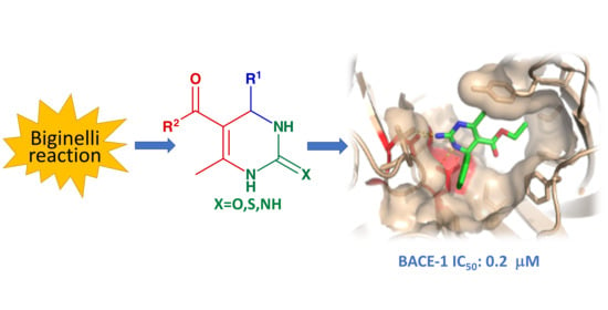
One Pot Synthesis of Micromolar BACE-1 Inhibitors Based on the Dihydropyrimidinone Scaffold and Their Thia and Imino Analogues

1
Dipartimento di Scienze Chimiche e Farmaceutiche, Università di Trieste, via Licio Giorgieri 1, 34127 Trieste, Italy
2
Dipartimento di Scienze, Università della Basilicata, Viale dell’Ateneo Lucano 10, 85100 Potenza, Italy
*
Author to whom correspondence should be addressed.
Molecules 2020, 25(18), 4152; https://doi.org/10.3390/molecules25184152
Submission received: 30 July 2020 / Revised: 31 August 2020 / Accepted: 2 September 2020 / Published: 10 September 2020
(This article belongs to the Special Issue Organic Synthesis in Natural Products and Bioactive Compounds)

Abstract

:
A library of dihydropyrimidinones was synthesized via a “one-pot” three component Biginelli reaction using different aldehydes in combination with β-dicarbonyl compounds and urea. Selected 2-thiooxo and 2-imino analogs were also obtained with the Biginelli reaction from thiourea and guanidine hydrochloride, respectively. The products were screened in vitro for their β-secretase inhibitory activity. The majority of the compounds resulted to be active, with IC50 in the range 100 nM–50 μM.

Graphical Abstract

1. Introduction

The 3,4-Dihydro-2-pyrimidinones (DHPMs), readily accessible in a single step via the Biginelli reaction [1,2,3] (Scheme 1, X = O), have recently attracted considerable attention for their multifaceted pharmacological properties [4,5]. Biologically active compounds based on this scaffold include HIV integrase inhibitors [6], human lactate hydrogenase inhibitors [7], calcium channel modulators [8], inhibitors of mitotic kinesin Eg5 [9], α1A adrenoceptor antagonists [10], inhibitors of prostaglandin E2 synthase [11], and other compounds with anticancer, antibacterial, antiviral, anti-inflammatory, antihyperglycemic, and analgesic activity [4,12,13].
The varied activity of DHPMs, combined with their synthetic accessibility and the possibility to rapidly assemble diverse substituents on the heterocyclic scaffold in a single multicomponent reaction, make the Biginelli reaction an attractive approach for the discovery of new biological activities. Multicomponent reactions, in particular, show great promise in the discovery of small molecule protease inhibitors possessing better pharmacological properties than peptidomimetic inhibitors [14,15,16].
Recently, there has been increasing interest in the development of small, non-peptide inhibitors of the β-amyloid cleaving enzyme 1 (BACE-1, β-secretase) [17,18,19,20,21,22,23,24], following the suggestion that this enzyme may be involved in the pathogenesis of Alzheimer’s Disease (AD) [19,25]. BACE-1 is an aspartyl protease involved in the proteolytic degradation of the β-amyloid precursor protein (APP) to form amyloid-β peptide (Aβ), a small protein with a high tendency to form aggregates that are found in the AD brain [19]. Nanomolar inhibitors based on several heterocyclic scaffolds have been developed and a number have progressed to clinical trials [17,18,26]. Although some programs have been discontinued due to phase III failure, interest in BACE-1 inhibitors is still high, in particular for the development of modifying therapies for early stage AD [27,28].
A structural feature common to these scaffolds is the presence of functional groups capable of establishing hydrogen bond interactions with the enzyme’s catalytic aspartates [18,19]. DHPMs similarly possess neighboring hydrogen bond donor and acceptor groups; we thus decided to explore the potential of the Biginelli reaction for the synthesis of new β-secretase inhibitors. In this work we report on the synthesis and preliminary evaluation of a set of dihydropyrimidinones and their thia and aza analogues (Scheme 1, X=S, NH) as BACE-1 inhibitors.
The analysis of the BACE-1 active site in complex with crystallized inhibitors [18,19], allowed for a preliminary assessment of the potential of Biginelli products as inhibitors. The cyclic urea motif embedded in the DHMP scaffold and its thiourea and guanidine analogues are in principle capable to establish multiple hydrogen bond interactions with the dyad of catalytic aspartates (Asp 32, Asp 228), while it is possible to introduce diversity at R1 and R2 (Scheme 1), to allow interactions at least with the closer subsites S1 (wider) and S1′ (narrow), which are both hydrophobic. We therefore planned to synthesize a first set of potential inhibitors 117 reported in Table 1, bearing different aromatic or aliphatic residues at R1 (including also polar groups) and ester, amide, or free carboxylic groups at R2.

2. Results and Discussion

The synthetic work started with the preparation of 3,4-dihydropyrimidin-2(1H)-ones 1a16a, and the corresponding thiones 1b3b, 9b, 10b, 12b and 16b by the Biginelli reaction of aldehydes and β-dicarbonyl compounds with urea or thiourea, respectively (Table 1). A wide variety of experimental conditions have been described for this reaction, with different solvents and acid catalysts [2,3], including microwave irradiation [30], solid phase [31,32], and mechanochemical [33] methods. Following a solvent free protocol [34] we carried out the reactions at 100 °C for three hours in the presence of ammonium chloride, obtaining the target compounds with yields from 50% to 92%. The carboxylic acids 16a and 16b were obtained by NaOH hydrolysis of 1a and 1b, respectively. Most of these compounds were already known in the literature, except for 9a, 10a, and 10b.
The inhibitory activity of the compounds thus obtained was determined in vitro on recombinant BACE-1 using a fluorogenic substrate analogue [35]. The results are reported in Table 1.
All the DHPMs and their thia derivatives inhibit BACE-1, with IC50s in the low μM range, except for 11a (71.3 μM) and 16a (34.0 μM). The most active compound among DHPMs is 2a, with IC50 0.32 μM. This inhibitor carries a phenyl group at R1, and a methyl group at R2. Compounds in which the phenyl group is replaced by other aromatic or aliphatic groups generally retain a significant IC50, although no improvement in activity is observed. No clear-cut electronic effect can be observed for aromatic systems with electron-withdrawing or donating groups (3a, 4a, 5a, and 8a). Larger aromatic groups such as naphthyl (9a, 10a) and benzothienyl (11a, 12a) appear to be tolerated by the enzyme; in this case, orientation seems to be an important factor, as shown by the comparison between 9a and 10a and, much more so, between 11a and 12a. Inhibition appears to be more sensitive to substitution at R2: the methyl ester 2a is more active than the corresponding ethyl ester 1a, and amides 14a, 15a, while the presence of a free carboxylic group in 16a lead to a major loss of activity. The latter may be related to the ionization of this molecule leading to mismatching interactions with the anionic catalytic site.
Thia derivatives, on average, are more active than DHPMs, and this is particularly evident in 16b, where sulfur is able to restore the poor activity of 16a. Better activities of sulfur containing compounds with respect to oxygen analogues are often observed in the interactions of small molecules with proteins. This has been explained with the reduced desolvation penalty that, in general, must be paid by sulfur compounds on entering a protein binding site [36].
Following on from the promising results observed with DHPMs and their 2-thia derivatives, we extended the study to a small selection of 2-imino-derivatives (Table 1: 1c3c, 9c, 10c, 16c). The Biginelli condensation of guanidine has been much less investigated than the corresponding reactions with urea and thiourea. Kappe, in 2001 [37], reported the one-pot three-component reaction with guanidine hydrochloride, giving the cyclocondensation products in satisfactory yields; the method, however, was limited to α-benzoylethylacetate as the β-dicarbonyl partner. Reduced reaction times were recently reported under ultrasonic irradiation [38] while a general procedure for the Biginelli reaction using a phase transfer catalyst, compatible also with guanidine, has been described [39].
Ethyl and methyl acetoacetate reacted smoothly with aldehydes under microwave irradiation [29] (EtOH, MW 120 °C, 10 min, excess NaHCO3) to give the corresponding 2-iminodihydropyrimidines 1c3c, 9c and 10c with good yields. Finally, the acid 16c was obtained by Pd/C hydrogenolysis of the corresponding benzyl ester 17c. 3,4-Dihydro-pyrimidin-2(1H)-imine derivatives (or their 2-amino tautomers) can, in principle, establish better interactions with the enzyme’s active site than their 2-oxo or 2-thia analogues. The more basic guanidine system is protonated at the enzyme’s optimal pH and can thus interact with the catalytic aspartate dyad by both charge complementarity and an extended network of hydrogen bonds [40]. Accordingly, imine derivatives 1c3c, 9c, 10c, and 16c are better inhibitors than their oxygen and thia analogs in nearly all cases. Only 9c is less active than the corresponding thia-derivative 9b, while 3b and 3c have comparable activities. Compound 10c is the most active inhibitor overall showing a higher activity that the isomer 9c; this confirms that the orientation of the large aromatic group with respect to the dihydropyrimidine group is important, as already observed for inhibitors 9a, 10a 11a, and 12a. The improvement of the activity is very marked on going from 16a to 16c. In this case, however, the net charge of the urea, thiourea, and guanidine derivatives are different, as 16a and 16b are anions, while 16c is, presumably, a neutral molecule at the enzyme’s optimal pH.
BACE-1 inhibitors must cross the blood-brain barrier in order to reach their target. Thus, permeability is an important factor to assess their pharmacological potential. BBB permeation is a complex process, depending on the interplay of several physicochemical parameters, among which lipophilicity, as defined by the octanol-water partition coefficient, is generally considered one of the most important [41]. Calculated LogP values (LogD for ionizable compounds) for inhibitors 116 (Table 1) show that, for several inhibitors, this value falls close to the 2.8 median value for marketed CNS drugs (1.7 for LogD) [41]. The more sophisticated BOILED-Egg model, taking into account also the polar surface area [42], assigns 3a, 4a, 9a,c and 10a,c as BBB+, thus predicting a good permeation for these compounds. These data suggest the possibility to develop the active compounds described in this paper towards more efficient inhibitors with favorable ADME properties.

3. Docking Analysis

The IC50 values of Table 1 were obtained for the racemic products of the Biginelli reaction. To preliminary assess the affinity of enantiomerically pure inhibitors for the enzyme’s catalytic site, we have built computational models for a small set of guanidines by a molecular dynamics-based docking protocol. The set comprises both the (R) and (S) enantiomers of the most active compound 10c, the (R) enantiomers of its isomer 9c, and of compound 1c. The models were built starting from the crystallographic structure of BACE-1 complexed with Baxter’s inhibitor [43]. This inhibitor (IC50 11nM) has a guanidine fragment that, in its protonated form, establishes four hydrogen bonds with the enzyme’s catalytic aspartate diad (Figure 1).
The models were built by manually docking the protonated inhibitors, to the enzyme’s binding site and a starting geometry was obtained by superimposing the guanidinium group of each inhibitor onto that of the Baxter inhibitor in the reference complex. The starting models were then optimized by molecular dynamics and molecular mechanics with the OPLS3 forcefield. Binding energies ( Δ E b , Equation (1)) were calculated as the difference between the energy of the enzyme-inhibitor complex (EEI) and the energies of the uncomplexed enzyme ( E E 0 ) and inhibitor ( E I 0 ) .
Δ E b =   E EI E E 0 E I 0
The resulting binding energies are reported in Table 2.
Inhibitor (R)-10c ranks first in binding energy within the series, and its pose inside the catalytic site resembles strictly that of the Baxter inhibitor (Figure 2), with the 2-naphthyl group well placed inside the S1 subsite of the catalytic site.
The main interactions of (R)-10c are shown in Figure 3. The guanidinium system establishes a network of hydrogen bonds with the two aspartyl residues, resembling that of Baxter’s inhibitor. The 2-naphthyl group is at hydrophobic contact distance with most of the residues of the S1 subsite, namely Phe 108 and Tyr 71 on the upper flap, Leu 30, Trp 115, and Ile 118 at the bottom of the subsite. The ethyl ester points towards the surface of S3′, with some minor steric clashes that lead to a certain change in the local conformation of the flap. It is likely that these clashes are relieved in the smaller methyl esters and this may be the reason for the slight improvement of IC50 which is observed on going from the ethyl ester 1a to the corresponding methyl ester 2a.
The enantiomeric inhibitor (S)-10c is predicted to bind less favorably (Table 2). Actually, none of the poses found for this enantiomer leads to optimal interactions. With respect to the (R) enantiomer, the hydrogen atom and the naphthalene ring bonded to the stereogenic center (red arrow in Figure 3), exchange positions, and the large aromatic systems points towards the bottom residues of S1 (Asn 37, Leu 119, Ile 118), leading to severe steric clashes (Supplementary Figure S20). Any solution to this bumping leads to a loss in binding energy, either at the aspartyl residues level, or at the S1 subsite that cannot be filled by the naphthyl group in the same way as in the (R) enantiomer. The measured activity of racemic 10c is thus likely due to the (R) enantiomer alone.
Having thus predicted that (R)-10c is a better binder than its (S)-enantiomer, the docking analyses on the remaining compounds were carried out for this configuration only. The 1-naphthyl derivative 9c, is about one order of magnitude less active than 10c (Table 1) and, accordingly, the calculated binding energy of (R)-9c is less favorable than that of (R)-10c by over 5 Kcal/mol (Table 2). In this case, the different orientation of the naphthyl system makes it impossible to preserve the optimal hydrogen bond network of the guanidinium system without clashes with Ile118 in the S1 subsite (Figure S21). Finally, in the complex with (R)-1c, the phenyl ring of the inhibitor occupies the same position in the catalytic site as the naphthyl ring of (R)-10c, but the hydrophobic contact with S1 is reduced due to its smaller size. This results in a slightly less favorable binding energy (Table 2), in agreement with the observed lower activity of 1c.
In conclusion, we show here that 3,4-dihydro-(1H)-pyrimidin-2-ones (DHPMs) and their 2-thia and 2-imino derivatives, easily obtained in one pot by the Biginelli reaction, show promising inhibitory activity against β-secretase. Notwithstanding the simple structure, that only allows interactions with the enzyme’s S1 and S3′ subsites (Figure 3), most of the compounds synthesized in this work inhibit BACE-1 at the μM and sub-μM level. Docking studies reveal that (R) enantiomers fit better in the enzyme’s binding site than (S) enantiomers and thus should be better inhibitors. This would imply that the activities observed for the racemic mixtures (Table 1) are due, mostly or exclusively, to one enantiomer. However, as was pointed out by a referee, the contributions of enantiomers may vary to some extent. This may explain deviations from a general trend, as observed, for example, in compounds 2 and 10, where the sulfur derivatives 2b and 10b are less active than the corresponding oxygen compounds 2a and 10a. The development of this approach toward more efficient inhibitors is currently underway in our laboratory and includes the synthesis of enantiomerically pure inhibitors and the introduction of substituents on the scaffold’s accessible positions, in order to target the other enzyme’s subsites.

4. Materials and Methods

4.1. General Experimental Information

NMR spectra were recorded on a Varian 500 spectrometer (Palo Alto, CA, USA) in DMSO-d6 or in MeOD, at 500 MHz (1H) and 125.68 MHz (13C); chemical shifts are in ppm (δ) with DMSO (δ = 2.50 for 1H-NMR and 39.50 for 13C-NMR) or with MeOD (δ = 3.34 for 1H-NMR and 47.60 for 13C-NMR) as the reference. Coupling constants J are given in Hertz. 1H and 13C-NMR resonances were assigned using a combination of DEPT, COSY, and HSQC spectra. Infrared (IR) spectra were recorded as thin film or Nujol mull on NaCl plates on a Nicolet Avatar FT-IR spectrometer (Thermo Fisher Scientific, Waltham, MA, USA). Melting points are uncorrected. Electrospray (ESI) mass spectra were obtained on a Bruker Daltonics Esquire 4000 spectrometer (Billerica, MA, USA). HRMS were obtained on a Bruker micrOTOF-Q. Fluorimetric assays were run on a Perkin Elmer LS50B spectrofluorimeter (Waltham, MA, USA). Yields refer to spectroscopically (1H-NMR) homogeneous materials. Commercial reagents and solvents were purchased from Sigma-Aldrich.

4.2. Synthesis and Characterization

4.2.1. General Method for the Synthesis of 3,4-Diihydropyrimidin-2(1H)-Ones 1a16a and 3,4-Dihydropyrimidin-2(1H)-Thiones 1b3b; 9b,10b; 13b

Following a literature procedure [34], a mixture of the appropriate aldehyde (1 mmol), β-ketoester, or β-ketoamide (1 mmol), urea or thiourea (1.5 mmol) and NH4Cl (0.4 mmol) was heated with stirring at 100 °C for 3 h. After cooling, cold water was added (25 mL) and the resulting solid was recrystallized from ethyl acetate/n-hexane (1:3).
5-Ethoxycarbonyl-6-methyl-4-phenyl-3,4-dihydropyrimidin-2(1H)-one1a [44]. From benzaldehyde, ethylacetoacetate, urea; 0.213 g, 82%, white solid, m.p. 205–206 °C; IR: 3420, 3220, 1724, 1700, 1646 cm−1; 1H-NMR (500 MHz, DMSO-d6): δ 1.09 (t, 3H, J = 10.0 Hz, CH3CH2O), 2.24 (s, 3H, C(6)CH3), 3.91 (q, 2H, CH3CH2O), 5.14 (d, 1H, J = 2.5 Hz, H-4), 7.20–7.36 (m, 5H, PhH), 7.74 (s, 1H, H-3), 9.20 (s, 1H, H-1) ppm; 13C-NMR (125.68 MHz, DMSO-d6): δ 14.0, 17.8, 54.0, 59.3, 99.4, 126.4, 127.5, 128.6, 145.1, 148.6, 152.4, 165.6 ppm; ESI-MS, m/z: 261.5 [M+H]+, 283.4 [M+Na]+; Anal. Calcd. for C14H16N2O3 C, 64.60; H, 6.20; N, 10.76; Found: C, 64.71; H, 6.18; N, 10.70.
5-Methoxycarbonyl-6-methyl-4-phenyl-3,4-dihydropyrimidin-2(1H)-one2a [33]. From benzaldehyde, methylacetoacetate, urea; 0.204 g, 83%, white solid, m.p. 207–209 °C; IR: 3420, 3220, 1693, 1642 cm−1; 1H-NMR (500 MHz, DMSO-d6): δ 2.25 (s, 3H, C(6)CH3), 3.53 (s, 3H, CH3O), 5.14 (d, J = 1.0 Hz, 1H, H-4), 7.23–7.34 (m, 5H, Ph), 7.74 (s, 1H, H-3), 9.20 (s, 1H, H-1); 13C-NMR (125.68 MHz, DMSO-d6): δ 17.9, 50.7, 53.7, 99.0, 126.2, 127.3, 128.3, 144.7, 148.7, 152.2, 165.8 ppm; ESI/MS, m/z: 269.4 [M+Na]+; Anal. Calcd. for C13H14N2O3 C, 63.40; H, 5.73; N, 11.38; Found: C, 63.51; H, 5.68; N, 11.45.
5-Ethoxycarbonyl-4-(4-fluorophenyl)-6-methyl-3,4-dihydropyrimidine-2(1H)-one3a [44]. From 4-fluorobenzaldehyde, ethylacetoacetate, urea; 0.195 g, 70%, yellow solid, m.p. 199–200 °C; IR: 3285, 2374, 1700, 1088 cm−1; 1H-NMR (500 MHz, DMSO-d6): δ 1.09 (t, 3H, J = 7.2 Hz, CH3CH2O), 2.25 (s, 3H, C(6)CH3), 3.98 (q, 2H, J = 7.2 Hz, CH3CH2O), 5.15 (d, 1H, J = 4.5 Hz, H-4), 7.11-7.18 (m, 2H, PhH), 7.24–7.29 (m, 2H, PhH), 7.75 (s, 1H, H-3), 9.22 (s, 1H, H-1) ppm. 13C-NMR (125.68 MHz, DMSO-d6): δ 14.1, 17.8, 53.3, 59.2, 99.1, 115.1 (d, 2JCF = 21.0 Hz), 128.2 (d, 3JCF = 8.0 Hz), 141.1 (d, 4JCF = 3.0 Hz), 150.3 (d, 1JCF = 345.0 Hz), 160.1, 165.2 ppm. ESI/MS, m/z: 279.1 [M+H]+; Anal. Calcd. for C14H15FN2O3 C, 60.43; H, 5.43; N, 10.07; Found: C, 60.31; H, 5.28; N, 10.27.
5-Ethoxycarbonyl-4-((4-trifluoromethyl)phenyl)-6-methyl-3,4-dihydropyrimidine-2(1H)-one4a [45]. From 4-trifluoromethylbenzaldehyde, ethylacetoacetate, urea; 0.266 g, 81%, m.p. 177–179 °C; IR: 3285, 2374, 1705, 1086 cm−1; 1H-NMR (500 MHz, DMSO-d6): δ 1.08 (t, 3H, J = 7.2 Hz, CH3CH2O), 2.26 (s, 3H, C(6)CH3), 3.98 (q, 2H, J = 7.2 Hz, CH3CH2O), 5.23 (d, 1H, J = 1.0 Hz, H-4), 7.45 (2H, o-CF3-ArH), 7.70 (2H, m-CF3-ArH), 7.74 (s, 1H, H-3), 9.23 (s, 1H, H-1) ppm; 13C-NMR (125.68 MHz, DMSO-d6): δ 14.05, 17.8, 53.7, 59.3, 98.5, 125.45 (q, 3JCF = 5.0 Hz), 127.1, 127.9 (d, 2JCF = 19.0 Hz), 126.4 (q, 1JCF = 193.0 Hz), 149.1, 149.3, 151.89, 165.1 ppm. ESI/MS, m/z: 329.1 [M+H]+; Anal. Calcd. for C15H15F3N2O3 C, 54.88; H, 4.61; N, 8.53; Found C, 55.02; H, 4.49; N, 8.58.
5-Ethoxycarbonyl 4-(4-hydroxy-3-methoxyphenyl)-6-methyl-3,4-dihydropyrimidine-2(1H)-one5a [44]. From 4-hydroxy-3-methoxybenzaldehyde, ethylacetoacetate, urea; 0.239 g, 78%, yellow solid, m.p. 228–230 °C; IR: 3248, 3114, 1701, 1647, 1518 cm−1; 1H-NMR (500 MHz, DMSO-d6): δ 1.11 (t, 3H, J = 10.0 Hz, CH3CH2O), 2.23 (s, 3H, C(6)CH3), 3.72 (s, 3H, CH3O), 3.99 (q, 2H, J = 9.0 Hz, CH3CH2O), 5.05 (d, 1H, J = 4.0 Hz, H-4), 6.55-6.83 (m, 3H, ArH), 7.62 (bs, 1H, H-3), 8.89 (s, 1H, OH), 9.11 (s, 1H, H-1) ppm; 13C-NMR (125.68 MHz, DMSO-d6): δ 14.2, 17.7, 53.6, 55.6, 59.1, 99.5, 135.9, 145.8, 147.2, 147.9, 152.2, 165.5 ppm; ESI-MS, m/z: 307.0 [M+H]+; Anal. Calcd. for C15H18N2O5 C, 58.82; H, 5.92; N, 9.15; Found C, 58.64; H, 5.95; N, 8.96.
5-Ethoxycarbonyl-4-(thiophen-3-yl)-6-methyl-3,4-dihydropyrimidin-2(1H)-one6a [46]. From 3-thiophenecarboxaldehyde, ethylacetoacetate, urea; 0.154 g, 58%, yellow solid. m.p. 230–232 °C; 1H-NMR (500 MHz): δ 1.14 (t, 3H, J = 7.2 Hz, CH3CH2O), 2.20 (s, 3H, C(6)CH3), 4.03 (q, 2H, J = 7.2 Hz, CH3CH2O), 5.19 (d, 1H, J = 3.2 Hz, C(4)H), 6.97 (d, 1H, J = 4.8 Hz, H-4′), 7.14 (s, 1H, H-2′), 7.45 (d, 1H, J = 4.8 Hz, H-5′), 7.75 (bs, 1H, H-3); 9.19 (s, 1H, H-1) ppm; 13C-NMR (125.68 MHz): δ 14.5, 18.1, 49.7, 59.6, 99.8, 121.1, 126.5, 127.0, 146.1, 148.8, 153.0, 165.0 ppm; HRMS-ESI, m/z (S7): 355.0520 [M+K]+; Calcd. for [C16H16KN2O3S]+: 355.0513; ESI/MS, m/z: 267.1 [M+H]+; Anal. Calcd. for C12H14N2O3S C, 54.12; H, 5.30; N, 10.52; Found C, 54.00; H, 5.45; N, 10.43.
5-Ethoxycarbonyl-4-(thiophen-2-yl)-6-methyl-3,4-dihydropyrimidin-2(1H)-one7a [46]. From 2-thiophenecarboxaldehyde, ethylacetoacetate, urea; 0.162 g, 61%, yellow solid, m.p. 220–222 °C; 1H-NMR (500 MHz, DMSO-d6): δ 1.15 (t, 3H, J = 7.2 Hz, CH3CH2O), 2.20 (s, 3H, C(6)CH3), 4.05 (q, 2H, J = 7.2 Hz, CH3CH2O), 5.40 (d, 1H, J = 3.2 Hz, H-4), 6.87-6.89 (m, 2H, H-3′ and H-4′), 7.35 (m, 1H, H-5′), 7.90 (s, 1H, H-3), 9.31 (s, 1H, H-1) ppm; 13C-NMR (125.68 MHz, DMSO-d6): δ 14.6, 18.1, 49.8, 59.8, 79.6, 100.2, 124.0, 125.1, 127.15, 149.1, 152.7, 165.5. ppm; ESI/MS, m/z: 267.1 [M+H]+; 289.1 [M+Na]+; Anal. Calcd. for C12H14N2O3S: C, 54.12; H, 5.30; N, 10.52; Found C, 54.10; H, 5.41; N, 10.40.
5-Ethoxycarbonyl-4-(5-nitrothiophen-2-yl)-6-methyl-3,4-dihydropyrimidin-2(1H)-one8a. From 5-nitro-2-thiophenecarboxaldehyde, ethylacetoacetate, urea; 0.156 g, 50%, yellow solid, m.p. 221–222 °C; 1H-NMR (500 MHz) (Figure S2): δ 1.17 (t, 3H, J = 7.2 Hz, CH3CH2O), 2.23 (s, 3H, C(6)CH3), 4.09 (q, 2H, J = 7.2 Hz, CH3CH2O), 5.39 (d, 1H, J = 2.2 Hz, H-4), 7.01 (d, 1H, J = 4.3 Hz, H-3′), 7.98 (d, 1H, J = 4.3 Hz, H-4′), 8.13 (bs, 1H, H-3), 9.53 (s, 1H, H-1) ppm; 13C-NMR (125.68 MHz) (Figure S3): δ 14.6, 18.2, 50.2, 60.2, 98.6, 124.5, 130.5, 149.5, 150.7, 152.3, 158.2, 165.2 ppm; HRMS-ESI, m/z (Figure S4): 312.0647 [M+H]+; Calcd. For [C12H14N3O5S]+: 312.0649; Anal. Calcd. for C12H13N3O5S C, 46.30; H, 4.21; N, 13.50; Found C, 46.43; H, 4.20; N, 13.43.
5-Ethoxycarbonyl-6-methyl-4-(naphthalen-1-yl)-3,4-dihydropyrimidin-2(1H)-one9a [47,48]. From 1-naphtaldehyde, ethylacetoacetate, urea; 0.276 g, 89%, white solid, m.p. 251–252 °C; IR: 3239, 3110, 2987, 2931, 1707, 1649, 1467, 1315, 1223, 1086, 793, 779 cm−1; 1H-NMR (500 MHz, DMSO-d6): δ 0.81 (t, 3H, J = 7.0 Hz, CH3CH2O), 2.35 (s, 3H, C(6)CH3), 3.80 (m, 2H, CH3CH2O), 6.06 (d, 1H, J = 3.1, H-4), 7.41 (d, 1H, ArH), 7.74 (bs, 1H, H-3), 7.46-7.62 (m, 3H, ArH), 7.94 (d, 1H, ArH), 8.02 (d, 1H, ArH), 8.30 (d, 1H, ArH), 9.25 (s, 1H, H-1) ppm. 13C-NMR (125.68 MHz, DMSO-d6): δ 13.8, 17.8, 49.8, 59.0, 99.1, 123.6, 124.2, 125.6, 125.7, 126.0, 127.9, 128.5, 133.5, 140.4, 148.7, 151.6, 157.3, 165.3 ppm; ESI/MS, m/z 311.4 [M+H]+ 333.4 [M+Na]+; Anal. Calcd. for C18H18N2O3 C, 69.66; H, 5.85; N, 9.03; Found C, 69,81; H, 5.66; N, 9.13.
5-Ethoxycarbonyl-6-methyl-4-(naphthalen-2-yl)-3,4-dihydropyrimidin-2(1H)-one10a [47]. From 2-naphtaldehyde, ethylacetoacetate, urea; 0.270 g, 87%, yellow solid, m.p. 211–213 °C; IR: 3247, 3123, 2975, 1719, 1658, 1459, 1292, 1226, 1088 cm−1; 1H-NMR (500 MHz, DMSO-d6): δ 1.08 (t, 3H, J = 7.0 Hz, CH3CH2O), 2.29 (s, 3H, C(6)CH3), 3.97 (q, 2H, J = 7.0 Hz, CH3CH2O), 5.32 (d, 1H, J = 3.0, H-4), 7.42–7.53 (m, 3H, ArH), 7.67 (s, 1H, ArH), 7.82 (bs, 1H, H-3), 7.85-7.91 (m, 3H, ArH), 9.24 (s, 1H, H-1) ppm; 13C-NMR (125.68 MHz, DMSO-d6): δ 14.1, 17.9, 54.3, 59.2, 99.0, 124.6, 124.9, 125.9, 126.3, 127.5, 127.8, 128.3, 132.3, 132.7, 142.15, 148.5, 152.01, 165.3 ppm; ESI/MS, m/z: 311 [M+H]+; 333.5 [M+Na]+; Anal. Calcd. for C18H18N2O3 C, 69.66; H, 5.85; N, 9.03; Found C, 69.60; H, 5.96; N, 9.08.
5-Ethoxycarbonyl-4-(benzo[b]thiophen-3-yl)-6-methyl-3,4-dihydropyrimidin-2(1H)-one11a. From benzo[b]thiophene-3-carboxaldehyde, ethylacetoacetate, urea; 0.231 g, 73%, yellow solid, m.p. 195–197 °C; 1H-NMR (500 MHz, DMSO-d6) (Figure S5): 0.98 (t, J = 7.2 Hz, 3H, CH3CH2O), 2.30 (s, 3H, C(6)CH3), 3.92 (m, 2H, CH3CH2O), 5.58 (s, 1H, C(4)H), 7.33-7.41 (m, 2H, ArH), 7.42 (s, 1H, thiophenyl CH=C), 7.82 (dd, J = 2.0 and 3.4 Hz, 1H, H-3), 7.96 (m, 2H, ArH), 9.29 (d, J = 2.0 Hz, 1H, H-1); 13C-NMR (125.68 MHz, DMSO-d6) (Figure S6): δ 14.48, 18.26, 48.81, 59.60, 98.75, 122.63, 123.44, 124.46, 124.76, 137.33, 139.30, 140.71, 149.28, 152.44, 165.65 ppm; HRMS-ESI, m/z (Figure S7): Found: 355.0520 [M+K]+; Calcd for [C16H16N2O3SK]+: 355.0513; Anal. Calcd. for C16H16N2O3S C, 60.74; H, 5.10; N, 8.85; Found C, 60.70; H, 5.26; N, 8.91.
5-Ethoxycarbonyl-4-(benzo[b]thiophen-2-yl)-6-methyl-3,4-dihydropyrimidin-2(1H)-one12a. From benzo[b]thiophene-2-carboxaldehyde, ethylacetoacetate, urea; 0.269 g, 85%, white solid, m.p. 260–262 °C (dec.); IR: 3188, 3091, 2916, 1704, 1645, 1285, 1221, 1089 cm−1; 1H-NMR (500 MHz, DMSO-d6) (Figure S8): δ 1.19 (t, 2H, J = 7.1 Hz, CH3CH2O), 2.25 (s, 3H, C(6)CH3), 4.09 (q, 3H, J = 7.1 Hz, CH3CH2O), 5.50 (d, 1H, J = 3.5 Hz, H-4), 7.19 (s, 1H, thiophenyl CH=C), 7.28–7.36 (m, 2H, ArH), 7.79 (d, 1H, J = 7.2 Hz, ArH), 7.88 (d, 1H, J = 7.2 Hz, ArH), 8.01 (bs, 1H, H-3), 9.39 (s, 1H, H-1); 13C-NMR (125.68 MHz, DMSO-d6) (Figure S9): δ 14.2, 17.7, 50.0, 59.5, 99.0, 119.9, 122.5, 123.5, 124.2, 124.4, 138.6, 139.0, 149.2, 149.25, 152.2, 159.6, 164.0 ppm; HRMS, m/z (Figure S10): Found: 339.0784 [M+Na]+; Calcd for [C16H16N2O3SNa]+: 339.0774; Found: 355.0511 [M+K]+; Calcd for: [C16H16N2O3SK]+: 355.0513; Anal. Calcd. for C16H16N2O3S C, 60.74; H, 5.10; N, 8.85; Found C, 60.60; H, 5.16; N, 9.00.
4-n-Butyl-5-ethoxycarbonyl-6-methyl-3,4-dihydropyrimidin-2(1H)-one13a [49]. From pentanal, ethylacetoacetate, urea; 0.120 g, 50%, white solid, m.p. 178–183 °C; IR: 3255, 3123, 2938, 1720, 1655 cm−1; 1H-NMR (500 MHz, DMSO-d6): δ 0.83 (t, 3H, J = 8.5 Hz, CH3(CH2)3, 1.16 (t, 3H, J = 9.0 Hz, CH3CH2O), 1.22 (m, 4H, CH3(CH2)2CH2), 1.36 (m, 2H, CH3(CH2)2CH2), 2.14 (s, 3H, C(6)CH3), 4.01 (m, 3H, CH3CH2O and H-4), 7.29 (bs, 1H, H-3), 8.90 (s, 1H, H-1) ppm; 13C-NMR (125.68 MHz, DMSO-d6): δ 14.0, 14.2, 17.7, 21.9, 25.9, 36.4, 50.0, 59.0, 99.4, 148.3, 152.8, 165.4 ppm; ESI-MS, m/z: 241.4 [M+H]+, 263.2 [M+Na]+, 279.3 [M+K]+; Anal. Calcd. for C12H20N2O3 C, 59.98; H, 8.39; N, 11.66; Found C, 60.12; H, 8.20; N, 11.70.
5-Aminocarbonyl-6-methyl-2-oxo-4-phenyl-3,4-dihydropyrimidin-2(1H)-one14a [50]. From benzaldehyde, acetoacetamide, urea; 0.109 g, 47%, white solid, m.p. 220–222 °C; 1H-NMR (500 MHz, DMSO-d6): δ 2.06 (s, 3H, C(6)CH3), 5.30 (d, 1H, J = 3.0 Hz, H-4), 6.90 (bs, 2H, NH2), 7.21–7.35 (m, 6H, Ph), 7.49 (bs, 1H, H-3), 8.55 (s, 1H, H-1) ppm; 13C-NMR (125.68 MHz, DMSO-d6): δ 17.5, 55.1, 126.8, 127.6, 128.75, 139.2, 144.9, 153.1, 168.6 ppm; ESI-MS, m/z: 232.1 [M+H]+; Anal. Calcd. for C12H13N3O2 C, 62.33; H, 5.67; N, 18.17; Found C, 62.40; H, 5.72; N, 18.10.
6-Methyl-4-phenyl-5-(piperidine-1-carbonyl)-3,4-dihydropyrimidin-2(1H)-one15a [50,51]. From benzaldehyde, 1-(piperidin-1-yl)-1,3-butanedione, prepared as in ref. [52], urea; 0.180 g, 60%, white solid, m.p. 267–269 °C; 1H-NMR (400 MHz, CDCl3): δ 1.10 (s, 2H, CH2), 1.33 (s, 2H, CH2), 2.95 (s, 4H, CH2NCH2), 5.17 (s, 1H, H-4), 7.19–7.35 (m, 5H, PhH), 8.44 (br, 1H, H-3) ppm; 13C-NMR (100 MHz, CDCl3): δ 15.7, 23.8, 25.2, 57.2, 100.8, 104.0, 126.3, 127.6, 128.0, 128.7, 144.2, 152.7, 166.7 ppm. ESI-MS, m/z: 322.2 [M+Na]+; Anal. Calcd. for C17H21N3O2 C, 68.20; H, 7.07; N, 14.04; Found C, 68.40; H, 6.97; N, 13.96.
5-Carboxy-6-methyl-4-phenyl-3,4-dihydropyrimidin-2(1H)-one16a [51,53]. By NaOH hydrolysis of the ethylester (1a), following a reported procedure [10,12]. 0.105 g, 45%, white solid, m.p. 210–211 °C; IR: 3433, 3227, 1706, 1645 cm−1. 1H-NMR (500 MHz, DMSO-d6): δ 2.22 (s, 3H, C(6)CH3), 5.10 (d, J = 4.0 Hz, 1H, H-4), 7.18-7.34 (m, 5H, PhH), 7.64 (bs, 1H, H-3), 9.05 (s, 1H, H-1), 11.83 (s, 1H, COOH); 13C-NMR (125.68 MHz, DMSO-d6): δ 18.2, 54.4, 100.25, 126.7, 127.6, 128.8, 145.3, 148.2, 152.8, 167.6 ppm. ESI/MS, m/z: 233.1 [M+Na]+; Anal. Calcd. for C12H12N2O3 C, 62.06; H, 5.21; N, 12.06; Found C, 62.24; H, 5.12; N, 12.10.
5-Ethoxycarbonyl-6-methyl-4-phenyl-3,4-dihydropyrimidin-2(1H)-thione1b [48]. From benzaldehyde, ethylacetoacetate, thiourea; 0.235 g, 85%, white solid, m.p. 203–204 °C; IR: 3356, 3251, 1668, 1573, 1462, 1283, 1195, 1117 cm−1; 1H-NMR (500 MHz, DMSO-d6): δ 1.10 (t, 3H, J = 7.0 Hz, CH3CH2O), 2.29 (s, 3H, C(6)CH3); 4.01 (q, 2H, J = 7.0 Hz, CH3CH2O); 5.17 (d, 1H, J = 3.5 Hz, H-4); 7.21-7.36 (m, 5H, PhH), 9.64 (bs, 1H, H-3); 10.32 (s, 1H, H-1) ppm; 13C-NMR (125.68 MHz, DMSO-d6): δ 14.5, 18.2, 54.5, 60.0, 101.1, 128.8, 128.1, 129.0, 143.9, 145.5, 165.6, 174.7 ppm; ESI-MS, m/z: 277.4 [M+H]+; 299.4 [M+Na]+; Anal. Calcd. for C14H16N2O2S C, 60.85; H, 5.84; N, 10.14; Found C, 60.80; H, 5.97; N, 10.10.
5-Methoxycarbonyl-6-methyl-4-phenyl-3,4-dihydropyrimidin-2(1H)-thione2b [48]. From benzaldehyde, methylacetoacetate, thiourea; 0.228 g, 87 %, light yellow solid, m.p. 205–207 °C; IR: 1711, 1572, 1459, 1318, 1280, 1180, 1113 cm−1; 1H-NMR (500 MHz, DMSO-d6): δ 2.29 (s, 3H, C(6)CH3), 3.56 (s, 3H, CH3O), 5.18 (d, 1H, J = 3.5 Hz, H-4), 7.21–7.36 (m, 5H, PhH), 9.66 (bs, 1H, H-3), 10.35 (s, 1H, H-1) ppm; 13C-NMR (125.68 MHz, DMSO-d6): δ 17.2, 51.1, 53.9, 100.4, 126.3, 127.7, 128.6, 143.3, 145.3, 165.6, 174.25 ppm; ESI-MS, m/z: 263.4 [M+H]+; 285.4 [M+Na]+. Anal. Calcd. for C13H14N2O2S C, 59.52; H, 5.38; N, 10.68; Found C, 59.40; H, 5.47; N, 10.81.
5-Ethoxycarbonyl-4-(4-fluorophenyl)-6-methyl-3,4-dihydropyrimidine-2(1H)-thione3b [46]. From 4-fluorobenzaldehyde, ethylacetoacetate, thiourea; 0.200 g, 68%, light yellow solid, m.p. 205–206 °C; IR: 3391, 3248, 1711, 1586 cm−1; 1H-NMR (400 MHz, DMSO-d6): δ 1.09 (t, 3H, J = 7.0 Hz, CH3CH2O), 2.29 (s, 3H, C(6)CH3), 4.00 (m, 2H, CH3CH2O), 5.17 (d, 1H, J = 4.0 Hz, H-4), 7.11–7.29 (m, 5H, ArH), 9.65 (bs, 1H, H-3), 10.36 (s, 1H, H-1) ppm; 13C-NMR (100 MHz, DMSO-d6): δ 14.0, 17.2, 53.4, 59.6, 100.6, 115.3 (d, 2J = 17.0 Hz), 128.4 (d, 3J = 7.0 Hz), 139.8 (d, 4J = 2.0 Hz), 161.5 (d, 1J = 194.0 Hz), 165.0, 174.2 ppm; ESI-MS, m/z: 295 [M+H]+; Anal. Calcd. for C14H15FN2O2S C, 57.13; H, 5.14; N, 9.52; Found C, 57.33; H, 5.087; N, 9.41.
5-Ethoxycarbonyl-6-methyl-4-(naphth-1-yl)-3,4-dihydropyrimidin-2(1H)-thione9b [48]. From 1-naphtaldehyde, ethylacetoacetate, thiourea; 0.284 g, 87%, yellow powder, m.p. 223–225 °C; IR: 3427, 3208, 2967, 1695, 1562, 1454, 1334, 1210, 1143, 1021 cm−1. 1H-NMR (500 MHz, DMSO-d6): δ 0.83 (t, 3H, J = 7.0 Hz, CH3CH2O), 2.39 (s, 3H, C(6)CH3), 3.83 (m, 2H, CH3CH2O), 5.08 (d, 1H, J = 3.5 Hz, H-4), 7.38 (d, J = 7.0 Hz, 1H, ArH), 7.55 (m, 3H, ArH), 7.88 (d, J = 8.0 Hz, 1H, ArH), 7.95 (d, J = 8.0 Hz, 1H, ArH), 8.37 (d, J = 8.0 Hz, 1H, ArH), 9.64 (bs, 1H, H-3), 10.37 (s, 1H, H-1) ppm; 13C-NMR (125.68 MHz, DMSO-d6): δ 13.7, 17.1, 14.1, 49.7, 59.4, 100.8, 130.0, 133.4, 139.2, 145.3, 165.0, 173.7 ppm; ESI-MS, m/z: 327.3 [M+H]+, 349.2 [M+Na]+, 365.1 [M+K]+; Anal. Calcd. for C18H18N2O2S C, 66.23; H, 5.56; N, 8.58; Found C, 66,43; H, 5.56; N, 8.62.
5-Ethoxycarbonyl-6-methyl-4-(naphth-2-yl)-3,4-dihydropyrimidin-2(1H)-thione10b [49]. From 2-naphtaldehyde, ethylacetoacetate, thiourea; 0.287 g, 88%, yellow solid, m.p. 181–183 °C; IR: 3435, 3209, 3097, 2961, 1693, 1559, 1461, 1335, 1214, 1146, 1023 cm−1; 1H-NMR (500 MHz, DMSO-d6): δ 1.11 (t, 3H, J = 7.0 Hz, CH3CH2O), 2.33 (s, 3H, C(6)CH3), 4.00 (m, 2H, CH3CH2O), 5.34 (d, J = 3.5 Hz, H-4), 7.45 (m, 1H, ArH), 7.54 (m, 2H, ArH), 7.71 (s, 1H, ArH), 7.93 (m, 1H, ArH), 7.96 (m, 3H, ArH), 9.73 (bs, 1H, H-3), 10.37 (s, 1H, H-1) ppm; 13C-NMR (125.68 MHz, DMSO-d6): δ 14.5, 17.7, 54.8, 60.0, 100.9, 125.2, 125.4, 126.9, 126.6, 128.0, 128.3, 129.0, 132.9, 133.1, 141.2, 145.7, 165.6, 174.6 ppm; ESI-MS, m/z: 327 [M+H]+ 349 [M+Na]+; Anal. Calcd. for C18H18N2O2S C, 66.23; H, 5.56; N, 8.58; Found C, 66.37; H, 5.50; N, 8.51.
4-(Benzo[b]thiophen-2-yl)-5-ethoxycarbonyl-6-methyl-3,4-dihydropyrimidin-2(1H)-thione12b. From benzo[b]thiophene-2-carboxaldehyde, ethylacetoacetate, thiourea; 0.243 g, 73%, pale orange solid, m.p. 213 °C (dec); IR: 3272, 3167, 2978, 1705, 1558, 1310, 1182, 1100 cm−1; 1H-NMR (500 MHz, DMSO-d6) (Figure S11): δ 1.19 (t, 2H, J = 7.1 Hz, CH3CH2O), 2.30 (s, 3H, C(6)CH3), 4.11 (q, 3H, J = 7.1 Hz, CH3CH2O), 5.52 (d, 1H, J = 3.5 Hz, H-4), 7.19 (s, 1H, thiophenyl CH = C), 7.29–7.38 (m, 2H, ArH), 7.81 (d, J = 8.2 Hz, 1H, ArH), 7.90 (d, J = 8.2 Hz, 1H, ArH), 9.87 (s, 1H, H-3), 10.56 (s, 1H, H-1) ppm; 13C NMR (125.68 MHz, DMSO-d6) (Figure S12): δ 14.1, 17.1, 50.0, 59.8, 100.5, 120.5, 122.6, 123.7, 124. 4, 124.5, 138.7, 138.9, 145.8, 147.4, 164.7, 174.9 ppm; HRMS-ESI, m/z (Figure S13): Found: 333.0722 [M+H]+; Calcd for [C16H17N2O2S2]+ 333.0726; Found: 355.0544 [M+Na]+; calcd for [C16H16N2O2S2Na]+: 355.0545; Anal. Calcd. for C16H16N2O2S2 C, 57.81; H, 4.85; N, 8.43; Found C, 57.87; H, 4.80; N, 8.51.
5-Carboxy-6-methyl-4-phenyl-3,4-dihydropyrimidin-2(1H)-thione16b [53]. By NaOH hydrolysis of the ethylester 1b according to the literature [12]; 0.199 g, 80%, yellowish solid, m.p. 204–206 °C; IR (nujol): 3240, 1685, 1460, 1176, 689 cm−1; 1H-NMR (500 MHz, DMSO-d6): δ 2.28 (s, 3H, C(6)CH3), 5.15 (s, 1H, H-4), 7.20-7.35 (m, 5H, PhH), 9.57 (s, 1H, H-3), 10.22 (s, 1H, H-1) ppm; 13C-NMR (125.68 MHz, DMSO-d6): 17.9, 53.5, 98.6, 127.6, 128.4, 129.5, 144.8, 146.9, 173.7, 178.3; ESI-MS, m/z: 249.3 [M+H]+; Anal. Calcd. for C12H12N2O2S C, 58.05; H, 4.87; N, 11.28; Found C, 58.17; H, 4.80; N, 11.34.

4.2.2. General Method for the Synthesis 3,4-Diihydropyrimidin-2(1H)-Imines 1c3c, 9c, and 10c

Compounds 1c3c, 9c, and 10c were prepared using a literature described protocol [29]. To a 0.5 M EtOH solution of the appropriate aldehyde (1mmol), β-ketoester (1.1 mmol), guanidine hydrochloride (1.5 mmol), and NaHCO3 (4 mmol) were added and the mixture irradiated at 120 °C for 10 min under microwaves conditions. After cooling to room temperature, the mixture was added to cold water to dissolve NaHCO3, and left at 5 °C for 30 min. The solid residue was filtered, washed with cold water, and finally triturated with diisopropyl ether or crystallized from hexane/ethyl acetate (3:1). Full characterization of these compounds is reported in [29].

4.2.3. Synthesis of 5-Carboxy-4-Phenyl-6-Methyl-3,4-Dihydropyrimidine-2(1H)-Imine 16c

Compound 16c was prepared in 47% yield by the following procedure, involving steps (a) and (b).
(a)
Synthesis of 5-benzyloxycarbonyl-6-methyl-4-phenyl-3,4-dihydropyrimidin-2(1H)-imine17c. From benzaldehyde, benzylacetoacetate, guanidine hydrochloride under the same conditions as above [38]; 0.151 g, 47%; white solid, m.p. 168–170 °C; IR: 3359, 3500–2500 (broad), 1701, 1629, cm−1; 1H-NMR (500 MHz, CDCl3) (Figure S14): δ 2.20 (s, 3H, C(6)CH3), 4.92, 4.98 (AB system, J = 12.6 Hz, PhCH2), 5.22 (s, 1H, H-4), 6.21 (bs, 2H, H-1 and C=NH), 7.09-7.29 (m, 10H, 2xPhH) 7.33 (s, 1H, H-3) ppm; 13C-NMR (125.68 MHz, CDCl3) (Figure S15): δ 24.2, 53.0, 64.3, 96.9, 126.4, 127.4, 127.7, 127.8, 128.6, 137.6 146.8, 156.0, 162.4, 166.2 ppm; HRMS-ESI, m/z (Figure S16): Found: 322.1544 [M+H]+; Calcd for [C19H20N3O2]+ 322.1550.
(b)
Hydrogenolysis of 17c. A MeOH solution of the benzylester 17c (0.100 g, 3.1 mmol) was added of 20 mg 10% Pd/C. The mixture was stirred overnight under a H2 atmosphere, then the solvent was removed in vacuo, to give the corresponding carboxylic acid 18c (0.109 g) in a quantitative yield, m.p. 175–176 °C. IR: 3700–2300 (broad), 1700–1600 (multiple bands) cm−1; 1H-NMR (500 MHz, MeOD) (Figure S17): δ 2.22 (s, 3H, C(6)CH3), 5.50 (s, 1H, H-4), 6.16 (br, 2H, H-3 and C=NH), 7.15–7.30 (m, 5H, PhH), 7.39 (s, 1H, H-1) ppm; 13C-NMR (125.68 MHz, MeOD) (Figure S18): δ 21.5, 55.2, 109.0, 126.4, 127.8, 128.5, 145.6, 153.3, 160.0, 175.0 ppm; HRMS-ESI, m/z: Found: 232.1082 [M+H]+; Calcd for [C12H14N3O2]+ 232.1081; Anal. Calcd. for C12H13N3O2 C, 62.33; H, 5.67; N, 18.17; Found C, 62.37; H, 5.80; N, 18.08.

4.3. BACE1 Inhibition Assay

CE1 substrate (Arg-Glu(EDANS)-(Asn670,Leu671)-Amyloid β/A4 Protein Precursor770 (668-675)-Lys(DABCYL)-Arg trifluoroacetate salt) was purchased from Bachem. (Art. N. 4033536.000). Recombinant human β-Secretase, expressed in HEK 293 cells (C-terminal FLAG tagged), extracellular domain, ≥10,000 units/mg protein, was purchased from Aldrich.
A stock solution of the enzyme was prepared by diluting 5 µL of the commercial enzyme preparation in 995 µL of 20 mM acetate buffer at pH = 4.5, containing 4% DMSO. The solution was kept at 4 °C for 2 h and further 30 min at room temperature to allow folding of the protein, before starting the kinetic assay. In total, 200 μL of the enzyme solution were placed in a square fluorimetric cuvette (5 mm optical path) and 2 μL of a 10 mM DMSO solution of the substrate were added. The fluorescence emission was recorded for 30 min at 345 nm excitation and 505 nm emission (excitation and emission slits: 10 nm). The emission increases linearly during this time, and the slope obtained by plotting the emission vs. time was taken as the non-inhibited enzyme reaction rate. In total, 2 μL of a solution of the inhibitor were then added, and the reaction was followed again for 30 min, to measure the rate of the inhibited reaction. The inhibitor solutions were prepared from 10mM stock solutions of each inhibitor in DMSO, further diluted to 1 mM, 0.1 mM, 0.5 mM, 50 μM, 25 μM, and 5 μM mother solutions. Each mother solution was finally diluted 100 times when added to the cuvette. The measured inhibited rates were then plotted vs. the log of the inhibitor concentrations. The experimental points were fitted to a tetraparametric logistic function (Sigma Plot 13, SPSS inc) to obtain the IC50 values. The experiments were repeated three times for each inhibitor.
The assay was validated with a reference inhibitor (CAS 797035-11-1) purchased from Sigma Aldrich: theor. IC50 15 nM [54]; found IC50 19 nM.

4.4. Docking Studies

To build the models, file 2Q15 was downloaded from the Protein Data Bank, and after adding all the hydrogens and adjusting the protonation state of ionizable residues to pH 5, the structure was relaxed by an energy minimization of 50,000 steps of steepest descent method, keeping initially frozen the protein backbone and then allowing the whole structure to relax. The minimization was carried out with the OPLS3 forcefield [55] as implemented in the Schrödinger suite [56]. The ligands were then manually docked to the enzyme’s binding site, and a starting geometry was obtained by superimposing the guanidine group of each inhibitor onto that of the Baxter inhibitor in the relaxed structure of the model. At least two starting models for each inhibitor were obtained by different superimposition modes. The starting models of each inhibitor-BACE-1 complex were then thermalized by a molecular dynamic run carried out in the NTP ensemble at 300 °K for 100 ns. The dynamics outcomes were then analyzed, and the lowest energy conformation for each run was chosen for the final optimization of the complexes, carried out as described above for the initial model, using the conjugate gradient optimization algorithm. Binding energies were calculated according to Equation (2).
Δ E b =   E EI E E 0 E I 0  
where EEI is the OPLS3 energy of the optimized enzyme–inhibitor complex, E E 0 is the OPLS3 energy of the empty enzyme after relaxation to its closest energy minimum (the same for each model), and E I 0 is the OPLS3 energy of the free ligand in its absolute minimum energy conformation as obtained from a conformational search. In this preliminary study all the optimizations were carried out in vacuo and no solvation effects were considered.

4.5. Prediction of ADME Properties

Chemicalize was used for prediction of LogP and LogD, August 2020, developed by ChemAxon [57]. Predictions of Blood brain barrier permeation (BBB +/−) based on the BOILED-Egg model [42] were obtained with the SwissADME web tool [58].

Supplementary Materials

The following are available online. Figures S2–S19: 1H-NMR, 13C-NMR and MS spectra of literature unknown compounds; Figures S20 and S21 supplementary docking figures.

Author Contributions

F.B. (Fabio Benedetti), F.B. (Federico Berti), F.F. and M.F. conceived and designed the experiments; J.B., I.C., S.D., G.R. and M.V. performed the experiments and analyzed the data. F.B. (Fabio Benedetti), F.B. (Federico Berti) and F.F. wrote the paper. All authors have read and agreed to the published version of the manuscript.

Funding

Funding by Università degli Studi di Trieste—Finanziamento di Ateneo per progetti di ricerca scientifica (FRA 2016)—is gratefully acknowledged.

Conflicts of Interest

The authors declare no conflict of interest.

References

  1. Biginelli, P. Aldureides of ethylic acetoacetate and ethylic oxaloacetate. Gazz. Chim. Ital. 1893, 23, 360–413. [Google Scholar]
  2. Kappe, C.O.; Stadler, A. The Biginelli dihydropyrimidinone synthesis. Org. React. 2004, 63, 1–116. [Google Scholar] [CrossRef]
  3. Nagarajaiah, H.; Mukhopadhyay, A.; Narasimha Moorth, J. Biginelli reaction: An overview. Tetrahedron Lett. 2016, 57, 5135–5148. [Google Scholar] [CrossRef]
  4. Kaur, R.; Sandeep, C.; Kumar, K.; Gupta, M.K.; Rawal, R.K. Recent synthetic and medicinal perspectives of dihydropyrimidinones: A review. Eur. J. Med. Chem. 2017, 132, 108–134. [Google Scholar] [CrossRef] [PubMed]
  5. Neto, B.A.D.; de Fernandes, T.A.; Correia, M.V. Chemistry and Biology of 3,4-Dihydropyrimidin-2(1H)-one (or thione) derivatives obtained by the Biginelli Multicomponent Reaction. Targets Heterocycl. Syst. 2018, 22, 356–376. [Google Scholar] [CrossRef]
  6. Naidu, B.N.; Sorenson, M.E.; Patel, M.; Ueda, Y.; Banville, J.; Beaulieu, F.; Bollini, S.; Dicker, I.B.; Higley, H.; Lin, Z. Synthesis and evaluation of C2-carbon-linked heterocyclic-5-hydroxy-6-oxo-dihydropyrimidine-4-carboxamides as HIV-1 integrase inhibitors. Bioorg. Med. Chem. Lett. 2015, 25, 717–720. [Google Scholar] [CrossRef]
  7. Dragovich, P.S.; Fauber, B.P.; Corson, L.B.; Ding, C.Z.; Eigenbrot, C.; Ge, H.; Giannetti, A.M.; Hunsaker, T.; Labadie, S.; Liu, Y. Identification of substituted 2-thio-6-oxo-1,6-dihydropyrimidines as inhibitors of human lactate dehydrogenase. Bioorg. Med. Chem. Lett. 2013, 23, 3186–3194. [Google Scholar] [CrossRef]
  8. Atwal, K.S.; Swanson, B.N.; Unger, S.E.; Floyd, D.M.; Moreland, S.; Hedberg, A.; O’Reilly, B.C. Dihydropyrimidine calcium channel blockers. 3. 3-Carbamoyl-4-aryl-1,2,3,4-tetrahydro-6-methyl-5-pyrimidinecarboxylic acid esters as orally effective antihypertensive agents. J. Med. Chem. 1991, 34, 806–811. [Google Scholar] [CrossRef]
  9. Mayer, U.; Kapoor, T.M.; Haggarty, S.J.; King, R.W.; Schreiber, S.L.; Mitchison, T.J. Small molecule inhibitor of mitotic spindle bipolarity identified in a phenotype-based screen. Science 1999, 286, 971–974. [Google Scholar] [CrossRef] [Green Version]
  10. Chang, R.S.; Chen, T.B.; O’Malley, S.S.; Pettibone, D.J.; DiSalvo, J.; Francis, B.; Bock, M.G.; Freidinger, R.; Nagarathnam, D.; Miao, S.W.; et al. In vitro studies on L-771,688 (SNAP 6383), a new potent and selective α1A-adrenoceptor antagonist. Eur. J. Pharmacol. 2000, 409, 301–312. [Google Scholar] [CrossRef]
  11. Lauro, G.; Strocchia, M.; Terracciano, S.; Bruno, I.; Fischer, K.; Pergola, C.; Werz, O.; Riccio, R.; Bifulco, G. Exploration of the dihydropyrimidine scaffold for the development of new potential anti-inflammatory agents blocking prostaglandin E2 synthase-1 enzyme (mPGES-1). Eur. J. Med. Chem. 2014, 80, 407–415. [Google Scholar] [CrossRef] [PubMed]
  12. Kappe, C.O. Biologically active dihydropyrimidones of the Biginelli type—A literature survey. Eur. J. Med. Chem. 2000, 35, 1043–1052. [Google Scholar] [CrossRef]
  13. Safari, S.; Ghavimi, R.; Razzaghi-Asl, N.; Sepehri, S. Synthesis, biological evaluation and molecular docking study of dihydropyrimidine derivatives as potential anticancer agents. J. Heterocycl. Chem. 2020, 57, 1023–1033. [Google Scholar] [CrossRef]
  14. Graebin, C.S.; Ribeiro, F.V.; Rogerio, K.R.; Kummerle, A.E. Multicomponent Reactions for the Synthesis of Bioactive Compounds: A Review. Curr. Org. Synth. 2019, 16, 855–899. [Google Scholar] [CrossRef] [PubMed]
  15. Dömling, A.; Wang, W.; Wang, K. Chemistry and biology of multicomponent reactions. Chem. Rev. 2012, 112, 3083–3135. [Google Scholar] [CrossRef] [Green Version]
  16. Costanzo, P.; Nardi, M.; Oliverio, M. Similarity and Competition between Biginelli and Hantzsch Reactions: An Opportunity for Modern Medicinal Chemistry. Eur. J. Org. Chem. 2020, 2020, 3954–3964. [Google Scholar] [CrossRef]
  17. Hsiao, C.-C.; Rombouts, F.; Gijsen, H.J.M. New evolutions in the BACE1 inhibitor field from 2014 to 2018. Bioorg. Med. Chem. Lett. 2019, 29, 761–777. [Google Scholar] [CrossRef]
  18. Silvestri, R. Boom in the development of non-peptidic β-secretase (BACE1) inhibitors for the treatment of Alzheimer’s disease. Med. Res. Rev. 2009, 29, 295–338. [Google Scholar] [CrossRef]
  19. Ghosh, A.K.; Osswald, H.L. BACE1 (β-secretase) inhibitors for the treatment of Alzheimer’s disease. Chem. Soc. Rev. 2014, 43, 6765–6813. [Google Scholar] [CrossRef] [Green Version]
  20. Ghosh, A.K.; Cárdenas, E.L.; Osswald, H.L. The design, development, and evaluation of BACE1 inhibitors for the treatment of Alzheimer’s disease. In Topics in Medicinal Chemistry; Wolfe, M.S., Ed.; Springer International Publishing: Berlin, Germany, 2017; Volume 24, pp. 27–86. [Google Scholar]
  21. Calugia, L.; Lencia, E.; Innocenti, R.; Trabocchi, A. Synthesis of morpholine derivatives using the Castagnoli-Cushman reaction as BACE1 inhibitors: Unexpected binding activity of cyclic thioamides. Bioorg. Med. Chem. Lett. 2020, 30, 127211–127214. [Google Scholar] [CrossRef]
  22. Pettus, L.H.; Bourbeau, M.P.; Bradley, J.; Bartberger, M.D.; Chen, K.; Hickman, D.; Johnson, M.; Liu, Q.; Manning, J.R.; Nanez, A.; et al. Discovery of AM-6494: A Potent and Orally Efficacious β-Site Amyloid Precursor Protein Cleaving Enzyme 1 (BACE1) Inhibitor with in Vivo Selectivity over BACE2. J. Med. Chem. 2020, 63, 2263–2281. [Google Scholar] [CrossRef] [PubMed]
  23. Jagtap, A.D.; Kondekar, N.B.; Hung, P.-Y.; Hsieh, C.-E.; Yang, C.-R.; Chen, G.S.; Cher, J.-W. 4-Substituted 2-amino-3,4-dihydroquinazolines with a 3-hairpin turn side chain as novel inhibitors of BACE-1. Bioorg. Chem. 2020, 95, 103135–103149. [Google Scholar] [CrossRef] [PubMed]
  24. Iraji, A.; Khoshneviszadeh, M.; Firuzi, O.; Khoshneviszadeh, M.; Edraki, N. Novel small molecule therapeutic agents for Alzheimer disease: Focusing on BACE1 and multi-target directed ligands. Bioorg. Chem. 2020, 97, 103649–103678. [Google Scholar] [CrossRef] [PubMed]
  25. Finder, V.H.J. Alzheimer’s disease: A general introduction and pathomechanism. Alzheimer’s Dis. 2010, 22, 5–19. [Google Scholar] [CrossRef] [PubMed]
  26. Moussa-Pacha, N.M.; Abdin, S.N.; Omar, H.A.; Alniss, H.; Al Tel, T.H. BACE1 inhibitors: Current Status and future directions in treating Alzheimer’s disease. Med. Res. Rev. 2020, 40, 339–384. [Google Scholar] [CrossRef]
  27. Panza, F.; Lozupone, M.; Watling, M.; Imbimbo, B.P. Do BACE inhibitor failures in Alzheimer patients challenge the amyloid hypothesis of the disease? Expert Rev. Neurother. 2019, 19, 599–602. [Google Scholar] [CrossRef]
  28. Maia, M.A.; Sousa, E. BACE-1 and γ-Secretase as Therapeutic Targets for Alzheimer’s Disease. Pharmaceuticals 2019, 12, 41. [Google Scholar] [CrossRef] [Green Version]
  29. Felluga, F.; Benedetti, F.; Berti, F.; Drioli, S.; Regini, G. Efficient Biginelli Synthesis of 2-Aminopyrimidines under Microwave Irradiation. Synlett 2018, 29, 1047–1054. [Google Scholar] [CrossRef]
  30. Nüchter, M.; Ondruschka, B. Tools for microwave-assisted parallel syntheses and combinatorial chemistry. Mol. Divers. 2003, 7, 253–264. [Google Scholar] [CrossRef]
  31. Wipf, P.; Cunningham, A. A solid phase protocol of the Biginelli dihydropyrimidine synthesis suitable for combinatorial chemistry. Tetrahedron Lett. 1995, 36, 7819–7822. [Google Scholar] [CrossRef]
  32. Kappe, C.O. Synthesis and reactions of Biginelli compounds. Part 17. Highly versatile solid phase synthesis of biofunctional 4-aryl-3,4-dihydropyrimidines using resin-bound isothiourea building blocks and multidirectional resin cleavage. Bioorg. Med. Chem. Lett. 2000, 10, 49–51. [Google Scholar] [CrossRef]
  33. Gomes, C.; Vinagreiro, C.S.; Damas, L.; Aquino, G.; Quaresma, J.; Chaves, C.; Pimenta, J.; Campos, J.; Pereira, M.; Pineiro, M. Advanced Mechanochemistry Device for Sustainable Synthetic Processes. ACS Omega 2020, 5, 10868–10877. [Google Scholar] [CrossRef] [PubMed]
  34. Shaabani, A.; Bazgir, A.; Teimouri, F. Ammonium chloride-catalyzed one-pot synthesis of 3,4-dihydropyrimidin-2(1H)-ones under solvent-free conditions. Tetrahedron Lett. 2003, 44, 857–859. [Google Scholar] [CrossRef]
  35. Niu, Y.; Gao, H.; Xu, F.; Wang, C.; Liu, P.; Yang, G.; Sun, Q.; Xu, P. Synthesis, in vitro biological evaluation and molecular docking studies of benzimidamides as potential BACE1 inhibitors. Chem. Biol. Drug Des. 2012, 80, 775–780. [Google Scholar] [CrossRef]
  36. Chiummiento, L.; Funicello, M.; Lupattelli, P.; Tramutola, F.; Berti, F. Synthesis and biological evaluation of novel small non-peptidic HIV-1 PIs: The benzothiophene ring as an effective moiety. Bioorg. Med. Chem. Lett. 2012, 22, 2948–2950. [Google Scholar] [CrossRef]
  37. Vanden Eynde, J.J.; Hecq, N.; Kataeva, O.; Kappe, C.O. Microwave-mediated regioselective synthesis of novel pyrimido[1,2-a]pyrimidines under solvent-free conditions. Tetrahedron 2001, 57, 1785–1791. [Google Scholar] [CrossRef]
  38. Ahmad, M.J.; Hassan, S.F.; Nisa, R.U.; Ayub, K.; Nadeem, M.S.; Nazir, S.; Ansari, F.L.; Qureshi, N.A.; Rashid, U. Synthesis, in vitro potential and computational studies on 2-amino-1, 4-dihydropyrimidines as multitarget antibacterial ligands. Med. Chem. Res. 2016, 25, 1877–1894. [Google Scholar] [CrossRef]
  39. Ahmeda, B.; Khan, R.A.; Habibullah; Keshari, M. An improved synthesis of Biginelli-type compounds via phase-transfer catalysis. Tetrahedron Lett. 2009, 50, 2889–2892. [Google Scholar] [CrossRef]
  40. Oehlrich, D.; Prokopcova, H.; Gijsen, J.M. The evolution of amidine-based brain penetrant BACE1 inhibitors. Bioorg. Med. Chem. Lett. 2014, 24, 2033–2045. [Google Scholar] [CrossRef] [Green Version]
  41. Rankovic, Z. CNS Drug Design: Balancing Physicochemical Properties for Optimal Brain Exposure. J. Med. Chem. 2015, 58, 2584–2608. [Google Scholar] [CrossRef]
  42. Daina, A.; Zoete, V. A BOILED-Egg to Predict Gastrointestinal Absorption and Brain Penetration of Small Molecules. ChemMedChem 2016, 11, 1117–1121. [Google Scholar] [CrossRef] [PubMed] [Green Version]
  43. Baxter, E.W.; Conway, A.K.; Kennis, L.; Bischoff, F.; Mercken, M.H.; De Winter, H.L.; Reynolds, C.H.; Tounge, B.A.; Luo, C.; Scott, M.K.; et al. 2-Amino-3,4-dihydroquinazolines as Inhibitors of BACE-1 (-Site APP Cleaving Enzyme): Use of Structure Based Design to Convert a Micromolar Hit into a Nanomolar Lead. J. Med. Chem. 2007, 50, 4261–4264. [Google Scholar] [CrossRef]
  44. Hua, K.M.; Tran, P.H.; Le, T.N. An efficient and recyclable L-proline triflate ionic liquid catalyst for one-pot synthesis of 3,4-dihydropyrimidin-2(1H)-ones via the multi-component Biginelli reaction. Arkivoc 2019, 6, 406–415. [Google Scholar] [CrossRef]
  45. Khorshidi, A.; Tabatabaeian, K.; Azizi, H.; Aghaei-Hashjin, M.; Abbaspour-Gilandeh, E. Efficient one-pot synthesis of 3,4-dihydropyrimidin-2(1H)-ones catalyzed by a new heterogeneous catalyst based on Co-functionalized Na+-montmorillonite. RSC Adv. 2017, 7, 17732–17740. [Google Scholar] [CrossRef] [Green Version]
  46. Crespo, A.; El Maatougui, A.; Biagini, P.; Azuaje, J.; Coelho, A.; Brea, J.; Loza, M.I.; Cadavid, M.I.; García-Mera, X.; Gutierrez de Teran, H.; et al. Discovery of 3,4-Dihydropyrimidin-2(1H)-ones as a novel class of potent and selective A2B adenosine receptor antagonists. ACS Med. Chem. Lett. 2013, 4, 1031–1036. [Google Scholar] [CrossRef] [Green Version]
  47. Pasunooti, K.K.; Chai, H.; Jensen, C.N.; Gorityala, B.K.; Wang, S.; Liu, X.W. A Microwave-assisted, Copper-Catalyzed Three-Component Synthesis of Dihydropyrimidinones under Mild Conditions. Tetrahedron Lett. 2011, 52, 80–84. [Google Scholar] [CrossRef]
  48. Gadkari, Y.U.; Hatvate, N.T.; Takale, B.S.; Telvekar, V.N. Concentrated solar radiation as a renewable heat source for a preparative-scale and solvent-free Biginelli reaction. New J. Chem. 2020, 44, 8167–8170. [Google Scholar] [CrossRef]
  49. Fu, L.-H.; Xie, Z.-B.; Lan, J.; Li, H.-X.; Liu, L.-S.; Le, Z.-G. Biginelli reaction of aliphatic aldehydes catalyzed by α-chymotrypsin: One-pot biocatalytic synthesis of dihydropyrimidinones. Heterocycles 2018, 96, 1808–1820. [Google Scholar] [CrossRef] [Green Version]
  50. Couto, I.; Tellitu, I.; Domínguez, E. Searching for a direct preparation of dihydropyrimidine-5-carboxamides under Biginelli reaction conditions. Arkivoc 2011, 2, 115–126. [Google Scholar] [CrossRef] [Green Version]
  51. Soumyanarayanan, U.; Bhat, V.G.; Kar, S.S.; Mathew, J.A. Monastrol mimic Biginelli dihydropyrimidinone derivatives: Synthesis, cytotoxicity screening against HepG2 and HeLa cell lines and molecular modeling study. Org. Med. Chem. Lett. 2012, 2, 23. [Google Scholar] [CrossRef] [Green Version]
  52. Roßbach, J.; Baumeister, J.; Harms, K.; Koert, U. Regio- and Diastereoselective Crotylboration of vic-Tricarbonyl Compounds. Eur. J. Org. Chem. 2013, 6, 662–665. [Google Scholar] [CrossRef]
  53. Desai, B.; Dallinger, D.; Kappe, C.O. Microwave-assisted solution phase synthesis of dihydropyrimidine C5 amides and esters. Tetrahedron 2006, 62, 4651–4664. [Google Scholar] [CrossRef]
  54. Stachel, S.J.; Coburn, C.A.; Steele, T.G.; Jones, K.G.; Loutzenhiser, E.F.; Gregro, A.R.; Rajapakse, H.A.; Lai, M.-T.; Crouthamel, M.-C.; Xu, M.; et al. Structure-Based Design of Potent and Selective Cell-Permeable Inhibitors of Human β-Secretase (BACE-1). J. Med. Chem. 2004, 47, 6447–6450. [Google Scholar] [CrossRef] [PubMed]
  55. Harder, E.; Damm, W.; Maple, J.; Wu, C.; Reboul, M.; Xiang, J.Y.; Wang, L.; Lupyan, D.; Dahlgren, M.K.; Knight, J.L.; et al. OPLS3: A Force Field Providing Broad Coverage of Drug-like Small Molecules and Proteins. J. Chem. Theory Comput. 2015, 12, 281–296. [Google Scholar] [CrossRef]
  56. Maestro. Schrödinger Release, 2016-4; Schrödinger, Inc.: New York, NY, USA, 2016. [Google Scholar]
  57. Chemicaliase. Available online: https://chemicalize.com/ (accessed on 30 June 2020).
  58. Daina, A.; Michielin, A.; Zoete, V. SwissADME: A free web tool to evaluate pharmacokinetics, drug-likeness and medicinal chemistry friendliness of small molecules. Sci. Rep. 2017, 7, 42717. [Google Scholar] [CrossRef] [Green Version]
Sample Availability: Samples of the compounds are not available from the authors.
Scheme 1. General scheme for the Biginelli reaction.
Molecules 25 04152 sch001
Figure 1. Baxter’s inhibitor and network of hydrogen bonds with the BACE-1 catalytic diad.
Molecules 25 04152 g001
Figure 2. Overlay of the crystal structure of the complex of BACE-1 with the Baxter inhibitor (blue) and the best calculated pose of (R)-10c (green). The dihydropyrimidine rings of the two inhibitors are nearly perfectly superimposed; the naphthyl group of (R)-10c is superimposed to the terminal cyclohexyl group of Baxter inhibitor’s side chain and the ester group is partially superimposed to the fused benzene ring.
Molecules 25 04152 g002
Figure 3. Best docking pose of (R)-10c and its interactions with BACE-1. Red arrow: the stereogenic center of the inhibitor.
Molecules 25 04152 g003
Table 1. Synthesis1 and BACE-1 inhibitory activity of DHPMs and their thia and imino derivatives obtained as in Scheme 1.
DihydropyrimidineYield (%)IC50 (μM)LogP2DihydropyrimidineYield (%)IC50 (μM)LogP2
Molecules 25 04152 i001 Molecules 25 04152 i002
1aX=O822.8 ± 0.11.29aX=O893.1 ± 0.42.2
1bX=S851.5 ± 0.42.19bX=S870.3 ± 0.13.1
1cX=NH830.5 ± 0.11.59cX=NH881.5 ± 0.42.5
(−0.8)3 (0.1)3
Molecules 25 04152 i003 Molecules 25 04152 i004
2aX=O830.32 ± 0.050.910aX=O870.50 ± 0.052.2
2bX=S870.85 ± 0.031.810bX=S881.1 ± 0.13.1
2cX=NH800.52 ± 0.111.110cX=NH900.2 ± 0.12.5
(−1.2)3 (0.2)3
Molecules 25 04152 i005 Molecules 25 04152 i00611aX=O7371.3 ± 1.22.1
3aX=O701.7 ± 0.41.4
3bX=S680.24 ± 0.052.3
3cX=NH700.30 ± 0.151.6
(−0.6)3
Molecules 25 04152 i0074aX=O810.6 ± 0.32.1 Molecules 25 04152 i00812aX=O851.35 ± 0.102.3
12bX=S730.7 ± 0.13.1
Molecules 25 04152 i0095aX=O780.65 ± 0.030.8 Molecules 25 04152 i01013aX=O502.3 ± 0.21.3
Molecules 25 04152 i0116aX=O583.2 ± 0.31.0 Molecules 25 04152 i01214aX=O471.6 ± 0.1−0.3
Molecules 25 04152 i0137aX=O612.2 ± 0.21.2 Molecules 25 04152 i01415aX=O601.6 ± 0.1−0.1
Molecules 25 04152 i0158aX=O501.2 ± 0.41.3 Molecules 25 04152 i01616aX=O45434.0 ± 8.0−2.93
16bX=S8041.2 ± 0.3−1.93
16cX=NH4750.7 ± 0.2−0.91
1 Conditions: X=O,S, solvent-free, NH4Cl, 100 °C, 3–12 h; X = NH: EtOH, NaHCO3 (4 eqv), MW 120°, 10 min [29]; 2 LogP and LogD (for ionizable compounds) values were calculated with Chemicalize by ChemAxon. 3 LogD at pH 7.4. 4 By NaOH hydrolysis of esters 1a and 1b. 5 By H2/Pd-C hydrogenolysis of the corresponding benzyl ester.
Table 2. Calculated relative binding energies and experimental IC50.
InhibitorExperimental IC50 1
(nM)
Rel ΔEb 2
(Kcal mol−1)
(R)-10c2000
(S)-10c5.2
(R)-9c1505.6
(R)-1c5002.3
1 racemic mixture. 2 relative to the binding energy of (R)-10c.

