
Streptomyces-Derived Metabolites with Potential Photoprotective Properties—A Systematic Literature Review and Meta-Analysis on the Reported Chemodiversity


Abstract
:1. Introduction
2. Results
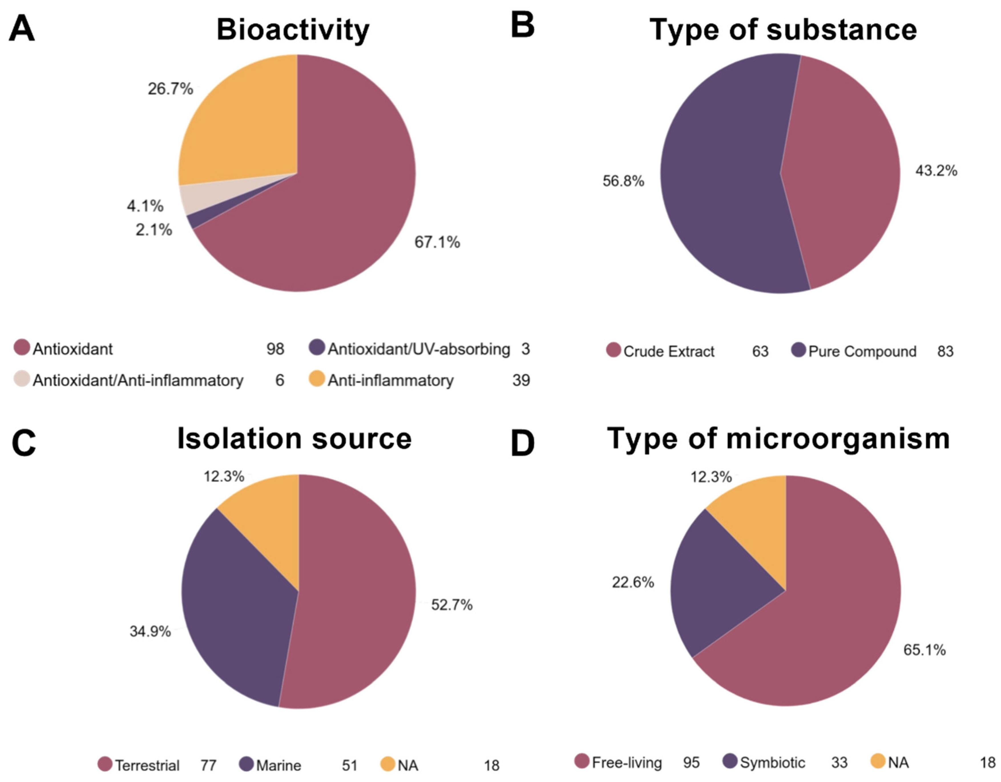
2.1. General Findings
2.2. Streptomyces as a Biological Source of Photoprotective Metabolites
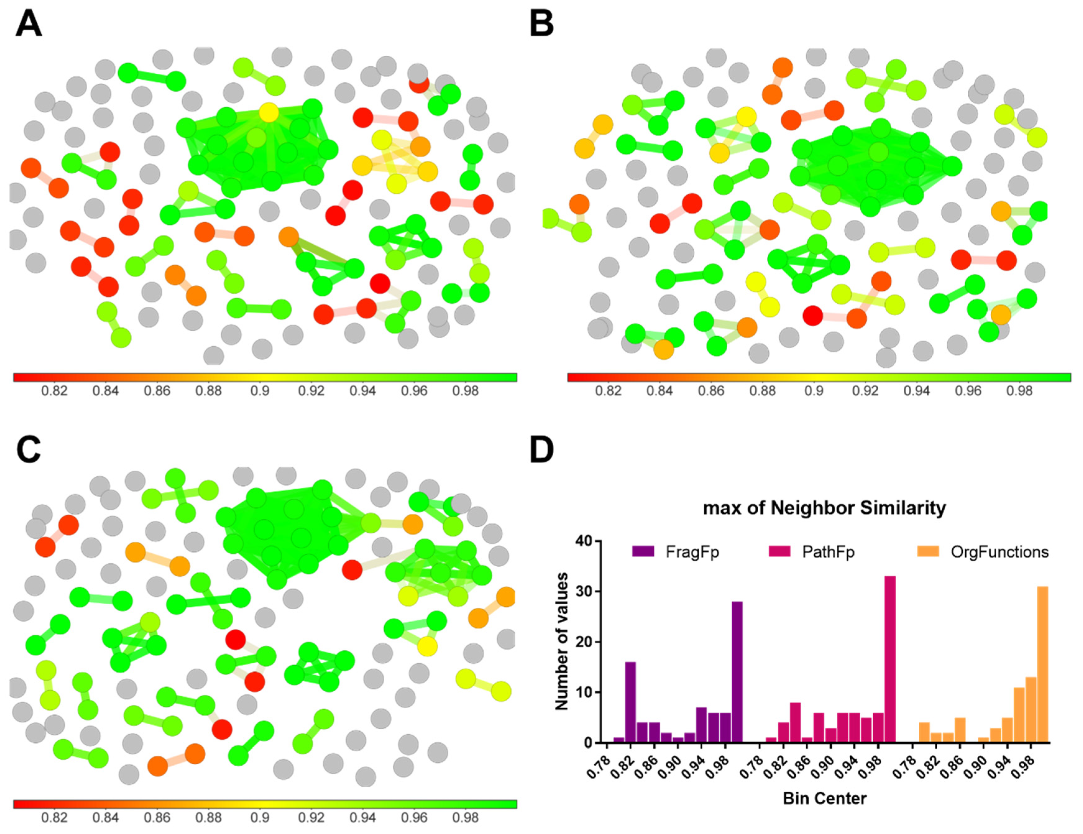
2.3. Chemical Space Analysis
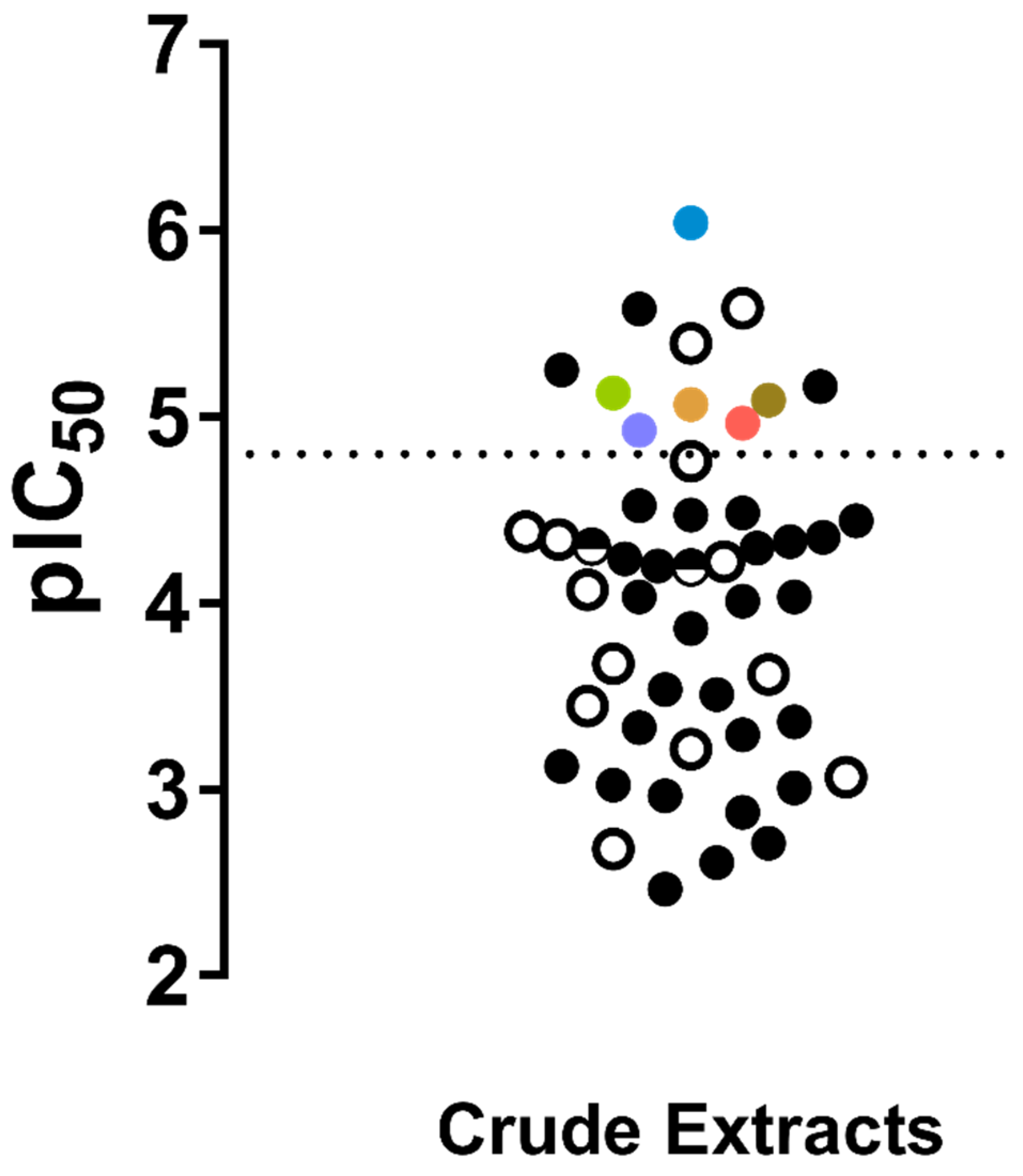
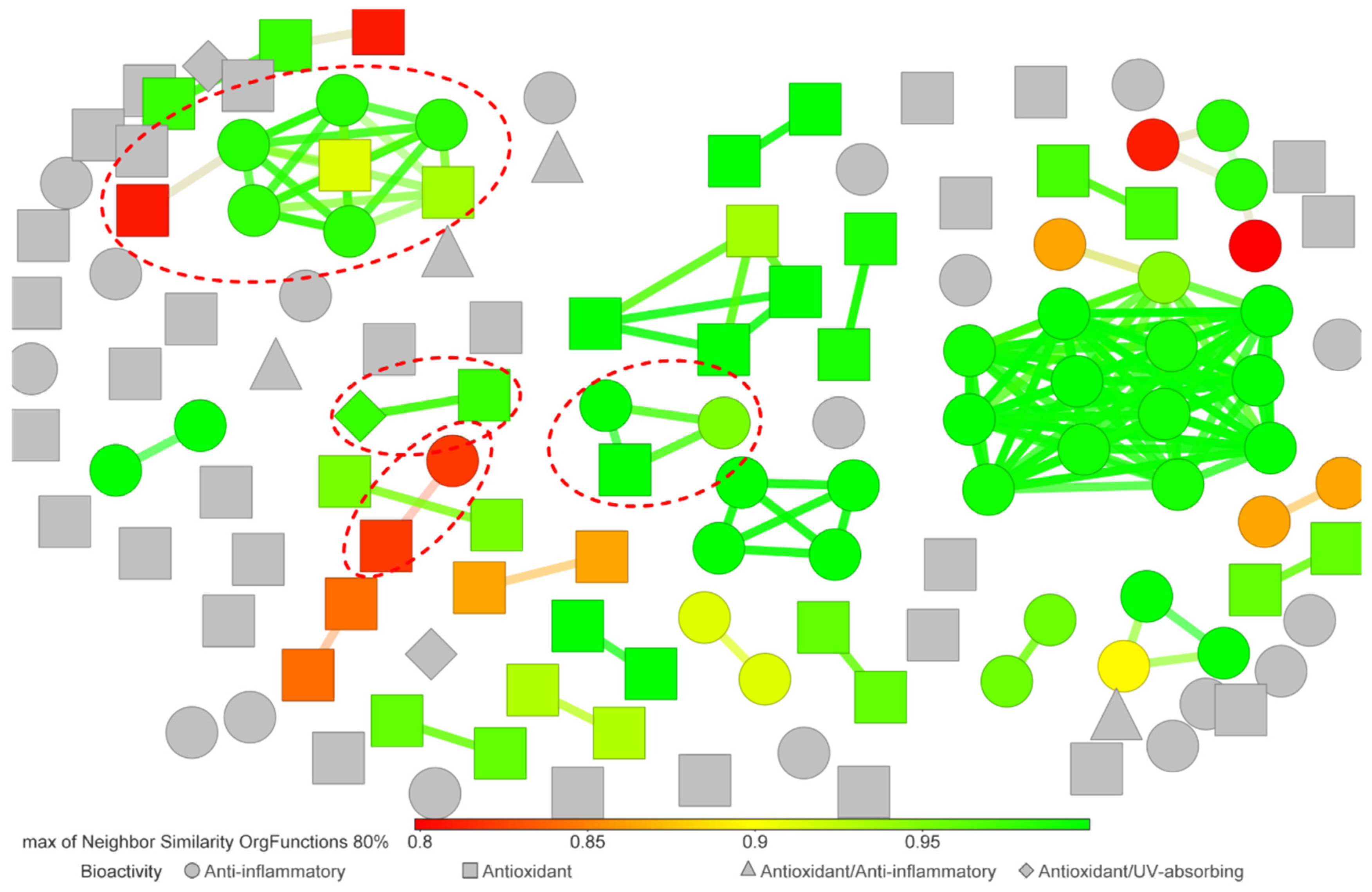
2.4. SPR and SAR Analysis
3. Discussion
4. Methods
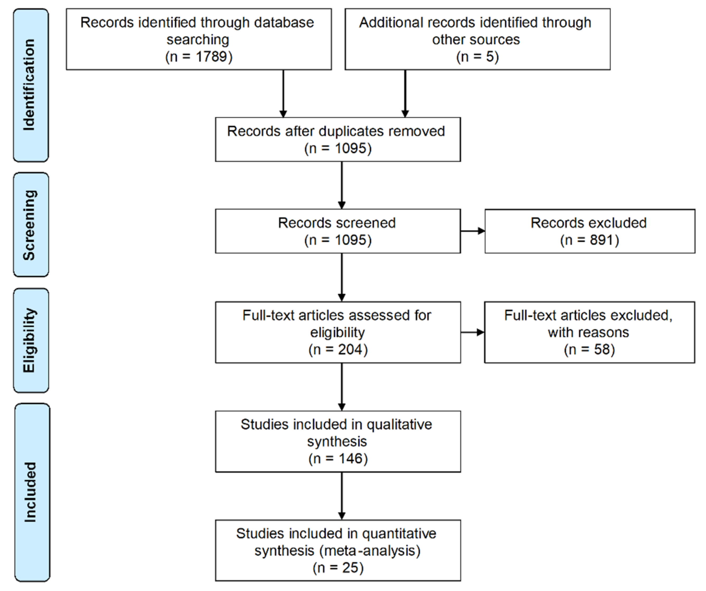
4.1. Databases and Search Strategy
4.2. Selection Procedure
4.3. Data Collection and Tabulation
4.4. Structure-Based Clustering
4.5. Data Analysis
5. Conclusions
Supplementary Materials
Author Contributions
Funding
Acknowledgments
Conflicts of Interest
References
- Gilchrest, B.A. Actinic Injury. Annu. Rev. Med. 1990, 41, 199–210. [Google Scholar] [CrossRef] [PubMed]
- Lucas, R.M.; Norval, M.; Neale, R.E.; Young, A.R.; de Gruijl, F.R.; Takizawa, Y.; van der Leun, J.C. The consequences for human health of stratospheric ozone depletion in association with other environmental factors. Photochem. Photobiol. Sci. 2015, 14, 53–87. [Google Scholar] [CrossRef] [PubMed] [Green Version]
- U.S. Department of Health and Human Services. The Surgeon General’s Call to Action to Prevent Skin Cancer; U.S. Department of Health and Human Services, Office of the Surgeon General: Washington, DC, USA, 2014.
- Mancuso, J.B.; Maruthi, R.; Wang, S.Q.; Lim, H.W. Sunscreens: An Update. Am. J. Clin. Dermatol. 2017, 18, 643–650. [Google Scholar] [CrossRef] [PubMed]
- Serpone, N.; Dondi, D.; Albini, A. Inorganic and organic UV filters: Their role and efficacy in sunscreens and suncare products. Inorg. Chim. Acta 2007, 360, 794–802. [Google Scholar] [CrossRef]
- Narla, S.; Lim, H.W. Sunscreen: FDA regulation, and environmental and health impact. Photochem. Photobiol. Sci. 2020, 19, 66–70. [Google Scholar] [CrossRef] [PubMed]
- Cole, C.; Shyr, T.; Ou-Yang, H. Metal oxide sunscreens protect skin by absorption, not by reflection or scattering. Photodermatol. Photoimmunol. Photomed. 2016, 32, 5–10. [Google Scholar] [CrossRef] [PubMed] [Green Version]
- Osterwalder, U.; Hareng, L. Global UV Filters: Current Technologies and Future Innovations. In Principles and Practice of Photoprotection; Springer International Publishing: Cham, Switzerland, 2016; pp. 179–197. [Google Scholar]
- Raffa, R.B.; Pergolizzi, J.V.; Taylor, R.; Kitzen, J.M. Sunscreen bans: Coral reefs and skin cancer. J. Clin. Pharm. Ther. 2019, 44, 134–139. [Google Scholar] [CrossRef] [PubMed] [Green Version]
- Hamzelou, J. Sunscreen safety fears. N. Sci. 2019, 243, 20–21. [Google Scholar] [CrossRef]
- Siller, A.; Blaszak, S.C.; Lazar, M.; Olasz Harken, E. Update About the Effects of the Sunscreen Ingredients Oxybenzone and Octinoxate on Humans and the Environment. Plast. Surg. Nurs. 2019, 39, 157–160. [Google Scholar] [CrossRef]
- Krause, M.; Klit, A.; Blomberg Jensen, M.; Søeborg, T.; Frederiksen, H.; Schlumpf, M.; Lichtensteiger, W.; Skakkebaek, N.E.; Drzewiecki, K.T. Sunscreens: Are they beneficial for health? An overview of endocrine disrupting properties of UV-filters. Int. J. Androl. 2012, 35, 424–436. [Google Scholar] [CrossRef]
- Tsui, M.M.P.; Leung, H.W.; Wai, T.C.; Yamashita, N.; Taniyasu, S.; Liu, W.; Lam, P.K.S.; Murphy, M.B. Occurrence, distribution and ecological risk assessment of multiple classes of UV filters in surface waters from different countries. Water Res. 2014, 67, 55–65. [Google Scholar] [CrossRef] [PubMed]
- Rainieri, S.; Barranco, A.; Primec, M.; Langerholc, T. Occurrence and toxicity of musks and UV filters in the marine environment. Food Chem. Toxicol. 2017, 104, 57–68. [Google Scholar] [CrossRef]
- Danovaro, R.; Bongiorni, L.; Corinaldesi, C.; Giovannelli, D.; Damiani, E.; Astolfi, P.; Greci, L.; Pusceddu, A. Sunscreens cause coral bleaching by promoting viral infections. Environ. Health Perspect. 2008, 116, 441–447. [Google Scholar] [CrossRef] [PubMed] [Green Version]
- Downs, C.A.; Kramarsky-Winter, E.; Segal, R.; Fauth, J.; Knutson, S.; Bronstein, O.; Ciner, F.R.; Jeger, R.; Lichtenfeld, Y.; Woodley, C.M.; et al. Toxicopathological Effects of the Sunscreen UV Filter, Oxybenzone (Benzophenone-3), on Coral Planulae and Cultured Primary Cells and Its Environmental Contamination in Hawaii and the U.S. Virgin Islands. Arch. Environ. Contam. Toxicol. 2016, 70, 265–288. [Google Scholar] [CrossRef] [PubMed]
- DeBuys, H.V.; Levy, S.B.; Murray, J.C.; Madey, D.L.; Pinnell, S.R. Modern approaches to photoprotection. Dermatol. Clin. 2000, 18, 577–590. [Google Scholar] [CrossRef]
- Kullavanijaya, P.; Lim, H.W. Photoprotection. J. Am. Acad. Dermatol. 2005, 52, 937–958. [Google Scholar] [CrossRef]
- Singer, S.; Karrer, S.; Berneburg, M. Modern sun protection. Curr. Opin. Pharmacol. 2019, 46, 24–28. [Google Scholar] [CrossRef]
- Tomazelli, L.C.; de Assis Ramos, M.M.; Sauce, R.; Cândido, T.M.; Sarruf, F.D.; de Oliveira Pinto, C.A.S.; de Oliveira, C.A.; Rosado, C.; Velasco, M.V.R.; Baby, A.R. SPF enhancement provided by rutin in a multifunctional sunscreen. Int. J. Pharm. 2018, 552, 401–406. [Google Scholar] [CrossRef]
- Batista, C.M.; Alves, A.V.F.; Queiroz, L.A.; Lima, B.S.; Filho, R.N.P.; Araújo, A.A.S.; de Albuquerque Júnior, R.L.C.; Cardoso, J.C. The photoprotective and anti-inflammatory activity of red propolis extract in rats. J. Photochem. Photobiol. B Biol. 2018, 180, 198–207. [Google Scholar] [CrossRef]
- Bickers, D.R.; Athar, M. Oxidative stress in the pathogenesis of skin disease. J. Investig. Dermatol. 2006, 126, 2565–2575. [Google Scholar] [CrossRef] [Green Version]
- Bosch, R.; Philips, N.; Suárez-Pérez, J.; Juarranz, A.; Devmurari, A.; Chalensouk-Khaosaat, J.; González, S. Mechanisms of Photoaging and Cutaneous Photocarcinogenesis, and Photoprotective Strategies with Phytochemicals. Antioxidants 2015, 4, 248–268. [Google Scholar] [CrossRef] [Green Version]
- Mounessa, J.; Buntinx-Krieg, T.; Qin, R.; Dunnick, C.A.; Dellavalle, R.P. Primary and Secondary Chemoprevention of Malignant Melanoma. Am. J. Clin. Dermatol. 2016, 17, 625–634. [Google Scholar] [CrossRef] [PubMed]
- Rodrigues, T.; Reker, D.; Schneider, P.; Schneider, G. Counting on natural products for drug design. Nat. Chem. 2016, 8, 531–541. [Google Scholar] [CrossRef] [PubMed]
- Mahesh, S.K.; Fathima, J.; Veena, V.G. Cosmetic Potential of Natural Products: Industrial Applications. In Natural Bio-Active Compounds; Swamy, M.K., Akhtar, M.S., Eds.; Springer: Singapore, 2019; pp. 215–250. ISBN 978-981-13-7204-9. [Google Scholar]
- Eastgate, M.D.; Schmidt, M.A.; Fandrick, K.R. On the design of complex drug candidate syntheses in the pharmaceutical industry. Nat. Rev. Chem. 2017, 1, 1–16. [Google Scholar] [CrossRef]
- Wallace, M.A.; Cheng, Y.-Q.; Currens, G.C. Construction of a Texas Microbes-Derived New Natural Product Library for Novel Drug Discovery. In Proceedings of the Summer Research Internship Program—2017; University of North Texas: Denton, TX, USA, 2017. [Google Scholar]
- Chang, M.C.Y.; Keasling, J.D. Production of isoprenoid pharmaceuticals by engineered microbes. Nat. Chem. Biol. 2006, 2, 674–681. [Google Scholar] [CrossRef]
- Demain, A.L. Microbial biotechnology. Trends Biotechnol. 2000, 18, 26–31. [Google Scholar] [CrossRef] [Green Version]
- Pessôa, M.G.; Vespermann, K.A.C.; Paulino, B.N.; Barcelos, M.C.S.; Pastore, G.M.; Molina, G. Newly isolated microorganisms with potential application in biotechnology. Biotechnol. Adv. 2019, 37, 319–339. [Google Scholar] [CrossRef] [PubMed]
- Mishra, M.; Vishwakarma, K.; Singh, J.; Jain, S.; Kumar, V.; Tripathi, D.K.; Sharma, S. Exploring the Multifaceted Role of Microbes in Pharmacology. In Microbial Biotechnology: Volume 2. Application in Food and Pharmacology; Patra, J.K., Das, G., Shin, H.-S., Eds.; Springer: Singapore, 2018; pp. 319–329. ISBN 978-981-10-7140-9. [Google Scholar]
- Du, J.; Shao, Z.; Zhao, H. Engineering microbial factories for synthesis of value-added products. J. Ind. Microbiol. Biotechnol. 2011, 38, 873–890. [Google Scholar] [CrossRef] [PubMed] [Green Version]
- Gill, S.R.; Pop, M.; DeBoy, R.T.; Eckburg, P.B.; Turnbaugh, P.J.; Samuel, B.S.; Gordon, J.I.; Relman, D.A.; Fraser-Liggett, C.M.; Nelson, K.E. Metagenomic Analysis of the Human Distal Gut Microbiome. Science 2006, 312, 1355–1359. [Google Scholar] [CrossRef] [Green Version]
- Yang, X.; Xie, L.; Li, Y.; Wei, C. More than 9,000,000 Unique Genes in Human Gut Bacterial Community: Estimating Gene Numbers Inside a Human Body. PLoS ONE 2009, 4, e6074. [Google Scholar] [CrossRef] [Green Version]
- Tierney, B.T.; Yang, Z.; Luber, J.M.; Beaudin, M.; Wibowo, M.C.; Baek, C.; Mehlenbacher, E.; Patel, C.J.; Kostic, A.D. The Landscape of Genetic Content in the Gut and Oral Human Microbiome. Cell Host Microbe 2019, 26, 283–295.e8. [Google Scholar] [CrossRef] [PubMed]
- Jenke-Kodama, H.; Börner, T.; Dittmann, E. Natural Biocombinatorics in the Polyketide Synthase Genes of the Actinobacterium Streptomyces avermitilis. PLoS Comput. Biol. 2006, 2, e132. [Google Scholar] [CrossRef] [PubMed]
- Hillenmeyer, M.E.; Vandova, G.A.; Berlew, E.E.; Charkoudian, L.K. Evolution of chemical diversity by coordinated gene swaps in type II polyketide gene clusters. Proc. Natl. Acad. Sci. USA 2015, 112, 13952–13957. [Google Scholar] [CrossRef] [PubMed] [Green Version]
- Barka, E.A.; Vatsa, P.; Sanchez, L.; Gaveau-Vaillant, N.; Jacquard, C.; Meier-Kolthoff, J.P.; Klenk, H.-P.; Clément, C.; Ouhdouch, Y.; van Wezel, G.P. Taxonomy, Physiology, and Natural Products of Actinobacteria. Microbiol. Mol. Biol. Rev. 2016, 80, 1–43. [Google Scholar] [CrossRef] [PubMed] [Green Version]
- Watve, M.G.; Tickoo, R.; Jog, M.M.; Bhole, B.D. How many antibiotics are produced by the genus Streptomyces? Arch. Microbiol. 2001, 176, 386–390. [Google Scholar] [CrossRef]
- De Lima Procópio, R.E.; da Silva, I.R.; Martins, M.K.; de Azevedo, J.L.; de Araújo, J.M. Antibiotics produced by Streptomyces. Braz. J. Infect. Dis. 2012, 16, 466–471. [Google Scholar] [CrossRef] [PubMed] [Green Version]
- Takahashi, Y.; Nakashima, T. Actinomycetes, an Inexhaustible Source of Naturally Occurring Antibiotics. Antibiotics 2018, 7, 45. [Google Scholar] [CrossRef] [Green Version]
- Sinha, R.P.; Singh, S.P.; Häder, D.P. Database on mycosporines and mycosporine-like amino acids (MAAs) in fungi, cyanobacteria, macroalgae, phytoplankton and animals. J. Photochem. Photobiol. B Biol. 2007, 89, 29–35. [Google Scholar] [CrossRef] [PubMed]
- Miyamoto, K.T.; Komatsu, M.; Ikeda, H. Discovery of gene cluster for mycosporine-like amino acid biosynthesis from Actinomycetales microorganisms and production of a novel mycosporine-like amino acid by heterologous expression. Appl. Environ. Microbiol. 2014, 80, 5028–5036. [Google Scholar] [CrossRef] [PubMed] [Green Version]
- Volkmann, M.; Gorbushina, A.A. A broadly applicable method for extraction and characterization of mycosporines and mycosporine-like amino acids of terrestrial, marine and freshwater origin. FEMS Microbiol. Lett. 2006, 255, 286–295. [Google Scholar] [CrossRef] [PubMed]
- Madhusudhan, D.N.; Agsar, D.; Sulochana, M.B. Water Soluble Melanin of Streptomyces lusitanus DMZ3 Persuade Synthesis of Enhanced Bio-medically Active Silver Nanoparticles. J. Clust. Sci. 2015, 26, 1077–1089. [Google Scholar] [CrossRef]
- Omura, S.; Ikeda, H.; Ishikawa, J.; Hanamoto, A.; Takahashi, C.; Shinose, M.; Takahashi, Y.; Horikawa, H.; Nakazawa, H.; Osonoe, T.; et al. Genome sequence of an industrial microorganism Streptomyces avermitilis: Deducing the ability of producing secondary metabolites. Proc. Natl. Acad. Sci. USA 2001, 98, 12215–12220. [Google Scholar] [CrossRef] [PubMed] [Green Version]
- Romero-Rodríguez, A.; Robledo-Casados, I.; Sánchez, S. An overview on transcriptional regulators in Streptomyces. Biochim. Biophys. Acta Gene Regul. Mech. 2015, 1849, 1017–1039. [Google Scholar] [CrossRef] [PubMed]
- Seipke, R.F.; Kaltenpoth, M.; Hutchings, M.I. Streptomyces as symbionts: An emerging and widespread theme? FEMS Microbiol. Rev. 2012, 36, 862–876. [Google Scholar] [CrossRef] [PubMed] [Green Version]
- Challis, G.L.; Hopwood, D.A. Synergy and contingency as driving forces for the evolution of multiple secondary metabolite production by Streptomyces species. Proc. Natl. Acad. Sci. USA 2003, 100, 14555–14561. [Google Scholar] [CrossRef] [PubMed] [Green Version]
- Lewin, G.R.; Carlos, C.; Chevrette, M.G.; Horn, H.A.; McDonald, B.R.; Stankey, R.J.; Fox, B.G.; Currie, C.R. Evolution and Ecology of Actinobacteria and Their Bioenergy Applications. Annu. Rev. Microbiol. 2016, 70, 235–254. [Google Scholar] [CrossRef] [PubMed] [Green Version]
- Moher, D.; Liberati, A.; Tetzlaff, J.; Altman, D.G. Preferred Reporting Items for Systematic Reviews and Meta-Analyses: The PRISMA Statement. PLoS Med. 2009, 6, e1000097. [Google Scholar] [CrossRef] [Green Version]
- Tarkka, M.; Hampp, R. Secondary Metabolites of Soil Streptomycetes in Biotic Interactions. In Secondary Metabolites in Soil Ecology. Soil Biology; Springer: Berlin/Heidelberg, Germany, 2008; Volume 14, pp. 107–126. [Google Scholar]
- Dharmaraj, S. Marine Streptomyces as a novel source of bioactive substances. World J. Microbiol. Biotechnol. 2010, 26, 2123–2139. [Google Scholar] [CrossRef]
- Peres, D.D.A.; Sarruf, F.D.; de Oliveira, C.A.; Velasco, M.V.R.; Baby, A.R. Ferulic acid photoprotective properties in association with UV filters: Multifunctional sunscreen with improved SPF and UVA-PF. J. Photochem. Photobiol. B Biol. 2018, 185, 46–49. [Google Scholar] [CrossRef]
- De Oliveira, C.A.; Peres, D.D.; Rugno, C.M.; Kojima, M.; De Oliveira Pinto, C.A.S.; Consiglieri, V.O.; Kaneko, T.M.; Rosado, C.; Mota, J.; Velasco, M.V.R.; et al. Functional photostability and cutaneous compatibility of bioactive UVA sun care products. J. Photochem. Photobiol. B Biol. 2015, 148, 154–159. [Google Scholar] [CrossRef]
- Bhosale, H.; Bismile, P.; Kadam, T.; Shaheen, U. Antioxidant, enzyme inhibitory and antifungal activities of actinomycetes isolated from Curcuma longa rhizosphere. Int. J. Pharm. Pharm. Sci. 2016, 8, 307–311. [Google Scholar]
- Narendhran, S.; Rajiv, P.; Vanathi, P.; Sivaraj, R. Spectroscopic analysis of bioactive compounds from Streptomyces Cavouresis KUV39: Evaluation of antioxidant and cytotoxicity activity. Int. J. Pharm. Pharm. Sci. 2014, 6, 319–322. [Google Scholar]
- Subathra Devi, C.; Kumari, A.; Jain, N.; Naine, S.J.; Mohanasrinivasan, V. Screening of actinomycetes isolated from soil samples for antibacterial and antioxidant activity. Int. J. Pharm. Pharm. Sci. 2013, 5, 483–489. [Google Scholar]
- Radhakrishnan, M.; Gopikrishnan, V.; Vijayalakshmi, G.; Kumar, V. In vitro antioxidant activity and antimicrobial activity against biofilm forming bacteria by the pigment from Desert soil Streptomyces sp. D25. J. Appl. Pharm. Sci. 2016, 6, 148–150. [Google Scholar] [CrossRef] [Green Version]
- Kai, Z.; Xia Ling, G.; Zheng Jun, X.; Li Hua, L.; Rong Jun, C.X.J.; Hong, G.; Kai, J.; Isomaro, Y. Isolation and characterization of a novel streptomyces strain Eri11 exhibiting antioxidant activity from the rhizosphere of Rhizoma Curcumae Longae. Afr. J. Microbiol. Res. 2011, 5, 1291–1297. [Google Scholar] [CrossRef] [Green Version]
- Praveen Kumar, P.; Preetam Raj, J.P.; Nimal Christhudas, I.V.S.; Sagaya Jansi, R.; Narbert Raj, M.; Agastian, P. α-Glucosidase Inhibition and Antioxidant Properties of Streptomyces sp.: In Vitro. Appl. Biochem. Biotechnol. 2014, 172, 1687–1698. [Google Scholar] [CrossRef]
- Lee, D.-R.; Lee, S.-K.; Choi, B.-K.; Cheng, J.; Lee, Y.-S.; Yang, S.H.; Suh, J.-W. Antioxidant activity and free radical scavenging activities of Streptomyces sp. strain MJM 10778. Asian Pac. J. Trop. Med. 2014, 7, 962–967. [Google Scholar] [CrossRef]
- Kaur, J.; Manhas, R.K.; Rani, R.; Arora, S. Actinobacteria from soil as potential free radical scavengers. Malays. J. Microbiol. 2013, 13, 217–227. [Google Scholar]
- Geo, H.N.; Panneerselvam, A. Studies on bioactive potential of Streptomyces spp. (KX710212). Isolated from environmental polluted sample, Ranipet, Vellore, India. Asian J. Pharm. Clin. Res. 2016, 9, 356–359. [Google Scholar] [CrossRef] [Green Version]
- Prashith, K.T.R.; Dileep, N.; Syed, J.; Rakesh, K.N.; Sunita, C.M.; Onkarappa, R. Biological activities of Streptomyces species SRDP-07 isolated from soil of Thirthahalli, Karnataka, India. Int. J. Drug Dev. Res. 2013, 5, 268–285. [Google Scholar]
- Revathy, T.; Jayasri, M.A.; Suthindhiran, K. Anti-oxidant and enzyme-inhibitory potential of marine Streptomyces. Am. J. Biochem. Biotechnol. 2013, 9, 282–290. [Google Scholar] [CrossRef] [Green Version]
- Jemimah Naine, S.; Nasimunislam, N.; Vaishnavi, B.; Mohanasrinivasan, V.; Subathra Devi, C. Isolation of soil actinomycetes inhabiting amrithi forest for the potential source of bioactive compounds. Asian J. Pharm. Clin. Res. 2012, 5, 189–192. [Google Scholar]
- Rani, R.; Arora, S.; Kaur, J.; Manhas, R.K. Phenolic compounds as antioxidants and chemopreventive drugs from Streptomyces cellulosae strain TES17 isolated from rhizosphere of Camellia sinensis. BMC Complement. Altern. Med. 2018, 18, 1–15. [Google Scholar] [CrossRef] [PubMed] [Green Version]
- Subathradevi, C.; Devi, P.; Jemimahnaine, S.; Mohanasrinivasan, V. Antibacterial and Antioxidant Property of Streptomyces cinnamonensis VITNS1 Isolated from Serkadu Region, Vellore, Tamil Nadu, India. Anti Infect. Agents 2014, 12, 206–212. [Google Scholar] [CrossRef]
- Law, J.W.F.; Ser, H.L.; Duangjai, A.; Saokaew, S.; Bukhari, S.I.; Khan, T.M.; Ab Mutalib, N.S.; Chan, K.G.; Goh, B.H.; Lee, L.H. Streptomyces colonosanans sp. nov., a novel actinobacterium isolated from Malaysia mangrove soil exhibiting antioxidative activity and cytotoxic potential against human colon cancer cell lines. Front. Microbiol. 2017, 8, 1–15. [Google Scholar] [CrossRef] [PubMed]
- Chakraborty, I.; Redkar, P.; Munjal, M.; Sathish Kumar, S.R.; Bhaskara Rao, K.V. Isolation and characterization of pigment producing marine actinobacteria from mangrove soil and applications of bio-pigments. Der Pharm. Lett. 2015, 7, 93–100. [Google Scholar]
- Parimala, G.S.A.; Manon, M.V.; Karthiyaini, D.; Priyadharshini, U.; Jeeva, S.; Brindha, P. V Bioprospecting fungicidal metabolite producing marine actinomycetes from southern coastal regions of India. Int. J. Life Sci. Pharma Res. 2017, 7, 55–64. [Google Scholar]
- Kumar, P.S.; Abdullah Al-Dhabi, N.; Duraipandiyan, V.; Balachandran, C.; Kumar, P.P.; Ignacimuthu, S. In vitro antimicrobial, antioxidant and cytotoxic properties of Streptomyces lavendulae strain SCA5. BMC Microbiol. 2014, 14, 291. [Google Scholar] [CrossRef] [Green Version]
- Lertcanawanichakul, M.; Pondet, K.; Kwantep, J. In vitro antimicrobial and antioxidant activities of bioactive compounds (secondary metabolites) extracted from Streptomyces lydicus A2. J. Appl. Pharm. Sci. 2015, 5, 17–21. [Google Scholar] [CrossRef] [Green Version]
- Jacob, J.; Rajendran, R.U.; Priya, S.H.; Purushothaman, J.; Saraswathy Amma, D.K.B.N. Enhanced antibacterial metabolite production through the application of statistical methodologies by a Streptomyces nogalater NIIST A30 isolated from Western Ghats forest soil. PLoS ONE 2017, 12, 1–21. [Google Scholar] [CrossRef] [PubMed]
- Gautham, S.A.; Onkarappa, R. In vitro antioxidant activity of metabolite from Streptomyces fradiae strain GOS1. Int. J. Drug Dev. Res. 2013, 5, 235–244. [Google Scholar]
- Gautham, S.A.; Onkarappa, R.; Kuppast, I.J. Pharmacological activities of metabolite from Streptomyces fradiae strain GOS 1. Int. J. Chem. Sci. 2013, 11, 583–590. [Google Scholar]
- Lee, S.K.; Lee, D.R.; Choi, B.K.; Palaniyandi, S.A.; Yang, S.H.; Suh, J.W. Glutathione S-transferase pi (GST-pi) inhibition and anti-inflammation activity of the ethyl acetate extract of Streptomyces sp. strain MJM 8637. Saudi J. Biol. Sci. 2015, 22, 744–751. [Google Scholar] [CrossRef] [PubMed] [Green Version]
- Saini, P.; Gangwar, M. Enzyme and free radical inhibitory potentials of ethyl acetate extract of endophytic actinomycete from Syzygium cumini. Indian J. Biochem. Biophys. 2017, 54, 207–213. [Google Scholar]
- Akshatha, J.V.; Prakash, H.S.; Nalini, M.S. Actinomycete Endophytes from the Ethno Medicinal Plants of Southern India: Antioxidant Activity and Characterization Studies. J. Biol. Act. Prod. Nat. 2016, 6, 166–172. [Google Scholar] [CrossRef]
- Nafis, A.; Kasrati, A.; Azmani, A.; Ouhdouch, Y.; Hassani, L. Endophytic actinobacteria of medicinal plant Aloe vera: Isolation, antimicrobial, antioxidant, cytotoxicity assays and taxonomic study. Asian Pac. J. Trop. Biomed. 2018, 8, 513–518. [Google Scholar] [CrossRef]
- Wang, L.; Qiu, P.; Long, X.F.; Zhang, S.; Zeng, Z.G.; Tian, Y.Q. Comparative analysis of chemical constituents, antimicrobial and antioxidant activities of ethylacetate extracts of Polygonum cuspidatum and its endophytic actinomycete, Streptomyces sp. A0916. Chin. J. Nat. Med. 2016, 14, 117–123. [Google Scholar] [CrossRef]
- Axenov-Gribanov, D.V.; Voytsekhovskaya, I.V.; Rebets, Y.V.; Tokovenko, B.T.; Penzina, T.A.; Gornostay, T.G.; Adelshin, R.V.; Protasov, E.S.; Luzhetskyy, A.N.; Timofeyev, M.A. Actinobacteria possessing antimicrobial and antioxidant activities isolated from the pollen of scots pine (Pinus sylvestris) grown on the Baikal shore. Antonie Van Leeuwenhoek 2016, 109, 1307–1322. [Google Scholar] [CrossRef]
- Nimal Christhudas, I.V.S.; Praveen Kumar, P.; Agastian, P. In Vitro α-Glucosidase Inhibition and Antioxidative Potential of an Endophyte Species (Streptomyces sp. Loyola UGC) Isolated from Datura stramonium L. Curr. Microbiol. 2013, 67, 69–76. [Google Scholar] [CrossRef]
- Jasim, B.; Soumya, R.; Jyothis, M.; Radhakrishnan, E.K. Exploration of actinomycetes endophytically associated with Piper nigrum for potential bioactivity. J. Microbiol. Biotechnol. Food Sci. 2015, 4, 282–286. [Google Scholar] [CrossRef] [Green Version]
- Tanvir, R.; Sajid, I.; Hasnain, S. Biotechnological potential of endophytic actinomycetes associated with Asteraceae plants: Isolation, biodiversity and bioactivities. Biotechnol. Lett. 2014, 36, 767–773. [Google Scholar] [CrossRef] [PubMed]
- Khanam, W.; Vootla, S.K. Comparative study of Ocimum basillicum and its endophytic actinomycetes Streptomyces flavoviridis a3wk: Evaluation of antioxidant, anti-inflammatory and antimicrobial activity. Int. J. Pharm. Sci. Res. 2018, 9, 1023–1034. [Google Scholar] [CrossRef]
- Ser, H.L.; Tan, L.T.H.; Palanisamy, U.D.; Abd Malek, S.N.; Yin, W.F.; Chan, K.G.; Goh, B.H.; Lee, L.H. Streptomyces antioxidans sp. nov., a novel mangrove soil actinobacterium with antioxidative and neuroprotective potentials. Front. Microbiol. 2016, 7, 1–14. [Google Scholar] [CrossRef] [PubMed] [Green Version]
- Veena, S.; Swetha, D.; Karthik, L.; Bhaskara Rao, K.V. Assessment of anti-typhoid and antioxidant activity of marine actinobacteria isolated from Chennai marine sediments. Der Pharm. Lett. 2016, 8, 166–172. [Google Scholar]
- Jemimah Naine, S.; Subathra Devi, C.; Mohanasrinivasan, V.; Vaishnavi, B. Antimicrobial, antioxidant and cytotoxic activity of marine Streptomyces parvulus VITJS11 crude extract. Braz. Arch. Biol. Technol. 2015, 58, 198–207. [Google Scholar] [CrossRef] [Green Version]
- Ser, H.L.; Mutalib, N.S.A.; Yin, W.F.; Chan, K.G.; Goh, B.H.; Lee, L.H. Evaluation of antioxidative and cytotoxic activities of Streptomyces pluripotens MUSC 137 isolated from mangrove soil in Malaysia. Front. Microbiol. 2015, 6, 1–11. [Google Scholar] [CrossRef] [PubMed] [Green Version]
- Selvakumar, J.; Chandrasekaran, S.; Vaithilingam, M. Bio prospecting of marine-derived Streptomyces spectabilis VITJS10 and exploring its cytotoxicity against human liver cancer cell lines. Pharmacogn. Mag. 2015, 11, 469. [Google Scholar] [CrossRef] [Green Version]
- Mandal, S.; Divya, V.S.; Rao, K.V.B. Bioactive potential of Streptomyces variabilis—DV-35 isolated from thottada marine sediments, Kannur, Kerala. Asian J. Pharm. Clin. Res. 2016, 9, 67–71. [Google Scholar] [CrossRef]
- Eva, S.M.; Baraka, S.; Ken, H.M.M. Cytotoxicity and antioxidant activity of a Streptomyces sp. from mangrove sediments of dar es salaam, Tanzania. Int. J. Pharm. Pharm. Sci. 2014, 6, 563–566. [Google Scholar]
- Karthik, L.; Kumar, G.; Rao, K.V.B. Antioxidant activity of newly discovered lineage of marine actinobacteria. Asian Pac. J. Trop. Med. 2013, 6, 325–332. [Google Scholar] [CrossRef] [Green Version]
- Tan, L.T.H.; Chan, K.G.; Khan, T.M.; Bukhari, S.I.; Saokaew, S.; Duangjai, A.; Pusparajah, P.; Lee, L.H.; Goh, B.H. Streptomyces sp. MUM212 as a source of antioxidants with radical scavenging and metal chelating properties. Front. Pharmacol. 2017, 8, 1–18. [Google Scholar] [CrossRef]
- Tan, L.T.H.; Ser, H.L.; Yin, W.F.; Chan, K.G.; Lee, L.H.; Goh, B.H. Investigation of antioxidative and anticancer potentials of Streptomyces sp. MUM256 isolated from Malaysia mangrove soil. Front. Microbiol. 2015, 6. [Google Scholar] [CrossRef] [PubMed]
- Tan, L.T.H.; Chan, K.G.; Pusparajah, P.; Yin, W.F.; Khan, T.M.; Lee, L.H.; Goh, B.H. Mangrove derived Streptomyces sp. MUM265 as a potential source of antioxidant and anticolon-cancer agents. BMC Microbiol. 2019, 19, 1–16. [Google Scholar] [CrossRef] [PubMed] [Green Version]
- Tan, L.T.H.; Chan, K.G.; Chan, C.K.; Khan, T.M.; Lee, L.H.; Goh, B.H. Antioxidative potential of a Streptomyces sp. MUM292 isolated from mangrove soil. Biomed. Res. Int. 2018, 2018. [Google Scholar] [CrossRef] [PubMed] [Green Version]
- Subramanian, D.; Kim, M.S.; Kim, D.H.; Heo, M.S. Isolation, characterization, antioxidant, antimicrobial and cytotoxic effect of marine Actinomycete, Streptomyces carpaticus MK-01, against fish pathogens. Braz. Arch. Biol. Technol. 2017, 60, 1–9. [Google Scholar] [CrossRef]
- Kamala, K.; Karuppiah, V.; Sivakumar, K. Comparative evaluation of in vitro antioxidant potent of the marine actinobacteria from gulf of Mannar Biosphere Reserve. Int. J. Pharma Bio Sci. 2013, 4, 207–216. [Google Scholar]
- Sengupta, S.; Pramanik, A.; Ghosh, A.; Bhattacharyya, M. Antimicrobial activities of actinomycetes isolated from unexplored regions of Sundarbans mangrove ecosystem. BMC Microbiol. 2015, 15, 170. [Google Scholar] [CrossRef] [Green Version]
- Almasi, F.; Mohammadipanah, F.; Adhami, H.R.; Hamedi, J. Introduction of marine-derived Streptomyces sp. UTMC 1334 as a source of pyrrole derivatives with anti-acetylcholinesterase activity. J. Appl. Microbiol. 2018, 125, 1370–1382. [Google Scholar] [CrossRef] [PubMed]
- Jemimah Naine, S.; Subathra Devi, C.; Mohanasrinivasan, V.; George Priya Doss, C. Bioactivity of Marine Streptomyces sp. VITJS4: Interactions of Cytotoxic Phthalate Derivatives with Human Topoisomerase II α: An In Silico Molecular Docking Analysis. Interdiscip. Sci. Comput. Life Sci. 2018, 10, 261–270. [Google Scholar] [CrossRef] [PubMed]
- Duraikannu, D.; Chandrasekaran, D.S.; Selvakumar, N.J.; Vaithilingam, M. A Preliminary Study of In vitro Antioxidant and Antibacterial Activity of Streptomyces gancidicus VITSD1 Isolated from Marine Soil. Curr. Bioact. Compd. 2014, 10, 292–297. [Google Scholar] [CrossRef]
- Raghava Rao, K.V.; Raghava Rao, T. Molecular characterization and its antioxidant activity of a newly isolated Streptomyces coelicoflavus BC 01 from mangrove soil. J. Young Pharm. 2013, 5, 121–126. [Google Scholar] [CrossRef] [PubMed] [Green Version]
- Siddharth, S.; Vittal, R. Evaluation of Antimicrobial, Enzyme Inhibitory, Antioxidant and Cytotoxic Activities of Partially Purified Volatile Metabolites of Marine Streptomyces sp.S2A. Microorganisms 2018, 6, 72. [Google Scholar] [CrossRef] [PubMed] [Green Version]
- Singhania, M.; Ravichander, P.; Swaroop, S.; Naine Selvakumar, J.; Vaithilingam, M.; Devi Chandrasekaran, S. Anti-bacterial and anti-oxidant property of Streptomyces laurentii VITMPS isolated from marine soil. Curr. Bioact. Compd. 2016, 13, 78–81. [Google Scholar] [CrossRef]
- Ser, H.L.; Palanisamy, U.D.; Yin, W.F.; Chan, K.G.; Goh, B.H.; Lee, L.H. Streptomyces malaysiense sp. nov.: A novel Malaysian mangrove soil actinobacterium with antioxidative activity and cytotoxic potential against human cancer cell lines. Sci. Rep. 2016, 6, 1–12. [Google Scholar] [CrossRef] [PubMed] [Green Version]
- Ser, H.L.; Palanisamy, U.D.; Yin, W.F.; Abd Malek, S.N.; Chan, K.G.; Goh, B.H.; Lee, L.H. Presence of antioxidative agent, Pyrrolo [1,2-a]pyrazine-1,4-dione, hexahydro- in newly isolated Streptomyces mangrovisoli sp. nov. Front. Microbiol. 2015, 6, 1–11. [Google Scholar] [CrossRef] [PubMed] [Green Version]
- Law, J.W.F.; Ser, H.L.; Ab Mutalib, N.S.; Saokaew, S.; Duangjai, A.; Khan, T.M.; Chan, K.G.; Goh, B.H.; Lee, L.H. Streptomyces monashensis sp. nov., a novel mangrove soil actinobacterium from East Malaysia with antioxidative potential. Sci. Rep. 2019, 9, 1–18. [Google Scholar] [CrossRef] [PubMed]
- Tangjitjaroenkun, J. Evaluation of antioxidant, antibacterial, and gas chromatography-mass spectrometry analysis of ethyl acetate extract of streptomyces omiyaensis SCH2. Asian J. Pharm. Clin. Res. 2018, 11, 271–276. [Google Scholar] [CrossRef]
- Sanjivkumar, M.; Babu, D.R.; Suganya, A.M.; Silambarasan, T.; Balagurunathan, R.; Immanuel, G. Investigation on pharmacological activities of secondary metabolite extracted from a mangrove associated actinobacterium Streptomyces olivaceus (MSU3). Biocatal. Agric. Biotechnol. 2016, 6, 82–90. [Google Scholar] [CrossRef]
- Pooja, S.; Aditi, T.; Naine, S.J.; Subathra Devi, C. Bioactive compounds from marine Streptomyces sp. VITPSA as therapeutics. Front. Biol. 2017, 12, 280–289. [Google Scholar] [CrossRef]
- El-Shenawy, N.S. Effect of Streptomyces 23-2B metabolites on hepatic lipid peroxidation and some antioxidant parameters in Wister rats. World J. Microbiol. Biotechnol. 2010, 26, 2185–2191. [Google Scholar] [CrossRef]
- Gozari, M.; Bahador, N.; Jassbi, A.R.; Mortazavi, M.S.; Eftekhar, E. Antioxidant and cytotoxic activities of metabolites produced by a new marine Streptomyces sp. isolated from the sea cucumber Holothuria leucospilota. Iran. J. Fish. Sci. 2018, 17, 413–426. [Google Scholar] [CrossRef]
- Lertcanawanichakul, M.; Chawawisit, K.; Pondet, K.; Kwantep, J. Quantitation of total phenolic contents of bioactive compounds fractions Streptomyces species. Int. J. PharmTech Res. 2015, 7, 320–324. [Google Scholar]
- Latha, Y.; Vasavithirumalanadhuni, V.M.; Devi, P.U.M. In vivo anti-inflammatory activity of ethyl acetate extract derived from marine Streptomyces carpaticus. Int. J. Pharm. Sci. Res. 2017, 8, 5221–5226. [Google Scholar] [CrossRef]
- Zhao, H. Scaffold selection and scaffold hopping in lead generation: A medicinal chemistry perspective. Drug Discov. Today 2007, 12, 149–155. [Google Scholar] [CrossRef] [PubMed]
- Kato, S.; Shindo, K.; Kawai, H.; Odagawa, A.; Matsuoka, M.; Mochizuki, J. Pyrrolostatin, a novel lipid peroxidation inhibitor from Streptomyces chrestomyceticus. Taxonomy, fermentation, isolation, structure elucidation and biological properties. J. Antibiot. 1993, 46, 892–899. [Google Scholar] [CrossRef] [PubMed] [Green Version]
- Kim, W.; Kim, J.; Kim, C.-J.; Lee, K.-H.; Yoo, I.-D. Benzastatins A, B, C, and D: New Free Radical Scavengers from Streptomyces nitrosporeus 30643. I. Taxonomy, Fermentation, Isolation, Physico-chemical Properties and Biological Activities. J. Antibiot. 1996, 49, 20–25. [Google Scholar] [CrossRef] [PubMed] [Green Version]
- Yang, L.; Mahal, A.; Liu, Y.; Li, H.; Wu, P.; Xue, J.; Xu, L.; Wei, X. Two new 2,5-diketopiperazines produced by Streptomyces sp. SC0581. Phytochem. Lett. 2017, 20, 89–92. [Google Scholar] [CrossRef]
- El Sayed, O.H.; Asker, M.M.S.; Swelim, M.A.; Abbas, I.H.; Attwa, A.I.; El Awady, M.E. Production of hydroxy marilone C as a bioactive compound from Streptomyces badius. J. Genet. Eng. Biotechnol. 2016, 14, 161–168. [Google Scholar] [CrossRef] [Green Version]
- Izumikawa, M.; Satou, R.; Motohashi, K.; Nagai, A.; Ohnishi, Y.; Takagi, M.; Shin-Ya, K. Naphthoquinone-like polyketide isolated from Streptomyces sp. RI-77 and its predicted biosynthetic pathway. J. Nat. Prod. 2011, 74, 2588–2591. [Google Scholar] [CrossRef]
- Jaivel, N.; Uvarani, C.; Rajesh, R.; Velmurugan, D.; Marimuthu, P. Natural occurrence of organofluorine and other constituents from Streptomyces sp. TC1. J. Nat. Prod. 2014, 77, 2–8. [Google Scholar] [CrossRef]
- Kawahara, T.; Izumikawa, M.; Otoguro, M.; Yamamura, H.; Hayakawa, M.; Takagi, M.; Shin-Ya, K. JBIR-94 and JBIR-125, antioxidative phenolic compounds from Streptomyces sp. R56-07. J. Nat. Prod. 2012, 75, 107–110. [Google Scholar] [CrossRef] [PubMed]
- Komoda, T.; Saeki, N.; Koseki, Y.; Kiyota, H. 12T061A and 12T061C, two new julichrome family compounds, as radical scavengers from Streptomyces sp. J. Gen. Appl. Microbiol. 2016, 62, 1–6. [Google Scholar] [CrossRef] [PubMed]
- Kim, K.J.; Kim, M.A.; Jung, J.H. Antitumor and antioxidant activity of protocatechualdehyde produced from Streptomyces lincolnensis M-20. Arch. Pharm. Res. 2008, 31, 1572–1577. [Google Scholar] [CrossRef] [PubMed]
- Kim, K.J.; Lee, J.H.; Yang, Y.J. Effect of interaction between protocatechualdehyde produced from Streptomyces lincolnensis M-20 and copper ions on antioxidant and pro-oxidant activities. Korean J. Microbiol. 2014, 50, 22–26. [Google Scholar] [CrossRef]
- Sugiyama, Y.; Oya, A.; Kudo, T.; Hirota, A. Surugapyrone A from Streptomyces coelicoflavus strain usf-6280 as a new dpph radical-scavenger. J. Antibiot. 2010, 63, 365–369. [Google Scholar] [CrossRef] [PubMed]
- Sugiyama, Y.; Watanabe, K.; Hirota, A. Surugapyrroles A and B, two new N-hydroxypyrroles, as DPPH radical-scavengers from Streptomyces sp. USF-6280 strain. Biosci. Biotechnol. Biochem. 2009, 73, 230–232. [Google Scholar] [CrossRef] [PubMed]
- Komiyama, K.; Funayama, S.; Anraku, Y.; Mita, A.; Takahashi, Y.; Omura, S. Isolation of isoflavonoids possessing antioxidant activity from the fermentation broth of Streptomyces sp. J. Antibiot. 1989, 42, 1344–1349. [Google Scholar] [CrossRef] [Green Version]
- Solecka, J.; Guśpiel, A.; Postek, M.; Ziemska, J.; Kawęcki, R.; Łęczycka, K.; Osior, A.; Pietrzak, B.; Pypowski, K.; Wyrzykowska, A. New derivatives of 3,4-dihydroisoquinoline-3-carboxylic acid with free-radical scavenging, d-amino acid oxidase, acetylcholinesterase and butyrylcholinesterase inhibitory activity. Molecules 2014, 19, 15866–15890. [Google Scholar] [CrossRef] [PubMed] [Green Version]
- Guśpiel, A.; Ziemska, J.; Cześcik, A.; Kawecki, R.; Solecka, J. Intracellular antioxidant activity of a Streptomyces sp. 8812 secondary metabolite, 6,7-dihydroxy-3,4-dihydroisoquinoline-3-carboxylic acid, and its synthetic derivatives. Acta Pol. Pharm. Drug Res. 2016, 73, 645–651. [Google Scholar]
- Kim, W.G.; Ryoo, I.J.; Yun, B.S.; Shin-ya, K.; Seto, H.; Yoo, I.D. New diphenazines with neuronal cell protecting activity, phenazostatins A and B, produced by Streptomyces sp. J. Antibiot. 1997, 50, 715–721. [Google Scholar] [CrossRef] [Green Version]
- Shinichiro, K.; Kazutoshi, S.; Yuji, Y.; Michiko, M.; Hiroyuki, K.; Junichiro, M. Phenazoviridin, a novel free radical scavenger from Streptomyces sp. Taxonomy, fermentation, isolation, structure elucidation and biological properties. J. Antibiot. 1993, 46, 1485–1493. [Google Scholar] [CrossRef]
- Chang, H.B.; Kim, J.H. Antioxidant properties of dihydroherbimycin A from a newly isolated Streptomyces sp. Biotechnol. Lett. 2007, 29, 599–603. [Google Scholar] [CrossRef] [PubMed]
- Morimitsu, Y.; Hirota, A. Ansamycin Antibiotics as Free Radical Scavengers Isolated from Streptomyces by Using the Bactericidal Action of the Hydroxyl Radical. Biosci. Biotechnol. Biochem. 1996, 60, 1507–1509. [Google Scholar] [CrossRef] [PubMed]
- Jaivel, N.; Rajesh, R.; Uvarani, C.; Marimuthu, P. In vitro antimicrobial evaluation of compound derived from Streptomyces sp. TC1 against xanthomonas oryzae pv. Oryzae. J. Pure Appl. Microbiol. 2014, 8, 1299–1310. [Google Scholar]
- Kato, S.; Kawasaki, T.; Urata, T.; Mochizuki, J. In vitro and ex vivo free radical scavenging activities of carazostatin, carbazomycin B and their derivatives. J. Antibiot. 1993, 46, 1859–1865. [Google Scholar] [CrossRef] [Green Version]
- Stankovic, N.; Radulovic, V.; Petkovic, M.; Vuckovic, I.; Jadranin, M.; Vasiljevic, B.; Nikodinovic-Runic, J. Streptomyces sp. JS520 produces exceptionally high quantities of undecylprodigiosin with antibacterial, antioxidative, and UV-protective properties. Appl. Microbiol. Biotechnol. 2012, 96, 1217–1231. [Google Scholar] [CrossRef]
- Sajjad, W.; Ahmad, S.; Aziz, I.; Azam, S.S.; Hasan, F.; Shah, A.A. Antiproliferative, antioxidant and binding mechanism analysis of prodigiosin from newly isolated radio-resistant Streptomyces sp. strain WMA-LM31. Mol. Biol. Rep. 2018, 45, 1787–1798. [Google Scholar] [CrossRef]
- Deng, H.; Zhang, N.; Wang, Y.; Chen, J.; Shen, J.; Wang, Z.; Xu, R.; Zhang, J.; Song, D.; Li, D. S632A3, a new glutarimide antibiotic, suppresses lipopolysaccharide-induced pro-inflammatory responses via inhibiting the activation of glycogen synthase kinase 3Β. Exp. Cell Res. 2012, 318, 2592–2603. [Google Scholar] [CrossRef]
- Usuki, Y.; Ishii, S.; Ijiri, M.; Yoshida, K.I.; Satoh, T.; Horigome, S.; Yoshida, I.; Mishima, T.; Fujita, K.I. Evaluation of Inhibitory Activities of UK-2A, an Antimycin-Type Antibiotic, and Its Synthetic Analogues against the Production of Anti-inflammatory Cytokine IL-4. J. Nat. Prod. 2018, 81, 2590–2594. [Google Scholar] [CrossRef] [Green Version]
- Nakayama, O.; Yagi, M.; Tanaka, M.; Kiyoto, S.; Uchida, I.; Hashimoto, M.; Okuhara, M.; Kohsaka, M. WS-7528, a new isoflavanone with estrogen activity isolated from Streptomyces sp. No. 7528. Taxonomy, fermentation, isolation, physico-chemical properties and biological activities. J. Antibiot. 1990, 43, 1394–1402. [Google Scholar] [CrossRef] [Green Version]
- Petříčková, K.; Pospíšil, S.; Kuzma, M.; Tylová, T.; Jágr, M.; Tomek, P.; Chroňáková, A.; Brabcová, E.; Anděra, L.; Krištůfek, V.; et al. Biosynthesis of Colabomycin E, a New Manumycin-Family Metabolite, Involves an Unusual Chain-Length Factor. ChemBioChem 2014, 15, 1334–1345. [Google Scholar] [CrossRef] [PubMed]
- Graziani, E.I.; Ritacco, F.V.; Bernan, V.S.; Telliez, J.-B. Phaeochromycins A−E, Anti-inflammatory Polyketides Isolated from the Soil Actinomycete Streptomyces phaeochromogenes LL-P018. J. Nat. Prod. 2005, 68, 1262–1265. [Google Scholar] [CrossRef] [PubMed]
- Gullo, V.; Conover, M.; Cooper, R.; Federbush, C.; Horan, A.C.; Kung, T.; Marquez, J.; Patel, M.; Watnick, A. Sch 36605, a novel anti-inflammatory compound. Taxonomy, fermentation, isolation and biological properties. J. Antibiot. 1988, 41, 20–24. [Google Scholar] [CrossRef] [PubMed] [Green Version]
- Kino, T.; Hatanaka, H.; Hashimoto, M.; Nishiyama, M.; Goto, T.; Okuhara, M.; Kohsaka, M.; Aoki, H.; Imanaka, H. FK-506, a novel immunosuppressant isolated from a Streptomyces. I. Fermentation, isolation, and physico-chemical and biological characteristics. J. Antibiot. 1987, 40, 1249–1255. [Google Scholar] [CrossRef] [PubMed] [Green Version]
- Kino, T.; Hatanaka, H.; Miyata, S.; Inamura, N.; Nishiyama, M.; Yajima, T.; Goto, T.; Okuhara, M.; Kohsaka, M.; Aoki, H.; et al. Fk-506, A Novel Immunosuppressant Isolated From A Streptomyces II. Immunosuppressive Effect Of Fk-506 In Vitro. J. Antibiot. 1987, 40, 1256–1265. [Google Scholar] [CrossRef] [PubMed]
- Pereira, R.; Santos Medeiros, Y.; Fröde, T.S. Antiinflammatory effects of Tacrolimus in a mouse model of pleurisy. Transpl. Immunol. 2006, 16, 105–111. [Google Scholar] [CrossRef] [PubMed]
- Vigil, S.V.G.; de Liz, R.; Medeiros, Y.S.; Fröde, T.S. Efficacy of tacrolimus in inhibiting inflammation caused by carrageenan in a murine model of air pouch. Transpl. Immunol. 2008, 19, 25–29. [Google Scholar] [CrossRef]
- Lee, S.J.; Kim, H.P.; Park, B.K.; Ahn, S.C.; Lee, H.S.; Ahn, J.S. Topical anti-inflammatory activity of dianemycin isolated from Streptomyces sp. MT 2705-4. Arch. Pharm. Res. 1997, 20, 372–374. [Google Scholar] [CrossRef] [PubMed]
- Hori, R.; Yamaguchi, K.; Sato, H.; Watanabe, M.; Tsutsumi, K.; Iwamoto, S.; Abe, M.; Onodera, H.; Nakamura, S.; Nakai, R. The discovery and characterization of K-563, a novel inhibitor of the Keap1/Nrf2 pathway produced by Streptomyces sp. Cancer Med. 2019, 8, 1157–1168. [Google Scholar] [CrossRef]
- Jiménez, J.T.; Sturdíková, M.; Brezová, V.; Svajdlenka, E.; Novotová, M. Screening of mutant strain Streptomyces mediolani sp. AC37 for (-)-8-O-methyltetrangomycin production enhancement. J. Microbiol. 2012, 50, 1014–1023. [Google Scholar] [CrossRef] [PubMed]
- Taechowisan, T.; Chaisaeng, S.; Phutdhawong, W.S. Antibacterial, antioxidant and anticancer activities of biphenyls from Streptomyces sp. BO-07: An endophyte in Boesenbergia rotunda (L.) Mansf A. Food Agric. Immunol. 2017, 28, 1330–1346. [Google Scholar] [CrossRef] [Green Version]
- Taechowisan, T.; Singtotong, C.; Phutdhawong, W.S. Antibacterial and antioxidant activities of acetogenins from Streptomyces sp. VE2; an endophyte in Vernonia cinerea (L.) less. J. Appl. Pharm. Sci. 2016, 6, 67–72. [Google Scholar] [CrossRef] [Green Version]
- Yang, Y.; Yang, X.; Zhang, Y.; Zhou, H.; Zhang, J.; Xu, L.; Ding, Z. A new daidzein derivative from endophytic Streptomyces sp. YIM 65408. Nat. Prod. Res. 2013, 27, 1727–1731. [Google Scholar] [CrossRef] [PubMed]
- Yang, X.; Peng, T.; Yang, Y.; Li, W.; Xiong, J.; Zhao, L.; Ding, Z. Antimicrobial and antioxidant activities of a new benzamide from endophytic Streptomyces sp. YIM 67086. Nat. Prod. Res. 2015, 29, 331–335. [Google Scholar] [CrossRef] [PubMed]
- Zhou, H.; Yang, Y.; Peng, T.; Li, W.; Zhao, L.; Xu, L.; Ding, Z. Metabolites of Streptomyces sp., an endophytic actinomycete from Alpinia oxyphylla. Nat. Prod. Res. 2014, 28, 265–267. [Google Scholar] [CrossRef] [PubMed]
- Hong, S.H.; Ban, Y.H.; Byun, W.S.; Kim, D.; Jang, Y.J.; An, J.S.; Shin, B.; Lee, S.K.; Shin, J.; Yoon, Y.J.; et al. Camporidines A and B: Antimetastatic and Anti-inflammatory Polyketide Alkaloids from a Gut Bacterium of Camponotus kiusiuensis. J. Nat. Prod. 2019, 82, 903–910. [Google Scholar] [CrossRef]
- Ma, J.; Lei, H.; Chen, X.; Bi, X.; Jiang, Y.; Han, L.; Huang, X. New anti-inflammatory metabolites produced by Streptomyces violaceoruber isolated from Equus burchelli feces. J. Antibiot. 2017, 70, 991–994. [Google Scholar] [CrossRef]
- Ma, J.; Cao, B.; Liu, C.; Guan, P.; Mu, Y.; Jiang, Y.; Han, L.; Huang, X. Actinofuranones D-I from a lichen-associated actinomycetes, streptomyces gramineus, and their anti-inflammatory effects. Molecules 2018, 23, 2393. [Google Scholar] [CrossRef] [Green Version]
- Ma, J.; Cao, B.; Chen, X.; Xu, M.; Bi, X.; Guan, P.; Jiang, Y.; Xu, J.; Han, L.; Huang, X. Violacin A, a new chromanone produced by Streptomyces violaceoruber and its anti-inflammatory activity. Bioorg. Med. Chem. Lett. 2018, 28, 947–951. [Google Scholar] [CrossRef]
- Taechowisan, T.; Lu, C.; Shen, Y.; Lumyong, S. Anti-inflammatory effects of 4-arylcoumarins in LPS-induced murine macrophage RAW 264.7 cells. Pharm. Biol. 2006, 44, 576–580. [Google Scholar] [CrossRef]
- Taechowisan, T.; Wanbanjob, A.; Tuntiwachwuttikul, P.; Liu, J. Anti-inflammatory activity of lansais from endophytic Streptomyces sp. SUC1 in LPS-induced RAW 264.7 cells. Food Agric. Immunol. 2009, 20, 67–77. [Google Scholar] [CrossRef]
- Taechowisan, T.; Wanbanjob, A.; Tuntiwachwuttikul, P.; Liu, J. Anti-inflammatory effects of lansai C and D cause inhibition of STAT-1 and NF-κB activations in LPS-induced RAW 264.7 cells. Food Agric. Immunol. 2010, 21, 57–64. [Google Scholar] [CrossRef] [Green Version]
- Saurav, K.; Kannabiran, K. Cytotoxicity and antioxidant activity of 5-(2,4-dimethylbenzyl)pyrrolidin-2-one extracted from marine Streptomyces VITSVK5 spp. Saudi J. Biol. Sci. 2012, 19, 81–86. [Google Scholar] [CrossRef] [PubMed] [Green Version]
- Arumugam, M.; Mitra, A.; Jaisankar, P.; Dasgupta, S.; Sen, T.; Gachhui, R.; Kumar Mukhopadhyay, U.; Mukherjee, J. Isolation of an unusual metabolite 2-allyloxyphenol from a marine actinobacterium, its biological activities and applications. Appl. Microbiol. Biotechnol. 2010, 86, 109–117. [Google Scholar] [CrossRef] [PubMed]
- Naine, S.J.; Devi, C.S.; Mohanasrinivasan, V.; Doss, C.G.P.; Kumar, D.T. Binding and molecular dynamic studies of sesquiterpenes (2R-acetoxymethyl-1,3,3-trimethyl-4t-(3-methyl-2-buten-1-yl)-1t-cyclohexanol) derived from marine Streptomyces sp. VITJS8 as potential anticancer agent. Appl. Microbiol. Biotechnol. 2016, 100, 2869–2882. [Google Scholar] [CrossRef]
- Alvarino, R.; Alonso, E.; Lacret, R.; Oves-Costales, D.; Genilloud, O.; Reyes, F.; Alfonso, A.; Botana, L.M. Caniferolide A, a Macrolide from Streptomyces caniferus, Attenuates Neuroinflammation, Oxidative Stress, Amyloid-Beta, and Tau Pathology in vitro. Mol. Pharm. 2019, 16, 1456–1466. [Google Scholar] [CrossRef] [PubMed]
- Raghava Rao, K.V.; Mani, P.; Satyanarayana, B.; Raghava Rao, T. Purification and structural elucidation of three bioactive compounds isolated from Streptomyces coelicoflavus BC 01 and their biological activity. 3 Biotech 2017, 7. [Google Scholar] [CrossRef] [PubMed] [Green Version]
- Bae, M.; Park, S.H.; Kwon, Y.; Lee, S.K.; Shin, J.; Nam, J.W.; Oh, D.C. QM-HiFSA-aided structure determination of succinilenes A-D, new triene polyols from a marine-derived Streptomyces sp. Mar. Drugs 2017, 15, 38. [Google Scholar] [CrossRef] [PubMed] [Green Version]
- Hassan, H.M.; Boonlarppradab, C.; Fenical, W. Actinoquinolines A and B, anti-inflammatory quinoline alkaloids from a marine-derived Streptomyces sp., strain CNP975. J. Antibiot. 2016, 69, 511–514. [Google Scholar] [CrossRef]
- Lee, J.; Kim, H.; Lee, T.G.; Yang, I.; Won, D.H.; Choi, H.; Nam, S.J.; Kang, H. Anmindenols A and B, inducible nitric oxide synthase inhibitors from a marine-derived Streptomyces sp. J. Nat. Prod. 2014, 77, 1528–1531. [Google Scholar] [CrossRef]
- Lee, D.S.; Yoon, C.S.; Jung, Y.T.; Yoon, J.H.; Kim, Y.C.; Oh, H. Marine-Derived Secondary Metabolite, Griseusrazin A, Suppresses Inflammation through Heme Oxygenase-1 Induction in Activated RAW264.7 Macrophages. J. Nat. Prod. 2016, 79, 1105–1111. [Google Scholar] [CrossRef] [PubMed]
- Li, H.; Huang, H.; Hou, L.; Ju, J.; Li, W. Discovery of antimycin-type depsipeptides from a wbl gene mutant strain of deepsea-derived Streptomyces somaliensis SCSIO ZH66 and their effects on pro-inflammatory cytokine production. Front. Microbiol. 2017, 8, 1–8. [Google Scholar] [CrossRef] [PubMed]
- Strangman, W.K.; Kwon, H.C.; Broide, D.; Jensen, P.R.; Fenical, W. Potent inhibitors of pro-inflammatory cytokine production produced by a marine-derived bacterium. J. Med. Chem. 2009, 52, 2317–2327. [Google Scholar] [CrossRef] [Green Version]
- Yang, X.W.; Peng, K.; Liu, Z.; Zhang, G.Y.; Li, J.; Wang, N.; Steinmetz, A.; Liu, Y. Strepsesquitriol, a rearranged zizaane-type sesquiterpenoid from the deep-sea-derived actinomycete Streptomyces sp. SCSIO 10355. J. Nat. Prod. 2013, 76, 2360–2363. [Google Scholar] [CrossRef] [PubMed]
- Park, E.J.; Pezzuto, J.M.; Jang, K.H.; Nam, S.J.; Bucarey, S.A.; Fenical, W. Suppression of nitric oxide synthase by thienodolin in lipopolysaccharide- stimulated RAW 264.7 murine macrophage cells. Nat. Prod. Commun. 2012, 7, 789–794. [Google Scholar] [CrossRef] [PubMed]
- Renner, M.K.; Shen, Y.C.; Cheng, X.C.; Jensen, P.R.; Frankmoelle, W.; Kauffman, C.A.; Fenical, W.; Lobkovsky, E.; Clardy, J. Cyclomarins A-C, new antiinflammatory cyclic peptides produced by a marine bacterium (Streptomyces sp.). J. Am. Chem. Soc. 1999, 121, 11273–11276. [Google Scholar] [CrossRef]
- Cheng, C.; Othman, E.M.; Reimer, A.; Grüne, M.; Kozjak-Pavlovic, V.; Stopper, H.; Hentschel, U.; Abdelmohsen, U.R. Ageloline A, new antioxidant and antichlamydial quinolone from the marine sponge-derived bacterium Streptomyces sp. SBT345. Tetrahedron Lett. 2016, 57, 2786–2789. [Google Scholar] [CrossRef]
- Wen, L.; Chen, G.; Zhang, S.; You, T.; Liu, F.; Fu, Y.; Yao, X. In vitro antioxidant and acetylcholinesterase inhibitory activities of the sesquiterpenes of a symbiotic actinomycete Streptomyces sp. from South China sea. Asian J. Chem. 2013, 25, 6865–6869. [Google Scholar] [CrossRef]
- Ramalingam, V.; Rajaram, R. Antioxidant activity of 1-hydroxy-1-norresistomycin derived from Streptomyces variabilis KP149559 and evaluation of its toxicity against zebra fish Danio rerio. RSC Adv. 2016, 6, 16615–16623. [Google Scholar] [CrossRef]
- Ai, W.; Lin, X.P.; Tu, Z.; Tian, X.P.; Lu, X.; Mangaladoss, F.; Zhong, Z.L.; Liu, Y. Axinelline A, a new COX-2 inhibitor from Streptomyces axinellae SCSIO02208. Nat. Prod. Res. 2014, 28, 1219–1224. [Google Scholar] [CrossRef]
- Ali, A.; Khajuria, A.; Sidiq, T.; Kumar, A.; Thakur, N.L.; Naik, D.; Vishwakarma, R.A. Modulation of LPS induced inflammatory response by Lawsonyl monocyclic terpene from the marine derived Streptomyces sp. Immunol. Lett. 2013, 150, 79–86. [Google Scholar] [CrossRef] [PubMed]
- Lee, H.S.; An, B.J.; Kim, H.J.; Cho, Y.H.; Kim, D.I.; Jang, J.Y.; Kwak, J.H.; Lee, H.S.; Lee, Y.J.; Lee, J.S.; et al. Anti-inflammatory effect of violapyrones B and C from a marine-derived Streptomyces sp. Nat. Prod. Sci. 2015, 21, 251–254. [Google Scholar] [CrossRef] [Green Version]
- Nalli, Y.; Gupta, S.; Khajuria, V.; Singh, V.P.; Sajgotra, M.; Ahmed, Z.; Thakur, N.L.; Ali, A. TNF-α and IL-6 inhibitory effects of cyclic dipeptides isolated from marine bacteria Streptomyces sp. Med. Chem. Res. 2017, 26, 93–100. [Google Scholar] [CrossRef]
- Trischman, J.A.; Tapiolas, D.M.; Jensen, P.R.; Dwight, R.; Fenical, W.; McKee, T.C.; Ireland, C.M.; Stout, T.J.; Clardy, J. Salinamides A and B: Anti-inflammatory depsipeptides from a marine streptomycete. J. Am. Chem. Soc. 1994, 116, 757–758. [Google Scholar] [CrossRef]
- Leirós, M.; Alonso, E.; Sanchez, J.A.; Rateb, M.E.; Ebel, R.; Houssen, W.E.; Jaspars, M.; Alfonso, A.; Botana, L.M. Mitigation of ROS insults by streptomyces secondary metabolites in primary cortical neurons. ACS Chem. Neurosci. 2014, 5, 71–80. [Google Scholar] [CrossRef] [PubMed] [Green Version]
- Kim, K.-Y.; Lee, S.-G.; Baek, S.Y.; Lee, E.H.; Jang, E.J.; Lee, J.-H.; Ahn, S.-C.; Chang, J.-H.; Oh, T.W.; Kim, S.-H.; et al. Salinomycin ameliorates oxidative hepatic damage through AMP-activated protein kinase, facilitating autophagy. Toxicol. Appl. Pharmacol. 2018, 360, 141–149. [Google Scholar] [CrossRef]
- Shin-ya, K.; Furihata, K.; Teshima, Y.; Hayakawa, Y.; Seto, H. Structures of stealthins A and B, new free radical scavengers of microbial origin. Tetrahedron Lett. 1992, 33, 7025–7028. [Google Scholar] [CrossRef]
- Shin-ya, K.; Tanaka, M.; Furihata, K.; Hayakawa, Y.; Seto, H. Structure of carquinostatin a, a new neuronal cell protecting substance produced by Streptomyces exfoliatus. Tetrahedron Lett. 1993, 34, 4943–4944. [Google Scholar] [CrossRef]
- Takagi, H.; Motohashi, K.; Miyamoto, T.; Shin-ya, K.; Furihata, K.; Seto, H. Studies on terpenoids produced by actinomycetes isolation and structural elucidation of antioxidative agents, naphterpins B and C. J. Antibiot. 2005, 58, 275–278. [Google Scholar] [CrossRef]
- Shin-Ya, K.; Imai, S.; Furihata, K.; Hayakawa, Y.; Kato, Y.; Vanduyne, G.D.; Clardy, J.; Seto, H. Isolation and structural elucidation of an antioxidative agent, naphterpin. J. Antibiot. 1990, 43, 444–447. [Google Scholar] [CrossRef] [Green Version]
- Komoda, T.; Tamiya, Y.; Nishikawa, M. 10T024A, a New phenazine derivative, as a radical scavenger and a prostaglandin, leukotriene release suppressor. Biosci. Biotechnol. Biochem. 2011, 75, 2056–2058. [Google Scholar] [CrossRef] [PubMed]
- Takeiri, M.; Ota, E.; Nishiyama, S.; Kiyota, H.; Umezawa, K. Structure-activity relationship of 9-methylstreptimidone, a compound that induces apoptosis selectively in adult T-cell leukemia cells. Oncol. Res. 2012, 20, 7–14. [Google Scholar] [CrossRef] [PubMed]
- Ding, R.; Tang, J.; Gao, H.; Li, T.; Zhou, H.; Liu, L.; Yao, X.S. New methymycin derivatives of Streptomyces venezuelae ATCC 15439 and their inhibitory effects on human T cell proliferation mediated by PMA/ionomycin. Arch. Pharm. Res. 2012, 35, 1567–1572. [Google Scholar] [CrossRef] [PubMed]
- Kuriyama, K.; Fujiwara, A.; Inagaki, K.; Abe, Y. Anti-inflammatory action of a novel peptide, SEK-1005, isolated from a Streptomyces. Eur. J. Pharmacol. 2000, 390, 223–228. [Google Scholar] [CrossRef]
- Mori, A.; Kaminuma, O.; Ogawa, K.; Nakata, A.; Egan, R.W.; Akiyama, K.; Okudaira, H. Control of IL-5 production by human helper T cells as a treatment for eosinophilic inflammation: Comparison of in vitro and in vivo effects between selective and nonselective cytokine synthesis inhibitors. J. Allergy Clin. Immunol. 2000, 106, S58–S64. [Google Scholar] [CrossRef]
- Kondratyuk, T.P.; Park, E.J.; Yu, R.; Van Breemen, R.B.; Asolkar, R.N.; Murphy, B.T.; Fenical, W.; Pezzuto, J.M. Novel marine phenazines as potential cancer chemopreventive and anti-inflammatory agents. Mar. Drugs 2012, 10, 451–464. [Google Scholar] [CrossRef] [PubMed] [Green Version]
- Sander, T.; Freyss, J.; Von Korff, M.; Rufener, C. DataWarrior: An open-source program for chemistry aware data visualization and analysis. J. Chem. Inf. Model. 2015, 55, 460–473. [Google Scholar] [CrossRef] [PubMed]
- Gibbs, A.C.; Agrafiotis, D.K. Chemical diversity: Definition and quantification. In Exploiting Chemical Diversity for Drug Discovery; Bartlett, P.A., Entzeroth, M., Eds.; RSC Publishing: Cambridge, UK, 2006; pp. 136–159. [Google Scholar]
- Salomon, C.E.; Magarvey, N.A.; Sherman, D.H. Merging the potential of microbial genetics with biological and chemical diversity: An even brighter future for marine natural product drug discovery. Nat. Prod. Rep. 2004, 21, 105. [Google Scholar] [CrossRef]
- Aniszewski, T. Evolution of alkaloids and alkaloids in evolution. In Alkaloids; Elsevier: Amsterdam, The Netherlands, 2015; pp. 291–344. [Google Scholar]
- Couteau, C.; Chauvet, C.; Paparis, E.; Coiffard, L. UV Filters, Ingredients with a Recognized Anti-Inflammatory Effect. PLoS ONE 2012, 7, 2–7. [Google Scholar] [CrossRef]
- Sayre, R.M.; Dowdy, J.C.; Rosenberg, E.W. Sun-protection factor confounded by anti-inflammatory activity of sunscreen agents? J. Am. Acad. Dermatol. 2013, 69, 481. [Google Scholar] [CrossRef]
- Lecumberri Lima, E.; Calderón Muñoz, L.; Expósito Harris, R.; Heras Caballero, Á.M. Potential applications of chitosan as a marine cosmeceutical. In Marine Cosmeceuticals: Trends and Prospects; Kim, S.-K., Ed.; CRC Press: Boca Raton, FL, USA, 2011; pp. 319–333. [Google Scholar]
- Von Korff, M.; Sander, T. Toxicity-indicating structural patterns. J. Chem. Inf. Model. 2006, 46, 536–544. [Google Scholar] [CrossRef] [PubMed]
- Khan, T.; Ahmad, R.; Azad, I.; Raza, S.; Joshi, S.; Khan, A.R. Computer-aided drug design and virtual screening of targeted combinatorial libraries of mixed-ligand transition metal complexes of 2-butanone thiosemicarbazone. Comput. Biol. Chem. 2018, 75, 178–195. [Google Scholar] [CrossRef] [PubMed]
- Moloney, F.J.; Collins, S.; Murphy, G.M. Sunscreens: Safety, efficacy and appropriate use. Am. J. Clin. Dermatol. 2002, 3, 185–191. [Google Scholar] [CrossRef] [PubMed]
- Hu, Y.; Stumpfe, D.; Bajorath, J. Advancing the activity cliff concept. F1000Research 2013, 2, 199. [Google Scholar] [CrossRef] [PubMed] [Green Version]
- Özakin, S.; Davis, R.W.; Umile, T.P.; Pirinccioglu, N.; Kizil, M.; Celik, G.; Sen, A.; Minbiole, K.P.C.; İnce, E. The isolation of tetrangomycin from terrestrial Streptomyces sp. CAH29: Evaluation of antioxidant, anticancer, and anti-MRSA activity. Med. Chem. Res. 2016, 25, 2872–2881. [Google Scholar] [CrossRef]
- Guha, R.; Van Drie, J.H. Structure—Activity landscape index: Identifying and quantifying activity cliffs. J. Chem. Inf. Model. 2008, 48, 646–658. [Google Scholar] [CrossRef]
- Galano, A.; Mazzone, G.; Alvarez-Diduk, R.; Marino, T.; Alvarez-Idaboy, J.R.; Russo, N. Food Antioxidants: Chemical Insights at the Molecular Level. Annu. Rev. Food Sci. Technol. 2016, 7, 335–352. [Google Scholar] [CrossRef]
- Apak, R.; Özyürek, M.; Güçlü, K.; Çapanoğlu, E. Antioxidant activity/capacity measurement. 1. Classification, physicochemical principles, mechanisms, and electron transfer (ET)-based assays. J. Agric. Food Chem. 2016, 64, 997–1027. [Google Scholar] [CrossRef]
- Kemung, H.M.; Tan, L.T.H.; Khan, T.M.; Chan, K.G.; Pusparajah, P.; Goh, B.H.; Lee, L.H. Streptomyces as a prominent resource of future anti-MRSA drugs. Front. Microbiol. 2018, 9, 1–26. [Google Scholar] [CrossRef]
- Hassan, Q.P.; Bhat, A.M.; Shah, A.M. Bioprospecting Actinobacteria for Bioactive Secondary Metabolites From Untapped Ecoregions of the Northwestern Himalayas. In New and Future Developments in Microbial Biotechnology and Bioengineering; Elsevier: Amsterdam, The Netherlands, 2019; pp. 77–85. ISBN 9780444635044. [Google Scholar]
- Vicente, C.M.; Thibessard, A.; Lorenzi, J.N.; Benhadj, M.; Hôtel, L.; Gacemi-Kirane, D.; Lespinet, O.; Leblond, P.; Aigle, B. Comparative genomics among closely related streptomyces strains revealed specialized metabolite biosynthetic gene cluster diversity. Antibiotics 2018, 7, 86. [Google Scholar] [CrossRef] [Green Version]
- Seipke, R.F. Strain-level diversity of secondary metabolism in Streptomyces albus. PLoS ONE 2015, 10, 1–14. [Google Scholar] [CrossRef] [PubMed] [Green Version]
- O’Brien, J.; Wright, G.D. An ecological perspective of microbial secondary metabolism. Curr. Opin. Biotechnol. 2011, 22, 552–558. [Google Scholar] [CrossRef] [PubMed]
- O’Brien, P.; Carrasco-Pozo, C.; Speisky, H. Boldine and its antioxidant or health-promoting properties. Chem. Biol. Interact. 2006, 159, 1–17. [Google Scholar] [CrossRef] [PubMed]
- Fan, H.; Peng, J.; Hamann, M.T.; Hu, J.-F. Lamellarins and Related Pyrrole-Derived Alkaloids from Marine Organisms. Chem. Rev. 2008, 108, 264–287. [Google Scholar] [CrossRef] [PubMed] [Green Version]
- Gill, S.S.; Tuteja, N. Reactive oxygen species and antioxidant machinery in abiotic stress tolerance in crop plants. Plant Physiol. Biochem. 2010, 48, 909–930. [Google Scholar] [CrossRef] [PubMed]
- Baltina, L. Chemical Modification of Glycyrrhizic Acid as a Route to New Bioactive Compounds for Medicine. Curr. Med. Chem. 2003, 10, 155–171. [Google Scholar] [CrossRef] [PubMed]
- Banoglu, E.; Akoğlu, Ç.; Ünlü, S.; Küpeli, E.; Yeşilada, E.; Şahin, M.F. Amide Derivatives of [6-(5-Methyl-3-phenylpyrazole-1-yl)-3(2H)-pyridazinone-2-yl]acetic Acids as Potential Analgesic and Anti-Inflammatory Compounds. Arch. Pharm. 2004, 337, 7–14. [Google Scholar] [CrossRef] [PubMed]
- Re, G.; Barbero, R.; Miolo, A.; Di Marzo, V. Palmitoylethanolamide, endocannabinoids and related cannabimimetic compounds in protection against tissue inflammation and pain: Potential use in companion animals. Vet. J. 2007, 173, 21–30. [Google Scholar] [CrossRef]
- Lee, Y.-T.; Hsieh, Y.-L.; Yeh, Y.-H.; Huang, C.-Y. Synthesis of phenolic amides and evaluation of their antioxidant and anti-inflammatory activity in vitro and in vivo. RSC Adv. 2015, 5, 85806–85815. [Google Scholar] [CrossRef]
- Hellberg, M.R.; Namil, A.; Delgado, P.; David, K.C.; Kessler, T.L.; Graff, G.; Haggard, K.S.; Nixon, J.C. Novel Esters and Amides of Nonsteroidal Antiinflammatory Carboxylic Acids as Antioxidants and Antiproliferative Agents. J. Med. Chem. 1999, 42, 267–276. [Google Scholar] [CrossRef]
- Braun-Falco, O.; Plewig, G.; Wolff, H.H.; Winkelmann, R.K. Dermatology; Springer: Berlin/Heidelberg, Germany, 1991; ISBN 978-3-662-00183-7. [Google Scholar]
- Hwang, K.S.; Kim, H.U.; Charusanti, P.; Palsson, B.T.; Lee, S.Y. Systems biology and biotechnology of Streptomyces species for the production of secondary metabolites. Biotechnol. Adv. 2014, 32, 255–268. [Google Scholar] [CrossRef] [PubMed]
- Weber, T.; Charusanti, P.; Musiol-Kroll, E.M.; Jiang, X.; Tong, Y.; Kim, H.U.; Lee, S.Y. Metabolic engineering of antibiotic factories: New tools for antibiotic production in actinomycetes. Trends Biotechnol. 2015, 33, 15–26. [Google Scholar] [CrossRef] [PubMed]
- Nieselt, K.; Battke, F.; Herbig, A.; Bruheim, P.; Wentzel, A.; Jakobsen, Ø.M.; Sletta, H.; Alam, M.T.; Merlo, M.E.; Moore, J.; et al. The dynamic architecture of the metabolic switch in Streptomyces coelicolor. BMC Genom. 2010, 11, 10. [Google Scholar] [CrossRef] [PubMed] [Green Version]
- Liu, G.; Chater, K.F.; Chandra, G.; Niu, G.; Tan, H. Molecular Regulation of Antibiotic Biosynthesis in Streptomyces. Microbiol. Mol. Biol. Rev. 2013, 77, 112–143. [Google Scholar] [CrossRef] [PubMed] [Green Version]
- Luo, Y.; Huang, H.; Liang, J.; Wang, M.; Lu, L.; Shao, Z.; Cobb, R.E.; Zhao, H. Activation and characterization of a cryptic polycyclic tetramate macrolactam biosynthetic gene cluster. Nat. Commun. 2013, 4, 2894. [Google Scholar] [CrossRef] [Green Version]
- Bai, C.; Zhang, Y.; Zhao, X.; Hu, Y.; Xiang, S.; Miao, J.; Lou, C.; Zhang, L. Exploiting a precise design of universal synthetic modular regulatory elements to unlock the microbial natural products in Streptomyces. Proc. Natl. Acad. Sci. USA 2015, 112, 12181–12186. [Google Scholar] [CrossRef] [Green Version]
- Apak, R.; Gorinstein, S.; Böhm, V.; Schaich, K.M.; Özyürek, M.; Güçlü, K. Methods of measurement and evaluation of natural antioxidant capacity/activity (IUPAC Technical Report). Pure Appl. Chem. 2013, 85, 957–998. [Google Scholar] [CrossRef] [Green Version]
- Sharma, O.P.; Bhat, T.K. DPPH antioxidant assay revisited. Food Chem. 2009, 113, 1202–1205. [Google Scholar] [CrossRef]
- Perricone, N.V.; DiNardo, J.C. Photoprotective and Antiinflammatory Effects of Topical Glycolic Acid. Dermatol. Surg. 1996, 22, 435–437. [Google Scholar] [CrossRef]
- Edlich, R.F.; Winters, K.L.; Lim, H.W.; Cox, M.J.; Becker, D.G.; Horowitz, J.H.; Nichter, L.S.; Britt, L.D.; Long, W.B. Photoprotection by Sunscreens with Topical Antioxidants and Systemic Antioxidants to Reduce Sun Exposure. J. Long. Term. Eff. Med. Implants 2004, 14, 317–340. [Google Scholar] [CrossRef]
- Matsui, M.S.; Hsia, A.; Miller, J.D.; Hanneman, K.; Scull, H.; Cooper, K.D.; Baron, E. Non-Sunscreen Photoprotection: Antioxidants Add Value to a Sunscreen. J. Investig. Dermatol. Symp. Proc. 2009, 14, 56–59. [Google Scholar] [CrossRef] [PubMed] [Green Version]
- Stevanato, R.; Bertelle, M.; Fabris, S. Photoprotective characteristics of natural antioxidant polyphenols. Regul. Toxicol. Pharmacol. 2014, 69, 71–77. [Google Scholar] [CrossRef] [PubMed]


| Source | Bioactivity | No. Strains | Ref. |
|---|---|---|---|
| Terrestrial (Free-living) | Antioxidant | 24 | [57,58,59,60,61,62,63,64,65,66,67,68,69,70,71,72,73,74,75,76] |
| Antioxidant/Anti-inflammatory | 2 | [77,78,79] | |
| Terrestrial (Symbiont) | Antioxidant | 26 | [80,81,82,83,84,85,86,87] |
| Antioxidant/Anti-inflammatory | 1 | [88] | |
| Marine (Free-living) | Antioxidant | 29 | [89,90,91,92,93,94,95,96,97,98,99,100,101,102,103,104,105,106,107,108,109,110,111,112,113] |
| Antioxidant/Anti-inflammatory | 2 | [114,115] | |
| Marine (Symbiont) | Antioxidant | 2 | [116,117] |
| NA a | Antioxidant | 2 | [118] |
| Anti-inflammatory | 1 | [119] | |
| Total | 89 | 63 | |
| Strain Name | IC50 μg mL–1 | pIC50 a | Ref. |
|---|---|---|---|
| Streptomyces sp. KB3 | 64.24 | 4.192 | [118] |
| Streptomyces sp. KB1 | 49.65 | 4.304 | |
| Streptomyces sp. LK3 | 41.09 | 4.386 | [96] |
| Streptomyces sp. Loyola AR1 | 750.50 | 3.125 | [62] |
| Streptomyces sp. Loyola UGC | 435.31 | 3.361 | [85] |
| Streptomyces sp. MJM 8637 | 977.20 | 3.010 | [79] |
| Streptomyces sp. MJM 10778 | 92.80 | 4.032 | [63] |
| Streptomyces sp. OS-6 | 2.63 | 5.580 | [64] |
| Streptomyces sp. TES-25 | 46.61 | 4.332 | |
| Streptomyces sp. PS4 | 950.00 | 3.022 | [65] |
| Streptomyces sp. SC 156 | 211.20 | 3.675 | [117] |
| Streptomyces sp. SMS_7 | 609.00 | 3.215 | [103] |
| Streptomyces sp. SMS_SU13 | 356.00 | 3.449 | |
| Streptomyces sp. SMS_SU21 | 242.00 | 3.616 | |
| Streptomyces sp. UTMC 1334 | 45.67 | 4.340 | [104] |
| Streptomyces sp. VITMSS05 | 92.49 | 4.034 | [67] |
| Streptomyces sp. IB 2014 I 73-1 | 50.67 | 4.295 | [84] |
| Streptomyces sp. IB 2014 I 73-1HS | 33.79 | 4.471 | |
| Streptomyces sp. IB 2014 I 73-2HS | 63.39 | 4.198 | |
| Streptomyces sp. IB 2014 I 74-1 | 57.65 | 4.239 | |
| Streptomyces sp. IB 2014 I 74-2HS | 44.22 | 4.354 | |
| Streptomyces sp. IB 2014 I 74-3 | 32.87 | 4.483 | |
| Streptomyces sp. IB 2014 I 74-4 HS | 292.11 | 3.534 | |
| Streptomyces sp. IB 2014 I 74-7HS | 6.89 | 5.162 | |
| Streptomyces sp. IB 2014 I 75-1HS | 136.86 | 3.864 | |
| Streptomyces sp. IB 2014 I 75-2HS | 36.12 | 4.442 | |
| Streptomyces sp. IB 2014 I 75-4HS | 1940.70 | 2.712 | |
| Streptomyces sp. IB 2014 I 77-1 | 310.05 | 3.509 | |
| Streptomyces sp. A071 | 466.17 | 3.331 | [81] |
| S. globosus A012 | 97.60 | 4.011 | |
| S. hypolithicus A100 | 1080.00 | 2.967 | |
| S. phaeochromogenes A009 | 1325.00 | 2.878 | |
| S. carpaticus MK-01 | 84.50 | 4.073 | [101] |
| S. cellulosae TES17 | 2480.00 | 2.606 | [69] |
| S. coelicoflavus BC 01 | 2.61 b | 5.583 | [107] |
| S. coelicoflavus BC 02 | 4.02 b | 5.396 | |
| S. coelicoflavus BC 04 | 17.28 b | 4.762 | |
| S. flavoviridis A3WK | 3430.00 | 2.465 | [88] |
| S. griesoruber S2A | 860.00 | 3.066 | [108] |
| S. hydrogenans NAF-1 | 5.58 | 5.253 | [82] |
| S. lavendulae SCA5 | 507.61 | 3.294 | [74] |
| S. nogalater NIIST A30 | 30.00 | 4.523 | [76] |
| S. omiyaensis SCH2 | 2078.13 | 2.682 | [113] |
| S. variabilis DV-35 | 60.00 | 4.222 | [94] |
| Source | Bioactivity | No. Strains | No. Compounds | Ref. |
|---|---|---|---|---|
| Terrestrial (Free-living) | Antioxidant | 19 | 35 | [121,122,123,124,125,126,127,128,129,130,131,132,133,134,135,136,137,138,139,140,141] |
| Antioxidant/UV-absorbing | 2 | 2 | [142,143] | |
| Anti-inflammatory | 8 | 9 | [144,145,146,147,148,149,150,151,152,153,154] | |
| Terrestrial (Symbiotic) | Antioxidant | 7 | 10 | [155,156,157,158,159,160,161] |
| Anti-inflammatory | 5 | 11 | [162,163,164,165,166,167,168] | |
| Marine (Free-living) | Antioxidant | 2 | 2 | [169,170] |
| Antioxidant/UV-absorbing | 1 | 1 | [171] | |
| Antioxidant/Anti-inflammatory | 2 | 4 | [172,173] | |
| Anti-inflammatory | 9 | 24 | [174,175,176,177,178,179,180,181,182] | |
| Marine (Symbiotic) | Antioxidant | 3 | 3 | [183,184,185] |
| Anti-inflammatory | 5 | 11 | [186,187,188,189,190] | |
| NA a | Antioxidant | 8 | 14 | [191,192,193,194,195,196,197] |
| Anti-inflammatory | 4 | 7 | [198,199,200,201,202] | |
| Total | 75 | 133 | 82 | |
| Compound Number | Molecular Weight (g mol−1) | IC50 (μM) | pIC50 a | Ref. |
|---|---|---|---|---|
| 61 | 411.342 | 486.21 | 3.3132 | [202] |
| 62 | 343.223 | 582.71 | 3.2345 | [202] |
| 63 | 332.446 | 601.60 | 3.2207 | [202] |
| 64 | 295.421 | 67.06 b | 4.1736 | [173] |
| 65 | 411.581 | 29.33 b | 4.5327 | [173] |
| 66 | 392.578 | 27.41 b | 4.5621 | [173] |
| 68 | 282.422 | 1770.40 | 2.7519 | [171] |
| 69 | 323.439 | 25.96 b | 4.5858 | [143] |
| 100 | 322.315 | 465.38 | 3.3322 | [214] |
| 101 | 182.174 | 1372.31 | 2.8625 | [161] |
| 102 | 243.265 | 1027.69 | 2.9881 | [161] |
| 103 | 574.668 | 100.00 | 4.0000 | [138] |
| 104 | 576.684 | 1.30 | 5.8861 | [138] |
| 105 | 636.783 | 100.00 | 4.0000 | [139] |
| 106 | 622.800 | 100.00 | 4.0000 | [139] |
| 107 | 596.762 | 100.00 | 4.0000 | [139] |
| 108 | 638.799 | 16.50 | 4.7825 | [139] |
| 113 | 324.379 | 250.00 | 3.6021 | [123] |
| 114 | 340.378 | 37.20 | 4.4295 | [123] |
| 115 | 486.519 | 27.70 | 4.5575 | [123] |
| 117 | 150.176 | 146.49 | 3.8342 | [170] |
| 118 | 346.421 | 4329.99 | 2.3635 | [124] |
| 119 | 444.391 | 165.00 | 3.7825 | [125] |
| 120 | 280.381 | 8.40 | 5.0757 | [126] |
| 121 | 354.576 | 14.90 | 4.8268 | [126] |
| 122 | 260.337 | 34.70 | 4.4597 | [126] |
| 123 | 444.526 | 11.40 | 4.9431 | [127] |
| 124 | 483.607 | 35.10 | 4.4547 | [127] |
| 125 | 360.361 | 370.00 | 3.4318 | [128] |
| 126 | 374.388 | 18.00 | 4.7447 | [128] |
| 127 | 284.270 | 125.00 | 3.9031 | [197] |
| 128 | 378.335 | 792.95 | 3.1008 | [185] |
| 129 | 244.245 | 351.45 | 3.4541 | [157] |
| 130 | 274.271 | 321.80 | 3.4924 | [157] |
| 131 | 622.924 | 139.28 | 3.8561 | [158] |
| 132 | 638.923 | 92.22 | 4.0352 | [158] |
| 133 | 298.293 | 600.00 | 3.2218 | [159] |
| 134 | 198.177 | 346.16 | 3.4607 | [160] |
| 135 | 226.183 | 20.38 | 4.6908 | [161] |
| 136 | 206.196 | 277.02 | 3.5575 | [161] |
| 137 | 138.122 | 200.00 | 3.6990 | [129] |
| 138 | 168.191 | 500.00 | 3.3010 | [131] |
| 139 | 212.204 | 50.30 | 4.2984 | [132] |
| 140 | 198.177 | 75.80 | 4.1203 | [132] |
| 141 | 207.184 | 23.26 | 4.6334 | [134] |
| Pair No | ID 1 | ID 2 | Similarity | Activity 1 | Activity 2 | Delta Activity | SALI |
|---|---|---|---|---|---|---|---|
| 1 | 106 | 108 | 0.994 | 4.000 | 4.783 | 0.783 | 120.940 |
| 2 | 107 | 108 | 0.994 | 4.000 | 4.783 | 0.783 | 120.940 |
| 3 | 113 | 114 | 0.987 | 3.602 | 4.429 | 0.827 | 64.979 |
| 4 | 125 | 126 | 0.962 | 3.432 | 4.745 | 1.313 | 34.102 |
| 5 | 103 | 104 | 0.851 | 4.000 | 5.886 | 1.886 | 12.630 |
| 6 | 131 | 132 | 0.973 | 3.856 | 4.035 | 0.179 | 6.710 |
| 7 | 105 | 108 | 0.878 | 4.000 | 4.783 | 0.783 | 6.426 |
| 8 | 113 | 115 | 0.809 | 3.602 | 4.558 | 0.955 | 4.993 |
| 9 | 123 | 124 | 0.842 | 4.943 | 4.455 | 0.488 | 3.087 |
| 10 | 61 | 62 | 0.962 | 3.313 | 3.235 | 0.079 | 2.042 |
| 11 | 61 | 63 | 0.930 | 3.313 | 3.221 | 0.092 | 1.323 |
| 12 | 129 | 130 | 0.962 | 3.454 | 3.492 | 0.038 | 0.994 |
| 13 | 62 | 63 | 0.864 | 3.235 | 3.221 | 0.014 | 0.102 |
| 14 | 139 | 140 | 1.000 | 4.298 | 4.120 | 0.178 | 0.000 |
| 15 | 105 | 107 | 0.893 | 4.000 | 4.000 | 0.000 | 0.000 |
| 16 | 105 | 106 | 0.893 | 4.000 | 4.000 | 0.000 | 0.000 |
| 17 | 106 | 107 | 1.000 | 4.000 | 4.000 | 0.000 | NaN a |
© 2020 by the authors. Licensee MDPI, Basel, Switzerland. This article is an open access article distributed under the terms and conditions of the Creative Commons Attribution (CC BY) license (http://creativecommons.org/licenses/by/4.0/).
Share and Cite
Sánchez-Suárez, J.; Coy-Barrera, E.; Villamil, L.; Díaz, L. Streptomyces-Derived Metabolites with Potential Photoprotective Properties—A Systematic Literature Review and Meta-Analysis on the Reported Chemodiversity. Molecules 2020, 25, 3221. https://doi.org/10.3390/molecules25143221
Sánchez-Suárez J, Coy-Barrera E, Villamil L, Díaz L. Streptomyces-Derived Metabolites with Potential Photoprotective Properties—A Systematic Literature Review and Meta-Analysis on the Reported Chemodiversity. Molecules. 2020; 25(14):3221. https://doi.org/10.3390/molecules25143221
Chicago/Turabian StyleSánchez-Suárez, Jeysson, Ericsson Coy-Barrera, Luisa Villamil, and Luis Díaz. 2020. "Streptomyces-Derived Metabolites with Potential Photoprotective Properties—A Systematic Literature Review and Meta-Analysis on the Reported Chemodiversity" Molecules 25, no. 14: 3221. https://doi.org/10.3390/molecules25143221