Article

New Paracyclophanylthiazoles with Anti-Leukemia Activity: Design, Synthesis, Molecular Docking, and Mechanistic Studies

1
Chemistry Department, Faculty of Science, Minia University, 61519-El-Minia, Egypt
2
Institute of Organic Chemistry, Karlsruhe Institute of Technology, 76131 Karlsruhe, Germany
3
Institute of Biological and Chemical Systems–Functional Molecular Systems (IBCS-FMS), Karlsruhe Institute of Technology, Hermann-von-Helmholtz-Platz 1, D-76344 Eggenstein-Leopoldshafen, Germany
4
Department of Chemistry, University of Helsinki, P.O. Box 55 (A. I. Virtasen aukio I), 00014 Helsinki, Finland
5
Organometallic and Organometalloid Chemistry Department, National Research Centre, Dokki, 12622 Cairo, Egypt
6
Department of Medicinal Chemistry, Faculty of Pharmacy, Minia University, 61519-El Minia, Egypt
*
Authors to whom correspondence should be addressed.
Molecules 2020, 25(13), 3089; https://doi.org/10.3390/molecules25133089
Submission received: 11 April 2020 / Revised: 12 May 2020 / Accepted: 13 May 2020 / Published: 7 July 2020

Abstract

:
A new series of methyl 2-(2-(4′-[2.2]paracyclophanyl)-hydrazinylidene)-3-substituted-4-oxothiazolidin-5-ylidene)acetates 3af were synthesized from the reaction of paracyclophanyl-acylthiosemicarbazides 2af with dimethyl acetylenedicarboxylate. Based upon nuclear magnetic resonance (NMR), infrared (IR), and mass spectra (HRMS), the structure of the obtained products was elucidated. X-ray structure analysis was also used as unambiguous tool to elucidate the structure of the products. The target compounds 3af were screened against 60 cancer cell lines. They displayed anticancer activity against a leukemia subpanel, namely, RPMI-8226 and SR cell lines. The activity of compound 3a was found as the most cytotoxic potency against 60 cancer cell lines. Consequently, it was selected for further five doses analysis according to National Cancer Institute (NCI) protocol. The cytotoxic effect showed selectivity ratios ranging between 0.63 and 1.28 and between 0.58 and 5.89 at the GI50 and total growth inhibition (TGI) levels, respectively. Accordingly, compound 3a underwent further mechanistic study against the most sensitive leukemia RPMI-8226 and SR cell lines. It showed antiproliferation with IC50 = 1.61 ± 0.04 and 1.11 ± 0.03 µM against RPMI-8226 and SR cell lines, respectively. It also revealed a remarkable tubulin inhibitory activity, compared to colchicine with IC50 = 4.97 µM/mL. Caspase-3, BAX, and Bcl-2 assays for 3a using annexin V-FITC staining revealed significant pro-apoptotic activity. Furthermore, multidrug-resistant leukemia SR cells were used to show better resistance indices (1.285 ng/mL, 1.15-fold) than the reference. Docking studies with β-tubulin indicate that most of the tested compounds illustrated good binding at the colchicine binding site of the enzyme, especially for compound 3a, which made several interactions better than that of the reference colchicine.

Graphical Abstract

Highlights

-
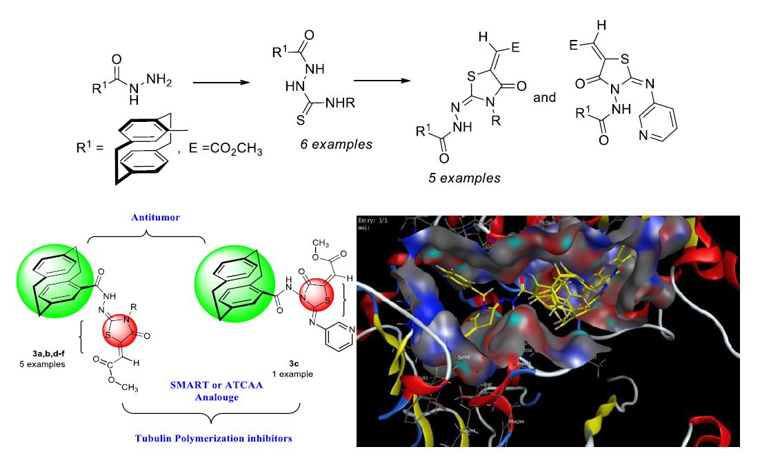
Synthesis of methyl 2-(2-(4′-[2.2]paracyclophanyl)-hydrazine-ylidene)-4-oxothiazolidin-5-ylidene)acetates was here reported.
-
Cytotoxic activity of the synthesized compounds toward the NCI-60 panel of cancer cell lines was determined, and the cellular mechanism of the most potent inhibitors was further investigated in leukemia cell lines.
-
Compound 3a was found as the most cytotoxic one, and it was selected for further five-dose analysis according to NCI protocol.
-
Compound 3a, in comparison to other derivatives, exhibited high specificity against leukemia RPMI-8226 and SR cell lines, and it also showed a remarkable tubulin inhibitory activity in relation to colchicine with IC50 = 4.97 µM/mL.
-
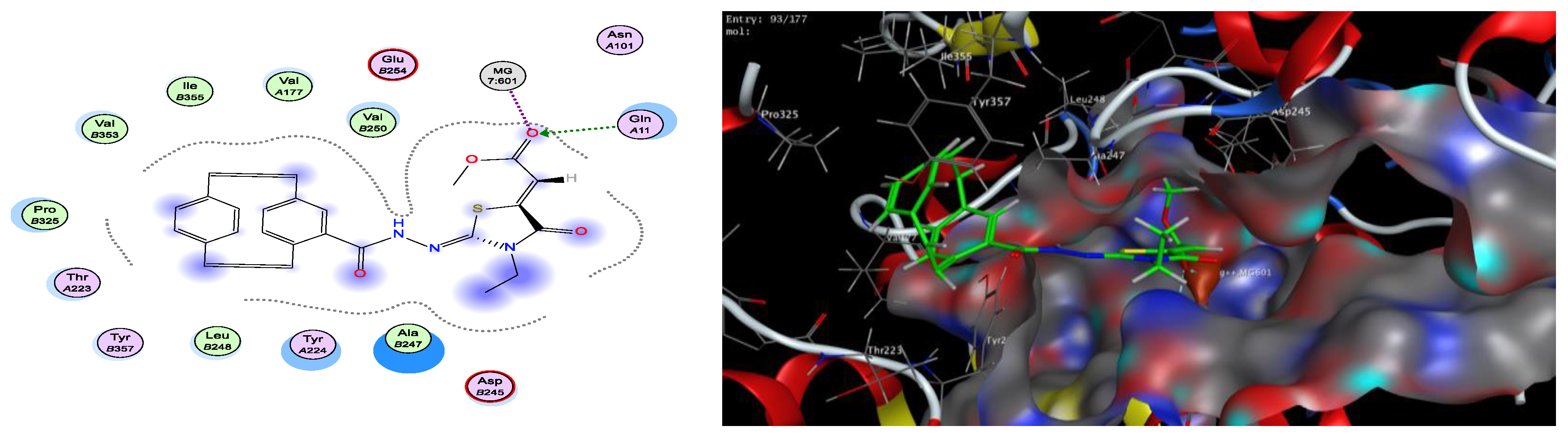
Docking studies with β-tubulin revealed that most of the tested compounds showed good binding at the colchicine binding site of the enzyme, especially for compound 3a, which made several interactions better than that of the reference colchicine.

1. Introduction

Cyclophane chemistry is rapidly growing in the field of stereoselective synthesis with its incorporation into heterocyclic and/or polymer chemistry [1,2,3,4,5,6,7,8]. A great deal of attention is focused on developing new synthetic tools for synthesizing functionalized [2.2]paracyclophanes. Substituted [2.2]paracyclophanes can also serve as chiral templates and/or as auxiliaries [9]. The synthesis and application of heterocycles based on [2.2]paracyclophane [10,11] can be organized into five structural classes (Figure 1): heterocycle derived by paracyclophanyl group (type I), heterocycle derived by bridge (type II), heterocycle fused to ethano bridge (type III), fused heterocycle to the benzene moiety (type IV), and heterocycle between the two benzene rings of paracyclophane (type V).
A [2.2]paracyclophanyl acetic acid enantiomer was tested as anti-inflammatory agent [12]. Recent studies reported that 1,3-thiazoles derivatives showed in vitro α-glucosidase inhibitory activity [13,14].
Recently, we have an interest in the preparation of heterocycles of similar structural features [15,16,17,18,19,20,21,22,23]. In addition, we prepared metal complexes of thiosemicarbazones with Cu(I) and Cu(II) and the tridentate and bidentate structures of the paracyclophanyl–thiosemicarbazone–metal complexes were elucidated [24].
The thiazole scaffold possesses potent cytotoxic activity in cancer cell lines. For example, thiazolidine-4-carboxylic acid amides (ATCAA) I showed activity against prostate cancer cells with an average IC50 in the range from 0.7 to 1.0 μM, while the average IC50 against melanoma cells ranged from 1.8 to 2.6 μM (Figure 2) [25].
Moreover, gold(I) complexes of phosphino [2.2]paracyclophane ligands III exhibited their cytotoxic activity in the HeLa S3 cell line (LD50 = 22.15 μm) in comparison to cisplatin (LD50 = 7.65 μm). Their cytotoxicity and their mechanisms of action are different and involve apoptosis, necrosis, and DNA damage (Figure 2) [26]. Studies reported that methoxylbenzoylaryl-thiazole (SMART) II compounds showed nanomolar activity in inhibiting melanoma and prostate cancer cell growth [27,28,29]. Due to the interest in developing new anti-cancer agents, we designed a strategy of gathering both thiazole and paracyclophane skeletons for designing new anti-tumor agents (Figure 3).

2. Results and Discussion

2.1. Chemistry Section

Starting with acyl hydrazide 1 [30], the acylthiosemicarbazides 2af could be prepared (Scheme 1). Compounds 2af were used as the target compounds for the formation of thiazolidinones, 3a,b,df, and 3c (Scheme 1).
Synthesis of compound 1 can be achieved from the reported route starting with the commercial hydrocarbon 5 (Scheme 2). The procedure consisted firstly of the conversion of 5 into the keto acid chloride 6 with oxalyl chloride/aluminum trichloride. Heating of 6 in chlorobenzene caused decarbonylation to give 7, and, when the resulting acid chloride 7 was quenched with ethanol, the ester 8 [31] was obtained (Scheme 2). On refluxing the α-ketoester 8 with hydrazine hydrate in different solvents, the reaction failed to give the target hydrazide 1 in good yields (Scheme 2). However, heating 8 in an excess of hydrazine hydrate afforded the corresponding compound carbohydrazide 1 in 80% yield (Scheme 2).

Preparation of 2-(4′-[2.2]Paracyclophanyl-4H-hydrazinecarbothioamides 2af

In the present work, 2-(4′-[2.2]paracyclophanyl-4H-N-substituted-hydrazinecarbothioamides 2af were prepared by refluxing compound 1 with the isothiocyanates in EtOH as a solvent (Scheme 3). The infrared (IR) spectroscopy of hydrazinecarbothioamides 2af showed that the carbonyl–amide and its N–H stretching bands were present at = 1656–1667 and 3360–3210 cm–1, respectively, in addition to a band at = 1390–1360 cm−1 due to the stretching vibration of the C=S groups. This fact was confirmed by the appearance of the carbon signal in the 13C-NMR at δ = 181.2–182.9 ppm. Spectroscopic details are shown from compound 2d, as an example. The 1H-NMR spectrum of 2d showed the two thiourea-NH protons at δ = 9.01 and 8.65 ppm. The paracyclophanyl protons resonated at δ = 6.96 as a doublet (J = 2.0 Hz) for 1H, a triplet at δ = 6.72 (J = 7.8, 1.9 Hz) for 2H, and at δ = 6.66–6.22 ppm as a multiplet for 4H. The allyl protons resonated as three multiplets at δ = 5.97–5.87 (CH=), 5.09–5.05 (CH2=), and 4.32–4.24 (CH2N).
In the 13C-NMR spectrum, the C=S carbon appeared at δ = 182.7, whereas the C=O carbon appeared at δ = 167.7 (C=O). The allyl carbons resonated at δ = 134.8, 115.0, and 56.0 for the allyl-CH=, allyl-CH2=, and allyl-CH2, respectively. The four carbons of the paracyclophanyl CH2 appeared at δ = 34.8 (1C), 34.7 (1C), and 34.5 (2C) ppm.
The X-ray structure analysis of compounds 2a,b,d strongly confirmed the proposed structures as shown in Figure 4, Figure 5 and Figure 6, respectively. One can note that the dihedral angle of CS–NH–NH–CO was nearly 90°, and that angle was also seen in an example reported in reference [32].
Refluxing 2af with dimethyl acetylenedicarboxylate (9) in EtOH afforded the expected thiazolidinones 3a,b,df in 67%–78% yield together with 3c in 65% yield (Scheme 4).
Mass spectroscopy and elemental analysis identified the molecular formula of 3b as C30H27N3O4S. Moreover, the 1H-NMR spectrum revealed three singlets at δ = 10.88 (NH), 6.93 (vinyl-CH), and 5.44 (benzylic-CH2). The 13C-NMR spectrum of 3b showed three carbonyl carbon signals at δ = 166.0 (ester-CO), 165.6 (cyclic-C═O), and 161.5 (hydrazide-CO). The thiazolidine-4-one moiety showed carbon signals at δ =146.2 for C-2 and 140.2 for C-5. In addition, the vinyl-CH, OCH3-ester, and benzyl-CH2 resonated at δ =116.6, 52.6, and 51.3 ppm, respectively. The X-ray structure analysis of 3b identified the structure as methyl (E)-2-((E)(2′-4-[2.2]paracyclohanyl)-hydrazinylidene)-3-benzyl-4-oxathiazolidin-5-ylidene)acetate (Figure 7).
The 1H-NMR spectrum showed two singlets at δ = 10.12 (NH) and 6.57 (vinyl-CH). The CO-ester, cyclic-CO, hydrazide-CO, thiazolidine-C-,2 and thiazolide-C-4 appeared in the 13C-NMR spectrum of 3c at δ = 167.0, 166.6, 162.5, 151.3, and 141.1, respectively (see Section 3). Finally, the structure of 3c was ambiguously identified by X-ray structure analysis (Figure 8).
The abnormal behavior of 2c toward 9 might be attributed to the expected resonance structure of 2c that would decrease the basicity of the N3-thioamide compared with the N2-hydrazide and, therefore, the N2-hydrazide would be more reactive toward nucleophilic addition (Scheme 5).

2.2. Biological Investigation

2.2.1. Anti-Proliferative Investigation against 60 Cancer Cell Lines at the National Cancer Institute (NCI), USA.

The methodology of the NCI anticancer screening was described in detail elsewhere (http://www.dtp.nci.nih.gov). Briefly, the primary anticancer assay was performed using approximately 60 human tumor cell lines derived from nine neoplastic diseases, in accordance with the protocol of the Drug Evaluation Branch, National Cancer Institute, Bethesda. Tested compounds were added to the culture at a single concentration (10−5 M) and the cultures were incubated for 48 h. End-point determinations were made with a protein binding dye, SRB. Results for each tested compound were reported as the percentage growth of the treated cells when compared to the untreated control cells. The percentage growth was evaluated spectrophotometrically versus controls not treated with test agents. All experiments were repeated three times (Table 1).
Compounds 3af revealed that compound 3a achieved complete cell death on the nine tested cancer cell lines. It is noteworthy that compounds 3a, 3b, 3d, and 3e were the most potent tested derivatives on the leukemia cell line. They showed growth inhibition percentages greater than 100%, which means that they were cytotoxic and displayed complete cancer cell death that killed all cells, including cancer cells. They may stop cancer cells from dividing and growing and may cause tumors to shrink in size. The complete cell death was against leukemia RRMI-8226 with inhibitions of 120.89%, 147.00%, 109.36%, and 114.28%, respectively, and against SR with inhibitions of 115.60%, 114.70%, 98.21%, and 113.40%, respectively. Compound 3e showed remarkable activity on the other tested cell lines. Although 3f showed moderate to weak activity on most of tested cancer cell lines, compound 3b exhibited significant inhibition against non-small-cell lung cancer NCI-H522, colon cancer HT29 and SW-620, melanoma LOX IMVI, ovarian cancer OVCAR-3, renal cancer CAKI-1, prostate cancer PC-3, and breast cancer BT-549, T-47D, and MDA-MB-468 with inhibitions of 84.06%, 87.62%, 81.83%, 93.79%, 79.71%, 84.43%, 82.23%, 89.68%, 106.25%, and 78.91%, respectively. Furthermore, compounds 3c and 3d displayed mild to moderate activity on most of the cancer panel cell lines

Structure Activity Relationship (SAR)

It is notable that the new prepared paracyclophane/thiazole conjugates showed significant anti-cancer activity with different growth inhibition percentages. This disparity among the different derivatives may be attributed to the type of substitution on the thiazole ring, whether on nitrogen (compounds 3a,b,df) or at position 2 (compound 3c). It is expected that increasing thiazole flexibility through inserting a phenyl or benzyl group (compounds 3a and 3b, respectively) into the structure would improve binding to the target protein and, hence, show higher antiproliferative activity on all tested cell lines than those containing an aliphatic group, i.e., either compound 3d (allyl) or 3e (ethyl) of the same series, which provides less flexibility. Interestingly, the other paracyclophane/thiazole derivative 3c bearing a pyridinyl amine moiety at position 2 of the thiazole ring altered the activity, showing lower cytotoxicity.

2.2.2. In Vitro Five-Dose Full NCI 60 Cell Panel Assay

Compound 3a was selected by NCI for five-dose investigation against 60 human tumor cell lines that were incubated at five different concentrations (0.01, 0.1, 1, 10, and 100 μM) (Figure 9). The outcomes were used to form log concentration vs. percentage growth inhibition curves and three response parameters (GI50, total growth inhibition (TGI), and LC50) were calculated for each cell line (Table 2). The GI50 value (growth inhibitory activity) corresponds to the concentration of the compound causing 50% decrease in net cell growth, the TGI value (cytostatic activity) is the concentration of the compound resulting in total growth inhibition (TGI), and the LC50 value (cytotoxic activity) is the concentration of the compound causing a net 50% loss of initial cells at the end of the incubation period of 48 h.
The criterion for selectivity of a compound depends upon the ratio obtained by dividing the full-panel MID (the average sensitivity of all cell lines toward the test agent) (μM) by the individual subpanel MIDs (μM). Ratios between 3 and 6 refer to moderate selectivity, and ratios greater than 6 indicate high selectivity toward the corresponding cell line, while compounds not meeting either of these criteria are rated non-selective.
Compound 3a under investigation exhibited remarkable anticancer activity against most of the tested cell lines representing nine different subpanels with GI50 ranging from 1.52–7.56 μM (Table 2). Results indicate that 3a showed high activity against renal cancer RXF-393, melanoma LOX IMIV, colon cancer HCC-2998, non-small-cell lung cancer EKVX, and leukemia RPMI-8226 with GI50 values of 1.52, 1.69, 1.78, 1.91, and 2.15 μM, respectively. An obvious sensitivity profile was seen toward the leukemia subpanel (GI50 values ranging from 2.15 to 3.15 μM), colon cancer subpanel (GI50 values ranging from 1.78 to 2.13 μM), breast cancer subpanel (GI50 values ranging from 1.54 to 1.87 μM), and ovarian cancer subpanel (GI50 values ranging from 1.66 to 3.55 μM). In this context, compound 3a was found to have broad-spectrum antitumor activity against the nine tumor subpanels tested with selectivity ratios ranging from 0.63–1.28 and 0.58–5.89 at the GI50 and TGI levels, respectively; however, it exhibited noteworthy antiproliferative activity against all cancer cell lines with low selectivity ratios.

2.2.3. Evaluation of In Vitro Antiproliferative Activities against Leukemia RPMI-8226 and SR

Since the antiproliferative investigation results against 60 cell lines at the NCI revealed greater activity toward leukemia cancer, especially leukemia RPMI-8226 and SR, we were encouraged to perform further in vitro antiproliferative studies against those two cell lines. Compounds 3ae were evaluated for their antiproliferative activity by performing an MTT assay against a panel of two human tumor cell lines, leukemia RPMI-8226 and SR, compared with colchicine as a reference. As shown in Table 3, the antiproliferative activities of the tested compounds were generally more pronounced against the two panels of leukemia cancer cells as compared with the reference. The calculated results were subjected to statistical analysis using GraphPad Prism 7 with the one-way ANOVA and non-parametric program. The difference in the results was considered significant when the p-values were less than 0.05. All the tested compounds were significant, as well as the reference (*** p < 0.05), in comparison to control. Compound 3a exhibited the highest antiproliferation compared to reference and the other tested compounds, whereas it showed IC50 values 1.61 and 1.11 µM better than colchicine (i.e., the reference compound) of 4.05 and 1.81 µM against leukemia RPMI-8226 and SR, respectively. On the other hand, compound 3e showed a significant antiproliferative activity with an IC50 value 3.17 µM better than the reference of 4.05 µM against leukemia RPMI-8226 only. This may be attributed to both compounds 3a and 3b having electron-withdrawing substitution of phenyl and benzyl, respectively, which positively affected their permeability to cancer cells. Compound 3b showed comparable IC50 values of 4.62 and 2.02 µM to colchicine.
Additionally, compound 3c bearing a pyridinyl amine moiety at position 2 of the thiazole ring showed weak anti-proliferation activity with IC50 values of 9.69 and 4.84 µM, which explains its low cytotoxicity. It is interesting to mention that the proliferation inhibitory results were positively correlated with the anticancer results obtained from NCI.

2.2.4. Evaluation of In Vitro Tubulin Polymerization Inhibitory Activity

To investigate whether the antiproliferative activities of these target compounds 3ae were related to their interaction with tubulin, these compounds were tested for their ability to inhibit tubulin polymerization at their IC50 concentrations using an ELISA assay for β-tubulin.
The in vitro kinetics of microtubule assembly was measured using an ELISA kit for TUBb (Cloud-Clone. Corp.) on the leukemia SR cell line. The compounds tested were 3a–f and colchicine. Briefly, growing cells from the SR cell line were trypsinized, counted, and seeded at the appropriate densities into 96-well microtiter plates. Cells were then incubated in a humidified atmosphere at 37 °C for 24 h.
The assay revealed that all the tested compounds 3ae showed tubulin polymerization inhibitory activity compared to colchicine as a reference (Table 4). Again, compound 3a showed the highest ability to inhibit tubulin polymerization with an IC50 value of 4.97 µM compared to the reference with an IC50 value 3.76 µM and the other tested compounds. On the other hand, compounds 3e and 3c showed remarkable tubulin polymerization inhibition with IC50 values of 6.61 and 8.38 µM, while 3d displayed weak inhibition with an IC50 value of 14.79 µM. Results are in agreement with the previous mentioned anti-proliferative activity

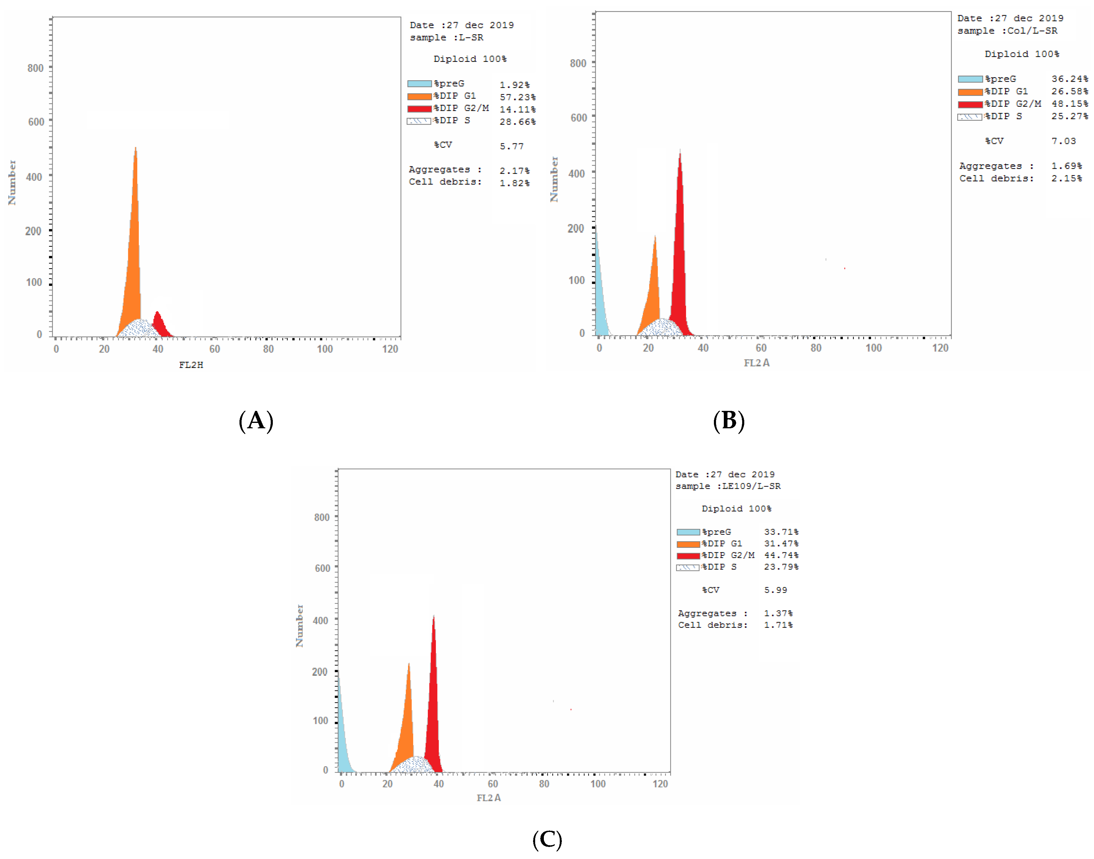
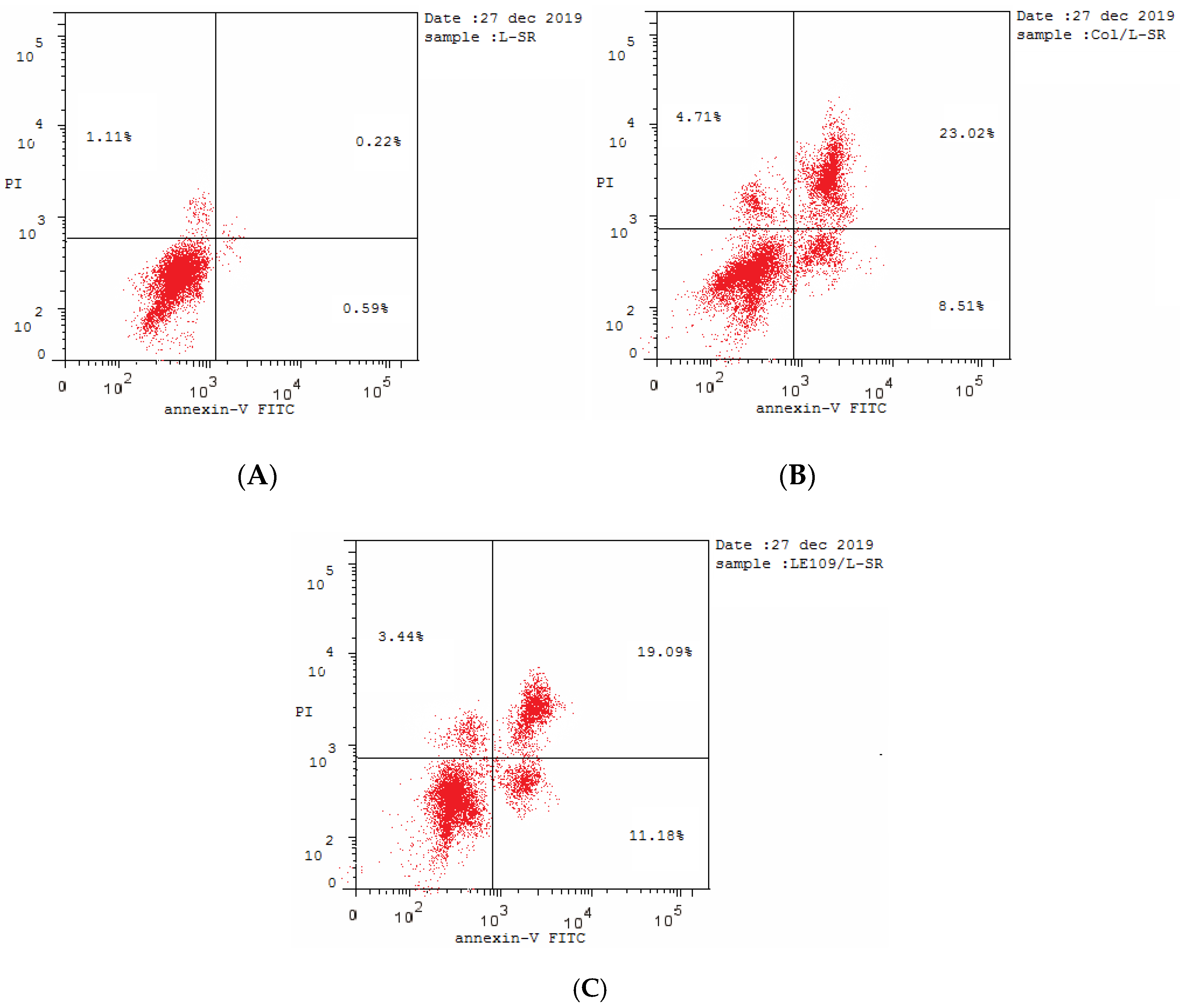
2.2.5. Cell Cycle Analysis

Cell cycle analysis was performed using cytometers from Becton Dickinson Immunocytometry Systems, Beckman/Coulter Inc., DACO/Cytomation, and PARTEC GmbH. Regulation of cell growth is mainly controlled through cell-cycle control mechanisms. Proliferation inhibition can be triggered by cell-cycle arrest in cancer cells. During the cell cycle, the G2/M checkpoint is a potential target for cancer therapy.
This prevents DNA-damaged cells from entering mitosis and allows for the repair of DNA that was damaged in late S or G2 phases prior to mitosis (Table 5). Induction of cell-cycle arrest is a common mechanism proposed for the cytotoxic effects of anticancer drugs containing paracyclophane/thiazole derivatives. The analysis indicated that leukemia SR cells treated with compound 3a showed significant growth arrest at the G2/M phase compared to control cells, where the S-phase progression of SR cells was substantially delayed (Figure 10). The annexin V/PI flow cytometry of SR cells was repeated three times after treatment with the IC50 value (1.11 µM) of 3a, which showed an increase in percentage of the necrotic cells in late apoptosis to 19% (upper right quadrant of the cytogram) (Figure 11). Hence, compound 3a showed a considerable ability to dissipate cell membrane integrity, whereas the lower right quadrant illustrating the early apoptotic cells which kept their membrane integrity indicated the ability of 3a to initiate apoptosis.

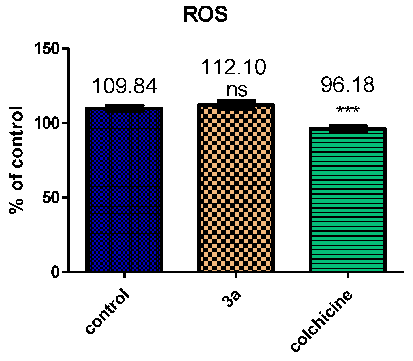
2.2.6. Compound 3b Induced Mitochondrial Depolarization and ROS Production

Mitochondria play an essential role in the propagation of apoptosis [33]. It is reported that, at an early stage, apoptotic stimuli are capable of modifying the mitochondrial transmembrane potential (Δψmt). Δψmt was recorded by the fluorescence of the dye JC-1.20. SR cells treated with 3a displayed a non-significant shift in fluorescence compared with control cells, explaining the weak depolarization of the mitochondrial membrane potential. In contrast, colchicine showed significant results (Figure 12). The disruption of Δψmt is associated with the appearance of annexin V positivity in the treated cells when they are in an early apoptotic stage. It is documented that the dissipation of Δψmt is characteristic of apoptosis, and this was indicated with both microtubule-stabilizing and -destabilizing agents in different cell types [34]. Induction of cell-cycle arrest is a common mechanism proposed for the cytotoxic effects of anticancer drugs containing paracyclophane/thiazole derivatives. Mitochondrial membrane depolarization is associated with mitochondrial production of ROS [35]. Therefore, we investigated whether ROS production increased after treatment with the test compounds.
The presented results in Figure 13 show that 3a induced the production of significant amounts of ROS (109.84%) in comparison with control cells and the colchicine reference. This result is compatible with the dissipation of Δψmt described above.

2.2.7. Effect of Compound 3a on Multidrug-Resistant (MDR) Leukemia SR Cells

A previous study reported that multidrug resistance was observed for numerous chemotherapeutic agents and could be identified through overexpression of P-glycoprotein (Pgp) [36,37].
Compound 3a and tubulin-targeting agent colchicine were tested on the MDR-SR Leukemia cell line. Compound 3a showed Pgp-mediated MDR expression at 1.285 ng/mL (1.15-fold change), which means that it had much better resistance indices comparable to control at 1.121 ng/mL (1-fold) than colchicine at 1.726 ng/mL (1.54-fold change). Results of compound 3a (Table 6) showed the ability of overcoming the assumed glycoprotein overexpression of the cell line; thus, 3a could be considered as an important substrate candidate against MDR cells.

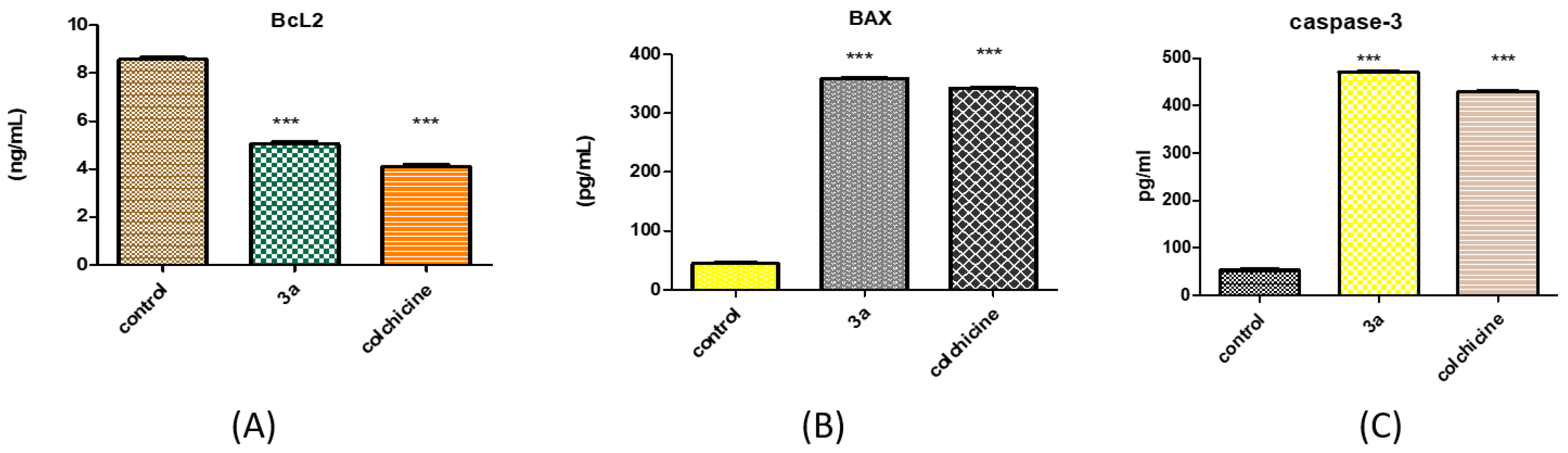
2.2.8. Effect of 3a on Caspase-3 Activation

Since caspase-3 is important for spreading the apoptotic signal after exposure to antimitotic compounds [38], the effect of compound 3a on the caspase-3 activated enzyme was evaluated using ELISA and replicated three times. The reference compound and 3a were incubated for 30 min at room temperature in the dark on the leukemia SR cell line. Results revealed that 3a is a potential caspase-3 activator with a slight increase in the level of active caspase-3 at a concentration of 471.2 ng/mL (8.84-fold) compared to colchicine at a concentration of 428.9 ng/mL (8.05-fold) as shown in Table 7.

2.2.9. Effect of 3a on BAX and Bcl-2 Proteins

The proteins of the Bcl2 family [39] play a major role in controlling apoptosis through the regulation of mitochondrial processes and the release of mitochondrial proapoptotic molecules that are important for the cell death pathway [40]. Compound 3a caused nearly 7.98-fold upregulation (Table 8), while it showed markedly higher levels of the antiapoptotic Bcl-2 proteins up to 0.59-fold compared to the control untreated cells (Figure 14).

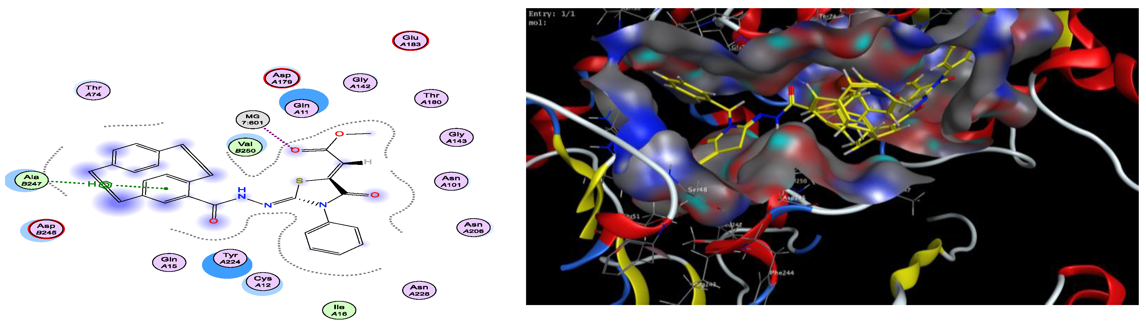
2.3. Docking Studies

The molecular modeling of the possible binding modes for the newly synthesized thiazole/paracyclophane hybrids 3ae and colchicine as a reference was done to predict the binding interactions between them and β-tubulin at the colchicine binding site, which was obtained from the protein data bank (PDB: 3HKC). Docking studies were carried out using Molecular Operating Environment (MOE®) version 2014.09 (Chemical Computing Group Inc., Montreal, QC, Canada) using colchicine as a reference for validation of the method. The theoretical predictions from the molecular docking study agreed for some highly active derivatives such as 3a, 3b, 3d, and 3e with the experimentally observed tubulin polymerization inhibition. All derivatives were successfully docked into the colchicine binding site of β-tubulin. The binding free energies from the major docked poses are listed in Table 9, and the most favorable poses of the tested compounds are shown in Figure 15, Figure 16, Figure 17, Figure 18, Figure 19 and Figure 20. Most of the tested compounds had high binding affinity to the enzyme as the binding free energy (ΔG) values of them ranged from −0.5 to −3.4 kcal/mol, which was comparable to the reference colchicine (ΔG = −0.6 to −2.3 kcal/mol). The docking result of the reference compound colchicine was completely consistent with the mode of action of thiazole/paracyclophane derivatives (Figure 15).
The two-dimensional (2D) diagram showed crucial binding involving Ala 247, Tyr 224, and Mg 601 through the [2.2]paracyclophane ring, thiazole ring, and ester C=O functionality. Moreover, stabilization of the reference colchicine within the active site occurred through one strong hydrogen bond interaction with amino-acid residue Glu 71 and metal intercalation with Mg 601. Docking results with the colchicine binding site revealed that most of the tested compounds showed good binding with the enzyme and made several interactions comparable to that of the reference colchicine (Figure 15).
Compounds 3a, 3c, 3d, and 3e exhibited the same interaction as the reference with Mg 601; however, both 3d and 3b showed hydrogen bonding interactions with Glu 71 (Figure 16, Figure 17, Figure 18, Figure 19 and Figure 20). On the other hand, compound 3d possessed greater interactions than the reference with the same amino acids with additional one H–pi bond with Arg 2 (Figure 20); however, compounds 3a and 3c displayed an extra H–pi interaction with Ala 247 (Figure 16 and Figure 18). Compound 3c (Figure 18) did not feature hydrogen binding interactions with the amino acid residue Glu 71, but kept an H–Pi interaction with Tyr 224.

3. Experimental

3.1. Chemistry Part

3.1.1. Materials and Methods

Melting points were taken in open capillaries on a Gallenkamp melting point apparatus (Weiss-Gallenkamp, Loughborough, UK) and are uncorrected. The IR spectra were recorded using the attenuated total reflection (ATR) technique with a FT device (FT-IR Bruker IFS 88, Bruker, Leiderdorp, The Netherlands), Institute of Organic Chemistry, Karlsruhe Institute of Technology, Karlsruhe, Germany. The NMR spectra were measured in DMSO-d6 and acetone-d6 on a Bruker AV-400 spectrometer at 400 MHz for 1H and 100 MHz for 13C; the chemical shifts are expressed in δ (ppm) versus internal tetramethylsilane (TMS) = 0 for 1H and 13C, and external liquid ammonia = 0. The description of signals includes s = singlet, d = doublet, t = triplet, q = quartet, m = multiplet, dd = doublet of doublet, ddd = doublet of dd, dt = doublet of triplet, td = triplet of doublet, bs = broad singlet, and m = multiplet. Mass spectra were recorded on a FAB (fast atom bombardment) Thermo Finnigan Mat 95 (70 eV). For the high-resolution mass, the following abbreviations were used: calc.= theoretical calculated mass; found = mass found in analysis at the Institute of Organic Chemistry, Karlsruhe University, Karlsruhe, Germany. Thin Layer Chromatography (TLC) was performed on analytical Merck 9385 silica aluminum sheets (Kieselgel 60) with Pf254 indicator; TLCs were viewed at λmax = 254 nm. Crude products were purified by flash chromatography with silica gel 60 (0.040 × 0.063 mm, Geduran®) (Merck, Darmstadt, Germany). Compounds 2 and 58 were prepared according to the methodology mentioned in References [30] and [31], respectively.

3.1.2. Preparation of Paracyclophanyl-N-substituted Hydrazinecarbothioamides 2af

A mixture of carbohydrazide paracyclophane (1) (1.00 g, 3.7 mmol) in 60 mL of ethanol and different derivatives of isothiocyanate (3.7 mmol) was refluxed for 4–8 h. The reaction mixture was poured into a beaker and was allowed to stand until a precipitate was formed. Then, the precipitate was filtered and washed with heptane several times (3 × 100 mL).
2-(4′-[2.2]Paracyclophanyl)-N-phenylhydrazinecarbothioamide (2a). Colorless crystals (ethanol), 1.33 g (88%), melting point (m.p.) 190–92 °C, 1H-NMR (400 MHz, DMSO-d6, ppm): δ = 9.86 (s, 1H, NHNHCS), 9.63 (s, 1H, NH-CO), 7.55 (d, J = 7.8 Hz, 1H, Ph-H), 7.50 (s, 1H, NHCS), 7.33 (t, J = 7.9 Hz, 2H, Ph-H), 7.14 (t, J = 7.6 Hz, 2H, Ph-H), 6.95 (s, 1H, PC-H), 6.63 (dd, J = 7.8, 1.8 Hz, 2H, PC-H), 6.52 (d, J = 8.0 Hz, 3H, PC-H), 6.47 (d, J = 7.8 Hz, 1H, PC-H), 3.76 (s, 1H, PC-CH2-2′), 3.33–2.79 (m, 7H, PC-CH2-CH2). 13C-NMR (100 MHz, DMSO-d6, ppm): δ = 181.2 (C=S), 171.8 (C=O), 140.1 (Ph-CH), 140.0 (PC-C-6′), 139.5 (PC-C-11′), 139.4 (PC-C-14′), 139.3 (PC-C-3′), 139.0 (PC-2CH), 135.7 (PC-2CH), 135.1, 132.6 (PC-CH), 132.5 (Ph-2CH), 132.4 (Ph-2CH), 131.6 (PC-CH),129.0 (Ph-C), 128.2 (PC-C-4′), 34.8 (PC-CH2-1′), 34.7 (PC-CH2-10′), 34.5 (PC-CH2-9′), 34.4 (PC-CH2-2′). IR (ATR, cm−1) = 3250–3210 (br, m, NH), 3009 (m, Ar-CH), 2890 (w, Aliph-CH), 1656 (s, CO), 1390 (s, C=S), 980 (s, C-N str). MS (FAB) m/z (%) = 402.2 ([M + H]+ (85). HRMS ([M + H]+, C24H24O1N332S1) Calcd.: 402.1635, Found: 402.1633.
2-(4′-[2.2]Paracyclophanyl)-N-benzylhydrazine-1-carbothioamide (2b). Colorless crystal (ethanol), 1.33 g (85%), m.p. 172–174 °C, 1H-NMR (400 MHz, DMSO-d6, ppm) δ = 9.76 (s, 1H, NHNHCS), 9.41 (s, 1H, NH-CO), 8.34 (s, 1H, NHCS), 7.34–7.32 (m, 2H, Ph-H), 7.31–7.28 (m, 2H, Ph-H), 7.22-7.20 (m, 1H, Ph-H), 6.93 (s, 1H, PC-H), 6.64–6.61 (m, 2H, PC-H), 6.53–6.40 (m, 4H, PC-H), 4.81–4.74 (m, 2H, Benzyl-CH2), 3.70–3.68 (m, 1H, PC-CH2-2′), 3.14–2.78(m, 7H, PC-CH2-CH2). 13C-NMR (100 MHz, DMSO-d6, ppm) δ = 182.9 (C=S), 168.3 (C=O), 140.6 (Ph-H), 140.0 (PC-C-6′), 139.9 (PC-C-11′), 139.7 (PC-C-14′), 139.4 (PC-C-3′), 136.2 (PC-2CH), 135.7 (PC-CH), 133.1 (Ph-C), 133.0, 132.9 (PC-2CH), 131.9 (PC-C-4′), 128.5 (Ph-2CH), 127.5, 127.1 (Ph-CH), 47.2 (Benzyl-CH2), 35.3 (PC-CH2-1′), 35.2 (PC-CH2-10′), 35.0 (PC-2CH2-9′,2′). IR (ATR, cm−1) = 3346 (s, NH), 3199 (s, Ar-CH), 2980 (w, Aliph-CH), 1653 (s, CO), 1365 (s, C=S), 978 (C-N str). MS (FAB) m/z (%) = 416.2 ([M + H]+ (70)]. HRMS ([M + H]+, C25H26O1N332S1) Calcd.: 416.1797, Found: 416.1799.
2-(4′-[2.2]Paracyclophanyl)-N-(pyridin-3-yl)hydrazine-1-carbothioamide (2c). Colorless crystal (methanol), 1.24 g (82%), 152–154 °C, 1H-NMR (400 MHz, DMSO-d6, ppm) δ = 9.93 (s, 1H, NHNHCS), 9.72 (s, 1H, NHCO), 8.57 (s, 1H, NHCS), 8.34 (dd, J = 4.8, 1.5 Hz, 1H, Pyr-H), 7.90–7.40 (m, 2H, Pyr-H), 7.38 (dd, J = 8.2, 4.7 Hz, 1H, Pyr-H), 7.12–6.87 (m, 2H, PC-H), 6.84–6.57 (m, 2H, PC-H), 6.58–6.48 (m, 2H, PC-H), 6.46 (d, J = 7.8 Hz, 1H, PC-H), 3.75–3.34 (m, 1H, PC-CH2-2′), 3.27–3.05 (m, 3H, PC-CH2-CH2), 3.05–2.90 (m, 3H, PC-CH2-CH2), 2.90–2.77 (m, 1H, PC-CH2-CH2). 13C-NMR (100 MHz, DMSO-d6, ppm) δ = 181.5 (C=S), 167.7 (C=O), 147.0, 145.7 (Pyr-CH), 140.2 (PC-C-6′), 139.4 (PC-C-11′), 139.2 (PC-C-14′), 138.9 (PC-C-3′), 136.1 (Pyr-C), 135.7 (PC-2CH), 135.2 (PC-CH), 133.4, 132.5 (PC-CH), 132.4 (Pyr-CH), 132.3 (PC-2CH), 131.4 (PC-C-4′), 122.9 (Pyr-CH), 38.8 (PC-CH2-1′), 34.7 (PC-CH2-10′), 34.6 (PC-CH2-9′), 34.4 (PC-CH2-4′). IR (ATR, cm−1) = 3206 (m, NH), 3091 (w, Ar-CH), 2925 (w, aliph-CH), 1645 (m, CO), 1376 (s, C=S), 980 (s, C-N str). MS (FAB) m/z (%) = 403.2 [M + H]+ (55). HRMS ([M + H]+, C23H23O1N432S1) Calcd.: 403.1593, Found: 403.1591.
N-Allyl-2-(4′-[2.2]paracyclophanyl)hydrazine-1-carbothioamide (2d). Colorless crystal (methanol), 1.18 g (86%), m.p. 162–164 °C, 1H-NMR (400 MHz, DMSO-d6, ppm) δ = 9.01 (s, 1H, NHNHCS), 8.65 (s, 1H, NHCO), 7.74 (s, 1H, NHCS), 6.96 (d, J = 2.0 Hz, 1H, PC-H), 6.72 (t, J = 7.8, 1.9 Hz, 2H, PC-H), 6.66–6.22 (m, 4H, PC-H), 5.97–5.87 (m, 1H, allyl-CH=), 5.09–5.05 (m, 1H, allyl-CH2=), 4.32–4.24 (m, 2H, allyl-CH2) 3.84–3.74 (m, 1H, PC-CH2-2′), 3.28–2.78 (m, 7H, PC-CH2-CH2). 13C-NMR (100 MHz, DMSO-d6, ppm) δ = 182.7 (C=S), 167.7 (C=O), 140.1 (PC-C-6′), 139.5 (PC-C-11′), 139.3 (PC-C-14′), 139.0 (PC-C-3′), 135.7, 135.2, 135.1 (PC-CH), 134.8 (allyl-CH=), 132.6, 132.5, 132.3 (PC-CH), 131.5 (PC-CH), 128.9 (PC-C-4′) 115.0 (allyl-CH2=), 56.0 (allyl-CH2), 34.8 (PC-CH2-1′), 34.7 (PC-CH2-10′), 34.5 (PC-2CH2-9′,2′). IR (ATR, cm−1) = 3360 (s, NH), 3132 (m, Ar-CH), 2970 (w, Aliph-CH), 1660 (s, CO), 1380 (s, C=S), 982 (s, C-N str). MS (FAB) m/z (%) = 366.2 ([M + H]+ (100). HRMS ([M + H]+, C21H24O1N332S1) Calcd.: 366.1640, Found: 366.1638.
2-(4′-[2.2]Paracyclophanyl)-N-ethylhydrazine-1-carbothioamide (2e). Colorless crystal (ethanol/cyclohexane), 1.08 g (81%), m.p. 130–132 °C, 1H-NMR (400 MHz, DMSO-d6, ppm) δ = 9.65 (s, 1H, NHNHCS), 9.22 (s, 1H, NH-CO), 7.81 (s, 1H, NHCS), 6.91 (d, J = 1.8 Hz, 1H, PC-H), 6.63 (dd, J = 7.7 Hz, J = 1.9 Hz, 2H, PC-H), 6.51 (d, J = 9.6 Hz, 3H, PC-H), 6.44 (d, J = 7.8 Hz, 1H, PC-H), 3.69 (m, 1H, PC-CH2-2′), 3.60–3.26 (m, 2H, ethyl-CH2), 3.26–2.68 (m, 7H, PC-CH2-CH2), 1.06 (t, J = 10.1 Hz, J = 7.0 Hz, 3H, ethyl-CH3). 13C-NMR (100 MHz, DMSO-d6, ppm) δ = 181.7 (C=S), 167.6 (C=O), 140.1 (PC-C-6′), 139.5 (PC-C-11′), 139.2 (PC-C-14′), 139.0 (PC-C-3′), 135.7 (PC-2CH), 135.1 (PC-CH), 132.7, 132.6, 132.5, 132.4 (PC-CH), 131.5 (PC-C-4′), 56.0 (ethyl-CH2), 35.0 (PC-CH2-1′), 34.9 (PC-CH2-10′), 34.6 (PC-2CH2-9′,2′), 14.7 (ethyl-CH3). IR (ATR, cm−1) = 3357 (m, NH), 3190 (m, Ar-CH), 2870 (w, Aliph-CH), 1667 (s, CO), 1365 (s, C=S), 980 (s, C-N str). MS (FAB) m/z (%) = 354.2 ([M + H]+ (100). HRMS ([M + H]+, C21H24O1N332S1) Calcd.: 354.1640, Found: 354.1640.
2-(4′-[2.2]Paracyclophanyl)-N-cyclopropylhydrazine-1-carbothioamide (2f). Colorless crystal (ethanol), 1.10 g (80%), m.p. 158–160 °C, 1H-NMR (400 MHz, DMSO-d6, ppm) δ = 9.66 (s, 1H, NHNHCS), 9.34 (s, 1H, NHCO), 6.90 (s, 1H, NHCS), 6.61 (dd, J = 7.7, 1.8 Hz, 2H, PC-H), 6.55–6.46 (m, 4H, PC-H), 6.43 (d, J = 7.8 Hz, 1H, PC-H), 3.74–3.43 (m, 1H, PC-CH2-CH2), 3.38 (d, J = 38.4 Hz, 2H, PC-CH2-CH2), 3.09–2.95 (m, 2H, PC-CH2-CH2), 2.94–2.80 (m, 3H, PC-CH2-CH2), 1.23–1.06 (m, 1H, cyclopropyl-CH), 1.05–1.03 (m, J = 7.0 Hz, 2H, cyclopropyl-CH2), 0.63–0.56 (m, J = 52.5 Hz, 2H, cyclopropyl-CH2). 13C-NMR (100 MHz, DMSO-d6, ppm) δ = 190.6 (C=S), 167.2 (C=O), 140.0 (PC-C-6′), 139.4 (PC-C-11′), 139.2 (PC-C-14′), 138.9 (PC-C-3′), 135.6 (PC-2CH), 135.0, 132.5, 132.4 (PC-CH), 132.3 (PC-2CH), 131.6 (PC-C-4′), 34.7 (PC-CH2-1′), 34.6 (PC-CH2-10′), 34.5 (PC-CH2-9′), 34.4 (PC-CH2-2′), 27.1 (cyclopropyl-CH), 6.7 (cyclopropyl-CH2), 6.5 (cyclopropyl-CH2). IR (ATR, cm−1) = 3208 (s, NH), 3008 (w, Ar-CH), 2919 (s, aliph-CH), 1672 (s, CO), 10 (s, C=S), 978 (s, C-N str). MS (FAB) m/z (%) = 366.2 ([M + H]+ (85). HRMS ([M + H]+, C21H24O1N332S1) Calcd.: 366.1640, Found: 366.1639.

3.1.3. Reactions of Hydrazinecarbothioamide Derivatives 2af with 9: Preparation of Compounds 3af

A mixture of hydrazinecarbothioamide derivatives (2af, 1 mmol) and 9 (0.142 g, 1 mmol) in 40 mL of absolute methanol was refluxed for 3–4 h (the reaction was monitored by thin-layer chromatography). After removal of the solvent on vacuum, the crude residue was purified by column chromatography (cyclohexane/ethyl acetate 10:6) as eluent to afford 3af.
Methyl (E)-2-((E)-2-(2-(4′-[2.2]paracyclophanyl)hydrazinylidene)-4-oxo-3-phenylthiazolidin-5-ylidene)acetate (3a). Colorless crystal (ethanol), 0.20 g (78%), m.p. 202–204 °C, 1H-NMR (400 MHz, DMSO-d6, ppm) δ = 10.77 (s, 1H, NH), 7.57 (d, J = 7.5 Hz, 2H, Ph-H), 7.5–7.41 (m, 2H, Ph-H), 7.16–7.00 (m, 1H, Ph-H), 7.05–6.97 (m, 1H,PC-H), 6.86 (s, 1H, vinyl-CH), 6.80–6.67 (m, 2H, PC-H), 6.65–6.41 (m, 4H, PC-H), 3.87 (s, 3H, OCH3), 3.14–3.09 (m, 3H, PC-CH2-CH2), 3.05–2.94 (m, 2H, PC-CH2-CH2), 2.93–2.81 (m, 3H, PC-CH2-CH2). 13C-NMR (100 MHz, DMSO-d6, ppm) δ = 165.8 (ester-CO), 165.5 (cyclic-CO), 165.4 (hydrazide-CO), 140.9 (thiazolidine-C-2), 139.6 (thiazolidine-C-5), 139.5 (PC-C-6′), 139.3 (PC-C-11′), 139.0 (PC-C-14′), 138.9 (PC-C-3′), 137.6 (Ph-C), 135.8, 135.6, 134.0 (PC-CH), 132.6, 132.4, 132.2, 132.1 (PC-CH), 131.2, 129.6, 129.5, 129.0 (Ph-CH), 128.1 (Ph-CH), 125.3 (PC-C-4′), 117.5 (vinyl-CH), 52.8 (OCH3), 34.7 (PC-CH2-1′), 34.5 (PC-CH2-10′), 34.3 (PC-CH2-9′), 34.1 (PC-CH2-2′). IR (ATR, cm−1) = 38 (s, NH), 3190 (w, Ar-CH), 2927 (m, aliph-CH), 1724, 1704, 1649 (s, CO), 1613 (s, C═N), 1587 (m, Ar-C═C), 980 (s, C-N str). MS (FAB) m/z (%) = 512.2 ([M + H]+ (100). HRMS ([M + H]+, C29H26O4N332S1) Calcd.: 512.1644, Found: 512.1645.
Methyl (E)-2-((E)-2-(2-(4′-[2.2]paracyclophanyl)hydrazinylidene)-3-benzyl-4-oxothiazolidin-5-ylidene)acetate (3b). Colorless crystal (ethanol/cyclohexane), 0.20 g (76%), m.p. 175–177 °C, 1H-NMR (400 MHz, DMSO-d6, ppm) δ = 10.88 (s, 1H, NH), 7.43–7.31 (m, 3H, Ph-H), 7.28–7.23 (m, 1H, Ph-H), 6.99 (d, J = 4.8 Hz, 1H, Ph-H), 6.93 (s, 1H, vinyl-CH), 6.88–6.82 (m, 1H, PC-H), 6.71–6.55 (m, 2H, PC-H), 6.45 (d, J = 10.3 Hz, 2H, PC-H), 6.28 (d, J = 2.5 Hz, 2H, PC-H), 5.44 (s, 2H, benzyl-CH2), 3.83 (s, 3H, OCH3), 3.68–3.62 (m, 2H, PC-CH2-CH2), 3.18–3.05 (m, 2H, PC-CH2-CH2), 3.02–2.82 (m, 4H, PC-CH2-CH2). 13C-NMR (100 MHz, DMSO-d6, ppm) δ = 166.0 (ester-CO), 165.6 (cyclic-CO), 161.5 (hydrazide-CO), 146.2 (thiazolidine-C-2), 140.2 (thiazolidine-C-5), 139.6 (PC-C-6′), 139.3 (PC-C-11′), 139.2 (PC-C-14′), 139.1 (PC-C-3′), 139.0 (Ph-C), 138.9, 138.6, 138.4, 137.8 (PC-CH), 136.0, 135.9, 135.8 (PC-CH), 132.5, 132.3, 132.2, 131.4 (Ph-CH), 128.5 (Ph-CH), 127.3 (PC-C-4′), 116.6 (vinyl-CH), 52.6 (OCH3), 51.3 (benzyl-CH2), 34.8 (PC-CH2-1′), 34.6 (PC-CH2-10′), 34.5 (PC-CH2-9′), 34.4 (PC-CH2-2′). IR (ATR, cm−1) = 3390 (m, NH), 3091 (m, Ar-CH), 2927 (m, aliph-CH), 1749, 1695, 1659 (s, CO), 1626 (s, C═N), 1595 (m, Ar-C═C), 980 (s, C-N str). MS (FAB) m/z (%) = 526.2 ([M + H]+ (60). HRMS ([M + H]+, C30H28N3O432S1) Calcd.: 526.1801, Found: 526.1799.
Methyl (E)-2-((E)-3-[2.2]paracyclophanyl)amido-4-oxo-2-(pyridin-3-ylimino)-thiazolidin-5-ylidene)acetate (3c). Colorless crystal (ethanol), 0.17 g (65%), m.p. 262–264 (decomp) °C, 1H-NMR (400 MHz, Acetone-d6, ppm) δ = 10.12 (s, 1H, NH), 8.43–8.28 (m, 2H, Pyr-H), 7.48–7.44 (m, 2H, Pyr-H), 7.05–6.98 (m, 2H, PC-H), 6.86–6.74 (m, 2H, PC-H), 6.65–6.62 (m, 1H, PC-H), 6.57 (s, 1H, vinyl-CH), 6.54–6.43 (m, 2H, PC-H), 4.00–3.90 (m, 1H, PC-CH2-CH2), 3.82 (s, 3H, OCH3), 3.20–2.91 (m, 7H, PC-CH2-CH2). 13C-NMR (100 MHz, Acetone-d6, ppm) δ = 167.0 (ester-CO), 166.6 (cyclic-CO), 162.5 (hydrazide-CO), 151.3 (thiazolidine-C-2), 147.5, 144.3 (Pyr-CH), 143.1 (Pyr-C), 141.1 (thiazolidine-C-5), 140.7 (PC-C-6′), 140.2 (PC-C-14′), 138.9 (PC-C-11′), 137.1 (PC-C-3′), 137.0 (PC-3CH), 133.7 (PC-2CH), 133.4, 133.2 (PC-CH), 132.6 (PC-C-4′), 128.8 (Pyr-CH), 125.0 (Pyr-CH), 118.6 (vinyl-CH), 53.3 (OCH3), 35.8 (PC-CH2-1′), 35.7 (PC-CH2-10′), 35.6 (PC-CH2-9′), 35.5 (PC-CH2-2′). IR (ATR, cm−1) = 3187 (w, NH), 2924 (m, Ar-CH), 2852 (w, aliph-CH), 1727, 1693, 1649 (s, CO), 1602 (s, C═N), 1480 (s, Ar-C═C), 980 (s, C-N str). MS (FAB) m/z (%) = 513.2 ([M + H]+ (50). HRMS ([M + H]+, C28H25O4N432S1) Calcd.: 513.1597, Found: 513.1597.
Methyl (E)-2-((E)-3-allyl-2-(2-(4′-[2.2]paracyclophanyl)hydrazinylidene)-4-oxothiazolidin-5-ylidene)acetate (3d). Colorless crystal (ethanol), 0.17 g (79%), m.p. 241–243 °C, 1H-NMR (400 MHz, Acetone-d6, ppm) δ = 9.78 (s, 1H, NH), 7.08–6.88 (m, 2H, PC-H), 6.82 (s, 1H, Vinyl-CH), 6.79–6.68 (m, 2H, PC-H), 6.63–6.57 (m, 2H, PC-CH), 6.50 (d, J = 8.1 Hz, 1H, PC-H), 6.03–5.94 (m, 1H, allyl-CH=), 5.38–5.13 (m, 2H, allyl-CH2=), 4.55–4.43 (m, 2H, allyl-CH2), 4.28–4015 (m, 1H, PC-CH2-CH2), 3.87 (s, 3H, OCH3), 3.26–3.10 (m, 3H, PC-CH2-CH2), 3.09–2.99 (m, 3H, PC-CH2-CH2), 2.96–2.87 (m, 1H, PC-CH2-CH2). 13C-NMR (101 MHz, Acetone-d6, ppm) δ = 166.5 (ester-CO), 166.2 (cyclic-CO), 162.0 (hydrazide-CO), 141.6 (thiazolidine-C-2), 140.6 (thiazolidine-C-5), 140.2 (PC-C-6′), 139.8 (PC-C-11′), 139.1 (PC-C-14′), 136.6 (PC-C-3′), 136.4, 135.9, 133.3, 133.2, 133.1, 133.0, 132.8 (PC-CH), 132.2 (allyl-CH=), 131.3 (PC-C-4′), 118.0 (allyl-CH2=), 116.7 (vinyl-CH), 54.2 (OCH3), 52.5 (allyl-CH2), 35.6 (PC-CH2-1′), 35.3 (PC-CH2-10′), 35.2 (PC-CH2-9′), 35.1 (PC-CH2-2′). IR (ATR, cm−1) = 30 (w, NH), 3009 (m, Ar-CH), 2892 (w, aliph-CH), 1703, 1697, 1655 (s, CO), 1601 (s, C═N), 1583 (m, Ar-C═C), 980 (s, C-N str). MS (FAB) m/z (%) = 476.2 [M + H]+ (55). HRMS ([M + H]+, C26H26N3O432S1) Calcd.: 476,1644, Found: 476.1646.
Methyl (E)-2-((E)-2-(2-(4′-[2.2]paracyclophanyl)hydrazinylidene)-3-ethyl-4-oxothiazolidin-5-ylidene)acetate (3e). Colorless crystal (methanol), m.p. 220–222 °C, 0.16 g (69%), 1H-NMR (400 MHz, DMSO-d6, ppm) δ = 10.83 (s, 1H, NH), 6.80 (s, 1H, vinyl-CH), 6.67 (d, J = 7.8 Hz, 3H, PC-H), 6.57 (s, 2H, PC-H), 6.45 (d, J = 7.8 Hz, 2H, PC-H), 3.95–2.89 (m, 2H, ethyl-CH2), 3.78 (s, 3H, OCH3), 3.67–3.59 (m, 1H, PC-CH2-CH2), 3.16–3.05 (m, 4H, PC-CH2-CH2), 3.02–2.96 (m, 2H, PC-CH2-CH2), 2.94–2.87 (m, 1H, PC-CH2-CH2), 1.26 (t, J = 7.1 Hz, 3H, ethyl-CH3). 13C-NMR (100 MHz, DMSO-d6, ppm) δ = 165.9 (ester-CO), 164.8 (cyclic-CO), 163.4 (hydrazide-CO), 155.0 (thiazolidine-C-2), 141.0 (thiazolidine-C-5), 139.6 (PC-C-6′), 139.5 (PC-C-11′), 139.2 (PC-C-14′), 139.1 (PC-C-3′), 135.8 (PC-2CH), 135.2 (PC-C-4′), 132.6 (PC-2CH), 132.4, 132.3, 131.4 (PC-CH), 114.9 (vinyl-CH), 52.7 (OCH3), 38.1 (ethyl-CH2), 34.9 (PC-CH2-1′), 34.7 (PC-CH2-10′), 34.5 (PC-CH2-9′), 34.4 (PC-CH2-2′), 12.5 (ethyl-CH3). IR (ATR, cm−1) = 32 (w, NH), 2931 (m, Ar-CH), 2880 (w, aliph-CH), 1698, 1694, 1648 (s, CO), 1606 (s, C═N), 1545 (s, Ar-C═C), 976 (s, C-N str). MS (FAB) m/z (%) = 464.2 ([M + H]+ (100). HRMS ([M + H]+, C25H26N3O432S1) Calcd.: 464.1644, Found: 464.1643.
Methyl-(E)-2-((E)-2-(2-(4′-[2.2]paracyclophanyl)hydrazinylidene)-3-cyclopropyl-4-oxothiazolidin-5-ylidene)acetate (3f). Colorless crystal (ethanol), m.p. 180–182 °C, 0.16 g (67%), 1H-NMR (400 MHz, DMSO-d6, ppm) δ = 10.80 (s, 1H, NH), 6.85 (s, 1H, vinyl-CH), 6.75 (s, 1H, PC-H), 6.70–6.65 (m, 2H, PC-H), 6.63–6.52 (m, 3H, PC-H), 6.46 (d, J = 7.8 Hz, 1H, PC-H), 3.76 (s, 3H, OCH3), 3.74–3.34 (m, 1H, PC-CH2-CH2), 3.22–3.05 (m, 3H, PC-CH2-CH2), 3.03–2.96 (m, 2H, PC-CH2-CH2), 2.98–2.84 (m, 2H, PC-CH2-CH2), 1.08–1.04 (m, 1H, cyclopropyl-CH), 1.03–0.98 (m, 4H, cyclopropyl-CH2). 13C-NMR (100 MHz, DMSO-d6, ppm) δ = 165.9 (ester-CO), 164.9 (cyclic-CO), 164.1 (hydrazide-CO), 157.3 (thiazolidine-C-2), 141.3 (thiazolidine-C-5), 139.6 (PC-C-6′), 139.2 (PC-C-11′), 139.1 (PC-C-14′), 135.8 (PC-C-3′), 135.1, 133.0 (PC-CH), 132.6, 132.4 (PC-2CH), 132.3(PC-C-4′), 131.4 (PC-CH), 114.5 (vinyl-CH), 52.6 (OCH3), 34.9 (PC-CH2-1′), 34.7 (PC-CH2-10′), 34.5 (PC-CH2-9′), 34.4 (PC-CH2-2′), 25.7 (cyclopropyl-CH), 6.7 (cyclopropyl-2CH2). IR (ATR, cm−1) = 3223 (w, NH), 3010 (m, Ar-CH), 2891 (w, aliph-CH), 1700, 1697, 1652 (s, CO), 1605 (s, C═N), 1560 (s, Ar-C═C), 980 (s, C-N str). MS (FAB) m/z (%) = 476.2 ([M + H]+ (100). HRMS ([M + H]+, C26H26N3O432S1) Calcd.: 476.1644, Found: 476.1643.

3.2. Biology Part

NCI Screening Assay

As mentioned previously, the methodology of the NCI procedure for the primary anticancer assay is detailed on their site (http://www.dtp.nci.nih.gov). Briefly, the protocol was performed on 60 human tumor cell lines derived from different nine neoplastic diseases. NCI-60 testing was performed either as a single concentration, which was tested on all 60 cell lines at a single dose of 10−5 M, or with a concentration of 5 μg/mL. All of the assays were in accordance with the protocol of the Drug Evaluation Branch, National Cancer Institute, Bethesda, USA. If the results obtained met selection criteria, then the compound was tested again on all 60 cell lines in 5× 10-fold dilutions with the top dose being 10−4 M or 150 μg/mL. Detailed methods are described in the Supplementary Materials related to this article.
Other methods of biology items were reported such as the MTT cytotoxicity assay method [41], analysis of cell cycle by flow cytometry [42], caspase assay [43], BAX assay [44], Bcl-2 assay [45], assessment of mitochondrial changes [46], tubulin polymerization inhibitory activity [47], methods of detection of ROS in suspension, and adherent cells by microplate assay [48]. In addition, the methodology dealing with the multidrug resistance assay was reported in Reference [48].

3.3. Docking Studies

A docking simulation study is performed using Molecular Operating Environment (MOE®) version 2014.09, Chemical Computing Group Inc., Montreal, Canada. The computational software was operated under “Windows XP” installed on an Intel Pentium IV personal computer (PC) with a 1.6-GHz processor and 512 MB of memory. The target compounds were constructed into a three-dimensional (3D) model using the builder interface of the MOE program [49].

3.4. Crystallographic Structure Analyses

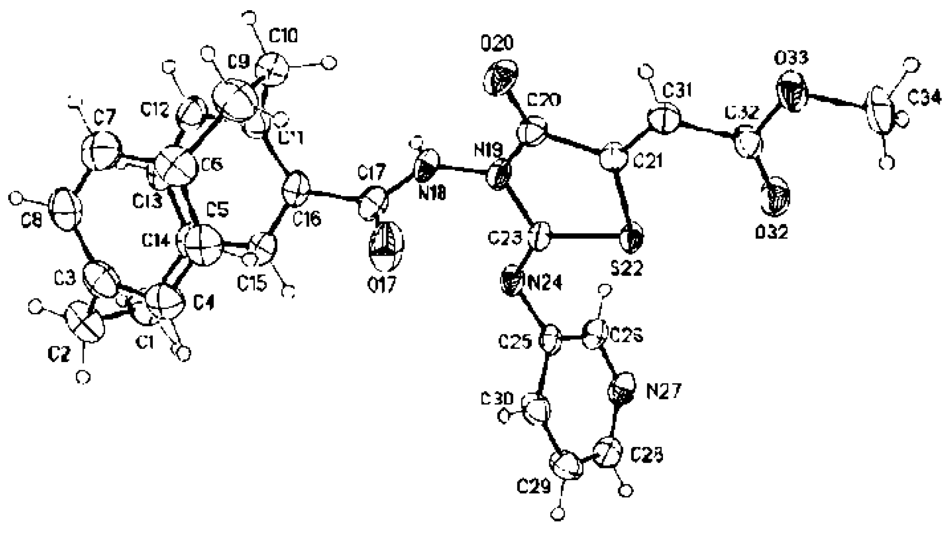
Crystal Structure Determinations of 2a, 2b, 2d, 3b, and 3c

The single-crystal X-ray diffraction studies were carried out on a Bruker D8 Venture diffractometer with PhotonII detector at 123(2) K using Cu-Kα radiation (λ = 1.54178 Å). Dual space methods (SHELXT) [50] were used for structure solution, and refinement was carried out using SHELXL-2014 (full-matrix least-squares on F2) [51]. Hydrogen atoms were localized by difference electron density determination and refined using a riding model (H(N) free). Semi-empirical absorption corrections were applied.
1: Colorless crystals, C17H18N2O, Mr = 266.33, crystal size 0.16 × 0.06 × 0.02 mm, monoclinic, space group C2/c (No. 15), a = 11.8196(4) Å, b = 7.9087(3) Å, c = 28.20(10) Å, β = 92.708(2)°, V = 2636.58(16) Å3, Z = 8, ρ = 1.342 Mg/m−3, µ(Cu-Kα) = 0.67 mm−1, F(000) = 1136, max = 144.6°, 10,645 measured reflections (2589 independent reflection in the HKLF 5 file, Rint = 0.000), 191 parameters, 3 restraints R1 = 0.071 (for 2452 I > 2σ(I)), wR2 = 0.174 (all data), S = 1.16, largest diff. peak/hole = 0.33/−0. e Å−3. Refined as a 2-component twin (BASF 0.139(4)). The option TwinRotMat of the program package PLATON [52] was used to create a HKLF 5 file, which was used for the refinement. Therefore, only unique reflections were used for the refinement (Rint = 0.00) (see cif-file for details).
2a: Colorless crystals, C24H23N3OS, Mr = 401.51, crystal size 0.16 × 0.12 × 0.04 mm, monoclinic, space group P21/c (No. 14), a = 12.2835(4) Å, b = 10.5257(3) Å, c = 15.5777(5) Å, β = 104.805(2)°, V = 1947.21(11) Å3, Z = 4, ρ = 1.0 Mg/m−3, µ(Cu-Kα) = 1.64 mm−1, F(000) = 848, max = 144.8°, 17,562 reflections, of which 3830 were independent (Rint = 0.032), 271 parameters, 3 restraints, R1 = 0.039 (for 3480 I > 2σ(I)), wR2 = 0.107 (all data), S = 1.04, largest diff. peak/hole = 0.34/−0.20 e Å−3.
2b: Colorless crystals, C25H25N3OS, Mr = 415.54, crystal size 0.28 × 0.06 × 0.03 mm, orthorhombic, space group Pccn (No. 56), a = 19.4444(6) Å, b = 25.2548(7) Å, c = 8.7599(2) Å, V = 4301.7(2) Å3, Z = 8, ρ = 1.283 Mg/m−3, µ(Cu-Kα) = 1.50 mm−1, F(000) = 1760, max = 144.4°, 48,155 reflections, of which 4241 were independent (Rint = 0.049), 283 parameters, 3 restraints, R1 = 0.038 (for 3846 I > 2σ(I)), wR2 = 0.096 (all data), S = 1.05, largest diff. peak/hole = 0.27/−0.19 e Å−3.
2d: Colorless crystals, C21H23N3OS, Mr = 365.48, crystal size 0.18 × 0.04 × 0.02 mm, orthorhombic, space group Pbca (No. 61), a = 19.5761(14) Å, b = 8.2709(7) Å, c = 24.0176(17) Å, V = 3888.7(5) Å3, Z = 8, ρ = 1.249 Mg/m−3, µ(Cu-Kα) = 1.58 mm−1, F(000) = 1552, max = 144.4°, 24,493 reflections, of which 3815 were independent (Rint = 0.087), 244 parameters, 198 restraints (a general RIGU restraint was applied, see cif.file for details), R1 = 0.059 (for 2913 I > 2σ(I)), wR2 = 0.141 (all data), S = 1.043, largest diff. peak/hole = 0.35/−0.27 e Å−3.
3b: Yellow crystals, C30H27N3O4S, Mr = 525.60, crystal size 0.14 × 0.12 × 0.04 mm, monoclinic, space group P21/c (No. 14), a = 13.0205(4) Å, b = 12.40(4) Å, c = 15.9584(5) Å, β = 92.8(1)°, V = 2574.16(14) Å3, Z = 4, ρ = 1.356 Mg/m−3, µ(Cu-Kα) = 1.46 mm−1, F(000) = 1104, max = 144.6°, 41,983 reflections, of which 5074 were independent (Rint = 0.038), 347 parameters, 1 restraint, R1 = 0.070 (for 4800 I > 2σ(I)), wR2 = 0.187 (all data), S = 1.06, largest diff. peak/hole = 1.13 (due to possible disorder in the [2.2]paracyclophane moiety)/−0.35 e Å−3.
3c: Colorless crystals, C28H24N4O4S ∙ 0.5 CH3OH ∙ 0.5 H2O, Mr = 5.60, crystal size 0.15 × 0.09 × 0.03 mm, monoclinic, space group C2/c (No. 15), a = 19.3130(6) Å, b = 12.4233(6) Å, c = 21.8993(8) Å, β = 90.008(2)°, V = 5254.3(4) Å3, Z = 8, ρ = 1.359 Mg/m−3, µ(Cu-Kα) = 1.49 mm−1, F(000) = 2256, max = 144.4°, 28,186 reflections, of which 5170 were independent (Rint = 0.041), 338 parameters, 1 restraint, R1 = 0.043 (for 4649 I > 2σ(I)), wR2 = 0.110 (all data), S = 1.04, largest diff. peak/hole = 0.42/−0.21 e Å−3.
Refinement with the listed atoms show residual electron density due to a heavily disordered methanol and water in four voids around a center of symmetry, which could not be refined with split atoms. Therefore, the option "SQUEEZE" of the program package PLATON [52] was used to create an hkl file taking into account the residual electron density in the void areas. Therefore, the atom list and unit card do not agree (see cif-file for details). CCDC 1,971,268 (1), 1,971,269 (2a), 1,971,270 (2b), 1,971,271 (2d), 1,971,272 (3b), and 1,971,273 (3c) contain the supplementary crystallographic data for this paper. These data can be obtained free of charge from The Cambridge Crystallographic Data Center via www.ccdc.cam.ac.uk/data_request/cif.

4. Conclusions

Herein, methyl 2-(2-(4′-[2.2]paracyclophanyl)-hydrazinylidene)-3-substituted-4-oxothiazolidin-5-ylidene)acetates were synthesized and screened against 60 cancer cell lines. Since the designed compounds showed moderate to high activity as anticancer agents, much work was done with the synthesis and biology of paracyclophane heterocyclic compounds in terms of proving the hypothesis that paracyclophane/thiazole conjugates may work through tubulin polymerization inhibition. Results indicated that compound 3a could be considered a good pharmacophore for further medicinal study.

Supplementary Materials

Supplementary data for this article can be found online.

Author Contributions

A.A.A. (Writing, Editing, and Submitting); S.B. (Writing and Editing); A.A.H. (Supervision); N.K.M. (Supervision); L.E.A.E.-H. (Experiment); M.N. (X-ray analysis); N.M.M. (Writing and Editing); E.M.N.A. (Biology, Writing, Editing and Revision). All authors have read and agreed to the published version of the manuscript.

Funding

No Funds.

Acknowledgments

The authors thank the Egyptian Mission, Ministry of Higher Education, Egypt or its financial support to Lamiaa E. Abd El-Haleem during her accommodation at the Institute für Technology, Karlsruhe, Germany. The authors also thank the DFG for providing Ashraf A. Aly with a one-month fellowship, enabling him to carry out the compound analysis at the Karlsruhe Institute of Technology, Karlsruhe, Germany in July–August 2019. We also acknowledge support by the KIT-Publication Fund of the Karlsruhe Institute of Technology.

Conflicts of Interest

The authors declare no conflicts of interest.

References

  1. Hopf, H.; Gleiter, R. Modern Cyclophane Chemistry; Wiley-VCH: Weinheim, Germany, 2004; ISBN 978-3-527-30713-5. [Google Scholar]
  2. Boekelheide, V. Syntheses and properties of the [2n]Cyclophanes. Top. Curr. Chem. 1983, 113, 87–143. [Google Scholar]
  3. Cram, D.J.; Steinberg, H. Macro Rings. I. Preparation and Spectra of the Paracyclophanes. J. Am. Chem. Soc. 1951, 73, 5691–5704. [Google Scholar]
  4. Vögtle, F.; Newmann, P. The Synthesis of [2.2]Phanes. Synthesis 1973, 2, 85–103. [Google Scholar] [CrossRef]
  5. Staab, H.A.; Knaus, G.H.; Henke, H.-E.; Krieger, C. Elektron-Donor-Acceptor-Verbindungen, XXIX. Elektron-Donor-Acceptor-Paracyclophane mit 7,7,8,8-Tetracyanchinodimethan (TCNQ) als Acceptor-Einheit. Chem. Ber. 1983, 116, 2785–2805. [Google Scholar] [CrossRef]
  6. Hopf, H.; Marquard, C. Strain Release in Aromatic Molecules: The [2n] Cyclophanes. In Strain and its Implications in Organic Chemistry; Springer: Dordrecht, The Netherlands, 1983; pp. 297–332. [Google Scholar]
  7. Vögtle, F. Cyclophane Chemistry: Synthesis, Structure and Reactions; Wiley: Chichester, UK, 1993; pp. 71–111. [Google Scholar]
  8. Rozenberg, V.I.; Sergeeve, E.V.; Hopf, H. Modern Cyclophane Chemistry; Gleiter, R., Hopf, H., Eds.; Wiley-VCH: Weinheim, Germany, 2004; Volume 17, pp. 435–462. [Google Scholar]
  9. Braun, C.; Nieger, M.; Thiel, W.R.; Bräse, S. [2.2]Paracyclophanes with N-Heterocycles as Ligands for Mono-and Dinuclear Ruthenium(II) Complexes. Chem. A Eur. J. 2017, 23, 15474–15483. [Google Scholar] [CrossRef] [PubMed]
  10. Aly, A.A.; Brown, A.A. Asymmetric and fused heterocycles based on [2.2]paracyclophane. Tetrahedron 2009, 65, 8055–8089. [Google Scholar] [CrossRef]
  11. Imming, P.; Graf, M.; Tries, S.; Hirschelmann, R.; Krause, E.; Pawlitzki, G. Anti-inflammatory planar chiral [2.2]paracyclophaneacetic acid enantiomers. Inflamm. Res. 2001, 50, 371–374. [Google Scholar] [CrossRef]
  12. Schlotter, K.; Boeckler, F.; Hübner, H.; Gmeiner, P. Fancy Bioisosteres: Novel Paracyclophane Derivatives As Super-Affinity Dopamine D3 Receptor Antagonists. J. Med. Chem. 2006, 49, 3628–3635. [Google Scholar] [CrossRef]
  13. Deep, A.; Jain, S.; Sharma, P.C. Synthesis and anti-inflammatory activity of some novel biphenyl-4-carboxylic acid 5-(arylidene)-2-(aryl)-4-oxothiazolidin-3-yl amides. Acta Pol. Pharm. Drug Res. 2010, 67, 63–67. [Google Scholar]
  14. Salar, U.; Taha, M.; Khan, K.M.; Ismail, N.H.; Imran, S.; Perveen, S.; Gul, S.; Wadood, A. Syntheses of new 3-thiazolyl coumarin derivatives, in vitro α-glucosidase inhibitory activity, and molecular modeling studies. Eur. J. Med. Chem. 2016, 122, 196–204. [Google Scholar] [CrossRef]
  15. Hassan, A.A.; Mohamed, N.K.; El-Haleem, L.E.A.; Braese, S.; Martin, N. Facile Synthesis of Naphtho [2,3-d]thiazoles, Naphtho[2,3-e][1,3,4]thiadiazines and Bis(naphtho[2,3-d]thiazolyl)-copper(II) Derivatives from Heteroylthiosemicarbazides. Chin. J. Chem. 2016, 34, 814–822. [Google Scholar] [CrossRef]
  16. Aly, A.A.; Hassan, A.A.; Mohamed, N.K.; El Shaieb, K.; Makhlouf, M.M.; Bräse, S.; Nieger, M. Reactive intermediates in the reaction of hydrazinecarbothioamides with 2-bis-(methylthio)-methylene)malononitrile and ethyl 2-cyano-3,3-bis(methylthio)acrylate. Res. Chem. Intermeds 2019, 45, 613–631. [Google Scholar] [CrossRef] [Green Version]
  17. Aly, A.A.; Ibrahim, M.A.A.; Shehata, E.M.; Hassan, A.A.M.; Brown, A.B. Prospective new amidinothiazoles as leukotriene B4 inhibitors. J. Mol. Struct. 2019, 1175, 414–427. [Google Scholar] [CrossRef]
  18. Hassan, A.A.; Aly, A.A.; Mostafa, S.M.; Döpp, D. Formation of thiadiazole, thiadiazine, thiadiazepine and pyrazole derivatives in the reaction of 2,4-disubstituted thiosemicarbazides with tetracyanoethylene. Arkivoc 2018, iii, 200–211. [Google Scholar]
  19. Aly, A.A.; Hassan, A.A.; AbdAl-Latif, E.-S.M.; Ibrahim, M.A.A.; Bräse, S.; Nieger, M. Reaction of N,N-disubstituted hydrazinecarbothioamides with 2-bromo-2-substituted acetophenones. Arkivoc 2018, iii, 102–111. [Google Scholar]
  20. Aly, A.A.; Hassan, A.A.; El-Latif, E.-S.M.A. Review on “An update of the use of thiocarbohydrazides and thiosemicarbazides in the preparation of heterocycles and their biological importance”. J. Heterocycl. Chem. 2018, 55, 2196–2223. [Google Scholar] [CrossRef]
  21. Aly, A.A.; Ishak, E.A.; El Malah, T.; Brown, A.B.; Elayah, W.M. Synthesis of potentially antioxidant and antibacterial biologically active thiazolidines. J. Heterocycl. Chem. 2015, 52, 1758–1764. [Google Scholar] [CrossRef]
  22. Aly, A.A.; Mohamed, N.K.; Hassan, A.A.; El-Shaieb, K.M.; Makhlouf, M.M.; Bräse, S.; Nieger, M.; Brown, A.B. Functionalized 1,3-Thiazolidin-4-Ones from 2-Oxo-Acenaphtho-quinylidene-and [2.2]Paracyclophanylidene-Thiosemicarbazones. Molecules 2019, 24, 3069. [Google Scholar] [CrossRef] [Green Version]
  23. Aly, A.A.; Mohamed, A.H.; Ramadan, M. Synthesis and colon anticancer activity of some novel thiazole/-2-quinolone derivatives. J. Mol. Struct. 2020, 1207, in press. [Google Scholar] [CrossRef]
  24. Aly, A.A.; Bräse, S.; Weis, P. Tridentate and bidentate copper complexes of [2.2]paracyclophanyl-substituted thiosemicarbazones, thiocarbazones, hydrazones and thioureas. J. Mol. Struct. 2019, 1178, 311–326. [Google Scholar] [CrossRef]
  25. Chen, J.; Wang, Z.; Li, C.-M.; Lu, Y.; Vaddady, P.K.; Meibohm, B.; Dalton, J.T.; Miller, D.D.; Li, W. Discovery of novel 2-aryl-4-benzoyl-imidazoles targeting the colchicines binding site in tubulin as potential anticancer agents. J. Med. Chem. 2010, 53, 7414–7427. [Google Scholar] [CrossRef] [PubMed] [Green Version]
  26. Bestgen, S.; Seidl, C.; Wiesner, T.; Zimmer, A.; Falk, M.; Köberle, B.; Austeri, M.; Paradies, J.; Bräse, S.; Schepers, U. Double-Strand DNA Breaks Induced by Paracyclophane Gold (I) Complexes. Chem. A Eur. J. 2017, 23, 6315–6322. [Google Scholar] [CrossRef] [PubMed]
  27. Li, C.-M.; Wang, Z.; Lu, Y.; Ahn, S.; Narayanan, R.; Kearbey, J.D.; Parke, D.N.; Li, W.; Mille, D.D.; Dalton, J.T. Biological activity of 4-substituted methoxybenzoyl-aryl-thiazole: An active microtubule inhibitor. Cancer Res. 2011, 71, 216–224. [Google Scholar] [CrossRef] [PubMed] [Green Version]
  28. Romagnoli, R.; Baraldi, P.G.; Salvador, M.K.; Preti, D.; Tabrizi, M.A.; Brancale, A.; Fu, X.-H.; Li, J.; Zhang, S.-Z.; Hamel, E. Discovery and optimization of a series of 2-aryl-4-amino-5-(3′,4′,5′-trimethoxybenzoyl)thiazoles as novel anticancer agents. J. Med. Chem. 2012, 55, 5433–5445. [Google Scholar] [CrossRef] [PubMed]
  29. Lu, Y.; Li, C.-M.; Wang, Z.; Ross, C.R.; Chen, J.; Dalton, J.T.; Li, W.; Miller, D.D. Discovery of 4-substituted methoxybenzoyl-aryl-thiazole as Novel Anticancer Agents: Synthesis, biological evaluation, and structure−activity relationships. J. Med. Chem. 2009, 52, 1701–1711. [Google Scholar] [CrossRef] [Green Version]
  30. Aly, A.A.; Bräse, S.; Hassan, A.A.; Mohamed, N.K.; El-Haleem, L.E.A. New planar-chiral linked [2.2]paracyclophanes-N-([2.2]-paracyclophanylcarbamoyl)-4-([2.2]paracyclophanylcarboxamide. Molecules 2020. under review. [Google Scholar]
  31. Zitt, H.; Dix, I.; Hopf, H.; Jones, P.G. 4,15-Diamino[2.2]paracyclophane, a Reusable Template for Topochemical Reaction Control in Solution. Eur. J. Org. Chem. 2002, 14, 2298–2307. [Google Scholar] [CrossRef]
  32. Holbeck, S.L.; Camalier, R.; Crowell, J.A.; Govindharajulu, J.P.; Hollingshead, M.; Anderson, L.W.; Polley, E.; Rubinstein, L.; Srivastava, A.; Wilsker, D. The National Cancer Institute ALMANAC: A comprehensive screening resource for the detection of anticancer drug pairs with enhanced therapeutic activity. Cancer Res. 2017, 77, 3564–3576. [Google Scholar] [CrossRef] [Green Version]
  33. Rostom, S.A. Synthesis and in vitro antitumor evaluation of some indeno[1,2-c]pyrazol (in) es substituted with sulfonamide, sulfonylurea (-thiourea) pharmacophores, and some derived thiazole ring systems. Biorg. Chem. Med. Chem. 2006, 14, 6475–6485. [Google Scholar] [CrossRef]
  34. Green, D.R.; Kroemer, G. The pathophysiology of mitochondrial cell death. Science 2004, 305, 626–629. [Google Scholar] [CrossRef]
  35. Nohl, H.; Gille, L.; Staniek, K. Intracellular generation of reactive oxygen species by mitochondria. Biochem. Pharmacol. 2005, 69, 719–723. [Google Scholar] [CrossRef]
  36. Rae, M.; Creighton, C.J.; Meck, J.M.; Haddad, B.R.; Johnson, M.D. MDA-MB-435 cells are derived from M14 Melanoma cells––a loss for breast cancer, but a boon for melanoma research. Breast Cancer Res. Treat. 2007, 104, 13–19. [Google Scholar] [CrossRef] [PubMed]
  37. Polgar, O.; Bates, S.E. ABC transporters in the balance: Is there a role in multidrug resistance? Biochem. Soc. Trans. 2005, 33, 241–245. [Google Scholar] [CrossRef] [PubMed]
  38. Kumar, S. Caspase function in programmed cell death. Cell Death Differ. 2007, 14, 32–43. [Google Scholar] [CrossRef] [PubMed]
  39. Matson, D.R.; Stukenberg, P.T. Spindle poisons and cell fate: A tale of two pathways. Mol. Interv. 2011, 11, 141–149. [Google Scholar] [CrossRef]
  40. Clarke, P.R.; Allan, L.A. Cell-cycle control in the face of damage–a matter of life or death. Trends Cell Boil. 2009, 19, 89–98. [Google Scholar] [CrossRef]
  41. Tolosa, L.; Donato, M.T.; Gomez-Lecchon, M.J. General Cytotoxicity Assessment by Means of the MTT Assay. Methods Mol. Biol. 2015, 1250, 333–348. [Google Scholar]
  42. Pozarowski, P.; Darzylienwicz, Z. Analysis of cell cycle by flow cytometry. Methods Mol. Biol. 2004, 281, 301–311. [Google Scholar]
  43. Niles, A.L.; Moravec, R.A.; Riss, T.L. Caspase activity assays. Methods Mol. Biol. 2008, 414, 137–150. [Google Scholar] [PubMed]
  44. Peng, L.X.; Wallace, M.; Andaloro, B.; Fallon, D.; L, L.F.; Delduco, D.; Tice, G. Modification of the BAX System PCR assay for detecting Salmonella in beef, produce, and soy protein isolate. Performance Tested Method 100201. J. AOAC Int. 2011, 94, 172–178. [Google Scholar] [CrossRef] [Green Version]
  45. Redmann, M.; Benavides, G.A.; Wani, W.Y.; Berryhill, T.F.; Ouyang, X.; Johnson, M.S.; Ravi, S.; Mitra, K.; Barnes, S.; Darley-Usmar, V.M.; et al. Methods for assessing mitochondrial quality control mechanisms and cellular consequences in cell culture. Redox Biol. 2018, 17, 59–69. [Google Scholar] [CrossRef] [PubMed]
  46. Lu, Y.; Chen, J.; Xiao, M.; Li, W.; Mille, D.D. An overview of tubulin inhibitors that interact with the colchicine binding site. Pharm Res. 2012, 29, 2943–2971. [Google Scholar] [CrossRef] [PubMed] [Green Version]
  47. Wu, D.; Yotnda, P. Production and detection of reactive oxygen species (ROS) in cancers. J Vis. Exp. 2011, 57, 3357–3365. [Google Scholar] [CrossRef] [PubMed]
  48. Gilpin, C.; Korobitsyn, A.; Weyer, K. Current tools available for the diagnosis of drug-resistant tuberculosis. Ther. Adv. Infect. Dis. 2016, 3, 145–151. [Google Scholar] [CrossRef] [PubMed]
  49. OpenEye Scientific Software Fast Rigid Exhaustive Docking (FRED) Receptor, Version 2.2.5. Available online: http://www.eyesopen.com (accessed on 27 May 2020).
  50. Sheldrick, G.M. SHELXT–Integrated space-group and crystal-structure determination. Acta Crystallogr. 2015, A71, 3–8. [Google Scholar] [CrossRef] [Green Version]
  51. Sheldrick, G.M. Crystal strturcture refinement with SHELXL. Acta Crystallogr. 2015, C71, 3–8. [Google Scholar]
  52. Spek, A.L. Structure validation in chemical crystallography. Acta Crystallogr. 2009, D65, 148–155. [Google Scholar] [CrossRef]
Figure 1. Five types of heterocycle-substituted [2.2]paracyclophanes.
Molecules 25 03089 g001
Figure 2. Structures of biologically active thiazole and paracyclophane derivatives.
Molecules 25 03089 g002
Figure 3. Design of the target compounds 3af.
Molecules 25 03089 g003
Scheme 1. Strategy of the structure features planned to obtain the following compounds from acylhydrazide 1: acylthiosemicarbzides 2af, thiazolidin-3-ones 3a,b,df, and the regioisomer 3c derived from [2.2]paracyclophane.
Molecules 25 03089 sch001
Scheme 2. The preparation of acylthiosemicarbazide 1 from [2.2]paracyclophane (5): (a) (COCl)2/AlCl3, −10 °C to 5 °C, 20 min; (b) PhCl, ∆, 40 h; (c) CH3CH2OH, ∆, 10 h; (d) NH2NH2 as a solvent, ∆, 5 h.
Molecules 25 03089 sch002
Scheme 3. Synthesis of 2-(4′-[2.2]paracyclophanyl-4H-hydrazinecarbothioamides 2af.
Molecules 25 03089 sch003
Figure 4. Molecular structure of compound 2a identified according to IUPAC nomenclature as 2-(1,4(1,4)-dibenzenacyclohexaphane-12-carbonyl)-N-phenylhydrazine-1-carbothioamide.
Molecules 25 03089 g004
Figure 5. Molecular structure of compound 2b identified according to IUPAC nomenclature as 2-(1,4(1,4)-dibenzenacyclohexaphane-12-carbonyl)-N-benzylhydrazine-1-carbothioamide.
Molecules 25 03089 g005
Figure 6. Molecular structure of compound 2d identified according to IUPAC nomenclature as 2-(1,4(1,4)-dibenzenacyclohexaphane-12-carbonyl)-N-vinylhydrazine-1-carbothioamide.
Molecules 25 03089 g006
Scheme 4. Reaction of 2af with dimethyl acetylenedicarboxylate (9): synthesis of different regioisomers thiazolidines 3a,b,df and 3c.
Molecules 25 03089 sch004
Figure 7. Molecular structure of compound 3b identified according to IUPAC nomenclature as methyl (E)-2-((E)-2-(2-(1,4(1,4)-dibenzen-cyclohexaphane-12-carbonyl)hydrazinylidene)-3-benzyl-4-oxothiazolidin-5-ylidene)acetate.
Molecules 25 03089 g007
Figure 8. Molecular structure of compound 3c identified according to IUPAC nomenclature as methyl (E)-2-((E)-3-(1,4(1,4)-dibenzenacyclohexaphane-12-carboxamido)-4-oxo-2-(pyridin-3-ylimino)thiazolidin-5-ylidene)acetate.
Molecules 25 03089 g008
Scheme 5. Resonance structures of compound 3c.
Molecules 25 03089 sch005
Figure 9. Dose–response curves of compound 3a (log10 of sample concentrations 0.01, 0.1, 1, 10, and 100 μM) against all nine different cancer cell line panels (leukemia, non-small-cell lung, colon, CNS, melanoma, ovarian, renal, prostate, and breast cancers).
Molecules 25 03089 g009
Figure 10. Cell-cycle analysis of SR cells treated with annexin PI at IC50. Concentration representing growth arrest at the pre-G1 (G0) and G2/M phases. (A) Untreated cells; (B) treated cells with colchicine; (C) treated cells with 3a. The test was repeated three times, while 3a and the reference were incubated for 24 h (2 × 10⁵ cells/well) at 37 °C.
Molecules 25 03089 g010
Figure 11. Contour diagram of annexin V/PI flow cytometry against SR cancer cell line. (A) Untreated cells; (B) treated cells with colchicine; (C) treated cells with 3a. The test was repeated three times. Following incubation for 24 h, cells were analyzed by flow cytometry after double staining with annexin V/PI.
Molecules 25 03089 g011
Figure 12. Contour diagram of annexin V/PI flow cytometry illustrating induction of the loss of mitochondrial membrane potential (Δψmt) against SR cancer cell line. (A) Untreated cells; (B) treated cells with colchicine; (C) treated cells with 3a. (B and C were performed at concentrations of 20, 10, 5, 2.5, 1.2, 0.63, and 0.31 ng/mL). The tests were repeated three times and the substrate was incubated at 37 °C in the dark for 15–30 min.
Molecules 25 03089 g012
Figure 13. Mitochondrial production of ROS in SR cells with 3a and colchicine reference compared to control. Mean ± standard deviation plotted for three replicates per condition. Results are significantly different from control at *** p < 0.05. The substrate was incubated for 30 min at 37 °C protected from light.
Molecules 25 03089 g013
Figure 14. Concentrations of (A) Bcl-2 (concentrations 32, 16, 8, 4, 2, 1, 0.5 ng/mL); (B) BAX (concentrations 2000, 1000, 500, 250, 125, 62.5, 31.25 pg/mL), and (C) caspase-3 expression (2500, 1250, 625, 313, 156, 78, 39 pg/mL) for 3a and colchicine on leukemia SR cell line relative to control. Results significantly different from control at *** p < 0.05.
Molecules 25 03089 g014
Figure 15. The two- and three-dimensional (2D and 3D) diagrams illustrate the binding mode of the reference colchicine interacting with the colchicine binding site of β-tubulin.
Molecules 25 03089 g015
Figure 16. The 2D and 3D diagrams illustrate the binding mode of 3a interacting with the colchicine binding site of β-tubulin.
Molecules 25 03089 g016
Figure 17. The 2D and 3D diagrams illustrate the binding mode of 3b interacting with the colchicine binding site of β-tubulin.
Molecules 25 03089 g017
Figure 18. The 2D and 3D diagrams illustrate the binding mode of 3c interacting with the colchicine binding site of β-tubulin.
Molecules 25 03089 g018
Figure 19. The 2D and 3D diagrams illustrate the binding mode of 3d interacting with the colchicine binding site of β-tubulin.
Molecules 25 03089 g019
Figure 20. The 2D and 3D diagrams illustrate the binding mode of 3e interacting with colchicine binding site of β-tubulin.
Molecules 25 03089 g020
Table 1. Growth inhibition percentage of compounds 3af (concentration 10−5 M) against different cell lines.
Panel/Cell Line3a3b3c3d3e3f
LeukemiaCCRF-CEM87.0496.1731.2777.0866.0841.43
HL-60(TB)83.60105.4648.648.1611.6426.38
K-56272.9581.1320.7248.1345.7830.52
MOLT-496.9798.9121.8664.5562.8555.61
RPMI-8226120.89147.0083.01109.36114.2849.83
SR115.60114.7074.1898.21113.4039.13
Non-Small-Cell Lung CancerA549/ATCC30.9222.952.045.203.0011.39
EKVX41.0322.302.619.166.4412.16
HOP-6297.0022.764.00000
HOP-9224.6624.508.1418.5918.4316.16
NCI-H22626.2235.207.4812.457.319.41
NCI-H2359.5722.649.1811.850.409.32
NCI-H322M31.546.701.722.947.754.43
NCI-H46039.356.290.75001.98
NCI-H52258.4784.0617.7439.2540.3130.01
Colon CancerCOLO 20552.2627.310004.48
HCC-299896.3620.490000
HCT-11695.9550.486.3546.0250.5424.59
HCT-1599.6672.1611.7533.1131.6029.43
HT29113.1387.627.7153.9954.7623.99
KM1291.5134.907.3218.4015.011.32
SW-62092.6181.8310.8455.4691.2112.94
CNS CancerSF-26827.4914.953.0802.182.56
SF-29541.0010.635.636.423.138.80
SF-539122.5056.7411.630.339.820
SNB-1960.0643.0816.6415.3114.4311.26
SNB-7554.2948.780.2521.9221.1824.73
U25184.2045.0414.6618.1715.6420.27
MelanomaLOX IMVI127.7793.799.7821.2226.519.41
MALME-3M21.2311.5707.568.0611.28
M1446.8836.597.9921.1412.8519.23
MDA-MB-43510.398.44000.728.06
SK-MEL-212.9814.4202.705.7813.53
SK-MEL-2817.5400.860010.28
SK-MEL-530.6517.723.647.537.5014.50
UACC-25730.7519.020.838.897.2616.33
UACC-6274.8548.1110.6722.8421.3917.97
Ovarian CancerIGROV194.0357.7215.4410.7611.627.20
OVCAR-3106.2579.714.5113.567.0915.87
OVCAR-470.6155.437.762.9310.0010.51
OVCAR-551.7534.650.613.9000
OVCAR-859.2728.001.5111.787.4213.28
NCI/ADR-RES68.6129.614.7010.5013.467.72
SK-OV-3000000
Renal Cancer786-0100.2850.458.528.779.4514.62
A49850.7553.923.5912.106.633.39
ACHN30.6734.1811.0212.2110.4517.78
CAKI-194.6584.434.785.23024.49
SN12C77.5138.768.8713.144.9717.07
TK-1084.834.53006.500
UO-3141.5154.9621.8624.6221.0321.73
Prostate CancerPC-3135.8882.2318.1951.6243.2818.95
DU-14549.1316.790006.20
Breast CancerMCF776.7967.2626.7548.4756.2936.38
MDA-MB-231/ATCC67.4743.1610.642.273.953.50
HS 578T22.0612.5806.411.035.01
BT-54964.5089.6818.4138.287.7326.81
T-47D138.25106.2531.1325.005.7723.00
MDA-MB-468100.3978.9118.0323.6812.7429.74
Table 2. Results of five in vitro doses (concentrations 0.01, 0.1, 1, 10, and 100 µM) tested with nine human cancer types and selectivity for compound 3a.
PanelCell LineGI50TGILC50
Concentration/Cell LineSubpanel MIDbSelectivity Ratio (MIDa: MIDb)Concentration/Cell LineSubpanel MIDdSelectivity Ratio (MIDc: MIDd)
LeukemiaCCRF-CEM2.712.720.88-81.143.85>100
HL-60(TB)2.70>100>100
K-5623.15>100>100
MOLT-43.12>100>100
RPMI-82262.155.69>100
SR2.46>100>100
Non-Small-Cell Lung CancerA549/ATCC2.832.740.878.0016.051.31>100
EKVX1.914.239.38
HOP-622.686.976.60
HOP-921.919.14>100
NCI-H2264.382.55>100
NCI-H231.954.158.85
NCI-H322M2.024.751.32
NCI-H4602.084.298.82
NCI-H5224.89>100>100
Colon CancerCOLO 2052.131.911.244.613.955.34
HCC-29981.783.456.68
HCT-1161.793.667.51
HCT-151.874.058.76
HT291.934.078.56
KM122.034.158.51
SW-6201.853.667.23
CNS CancerSF-2683.092.710.881.1219.481.08>100
SF-2952.074.198.50
SF-5391.793.326.18
SNB-191.954.259.26
SNB-755.44>100>100
U2511.894.008.47
MelanomaLOX IMVI1.693.770.633.36.540.586.75
MALME-3M3.272.14>100
M143.271.54>100
MDA-MB-4355.44100>100
SK-MEL-22.657.76>100
SK-MEL-282.979.87>100
SK-MEL-57.56>100>100
UACC-2575.14>100>100
UACC-621.964.219.02
Ovarian CancerIGROV11.662.590.923.2718.461.146.44
OVCAR-31.803.617.23
OVCAR-43.179.883.69
OVCAR-51.923.847.68
OVCAR-83.08>100>100
NCI/ADR-RES2.921.06>100
SK-OV-33.557.53>100
Renal Cancer786-02.001.861.284.3816.641.279.58
A498--8.36>100
ACHN1.783.296.08
RXF3933.743.86>100
CAKI-11.523.009.78
SN12C2.275.94>100
TK-102.035.602.92
UO-311.533.086.20
Prostate CancerPC-31.713.410.693.633.585.897.70
DU-1455.113.53>100
Breast CancerMCF71.872.271.054.9524.280.872.25
MDA-MB-231/ATCC2.637.46>100
HS 578T3.49>100>100
BT-5492.225.352.47
T-47D1.543.658.68
MDA-MB-4681.843.747.61
MIDa2.38 MIDc21.08
MIDa,c = average sensitivity of all cell lines in mM; MIDb,d = average sensitivity of all cell lines of a particular subpanel in mM.
Table 3. MTT assay for the antiproliferative IC50 ± SD (μM) activity of compounds 3ae and colchicine.
CompoundCytotoxicity IC50 (µM) a
RPMI-8226L.SR
3a1.61 ± 0.0654 ***1.11 ± 0.06849 ***
3b4.62 ± 0.0459 ***2.02 ± 0.06875 ***
3c9.96 ± 0.3451 ***4.84 ± 0.08425 ***
3d17.81 ± 0.0544 ***22.06 ± 0.06792 ***
3e3.17 ± 0.0489 ***5.04 ± 0.07908 ***
Colchicine4.05 ± 0.0478 ***1.81 ± 0.07009 ***
a IC50 = compound concentration required to inhibit tumor cell proliferation by 50%. Data are expressed as the mean ± SD from the dose–response curves of at least three independent experiments. Results significantly different from control at *** p < 0.05.
Table 4. Inhibition of tubulin polymerization displaying IC50 ± standard error of the mean (SEM) (μM) for compounds 3ae and colchicine as a reference.
CompoundTubulin Polymerization Inhibition
IC50 (µM) a
3a4.97 ± 0.11
3b8.38 ± 0.18
3c8.28 ± 0.18
3d14.79 ± 0.32
3e6.61 ± 0.14
Colchicine3.76 ± 0.08
a Tubulin, colchicine, and the tested compound concentrations were 1, 5, and 5 μM, respectively.
Table 5. DNA content % using propidium iodide flow cytometry.
CompoundDNA Content %
%G0-G1%S%G2-M%Pre G1Comment
3a/SR31.4723.7944.74 ± 0.1747 ***33.71cell growth arrest (G2/M)
Colchicine/SR26.5825.2748.15 ± 1.000 ***36.24cell growth arrest (G2/M)
cont.SR57.2328.6614.11 ± 0.10501.92---
Results significantly different from control at *** p < 0.05.
Table 6. In vitro growth inhibitory effects of 3a in comparison to colchicine on multidrug-resistant leukemia cell line (MDR cells). Pgp—P-glycoprotein.
Compound CodeIC50 ± SEM (ng/mL) (n = 3)
Pgp-Mediated MDRFold Change
3a/SR1.285 ± 0.061.15
Colchicine/SR1.726 ± 0.051.54
Control/SR1.121 ± 0.051
Table 7. Caspase-3 conc (pg/mL) ± SD and fold change levels for compounds 3a and colchicine on leukemia SR cell line.
CompoundCaspase 3
Conc pg/mLFold Change
3a/SR471.2 ± 6.118.84
Colchicine/SR428.9 ± 5.478.05
Control/SR53.28 ± 1.091
Table 8. Bax and Bcl-2 conc ± SD and fold change levels for compounds 3a and colchicine on leukemia SR cell line.
CompoundBAXBcl2
Concentration (pg/mL)Fold ChangeConcentration (ng/mL)Fold Change
3a/SR359 ± 3.830 ***7.985.045 ± 0.1918 ***0.59
Colchicine/SR341.8 ± 4.310 ***7.604.101 ± 0.1441 ***0.48
Control/SR44.96 ± 1.27018.569 ± 0.15651
Results significantly different from control at *** p < 0.05.
Table 9. Energy scores for the complexes formed by the tested compounds 3ae and the reference colchicine in the active site of β-tubulin enzyme (PDB: 3HKC).
CompoundS ScoreΔG (kcal/mol)aLigand–Receptor Interaction
ResidueTypeLength (Å)
Colchicine−5.62−3.4
−0.5
Glu 71
Mg 601
H–donor
metal
3.16
2.61
3a−7.05−2.2
−0.6
Mg 601
Ala 247
metal
pi–H
2.00
4.17
3b−7.16−0.9
−2.3
−0.9
Asp 245
Arg 2
Glu 71
H–donor
H–acceptor
pi–H
3.27
3.41
3.98
3c−6.29−1.8
−0.7
−0.7
Mg 601
Tyr 224
Ala 247
metal
pi–H
pi–H
2.40
3.79
4.50
3d−6.94−1.0
−2.1
−0.7
Glu 71
Mg 601
Arg 2
H–donor
metal
pi–H
3.90
2.51
4.69
3e−6.43−2.0
−2.2
Gln 11
Mg 601
H–acceptor
metal
3.09
2.33
ΔG (kcal/mol)a: the binding free energies.

