Article

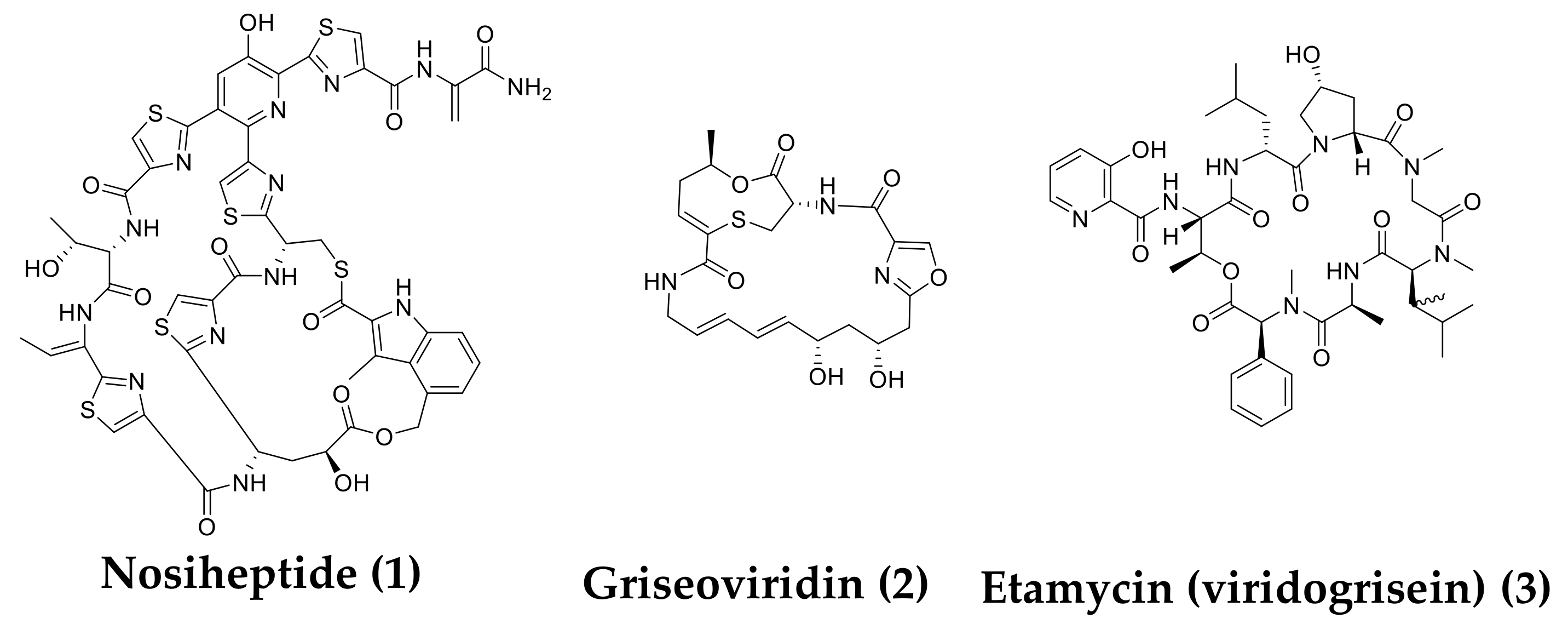
Discovery of Nosiheptide, Griseoviridin, and Etamycin as Potent Anti-Mycobacterial Agents against Mycobacterium avium Complex

1
Department of Microbial Chemistry, Graduate School of Pharmaceutical Sciences, Kitasato University, Tokyo 108-8641, Japan
2
Medicinal Research Laboratories, School of Pharmacy, Kitasato University, Tokyo 108-8641, Japan
3
OP BIO FACTORY Co., Ltd., 5-8 Suzaki, Uruma-shi, Okinawa 904-2234, Japan
*
Authors to whom correspondence should be addressed.
Molecules 2019, 24(8), 1495; https://doi.org/10.3390/molecules24081495
Submission received: 4 April 2019 / Revised: 11 April 2019 / Accepted: 15 April 2019 / Published: 16 April 2019
(This article belongs to the Collection Bioactive Compounds)

Abstract

:
Mycobacterium avium complex (MAC) is a serious disease mainly caused by M. avium and M. intracellulare. Although the incidence of MAC infection is increasing worldwide, only a few agents are clinically used, and their therapeutic effects are limited. Therefore, new anti-MAC agents are needed. Approximately 6600 microbial samples were screened for new anti-mycobacterial agents that inhibit the growth of both M. avium and M. intracellulare, and two culture broths derived from marine actinomycete strains OPMA1245 and OPMA1730 had strong activity. Nosiheptide (1) was isolated from the culture broth of OPMA1245, and griseoviridin (2) and etamycin (viridogrisein) (3) were isolated from the culture broth of OPMA1730. They had potent anti-mycobacterial activity against M. avium and M. intracellulare with minimum inhibitory concentrations (MICs) between 0.024 and 1.56 μg/mL. In addition, a combination of 2 and 3 markedly enhanced the anti-mycobacterial activity against both M. avium and M. intracellulare. Furthermore, a combination 2 and 3 had a therapeutic effect comparable to that of ethambutol in a silkworm infection assay with M. smegmatis.

1. Introduction

Mycobacterium avium complex (MAC) is a serious disease mainly caused by M. avium and M. intracellulare. In recent years, the number of MAC patients has exceeded the number of tuberculosis patients [1,2]. Although clarithromycin (CAM), ethambutol (EB), and rifampicin (RFP) are used to treat MAC infection, their therapeutic effects are limited, and patients must take them for a long time (over one year). In addition, mycobacteria resistant to these clinically used agents are increasingly reported [3,4,5]. Therefore, it is important to develop new drugs for MAC infection. Accordingly, we screened for microbial compounds that were effective for MAC infection by selecting microbial culture broths that inhibit the growth of both M. avium and M. intracellulare. We found two marine-derived actinomycetes Streptomyces spp. OPMA1245 and OPMA1730. A thiopeptide identified as nosiheptide (1) [6] was isolated from the culture broth of the former strain, and two streptogramins identified as griseoviridin (2) [7] and etamycin (viridogrisein) (3) [8] were isolated from the culture broth of the latter strain (Figure 1). These compounds were previously isolated, but we found that they had anti-MAC activity and described the anti-mycobacterial activities and therapeutic effects in an in vivo mimic silkworm infection model.

2. Results

2.1. In Vitro Anti-Mycobacterial Activities of 1–3

The anti-mycobacterial activities of 1–3 on M. avium, M. intracellulare, M. smegmatis, and M. bovis were evaluated according to a liquid microdilution method [9] (Table 1). Among the three compounds, 1 had the most potent anti-mycobacterial activity against M. avium, M. intracellulare, and M. bovis with the lowest minimum inhibitory concentrations (MICs; 0.024, 0.024, and 0.012 μg/mL, respectively) followed by those of 3, and compound 2 had modest MICs (1.56, 1.56, and 6.25 μg/mL, respectively). The MICs of 1 and 3 were comparable to those of clinically used agents, CAM and RFP. Furthermore, all the compounds had higher MICs against M. smegmatis than those against the other mycobacteria listed in Table 1.

2.2. Combination Effect of Streptogramins

Noeske et al. reported that streptogramins (combination of group A, dalfopristin, and group B quinupristin) have synergistic effects on the growth of gram-positive bacteria such as Enterococcus faecium, Staphylococcus aureus, and Streptococcus pyogenes [10]. In our case, 2 belongs to group A, and 3 belongs to group B. Therefore, we investigated whether a combination of 2 and 3 had a synergistic inhibitory effect on M. avium and M. intracellulare according to the checkerboard method [11]. For instance, the MIC of 2 on M. avium and M. intracellulare was reduced from 1.56 μg/mL to 0.019 μg/mL and from 1.56 μg/mL to 0.039 μg/mL in combination with 3 (0.007 μg/mL to 0.031 μg/mL), maximally yielding 80-fold and 40-fold potentiation, respectively (Table 2). Furthermore, the fractional inhibitory concentration (FIC) indexes in these combinations were below 0.5, which suggests that their interaction was synergistic. Furthermore, to confirm the synergistic activity of 2 and 3 against the three mycobacteria, the ratios of 2 and 3 (1:1 and 3:7) were set up based on a previous study using streptogramins. As shown in Table 3, the combinations of these ratios were synergistic against M. avium, M. intracellulare, and M. bovis BCG with reduced MICs ranging between 0.02 and 0.09 μg/mL.

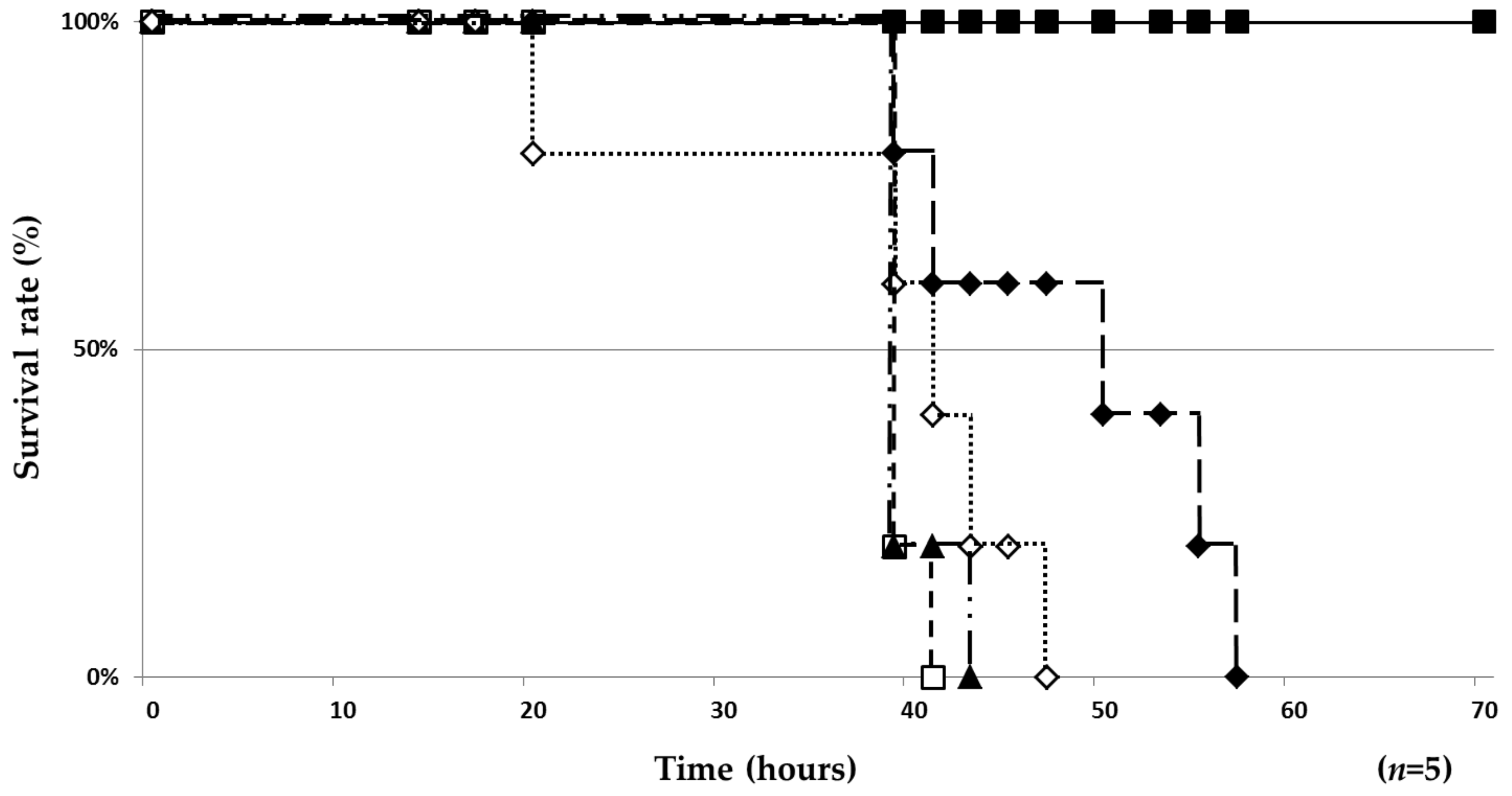
2.3. Therapeutic Effect of 1 and 2/3 in a Silkworm Infection Model

We previously established a mycobacterial silkworm infection assay with M. smegmatis [12]. M. smegmatis is a nonpathogenic bacterium. However, when M. smegmatis was injected to silkworms at a higher cell number (107 CFU/larva) and the injected silkworms were incubated at a higher temperature (37 °C), they all died within 50 h. In addition, clinically used anti-TB drugs, such as isoniazid, rifampicin, and ethambutol, showed a therapeutic effect in this silkworm assay [12]. Therefore, we evaluated 1 and a 2+3 mixture (in a 1:1 ratio) in this assay model. After being infected with M. smegmatis (n = 5), all silkworms died within 41 h. Under this condition, 1 showed a moderate therapeutic effect in the silkworm infection assay by prolonging survival time several hours (Figure 2). On the other hand, 2/3 increased the survival of silkworms in a dose-dependent manner, and silkworms survived for longer than 14 h at the maximal dose of 2/3 (Figure 2). Furthermore, the 50% effective dose (ED50) values of these compounds 41 h after infection were 35.4 µg/larva (Table 4) indicating that 2/3 (ED50, 35.4 µg/larva) had better efficacy than that of 1 and comparable efficacy to that of EB. In addition, 1 and 2/3 did not have toxicity on silkworms for at least 48 h.

3. Discussion

In the present study, approximately 6600 microbial culture broth samples were screened for new anti-MAC compounds using M. avium and M. intracellulare. Our screening results indicated that 0.06% of samples were active against only M. avium, 5.7% were active against only M. intracellulare, and 5.4% were active against both M. avium and M. intracellulare. We focused on samples active against both mycobacteria to obtain agents that may be clinically useful and selected samples with particularly potent activity. The selected strains were re-cultured, and the re-cultured broths that had reproducible activity were selected (0.03%).
Two culture broths from marine-derived actinomycetes OPMA1245 and OPMA1730 had potent activity against both mycobacteria. We isolated compounds from the culture broths and found three active compounds, nosiheptide (1), griseoviridin (2), and etamycin (viridogrisein) (3; Figure 1). These antibiotics were discovered in the 1950s and 1970s, but little was known about their anti-mycobacterial activity [13,14]. We found that they had potent activity against two MAC mycobacteria (Table 1). In an in vitro assay, 1 was the most potent followed by 3 and 2. As reported by Noeske et al., a combination of group A (containing an unsaturated lactam ring) and group B (containing a cyclohexadepsipeptide) of streptogramin antibiotics has a synergistic effect on the growth of Gram-positive bacteria [10]. Based on this finding, 2 (group A) and 3 (group B) were mixed in a 1:1 ratio to investigate anti-MAC activity. We demonstrated the synergistic activity of 2/3 on two mycobacteria (Table 3) and the in vivo efficacy in silkworms infected with M. smegmatis (Figure 2). Intriguingly, 1 was the most potent in vitro on the four mycobacteria (Table 1), but 2/3 had better efficacy than that of 1 in the silkworm infection assay. The in vitro potency of 2/3 on M. smegmatis (MIC, 6.25 µg/mL) was 8-fold weaker than that of EB (MIC, 0.78 µg/mL), whereas 2/3 had the same in vivo therapeutic effects (ED50, 35.4 µg/mL) in the silkworm assay (Table 4).
The therapeutic potency of these compounds has been reported. For example, 1 and 3 has therapeutic efficacy in the mice infection model with methicillin-resistant S. aureus [13,15]. However, chiefly due to unfavorable physico-chemical properties, 1 has been used previously only for applications in farm animals, mainly as a feed additive [16].
Hamamoto et al. previously reported that the ED50/MIC ratio of a compound is an index of drug potential, and the ratio is typically below 10 for clinically useful antibiotics [14]. As shown in Table 4, the ratio of 2/3 was below 10, indicating that 2/3 is a potential anti-MAC drug. Combination therapy of dalfopristin (group A) and quinupristin (group B) is clinically used for the treatment of vancomycin-resistant Enterococcus faecium [17]. Therefore, streptogramin combination therapy may be effective to treat MAC infection. Since effective antimicrobial therapies for MAC infection are limited, these antibiotics should be re-evaluated as lead compounds for anti-MAC drugs.

4. Materials and Methods

4.1. Assay for Anti-Mycobacterial Activity

The M. smegmatis M341 and M. bovis BCG Pasteur used were laboratory strains. M. avium JCM15430 and M. intracellulare JCM6384 were purchased from the Riken BioResource Research Center (Ibaraki, Japan). Anti-mycobacterial activities against these four strains were evaluated by the liquid microdilution method according to a previously established method [12,18].
M. avium and M. intracellulare were routinely cultured for 72 h and 96 h, respectively. The suspensions were diluted with Middlebrook 7H9 broth (Becton Dickinson, MD, USA) containing 0.5% Tween 80 (MP Biomedicals, Santa Ana, CA, USA) and 10% ADC enrichment (Sigma Aldrich, MO, USA) to adjust to 4.0 × 10⁶ CFU/mL. Diluted mycobacteria (95 μL) were added to each well of a 96-well microplate (Corning, NY, USA) with or without a test sample or drug (in 5 μL MeOH or DMSO) and incubated at 37 °C for 120 h. Then, MTT (3-(4,5-dimethyl-2-thiazolyl)-2,5-diphenyl-2H tetrazolium bromide, Sigma Aldrich) reagent (5.5 mg/mL, 5 µL H2O) was added to each well, and the cells were incubated for 16 h. Lysis buffer (40% N,N-dimethylformamide, 20% SDS, 2% CH3COOH, 95 μL) was added to each well to dissolve the formazan pigment. The absorbance was measured at a wavelength of 570 nm using a UV plate reader (BioTek, VT, USA) to evaluate the growth of the mycobacterium. The MIC was defined as the lowest drug concentration that inhibited mycobacterium growth by 90%.

4.2. Isolation of 13

The actinomycete strain Streptomyces sp. OPMA1245 was isolated from marine sediment collected in Okinawa prefecture, Japan. The 7-day-old culture broth of OPMA1245 (3.0 L) was extracted with an equal volume of EtOAc. After the EtOAc extracts were filtered, the EtOAc layer was concentrated to yield 204.3 mg of a yellow solid material. This material was dissolved in a small volume of DMSO, applied to an ODS column (10 g, 1.0 cm × 20 cm), and eluted stepwise with 100% H2O, 20%, 40%, 60%, 70%, 80%, and 100% CH3CN (80 mL each). Compound 1 was eluted with 60% CH3CN. The whole eluate was concentrated in vacuo to dryness to yield a yellow solid material (110.0 mg). Compound 1 was finally purified by preparative high-performance liquid chromatography (HPLC) under the following conditions: Column, PEGASIL ODS SP100 (i.d. 20 mm × 250 mm); mobile phase, 40-min gradient from 30% CH3CN to 70% CH3CN; flow rate, 6.0 mL/min; detection, UV at 210 nm. Under these conditions, 1 was eluted as a peak with a retention time of 31 min, yielding pure 1 (6.8 mg) as a yellow powder.
The actinomycete strain Streptomyces sp. OPMA1730 was isolated from marine algae collected in Okinawa prefecture, Japan. The 8-day-old culture broth of OPMA1730 (4.5 L) was extracted with an equal volume of EtOAc. After the EtOAc extracts were filtered, the EtOAc fraction was concentrated to yield 376.5 mg of a yellow solid material. This material was dissolved in a small volume of MeOH, applied to an ODS column (20 g, 2.0 cm × 20 cm), and eluted stepwise with 100% H2O, 20%, 30%, 40%, 50%, 60%, 70%, and 100% CH3CN (120 mL each). The 30% CH3CN fraction containing 2 was concentrated to give a yellow solid material (53.4 mg). Compound 2 was purified by preparative HPLC: Column, PEGASIL ODS SP100 (i.d. 20 mm × 250 mm); mobile phase, 23% CH3CN; flow rate, 6.0 mL/min; detection, UV at 210 nm. Under these conditions, 2 was eluted as a peak with a retention time of 37 min, yielding pure 2 (12.5 mg) as a white powder. The 50% CH3CN fraction containing 3 was concentrated to give a yellow solid material (54.5 mg). Compound 3 was also purified by preparative HPLC: Column, PEGASIL ODS SP100 (i.d. 20 × 250 mm); mobile phase, 65% CH3CN; flow rate, 6.0 mL/min; detection, UV at 210 nm. Under these conditions, 3 was eluted as a peak with a retention time of 20 min, yielding pure 3 (30.8 mg) as a pale yellow solid.
Their structures were elucidated by various spectroscopic analyses including NMR experiments (Figures S1–S6). Based on reference data described in the literature [19,20,21], compounds 1, 2, and 3 were identified as nosiheptide, griseoviridin, and etamycin (viridogrisein), respectively (see details in the Supplementary Information).

4.3. Measurement of FIC Indexes

The synergistic effects of the combination of 2/3 were evaluated according to the checkerboard method [10]. Briefly, two-fold serial dilution samples (5 μL) of 2 were added to rows 1 to 11 in a 96-well plate. Then, serial two-fold dilution samples (5 μL) of 3 were added to columns A to G in the 96-well plate. In addition, those of 2 were added to row 12, and those of 3 were added to column H to measure each MIC. After drying the samples and re-dissolving in MeOH (5 μL), mycobacterial culture (95 μL) was added to each well and incubated at 37 °C for 120 h. MICs of 2 and 3 in combination were determined. For the wells of the 96-well plate that corresponded to each MIC, an FIC index was calculated for each well with Equation (1) as below [11]:
Equation (1). FIC index:
FIC index = FIC2a + FIC3a = (C2b/MIC2c) + (C3b/MIC3c)
where a: FIC of 2 and 3; b: C2 and C3 are the MICs of 2 and 3 in combination, respectively; and c: MIC2 and MIC3 are the MICs of 2 and 3 alone, respectively

4.4. Silkworm Infection Assay with M. smegmatis

The assay for silkworm infection with M. smegmatis was performed according to our established method [11]. Hatched silkworm larvae (Ehime Sansyu, Ehime, Japan) were raised by feeding Silk Mate 2S (Nihon Nosan Kogyo, Kanagawa, Japan) in an incubator at 27 °C until the fourth molting stage. On the first day of fifth-instar larvae, silkworms (n = 5) were fed Silk Mate 2S. On the second day, M. smegmatis (2.5 × 10⁷ CFU/larva in 50 μL Middlebrook 7H9 broth) was injected into the hemolymph through the dorsal surface of the silkworm using a disposable 1-mL syringe with a 27-G needle (TERUMO, Tokyo, Japan). A sample solubilized in 50 μL of 10% DMSO was injected to the hemolymph within 1 h of infection with M. smegmatis. All M. smegmatis-infected silkworms died within 41 h when no sample was administered. After sample injection, the number of silkworms that survived was counted at the indicated time until 70 h. The survival rate at the indicated dose of each sample was calculated when all M. smegmatis-infected silkworms without sample injection had died (41 h). The ED50 values (at 41 h after infection) were calculated according to a previous method [12,15,22].

Supplementary Materials

The following are available online at https://www.mdpi.com/1420-3049/24/8/1495/s1, general experimental procedures, and structural elucidation of 13. Figure S1: 1H-NMR spectrum of 1 in CHCl3-d : MeOH-d4 = 9 : 1 (400 MHz), Figure S2: Comparison of the natural product and authentic sample of nosiheptide, Figure S3: 1H-NMR spectrum of 2 in DMSO-d6 (400 MHz), Figure S4: 13C-NMR spectrum of 2 in DMSO-d6 (100 MHz), Figure S5: 1H-NMR spectrum of 3 in CHCl3-d, Figure S6: 13C-NMR spectrum of 3 in CHCl3-d, Table S1: The 1H-NMR and 13C-NMR data of 2 and reported values of griseoviridin in DMSO-d6.

Author Contributions

N.K. and H.T. designed the research. A.K. collected the microbial samples. K.H. performed the experiments. K.H., N.K., and H.T. wrote the paper.

Funding

This work was supported by JSPS KAKENHI Grant Number 15K07867 (to NK) from the Ministry of Education, Culture, Sports, Science and Technology, Japan.

Acknowledgments

We express our thanks to Kenichiro Nagai and Noriko Sato, School of Pharmacy, Kitasato University, for measurements of mass spectra and NMR, respectively.

Conflicts of Interest

The authors declare no conflict of interest.

References

  1. Adjemian, J.; Olivier, K.N.; Seitz, A.E.; Holland, S.M.; Prevots, D.R. Prevalence of nontuberculous mycobacterial lung disease in U.S. Medicare beneficiaries. Am. J. Respir. Crit. Care. Med. 2012, 185, 881–886. [Google Scholar] [CrossRef] [PubMed]
  2. Namkoong, H.; Kurashima, A.; Morimoto, K.; Hoshino, Y.; Hasegawa, N.; Ato, M.; Mitarai, S. Epidemiology of Pulmonary Nontuberculous Mycobacterial Disease, Japan. Emerg. Infect. Dis. 2016, 22, 1116–1117. [Google Scholar] [CrossRef] [PubMed]
  3. Wagner, D.; Young, L.S. Nontuberculous Mycobacterial Infections: A Clinical Review. Infection 2004, 32, 257–270. [Google Scholar] [CrossRef]
  4. Griffith, D.E.; Aksamit, T.; Brown-Elliott, B.A.; Catanzaro, A.; Daley, C.; Gordin, F.; Holland, S.M.; Horsburgh, R.; Huitt, G.; Iademarco, M.F.; et al. An Official ATS/IDSA Statement: Diagnosis, Treatment, and Prevention of Nontuberculous Mycobacterial Diseases. Am. J. Respir. Crit. Care. Med. 2007, 175, 367–416. [Google Scholar] [CrossRef] [PubMed]
  5. Adelman, M.H.; Addrizzo-Harris, D.J. Management of nontuberculous mycobacterial pulmonary disease. Curr. Opin. Pulm. Med. 2018, 24, 212–219. [Google Scholar] [CrossRef]
  6. Pascard, C.; Ducruix, A.; Lunel, J.; Prangé, T. Highly modified cysteine-containing antibiotics. Chemical structure and configuration of nosiheptide. J. Am. Chem. Soc. 1977, 99, 6418–6423. [Google Scholar] [CrossRef]
  7. Ames, D.E.; Bowman, R.E.; Cavalla, J.F.; Evans, D.D. Griseoviridin. Part I. J. Chem. Soc. 1955, 0, 4260–4264. [Google Scholar] [CrossRef]
  8. Bartz, Q.R.; Standiford, J.; Mold, J.D.; Johannessen, D.W.; Ryder, A.; Maretzki, A.; Haskell, T.H. Griseoviridin and viridogrisein: isolation and characterization. Antibiotics. Annual. 1955, 2, 777–783. [Google Scholar]
  9. Kairo, S.K.; Bedwell, J.; Tyler, P.C.; Carter, A.; Corbel, M.J. Development of a tetrazolium salt assay for rapid determination of viability of BCG vaccines. Vaccine 1999, 17, 2423–2328. [Google Scholar] [CrossRef]
  10. Noeske, J.; Huang, J.; Olivier, N.B.; Giacobbe, R.A.; Zambrowski, M.; Cate, J.H. Synergy of Streptogramin Antibiotics Occurs Independently of Their Effects on Translation. Antimicrob. Agents. Chemother. 2014, 58, 5269–5279. [Google Scholar] [CrossRef]
  11. Meletiadis, J.; Pournaras, S.; Roilides, E.; Walsh, T.J. Defining Fractional Inhibitory Concentration Index Cutoffs for Additive Interactions Based on Self-Drug Additive Combinations, Monte Carlo Simulation Analysis, and In Vitro-In Vivo Correlation Data for Antifungal Drug Combinations against Aspergillus fumigatus. Antimicrob. Agents. Chemother. 2010, 54, 602–609. [Google Scholar] [PubMed]
  12. Yagi, A.; Uchida, R.; Hamamoto, H.; Sekimizu, K.; Kimura, K.; Tomoda, H. Anti-Mycobacterium activity of microbial peptides in a silkworm infection model with Mycobacterium smegmatis. J. Antibiot. 2017, 70, 685–690. [Google Scholar]
  13. Haste, N.M.; Thienphrapa, W.; Tran, D.N.; Loesgen, S.; Sun, P.; Nam, S.J.; Jensen, P.R.; Fenical, W.; Sakoulas, G.; Nizet, V.; et al. Activity of the thiopeptide antibiotic nosiheptide against contemporary strains of methicillin-resistant Staphylococcus aureus. J. Antibiot. 2012, 65, 593–598. [Google Scholar] [CrossRef]
  14. Tadeusz, K.; Zuzanna, K.G.; Wlodzimierz, K. Antibiotics: Origin, Nature and Properties; Elsevier: Amsterdam, The Netherlands, 1967; Volume 1, pp. 351–356. ISBN 9781483223056. [Google Scholar]
  15. Hamamoto, H.; Kurokawa, K.; Kaito, C.; Kamura, K.; Manitra, R.I.; Kusuhara, H.; Santa, T.; Sekimizu, K. Quantitative evaluation of the therapeutic effects of antibiotics using silkworms infected with human pathogenic microorganisms. Antimicrob. Agents. Chemother. 2004, 48, 774–779. [Google Scholar] [CrossRef]
  16. Benazet, F.; Cartier, J.R. Effect of Nosiheptide as a Feed Additive in Chicks on the Quantity, Duration, Prevalence of Excretion, and Resistance to Antibacterial Agents of Salmonella typhimurium; On the Proportion of Escherichia coli and other Coliforms Resistant to Antibacterial Agents; and on Their Degree and Spectrum of Resistance. Poult. Sci. 1980, 59, 1405–1415. [Google Scholar]
  17. Allington, D.R.; Rivey, M.P. Quinupristin/dalfopristin: A therapeutic review. Clin. Ther. 2001, 23, 24–44. [Google Scholar] [CrossRef]
  18. Koyama, N.; Kojima, S.; Nonaka, K.; Masuma, R.; Matsumoto, M.; Omura, S.; Tomoda, H. Calpinactam, a new anti-mycobacterial agent, produced by Mortierella alpina FKI-4905. J. Antibiot. 2010, 63, 183–186. [Google Scholar] [CrossRef]
  19. Haste, N.M.; Perera, V.R.; Maloney, K.N.; Tran, D.N.; Jensen, P.; Fenical, W.; Nizet, V.; Hensler, M.E. Activity of the streptogramin antibiotic etamycin against methicillin-resistant Staphylococcus aureus. J. Antibiot. 2010, 64, 219–224. [Google Scholar] [CrossRef] [PubMed]
  20. Wojts, K.P.; Riedrich, M.; Lu, J.Y.; Winter, P.; Winkler, T.; Walter, S.; Arndt, H.D. Total synthesis of nosiheptide. Angew. Chem. Int. Ed. Engl. 2016, 55, 9772–9776. [Google Scholar] [CrossRef] [PubMed]
  21. Xie, Y.; Li, Q.; Song, Y.; Ma, J.; Ju, J. Involvement of SgvP in carbon-sulfur bond formation during Griseoviridin biosynthesis. Chembiochem 2014, 15, 1183–1189. [Google Scholar] [CrossRef]
  22. Tominaga, T.; Uchida, R.; Koyama, N.; Tomoda, H. Anti-Rhizopus activity of tanzawaic acids produced by the hot spring-derived fungus Penicillium sp. BF-0005. J. Antibiot. 2018, 71, 626–632. [Google Scholar] [CrossRef] [PubMed]
Sample Availability: Samples of the 13 are available from the corresponding authors.
Figure 1. Structures of 13.
Molecules 24 01495 g001
Figure 2. Therapeutic effects of 1 and 2/3 in the silkworm infection assay with M. smegmatis. Dose: ◊ 50 µg/larva for 1. ▲ 12.5 and ◆ 50 µg/larva for 2/3. ☐ control (no drug). ■ no infection with M. smegmatis. Experiments were performed twice to confirm reproducibility.
Molecules 24 01495 g002
Table 1. Minimum inhibitory concentration (MIC) values of 1–3 against mycobacteria.
Test MicroorganismMIC (µg/mL)
123CAMRFPEB
Mycobacterium avium JCM154300.024 1.560.0970.190.7812.5
Mycobacterium intracellulare JCM63840.024 1.560.190.0240.0123.12
Mycobacterium smegmatis M3416.25>1002515.61.560.78
Mycobacterium bovis BCG Pasteur0.0126.250.780.120.0121.56
Table 2. MICs of 2 in combination with 3 against Mycobacterium avium and M. intracellulare.
In Combination with 3MIC of 2 (FIC Index *) Against
M. avium JCM15430M. intracellulare JCM6384
(µg/mL)(µg/mL)
01.561.56
+0.0070.15 (0.078)0.078 (0.093)
+0.0150.039 (0.046)0.078 (0.15)
+0.0310.019 (0.070)0.039 (0.26)
* The fractional inhibitory concentration (FIC) index was calculated by Equation (1). Synergistic: FIC index ≤ 0.5, Additive: 0.5 < FIC index ≤ 1.0, No synergy: 1.0 < FIC index ≤ 2.0.
Table 3. MICs of 2+3 mixtures against mycobacteria.
Test MicroorganismMIC (µg/mL)
2+3 (1:1)2+3 (3:7)
M. avium JCM154300.0240.097
M. intracellulare JCM63840.0480.097
M. smegmatis M3416.256.25
M. bovis BCG Pasteur0.0240.012
Table 4. ED50 values of 1 and 2/3 in the silkworm infection assay with M. smegmatis.
Test CompoundED50 (µg/larva) 1MIC (µg/mL)ED50/MIC
1>506.25>8
2/335.46.255.6
EB35.40.7845
1: 50% effective dose.

